28 November 2018 - pdfs.semanticscholar.org fileConsensus guidelines for the detection of...
62
28 November 2018 AperTO - Archivio Istituzionale Open Access dell'Università di Torino Consensus guidelines for the detection of immunogenic cell death / O Kepp; L Senovilla; I Vitale; E Vacchelli; S Adjemian; P Agostinis; L Apetoh; F Aranda; V Barnaba; N Bloy; L Bracci; K Breckpot; D Brough; A Buqué; MG Castro; M Cirone; MI Colombo; I Cremer; S Demaria; L Dini; A Eliopoulos; A Faggioni; SC Formenti; J Fu??íková; L Gabriele; US Gaipl; J Galon; A Garg; F Ghiringhelli; NA Giese; ZS Guo; A Hemminki; M Herrmann; JW Hodge; S Holdenrieder; J Honeychurch; HM Hu; X Huang; TM Illidge; K Kono; M Korbelik; DV Krysko; S Loi; PR Lowenstein; E Lugli; Y Ma; F Original Citation: Consensus guidelines for the detection of immunogenic cell death Published version: DOI:10.4161/21624011.2014.955691 Terms of use: Open Access (Article begins on next page) Anyone can freely access the full text of works made available as "Open Access". Works made available under a Creative Commons license can be used according to the terms and conditions of said license. Use of all other works requires consent of the right holder (author or publisher) if not exempted from copyright protection by the applicable law. Availability: This is a pre print version of the following article: This version is available http://hdl.handle.net/2318/152813 since
Transcript of 28 November 2018 - pdfs.semanticscholar.org fileConsensus guidelines for the detection of...

28 November 2018
AperTO - Archivio Istituzionale Open Access dell'Università di Torino
Consensus guidelines for the detection of immunogenic cell death / O Kepp; L Senovilla; I Vitale; E Vacchelli; SAdjemian; P Agostinis; L Apetoh; F Aranda; V Barnaba; N Bloy; L Bracci; K Breckpot; D Brough; A Buqué; MG Castro; MCirone; MI Colombo; I Cremer; S Demaria; L Dini; A Eliopoulos; A Faggioni; SC Formenti; J Fu??íková; L Gabriele; USGaipl; J Galon; A Garg; F Ghiringhelli; NA Giese; ZS Guo; A Hemminki; M Herrmann; JW Hodge; S Holdenrieder; JHoneychurch; HM Hu; X Huang; TM Illidge; K Kono; M Korbelik; DV Krysko; S Loi; PR Lowenstein; E Lugli; Y Ma; F
Original Citation:
Consensus guidelines for the detection of immunogenic cell death
Published version:
DOI:10.4161/21624011.2014.955691
Terms of use:
Open Access
(Article begins on next page)
Anyone can freely access the full text of works made available as "Open Access". Works made available under aCreative Commons license can be used according to the terms and conditions of said license. Use of all other worksrequires consent of the right holder (author or publisher) if not exempted from copyright protection by the applicable law.
Availability:
This is a pre print version of the following article:
This version is available http://hdl.handle.net/2318/152813 since

28 November 2018

This is an author version of the contribution published on:
Questa è la versione dell’autore dell’opera:
[Oncoimmunology, 3, 9:e955691, 2014, doi: 10.4161/21624011.2014.955691]
The definitive version is available at:
La versione definitiva è disponibile alla URL:
[http:www.tandfonline.com/loi/koni20]
Consensus guidelines for the detection of
immunogenic cell death
Oliver Kepp1-3,*, Laura Senovilla2-4,*, Ilio Vitale5, Erika Vacchelli1,2,6, Sandy Adjemian1,2,6,7,
Patrizia Agostinis8, Lionel Apetoh9-11, Fernando Aranda1,2,6, Vincenzo Barnaba12,13, Norma
Bloy1,2,6, Laura Bracci14, Karine Breckpot15, David Brough16, Aitziber Buqué1,2,6, Maria G.
Castro17, Mara Cirone18, Maria I. Colombo19, Isabelle Cremer2,20,21, Sandra Demaria22,
Luciana Dini23, Aristides Eliopoulos24,25, Alberto Faggioni18, Silvia C. Formenti26, Jitka
Fučíková27,28, Lucia Gabriele14, Udo S. Gaipl29, Jérôme Galon2,20,30,31, Abhishek Garg8,
François Ghiringhelli9-11, Nathalia A. Giese32, Zong Sheng Guo33, Akseli Hemminki34, Martin
Herrmann35, James W. Hodge36, Stefan Holdenrieder37, Jamie Honeychurch38, Hong-Min
Hu39,40, Xing Huang1-3, Tim M. Illidge38, Koji Kono41,42, Mladen Korbelik43, Dmitri V.
Krysko8, Sherene Loi44, Pedro R. Lowenstein17, Enrico Lugli45, Yuting Ma1,2,6, Frank
Madeo46, Angelo A. Manfredi47,48, Isabelle Martins6,49,50, Polly Matzinger51, Domenico
Mavilio45, Laurie Menger1,2,6,52, Nicolò Merendino53, Michael Michaud1,2,6, Gregoire Mignot54,
Karen L. Mossman55,56, Gabriele Multhoff57, Rudolf Oehler58, Fabio Palombo12,13, Theocharis
Panaretakis59, Jonathan Pol1,2,6, Enrico Proietti14, Jean-Ehrland Ricci60-63, Chiara Riganti64,
Patrizia Rovere-Querini47,48, Anna Rubartelli65, Antonella Sistigu5, Mark J. Smyth66,67,
Juergen Sonnemann68, Radek Spisek27,28, John Stagg69, Abdul Q. Sukkurwala1,2,6,70, Eric
Tartour71,72, Andrew Thorburn73, Stephen H. Thorne33, Peter Vandenabeele74-76, Francesca
Velotti53, Samuel T. Workenhe55,56, Haining Yang77, Wei-Xing Zong78, Laurence Zitvogel4,6,79,
Guido Kroemer1-3,30,72,** and Lorenzo Galluzzi1,2,6,30,**
1Equipe 11 labellisée par la Ligue Nationale contre le Cancer, Centre de Recherche des Cordeliers; Paris,
France; 2INSERM, U1138; Paris, France; 3Metabolomics and Cell Biology Plaforms, Gustave Roussy;
Villejuif, France; 4INSERM, U1015; Villejuif, France; 5Regina Elena National Cancer Institute; Rome,
Italy; 6Gustave Roussy Cancer Campus; Villejuif, France; 7Molecular Cell Biology Laboratory,
Department of Immunology, Institute of Biomedical Sciences, University of São Paulo; São Paulo,
Brazil; 8Cell Death Research and Therapy (CDRT) Laboratory, Department of Cellular and
Molecular Medicine, University of Leuven; Leuven, Belgium; 9INSERM, UMR866; Dijon, France; 10Centre Georges François Leclerc; Dijon, France; 11Université de Bourgogne; Dijon, France;
12Departement of Internal Medicine and Medical Sciences, University of Rome La Sapienza; Rome,
Italy; 13Istituto Pasteur, Fondazione Cenci Bolognetti; Rome, Italy; 14Department of Hematology,

Oncology and Molecular Medicine, Istituto Superiore di Sanità (ISS); Rome, Italy; 15Laboratory of
Molecular and Cellular Therapy (LMCT), Department of Biomedical Sciences Medical School of
the Free University of Brussels (VUB); Jette, Belgium; 16Faculty of Life Sciences, University of
Manchester; Manchester, UK; 17Department of Neurosurgery and Cell and Developmental Biology,
University of Michigan School of Medicine; Ann Arbor, MI, USA; 18Department of Experimental
Medicine, University of Rome La Sapienza; Rome, Italy; 19Laboratorio de Biología Celular y
Molecular, Instituto de Histología y Embriología (IHEM), Facultad de Ciencias Médicas,
Universidad Nacional de Cuyo, CONICET; Mendoza, Argentina; 20Université Pierre et Marie
Curie/Paris VI; Paris, France; 21Equipe 13, Centre de Recherche des Cordeliers; Paris, France;
22Department of Pathology, New York University School of Medicine; New York, NY, USA;
23Department of Biological and Environmental Science and Technology (DiSTeBA), University of
Salento; Lecce, Italy; 24Molecular and Cellular Biology Laboratory, Division of Basic Sciences,
University of Crete Medical School; Heraklion, Greece; 25Institute of Molecular Biology and
Biotechnology, Foundation of Research and Technology - Hellas; Heraklion, Greece; 26Department
of Radiation Oncology, NewYork University School of Medicine and Langone Medical Center,
New York, NY, USA; 27Department of Immunology, 2nd Faculty of Medicine and University
Hospital Motol, Charles University; Prague, Czech Republic; 28Sotio; Prague, Czech Republic;
29Department of Radiation Oncology, University Hospital Erlangen, University of Erlangen-
Nürnberg; Erlangen, Germany; 30Université Paris Descartes/Paris V; Sorbonne Paris Cité; Paris,
France; 31Laboratory of Integrative Cancer Immunology; Centre de Recherche des Cordeliers;
Paris, France; 32European Pancreas Centre, Department of Surgery, University Hospital Heidelberg;
Heidelberg, Germany; 33Department of Surgery, University of Pittsburgh; Pittsburgh, PA, USA;
34Cancer Gene Therapy Group, Transplantation laboratory, Haartman Institute, University of
Helsinki; Helsinki, Finland; 35Department of Internal Medicine 3, University of Erlangen-
Nuremberg; Erlangen, Germany; 36Laboratory of Tumor Immunology and Biology, Center for
Cancer Research, National Cancer Institute (NCI), National Institutes of Health (NIH); Bethesda,
MD, USA; 37Institute of Clinical Chemistry and Clinical Pharmacology, University Hospital Bonn;
Bonn, Germany; 38Faculty of Medical and Human Sciences, Institute of Cancer Studies, Manchester
Academic Health Sciences Centre, University of Manchester; Manchester, UK; 39Cancer Research
and Biotherapy Center, Second Affiliated Hospital of Southeast University; Nanjing, China; 40Laboratory of Cancer Immunobiology, Earle A. Chiles Research Institute, Providence Portland
Medical Center; Portland, OR, USA; 41Department of Surgery, National University of Singapore;
Singapore, Singapore; 42Cancer Science Institute of Singapore, National University of Singapore;
Singapore, Singapore; 43British Columbia Cancer Agency; Vancouver, Canada; 44Division of

Cancer Medicine and Division of Research, Peter MacCallum Cancer Centre; East Melbourne,
Victoria, Australia; 45Unit of Clinical and Experimental Immunology, Humanitas Clinical and
Research Center; Milan, Italy; 46Institute of Molecular Biosciences, University of Graz; Graz,
Austria; 47University Vita-Salute San Raffaele; Milano, Italy; 48San Raffaele Scientific Institute;
Milano, Italy; 49INSERM, U1030; Villejuif, France; 50Faculté de Médecine, Université Paris-
Sud/Paris XI; Kremlin-Bicêtre, France; 51Ghost Laboratory, Laboratory of Cellular and Molecular
Immunology, National Institute of Allergy and Infectious Diseases (NIAID), National Institutes of
Health (NIH); Bethesda, MD, US; 52Cancer Immunology Unit, Research Department of
Haematology, University College London (UCL) Cancer Institute; London, UK; 53Department of
Ecological and Biological Sciences (DEB), Tuscia University; Viterbo, Italy; 54Cellular and
Molecular Immunology and Endocrinology, Oniris; Nantes, France; 55Department of Pathology and
Molecular Medicine, McMaster Immunology Research Centre; Hamilton, Canada; 56Institute for
Infectious Disease Research, McMaster University; Hamilton, Canada; 57Department of Radiation
Oncology, Klinikum rechts der Isar, Technical University of Munich; Munich, Germany; 58Comprehensive Camper Center, Medical University of Vienna; Vienna, Austria; 59Department of
Oncology-Pathology, Karolinska Institute; Stockholm, Sweden; 60INSERM, U1065; Nice, France; 61Equipe “Contrôle Métabolique des Morts Cellulaires”, Centre Méditerranéen de Médecine
Moléculaire (C3M); Nice, France; 62Faculté de Médecine, Université de Nice Sophia Antipolis;
Nice, France; 63Centre Hospitalier Universitaire de Nice; Nice, France; 64Department of Oncology
and Subalpine Center for Research and Experimental Medicine (CeRMS), University of Turin;
Turin, Italy; 65Cell Biology Unit, Azienza Ospitaliera Universitaria San Martino, Istituto Nazionale
per la Ricerca sul Cancro; Genova, Italy; 66Immunology in Cancer and Infection Laboratory, QIMR
Berghofer Medical Research Institute; Herston, Australia; 67School of Medicine, University of
Queensland; Herston, Australia; 68Department of Paediatric Haematology and Oncology, Jena
University Hospital, Children's Clinic; Jena, Germany; 69Centre de Recherche du Centre Hospitalier
de l'Université de Montréal, Faculté de Pharmacie, Université de Montréal; Montréal, Canada; 70Department of Pathology, Dow International Medical College, Dow University of Health
Sciences; Karachi, Pakistan; 71INSERM, U970; Paris, France; 72Pôle de Biologie, Hôpital Européen
Georges Pompidou, AP-HP; Paris, France; 73Deptartment of Pharmacology, University of Colorado
School of Medicine; Aurora, CO, USA; 74VIB Inflammation Research Center; Gent, Belgium; 75Department of Biomedical Molecular Biology, Ghent University; Gent, Belgium; 76Methusalem
Program, Ghent University; Gent, Belgium; 77University of Hawaii Cancer Center; Honolulu, HI,
USA; 78Department of Molecular Genetics and Microbiology, Stony Brook University; Stony

Brook, NY, USA; 79Centre d'Investigation Clinique Biothérapie 507 (CICBT507), Gustave Roussy
Cancer Campus, Villejuif, France.
*equally contributed to this work
**share senior co-authorship
Correspondence to: Dr. Lorenzo Galluzzi or Dr. Guido Kroemer INSERM, U848 Gustave Roussy Comprehensive Cancer Center, PR1 39, rue Camille Desmoulins F-94805 Villejuif France Tel. +33 1 4211 4516 or +33 1 4211 6046 Fax +33 1 4211 6047 e-mail: [email protected] or [email protected]
Running title: Definition and assessment of ICD
Keywords: ATP release; autophagy; calreticulin, endoplasmic reticulum stress; HMGB1;
immunotherapy.
Conflict of interest: AH owns a share of Oncos Therapeutics Ltd. (Helsinki, Finland) and TILT
Biotherapeutics Ltd. (Helsinki, Finalnd), and is a legal employee of the latter.
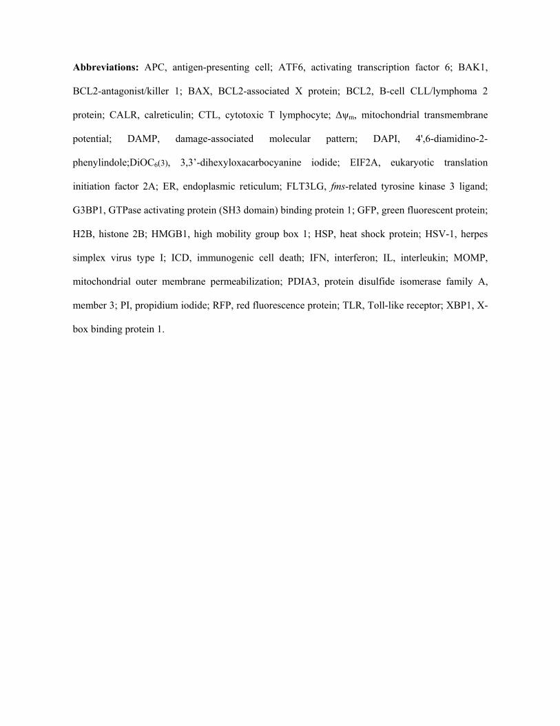
Abbreviations: APC, antigen-presenting cell; ATF6, activating transcription factor 6; BAK1,
BCL2-antagonist/killer 1; BAX, BCL2-associated X protein; BCL2, B-cell CLL/lymphoma 2
protein; CALR, calreticulin; CTL, cytotoxic T lymphocyte; Δψm, mitochondrial transmembrane
potential; DAMP, damage-associated molecular pattern; DAPI, 4',6-diamidino-2-
phenylindole;DiOC6(3), 3,3’-dihexyloxacarbocyanine iodide; EIF2A, eukaryotic translation
initiation factor 2A; ER, endoplasmic reticulum; FLT3LG, fms-related tyrosine kinase 3 ligand;
G3BP1, GTPase activating protein (SH3 domain) binding protein 1; GFP, green fluorescent protein;
H2B, histone 2B; HMGB1, high mobility group box 1; HSP, heat shock protein; HSV-1, herpes
simplex virus type I; ICD, immunogenic cell death; IFN, interferon; IL, interleukin; MOMP,
mitochondrial outer membrane permeabilization; PDIA3, protein disulfide isomerase family A,
member 3; PI, propidium iodide; RFP, red fluorescence protein; TLR, Toll-like receptor; XBP1, X-
box binding protein 1.
Abstract
For long, apoptotic cells have been considered as intrinsically tolerogenic or unable to elicit
immune responses specific for dead cell-associated antigens. However, multiple stimuli can trigger
a functionally peculiar type of apoptotic demise that does not go unnoticed by the adaptive arm of
the immune system, which we named “immunogenic cell death” (ICD). ICD is preceded or
accompanied by the emission of a series of immunostimulatory damage-associated molecular
patterns (DAMPs) in a precise spatiotemporal configuration. Several anticancer agents that have
been successfully employed in the clinic for decades, including various chemotherapeutics and
radiotherapy, can elicit ICD. Moreover, defects in the components that underlie the capacity of the
immune system to perceive cell death as immunogenic negatively influence disease outcome among
cancer patients treated with ICD inducers. Thus, ICD has profound clinical and therapeutic
implications. Unfortunately, the gold-standard approach to detect ICD relies on vaccination
experiments involving immunocompetent murine models and syngeneic cancer cells, an approach
that is incompatible with large screening campaigns. Here, we outline strategies conceived to detect
surrogate markers of ICD in vitro and to screen large chemical libraries for putative ICD inducers,
based on a high-content, high-throughput platform that we recently developed. Such platform
allows for the detection of multiple DAMPs, like cell surface-exposed calreticulin, extracellular
ATP and high mobility group box 1 (HMGB1), and/or the processes that underlie their emission,
such as endoplasmic reticulum stress, autophagy and necrotic plasma membrane permeabilization.
We surmise that this technology will facilitate the development of next-generation anticancer
regimens, which kill malignant cells and simultaneously convert them into a cancer-specific
therapeutic vaccine.
Introduction
Cell death can be classified based on several parameters, including morphological manifestations,
biochemical features, kinetic considerations and functional outcomes.1-7 This said, how cell death
has been investigated and conceived since its pristine descriptions (dating back to the mid-19th
century)8 has obviously evolved along with the technological advances that have been made
throughout the last one and a half centuries.9,10 Thus, morphology-based classifications postulating
the existence of three cell death subroutines (i.e., type I, type II and type III cell death)2,11-14 have
been progressively abandoned in favor of definitions that rely on objectively quantifiable functional
features.3,15-19 Alongside, the long-standing conception according to which distinct types of cell
death like apoptosis and necrosis would constitute mutually exclusive and diametrically opposed
entities has been refuted. In particular, throughout the past two decades it has become clear that: (1)
apoptosis is not the sole type of regulated cell death that contributes to (post-)embryonic
development and adult tissue homeostasis;20 (2) similar to apoptosis, necrosis can occur in a
regulated fashion, i.e., it can involve a genetically encoded molecular machinery;4,5,21 (3) similar to
their necrotic counterparts, apoptotic cells can sometimes be detected by the immune system and
elicit an adaptive immune response specific for dead cell-associated antigens.6,7,22,23 Thus, although
apoptosis as a physiological process involved in (post-)embryonic development and tissue
homeostasis invariably fails to engage the adaptive branch of the immune system,24,25 specific
stimuli can promote an immunogenic variant of regulated cell death that manifests with both
morphological and biochemical features of apoptosis.2,3,6 Of note, defects in the clearance of
apoptotic cells by professional phagocytes have been associated with autoimmune conditions such
as systemic lupus erythematosus (SLE) and chronic inflammation.26,27 However, it remains unclear
whether this reflects the immunogenic potential of intact apoptotic corpses or the insurgence of
secondary necrosis.
Back in 2005, we were the first to report the unexpected finding that murine colorectal carcinoma

CT26 cells as well as murine fibrosarcoma MCA205 cells exposed to a lethal dose of doxorubicin
in vitro are capable of vaccinating syngeneic mice against a subsequent challenge with living cells
of the same type.22 We dubbed such a functionally peculiar variant of cellular demise, manifesting
with an apoptotic morphology and depending on the activity of apoptotic caspases, “immunogenic
cell death” (ICD).22 It turned out that the unsuspected ability of doxorubicin (an anthracycline
employed for the treatment of various carcinomas) to trigger ICD as a standalone intervention,
hence converting dying cancer cells into a vaccine that is efficient in the absence of adjuvants, is
shared by a relatively restricted set of lethal triggers.28-33 These include, but perhaps are not limited
to, mitoxantrone and epirubicin (two other anthracyclines currently used in the clinic),34-37
bleomycin (a glycopeptide antibiotic endowed with antineoplastic properties),38 oxaliplatin (a
platinum derivative generally employed against colorectal carcinoma),39-42 cyclophosphamide (an
alkylating agent approved for the treatment of neoplastic and autoimmune conditions),43-48
etoposide (a topoisomerase inhibitor currently used for the treatment of several neoplasms)
combined with the chemical inhibitor of glycolysis 2-deoxyglucose,49,50 patupilone (a microtubular
inhibitor that has not yet been approved for use in humans),51-53 septacidin (an antifungal antibiotic
produced by Streptomyces fibriatus)54,55 specific forms of radiation therapy,34,56-64 photodynamic
therapy (a clinically approved anticancer intervention that involves the administration of a
photosensitizing agent followed by light irradiation),65-73 high hydrostatic pressure,74 multiple
oncolytic viruses,75-83 replication-defective viral vectors encoding a potentially cytotoxic product
(e.g., thymidine kinase from herpes simplex virus type I, HSV-1) combined with viruses expressing
an immunostimulatory molecule (e.g., fms-related tyrosine kinase 3 ligand, FLT3LG),84 the
clinically employed proteasomal inhibitor bortezomib,85-87 shikonin (a component of Chinese herbal
medicine),88 a monoclonal antibody specific for the epidermal growth factor receptor (EGFR),89
capsaicin (a neurotoxic derivative of homovanillic acid found in chili peppers),90,91 and perhaps the
n3-polyunsaturated fatty acid docosahexaenoic acid,92 as well as the transgene-driven expression of
SMAC mimetics.93,94 In addition, some interventions are capable of converting non-immunogenic

instances of cell death into bona fide ICD. These maneuvers include the administration of cardiac
glycosides, which are particularly powerful in this respect as they promote per se all major
manifestations of ICD (see below),95-97 or zoledronic acid (a bisphosphonate currently approved to
treat osteoporosis and to prevent skeletal fractures in cancer patients with bone metastases),98,99 as
well as the provision of co-stimulatory signals via CD40.100 This said, it should be kept in mind that
the capacity of a given agent to cause ICD or exacerbate the immunogenicity of apoptosis cannot be
predicted yet from its structural or chemical properties, since molecules as similar to each other as
oxaliplatin and cisplatin do not share this functional profile.39,40
The notion that apoptotic cancer cells do not always go undetected by the immune system has
profound clinical repercussions.101 First, it implies that the immune system, at least under specific
circumstances, can mount an adaptive immune response against (self) malignant cells, hence
mediating antineoplastic effects or contributing to the therapeutic activity of conventional
anticancer regimens. This concept represents the theoretical foundation of modern tumor
immunology and anticancer immunotherapy.22,102,103 As a matter of fact, many chemotherapeutics
that have been successfully used in the clinic throughout the past century have recently been
discovered to mediate off-target immunostimulatory effects, ICD being one of the underlying
mechanisms (though not the sole).104-106 Second, it implies that a large number of parameters
reflecting the immunological competence of the host, including the type, quantity and localization
of tumor-infiltrating lymphoid and myeloid cells,107-113 the amount of blood-borne memory T cells
that are able to recognize antigens associated with apoptotic cancer cells,114 the circulating levels of
various ICD-associated biomarkers, including the non-histone chromatin-binding protein high
mobility group box 1 (HMGB1),46,115-117 as well as genetic polymorphisms affecting virtually all
facets of the immune response,41,108,118,119 may be endowed with a robust prognostic or predictive
value. This notion has already been demonstrated in several ICD-related clinical scenarios. Thus,
the relative abundance of tumor-infiltrating CD8+ cytotoxic T lymphocytes (CTLs) and

CD4+CD25+FOXP3+ regulatory T cells reportedly predicts the propensity of breast carcinoma
patients to benefit from anthracycline- or oxaliplatin-based chemotherapy, respectively.52,120 Along
similar lines, single nucleotide polymorphisms in the genes coding for ICD-relevant receptors such
as Toll-like receptor 4 (TLR4) and purinergic receptor P2X, ligand-gated ion channel, 7 (P2RX7)
have been shown to influence disease outcome among breast carcinoma patients treated with
anthracycline-based chemotherapy.41,119 Taken together, these observations demonstrate that the
induction of ICD is a therapeutically relevant objective, calling for the identification of novel ICD
inducers and molecules that improve the immunogenicity of conventional variants of apoptosis.
After summarizing the main molecular and cellular determinants that underlie ICD, we discuss the
assays that are currently available for the detection of surrogate ICD markers and how these
methods can be combined into a platform that is compatible with high-content, high-throughput
applications. We surmise that this methodological approach will accelerate the discovery and
development of therapeutic regimens that kill malignant cells in an immunogenic fashion.

Immunogenic cell death signaling
According to current models, ICD relies on the ability of specific stimuli to kill cells while
provoking the spatiotemporally coordinated emission of immunogenic signals.7,121-129 Such signals
are conveyed by damage-associated molecular patterns (DAMPs), i.e., molecules that are not
accessible by the immune system in physiological conditions but are released or exposed on the
outer leaflet of the plasma membrane during cytoprotective stress responses or upon cell
death.103,130-133 Similar to their microbial counterparts, many (but not all) DAMPs exert robust
immunostimulatory effects upon binding to pattern recognition receptors (PRRs) expressed by
immune cells.121 So far, three DAMPs have been attributed a key role in the immunogenic potential
of virtually all ICD inducers: the endoplasmic reticulum (ER) chaperone calreticulin
(CALR),34,65,126,134-136 ATP,66,124,137-143 and HMGB1.41,46,115,116,144-147 In addition, many DAMPs
have been shown to contribute to the immunogenicity of cell death in a limited amount of
experimental scenarios. These include immunostimulatory cytokines like interferon α (IFNα),148,149
various chaperones of the heat-shock protein (HSP) family, notably heat shock 70kDa protein 1A
(HSPA1A, best known as HSP70) and heat shock protein 90kDa alpha (cytosolic), class A member
1 (HSP90AA1, best known as HSP90),65,71,85,90,145,150-153 sphingomyelin metabolites (e.g., ceramide
and sphingosine-1-phosphate),154 a plethora of mitochondrial products (e.g., mitochondrial DNA,
N-formylated peptides, cardiolipin),155-157 cytosolic components like urate and F-actin,158-161 as well
as products of the breakdown of the extracellular matrix (e.g., hyaluronan fragments).162,163
CALR gets exposed on the cell surface early in the course of ICD, i.e., before the apoptosis-
associated shuffling of phosphatidylserine between the inner and outer leaflet of the plasma
membrane.34,164,165 The molecular mechanisms underlying this ICD hallmark have been dissected in
detail and appear to involve three distinct signaling modules: (1) an ER stress module centered
around the phosphorylation of eukaryotic translation initiation factor 2A (EIF2A) and the resultant
arrest in protein synthesis; (2) an apoptotic module involving the activation of caspase-8 and the
consequent cleavage of B-cell receptor-associated protein 31 (BCAP31) as well as the pro-apoptotic
Bcl-2 family members BCL2-associated X protein (BAX) and BCL2-antagonist/killer 1 (BAK1);
and (3) an exocytosis module requiring the actin cytoskeleton as well as vesicle-associated
membrane protein 1 (VAMP1) and synaptosomal-associated protein, 25kDa (SNAP25), two
proteins involved in intracellular vesicular trafficking.36 Moreover, in some (but not all) models of
ICD,67 CALR obligatorily translocates to the cell surface together with another ER chaperone,
protein disulfide isomerase family A, member 3 (PDIA3).36,37 Upon binding to low density
lipoprotein receptor-related protein 1 (LRP1, also known as CD91), membrane-exposed CALR
delivers a major phagocytic signal to professional antigen-presenting cells (APCs) such as dendritic
cells, de facto improving their capacity to take up dead cells and their corpses.66,91,166-173
Interestingly, the phagocytosis-stimulatory effects of CALR is robustly counterbalanced by CD47,
which is highly expressed by a large panel of solid and hematopoietic tumors.166 This latter
observation suggests that various neoplasms benefit from avoiding the effects of CALR exposure,
perhaps as this prevents the elicitation of an adaptive immune response against the malignant cells
that “physiologically” succumb in the course of oncogenesis and tumor progression. Alternatively,
the phagocytosis-inhibitory activity of CD47 may confer tumors with an advantage by increasing
the local availability of macromolecules derived from the spontaneous demise (and degradation) of
some of their cellular constituents. This possibility has not yet experimentally addressed.
The ICD-associated release of ATP proceeds through a complex mechanism that involves (1) the
apparent relocalization of vesicular ATP stores from lysosomes to autolysosomes; (2) the
redistribution of lysosomal-associated membrane protein 1 (LAMP1) to the plasma membrane; (3)
Rho-associated, coiled-coil containing protein kinase 1 (ROCK1)-mediated, myosin II-dependent
cellular blebbing; and (4) the opening of pannexin 1 (PANX1) channels, 4 processes that rely on
caspases.140,142,174 In a vast majority of models, the secretion of ATP by cells exposed to ICD
inducers requires an intact autophagic machinery.83,138,139,175 In these settings, the genetic or

pharmacological inhibition of autophagy limits ATP release by cells succumbing to ICD and hence
negatively influences their ability to elicit an adaptive immune response upon inoculation in
immunocompetent syngeneic mice.60,138,139 Along similar lines, the chemical inducer of autophagy
STF-62247 increases the immunostimulatory potential of ICD as triggered by chlorin-e6-based
photodynamic therapy (MK, unpublished observations). However, this does not seem to apply to all
ICD inducers.68 Thus, the ability of hypericin-based photodynamic therapy to induce the secretion
of ATP does not appear to change in autophagy-deficient versus autophagy-proficient cells.68,70,176
Moreover, the former respond to hypericin-based photodynamic therapy by exposing higher
amounts of CALR on the plasma membrane than the latter, hence exhibiting a superior
immunogenic potential.68,70,176 Possibly, this reflects the incapacity of autophagy-deficient cells to
clear oxidized proteins, resulting in an aggravation of the ER stress response that underlies CALR
exposure in the course of ICD.68,70,176 Irrespective of these variations, extracellular ATP operates as
a strong chemoattractant and promotes not only the recruitment of immune cells to sites of ICD, but
also their differentiation, an effect that depends on purinergic receptor P2Y, G-protein coupled, 2
(P2RY2).141,177-179 Moreover, extracellular ATP promotes the activation of the NLR family, pyrin
domain containing 3 (NLRP3) inflammasome in APCs, hence stimulating the processing and
release of interleukin (IL)-1β and IL-18.119,180-189 In line with this notion, the immunogenic potential
of cells succumbing to ICD can be significantly reduced by pharmacological or genetic
interventions that limit the availability of ATP in the pericellular space, such as the administration
of recombinant apyrase (an ATP-degrading enzyme) or the transfection-enforced overexpression of
ectonucleoside triphosphate diphosphohydrolase 1 (ENTPD1, best known as CD39), which
converts ATP into ADP and AMP.190 Intriguingly, CD39 and 5'-nucleotidase, ecto (NT5E, best
known as CD73), which transforms AMP into adenosine, are often overexpressed by malignant
tissues. This reflects the advantage conferred to cancer cells by the conversion of extracellular ATP,
which promotes immunosurveillance, into adenosine, which exerts potent immunosuppressive
effects.191-197 Of note, autophagy is also important for the perception of cell death as immunogenic
because it contributes to several aspects of cellular immune responses, including the differentiation,
survival and activation of myeloid and lymphoid cells.198-200
The release of HMGB1 from cells succumbing to ICD requires the permeabilization of both the
nuclear and plasma membranes, de facto constituting a post-mortem event.3,41 Although autophagy
has been proposed to contribute to the release of HMGB1 from dying cells, at least under some
circumstances,201 the molecular machinery that underlies this crucial manifestation of ICD has not
yet been elucidated in detail. This said, extracellular HMGB1 is well known to mediate robust pro-
inflammatory effects upon binding to several receptors on the surface of immune cells, including
TLR2, TLR4 and advanced glycosylation end product-specific receptor (AGER, best known as
RAGE).202-210 Moreover, extracellular HMGB1 reportedly exerts a chemotactic activity by forming
a complex with chemokine (C-X-C motif) ligand 12 (CXCL12) that signals via chemokine (C-X-C
motif) receptor 4 (CXCR4).211 Finally, at least under some circumstances, endogenous HMGB1
appears to promote autophagy by interfering with the mutually inhibitory interaction between the
central autophagic regulator beclin 1 (BECN1) and the anti-apoptotic protein B-cell
CLL/lymphoma 2 (BCL2).212-214 It is therefore tempting to speculate, yet remains to be formally
demonstrated, that the nuclear release of HMGB1 may contribute to the autophagic response of
cells succumbing to ICD inducers. Of note, the biological activity of extracellular HMGB1 appears
to be regulated by its redox state.215-221 Moreover, HMGB1 binds not only to TLR2, TLR4 and
RAGE, but also to hepatitis A virus cellular receptor 2 (HAVCR2, best known as TIM-3), hence
mediating immunosuppressive (as opposed to immunostimulatory) effects.222-224 Taken together,
these observations suggest that the biological activity of HMGB1 exhibits a consistent-degree of
context-dependency. Nonetheless, HMGB1-deficient malignant cells exposed to ICD inducers fail
to elicit adaptive immune responses upon inoculation into immunocompetent syngeneic mice, a
defect that can be corrected by the co-administration of synthetic TLR4 ligands.225-228 Together with
the notion that Tlr4-/- mice fail to perceive anthracycline-treated syngeneic cells as

immunogenic,41,229 this observation demonstrates the importance of the HMGB1-TLR4 signaling
axis for ICD.
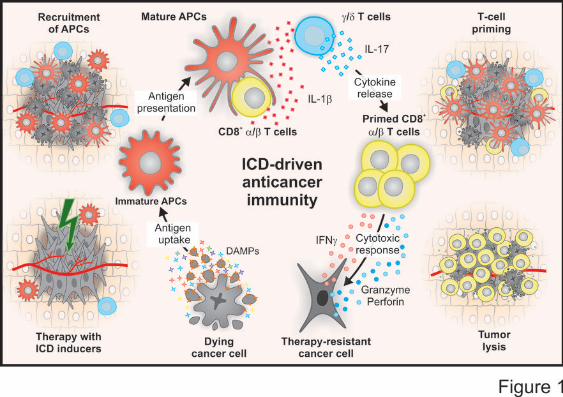
In summary, the spatiotemporally coordinated emission of specific DAMPs promotes the
recruitment of APCs to sites of ongoing ICD, their ability to take up dead cell-derived particulate
material, as well as their capacity to prime an adaptive immune response.6 This generally proceeds
in two phases, involving the sequential recruitment and activation of IL-17-secreting γδ T cells and
αβ CTLs.31,230 The latter not only mediate direct antineoplastic effects, mostly by secreting
interferon γ (IFNγ) and via the granzyme-perforin pathway, but also underlie the establishment of
protective immunological memory (Figure 1).231

Gold-standard methods to monitor ICD
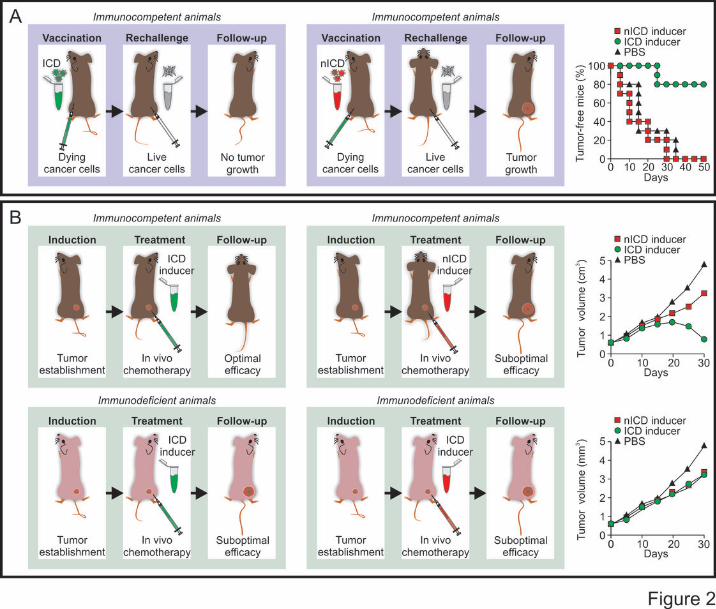
As it stands, the gold-standard approach to evaluate the ability of a specific stimulus to cause bona
fide ICD relies on vaccination assays.6,22,30 In this setting, malignant cells are exposed in vitro to the
lethal stimulus of choice, thoroughly washed (to remove the stimulus), resuspended in an adequate
volume of PBS, and then inoculated subcutaneously into the flank of immunocompetent syngeneic
mice. One week later, living cells of the same type are introduced subcutaneously into the opposite
flank, and mice are routinely monitored for the appearance of a palpable neoplastic lesion (Figure
2A). The proportion of mice that do not develop subcutaneous tumors reflects the degree of
immunogenicity of cell death as induced by the lethal trigger under evaluation. As a note, murine
cells succumbing to prototypic inducers of ICD such as doxorubicin and mitoxantrone effectively
vaccinate 80% of mice.34,95,232
As a confirmatory assay, putative ICD inducers can be assessed for their ability to mediate immune
system-dependent therapeutic effects against established neoplastic lesions.6,34,233 In this scenario,
grafted, genetically-driven or chemically-induced subcutaneous or orthotopic tumors are established
in both immunocompetent and immunodeficient mice. Malignant lesions are then allowed to
progress until a pre-determined size or time point, beyond which tumor-bearing mice are treated
with the compound under evaluation (Figure 2B). In this experimental setup, bona fide ICD
inducers mediate optimal therapeutic effects in immunocompetent, but not in immunodeficient,
mice.34,41,95,119,233 Importantly, this latter approach is suitable to validate the results of vaccination
experiments but cannot be employed alone to determine the capacity of a specific intervention to
cause ICD. Indeed, even the activity of antineoplastic regimens that fail to render dying cells
immunogenic but induce other immunostimulatory effects is negatively affected by the absence of a
functional immune system.104,105 Among other molecules, this applies to the microtubular inhibitor
paclitaxel and the nucleoside analogue gemcitabine.104,105

The main drawbacks of these types of assay relate to the use of rodents and syngeneic tumor
models: the need for a tightly controlled sterile facility (which is mandatory for working with
immunodeficient animals), prolonged times for the establishment/growth of neoplastic lesions, and
significant costs. Moreover, vaccination and therapeutic tests for the detection of ICD are limited by
the relatively restricted number of syngeneic tumor models that are currently available. Thus,
although they constitute the gold-standard approach for the detection of ICD, vaccination assays
relying on immunocompetent mice and syngeneic cancer cells are intrinsically incompatible with
large screening campaigns. To circumvent this issue, various techniques that allow for the detection
of one or more ICD manifestations in vitro and in vivo have been developed.6,234 Monitoring the
immunostimulatory activity of lead compounds (be it linked to the induction of ICD or reflecting
other mechanisms) early in the drug discovery pipeline may indeed speed up significantly the
development of novel anticancer agents.104

Detection of surrogate ICD biomarkers
A relatively ample panel of ICD-associated phenomena can be monitored in vitro to obtain insights
into the ability of a specific intervention to provoke ICD (Table 1).
Cell death. By definition, ICD inducers must be cytotoxic and provoke cell death above a minimal
threshold level. Cancer cells emit indeed a wide panel of DAMPs in response to non-lethal
perturbations of homeostasis. However, such DAMPs differ in both qualitative and quantitative
terms from those emitted by cells of the same type dying in response to the same stimulus applied
with a lethal intensity/duration. Living cells are less likely to be taken up by APCs and ignite an
adaptive immune response than their dying counterparts. Moreover, if the fraction of dying cells is
excessively low, neoplastic lesions develop at the vaccination site and protective immunity cannot
be established.34,95 Thus, agents that stimulate all the key manifestations of ICD including CALR
exposure, ATP secretion and HMGB1 release, but fail to exert robust cytotoxic effects cannot be
considered as authentic ICD inducers. This is the case of cardiac glycosides including digoxin and
digitoxin, which nonetheless are powerful at converting non-immunogenic instances of cell death
into bona fide ICD, hence operating as potent immune adjuvant.95-97,235
Several assays are commercially available to monitor cell death-associated parameters, the most
reliable indicator of cell death being end-stage plasma membrane permeabilization.9,236 This can be
conveniently monitored by so-called exclusion dyes like the DNA-binding chemicals propidium
iodide (PI) and 4',6-diamidino-2-phenylindole (DAPI), which only accumulate in cells with
permeabilized plasma membranes. PI and DAPI can be conveniently detected by flow cytometry or
fluorescence microscopy (absorption/emission peaks: 535/617 and 358/461 nm, respectively). On
flow cytometry, both PI and DAPI can be combined with fluorescence variants of the protein
annexin A5 (ANXA5), permitting the detection of phosphatidylserine exposure,9,237,238 as well as
with 3,3’-dihexyloxacarbocyanine iodide (DiOC6(3), absorption/emission peaks: 482/504 nm),

allowing for the quantification of mitochondrial transmembrane potential (Δψm).239-241 The
externalization of phosphatidylserine (a phospholipid normally restricted to the inner leaflet of the
plasma membrane) accompanies indeed multiple (though not all) instances of apoptotic cell
death,16,242-245 while the permanent dissipation of the Δψm as a result of mitochondrial outer
membrane permeabilization (MOMP) constitutes one of the major hallmarks of mitochondrial
apoptosis.17,18,246,247 Of note, DiOC6(3) is not compatible with fixation, but other Δψm-sensitive
probes that exist are, including chloromethyltetramethylrosamine (absorption/emission peaks:
554/576 nm).248 MOMP is accompanied by the massive activation of caspase-9 and -3, while
caspase-8 is required for ICD upstream of MOMP. The activation of caspases can be documented
by flow cytometry or fluorescence microscopy, either upon the immunostaining of cells with
monoclonal antibodies specific for active caspase fragments, or with cell-permeant caspase
substrates that become fluorescent upon cleavage.9,249,250 Alternatively, caspase activation can be
detected in a semi-quantitative manner by immunoblotting, with antibodies specific for caspases
(which are themselves activated by cleavage) or their substrates.250,251
As MOMP ensues the assembly of BAX/BAK1-containing oligomers across the outer
mitochondrial membrane, the process can also be monitored by means of green fluorescent protein
(GFP)-BAX chimeras (GFP absorption/emission peaks: 395/509 nm). In this setting, the
relocalization of BAX to mitochondria can be followed by fluorescence microscopy as a shift in
GFP fluorescence from a diffuse to a punctate or network-like pattern.40,252 Finally, one of the major
morphological modifications of apoptosis (and hence of ICD) is nuclear condensation
(pyknosis).1,2,95 Also this process can be conveniently monitored by fluorescence microscopy, either
in cells that constitutively express a GFP- or red fluorescent protein (RFP)-tagged variant of histone
2B (RFP-H2B, absorption/emission peaks: 584/607 nm) or upon fixation and staining with the
chromatinophilic dye Hoechst 33342 (absorption/emission peaks: 361/461 nm).40,95,235

CALR exposure. Several assays are available to directly monitor the ICD-associated translocation
of CALR on the outer leaflet of the plasma membrane. For instance, this can be achieved on flow
cytometry, by staining non-permeabilized cells with a CALR-specific antibody, or in cells that
stably express a CALR-HaloTag™ fusion protein.40,95 In the latter scenario, the HaloTag™ label
can be visualized by a cell-impermeant fluorescent chemical, resulting in the specific detection of
the CALR molecules that are effectively accessible for ligand binding from the extracellular
microenvironment.40,95 In both cases, it is imperative to remove from the analysis dead (PI+ or
DAPI+) cells, as the permeabilized plasma membrane allows both the CALR-specific antibody and
the normally cell-impermeant HaloTag™ ligand to access intracellular CALR.34,40 Alternatively,
CALR exposure can be monitored upon the biotinylation of cell surface proteins (which must be
performed in pre-apoptotic conditions, when plasma membranes are intact, to avoid false-positive
results owing to intracellular CALR), followed by streptavidin-mediated precipitation, and
detection by immunoblotting,34,66,253 or by fluorescence microscopy, in cells that constitutively
express a CALR-GFP fusion construct. For the sake of precision, it should be noted that the latter
system does not detect CALR-GFP exposure in itself, but the ER perinuclear clustering that
invariably accompanies exposure.20,232 We have also successfully employed a PDIA3-specific
antibody and flow cytometry as well as PDIA3-GFP-expressing cells and fluorescence microscopy
to (indirectly) assess CALR exposure in the course of ICD, as in our models PDIA3 invariably co-
translocates with CALR on the surface of cells exposed to ICD inducers.36,37,95 However, this does
not apply to all experimental settings,66,67 implying that the PDIA3-GFP fusion is a useful
confirmatory tool but cannot be employed as a standalone means to identify all instances of ICD.
In some instances, it may be important to monitor CALR exposure along with the proficiency of the
ER stress response. This may indeed allow for the identification of defects in the signaling pathway
that leads to the translocation of CALR to the outer leaflet of the plasma membrane. Several assays
are currently available for the detection of the different arms of the ER stress response.136,254-256 For
instance, the phosphorylation state of EIF2A and/or of the major EIF2A kinases, including EIF2A
kinase 1 (EIF2AK1, best known as HRI),257 EIF2AK2 (best known as PKR),258 and EIF2AK3 (best
known as PERK),259-261 can be assessed by immunoblotting, flow cytometry or
immunofluorescence microscopy with phosphoneoepitope-specific antibodies.260 The splicing
status of X-box binding protein 1 (XBP1) mRNA, reflecting the activation of the ER stress sensor
endoplasmic reticulum to nucleus signaling 1 (ERN1, best known as IRE1α), can be monitored by
quantitative real-time RT-PCR,262 as well as by flow cytometry or fluorescence microscopy, either
in cells that express a fluorescently-tagged version of XBP1263 or upon the administration of a self-
quenched RNA probe that can be cleaved by IRE1α.264 Finally, the nuclear redistribution of
activating transcription factor 6 (ATF6) can be easily evaluated by fluorescence microscopy in cells
that constitutively express GFP- or RFP-tagged variants of ATF6.52 As an alternative, ER stress can
be indirectly monitored upon the formation of GTPase activating protein (SH3 domain) binding
protein 1 (G3BP1)-containing granules in cells genetically modified to express a G3BP1-GFP
fusion.40,265 This said, G3BP1 appears to redistribute to granules in response to a wide panel of
stressful conditions that are not limited to specific perturbations of reticular homeostasis. Thus,
monitoring G3BP1 aggregation can be useful to determine whether cells mount a stress response to
a putative inducer of ICD, yet cannot be employed to formally imply the ER in this process.
ATP secretion. The ICD-associated secretion of ATP can be monitored by two complementary
approaches: directly, by quantification of extracellular ATP,137,180 or indirectly, by the assessment
of residual intracellular ATP.137,139 The most employed method currently available for the
quantification of ATP levels relies on the ability of eukaryotic luciferases to produce light while
oxidizing D-(-)-luciferin (which must be added exogenously) in a ATP-dependent manner.266,267
This can be applied to culture supernatants as well as to cell lysates, and hence is compatible with
both the direct and indirect assessment of ATP secretion in the course of ICD. The vesicular pool of

ATP can also be visualized by fluorescence microscopy upon staining cells with the ATP-binding
fluorochrome quinacrine (absorption/emission peaks: 436/525 nm).268 Alternatively, intracellular
ATP can be monitored in living cells by a fluorescence resonance energy transfer (FRET)-based
assay involving a yellow fluorescent protein-cyan fluorescent protein (YFP-CFP) fusion containing
a domain that changes its conformation upon ATP binding, hence shifting the spectral properties of
the probe.269
In some settings, it may be relevant to monitor the autophagic response that generally precedes and
is required for ICD-associated ATP release. This can be achieved by a wide panel of techniques,
whose detailed discussion goes largely beyond the scope of this set of recommendations.15,270,271
This said, one of the most convenient approaches to obtain insights into the autophagic response of
cells exposed to homeostatic perturbations relies on the use of a GFP- or RFP-tagged variant of
microtubule-associated protein 1 light chain 3 (MAP1LC3, best known as LC3).272 In the course of
autophagy, LC3 gets conjugated to phosphatidylethanolamine, hence acquiring the ability to
accumulate into forming autophagosomes.273,274 In line with this notion, GFP-LC3 redistributes
from a diffuse to a punctate pattern in cells mounting an autophagic response, a phenomenon that
can readily be monitored by fluorescence microscopy.
HMGB1 release. Similar to the secretion of ATP, the release of HMGB1 in the supernatant of cells
undergoing ICD can be monitored directly or indirectly, as a function of residual intracellular
HMGB1.41,207,275 The former approach relies on the immunoblotting-based assessment of HGMB1
in concentrated cell supernatants, or (most often) on commercially available enzyme-linked
immunosorbent assay (ELISA) kits specific for human or murine HMGB1. These kits generally
allow for the precise quantification of HMGB1 concentrations in a wide panel of biological
specimens, including culture supernatants, serum samples and interstitial fluids, yet may be
relatively expensive for use in large-scale screening campaigns.95,147,275 Alternatively, HMGB1

release can be assessed by fluorescence microscopy in cells expressing a GFP-tagged variant of
HMGB1, as the loss of colocalization between the GFP signal and a nuclear staining (e.g., Hoechst
33342, H2B-RFP).275 This said, the precise quantification of HMGB1 variants exhibiting
differential redox states requires mass spectroscopy.276
High-content, high-throughput platform. Cell death that is not accompanied by CALR exposure,
ATP secretion and HMGB1 release is generally not perceived as immunogenic.34,41,119 In other
words, the absence of only one such ICD-associated events often entails a consistent decrease in the
immunogenicity of cell death, if not its total loss. This implies that the ability of a given
intervention to promote ICD can be inferred in vitro only upon the concurrent evaluation of all ICD
hallmarks. Indeed, cells succumbing to homeostatic perturbations that stimulate ATP secretion and
HMGB1 release but not CALR exposure, such as the administration of cisplatin, fail to elicit
adaptive immune responses upon inoculation into immunocompetent mice.34,39,40 This said, a
platform that would allow for the simultaneous detection of cell death, CALR exposure, ATP
secretion and HMGB1 release in the context of large screening campaigns was missing. To
circumvent this obstacle to the identification of novel, perhaps clinically relevant bona fide inducers
of ICD, we recently developed a robotized cell biology platform that allows for entirely automated
compound handling and multiplex read-out capability (including fluorescence microscopy, flow
cytometry and bioluminescence detection) in sterile conditions. We then designed fully automated
workflows based on various combinations of the assays described above and including appropriate
procedures for data handling/normalization and statistical analysis. This approach is compatible
with the high-content, high-throughput screening of large chemical libraries, returning a cumulative
score that represents the ability of a specific compound to promote the four tenets of ICD.
Importantly, this integrated platform does not abolish the need to evaluate putative ICD inducers for
their capacity to elicit protective anticancer immune responses in gold-standard vaccination assays.
Nonetheless, it allows for the relatively straightforward identification of candidate molecules. By
means of this approach, septacidin has been identified as a bona fide ICD inducer.232 Moreover,
cardiac glycosides were found to robustly improve the immunogenic potential of cell death.95-97,235
We expect this platform not only to allow for the discovery of other ICD inducers, but also to
facilitate the understanding of the molecular pathways that underlie ICD and how these can be
modulated for therapeutic purposes.
Concluding remarks and future directions
As described above, the simultaneous detection of cell death, CALR exposure, ATP secretion and
HMGB1 release by means of a high-content-, high-throughput-compatible platform is useful for the
identification of candidate ICD inducers among large chemical libraries. Nonetheless, vaccination
assays involving immunocompetent mice and syngeneic cancer cells do not cease to constitute the
gold-standard approach to formally identify bona fide triggers of ICD.
Paradoxically, the major obstacle to the identification and development of clinically relevant ICD
inducers appears to be represented by the murine system itself, as rodent and human cells do not
necessarily respond to a specific stimulus in a comparable fashion. As a standalone example, mouse
cells are highly resistant to the cytotoxic activity of cardiac glycosides, owing to the expression of a
mutated subunit of their target, the Na2+/K+ ATPase.95,277 This implies that formally determining
whether a given intervention provokes ICD in the human system is complicated. Humanized rodent
models, i.e., immunodeficient mice reconstituted with a human immune system,278 may partially
circumvent this issue. However, the interaction between human immune cells and the murine
microenvironment may be negatively influenced by inter-species molecular variations that
compromise the ability of the former to mount an appropriate immune response.279,280 Thus,
although attempts are being made to limit such variations,281 experimental models that allow for the
proper evaluation of ICD in the human system require further improvements. Finally, the procedure
outlined above for the identification of novel ICD inducers assesses the biochemical processes that
are required for the immunogenicity of anthracycline-induced cell death. However, ICD might exist
in functionally distinct variants, implying that hitherto uncharacterized mechanisms might render
cell death immunogenic. This possibility should be actively investigated in future studies.
Irrespective of these caveats, we are confident that the screening of large chemical or small-
interfering RNA libraries combined with vaccination assays in the murine model will allow for the
identification of novel, therapeutically relevant interventions for the induction or modulation of
ICD. Moreover, the immunohistochemical detection of ICD-associated biomarkers in bioptic
specimens from cancer patients may convey robust predictive or prognostic indications, at least
under some circumstances.282,283 The implementation of well-designed, longitudinal
immunomonitoring procedures into the clinical development of antineoplastic agents is required to
ascertain the actual prognostic or predictive value of ICD-associated processes among oncological
patients.284-286 Of note, a phase I clinical study has recently been launched to investigate the safety
and preliminary therapeutic efficacy of adenoviral vectors genetically modified to trigger ICD, in
subjects with malignant glioma and glioblastoma multiforme (NCT01811992). In this setting,
serotype 5, replication-defective, first-generation adenoviruses encoding the HSV-1 thymidine
kinase and similar vectors coding for FLT3LG are co-infused at the time of surgical tumor
resection, followed by valacyclovir (a gancylovir-like prodrug converted by the viral thymidine
kinase and cellular kinases into its triphosphate cytotoxic variant)287,288 in the context of current
standard-of-care therapy (source https://clinicaltrials.gov/). The results of such a first-in-man study
relying on the genetic induction of ICD in cancer patients are urgently awaited.
Acknowledgements. GL and LG are supported by the Ligue contre le Cancer (équipe labelisée); Agence National de la
Recherche (ANR); Association pour la recherche sur le cancer (ARC); Cancéropôle Ile-de-France; AXA Chair for
Longevity Research; Institut National du Cancer (INCa); Fondation Bettencourt-Schueller; Fondation de France;
Fondation pour la Recherche Médicale (FRM); the European Commission (ArtForce); the European Research Council
(ERC); the LabEx Immuno-Oncology; the SIRIC Stratified Oncology Cell DNA Repair and Tumor Immune Elimination
(SOCRATE); the SIRIC Cancer Research and Personalized Medicine (CARPEM); and the Paris Alliance of Cancer
Research Institutes (PACRI).
References
1. Galluzzi L, Maiuri MC, Vitale I, Zischka H, Castedo M, Zitvogel L, et al. Cell death
modalities: classification and pathophysiological implications. Cell Death Differ 2007;
14:1237-43.
2. Kroemer G, Galluzzi L, Vandenabeele P, Abrams J, Alnemri ES, Baehrecke EH, et al.
Classification of cell death: recommendations of the Nomenclature Committee on Cell Death
2009. Cell Death Differ 2009; 16:3-11.
3. Galluzzi L, Vitale I, Abrams JM, Alnemri ES, Baehrecke EH, Blagosklonny MV, et al.
Molecular definitions of cell death subroutines: recommendations of the Nomenclature
Committee on Cell Death 2012. Cell Death Differ 2012; 19:107-20.
4. Vandenabeele P, Galluzzi L, Vanden Berghe T, Kroemer G. Molecular mechanisms of
necroptosis: an ordered cellular explosion. Nat Rev Mol Cell Biol 2010; 11:700-14.
5. Vanden Berghe T, Linkermann A, Jouan-Lanhouet S, Walczak H, Vandenabeele P. Regulated
necrosis: the expanding network of non-apoptotic cell death pathways. Nat Rev Mol Cell Biol
2014; 15:135-47.
6. Kroemer G, Galluzzi L, Kepp O, Zitvogel L. Immunogenic cell death in cancer therapy. Annu
Rev Immunol 2013; 31:51-72.
7. Krysko DV, Garg AD, Kaczmarek A, Krysko O, Agostinis P, Vandenabeele P. Immunogenic
cell death and DAMPs in cancer therapy. Nat Rev Cancer 2012; 12:860-75.
8. Vogt CI. Untersuchungen über die Entwicklungsgeschichte der Geburtshelferkröte (Alytes
obstetricans). Jent, 1842.
9. Galluzzi L, Aaronson SA, Abrams J, Alnemri ES, Andrews DW, Baehrecke EH, et al.
Guidelines for the use and interpretation of assays for monitoring cell death in higher
eukaryotes. Cell Death Differ 2009; 16:1093-107.
10. Galluzzi L, Vitale I, Michels J, Brenner C, Szabadkai G, Harel-Bellan A, et al. Systems
biology of cisplatin resistance: past, present and future. Cell Death Dis 2014; 5:e1257.

11. Kerr JF, Wyllie AH, Currie AR. Apoptosis: a basic biological phenomenon with wide-ranging
implications in tissue kinetics. Br J Cancer 1972; 26:239-57.
12. Levine B, Yuan J. Autophagy in cell death: an innocent convict? J Clin Invest 2005;
115:2679-88.
13. Ericsson JL. Studies on induced cellular autophagy. I. Electron microscopy of cells with in
vivo labelled lysosomes. Exp Cell Res 1969; 55:95-106.
14. Maximow AA. Studies on the Changes Produced by Roentgen Rays in Inflamed Connective
Tissue. J Exp Med 1923; 37:319-40.
15. Klionsky DJ, Abeliovich H, Agostinis P, Agrawal DK, Aliev G, Askew DS, et al. Guidelines
for the use and interpretation of assays for monitoring autophagy in higher eukaryotes.
Autophagy 2008; 4:151-75.
16. Martin SJ, Reutelingsperger CP, McGahon AJ, Rader JA, van Schie RC, LaFace DM, et al.
Early redistribution of plasma membrane phosphatidylserine is a general feature of apoptosis
regardless of the initiating stimulus: inhibition by overexpression of Bcl-2 and Abl. J Exp
Med 1995; 182:1545-56.
17. Tait SW, Green DR. Mitochondria and cell death: outer membrane permeabilization and
beyond. Nat Rev Mol Cell Biol 2010; 11:621-32.
18. Kroemer G, Galluzzi L, Brenner C. Mitochondrial membrane permeabilization in cell death.
Physiol Rev 2007; 87:99-163.
19. Taylor RC, Cullen SP, Martin SJ. Apoptosis: controlled demolition at the cellular level. Nat
Rev Mol Cell Biol 2008; 9:231-41.
20. Candi E, Schmidt R, Melino G. The cornified envelope: a model of cell death in the skin. Nat
Rev Mol Cell Biol 2005; 6:328-40.
21. Galluzzi L, Kepp O, Krautwald S, Kroemer G, Linkermann A. Molecular mechanisms of
regulated necrosis. Semin Cell Dev Biol 2014:IN PRESS.
22. Casares N, Pequignot MO, Tesniere A, Ghiringhelli F, Roux S, Chaput N, et al. Caspase-
dependent immunogenicity of doxorubicin-induced tumor cell death. J Exp Med 2005;
202:1691-701.

23. Cirone M, Di Renzo L, Lotti LV, Conte V, Trivedi P, Santarelli R, et al. Activation of
dendritic cells by tumor cell death. Oncoimmunology 2012; 1:1218-9.
24. Henson PM, Hume DA. Apoptotic cell removal in development and tissue homeostasis.
Trends Immunol 2006; 27:244-50.
25. Abud HE. Shaping developing tissues by apoptosis. Cell Death Differ 2004; 11:797-9.
26. Baumann I, Kolowos W, Voll RE, Manger B, Gaipl U, Neuhuber WL, et al. Impaired uptake
of apoptotic cells into tingible body macrophages in germinal centers of patients with
systemic lupus erythematosus. Arthritis Rheum 2002; 46:191-201.
27. Munoz LE, Lauber K, Schiller M, Manfredi AA, Herrmann M. The role of defective
clearance of apoptotic cells in systemic autoimmunity. Nat Rev Rheumatol 2010; 6:280-9.
28. Vacchelli E, Galluzzi L, Fridman WH, Galon J, Sautes-Fridman C, Tartour E, et al. Trial
watch: Chemotherapy with immunogenic cell death inducers. Oncoimmunology 2012; 1:179-
88.
29. Vacchelli E, Senovilla L, Eggermont A, Fridman WH, Galon J, Zitvogel L, et al. Trial watch:
Chemotherapy with immunogenic cell death inducers. Oncoimmunology 2013; 2:e23510.
30. Vacchelli E, Aranda F, Eggermont A, Galon J, Sautes-Fridman C, Cremer I, et al. Trial
Watch: Chemotherapy with immunogenic cell death inducers. Oncoimmunology 2014;
3:e27878.
31. Mattarollo SR, Loi S, Duret H, Ma Y, Zitvogel L, Smyth MJ. Pivotal role of innate and
adaptive immunity in anthracycline chemotherapy of established tumors. Cancer Res 2011;
71:4809-20.
32. Cirone M, Garufi A, Di Renzo L, Granato M, Faggioni A, D'Orazi G. Zinc supplementation is
required for the cytotoxic and immunogenic effects of chemotherapy in chemoresistant p53-
functionally deficient cells. Oncoimmunology 2013; 2:e26198.
33. Bracci L, Schiavoni G, Sistigu A, Belardelli F. Immune-based mechanisms of cytotoxic
chemotherapy: implications for the design of novel and rationale-based combined treatments
against cancer. Cell Death Differ 2014; 21:15-25.

34. Obeid M, Tesniere A, Ghiringhelli F, Fimia GM, Apetoh L, Perfettini JL, et al. Calreticulin
exposure dictates the immunogenicity of cancer cell death. Nat Med 2007; 13:54-61.
35. Fucikova J, Kralikova P, Fialova A, Brtnicky T, Rob L, Bartunkova J, et al. Human tumor
cells killed by anthracyclines induce a tumor-specific immune response. Cancer Res 2011;
71:4821-33.
36. Panaretakis T, Kepp O, Brockmeier U, Tesniere A, Bjorklund AC, Chapman DC, et al.
Mechanisms of pre-apoptotic calreticulin exposure in immunogenic cell death. EMBO J 2009;
28:578-90.
37. Panaretakis T, Joza N, Modjtahedi N, Tesniere A, Vitale I, Durchschlag M, et al. The co-
translocation of ERp57 and calreticulin determines the immunogenicity of cell death. Cell
Death Differ 2008; 15:1499-509.
38. Bugaut H, Bruchard M, Berger H, Derangere V, Odoul L, Euvrard R, et al. Bleomycin exerts
ambivalent antitumor immune effect by triggering both immunogenic cell death and
proliferation of regulatory T cells. PLoS One 2013; 8:e65181.
39. Tesniere A, Schlemmer F, Boige V, Kepp O, Martins I, Ghiringhelli F, et al. Immunogenic
death of colon cancer cells treated with oxaliplatin. Oncogene 2010; 29:482-91.
40. Martins I, Kepp O, Schlemmer F, Adjemian S, Tailler M, Shen S, et al. Restoration of the
immunogenicity of cisplatin-induced cancer cell death by endoplasmic reticulum stress.
Oncogene 2011; 30:1147-58.
41. Apetoh L, Ghiringhelli F, Tesniere A, Obeid M, Ortiz C, Criollo A, et al. Toll-like receptor 4-
dependent contribution of the immune system to anticancer chemotherapy and radiotherapy.
Nat Med 2007; 13:1050-9.
42. Gou HF, Huang J, Shi HS, Chen XC, Wang YS. Chemo-immunotherapy with oxaliplatin and
interleukin-7 inhibits colon cancer metastasis in mice. PLoS One 2014; 9:e85789.
43. Tongu M, Harashima N, Yamada T, Harada T, Harada M. Immunogenic chemotherapy with
cyclophosphamide and doxorubicin against established murine carcinoma. Cancer Immunol
Immunother 2010; 59:769-77.

44. Schiavoni G, Sistigu A, Valentini M, Mattei F, Sestili P, Spadaro F, et al. Cyclophosphamide
synergizes with type I interferons through systemic dendritic cell reactivation and induction of
immunogenic tumor apoptosis. Cancer Res 2011; 71:768-78.
45. Sistigu A, Viaud S, Chaput N, Bracci L, Proietti E, Zitvogel L. Immunomodulatory effects of
cyclophosphamide and implementations for vaccine design. Semin Immunopathol 2011;
33:369-83.
46. Stoetzer OJ, Fersching DM, Salat C, Steinkohl O, Gabka CJ, Hamann U, et al. Circulating
immunogenic cell death biomarkers HMGB1 and RAGE in breast cancer patients during
neoadjuvant chemotherapy. Tumour Biol 2013; 34:81-90.
47. Chen X, Yang Y, Zhou Q, Weiss JM, Howard OZ, McPherson JM, et al. Effective
chemoimmunotherapy with anti-TGFbeta antibody and cyclophosphamide in a mouse model
of breast cancer. PLoS One 2014; 9:e85398.
48. Guerriero JL, Ditsworth D, Fan Y, Zhao F, Crawford HC, Zong WX. Chemotherapy induces
tumor clearance independent of apoptosis. Cancer Res 2008; 68:9595-600.
49. Beneteau M, Zunino B, Jacquin MA, Meynet O, Chiche J, Pradelli LA, et al. Combination of
glycolysis inhibition with chemotherapy results in an antitumor immune response. Proc Natl
Acad Sci U S A 2012; 109:20071-6.
50. Galluzzi L, Kepp O, Vander Heiden MG, Kroemer G. Metabolic targets for cancer therapy.
Nat Rev Drug Discov 2013; 12:829-46.
51. Hoffmann J, Vitale I, Buchmann B, Galluzzi L, Schwede W, Senovilla L, et al. Improved
cellular pharmacokinetics and pharmacodynamics underlie the wide anticancer activity of
sagopilone. Cancer Res 2008; 68:5301-8.
52. Senovilla L, Vitale I, Martins I, Tailler M, Pailleret C, Michaud M, et al. An
immunosurveillance mechanism controls cancer cell ploidy. Science 2012; 337:1678-84.
53. Pellicciotta I, Yang CP, Goldberg GL, Shahabi S. Epothilone B enhances Class I HLA and
HLA-A2 surface molecule expression in ovarian cancer cells. Gynecol Oncol 2011; 122:625-
31.
54. Dutcher JD, Vonsaltza MH, Pansy FE. Septacidin, a New Antitumor and Antifungal
Antibiotic Produced by Streptomyces Fibriatus. Antimicrob Agents Chemother (Bethesda)
1963; 161:83-8.
55. Sukkurwala AQ, Martins I, Wang Y, Schlemmer F, Ruckenstuhl C, Durchschlag M, et al.
Immunogenic calreticulin exposure occurs through a phylogenetically conserved stress
pathway involving the chemokine CXCL8. Cell Death Differ 2014; 21:59-68.
56. Perez CA, Fu A, Onishko H, Hallahan DE, Geng L. Radiation induces an antitumour immune
response to mouse melanoma. Int J Radiat Biol 2009; 85:1126-36.
57. Vacchelli E, Vitale I, Tartour E, Eggermont A, Sautes-Fridman C, Galon J, et al. Trial Watch:
Anticancer radioimmunotherapy. Oncoimmunology 2013; 2:e25595.
58. Galluzzi L, Kepp O, Kroemer G. Immunogenic cell death in radiation therapy.
Oncoimmunology 2013; 2:e26536.
59. Suzuki Y, Mimura K, Yoshimoto Y, Watanabe M, Ohkubo Y, Izawa S, et al. Immunogenic
tumor cell death induced by chemoradiotherapy in patients with esophageal squamous cell
carcinoma. Cancer Res 2012; 72:3967-76.
60. Ko A, Kanehisa A, Martins I, Senovilla L, Chargari C, Dugue D, et al. Autophagy inhibition
radiosensitizes in vitro, yet reduces radioresponses in vivo due to deficient immunogenic
signalling. Cell Death Differ 2014; 21:92-9.
61. Formenti SC, Demaria S. Radiation therapy to convert the tumor into an in situ vaccine. Int J
Radiat Oncol Biol Phys 2012; 84:879-80.
62. Gameiro SR, Jammeh ML, Wattenberg MM, Tsang KY, Ferrone S, Hodge JW. Radiation-
induced immunogenic modulation of tumor enhances antigen processing and calreticulin
exposure, resulting in enhanced T-cell killing. Oncotarget 2014; 5:403-16.
63. Schildkopf P, Frey B, Ott OJ, Rubner Y, Multhoff G, Sauer R, et al. Radiation combined with
hyperthermia induces HSP70-dependent maturation of dendritic cells and release of pro-
inflammatory cytokines by dendritic cells and macrophages. Radiother Oncol 2011; 101:109-
15.
64. Gorin JB, Menager J, Gouard S, Maurel C, Guilloux Y, Faivre-Chauvet A, et al. Antitumor
immunity induced after alpha irradiation. Neoplasia 2014; 16:319-28.
65. Garg AD, Krysko DV, Vandenabeele P, Agostinis P. Hypericin-based photodynamic therapy
induces surface exposure of damage-associated molecular patterns like HSP70 and
calreticulin. Cancer Immunol Immunother 2012; 61:215-21.
66. Garg AD, Krysko DV, Verfaillie T, Kaczmarek A, Ferreira GB, Marysael T, et al. A novel
pathway combining calreticulin exposure and ATP secretion in immunogenic cancer cell
death. EMBO J 2012; 31:1062-79.
67. Galluzzi L, Kepp O, Kroemer G. Enlightening the impact of immunogenic cell death in
photodynamic cancer therapy. EMBO J 2012; 31:1055-7.
68. Garg AD, Dudek AM, Ferreira GB, Verfaillie T, Vandenabeele P, Krysko DV, et al. ROS-
induced autophagy in cancer cells assists in evasion from determinants of immunogenic cell
death. Autophagy 2013; 9:1292-307.
69. Garg AD, Agostinis P. ER stress, autophagy and immunogenic cell death in photodynamic
therapy-induced anti-cancer immune responses. Photochem Photobiol Sci 2014; 13:474-87.
70. Garg AD, Dudek AM, Agostinis P. Autophagy-dependent suppression of cancer
immunogenicity and effector mechanisms of innate and adaptive immunity.
Oncoimmunology 2013; 2:e26260.
71. Korbelik M, Sun J, Cecic I. Photodynamic therapy-induced cell surface expression and
release of heat shock proteins: relevance for tumor response. Cancer Res 2005; 65:1018-26.
72. Panzarini E, Inguscio V, Dini L. Immunogenic cell death: can it be exploited in
PhotoDynamic Therapy for cancer? Biomed Res Int 2013; 2013:482160.
73. Yu P, Fu YX. Targeting tumors with LIGHT to generate metastasis-clearing immunity.
Cytokine Growth Factor Rev 2008; 19:285-94.
74. Fucikova J, Moserova I, Truxova I, Hermanova I, Vancurova I, Partlova S, et al. High
hydrostatic pressure induces immunogenic cell death in human tumor cells. Int J Cancer 2014;
135:1165-77.
75. Pol J, Bloy N, Obrist F, Eggermont A, Galon J, Cremer I, et al. Trial Watch: Oncolytic
viruses for cancer therapy. Oncoimmunology 2014; 3:28694.

76. Vacchelli E, Eggermont A, Sautes-Fridman C, Galon J, Zitvogel L, Kroemer G, et al. Trial
watch: Oncolytic viruses for cancer therapy. Oncoimmunology 2013; 2:e24612.
77. Angelova AL, Grekova SP, Heller A, Kuhlmann O, Soyka E, Giese T, et al. Complementary
induction of immunogenic cell death by oncolytic parvovirus H-1PV and gemcitabine in
pancreatic cancer. J Virol 2014; 88:5263-76.
78. Workenhe ST, Mossman KL. Rewiring cancer cell death to enhance oncolytic viro-
immunotherapy. Oncoimmunology 2013; 2:e27138.
79. Workenhe ST, Pol JG, Lichty BD, Cummings DT, Mossman KL. Combining oncolytic HSV-
1 with immunogenic cell death-inducing drug mitoxantrone breaks cancer immune tolerance
and improves therapeutic efficacy. Cancer Immunol Res 2013; 1:309-19.
80. Bartlett DL, Liu Z, Sathaiah M, Ravindranathan R, Guo Z, He Y, et al. Oncolytic viruses as
therapeutic cancer vaccines. Mol Cancer 2013; 12:103.
81. Guo ZS, Liu Z, Bartlett DL. Oncolytic Immunotherapy: Dying the Right Way is a Key to
Eliciting Potent Antitumor Immunity. Front Oncol 2014; 4:74.
82. Huang B, Sikorski R, Kirn DH, Thorne SH. Synergistic anti-tumor effects between oncolytic
vaccinia virus and paclitaxel are mediated by the IFN response and HMGB1. Gene Ther
2011; 18:164-72.
83. Liikanen I, Ahtiainen L, Hirvinen ML, Bramante S, Cerullo V, Nokisalmi P, et al. Oncolytic
adenovirus with temozolomide induces autophagy and antitumor immune responses in cancer
patients. Mol Ther 2013; 21:1212-23.
84. Mineharu Y, King GD, Muhammad AK, Bannykh S, Kroeger KM, Liu C, et al. Engineering
the brain tumor microenvironment enhances the efficacy of dendritic cell vaccination:
implications for clinical trial design. Clin Cancer Res 2011; 17:4705-18.
85. Spisek R, Charalambous A, Mazumder A, Vesole DH, Jagannath S, Dhodapkar MV.
Bortezomib enhances dendritic cell (DC)-mediated induction of immunity to human myeloma
via exposure of cell surface heat shock protein 90 on dying tumor cells: therapeutic
implications. Blood 2007; 109:4839-45.
86. Demaria S, Santori FR, Ng B, Liebes L, Formenti SC, Vukmanovic S. Select forms of tumor
cell apoptosis induce dendritic cell maturation. J Leukoc Biol 2005; 77:361-8.

87. Cirone M, Di Renzo L, Lotti LV, Conte V, Trivedi P, Santarelli R, et al. Primary effusion
lymphoma cell death induced by bortezomib and AG 490 activates dendritic cells through
CD91. PLoS One 2012; 7:e31732.
88. Chen HM, Wang PH, Chen SS, Wen CC, Chen YH, Yang WC, et al. Shikonin induces
immunogenic cell death in tumor cells and enhances dendritic cell-based cancer vaccine.
Cancer Immunol Immunother 2012; 61:1989-2002.
89. Garrido G, Rabasa A, Sanchez B, Lopez MV, Blanco R, Lopez A, et al. Induction of
immunogenic apoptosis by blockade of epidermal growth factor receptor activation with a
specific antibody. J Immunol 2011; 187:4954-66.
90. D'Eliseo D, Manzi L, Velotti F. Capsaicin as an inducer of damage-associated molecular
patterns (DAMPs) of immunogenic cell death (ICD) in human bladder cancer cells. Cell
Stress Chaperones 2013; 18:801-8.
91. Gilardini Montani MS, D'Eliseo D, Cirone M, Di Renzo L, Faggioni A, Santoni A, et al.
Capsaicin-mediated apoptosis of human bladder cancer cells activates dendritic cells via
CD91. Nutrition 2014:IN PRESS.
92. Molinari R, D'Eliseo D, Manzi L, Zolla L, Velotti F, Merendino N. The n3-polyunsaturated
fatty acid docosahexaenoic acid induces immunogenic cell death in human cancer cell lines
via pre-apoptotic calreticulin exposure. Cancer Immunol Immunother 2011; 60:1503-7.
93. Emeagi PU, Van Lint S, Goyvaerts C, Maenhout S, Cauwels A, McNeish IA, et al.
Proinflammatory characteristics of SMAC/DIABLO-induced cell death in antitumor therapy.
Cancer Res 2012; 72:1342-52.
94. Emeagi PU, Thielemans K, Breckpot K. The role of SMAC mimetics in regulation of tumor
cell death and immunity. Oncoimmunology 2012; 1:965-7.
95. Menger L, Vacchelli E, Adjemian S, Martins I, Ma Y, Shen S, et al. Cardiac glycosides exert
anticancer effects by inducing immunogenic cell death. Sci Transl Med 2012; 4:143ra99.
96. Kepp O, Menger L, Vacchelli E, Adjemian S, Martins I, Ma Y, et al. Anticancer activity of
cardiac glycosides: At the frontier between cell-autonomous and immunological effects.
Oncoimmunology 2012; 1:1640-2.

97. Menger L, Vacchelli E, Kepp O, Eggermont A, Tartour E, Zitvogel L, et al. Trial watch:
Cardiac glycosides and cancer therapy. Oncoimmunology 2013; 2:e23082.
98. Riganti C, Castella B, Kopecka J, Campia I, Coscia M, Pescarmona G, et al. Zoledronic acid
restores doxorubicin chemosensitivity and immunogenic cell death in multidrug-resistant
human cancer cells. PLoS One 2013; 8:e60975.
99. Riganti C, Massaia M. Inhibition of the mevalonate pathway to override chemoresistance and
promote the immunogenic demise of cancer cells: Killing two birds with one stone.
Oncoimmunology 2013; 2:e25770.
100. Liljenfeldt L, Gkirtzimanaki K, Vyrla D, Svensson E, Loskog AS, Eliopoulos AG. Enhanced
therapeutic anti-tumor immunity induced by co-administration of 5-fluorouracil and
adenovirus expressing CD40 ligand. Cancer Immunol Immunother 2014; 63:273-82.
101. Palombo F, Focaccetti C, Barnaba V. Therapeutic implications of immunogenic cell death in
human cancer. Front Immunol 2014; 4:503.
102. Olsson L, Ebbesen P. Experimental induction of tumor growth control by immune adjuvants:
current status and some theories to be explored. Biomedicine 1978; 28:88-91.
103. Matzinger P. The danger model: a renewed sense of self. Science 2002; 296:301-5.
104. Zitvogel L, Galluzzi L, Smyth MJ, Kroemer G. Mechanism of action of conventional and
targeted anticancer therapies: reinstating immunosurveillance. Immunity 2013; 39:74-88.
105. Galluzzi L, Senovilla L, Zitvogel L, Kroemer G. The secret ally: immunostimulation by
anticancer drugs. Nat Rev Drug Discov 2012; 11:215-33.
106. Ghiringhelli F, Apetoh L. The interplay between the immune system and chemotherapy:
emerging methods for optimizing therapy. Expert Rev Clin Immunol 2014; 10:19-30.
107. Galon J, Costes A, Sanchez-Cabo F, Kirilovsky A, Mlecnik B, Lagorce-Pages C, et al. Type,
density, and location of immune cells within human colorectal tumors predict clinical
outcome. Science 2006; 313:1960-4.
108. Fridman WH, Pages F, Sautes-Fridman C, Galon J. The immune contexture in human
tumours: impact on clinical outcome. Nat Rev Cancer 2012; 12:298-306.
109. Senovilla L, Vacchelli E, Galon J, Adjemian S, Eggermont A, Fridman WH, et al. Trial
watch: Prognostic and predictive value of the immune infiltrate in cancer. Oncoimmunology
2012; 1:1323-43.
110. Galon J, Mlecnik B, Bindea G, Angell HK, Berger A, Lagorce C, et al. Towards the
introduction of the 'Immunoscore' in the classification of malignant tumours. J Pathol 2014;
232:199-209.
111. Bindea G, Mlecnik B, Tosolini M, Kirilovsky A, Waldner M, Obenauf AC, et al.
Spatiotemporal dynamics of intratumoral immune cells reveal the immune landscape in
human cancer. Immunity 2013; 39:782-95.
112. Anitei MG, Zeitoun G, Mlecnik B, Marliot F, Haicheur N, Todosi AM, et al. Prognostic and
predictive values of the immunoscore in patients with rectal cancer. Clin Cancer Res 2014;
20:1891-9.
113. Bindea G, Mlecnik B, Angell HK, Galon J. The immune landscape of human tumors:
Implications for cancer immunotherapy. Oncoimmunology 2014; 3:e27456.
114. Paroli M, Bellati F, Videtta M, Focaccetti C, Mancone C, Donato T, et al. Discovery of
chemotherapy-associated ovarian cancer antigens by interrogating memory T cells. Int J
Cancer 2014; 134:1823-34.
115. Kohles N, Nagel D, Jungst D, Stieber P, Holdenrieder S. Predictive value of immunogenic
cell death biomarkers HMGB1, sRAGE, and DNase in liver cancer patients receiving
transarterial chemoembolization therapy. Tumour Biol 2012; 33:2401-9.
116. Stoetzer OJ, Wittwer C, Lehner J, Fahmueller YN, Kohles N, Fersching DM, et al.
Circulating nucleosomes and biomarkers of immunogenic cell death as predictive and
prognostic markers in cancer patients undergoing cytotoxic therapy. Expert Opin Biol Ther
2012; 12 Suppl 1:S217-24.
117. Wittwer C, Boeck S, Heinemann V, Haas M, Stieber P, Nagel D, et al. Circulating
nucleosomes and immunogenic cell death markers HMGB1, sRAGE and DNAse in patients
with advanced pancreatic cancer undergoing chemotherapy. Int J Cancer 2013; 133:2619-30.
118. Stoll G, Enot D, Mlecnik B, Galon J, Zitvogel L, Kroemer G. Immune-related gene signatures
predict the outcome of neoadjuvant chemotherapy. Oncoimmunology 2014; 3:e27884.

119. Ghiringhelli F, Apetoh L, Tesniere A, Aymeric L, Ma Y, Ortiz C, et al. Activation of the
NLRP3 inflammasome in dendritic cells induces IL-1beta-dependent adaptive immunity
against tumors. Nat Med 2009; 15:1170-8.
120. Ladoire S, Mignot G, Dabakuyo S, Arnould L, Apetoh L, Rebe C, et al. In situ immune
response after neoadjuvant chemotherapy for breast cancer predicts survival. J Pathol 2011;
224:389-400.
121. Zitvogel L, Kepp O, Kroemer G. Decoding cell death signals in inflammation and immunity.
Cell 2010; 140:798-804.
122. Krysko O, Love Aaes T, Bachert C, Vandenabeele P, Krysko DV. Many faces of DAMPs in
cancer therapy. Cell Death Dis 2013; 4:e631.
123. Garg AD, Dudek AM, Agostinis P. Cancer immunogenicity, danger signals, and DAMPs:
what, when, and how? Biofactors 2013; 39:355-67.
124. Hou W, Zhang Q, Yan Z, Chen R, Zeh Iii HJ, Kang R, et al. Strange attractors: DAMPs and
autophagy link tumor cell death and immunity. Cell Death Dis 2013; 4:e966.
125. Garg AD, Martin S, Golab J, Agostinis P. Danger signalling during cancer cell death: origins,
plasticity and regulation. Cell Death Differ 2014; 21:26-38.
126. Garg AD, Nowis D, Golab J, Vandenabeele P, Krysko DV, Agostinis P. Immunogenic cell
death, DAMPs and anticancer therapeutics: an emerging amalgamation. Biochim Biophys
Acta 2010; 1805:53-71.
127. Honeychurch J, Dive C, Illidge TM. Synchronous apoptosis in established tumors leads to the
induction of adaptive immunity. Oncoimmunology 2013; 2:e24501.
128. Inoue H, Tani K. Multimodal immunogenic cancer cell death as a consequence of anticancer
cytotoxic treatments. Cell Death Differ 2014; 21:39-49.
129. Melis MH, Simpson KL, Dovedi SJ, Welman A, MacFarlane M, Dive C, et al. Sustained
tumour eradication after induced caspase-3 activation and synchronous tumour apoptosis
requires an intact host immune response. Cell Death Differ 2013; 20:765-73.
130. Matzinger P. An innate sense of danger. Ann N Y Acad Sci 2002; 961:341-2.
131. Lotze MT, Deisseroth A, Rubartelli A. Damage associated molecular pattern molecules. Clin
Immunol 2007; 124:1-4.
132. Pouwels SD, Heijink IH, ten Hacken NH, Vandenabeele P, Krysko DV, Nawijn MC, et al.
DAMPs activating innate and adaptive immune responses in COPD. Mucosal Immunol 2014;
7:215-26.
133. Seong SY, Matzinger P. Hydrophobicity: an ancient damage-associated molecular pattern that
initiates innate immune responses. Nat Rev Immunol 2004; 4:469-78.
134. Obeid M, Panaretakis T, Tesniere A, Joza N, Tufi R, Apetoh L, et al. Leveraging the immune
system during chemotherapy: moving calreticulin to the cell surface converts apoptotic death
from "silent" to immunogenic. Cancer Res 2007; 67:7941-4.
135. Kepp O, Galluzzi L, Martins I, Schlemmer F, Adjemian S, Michaud M, et al. Molecular
determinants of immunogenic cell death elicited by anticancer chemotherapy. Cancer
Metastasis Rev 2011; 30:61-9.
136. Kepp O, Menger L, Vacchelli E, Locher C, Adjemian S, Yamazaki T, et al. Crosstalk between
ER stress and immunogenic cell death. Cytokine Growth Factor Rev 2013; 24:311-8.
137. Martins I, Tesniere A, Kepp O, Michaud M, Schlemmer F, Senovilla L, et al. Chemotherapy
induces ATP release from tumor cells. Cell Cycle 2009; 8:3723-8.
138. Martins I, Michaud M, Sukkurwala AQ, Adjemian S, Ma Y, Shen S, et al. Premortem
autophagy determines the immunogenicity of chemotherapy-induced cancer cell death.
Autophagy 2012; 8:413-5.
139. Michaud M, Martins I, Sukkurwala AQ, Adjemian S, Ma Y, Pellegatti P, et al. Autophagy-
dependent anticancer immune responses induced by chemotherapeutic agents in mice.
Science 2011; 334:1573-7.
140. Wang Y, Martins I, Ma Y, Kepp O, Galluzzi L, Kroemer G. Autophagy-dependent ATP
release from dying cells via lysosomal exocytosis. Autophagy 2013; 9:1624-5.
141. Ma Y, Adjemian S, Yang H, Catani JP, Hannani D, Martins I, et al. ATP-dependent
recruitment, survival and differentiation of dendritic cell precursors in the tumor bed after
anticancer chemotherapy. Oncoimmunology 2013; 2:e24568.

142. Martins I, Wang Y, Michaud M, Ma Y, Sukkurwala AQ, Shen S, et al. Molecular
mechanisms of ATP secretion during immunogenic cell death. Cell Death Differ 2014; 21:79-
91.
143. Lavieri R, Piccioli P, Carta S, Delfino L, Castellani P, Rubartelli A. TLR costimulation
causes oxidative stress with unbalance of proinflammatory and anti-inflammatory cytokine
production. J Immunol 2014; 192:5373-81.
144. Rovere-Querini P, Capobianco A, Scaffidi P, Valentinis B, Catalanotti F, Giazzon M, et al.
HMGB1 is an endogenous immune adjuvant released by necrotic cells. EMBO Rep 2004;
5:825-30.
145. Brusa D, Migliore E, Garetto S, Simone M, Matera L. Immunogenicity of 56 degrees C and
UVC-treated prostate cancer is associated with release of HSP70 and HMGB1 from necrotic
cells. Prostate 2009; 69:1343-52.
146. Arnold T, Michlmayr A, Baumann S, Burghuber C, Pluschnig U, Bartsch R, et al. Plasma
HMGB-1 after the initial dose of epirubicin/docetaxel in cancer. Eur J Clin Invest 2013;
43:286-91.
147. Candolfi M, Yagiz K, Foulad D, Alzadeh GE, Tesarfreund M, Muhammad AK, et al. Release
of HMGB1 in response to proapoptotic glioma killing strategies: efficacy and neurotoxicity.
Clin Cancer Res 2009; 15:4401-14.
148. Gallucci S, Lolkema M, Matzinger P. Natural adjuvants: endogenous activators of dendritic
cells. Nat Med 1999; 5:1249-55.
149. Gallucci S, Matzinger P. Danger signals: SOS to the immune system. Curr Opin Immunol
2001; 13:114-9.
150. Fredly H, Ersvaer E, Gjertsen BT, Bruserud O. Immunogenic apoptosis in human acute
myeloid leukemia (AML): primary human AML cells expose calreticulin and release heat
shock protein (HSP) 70 and HSP90 during apoptosis. Oncol Rep 2011; 25:1549-56.
151. Aguilera R, Saffie C, Tittarelli A, Gonzalez FE, Ramirez M, Reyes D, et al. Heat-shock
induction of tumor-derived danger signals mediates rapid monocyte differentiation into
clinically effective dendritic cells. Clin Cancer Res 2011; 17:2474-83.
152. Lv LH, Wan YL, Lin Y, Zhang W, Yang M, Li GL, et al. Anticancer drugs cause release of
exosomes with heat shock proteins from human hepatocellular carcinoma cells that elicit
effective natural killer cell antitumor responses in vitro. J Biol Chem 2012; 287:15874-85.
153. Vega VL, Rodriguez-Silva M, Frey T, Gehrmann M, Diaz JC, Steinem C, et al. Hsp70
translocates into the plasma membrane after stress and is released into the extracellular
environment in a membrane-associated form that activates macrophages. J Immunol 2008;
180:4299-307.
154. Korbelik M, Banath J, Sun J, Canals D, Hannun YA, Separovic D. Ceramide and
sphingosine-1-phosphate act as photodynamic therapy-elicited damage-associated molecular
patterns: cell surface exposure. Int Immunopharmacol 2014; 20:359-65.
155. Galluzzi L, Kepp O, Kroemer G. Mitochondria: master regulators of danger signalling. Nat
Rev Mol Cell Biol 2012; 13:780-8.
156. Krysko DV, Agostinis P, Krysko O, Garg AD, Bachert C, Lambrecht BN, et al. Emerging
role of damage-associated molecular patterns derived from mitochondria in inflammation.
Trends Immunol 2011; 32:157-64.
157. Zhang Q, Raoof M, Chen Y, Sumi Y, Sursal T, Junger W, et al. Circulating mitochondrial
DAMPs cause inflammatory responses to injury. Nature 2010; 464:104-7.
158. Martinon F, Petrilli V, Mayor A, Tardivel A, Tschopp J. Gout-associated uric acid crystals
activate the NALP3 inflammasome. Nature 2006; 440:237-41.
159. Shi Y, Mucsi AD, Ng G. Monosodium urate crystals in inflammation and immunity. Immunol
Rev 2010; 233:203-17.
160. Franchi L, Eigenbrod T, Munoz-Planillo R, Nunez G. The inflammasome: a caspase-1-
activation platform that regulates immune responses and disease pathogenesis. Nat Immunol
2009; 10:241-7.
161. Ahrens S, Zelenay S, Sancho D, Hanc P, Kjaer S, Feest C, et al. F-actin is an evolutionarily
conserved damage-associated molecular pattern recognized by DNGR-1, a receptor for dead
cells. Immunity 2012; 36:635-45.

162. Taylor KR, Trowbridge JM, Rudisill JA, Termeer CC, Simon JC, Gallo RL. Hyaluronan
fragments stimulate endothelial recognition of injury through TLR4. J Biol Chem 2004;
279:17079-84.
163. Scheibner KA, Lutz MA, Boodoo S, Fenton MJ, Powell JD, Horton MR. Hyaluronan
fragments act as an endogenous danger signal by engaging TLR2. J Immunol 2006;
177:1272-81.
164. Tesniere A, Apetoh L, Ghiringhelli F, Joza N, Panaretakis T, Kepp O, et al. Immunogenic
cancer cell death: a key-lock paradigm. Curr Opin Immunol 2008; 20:504-11.
165. Sonnemann J, Gressmann S, Becker S, Wittig S, Schmudde M, Beck JF. The histone
deacetylase inhibitor vorinostat induces calreticulin exposure in childhood brain tumour cells
in vitro. Cancer Chemother Pharmacol 2010; 66:611-6.
166. Chao MP, Jaiswal S, Weissman-Tsukamoto R, Alizadeh AA, Gentles AJ, Volkmer J, et al.
Calreticulin is the dominant pro-phagocytic signal on multiple human cancers and is
counterbalanced by CD47. Sci Transl Med 2010; 2:63ra94.
167. Basu S, Binder RJ, Ramalingam T, Srivastava PK. CD91 is a common receptor for heat shock
proteins gp96, hsp90, hsp70, and calreticulin. Immunity 2001; 14:303-13.
168. Lauber K, Ernst A, Orth M, Herrmann M, Belka C. Dying cell clearance and its impact on the
outcome of tumor radiotherapy. Front Oncol 2012; 2:116.
169. Krysko DV, Vandenabeele P. Clearance of dead cells: mechanisms, immune responses and
implication in the development of diseases. Apoptosis 2010; 15:995-7.
170. Krysko DV, Vanden Berghe T, Parthoens E, D'Herde K, Vandenabeele P. Methods for
distinguishing apoptotic from necrotic cells and measuring their clearance. Methods Enzymol
2008; 442:307-41.
171. Krysko DV, Vandenabeele P. From regulation of dying cell engulfment to development of
anti-cancer therapy. Cell Death Differ 2008; 15:29-38.
172. Krysko DV, D'Herde K, Vandenabeele P. Clearance of apoptotic and necrotic cells and its
immunological consequences. Apoptosis 2006; 11:1709-26.

173. Weiss EM, Frey B, Rodel F, Herrmann M, Schlucker E, Voll RE, et al. Ex vivo- and in vivo-
induced dead tumor cells as modulators of antitumor responses. Ann N Y Acad Sci 2010;
1209:109-17.
174. Chekeni FB, Elliott MR, Sandilos JK, Walk SF, Kinchen JM, Lazarowski ER, et al. Pannexin
1 channels mediate 'find-me' signal release and membrane permeability during apoptosis.
Nature 2010; 467:863-7.
175. Ayna G, Krysko DV, Kaczmarek A, Petrovski G, Vandenabeele P, Fesus L. ATP release from
dying autophagic cells and their phagocytosis are crucial for inflammasome activation in
macrophages. PLoS One 2012; 7:e40069.
176. Garg AD, Krysko DV, Vandenabeele P, Agostinis P. The emergence of phox-ER stress
induced immunogenic apoptosis. Oncoimmunology 2012; 1:786-8.
177. Elliott MR, Chekeni FB, Trampont PC, Lazarowski ER, Kadl A, Walk SF, et al. Nucleotides
released by apoptotic cells act as a find-me signal to promote phagocytic clearance. Nature
2009; 461:282-6.
178. Ma Y, Adjemian S, Mattarollo SR, Yamazaki T, Aymeric L, Yang H, et al. Anticancer
chemotherapy-induced intratumoral recruitment and differentiation of antigen-presenting
cells. Immunity 2013; 38:729-41.
179. McDonald B, Pittman K, Menezes GB, Hirota SA, Slaba I, Waterhouse CC, et al.
Intravascular danger signals guide neutrophils to sites of sterile inflammation. Science 2010;
330:362-6.
180. Aymeric L, Apetoh L, Ghiringhelli F, Tesniere A, Martins I, Kroemer G, et al. Tumor cell
death and ATP release prime dendritic cells and efficient anticancer immunity. Cancer Res
2010; 70:855-8.
181. Di Virgilio F. Liaisons dangereuses: P2X(7) and the inflammasome. Trends Pharmacol Sci
2007; 28:465-72.
182. Riteau N, Gasse P, Fauconnier L, Gombault A, Couegnat M, Fick L, et al. Extracellular ATP
is a danger signal activating P2X7 receptor in lung inflammation and fibrosis. Am J Respir
Crit Care Med 2010; 182:774-83.

183. Cauwels A, Rogge E, Vandendriessche B, Shiva S, Brouckaert P. Extracellular ATP drives
systemic inflammation, tissue damage and mortality. Cell Death Dis 2014; 5:e1102.
184. Xiang Y, Wang X, Yan C, Gao Q, Li SA, Liu J, et al. Adenosine-5'-triphosphate (ATP)
protects mice against bacterial infection by activation of the NLRP3 inflammasome. PLoS
One 2013; 8:e63759.
185. Gombault A, Baron L, Couillin I. ATP release and purinergic signaling in NLRP3
inflammasome activation. Front Immunol 2012; 3:414.
186. Riteau N, Baron L, Villeret B, Guillou N, Savigny F, Ryffel B, et al. ATP release and
purinergic signaling: a common pathway for particle-mediated inflammasome activation. Cell
Death Dis 2012; 3:e403.
187. Iyer SS, Pulskens WP, Sadler JJ, Butter LM, Teske GJ, Ulland TK, et al. Necrotic cells
trigger a sterile inflammatory response through the Nlrp3 inflammasome. Proc Natl Acad Sci
U S A 2009; 106:20388-93.
188. England H, Summersgill HR, Edye ME, Rothwell NJ, Brough D. Release of Interleukin-
1alpha or Interleukin-1beta Depends on Mechanism of Cell Death. J Biol Chem 2014;
289:15942-50.
189. Steer SA, Scarim AL, Chambers KT, Corbett JA. Interleukin-1 stimulates beta-cell necrosis
and release of the immunological adjuvant HMGB1. PLoS Med 2006; 3:e17.
190. Michaud M, Sukkurwala AQ, Martins I, Shen S, Zitvogel L, Kroemer G. Subversion of the
chemotherapy-induced anticancer immune response by the ecto-ATPase CD39.
Oncoimmunology 2012; 1:393-5.
191. Aliagas E, Vidal A, Texido L, Ponce J, Condom E, Martin-Satue M. High expression of ecto-
nucleotidases CD39 and CD73 in human endometrial tumors. Mediators Inflamm 2014;
2014:509027.
192. Loi S, Pommey S, Haibe-Kains B, Beavis PA, Darcy PK, Smyth MJ, et al. CD73 promotes
anthracycline resistance and poor prognosis in triple negative breast cancer. Proc Natl Acad
Sci U S A 2013; 110:11091-6.
193. Antonioli L, Pacher P, Vizi ES, Hasko G. CD39 and CD73 in immunity and inflammation.
Trends Mol Med 2013; 19:355-67.
194. Ghiringhelli F, Bruchard M, Chalmin F, Rebe C. Production of adenosine by
ectonucleotidases: a key factor in tumor immunoescape. J Biomed Biotechnol 2012;
2012:473712.
195. Bastid J, Cottalorda-Regairaz A, Alberici G, Bonnefoy N, Eliaou JF, Bensussan A.
ENTPD1/CD39 is a promising therapeutic target in oncology. Oncogene 2013; 32:1743-51.
196. Clayton A, Al-Taei S, Webber J, Mason MD, Tabi Z. Cancer exosomes express CD39 and
CD73, which suppress T cells through adenosine production. J Immunol 2011; 187:676-83.
197. Rao S, Tortola L, Perlot T, Wirnsberger G, Novatchkova M, Nitsch R, et al. A dual role for
autophagy in a murine model of lung cancer. Nat Commun 2014; 5:3056.
198. Senovilla L, Aranda F, Galluzzi L, Kroemer G. Impact of myeloid cells on the efficacy of
anticancer chemotherapy. Curr Opin Immunol 2014; 30C:24-31.
199. Ma Y, Galluzzi L, Zitvogel L, Kroemer G. Autophagy and cellular immune responses.
Immunity 2013; 39:211-27.
200. Li H, Li Y, Jiao J, Hu HM. Alpha-alumina nanoparticles induce efficient autophagy-
dependent cross-presentation and potent antitumour response. Nat Nanotechnol 2011; 6:645-
50.
201. Thorburn J, Horita H, Redzic J, Hansen K, Frankel AE, Thorburn A. Autophagy regulates
selective HMGB1 release in tumor cells that are destined to die. Cell Death Differ 2009;
16:175-83.
202. Scaffidi P, Misteli T, Bianchi ME. Release of chromatin protein HMGB1 by necrotic cells
triggers inflammation. Nature 2002; 418:191-5.
203. Park JS, Gamboni-Robertson F, He Q, Svetkauskaite D, Kim JY, Strassheim D, et al. High
mobility group box 1 protein interacts with multiple Toll-like receptors. Am J Physiol Cell
Physiol 2006; 290:C917-24.
204. Yu M, Wang H, Ding A, Golenbock DT, Latz E, Czura CJ, et al. HMGB1 signals through
toll-like receptor (TLR) 4 and TLR2. Shock 2006; 26:174-9.
205. Lotze MT, Tracey KJ. High-mobility group box 1 protein (HMGB1): nuclear weapon in the
immune arsenal. Nat Rev Immunol 2005; 5:331-42.

206. Kokkola R, Andersson A, Mullins G, Ostberg T, Treutiger CJ, Arnold B, et al. RAGE is the
major receptor for the proinflammatory activity of HMGB1 in rodent macrophages. Scand J
Immunol 2005; 61:1-9.
207. Dong Xda E, Ito N, Lotze MT, Demarco RA, Popovic P, Shand SH, et al. High mobility
group box I (HMGB1) release from tumor cells after treatment: implications for development
of targeted chemoimmunotherapy. J Immunother 2007; 30:596-606.
208. Pathak SK, Skold AE, Mohanram V, Persson C, Johansson U, Spetz AL. Activated apoptotic
cells induce dendritic cell maturation via engagement of Toll-like receptor 4 (TLR4),
dendritic cell-specific intercellular adhesion molecule 3 (ICAM-3)-grabbing nonintegrin (DC-
SIGN), and beta2 integrins. J Biol Chem 2012; 287:13731-42.
209. Bianchi ME, Manfredi AA. High-mobility group box 1 (HMGB1) protein at the crossroads
between innate and adaptive immunity. Immunol Rev 2007; 220:35-46.
210. Curtin JF, Liu N, Candolfi M, Xiong W, Assi H, Yagiz K, et al. HMGB1 mediates
endogenous TLR2 activation and brain tumor regression. PLoS Med 2009; 6:e10.
211. Schiraldi M, Raucci A, Munoz LM, Livoti E, Celona B, Venereau E, et al. HMGB1 promotes
recruitment of inflammatory cells to damaged tissues by forming a complex with CXCL12
and signaling via CXCR4. J Exp Med 2012; 209:551-63.
212. Tang D, Kang R, Livesey KM, Cheh CW, Farkas A, Loughran P, et al. Endogenous HMGB1
regulates autophagy. J Cell Biol 2010; 190:881-92.
213. Kang R, Zeh HJ, Lotze MT, Tang D. The Beclin 1 network regulates autophagy and
apoptosis. Cell Death Differ 2011; 18:571-80.
214. Kang R, Livesey KM, Zeh HJ, 3rd, Lotze MT, Tang D. HMGB1 as an autophagy sensor in
oxidative stress. Autophagy 2011; 7:904-6.
215. Yang H, Lundback P, Ottosson L, Erlandsson-Harris H, Venereau E, Bianchi ME, et al.
Redox modification of cysteine residues regulates the cytokine activity of high mobility group
box-1 (HMGB1). Mol Med 2012; 18:250-9.
216. Tang D, Billiar TR, Lotze MT. A Janus tale of two active high mobility group box 1
(HMGB1) redox states. Mol Med 2012; 18:1360-2.
217. Venereau E, Casalgrandi M, Schiraldi M, Antoine DJ, Cattaneo A, De Marchis F, et al.
Mutually exclusive redox forms of HMGB1 promote cell recruitment or proinflammatory
cytokine release. J Exp Med 2012; 209:1519-28.
218. Tang D, Kang R, Cheh CW, Livesey KM, Liang X, Schapiro NE, et al. HMGB1 release and
redox regulates autophagy and apoptosis in cancer cells. Oncogene 2010; 29:5299-310.
219. Kazama H, Ricci JE, Herndon JM, Hoppe G, Green DR, Ferguson TA. Induction of
immunological tolerance by apoptotic cells requires caspase-dependent oxidation of high-
mobility group box-1 protein. Immunity 2008; 29:21-32.
220. Rubartelli A, Lotze MT. Inside, outside, upside down: damage-associated molecular-pattern
molecules (DAMPs) and redox. Trends Immunol 2007; 28:429-36.
221. Urbonaviciute V, Meister S, Furnrohr BG, Frey B, Guckel E, Schett G, et al. Oxidation of the
alarmin high-mobility group box 1 protein (HMGB1) during apoptosis. Autoimmunity 2009;
42:305-7.
222. Chiba S, Baghdadi M, Akiba H, Yoshiyama H, Kinoshita I, Dosaka-Akita H, et al. Tumor-
infiltrating DCs suppress nucleic acid-mediated innate immune responses through interactions
between the receptor TIM-3 and the alarmin HMGB1. Nat Immunol 2012; 13:832-42.
223. Tang D, Lotze MT. Tumor immunity times out: TIM-3 and HMGB1. Nat Immunol 2012;
13:808-10.
224. Patel J, Bozeman EN, Selvaraj P. Taming dendritic cells with TIM-3: another
immunosuppressive strategy used by tumors. Immunotherapy 2012; 4:1795-8.
225. Vacchelli E, Galluzzi L, Eggermont A, Fridman WH, Galon J, Sautes-Fridman C, et al. Trial
watch: FDA-approved Toll-like receptor agonists for cancer therapy. Oncoimmunology 2012;
1:894-907.
226. Vacchelli E, Eggermont A, Sautes-Fridman C, Galon J, Zitvogel L, Kroemer G, et al. Trial
Watch: Toll-like receptor agonists for cancer therapy. Oncoimmunology 2013; 2:e25238.
227. Yamazaki T, Hannani D, Poirier-Colame V, Ladoire S, Locher C, Sistigu A, et al. Defective
immunogenic cell death of HMGB1-deficient tumors: compensatory therapy with TLR4
agonists. Cell Death Differ 2014; 21:69-78.
228. Aranda F, Vacchelli E, Obrist F, Eggermont A, Galon J, Sautes-Fridman C, et al. Trial Watch:
Toll-like receptor agonists in oncological indications. Oncoimmunology 2014; 3:e29179.
229. Apetoh L, Ghiringhelli F, Tesniere A, Criollo A, Ortiz C, Lidereau R, et al. The interaction
between HMGB1 and TLR4 dictates the outcome of anticancer chemotherapy and
radiotherapy. Immunol Rev 2007; 220:47-59.
230. Ma Y, Aymeric L, Locher C, Mattarollo SR, Delahaye NF, Pereira P, et al. Contribution of
IL-17-producing gamma delta T cells to the efficacy of anticancer chemotherapy. J Exp Med
2011; 208:491-503.
231. Klionsky DJ, Abdalla FC, Abeliovich H, Abraham RT, Acevedo-Arozena A, Adeli K, et al.
Guidelines for the use and interpretation of assays for monitoring autophagy. Autophagy
2012; 8:445-544.
232. Sukkurwala AQ, Adjemian S, Senovilla L, Michaud M, Spaggiari S, Vacchelli E, et al.
Screening of novel immunogenic cell death inducers within the NCI Mechanistic Diversity
Set. Oncoimmunology 2014; 3:e28473.
233. Vesely MD, Kershaw MH, Schreiber RD, Smyth MJ. Natural innate and adaptive immunity
to cancer. Annu Rev Immunol 2011; 29:235-71.
234. Zirger JM, Puntel M, Bergeron J, Wibowo M, Moridzadeh R, Bondale N, et al. Immune-
mediated loss of transgene expression from virally transduced brain cells is irreversible,
mediated by IFNgamma, perforin, and TNFalpha, and due to the elimination of transduced
cells. Mol Ther 2012; 20:808-19.
235. Tailler M, Senovilla L, Lainey E, Thepot S, Metivier D, Sebert M, et al. Antineoplastic
activity of ouabain and pyrithione zinc in acute myeloid leukemia. Oncogene 2012; 31:3536-
46.
236. Kepp O, Galluzzi L, Lipinski M, Yuan J, Kroemer G. Cell death assays for drug discovery.
Nat Rev Drug Discov 2011; 10:221-37.
237. Vermes I, Haanen C, Steffens-Nakken H, Reutelingsperger C. A novel assay for apoptosis.
Flow cytometric detection of phosphatidylserine expression on early apoptotic cells using
fluorescein labelled Annexin V. J Immunol Methods 1995; 184:39-51.

238. Koopman G, Reutelingsperger CP, Kuijten GA, Keehnen RM, Pals ST, van Oers MH.
Annexin V for flow cytometric detection of phosphatidylserine expression on B cells
undergoing apoptosis. Blood 1994; 84:1415-20.
239. Galluzzi L, Vitale I, Kepp O, Seror C, Hangen E, Perfettini JL, et al. Methods to dissect
mitochondrial membrane permeabilization in the course of apoptosis. Methods Enzymol
2008; 442:355-74.
240. Galluzzi L, Zamzami N, de La Motte Rouge T, Lemaire C, Brenner C, Kroemer G. Methods
for the assessment of mitochondrial membrane permeabilization in apoptosis. Apoptosis
2007; 12:803-13.
241. Zamzami N, Marchetti P, Castedo M, Zanin C, Vayssiere JL, Petit PX, et al. Reduction in
mitochondrial potential constitutes an early irreversible step of programmed lymphocyte
death in vivo. J Exp Med 1995; 181:1661-72.
242. Mellen MA, de la Rosa EJ, Boya P. Autophagy is not universally required for phosphatidyl-
serine exposure and apoptotic cell engulfment during neural development. Autophagy 2009;
5:964-72.
243. Mellen MA, de la Rosa EJ, Boya P. The autophagic machinery is necessary for removal of
cell corpses from the developing retinal neuroepithelium. Cell Death Differ 2008; 15:1279-
90.
244. Segawa K, Kurata S, Yanagihashi Y, Brummelkamp TR, Matsuda F, Nagata S. Caspase-
mediated cleavage of phospholipid flippase for apoptotic phosphatidylserine exposure.
Science 2014; 344:1164-8.
245. Kenis H, Zandbergen HR, Hofstra L, Petrov AD, Dumont EA, Blankenberg FD, et al.
Annexin A5 uptake in ischemic myocardium: demonstration of reversible phosphatidylserine
externalization and feasibility of radionuclide imaging. J Nucl Med 2010; 51:259-67.
246. Galluzzi L, Kepp O, Trojel-Hansen C, Kroemer G. Mitochondrial control of cellular life,
stress, and death. Circ Res 2012; 111:1198-207.
247. Galluzzi L, Blomgren K, Kroemer G. Mitochondrial membrane permeabilization in neuronal
injury. Nat Rev Neurosci 2009; 10:481-94.
248. Metivier D, Dallaporta B, Zamzami N, Larochette N, Susin SA, Marzo I, et al.
Cytofluorometric detection of mitochondrial alterations in early CD95/Fas/APO-1-triggered
apoptosis of Jurkat T lymphoma cells. Comparison of seven mitochondrion-specific
fluorochromes. Immunol Lett 1998; 61:157-63.
249. Komoriya A, Packard BZ, Brown MJ, Wu ML, Henkart PA. Assessment of caspase activities
in intact apoptotic thymocytes using cell-permeable fluorogenic caspase substrates. J Exp
Med 2000; 191:1819-28.
250. Kepp O, Rajalingam K, Kimmig S, Rudel T. Bak and Bax are non-redundant during
infection- and DNA damage-induced apoptosis. EMBO J 2007; 26:825-34.
251. Tajeddine N, Galluzzi L, Kepp O, Hangen E, Morselli E, Senovilla L, et al. Hierarchical
involvement of Bak, VDAC1 and Bax in cisplatin-induced cell death. Oncogene 2008;
27:4221-32.
252. Pauleau AL, Larochette N, Giordanetto F, Scholz SR, Poncet D, Zamzami N, et al. Structure-
function analysis of the interaction between Bax and the cytomegalovirus-encoded protein
vMIA. Oncogene 2007; 26:7067-80.
253. Garg AD, Dudek AM, Agostinis P. Calreticulin surface exposure is abrogated in cells lacking,
chaperone-mediated autophagy-essential gene, LAMP2A. Cell Death Dis 2013; 4:e826.
254. Hetz C, Chevet E, Harding HP. Targeting the unfolded protein response in disease. Nat Rev
Drug Discov 2013; 12:703-19.
255. Hetz C, Martinon F, Rodriguez D, Glimcher LH. The unfolded protein response: integrating
stress signals through the stress sensor IRE1alpha. Physiol Rev 2011; 91:1219-43.
256. Todd DJ, Lee AH, Glimcher LH. The endoplasmic reticulum stress response in immunity and
autoimmunity. Nat Rev Immunol 2008; 8:663-74.
257. Omasa T, Chen YG, Mantalaris A, Tsai YC, Wu JH. Molecular cloning and sequencing of the
human heme-regulated eukaryotic initiation factor 2 alpha (eIF-2 alpha) kinase from bone
marrow culture. DNA Seq 2002; 13:133-7.
258. Samuel CE. The eIF-2 alpha protein kinases, regulators of translation in eukaryotes from
yeasts to humans. J Biol Chem 1993; 268:7603-6.
259. Shi Y, Vattem KM, Sood R, An J, Liang J, Stramm L, et al. Identification and
characterization of pancreatic eukaryotic initiation factor 2 alpha-subunit kinase, PEK,
involved in translational control. Mol Cell Biol 1998; 18:7499-509.
260. Harding HP, Zhang Y, Ron D. Protein translation and folding are coupled by an endoplasmic-
reticulum-resident kinase. Nature 1999; 397:271-4.
261. Yang Y, Li XJ, Chen Z, Zhu XX, Wang J, Zhang LB, et al. Wogonin induced
calreticulin/annexin A1 exposure dictates the immunogenicity of cancer cells in a PERK/AKT
dependent manner. PLoS One 2012; 7:e50811.
262. van Schadewijk A, van't Wout EF, Stolk J, Hiemstra PS. A quantitative method for detection
of spliced X-box binding protein-1 (XBP1) mRNA as a measure of endoplasmic reticulum
(ER) stress. Cell Stress Chaperones 2012; 17:275-9.
263. Hayashi A, Kasahara T, Iwamoto K, Ishiwata M, Kametani M, Kakiuchi C, et al. The role of
brain-derived neurotrophic factor (BDNF)-induced XBP1 splicing during brain development.
J Biol Chem 2007; 282:34525-34.
264. Prischi F, Nowak PR, Carrara M, Ali MM. Phosphoregulation of Ire1 RNase splicing activity.
Nat Commun 2014; 5:3554.
265. Reineke LC, Dougherty JD, Pierre P, Lloyd RE. Large G3BP-induced granules trigger
eIF2alpha phosphorylation. Mol Biol Cell 2012; 23:3499-510.
266. Lundin A, Thore A. Analytical information obtainable by evaluation of the time course of
firefly bioluminescence in the assay of ATP. Anal Biochem 1975; 66:47-63.
267. McElroy WD, DeLuca MA. Firefly and bacterial luminescence: basic science and
applications. J Appl Biochem 1983; 5:197-209.
268. Sorensen CE, Novak I. Visualization of ATP release in pancreatic acini in response to
cholinergic stimulus. Use of fluorescent probes and confocal microscopy. J Biol Chem 2001;
276:32925-32.
269. Imamura H, Nhat KP, Togawa H, Saito K, Iino R, Kato-Yamada Y, et al. Visualization of
ATP levels inside single living cells with fluorescence resonance energy transfer-based
genetically encoded indicators. Proc Natl Acad Sci U S A 2009; 106:15651-6.
270. Tasdemir E, Galluzzi L, Maiuri MC, Criollo A, Vitale I, Hangen E, et al. Methods for
assessing autophagy and autophagic cell death. Methods Mol Biol 2008; 445:29-76.
271. Klionsky DJ. A human autophagy interaction network. Autophagy 2012; 8:439-41.
272. Kabeya Y, Mizushima N, Ueno T, Yamamoto A, Kirisako T, Noda T, et al. LC3, a
mammalian homologue of yeast Apg8p, is localized in autophagosome membranes after
processing. EMBO J 2000; 19:5720-8.
273. Klionsky DJ. Autophagy in mammalian systems, Part B. Preface. Methods Enzymol 2009;
452:xxi-xxii.
274. Mizushima N, Yamamoto A, Hatano M, Kobayashi Y, Kabeya Y, Suzuki K, et al. Dissection
of autophagosome formation using Apg5-deficient mouse embryonic stem cells. J Cell Biol
2001; 152:657-68.
275. Martins I, Kepp O, Menger L, Michaud M, Adjemian S, Sukkurwala AQ, et al. Fluorescent
biosensors for the detection of HMGB1 release. Methods Mol Biol 2013; 1004:43-56.
276. Antoine DJ, Harris HE, Andersson U, Tracey KJ, Bianchi ME. A systematic nomenclature for
the redox states of high mobility group box (HMGB) proteins. Mol Med 2014; 20:135-7.
277. Perne A, Muellner MK, Steinrueck M, Craig-Mueller N, Mayerhofer J, Schwarzinger I, et al.
Cardiac glycosides induce cell death in human cells by inhibiting general protein synthesis.
PLoS One 2009; 4:e8292.
278. Vatakis DN, Koya RC, Nixon CC, Wei L, Kim SG, Avancena P, et al. Antitumor activity
from antigen-specific CD8 T cells generated in vivo from genetically engineered human
hematopoietic stem cells. Proc Natl Acad Sci U S A 2011; 108:E1408-16.
279. Shultz LD, Brehm MA, Garcia-Martinez JV, Greiner DL. Humanized mice for immune
system investigation: progress, promise and challenges. Nat Rev Immunol 2012; 12:786-98.
280. Payne KJ, Crooks GM. Immune-cell lineage commitment: translation from mice to humans.
Immunity 2007; 26:674-7.
281. Rongvaux A, Willinger T, Martinek J, Strowig T, Gearty SV, Teichmann LL, et al.
Development and function of human innate immune cells in a humanized mouse model. Nat
Biotechnol 2014; 32:364-72.

282. Ladoire S, Chaba K, Martins I, Sukkurwala AQ, Adjemian S, Michaud M, et al.
Immunohistochemical detection of cytoplasmic LC3 puncta in human cancer specimens.
Autophagy 2012; 8:1175-84.
283. Ladoire S, Hannani D, Vetizou M, Locher C, Aymeric L, Apetoh L, et al. Cell-death-
associated molecular patterns as determinants of cancer immunogenicity. Antioxid Redox
Signal 2014; 20:1098-116.
284. Zitvogel L, Tanchot C, Granier C, Tartour E. Following up tumor-specific regulatory T cells
in cancer patients. Oncoimmunology 2013; 2:e25444.
285. Popadic D, Anegon I, Baeten D, Eibel H, Giese T, Marits P, et al. Predictive
immunomonitoring -- the COST ENTIRE initiative. Clin Immunol 2013; 147:23-6.
286. Jager M, Schoberth A, Ruf P, Hess J, Hennig M, Schmalfeldt B, et al. Immunomonitoring
results of a phase II/III study of malignant ascites patients treated with the trifunctional
antibody catumaxomab (anti-EpCAM x anti-CD3). Cancer Res 2012; 72:24-32.
287. Rainov NG. A phase III clinical evaluation of herpes simplex virus type 1 thymidine kinase
and ganciclovir gene therapy as an adjuvant to surgical resection and radiation in adults with
previously untreated glioblastoma multiforme. Hum Gene Ther 2000; 11:2389-401.
288. Fillat C, Carrio M, Cascante A, Sangro B. Suicide gene therapy mediated by the Herpes
Simplex virus thymidine kinase gene/Ganciclovir system: fifteen years of application. Curr
Gene Ther 2003; 3:13-26.

Legends to Figures
Figure 1. Molecular and cellular mechanisms of immunogenic cell death. Cancer cells succumb
to specific stimuli (e.g., anthracyclines, oxaliplatin, some forms of radiation therapy, photodynamic
therapy) while emitting a spatiotemporally ordered combination of damage-associated molecular
patterns (DAMPs). These signals include (but are not limited to) the pre-apoptotic exposure of the
endoplasmic reticulum chaperone calreticulin (CALR) on the surface of dying cells, the secretion of
ATP during the blebbing phase of apoptosis, and the release of the nuclear protein high mobility
group box 1 (HMGB1) upon plasma membrane permeabilization. Upon binding to specific
receptors, immunogenic cell death (ICD)-associated DAMPs promote the recruitment of antigen-
presenting cells (APCs) and stimulate their ability to take up particulate material and cross-present
exogenous antigens to CD8+ cytotoxic T lymphocytes (CTLs) while secreting interleukin (IL)-1β.
The consequent adaptive immune response also involves γδ T lymphocytes that produce IL-17.
Both γδ T cells and αβ CTLs mediate direct antineoplastic effects by secreting interferon γ (IFNγ)
and via the granzyme-perforin pathway. In addition, some CTLs acquire a memory phenotype,
underlying the establishment of long-term immunological protection.
Figure 2. Assays for the evaluation of immunogenic cell death in vivo. A. Vaccination assays.
Murine cancer cells of choice are exposed in vitro to a putative inducer of immunogenic cell death
(ICD), 1 μM mitoxantrone (positive control) or 50 μM cisplatin (negative control) for a
predetermined time (normally 6-24 hours), then washed, resuspended in PBS, and eventually
injected s.c. into one flank (vaccination site) of immunocompetent syngeneic mice (ideally 5-10 per
group). One week later, mice are challenged with living cancer cells of the same type, which are
inoculated s.c. into the contralateral flank (challenge site). Tumor incidence and growth are
routinely monitored at both injection sites over a 1-2 months period. The development of neoplastic
lesions at the vaccination site indicates that the stimulus under investigation is unable to cause cell
death (under the circumstances under investigation) to a degree that is compatible with the

elicitation of adaptive immunity. Conversely, in the absence of tumors at the vaccination site, the
ability of the experimental maneuver under evaluation to promote bona fide ICD inversely
correlates with the number of neoplastic lesions developed at the challenge site. As an indication,
neoplastic cells exposed in vitro to 1 μM mitoxantrone for 6 hours and maintained in culture for
additional 18 hours vaccinate approximately 80% of mice against a challenge with living cells of
the same type. B. Therapeutic assays. Immunocompetent and immunodeficient syngeneic mice
bearing grafted, genetically-driven or chemically-induced subcutaneous or orthotopic tumors are
treated with a putative ICD inducer, mitoxantrone (positive control) or cisplatin (negative control)
at therapeutic doses, followed by the monitoring of tumor size over a 1-3 weeks period. In this
setting, bona fide ICD inducers mediate optimal antineoplastic effects in immunocompetent, but not
in immunodeficient, mice. Since this is also the case of therapeutic interventions that exert off-
target immunostimulatory effects, this assay cannot be employed alone to discriminate between
ICD and non-immunogenic cell death (nICD). Please note that all curves represented in this figure
do not depict primary data but have been created for the sake of exemplification.
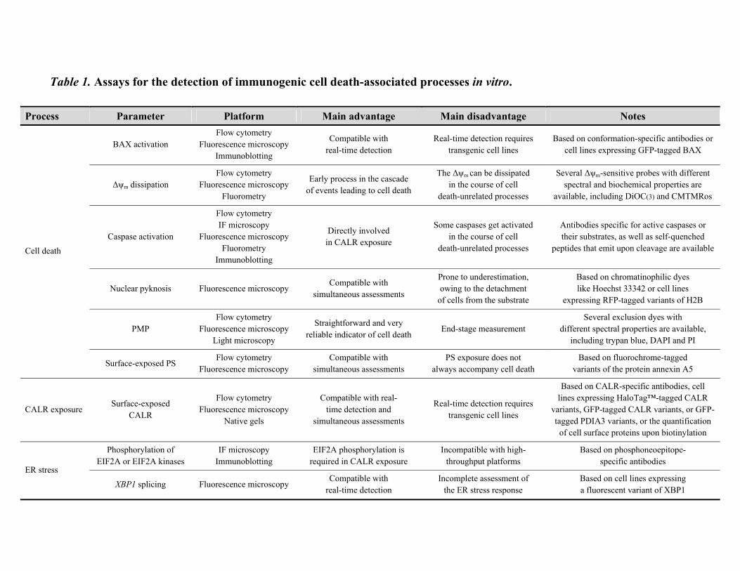
Table 1. Assays for the detection of immunogenic cell death-associated processes in vitro.
Process Parameter Platform Main advantage Main disadvantage Notes
Cell death
BAX activation Flow cytometry
Fluorescence microscopy Immunoblotting
Compatible with real-time detection
Real-time detection requires transgenic cell lines
Based on conformation-specific antibodies or cell lines expressing GFP-tagged BAX
Δψm dissipation Flow cytometry
Fluorescence microscopy Fluorometry
Early process in the cascade of events leading to cell death
The Δψm can be dissipated in the course of cell
death-unrelated processes
Several Δψm-sensitive probes with different spectral and biochemical properties are
available, including DiOC(3) and CMTMRos
Caspase activation
Flow cytometry IF microscopy
Fluorescence microscopy Fluorometry
Immunoblotting
Directly involved in CALR exposure
Some caspases get activated in the course of cell
death-unrelated processes
Antibodies specific for active caspases or their substrates, as well as self-quenched
peptides that emit upon cleavage are available
Nuclear pyknosis Fluorescence microscopy Compatible with
simultaneous assessments
Prone to underestimation, owing to the detachment
of cells from the substrate
Based on chromatinophilic dyes like Hoechst 33342 or cell lines
expressing RFP-tagged variants of H2B
PMP Flow cytometry
Fluorescence microscopy Light microscopy
Straightforward and very reliable indicator of cell death
End-stage measurement Several exclusion dyes with
different spectral properties are available, including trypan blue, DAPI and PI
Surface-exposed PS Flow cytometry
Fluorescence microscopy Compatible with
simultaneous assessments PS exposure does not
always accompany cell death Based on fluorochrome-tagged
variants of the protein annexin A5
CALR exposure Surface-exposed
CALR
Flow cytometry Fluorescence microscopy
Native gels
Compatible with real- time detection and
simultaneous assessments
Real-time detection requires transgenic cell lines
Based on CALR-specific antibodies, cell lines expressing HaloTag™-tagged CALR
variants, GFP-tagged CALR variants, or GFP-tagged PDIA3 variants, or the quantification
of cell surface proteins upon biotinylation
ER stress
Phosphorylation of EIF2A or EIF2A kinases
IF microscopy Immunoblotting
EIF2A phosphorylation is required in CALR exposure
Incompatible with high-throughput platforms
Based on phosphoneoepitope- specific antibodies
XBP1 splicing Fluorescence microscopy Compatible with
real-time detection Incomplete assessment of
the ER stress response Based on cell lines expressing a fluorescent variant of XBP1
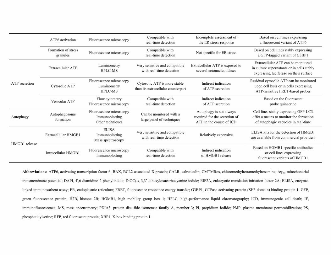
ATF6 activation Fluorescence microscopy Compatible with
real-time detection Incomplete assessment of
the ER stress response Based on cell lines expressing a fluorescent variant of ATF6
Formation of stress granules
Fluorescence microscopy Compatible with
real-time detection Not specific for ER stress
Based on cell lines stably expressing a GFP-tagged variant of G3BP1
ATP secretion
Extracellular ATP Luminometry
HPLC-MS Very sensitive and compatible
with real-time detection Extracellular ATP is exposed to
several ectonucleotidases
Extracellular ATP can be monitored in culture supernatants or in cells stably
expressing luciferase on their surface
Cytosolic ATP Fluorescence microscopy
Luminometry HPLC-MS
Cytosolic ATP is more stable than its extracellular counterpart
Indirect indication of ATP secretion
Residual cytosolic ATP can be monitored upon cell lysis or in cells expressing ATP-sensitive FRET-based probes
Vesicular ATP Flow cytometry
Fluorescence microscopy Compatible with
real-time detection Indirect indication of ATP secretion
Based on the fluorescent probe quinacrine
Autophagy Autophagosome
formation
Fluorescence microscopy Immunoblotting Other techniques
Can be monitored with a large panel of techniques
Autophagy is not always required for the secretion of ATP in the course of ICD
Cell lines stably expressing GFP-LC3 offer a means to monitor the formation
of autophagic vacuoles in real-time
HMGB1 release
Extracellular HMGB1 ELISA
Immunoblotting Mass spectroscopy
Very sensitive and compatible with real-time detection
Relatively expensive ELISA kits for the detection of HMGB1 are available from commercial providers
Intracellular HMGB1 Fluorescence microscopy
Immunoblotting Compatible with
real-time detection Indirect indication of HMGB1 release
Based on HGMB1-specific antibodies or cell lines expressing
fluorescent variants of HMGB1
Abbreviations: ATF6, activating transcription factor 6; BAX, BCL2-associated X protein; CALR, calreticulin; CMTMRos, chloromethyltetramethylrosamine; Δψm, mitochondrial
transmembrane potential; DAPI, 4',6-diamidino-2-phenylindole; DiOC(3), 3,3’-dihexyloxacarbocyanine iodide; EIF2A, eukaryotic translation initiation factor 2A; ELISA, enzyme-
linked immunosorbent assay; ER, endoplasmic reticulum; FRET, fluorescence resonance energy transfer; G3BP1, GTPase activating protein (SH3 domain) binding protein 1; GFP,
green fluorescence protein; H2B, histone 2B; HGMB1, high mobility group box 1; HPLC, high-performance liquid chromatography; ICD, immunogenic cell death; IF,
immunofluorescence; MS, mass spectrometry; PDIA3, protein disulfide isomerase family A, member 3; PI, propidium iodide; PMP, plasma membrane permeabilization; PS,
phosphatidylserine; RFP, red fluorescent protein; XBP1, X-box binding protein 1.