2.3 Peritoneal cavity 2.4 Liver and gallbladder 2.5 Stomach and spleen Abdomen 2 Albert van Schoor...
72
2.3 Peritoneal cavity 2.4 Liver and gallbladder 2.5 Stomach and spleen Abdomen 2 Albert van Schoor GNK 288 (SA4 Anatomy dissection)
-
Upload
barnard-norris -
Category
Documents
-
view
221 -
download
2
Transcript of 2.3 Peritoneal cavity 2.4 Liver and gallbladder 2.5 Stomach and spleen Abdomen 2 Albert van Schoor...
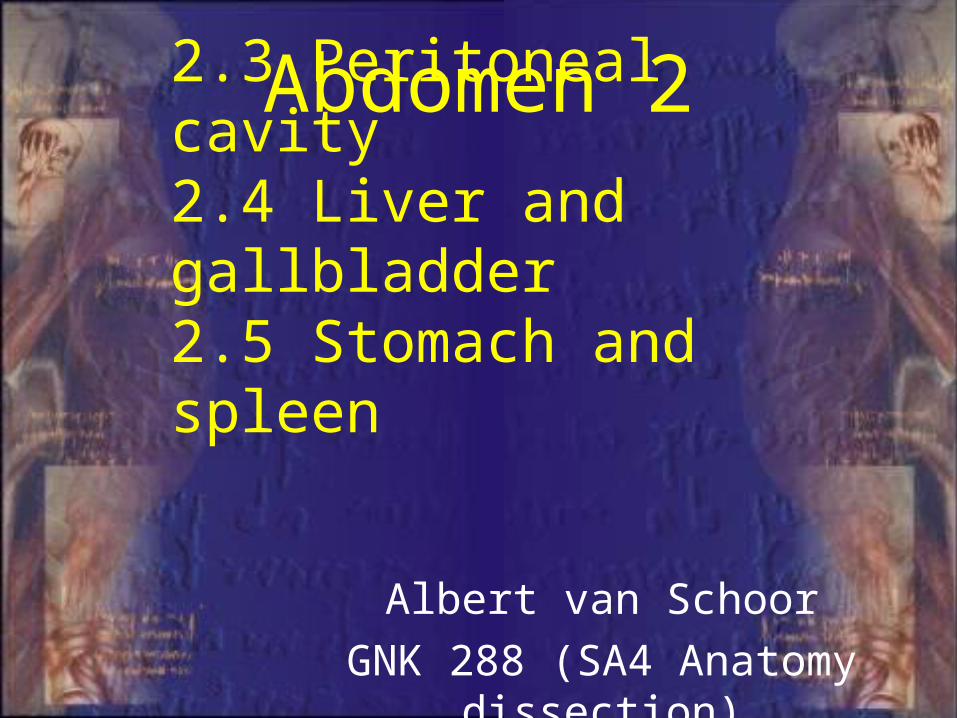
2.3 Peritoneal cavity2.4 Liver and gallbladder2.5 Stomach and spleen
Abdomen 2
Albert van Schoor
GNK 288 (SA4 Anatomy dissection)
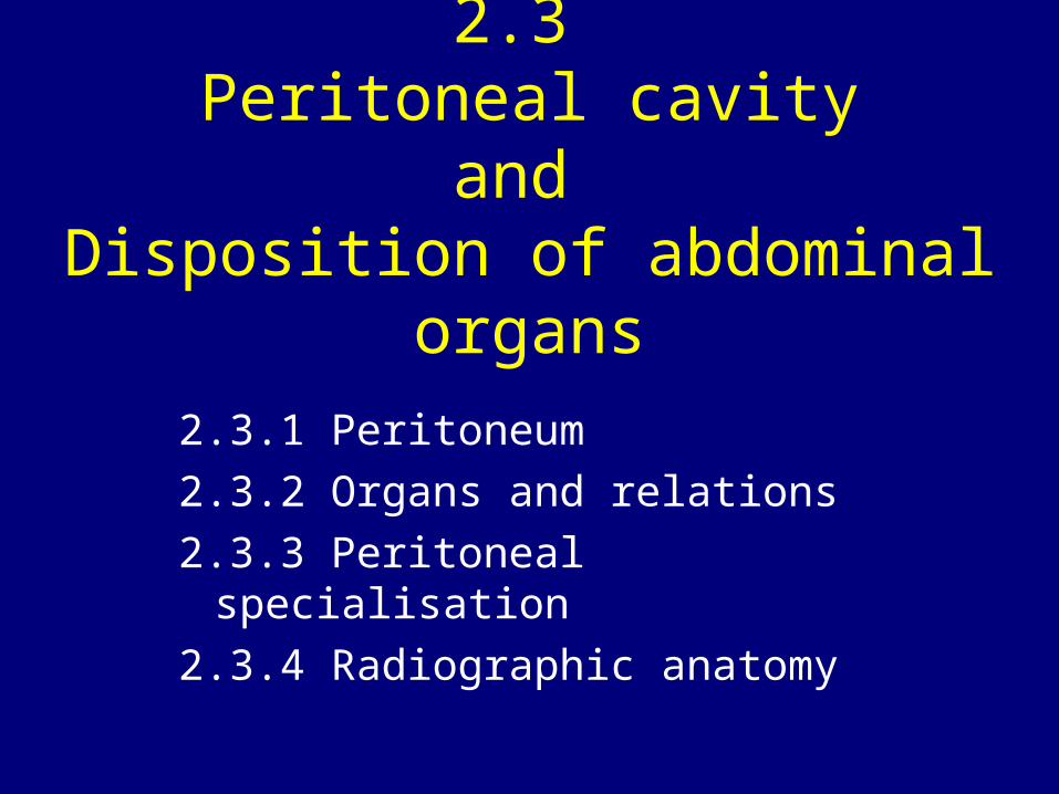
2.3 Peritoneal cavity
and Disposition of abdominal organs
2.3.1 Peritoneum
2.3.2 Organs and relations
2.3.3 Peritoneal specialisation
2.3.4 Radiographic anatomy
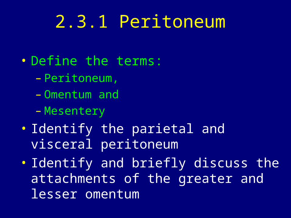
2.3.1 Peritoneum
• Define the terms: – Peritoneum, – Omentum and – Mesentery
• Identify the parietal and visceral peritoneum
• Identify and briefly discuss the attachments of the greater and lesser omentum
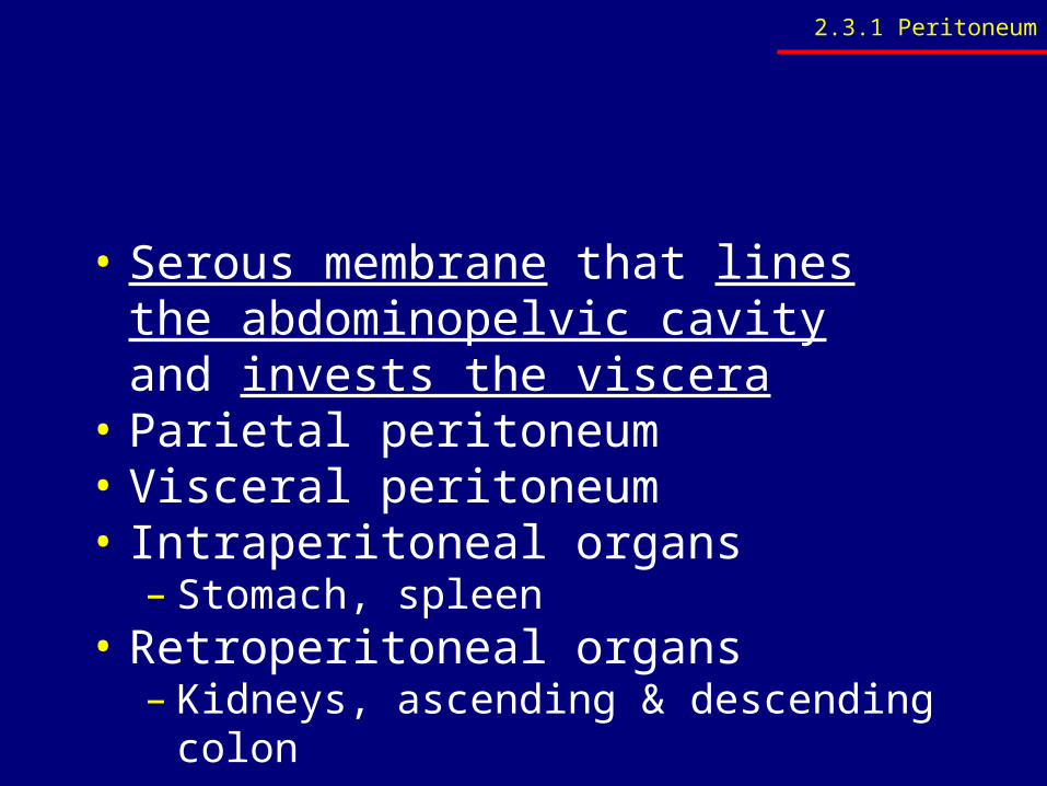
• Serous membrane that lines the abdominopelvic cavity and invests the viscera
• Parietal peritoneum• Visceral peritoneum• Intraperitoneal organs
– Stomach, spleen• Retroperitoneal organs
– Kidneys, ascending & descending colon
2.3.1 Peritoneum
2.3.1 Peritoneum
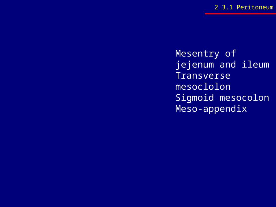
Mesentry of jejenum and ileumTransverse mesoclolonSigmoid mesocolonMeso-appendix
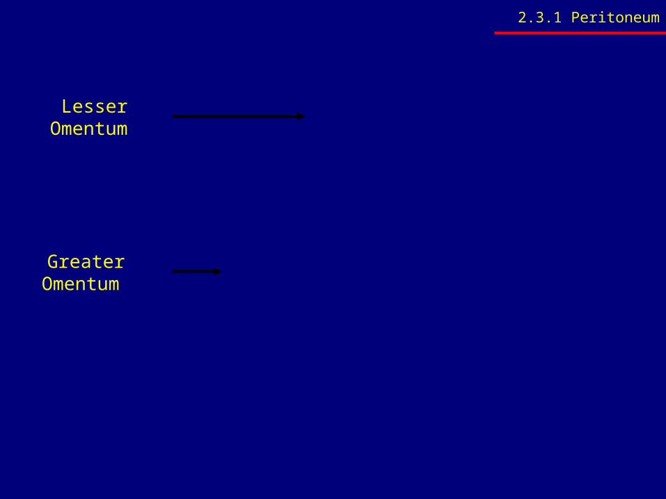
Lesser Omentum
Greater Omentum
2.3.1 Peritoneum
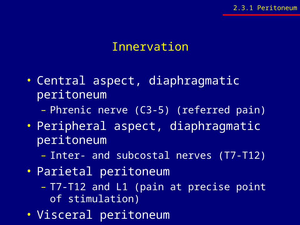
Innervation
• Central aspect, diaphragmatic peritoneum– Phrenic nerve (C3-5) (referred pain)
• Peripheral aspect, diaphragmatic peritoneum– Inter- and subcostal nerves (T7-T12)
• Parietal peritoneum– T7-T12 and L1 (pain at precise point of
stimulation)
• Visceral peritoneum– Insensitive to mechanical stimulation
2.3.1 Peritoneum
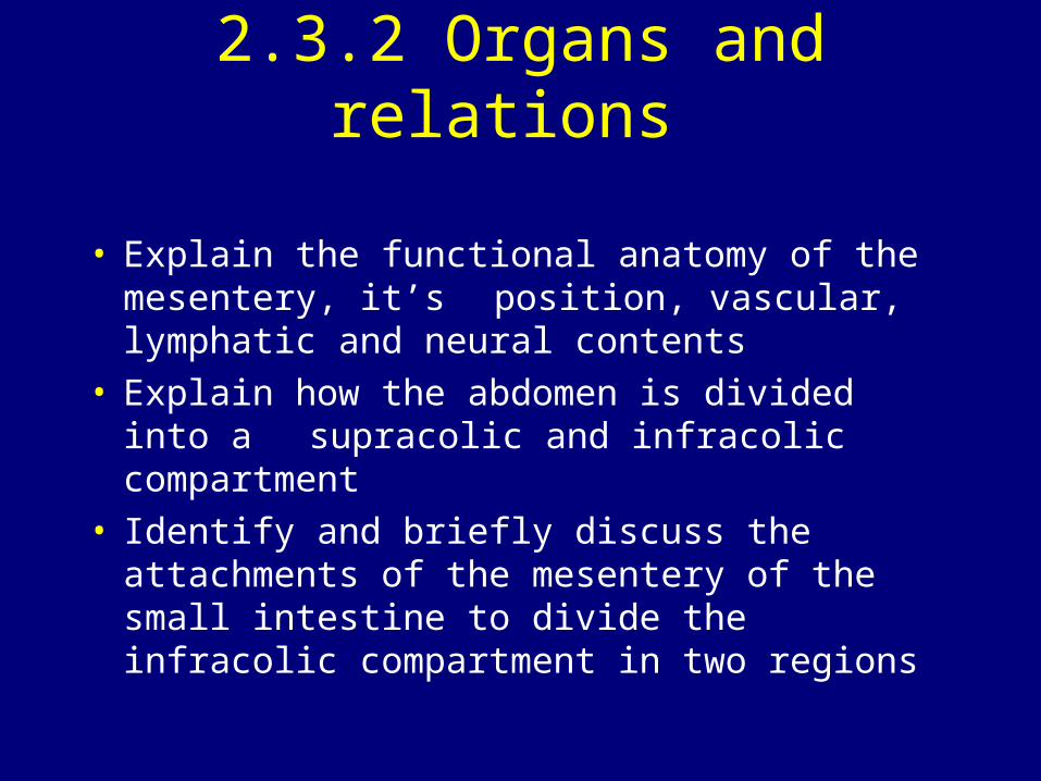
2.3.2 Organs and relations
• Explain the functional anatomy of the mesentery, it’s position, vascular, lymphatic and neural contents
• Explain how the abdomen is divided into a supracolic and infracolic compartment
• Identify and briefly discuss the attachments of the mesentery of the small intestine to divide the infracolic compartment in two regions
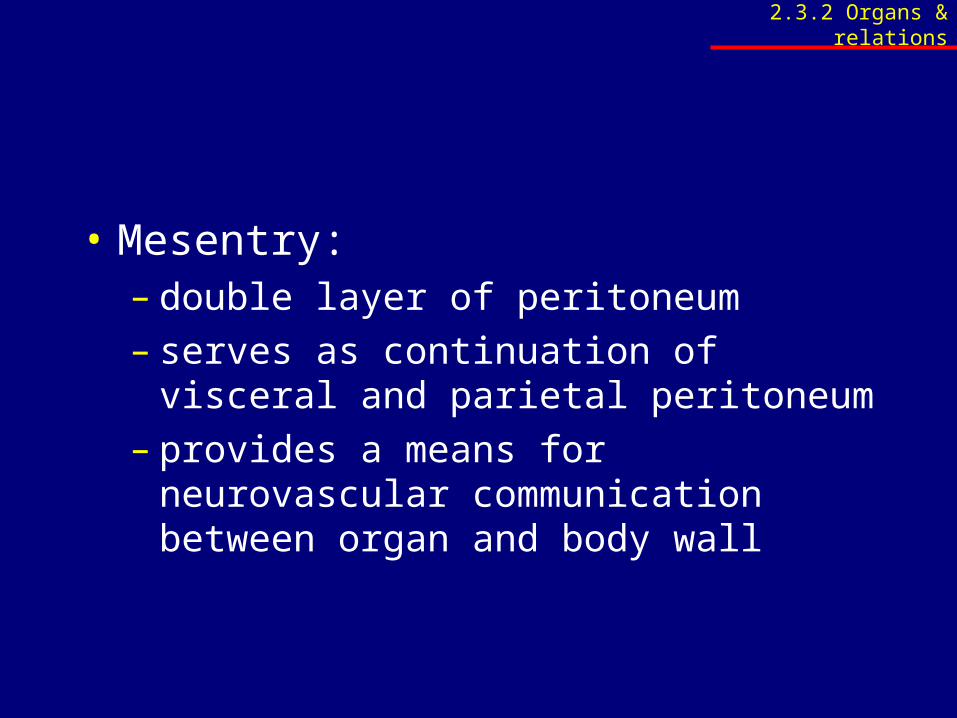
• Mesentry: – double layer of peritoneum– serves as continuation of visceral and
parietal peritoneum– provides a means for neurovascular
communication between organ and body wall
2.3.2 Organs & relations
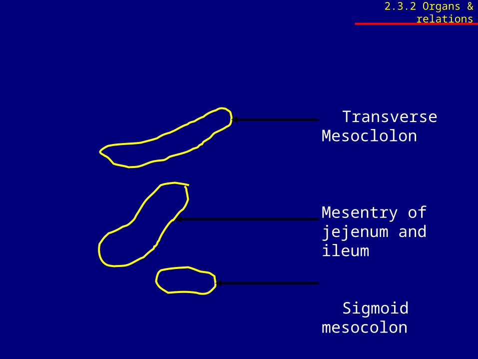
Transverse Mesoclolon
Mesentry of jejenum and ileum
Sigmoid mesocolon
2.3.2 Organs & relations
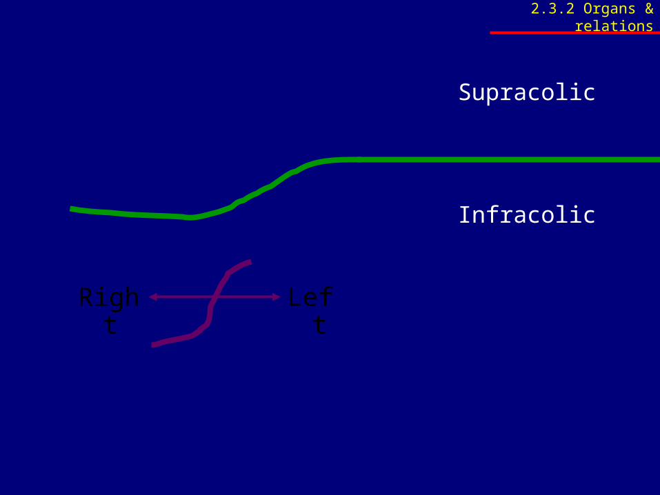
Supracolic
Infracolic
Right Left
2.3.2 Organs & relations
2.3.3 Peritoneal specialisation
• Name and identify the peritoneal folds• Name and identify the peritoneal fossae• Name and identify the paracolic gutters
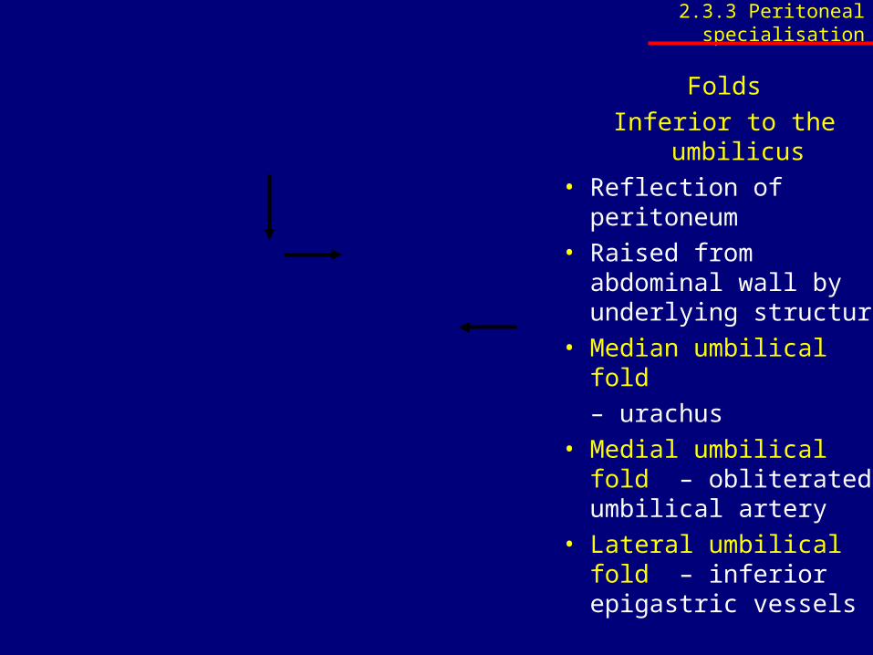
Folds
Inferior to the umbilicus• Reflection of
peritoneum• Raised from
abdominal wall by underlying structure
• Median umbilical fold
– urachus• Medial umbilical fold
– obliterated umbilical artery
• Lateral umbilical fold – inferior epigastric vessels
2.3.3 Peritoneal specialisation
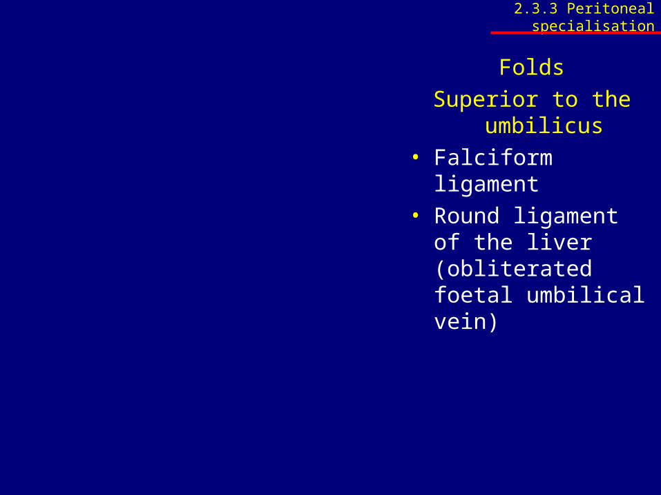
Folds
Superior to the umbilicus• Falciform ligament• Round ligament of the
liver (obliterated foetal umbilical vein)
2.3.3 Peritoneal specialisation
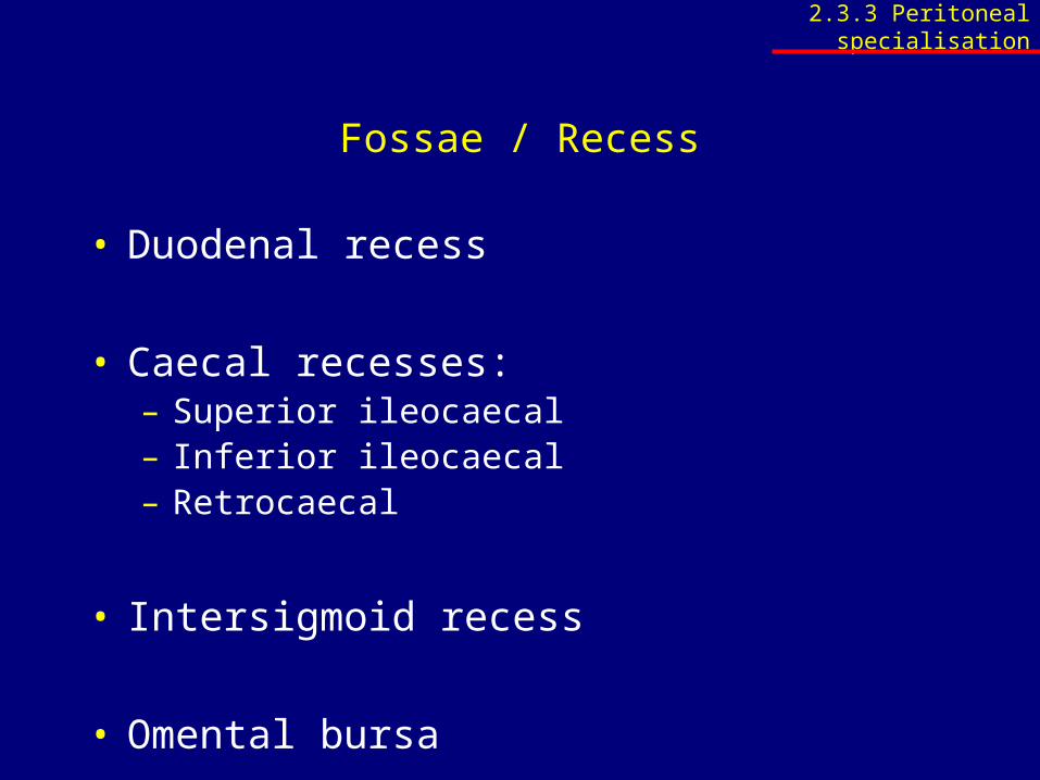
Fossae / Recess
• Duodenal recess
• Caecal recesses:– Superior ileocaecal– Inferior ileocaecal– Retrocaecal
• Intersigmoid recess
• Omental bursa
2.3.3 Peritoneal specialisation
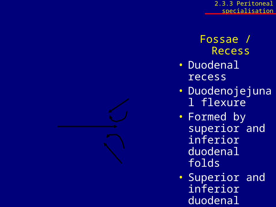
Fossae / Recess• Duodenal recess• Duodenojejunal
flexure• Formed by
superior and inferior duodenal folds
• Superior and inferior duodenal recesses
• Paraduodenal recess
2.3.3 Peritoneal specialisation

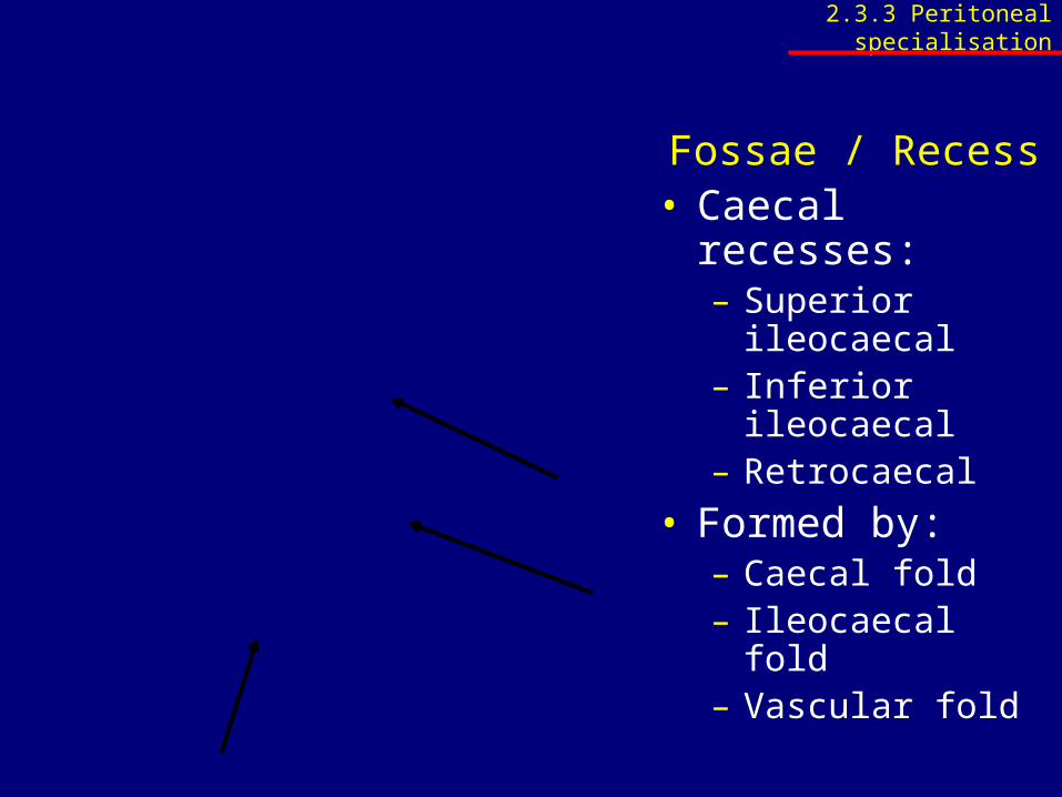
Fossae / Recess• Caecal recesses:
– Superior ileocaecal– Inferior ileocaecal– Retrocaecal
• Formed by:– Caecal fold– Ileocaecal fold– Vascular fold
2.3.3 Peritoneal specialisation
Fossae / Recess• Caecal recesses:
– Superior ileocaecal– Inferior ileocaecal– Retrocaecal
• Formed by:– Caecal fold– Ileocaecal fold– Vascular fold
2.3.3 Peritoneal specialisation
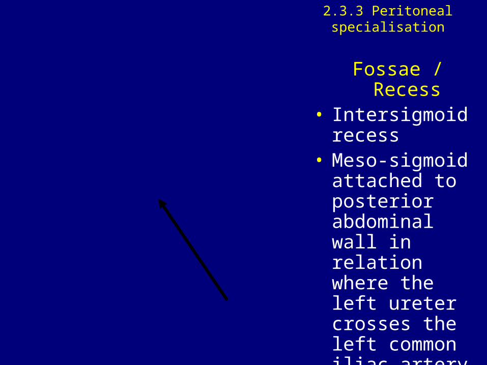
Fossae / Recess• Intersigmoid
recess• Meso-sigmoid
attached to posterior abdominal wall in relation where the left ureter crosses the left common iliac artery
2.3.3 Peritoneal specialisation
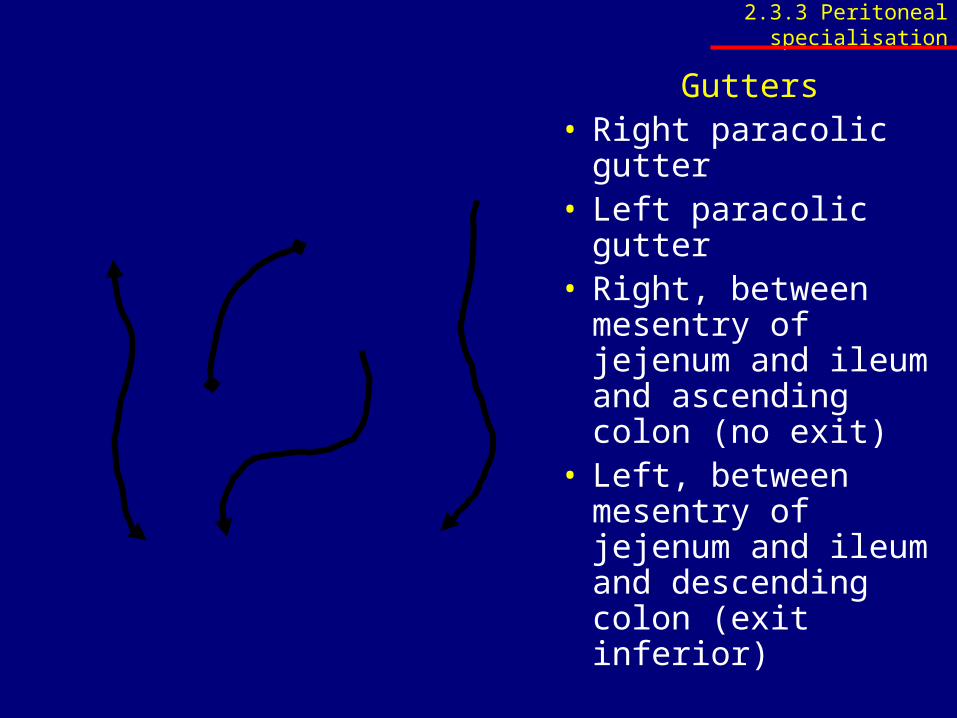
Gutters• Right paracolic
gutter• Left paracolic gutter• Right, between
mesentry of jejenum and ileum and ascending colon (no exit)
• Left, between mesentry of jejenum and ileum and descending colon (exit inferior)
2.3.3 Peritoneal specialisation
2.3.3 Peritoneal specialisation
• Identify the following: – Gastrosplenic ligament, – Splenorenal ligament, – Transverse and sigmoid mesocolon, – Ileocoecal fold, – Meso-appendix and – The mesenterium of the small intestine
2.3.3 Peritoneal specialisation
• Identify and describe the omental bursa (lesser sac) in respect of its relations, borders and entrance - the omental foramen
• Identify the structures forming the borders of the omental foramen
• Name and identify the subphrenic spaces
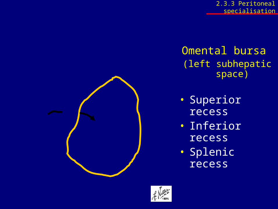
Omental bursa (left subhepatic space)
• Superior recess• Inferior recess• Splenic recess
2.3.3 Peritoneal specialisation
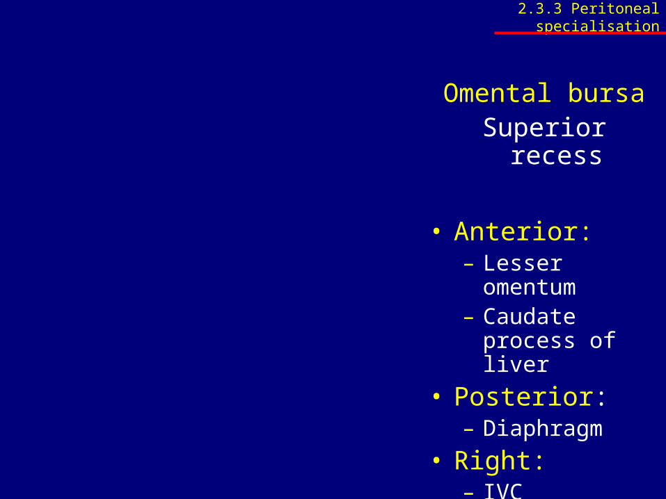
Omental bursaSuperior recess
• Anterior:– Lesser omentum– Caudate process
of liver
• Posterior:– Diaphragm
• Right:– IVC
• Left: – Oesophagus
2.3.3 Peritoneal specialisation
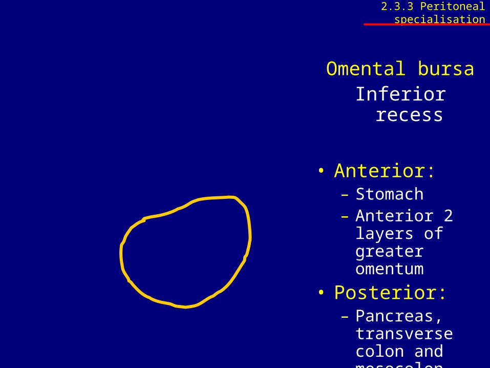
Omental bursaInferior recess
• Anterior:– Stomach– Anterior 2 layers
of greater omentum
• Posterior:– Pancreas,
transverse colon and mesocolon, poster 2 layers of greater omentum
2.3.3 Peritoneal specialisation
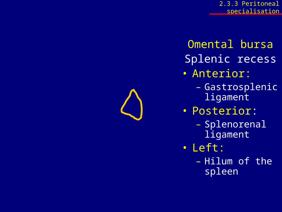
Omental bursaSplenic recess
• Anterior:– Gastrosplenic
ligament
• Posterior:– Splenorenal
ligament
• Left: – Hilum of the
spleen
2.3.3 Peritoneal specialisation
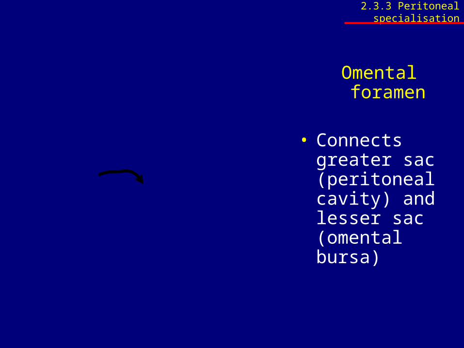
Omental foramen
• Connects greater sac (peritoneal cavity) and lesser sac (omental bursa)
2.3.3 Peritoneal specialisation
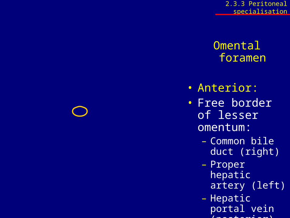
Omental foramen
• Anterior:• Free border of
lesser omentum:– Common bile
duct (right)– Proper hepatic
artery (left)– Hepatic portal
vein (posterior)
2.3.3 Peritoneal specialisation
Omental foramen
• Posterior:– Inferior vena cava
2.3.3 Peritoneal specialisation
Omental foramen
• Superior:– Caudate lobe of
liver
2.3.3 Peritoneal specialisation
Omental foramen
• Inferior:– 1st part of
duodenum– Common hepatic
artery
2.3.3 Peritoneal specialisation
2.3.3 Peritoneal specialisation
• Name and identify the subphrenic spaces
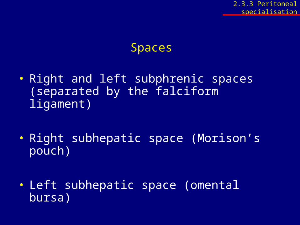
Spaces
• Right and left subphrenic spaces (separated by the falciform ligament)
• Right subhepatic space (Morison’s pouch)
• Left subhepatic space (omental bursa)
• Extraperitoneal subphrenic space
2.3.3 Peritoneal specialisation
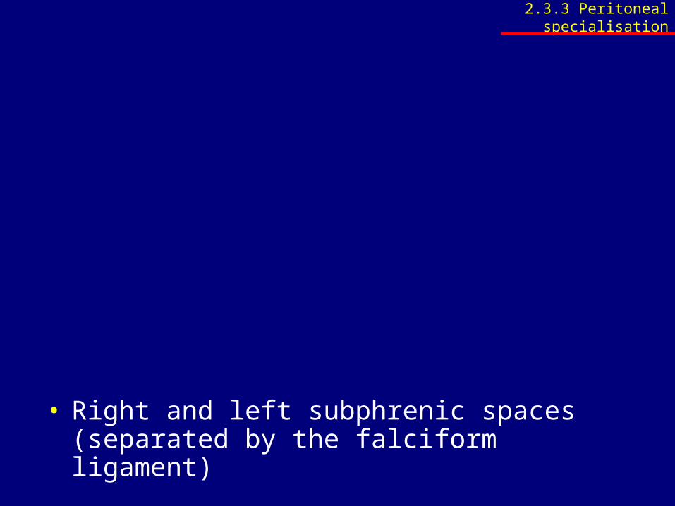
• Right and left subphrenic spaces (separated by the falciform ligament)
2.3.3 Peritoneal specialisation
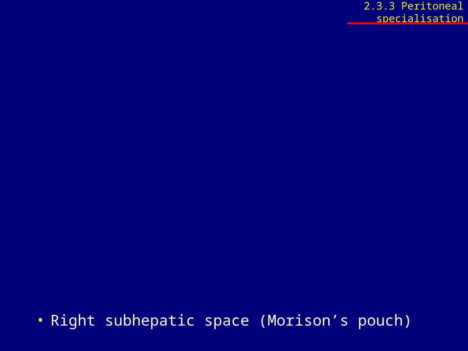
• Right subhepatic space (Morison’s pouch)
2.3.3 Peritoneal specialisation
• Extraperitoneal subphrenic space
2.3.3 Peritoneal specialisation
2.3.4 Radiographic anatomy
• Identify the following structures on a plain erect abdominal X-ray: – ASIS, – lumbar vertebrae, – SI-joint, – large intestine, – diaphragm, – stomach with air in fundus of stomach,– liver, – psoas line
www.up.ac.za/academic/medicine/anatomy/current/sa4/week01e.html#radio
2.4 Liver and gallbladder
2.4.1 Surface anatomy
2.4.2 Structure
2.4.3 Blood supply, nerve supply and lymph drainage
2.4.1 Surface anatomy
• Review the surface anatomy of the liver and gallbladder
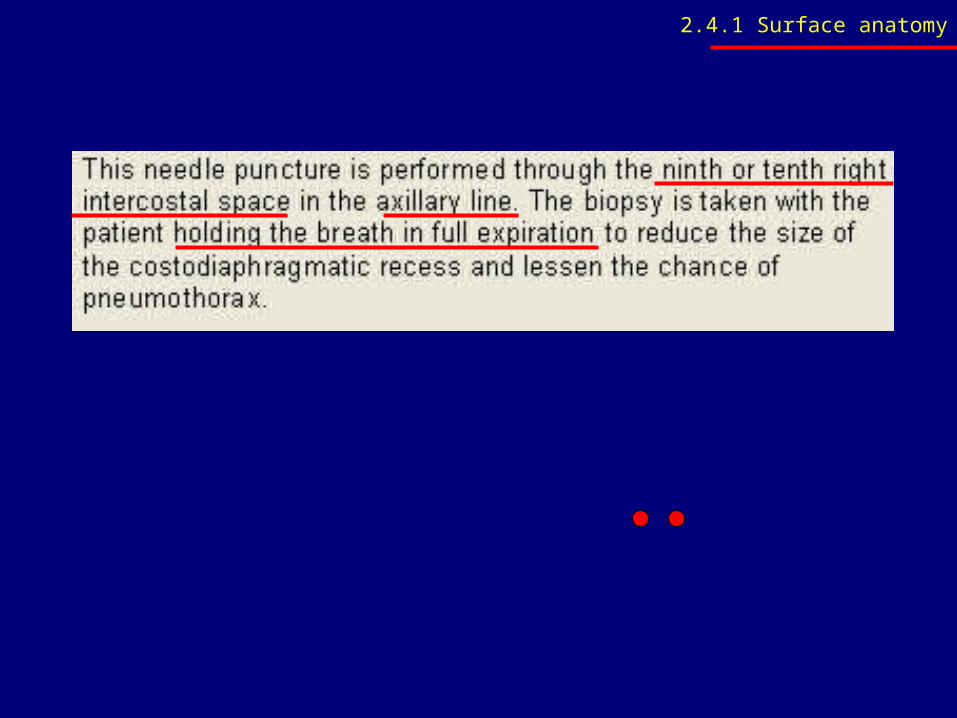
• Indicate where a liver biopsy should be done
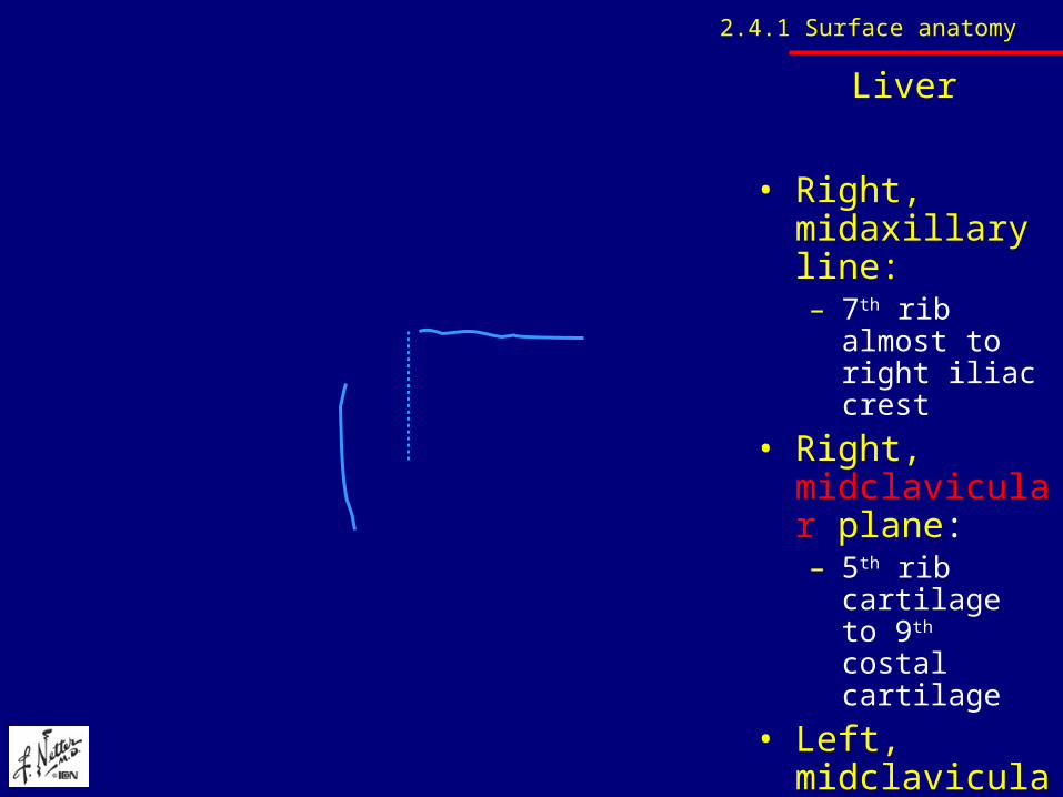
Liver
• Right, midaxillary line:– 7th rib almost to
right iliac crest
• Right, midclavicular plane:– 5th rib cartilage
to 9th costal cartilage
• Left, midclavicular plane: – 2.5cm short 5th
intercostal space and left nipple
2.4.1 Surface anatomy
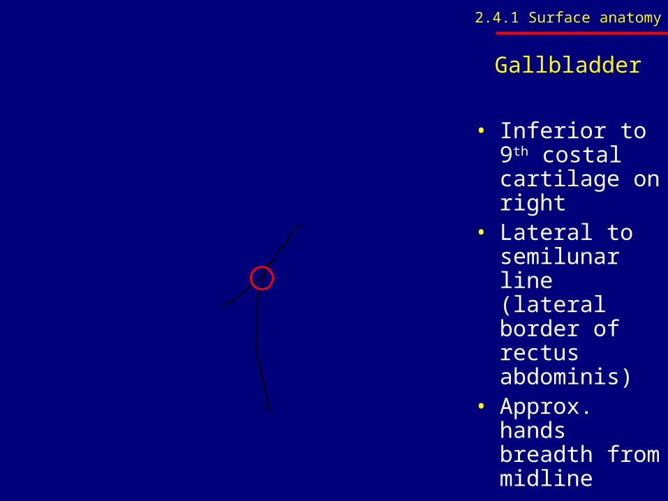
Gallbladder
• Inferior to 9th costal cartilage on right
• Lateral to semilunar line (lateral border of rectus abdominis)
• Approx. hands breadth from midline
2.4.1 Surface anatomy
2.4.1 Surface anatomy
2.4.2 Structure
• Name and identify the borders and surfaces of the liver
• Name and identify the lobes, segments, fissures with their contents Identify the subhepatic and subphrenic spaces, and their possible implication in the spread of infection
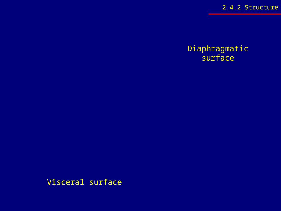
Diaphragmatic surface
Visceral surface
2.4.2 Structure
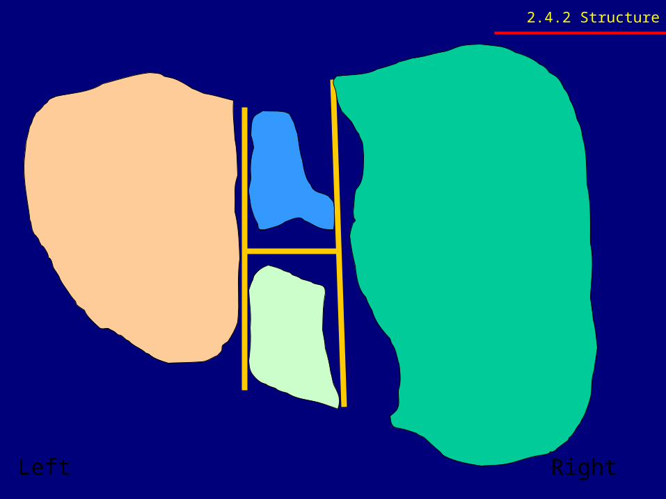
Left Right
2.4.2 Structure
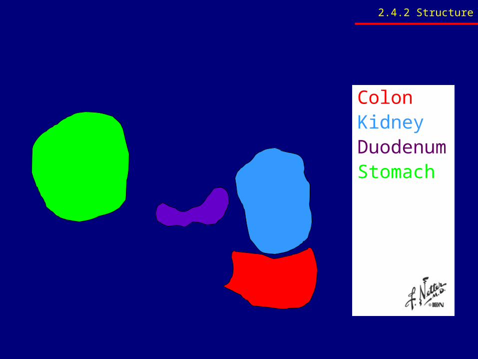
ColonKidneyDuodenumStomach
2.4.2 Structure
2.4.2 Structure
• Name and identify the following: – Triangular ligaments, – Coronary ligaments, – Falciform ligament, – Lesser omentum, – Round ligament of the liver and – Ligamentum venosum
2.4.2 Structure
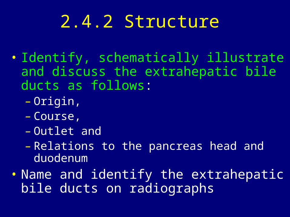
• Identify, schematically illustrate and discuss the extrahepatic bile ducts as follows: – Origin, – Course, – Outlet and – Relations to the pancreas head and duodenum
• Name and identify the extrahepatic bile ducts on radiographs
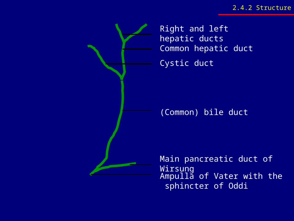
Right and left hepatic ducts
Common hepatic duct
Cystic duct
(Common) bile duct
Main pancreatic duct of Wirsung
Ampulla of Vater with the sphincter of Oddi
2.4.2 Structure
ERCPEndoscopic retrograde
cholangiopancreatography
2.4.2 Structure
2.4.3 Blood supply
• Discuss and identify the arterial blood supply and venous drainage of the liver and gallbladder
• Take note of variations in the arterial blood supply of the liver and gallbladder
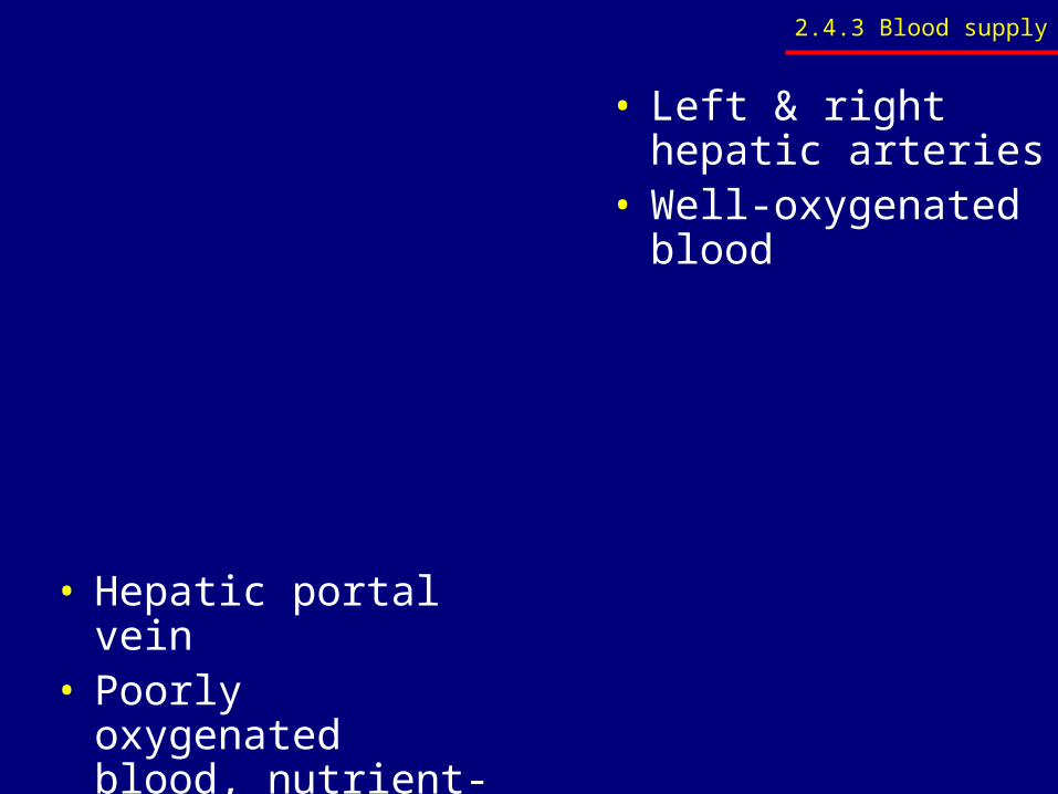
• Left & right hepatic arteries
• Well-oxygenated blood
• Hepatic portal vein• Poorly oxygenated
blood, nutrient-rich blood from GI tract
2.4.3 Blood supply
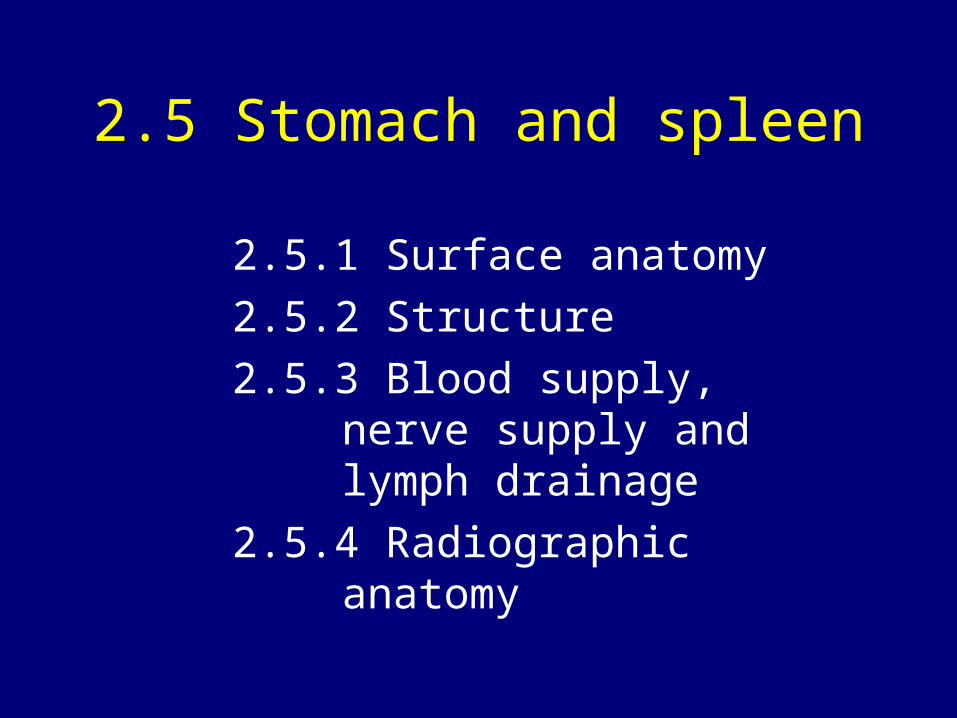
2.5 Stomach and spleen
2.5.1 Surface anatomy
2.5.2 Structure
2.5.3 Blood supply, nerve supply and lymph drainage
2.5.4 Radiographic anatomy
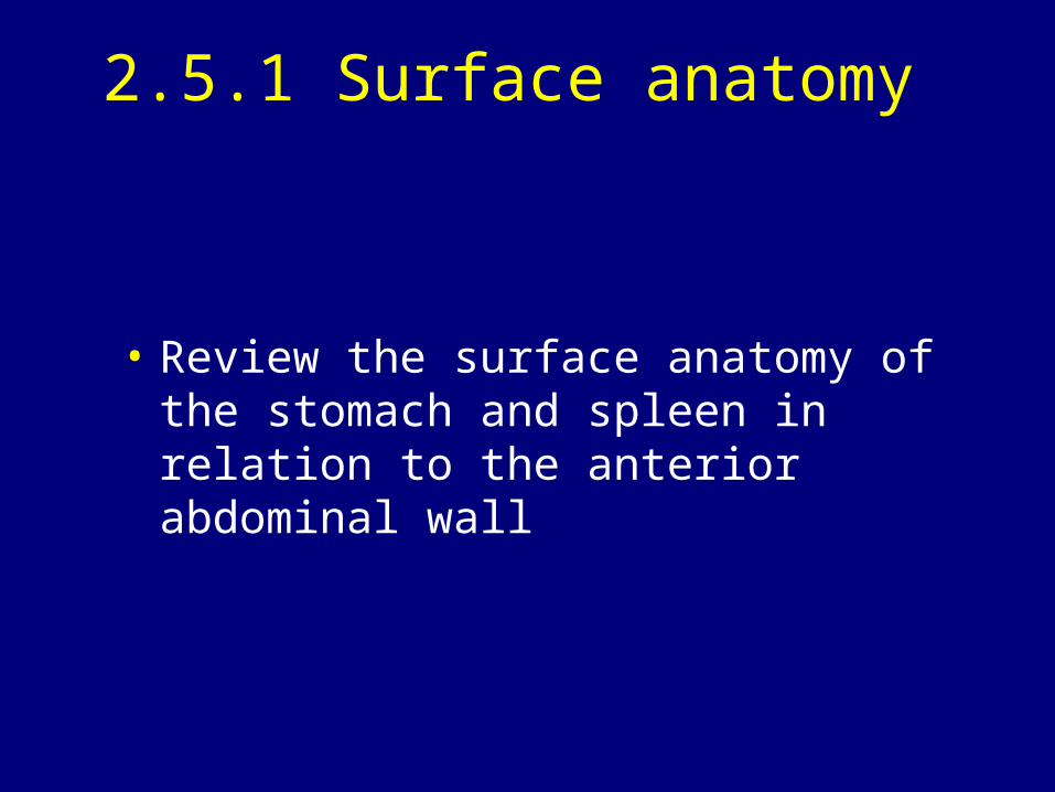
2.5.1 Surface anatomy
• Review the surface anatomy of the stomach and spleen in relation to the anterior abdominal wall
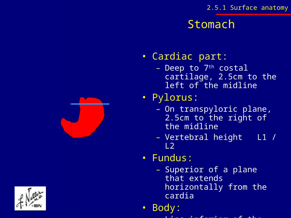
Stomach
• Cardiac part:– Deep to 7th costal cartilage,
2.5cm to the left of the midline
• Pylorus:– On transpyloric plane, 2.5cm to
the right of the midline– Vertebral height L1 / L2
• Fundus: – Superior of a plane that extends
horizontally from the cardia
• Body: – Lies inferior of the above-
mentioned plane
2.5.1 Surface anatomy
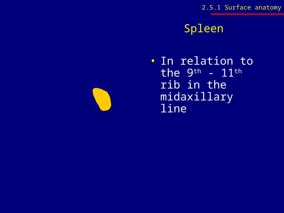
Spleen
• In relation to the 9th - 11th rib in the midaxillary line
2.5.1 Surface anatomy
2.5.2 Structure
• Name and identify the various regions, curvatures and surfaces of the stomach. Identify these also on radiographs
• Identify and briefly describe the general and peritoneal relations of the stomach
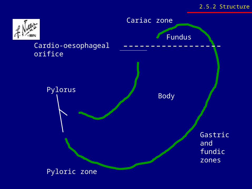
Pyloric zone
Gastric and fundic zones
Cariac zone
Fundus
Body
Cardio-oesophageal orifice
Pylorus
2.5.2 Structure
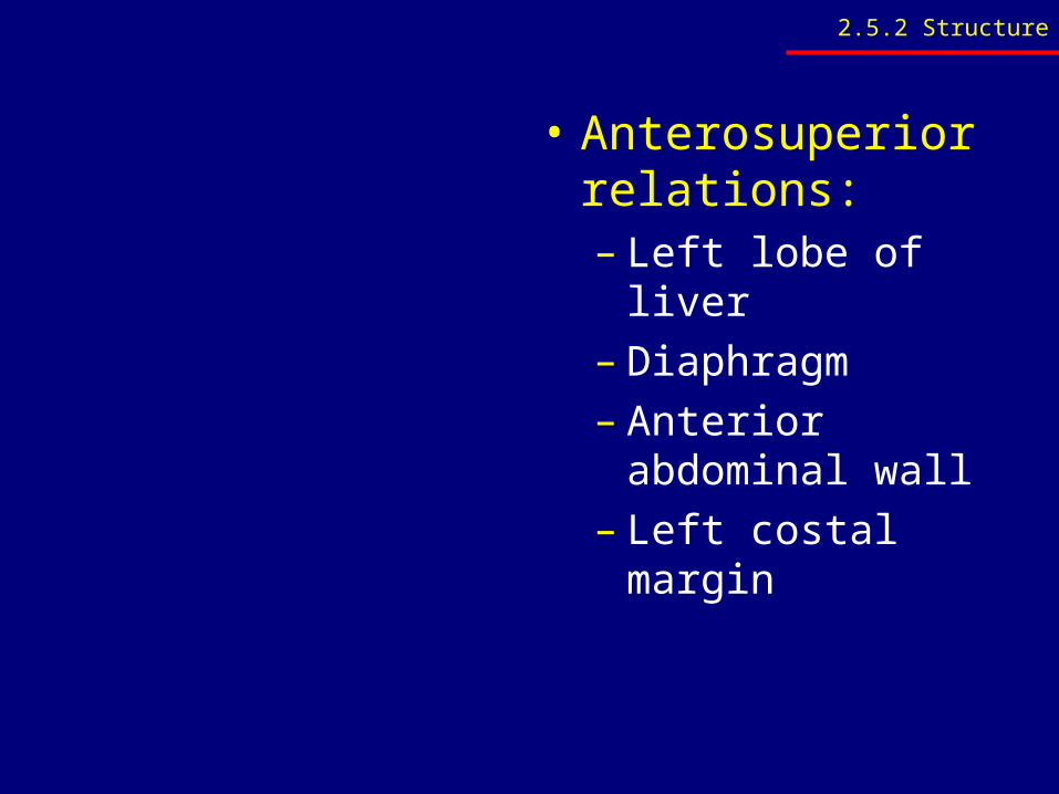
• Anterosuperior relations:– Left lobe of liver– Diaphragm– Anterior abdominal
wall– Left costal margin
2.5.2 Structure
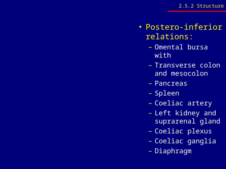
• Postero-inferior relations:– Omental bursa with– Transverse colon
and mesocolon– Pancreas– Spleen– Coeliac artery– Left kidney and
suprarenal gland– Coeliac plexus– Coeliac ganglia– Diaphragm
2.5.2 Structure
2.5.2 Structure
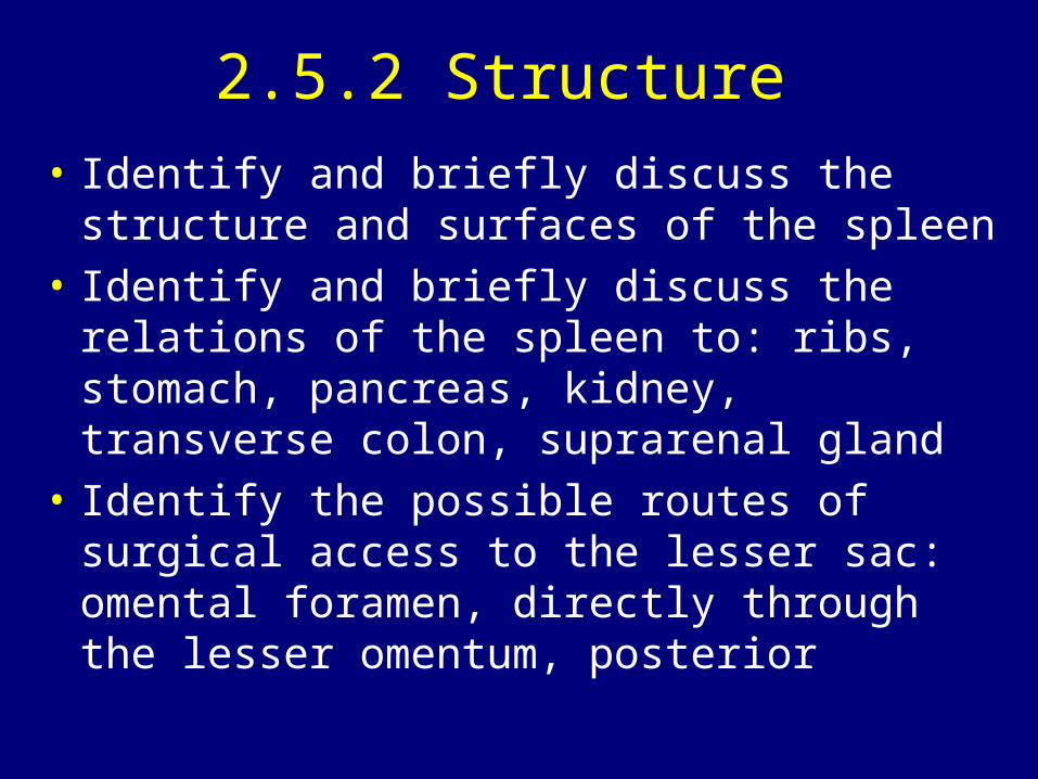
• Identify and briefly discuss the structure and surfaces of the spleen
• Identify and briefly discuss the relations of the spleen to: ribs, stomach, pancreas, kidney, transverse colon, suprarenal gland
• Identify the possible routes of surgical access to the lesser sac: omental foramen, directly through the lesser omentum, posterior

2.5.2 Structure
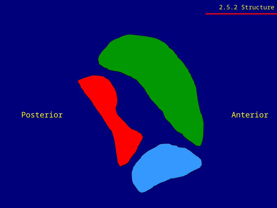
Posterior Anterior
2.5.2 Structure
2.5.3 Blood supply, nerve supply and lymph drainage
• Identify and briefly discuss the arterial supply and venous drainage of the stomach and spleen
• Schematically illustrate the coeliac trunk and it's branches
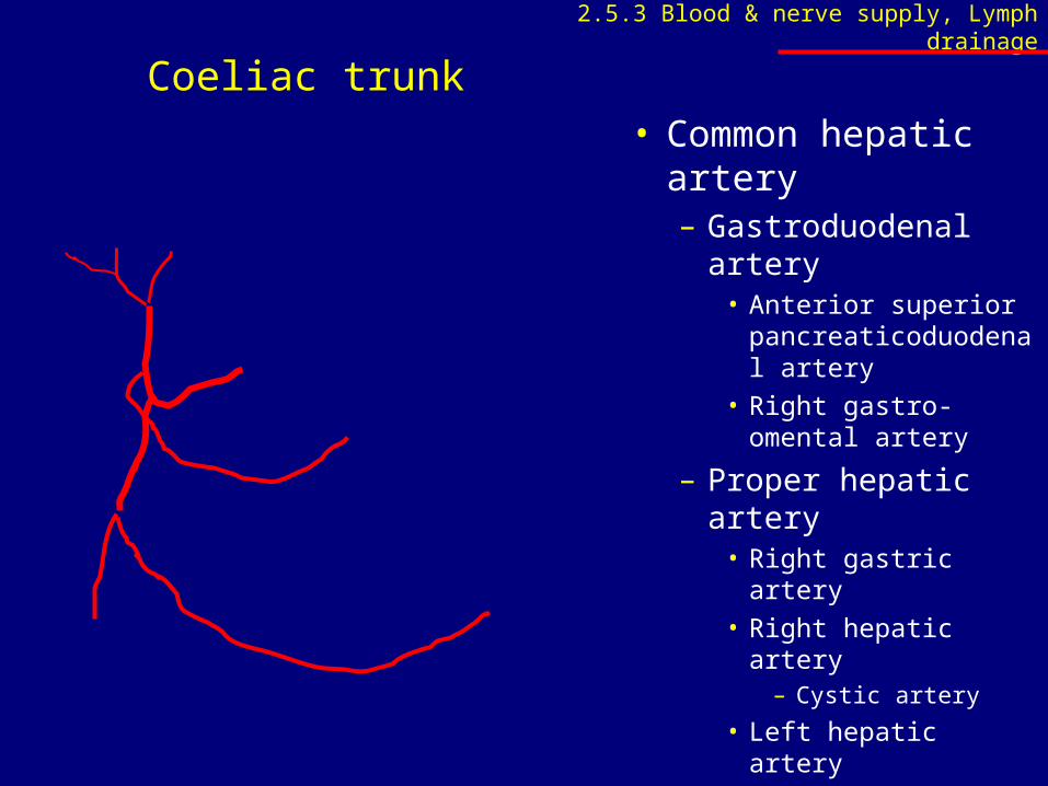
• Common hepatic artery– Gastroduodenal
artery• Anterior superior
pancreaticoduodenal artery
• Right gastro-omental artery
– Proper hepatic artery
• Right gastric artery• Right hepatic artery
– Cystic artery
• Left hepatic artery
Coeliac trunk2.5.3 Blood & nerve supply, Lymph drainage
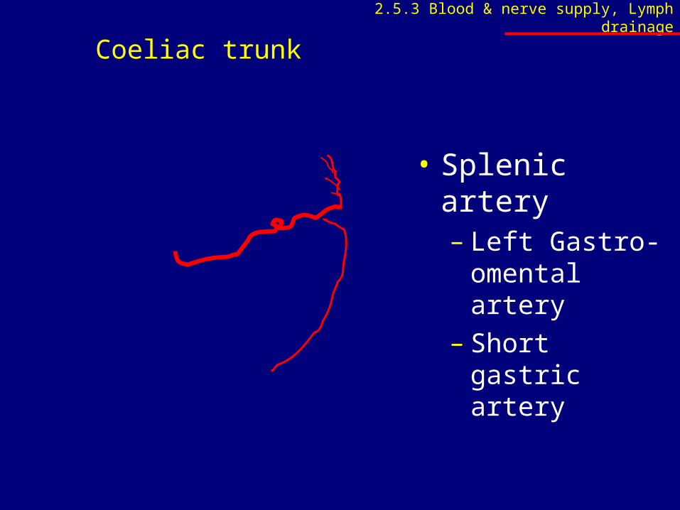
• Splenic artery– Left Gastro-
omental artery– Short gastric
artery
Coeliac trunk2.5.3 Blood & nerve supply, Lymph drainage
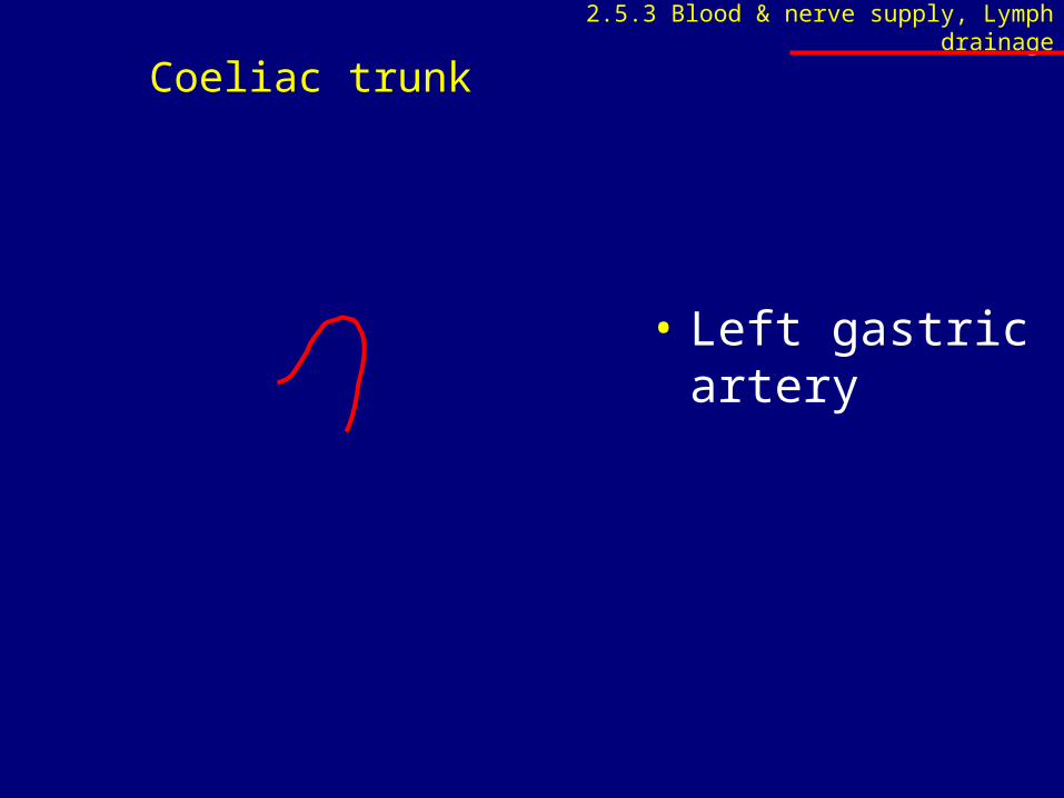
Coeliac trunk
• Left gastric artery
2.5.3 Blood & nerve supply, Lymph drainage
2.5.3 Blood supply, nerve supply and lymph drainage
• Identify and briefly discuss the vagus nerve as follows: – Abdominal entrance (anterior and posterior
vagus trunks), – Prominent plexuses and – Branches and extent of abdominal supply
• Identify the intra-abdominal part of the oesophagus
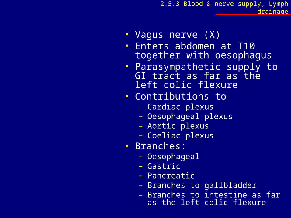
• Vagus nerve (X)• Enters abdomen at T10 together
with oesophagus• Parasympathetic supply to GI
tract as far as the left colic flexure• Contributions to
– Cardiac plexus– Oesophageal plexus– Aortic plexus– Coeliac plexus
• Branches:– Oesophageal– Gastric– Pancreatic– Branches to gallbladder– Branches to intestine as far as the left
colic flexure
2.5.3 Blood & nerve supply, Lymph drainage
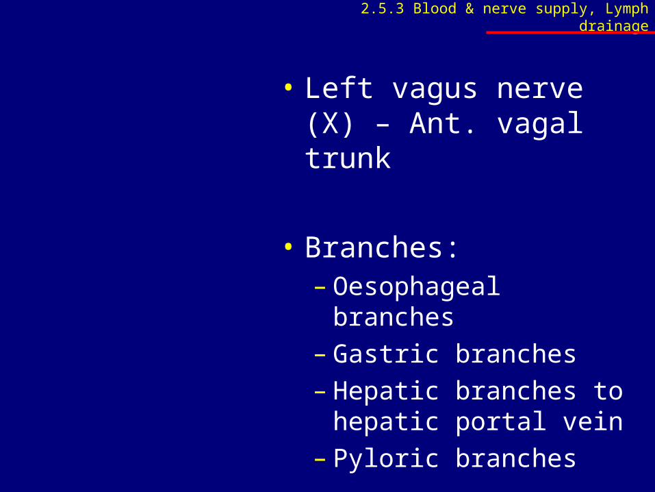
• Left vagus nerve (X) – Ant. vagal trunk
• Branches:– Oesophageal branches– Gastric branches– Hepatic branches to
hepatic portal vein– Pyloric branches
2.5.3 Blood & nerve supply, Lymph drainage
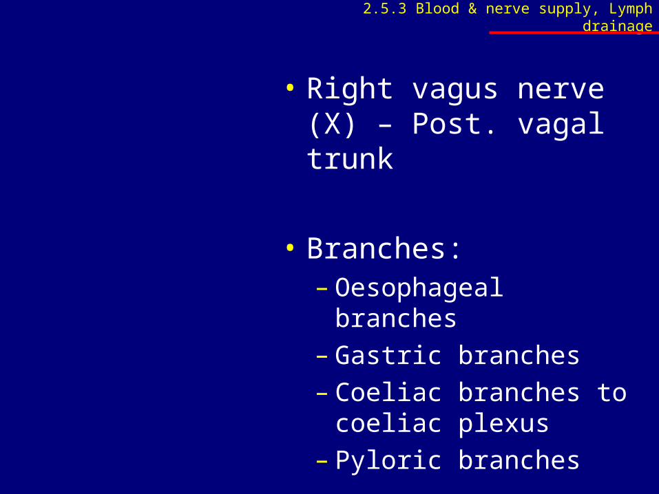
• Right vagus nerve (X) – Post. vagal trunk
• Branches:– Oesophageal branches– Gastric branches– Coeliac branches to
coeliac plexus– Pyloric branches
2.5.3 Blood & nerve supply, Lymph drainage
2.5.4 Radiographic anatomy
• Identify the stomach and air in the fundus of the stomach on a plain erect abdominal X-ray
• Identify the stomach on a barium meal
www.up.ac.za/academic/medicine/anatomy/current/sa4/week01e.html#radio