2246: 45 57 (2009) Article … · Accepted by G. Hormiga: 4 Sept. 2009; published: 5 Oct. 2009 45...
13
Accepted by G. Hormiga: 4 Sept. 2009; published: 5 Oct. 2009 45 ZOOTAXA ISSN 1175-5326 (print edition) ISSN 1175-5334 (online edition) Copyright © 2009 · Magnolia Press Zootaxa 2246: 45–57 (2009) www.mapress.com/ zootaxa/ Article First record of the spider family Symphytognathidae in Europe and description of Anapistula ataecina sp. n. (Araneae) PEDRO CARDOSO 1,2,3,4 & NIKOLAJ SCHARFF 2 1 Azorean Biodiversity Group – CITA–A, Departamento de Ciências Agrárias, Universidade dos Açores, Terra-Chã, 9701–851 Angra do Heroísmo, Portugal. E-mail: [email protected]. 2 Department of Entomology, Natural History Museum of Denmark, Universitetsparken 15, DK–2100 Copenhagen, Denmark. 3 Núcleo de Espeleologia da Costa Azul – FPE, Sesimbra, Portugal. 4 Corresponding author Abstract The spider family Symphytognathidae has never been recorded from Europe, being mostly present in tropical regions. Different collecting trips to a cave system in Portugal revealed several specimens of a new species of Symphytognathidae here described, Anapistula ataecina sp. n. This is one of the smallest spiders described to date. The species is almost exclusively known from the Frade Cave System in Portugal which is partly endangered by limestone quarries. No males were found to date despite many collecting trips to the caves during more than three years. Parthenogenesis could therefore be responsible for the species reproduction. Its web, with a sheet-like structure, seems atypical for the family and for the genus. Details on the eggsacs and spinneret morphology are also given. Key words: Araneoidea, cave, endangered species, morphology, new species, parthenogenesis, Portugal, spider, spinnerets, troglobite Introduction The spider family Symphytognathidae, with its limits defined by Griswold et al. (1998) and Schütt (2003), currently comprises six genera and 61 species (Lin & Li 2009; Miller et al. 2009; Platnick 2009). It includes minute, lungless spiders, with the chelicerae fused wholly or in part, female pedipalp reduced or absent, reduced number of eyes, reduced colulus, and the sternum broadly truncated posteriorly (Forster and Platnick 1977; Griswold 1987; Griswold et al. 1998; Schütt 2003; Miller et al. 2009). This combination of characters will diagnose the family, but most characters are shared with other spider families, so good synapomorphies for the family as currently circumscribed are still to be found and defined. Griswold et al. (1998) described the spinnerets of symphytognathids in details and pointed out that the two aggregate spigots (AG) on the posterior lateral spinnerets (PLS) share a common basis, and Miller et al. (2009) suggested that the AG spigots on a shared base could be a potential synapomorphy for the family. To this family belong the smallest spiders ever described, the male of Patu digua Forster & Platnick, 1977 with a total body length of only 0.37 mm and the females of Anapistula caecula Baert & Jocqué, 1993, A. bifurcata Harvey, 1998 and A. jerai Harvey, 1998, all with a total body length of 0.48 to 0.55 mm. Symphytognathids are mainly distributed in the tropics, but can also be found in south temperate regions (Fig. 1), where they live in the litter of rainforests and similar moist habitats. They are rarely collected, but due to their minute size they are easily overlooked and more species should certainly be expected from all parts of the world. A relatively low carapace and the existence of posterior tracheal spiracles set the genus Anapistula apart
Transcript of 2246: 45 57 (2009) Article … · Accepted by G. Hormiga: 4 Sept. 2009; published: 5 Oct. 2009 45...
ZOOTAXAISSN 1175-5326 (print edition)
ISSN 1175-5334 (online edition)Copyright © 2009 · Magnolia Press
Zootaxa 2246: 45–57 (2009) www.mapress.com/zootaxa/ Article
First record of the spider family Symphytognathidae in Europe and description of Anapistula ataecina sp. n. (Araneae)
PEDRO CARDOSO1,2,3,4 & NIKOLAJ SCHARFF2
1Azorean Biodiversity Group – CITA–A, Departamento de Ciências Agrárias, Universidade dos Açores, Terra-Chã, 9701–851 Angra do Heroísmo, Portugal. E-mail: [email protected] of Entomology, Natural History Museum of Denmark, Universitetsparken 15, DK–2100 Copenhagen, Denmark.3Núcleo de Espeleologia da Costa Azul – FPE, Sesimbra, Portugal.4Corresponding author
Abstract
The spider family Symphytognathidae has never been recorded from Europe, being mostly present in tropical regions. Different collecting trips to a cave system in Portugal revealed several specimens of a new species of Symphytognathidae here described, Anapistula ataecina sp. n. This is one of the smallest spiders described to date. The species is almost exclusively known from the Frade Cave System in Portugal which is partly endangered by limestone quarries. No males were found to date despite many collecting trips to the caves during more than three years. Parthenogenesis could therefore be responsible for the species reproduction. Its web, with a sheet-like structure, seems atypical for the family and for the genus. Details on the eggsacs and spinneret morphology are also given.
Key words: Araneoidea, cave, endangered species, morphology, new species, parthenogenesis, Portugal, spider, spinnerets, troglobite
Introduction
The spider family Symphytognathidae, with its limits defined by Griswold et al. (1998) and Schütt (2003), currently comprises six genera and 61 species (Lin & Li 2009; Miller et al. 2009; Platnick 2009). It includes minute, lungless spiders, with the chelicerae fused wholly or in part, female pedipalp reduced or absent, reduced number of eyes, reduced colulus, and the sternum broadly truncated posteriorly (Forster and Platnick 1977; Griswold 1987; Griswold et al. 1998; Schütt 2003; Miller et al. 2009). This combination of characters will diagnose the family, but most characters are shared with other spider families, so good synapomorphies for the family as currently circumscribed are still to be found and defined. Griswold et al. (1998) described the spinnerets of symphytognathids in details and pointed out that the two aggregate spigots (AG) on the posterior lateral spinnerets (PLS) share a common basis, and Miller et al. (2009) suggested that the AG spigots on a shared base could be a potential synapomorphy for the family.
To this family belong the smallest spiders ever described, the male of Patu digua Forster & Platnick, 1977 with a total body length of only 0.37 mm and the females of Anapistula caecula Baert & Jocqué, 1993, A. bifurcata Harvey, 1998 and A. jerai Harvey, 1998, all with a total body length of 0.48 to 0.55 mm. Symphytognathids are mainly distributed in the tropics, but can also be found in south temperate regions (Fig. 1), where they live in the litter of rainforests and similar moist habitats. They are rarely collected, but due to their minute size they are easily overlooked and more species should certainly be expected from all parts of the world.
A relatively low carapace and the existence of posterior tracheal spiracles set the genus Anapistula apart
Accepted by G. Hormiga: 4 Sept. 2009; published: 5 Oct. 2009 45

from all other symphytognathids (Forster & Platnick 1977). The low carapace has been suggested as a synapomorphy for Anapistula (Harvey 1998). Almost all Anapistula species have only the four lateral eyes, with the exception of A. boneti Forster, 1958 which has six and could therefore belong in a different genus or even family (Harvey 1998). Two blind (eyeless) Anapistula cave species have also been described, A. troglobia Harvey, 1998 and A. cuttacutta Harvey, 1998.
Despite the recent discoveries of new species of Anapistula in Asia (Ono 2002; Tong & Li 2006) the family Symphytognathidae remained unknown to Europe. It may therefore not be surprising that a new species of Anapistula turned up in connection with a biological survey of caves in Portugal. This is the first record of the family in Europe and also the smallest European spider species found to date. With a female average size of 0.53 mm and a recorded minimum of 0.43 mm, we predict that the males, if existing, will turn out to be one of the smallest adult spiders worldwide.
FIGURE 1. Known worldwide distribution of the family Symphytognathidae by countries or regions (according to Platnick 2009 and with new additions by Miller et al. 2009).
Materials and methods
Specimens were examined and illustrated using a Leica MZ16A stereo microscope. Further details, such as female genitalia, were studied with a Leica DMRXE compound microscope equipped with a drawing tube. Digital habitus images (Figs 2D–F) were recorded using a BK+ Imaging System from Visionary Digital (http://www.visionarydigital.com) equipped with a Canon EOS 40D camera. Single images were combined with Helicon Focus (version 4.77.4) software from Helicon Soft Ltd., to increase depth of field. A JEOL JSM840A scanning electron microscope was also used to study and photograph morphological structures. For SEM study, specimens were transferred to absolute ethanol and left overnight. After critical-point drying, the specimens were glued to rounded aluminium rivets using transparent nail polish and then coated with Platinum-Palladium for examination in the SEM. Somatic morphological measurements were taken using a scale reticule in the stereo microscope. All morphological measurements are given in millimetres. The cephalothorax length and height were measured in lateral view and its width was taken at the widest point. Similarly, the abdomen length was measured in lateral view and the width as the widest point as seen in dorsal view. The measurements of the abdomen are only approximations, because the abdomen size changes more easily in preserved specimens than do other more sclerotized parts (e.g., the cephalothorax). The total length was measured in lateral view and is also an approximation, because it involves the size of the abdomen and its relative position. Separation of posterior lateral eyes (PLE) was measured in dorsal view and the diameter of PLE was measured at its widest point. Diameter of spermathecae was measured at its widest point and the
CARDOSO & SCHARFF46 · Zootaxa 2246 © 2009 Magnolia Press
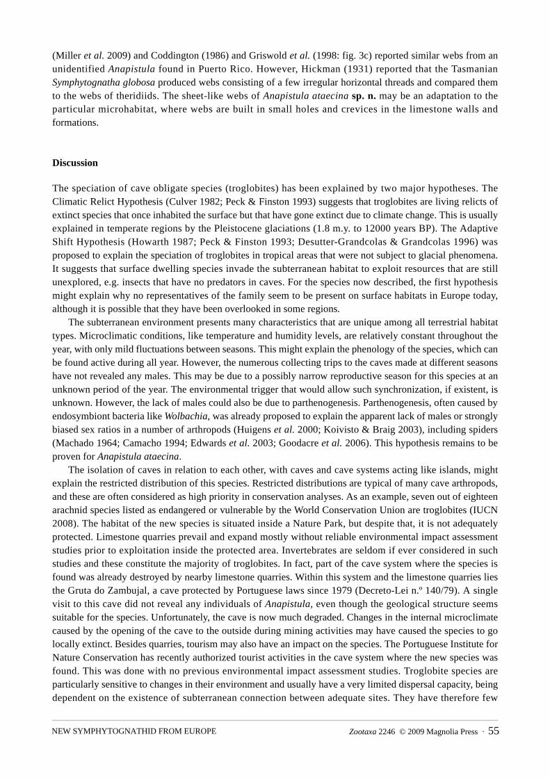
length of the lateral ducts was measured as the distance between A–B on the anterior border of the duct (see Fig. 7 – A–B). The length of the median duct was measured as the distance between B–C (Fig. 7). Approximate leg article lengths were measured in dorsal view, without detaching the legs from the animals, by positioning the article being measured perpendicularly. The female genitalia was too small to be excised from the abdomen, and we therefore prepared the whole abdomen. The abdomen was punctured dorsally and then digested with SIGMA Pancreatin LP1750 enzyme complex, in a solution of sodium borate following the concentrations described by Álvarez-Padilla and Hormiga (2008). The solution digested the no-chitinous internal parts of the abdomen within 2–4 hours at room temperature. The digested abdomen was then transferred to lactic acid and temporarily mounted on a slide, as described by Grandjean (1949). All material, including type material, is deposited in the Natural History Museum of Denmark, Zoological Museum, University of Copenhagen (acronym ZMUC). Catalogue numbers are provided in parenthesis. Using the same preparation technique we also tried to prepare a female abdomen for SEM in order to observe internal structures such as spermathecae, fertilization and copulatory ducts, but the preparations all failed due to the collapse of the digested abdomen.
Abbreviations used in text and figures
Epigynum. CD—copulatory duct; ld—lateral duct; md—median duct; Sp—spermatheca. Somatic morphology. AC— aciniform gland spigot(s); AG—aggregate gland spigot(s); ALE—anterior lateral eye(s); ALS—anterior lateral spinneret; CY—cylindrical gland spigot(s); MAP—major ampullate gland spigot; mAP —minor ampullate gland spigot; PI—piriform gland spigot(s); PLE—posterior lateral eye(s); PLS—posterior lateral spinneret; PMS—posterior median spinneret.
Taxonomy
Symphytognathidae Hickman, 1931
Anapistula Gertsch, 1941
Anapistula ataecina sp. n.(Figs 2–7)
Type material. Female holotype from Gruta do Fumo, Sesimbra, Portugal (N38°26.050 W009°08.650, 210 m altitude); 23.XII.2007; P. Cardoso leg. (ZMUC000012700).
Other material (para types): 1 female from same locali ty; 27.II .2005; P. Cardoso leg. (ZMUC000012701). 1 female from same locality; 11.IV.2007; P. Cardoso leg. (ZMUC000012702). 1 female from same locality; 19.IV.2007; P. Cardoso leg. (ZMUC000012703). 7 females from same locality; 23.XII.2007; P. Cardoso leg. (ZMUC000012704). 3 females from Gruta do Coelho, Sesimbra, Portugal (N38°25.800 W009°08.200, 120m altitude); 30.IV.2005; P. Cardoso leg. (ZMUC000012705). 1 female from Gruta da Utopia, Sesimbra, Portugal (N38°26.050 W009°08.850, 180m altitude); 01.VI.2006; P. Cardoso leg. (ZMUC000012706). 1 female from Gruta da Furada, Sesimbra, Portugal (N38°25.700 W009°10.500, 150m altitude); 13.IV.2007; P. Cardoso leg. (ZMUC000012707).
Etymology. The species name is derived from “Ataecina” (also spelled “Ataegina”), a subterranean world deity in Lusitanian and Celtiberian mythology, goddess of nature, night, healing, death and rebirth.
Taxonomic justification: The relatively low cephalothorax, the presence of posterior spiracles, the complete lack of a palp, and four eyes in two diads, makes it easy to place the new species in Anapistula. However, it is more difficult to compare our new species to congeneric species, since illustrations in the literature are rather poor and inadequate. Only a few authors have described their species in such details as we
Zootaxa 2246 © 2009 Magnolia Press · 47NEW SYMPHYTOGNATHID FROM EUROPE

do here. Given the somatic and genital details presented in the paper, combined with the habitat andgeographic isolation of the specimens found in this Portuguese cave system, we do not hesitate to describe thismaterial as belonging to a new species.
In general, all known species of Anapistula have rather similar female genitalia with paired spermathecaeconnected by lateral ducts to a median duct (Fig. 7). Females of different species are separated on the 1) shapeand size of the spermathecae and the detailed proportion of the ducts; 2) presence or absence of epigynalatrium; 3) body size; 4) separation of posterior lateral eyes; and 5) number and distribution of setae on thecephalothorax.
FIGURE 2. Adult female of Anapistula ataecina sp. n.: A) live specimen hanging on a single silk line; B) web; C) eggsac; D) dorsal view; E) lateral view; F) ventral view, with spermathecae clearly visible. Live photos: Pedro Cardoso (2A–B).
CARDOSO & SCHARFF48 · Zootaxa 2246 © 2009 Magnolia Press
In order to decide whether our Portuguese specimens of Anapistula represented individuals of a known species or should be described as a new species, we compared the Portuguese specimens with the nineteen known species of Anapistula (Platnick 2009).
Some species can be disregarded right away, since they have no eyes (the cave species A. troglobia and A. cuttacutta) or six eyes (A. boneti) and two additional species are only known from males and therefore cannot be compared with Anapistula ataecina sp. n.. These are A. bebuia Rheims & Brescovit, 2003 from a cave and A. guyri Rheims & Brescovit, 2003 from litter, both in Brazil. Both species have males that are larger than the females of Anapistula ataecina sp. n. and since males of all known species of Anapistula are considerably smaller than the females, they are not considered to be same species as A. ataecina sp. n. Similarly, A. aquytabuera Rheims & Brescovit, 2003, A. pocaruguara Rheims & Brescovit, 2003 and A. ybyquyra Rheims & Brescovit, 2003 from Brazil and A. seychellensis Saaristo, 1996 from the Seychelles all have distinct epigynal atria, not seen in Anapistula ataecina sp. n. In a review of the Australasian Anapistula species, Harvey (1998) described the setal pattern of the cephalothorax of males and females of Australasian species and found an interesting pattern that he used in the descriptions of the species. Males and females have similar patterns within each species. All species had either eight or six setae on the cephalothorax and a pattern with either four setae on the clypeus, two setae adjacent to the eye diads, and two setae on the posterior part of thorax (4-2-2 pattern present in A. troglobia, A. bifurcata Harvey, 1998 and A. jerai Harvey, 1998) or with four setae on clypeus, two setae adjacent to eyes, and no setae on thorax (4-2-0 pattern present in A. tonga Harvey, 1998). Anapistula ataecina sp. n. have a unique pattern with ten setae on cephalothorax of which six are on clypeus, two adjacent to the eyes and two on the posterior part of thorax. Of the remaining seven species of Anapistula worldwide, A. appendix Tong & Li, 2006 from a cave in China are much larger (0.65 long), have lateral epigynal ducts with lobes not present in A. ataecina sp. n. and eight setae on cephalothorax (4-2-2 pattern). A. ayri Rheims & Brescovit, 2003 from Brazil have spermathecae with small lateral triangular projections and a sinouid posterior border of the epigynal plate, characters that are not present in Anapistula ataecina sp. n.
This leaves five species that are so similar to Anapistula ataecina sp. n. that they require special attention. These are A. caecula from forest litter in Ivory Coast, A. secreta from forest litter in Central America, A. benoiti from forest litter in Zaire, A. ishikawai from forest litter in Japan and A. australia from forest litter in Australia. We examined the type material of A. secreta Gertch, 1941 (the type species for the genus Anapistula), A.benoiti Forster & Platnick, 1977 and A.caecula Baert & Jocqué, 1993. Comparisons with borrowed material had to be limited to characters that could be observed without dissections, since many species are only known from the type material. Based on these comparisons, we concluded that the material from Portugal represents a new species. The characters that separate the new species from the five species mentioned above are given in the diagnosis below.
Of the nineteen species of Anapistula described worldwide, fourteen species have only been found in litter of various kind of vegetation, whereas five species have been found in caves only. None of the cave species are morphologically similar to Anapistula ataecina sp. n.
The notch on the fangs of Anapistula ataecina sp. n. (Fig. 4D) also seems to be present in Anapistula caecula (Baert & Jocqué 1993: fig. 2) from West Africa and in Anapistula ybyquyra (Rheims & Brescovit 2003: fig. 6) from southern Brazil, and could thus be a phylogenetic informative character at some higher level.
As far as we know, the spinnerets of Anapistula have never before been described and illustrated and it is therefore unknown whether the surprising lack of a flagelliform spigot on the PLS of the female is unique to Anapistula ataecina or perhaps common to all species of Anapistula. If so, this would be another potential synapomorphy for the genus. All other species of symphytognathids, where the webs are known, produce orb webs with sticky spirals. The lack of a flagelliform spigot in A. ataecina could be related to the modified web (sheet web) built by these spiders.
Diagnosis. Overall, Anapistula ataecina sp. n. is most similar to A. secreta, A. benoiti, A. caecula, A. australia, and A. ishikawai. It can be distinguished from these species by the following combination of
Zootaxa 2246 © 2009 Magnolia Press · 49NEW SYMPHYTOGNATHID FROM EUROPE

characters: adult females 0.43–0.57 long (A. benoiti 0.61 long; A. ishikawai 0.65 long; A. secreta 0.50; A. caecula 0.48–0.55; A. australia unpublished), spermathecae globular (Fig. 7)(kidney-shaped in A. caecula, fig. 1 in Baert & Jocqué 1993, globular in A. australia, A. secreta, A. ishikawai and A. benoiti), posterior lateral eyes separated by more than six times their diameter (Fig. 2D)(PLE separation three times in A. benoiti and A. secreta, six times in A. ishikawai, and eight times in A. australia), median duct (md) of epigynum broad, approx. 0.25 times the width of the spermathecae (A. secreta and A. benoiti 0.08, A. caecula 0.18, A. ishikawai 0.13, A. australia 0.07 (from Harvey, 1998)), and lateral ducts (ld) of epigynum short, approximately 0.4 times the length of the median duct (A. benoiti 0.25, A. australia and A. secreta 0.60, A. caecula 0.66, and A. ishikawai 1.2). Small pit present on mesal side of ALE (Fig. 3A – square box and 3C – arrow) not seen in any other described species of Anapistula with SEM images available.
FIGURE 3. SEM images of female Anapistula ataecina sp. n.: A) carapace and chelicerae, frontal view. White square delimits two lateral eyes (for close-up see 3C), white arrow points to setae mentioned in text; B) carapace and chelicerae, antero-lateral view, arrow points to area on endites where pedipalps would have inserted, if the females had pedipalps; C) close-up of lateral eye, with pit (arrow); D) serrula of endites (arrow). Scale bars: A, B, D) 0.01 mm; C) 0.001 mm.
CARDOSO & SCHARFF50 · Zootaxa 2246 © 2009 Magnolia Press
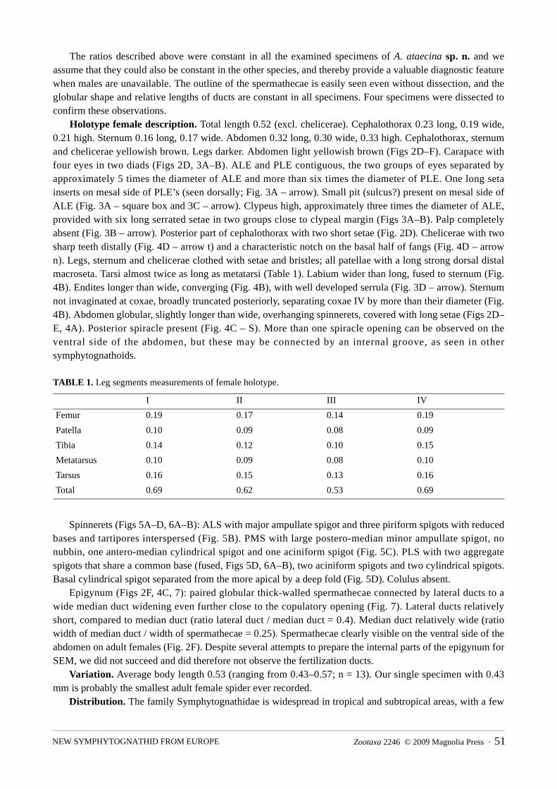
The ratios described above were constant in all the examined specimens of A. ataecina sp. n. and we assume that they could also be constant in the other species, and thereby provide a valuable diagnostic feature when males are unavailable. The outline of the spermathecae is easily seen even without dissection, and the globular shape and relative lengths of ducts are constant in all specimens. Four specimens were dissected to confirm these observations.
Holotype female description. Total length 0.52 (excl. chelicerae). Cephalothorax 0.23 long, 0.19 wide, 0.21 high. Sternum 0.16 long, 0.17 wide. Abdomen 0.32 long, 0.30 wide, 0.33 high. Cephalothorax, sternum and chelicerae yellowish brown. Legs darker. Abdomen light yellowish brown (Figs 2D–F). Carapace with four eyes in two diads (Figs 2D, 3A–B). ALE and PLE contiguous, the two groups of eyes separated by approximately 5 times the diameter of ALE and more than six times the diameter of PLE. One long seta inserts on mesal side of PLE’s (seen dorsally; Fig. 3A – arrow). Small pit (sulcus?) present on mesal side of ALE (Fig. 3A – square box and 3C – arrow). Clypeus high, approximately three times the diameter of ALE, provided with six long serrated setae in two groups close to clypeal margin (Figs 3A–B). Palp completely absent (Fig. 3B – arrow). Posterior part of cephalothorax with two short setae (Fig. 2D). Chelicerae with two sharp teeth distally (Fig. 4D – arrow t) and a characteristic notch on the basal half of fangs (Fig. 4D – arrow n). Legs, sternum and chelicerae clothed with setae and bristles; all patellae with a long strong dorsal distal macroseta. Tarsi almost twice as long as metatarsi (Table 1). Labium wider than long, fused to sternum (Fig. 4B). Endites longer than wide, converging (Fig. 4B), with well developed serrula (Fig. 3D – arrow). Sternum not invaginated at coxae, broadly truncated posteriorly, separating coxae IV by more than their diameter (Fig. 4B). Abdomen globular, slightly longer than wide, overhanging spinnerets, covered with long setae (Figs 2D–E, 4A). Posterior spiracle present (Fig. 4C – S). More than one spiracle opening can be observed on the ventral side of the abdomen, but these may be connected by an internal groove, as seen in other symphytognathoids.
TABLE 1. Leg segments measurements of female holotype.
Spinnerets (Figs 5A–D, 6A–B): ALS with major ampullate spigot and three piriform spigots with reduced bases and tartipores interspersed (Fig. 5B). PMS with large postero-median minor ampullate spigot, no nubbin, one antero-median cylindrical spigot and one aciniform spigot (Fig. 5C). PLS with two aggregate spigots that share a common base (fused, Figs 5D, 6A–B), two aciniform spigots and two cylindrical spigots. Basal cylindrical spigot separated from the more apical by a deep fold (Fig. 5D). Colulus absent.
Epigynum (Figs 2F, 4C, 7): paired globular thick-walled spermathecae connected by lateral ducts to a wide median duct widening even further close to the copulatory opening (Fig. 7). Lateral ducts relatively short, compared to median duct (ratio lateral duct / median duct = 0.4). Median duct relatively wide (ratio width of median duct / width of spermathecae = 0.25). Spermathecae clearly visible on the ventral side of the abdomen on adult females (Fig. 2F). Despite several attempts to prepare the internal parts of the epigynum for SEM, we did not succeed and did therefore not observe the fertilization ducts.
Variation. Average body length 0.53 (ranging from 0.43–0.57; n = 13). Our single specimen with 0.43 mm is probably the smallest adult female spider ever recorded.
Distribution. The family Symphytognathidae is widespread in tropical and subtropical areas, with a few
I II III IV
Femur 0.19 0.17 0.14 0.19
Patella 0.10 0.09 0.08 0.09
Tibia 0.14 0.12 0.10 0.15
Metatarsus 0.10 0.09 0.08 0.10
Tarsus 0.16 0.15 0.13 0.16
Total 0.69 0.62 0.53 0.69
Zootaxa 2246 © 2009 Magnolia Press · 51NEW SYMPHYTOGNATHID FROM EUROPE

species reaching southern USA, southern China and Japan (Fig. 1). It was not known from Europe or even northern Africa. The new species was only found in Sesimbra, inside the limits of the Arrábida Nature Park, about 30 km south of Lisbon, Portugal. It was exclusively found living in four caves: Gruta do Fumo, Gruta da Utopia, Gruta do Coelho and Gruta da Furada. All but the latter belong to the Frade cave system. The single female captured in Gruta da Furada, a cave about 2 km from the others, was the only specimen found during three independent visits to the cave, and therefore probably not representative of a self-supporting population in this cave. All the other caves are connected and less than one km apart, the whole cave system probably
covers about 1 km2. Although it is possible that the area of occupancy extends to other regions, extensive work has been made in about 20 caves nearby, without success. Extensive collecting of spiders was also carried out by António de Barros Machado in caves all over Portugal (Machado 1941; Machado & Machado 1941) during the 1930s. Being both a biospeleologist and a spider specialist, it is unlikely that such remarkable, albeit minute, species would have been overlooked by Barros Machado in other regions of the country.
Natural history. Adult females were found during most of the year, on all visits to the Frade cave system. Egg sacs were found from December to May, with either two or three eggs loosely embedded in a relatively large surrounding mesh of silk (Fig. 2C). Usually there was only a single egg sac in each web, but up to three were found in a single web. All known egg sacs in this family are similar, always with a very low number or even a single egg per eggsac (Harvey 1998, Griswold & Yan 2003). The egg sacs where always hanging from the web, either at the periphery or in a more central position.
FIGURE 4. SEM images of Anapistula ataecina sp. n.: A) habitus, lateral view; B) sternum, ventral view; C) abdomen, ventral view (S = Spiracle; Ep = Epigynum); D) chelicerae, with two sharp teeth (arrow – t) and notch (arrow – n). Scale bars: A, B) 0.1 mm; C, D) 0.01 mm.
CARDOSO & SCHARFF52 · Zootaxa 2246 © 2009 Magnolia Press

FIGURE 5. SEM images of spinnerets of Anapistula ataecina sp. n.: A) left spinnerets: ALS—Anterior lateral spinnerets, PMS—Posterior median spinnerets, PLS—Posterior lateral spinnerets; B) ALS: major ampullate spigot (MAP), piriform spigots (PI), tartipores (ta); C) PMS: minor ampullate spigot (mAP), cylindrical spigot (CY), aciniform spigot (AC); D) PLS: cylindrical spigots (CY), aciniform spigots (AC), aggregate spigots (AG). Scale bars: A) 0.01 mm; B–D) 0.001 mm.
Zootaxa 2246 © 2009 Magnolia Press · 53NEW SYMPHYTOGNATHID FROM EUROPE
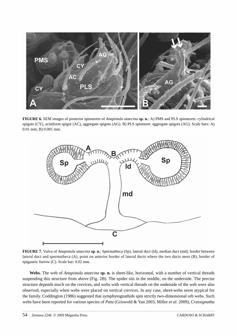
FIGURE 6. SEM images of posterior spinnerets of Anapistula ataecina sp. n.: A) PMS and PLS spinnerets: cylindrical spigots (CY), aciniform spigot (AC), aggregate spigots (AG). B) PLS spinneret: aggregate spigots (AG). Scale bars: A) 0.01 mm; B) 0.001 mm.
FIGURE 7. Vulva of Anapistula ataecina sp. n.: Spermatheca (Sp), lateral duct (ld), median duct (md), border between lateral duct and spermatheca (A), point on anterior border of lateral ducts where the two ducts meet (B), border of epigastric furrow (C). Scale bar: 0.02 mm.
Webs. The web of Anapistula ataecina sp. n. is sheet-like, horizontal, with a number of vertical threads suspending this structure from above (Fig. 2B). The spider sits in the middle, on the underside. The precise structure depends much on the crevices, and webs with vertical threads on the underside of the web were also observed, especially when webs were placed on vertical crevices. In any case, sheet-webs seem atypical for the family. Coddington (1986) suggested that symphytognathids spin strictly two-dimensional orb webs. Such webs have been reported for various species of Patu (Griswold & Yan 2003, Miller et al. 2009), Crassignatha
CARDOSO & SCHARFF54 · Zootaxa 2246 © 2009 Magnolia Press
(Miller et al. 2009) and Coddington (1986) and Griswold et al. (1998: fig. 3c) reported similar webs from an unidentified Anapistula found in Puerto Rico. However, Hickman (1931) reported that the Tasmanian Symphytognatha globosa produced webs consisting of a few irregular horizontal threads and compared them to the webs of theridiids. The sheet-like webs of Anapistula ataecina sp. n. may be an adaptation to the particular microhabitat, where webs are built in small holes and crevices in the limestone walls and formations.
Discussion
The speciation of cave obligate species (troglobites) has been explained by two major hypotheses. The Climatic Relict Hypothesis (Culver 1982; Peck & Finston 1993) suggests that troglobites are living relicts of extinct species that once inhabited the surface but that have gone extinct due to climate change. This is usually explained in temperate regions by the Pleistocene glaciations (1.8 m.y. to 12000 years BP). The Adaptive Shift Hypothesis (Howarth 1987; Peck & Finston 1993; Desutter-Grandcolas & Grandcolas 1996) was proposed to explain the speciation of troglobites in tropical areas that were not subject to glacial phenomena. It suggests that surface dwelling species invade the subterranean habitat to exploit resources that are still unexplored, e.g. insects that have no predators in caves. For the species now described, the first hypothesis might explain why no representatives of the family seem to be present on surface habitats in Europe today, although it is possible that they have been overlooked in some regions.
The subterranean environment presents many characteristics that are unique among all terrestrial habitat types. Microclimatic conditions, like temperature and humidity levels, are relatively constant throughout the year, with only mild fluctuations between seasons. This might explain the phenology of the species, which can be found active during all year. However, the numerous collecting trips to the caves made at different seasons have not revealed any males. This may be due to a possibly narrow reproductive season for this species at an unknown period of the year. The environmental trigger that would allow such synchronization, if existent, is unknown. However, the lack of males could also be due to parthenogenesis. Parthenogenesis, often caused by endosymbiont bacteria like Wolbachia, was already proposed to explain the apparent lack of males or strongly biased sex ratios in a number of arthropods (Huigens et al. 2000; Koivisto & Braig 2003), including spiders (Machado 1964; Camacho 1994; Edwards et al. 2003; Goodacre et al. 2006). This hypothesis remains to be proven for Anapistula ataecina.
The isolation of caves in relation to each other, with caves and cave systems acting like islands, might explain the restricted distribution of this species. Restricted distributions are typical of many cave arthropods, and these are often considered as high priority in conservation analyses. As an example, seven out of eighteen arachnid species listed as endangered or vulnerable by the World Conservation Union are troglobites (IUCN 2008). The habitat of the new species is situated inside a Nature Park, but despite that, it is not adequately protected. Limestone quarries prevail and expand mostly without reliable environmental impact assessment studies prior to exploitation inside the protected area. Invertebrates are seldom if ever considered in such studies and these constitute the majority of troglobites. In fact, part of the cave system where the species is found was already destroyed by nearby limestone quarries. Within this system and the limestone quarries lies the Gruta do Zambujal, a cave protected by Portuguese laws since 1979 (Decreto-Lei n.º 140/79). A single visit to this cave did not reveal any individuals of Anapistula, even though the geological structure seems suitable for the species. Unfortunately, the cave is now much degraded. Changes in the internal microclimate caused by the opening of the cave to the outside during mining activities may have caused the species to go locally extinct. Besides quarries, tourism may also have an impact on the species. The Portuguese Institute for Nature Conservation has recently authorized tourist activities in the cave system where the new species was found. This was done with no previous environmental impact assessment studies. Troglobite species are particularly sensitive to changes in their environment and usually have a very limited dispersal capacity, being dependent on the existence of subterranean connection between adequate sites. They have therefore few
Zootaxa 2246 © 2009 Magnolia Press · 55NEW SYMPHYTOGNATHID FROM EUROPE
means to escape any kind of threats to the habitat. The very restricted distribution range of this species and the current and predictable habitat degradation and disappearance, calls for an urgent classification of Anapistula ataecina according to the IUCN criteria (IUCN 2001). Although the criteria have been mainly established for vertebrates, they have been used for arthropods and other invertebrates in many cases, provided that sufficient information is available (IUCN 2008). This species is the single known representative of the family Symphytognathidae in Europe, further emphasizing its uniqueness in the region and the importance of its protection. The listing of a species as threatened is no guarantee that any kind of protection measures will be taken, as a threatened species list is not equivalent to a conservation priority species list. It can however serve as a useful tool for political lobbying for the protection of the species. Red listing also focuses the attention of stakeholders in some species that, as is the case for most arthropods, are usually not acknowledged in conservation politics and decisions.
Acknowledgements
We thank Francisco Rasteiro, Sérgio Henriques and all the members of Núcleo de Espeleologia da Costa Azul for the many cave trips made to look for Anapistula webs. Clara Gaspar and António Cardoso were also present during the first sightings of the species. Special thanks to Norman I. Platnick, Lara Lopardo and Gustavo Hormiga for comments and helpful suggestions to an earlier version of the manuscript. Thanks also to Jesper Birkedal Schmidt for first pointing out that this was an Anapistula and to Norman I. Platnick for confirming the identification. Tamás Szüts and Jan Pedersen for help in the laboratory. We would also like to thank Norman I. Platnick (American Museum of Natural History; AMNH) and Rudy Jocqué (Musee Royal de l’Afrique Centrale, Tervuren; MRAC) for making type material available for this study. Jonathan Coddington, Martín Ramírez, Lara Lopardo and Gustavo Hormiga for fruitful discussion during the writing of the paper. Jonathan Coddington and Roy Larimer for help with the habitus photographs of Anapistula (Figs 2D–F) and Jonathan Coddington for letting us use the BK+ Imaging System at the Smithsonian Institution, Washington DC, USA. PC was supported by a grant from the European Commission’s (FP6) Integrated Infrastructure Initiative programme Synthesys (DK–TAF–4426). Fieldwork was supported by Câmara Municipal de Sesimbra. NS was supported by the Danish Agency for Science, Technology and Innovation (grant no. 272–05–0431).
References
Álvarez-Padilla, F. & Hormiga, G. (2008) A protocol for digesting internal soft tissues and mounting spiders for scanning electron microscopy. Journal of Arachnology, 35, 538–542.
Baert, L. & Jocqué, R. (1993) Anapistula caecula sp. n., the smallest known female spider (Araneae, Symphytognathidae). Journal of African Zoology, 107, 187–189.
Camacho, J.P.M. (1994) Female-biased sex ratio in spiders caused by parthenogenesis? Hereditas, 120, 183–185.Coddington, J.A. (1986) The monophyletic origin of the orb web. In: Shear, W.A. (ed.) Spiders: webs, behavior, and
evolution. Stanford University Press, Stanford, California.Culver, D.C. (1982) Cave life: evolution and ecology. Harvard University Press, Cambridge, MA.Desutter-Grandcolas, L. & Grandcolas, P. (1996) The evolution toward troglobitic life: a phylogenetic reappraisal of
climatic relict and local habitat shift hypotheses. Mémoires de Biospéologie, 23, 57–63.Edwards, R.L., Edwards, E.H & Edwards, A.D. (2003) Observations of Theotima minutissimus (Araneae,
Ochyroceratidae), a parthenogenetic spider. Journal of Arachnology, 31, 274–277.Forster, R.R. & Platnick, N.I. (1977) A review of the spider family Symphytognathidae (Arachnida, Araneae). American
Museum Novitates, 2619, 1–19.Goodacre, S.L., Martin, O.Y., Thomas, C.F.G. & Hewitt, G.M. (2006) Wolbachia and other endosymbiont infections in
spiders. Molecular Ecology, 15, 517–527. Grandjean, F. (1949) Observation et conservation des trés petit arthropodes. Bulletin du Muséum National d’Histoire
Naturelle de Paris, 21, 363–370.
CARDOSO & SCHARFF56 · Zootaxa 2246 © 2009 Magnolia Press

Griswold, C. E. (1987) The spider genus Symphytognatha Hickman (Araneae: Symphytognathidae) newly described from Africa. Annals of the Natal Museum, 28, 133–136.
Griswold, C.E., Coddington, J.A., Hormiga, G., & Scharff, N. (1998) Phylogeny of the orb-web building spiders (Araneae, Orbiculariae: Deinopoidea, Araneoidea). Zoological Journal of the Linnean Society, 123, 1–99.
Griswold, C.E. & Yan, H.-M. (2003) On the egg-guarding behavior of a Chinese Symphytognathid spider of the genus Patu Marples, 1951 (Araneae, Araneoidea, Symphytognathidae). Proceedings of the California Academy of Sciences, 54, 356–360.
Harvey, M.S. (1998) A review of the Australasian species of Anapistula Gertsch (Araneae: Symphytognathidae). Records of the Western Australian Museum, 19, 111–120.
Hickman, V.V. (1931) A new family of spiders. Proceedings of the Zoological Society of London B, 1931, 1321–1328.Howarth, F.G. (1987) The evolution of non-relictual tropical troglobites. International Journal of Speleology, 16, 1–16.Huigens, M.E., Luck, R.F., Klaassen, R.H.G., Maas, M.F.P.M., Timmermans, M.J.T.N. & Stouthamer, R. (2000)
Infectious parthenogenesis. Nature, 405, 178–179.IUCN (2001) IUCN Red List categories and criteria: version 3.1. IUCN Species Survival Commission. IUCN, Gland,
Switzerland and Cambridge, UK.IUCN (2008) 2008 IUCN Red List of Threatened Species. Available from: www.iucnredlist.org (Date of access: 13 May
2009).Koivisto, R.K.K. & Braig, H.R. (2003) Microorganisms and parthenogenesis. Biological Journal of the Linnean Society,
79, 43–58.Lin, Y. & Li, S. (2009) First described Patu spiders (Araneae, Symphytognathidae) from Asia. Zootaxa, 2154, 47–68.Machado, A.B. (1941) As cavernas de Portugal sob o ponto de vista biológico. Boletim da Sociedade Portuguesa de
Ciências Naturais, 13, 639–642.Machado, A.B. (1964) Sur l’existence de la parthénogenese dans quelques espéces d’Araignées Ochyrocératides.
Comptes Rendus de L’Academie Science Paris, 2, 5056–5059.Machado, A.B. & Machado, B.B. (1941) Inventário das cavernas calcárias de Portugal. Boletim da Sociedade
Portuguesa de Ciências Naturais, 13, 444–473.Miller, J.A., Griswold, C.E. & Yin, C.M. (2009) The symphytognathoid spiders of the Gaoligongshan, Yunnan, China
(Araneae, Araneoidea): systematics and diversity of micro-orbweavers. Zookeys, 11, 9–195.Ono, H. (2002) First record of the genus Anapistula (Araneae, Symphytognathidae) from Asia. Bulletin of the Natural
Sciences Museum of Tokyo, 28, 61–64.Peck, S.B. & Finston, T. (1993) Galapagos islands troglobites: the questions of tropical troglobites, parapatric
distributions with eyed-sister-species, and their origin by parapatric speciation. Mémoires de Biospéologie, 20, 19–37.
Platnick, N.I. (2009) The world spider catalog, version 9.5. American Museum of Natural History. Available from: http://research.amnh.org/entomology/spiders/catalog/index.html (Date of access: 13 May 2009).
Rheims, C.A. & Brescovit, A.D. (2003) Description of six new species of Anapistula, Gertsch (Araneae, Symphytognathidae) from Brazil. Bulletin of the British Arachnological Society, 12, 324–330.
Schütt, K. (2003) Phylogeny of Symphytognathidae s.l. (Araneae, Araneoidea). Zoologica Scripta, 32, 129–151.Tong, Y.F. & Li, S.Q. (2006) Symphytognathidae (Araneae), a spider family newly recorded from China. Zootaxa, 1259,
33–38.
Zootaxa 2246 © 2009 Magnolia Press · 57NEW SYMPHYTOGNATHID FROM EUROPE