Hormonesbiyokimya.vet/documents/biyokimya/Hormones.pdf · 2019-11-01 · •Hormones, secreted by...
113
Serkan SAYINER, DVM PhD. Assist. Prof. Near East University, Faculty of Veterinary Medicine, Department of Biochemistry [email protected] Hormones
Transcript of Hormonesbiyokimya.vet/documents/biyokimya/Hormones.pdf · 2019-11-01 · •Hormones, secreted by...
Serkan SAYINER, DVM PhD. Assist. Prof.Near East University, Faculty of Veterinary Medicine, Department of Biochemistry
Hormones
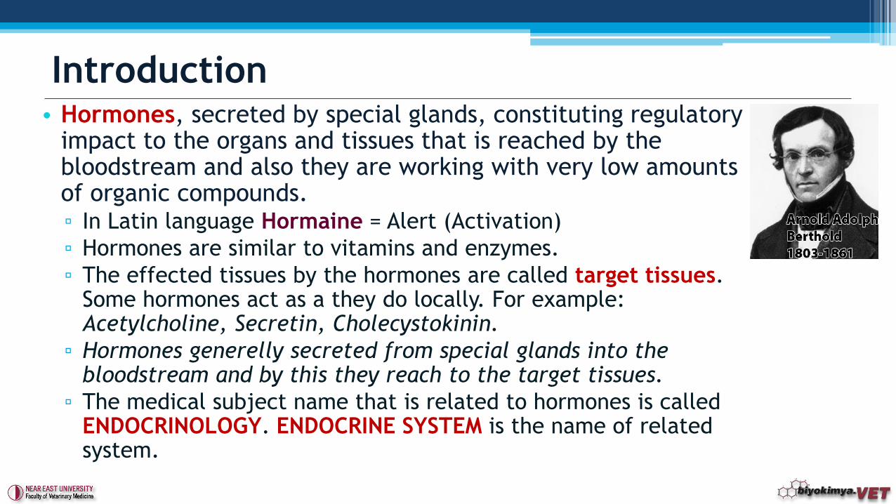
• Hormones, secreted by special glands, constituting regulatory impact to the organs and tissues that is reached by the bloodstream and also they are working with very low amounts of organic compounds.▫ In Latin language Hormaine = Alert (Activation)
▫ Hormones are similar to vitamins and enzymes.
▫ The effected tissues by the hormones are called target tissues. Some hormones act as a they do locally. For example: Acetylcholine, Secretin, Cholecystokinin.
▫ Hormones generelly secreted from special glands into the bloodstream and by this they reach to the target tissues.
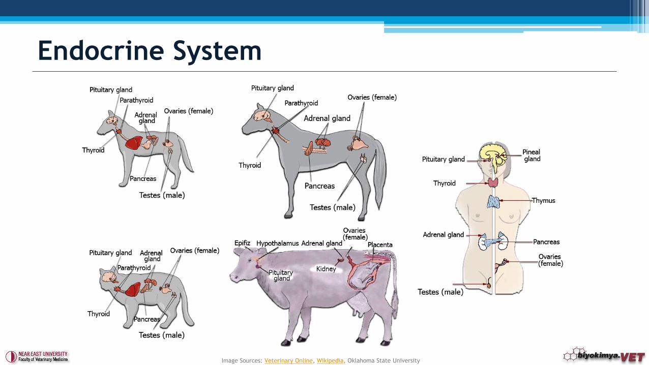
▫ The medical subject name that is related to hormones is called ENDOCRINOLOGY. ENDOCRINE SYSTEM is the name of related system.
Introduction
Endocrine System
Image Sources: Veterinary Online, Wikipedia, Oklahoma State University
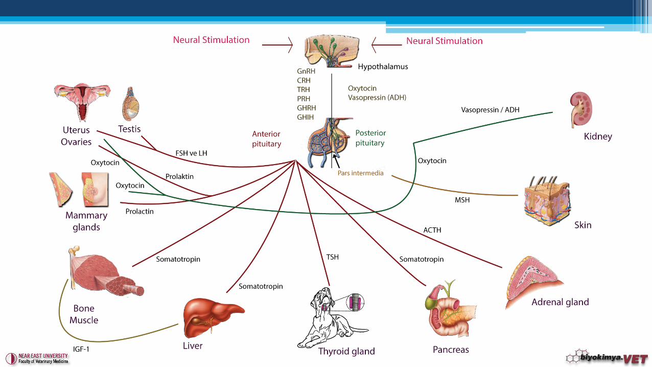
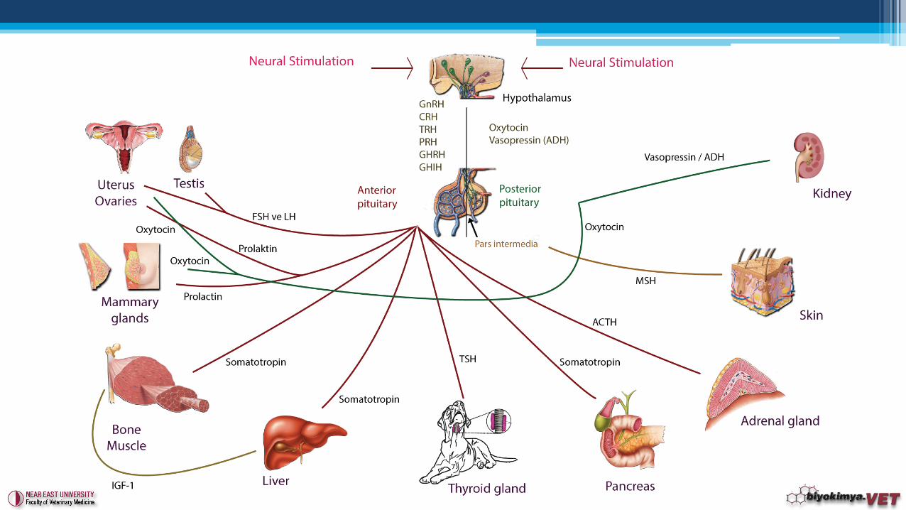
• Blood formation and secretion of hormones occur depending on
the hierarchical control mechanism.
• Large part of the group of hormones on the control mechanism
are released into the bloodstream as listed from top to bottom.
• The hypothalamus is located at the very top forming the base of
the brain. Any neural stimulation reached here, activates the
releasing factors (RFs) and leads to secretion of specific
hormones in very small amounts.
• RFs located in sella tursika alerts pituitary gland (Frontal lobe).
Each RFs stimulate different specific hormone secretion from
the pituitary gland.
Control Mechanism of Hormones
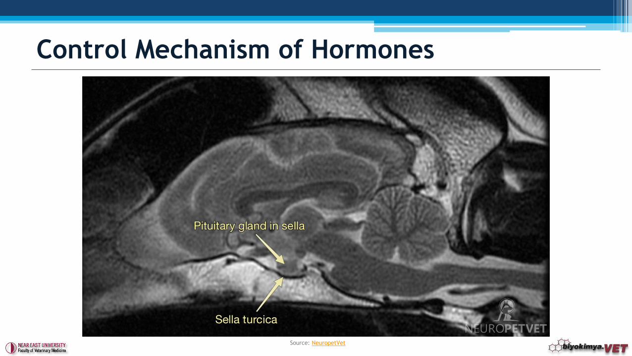
Control Mechanism of Hormones
Source: NeuropetVet
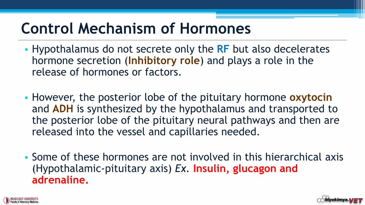
• Hypothalamus do not secrete only the RF but also decelerates hormone secretion (Inhibitory role) and plays a role in the release of hormones or factors.
• However, the posterior lobe of the pituitary hormone oxytocinand ADH is synthesized by the hypothalamus and transported to the posterior lobe of the pituitary neural pathways and then are released into the vessel and capillaries needed.
• Some of these hormones are not involved in this hierarchical axis (Hypothalamic-pituitary axis) Ex. Insulin, glucagon and adrenaline.
Control Mechanism of Hormones
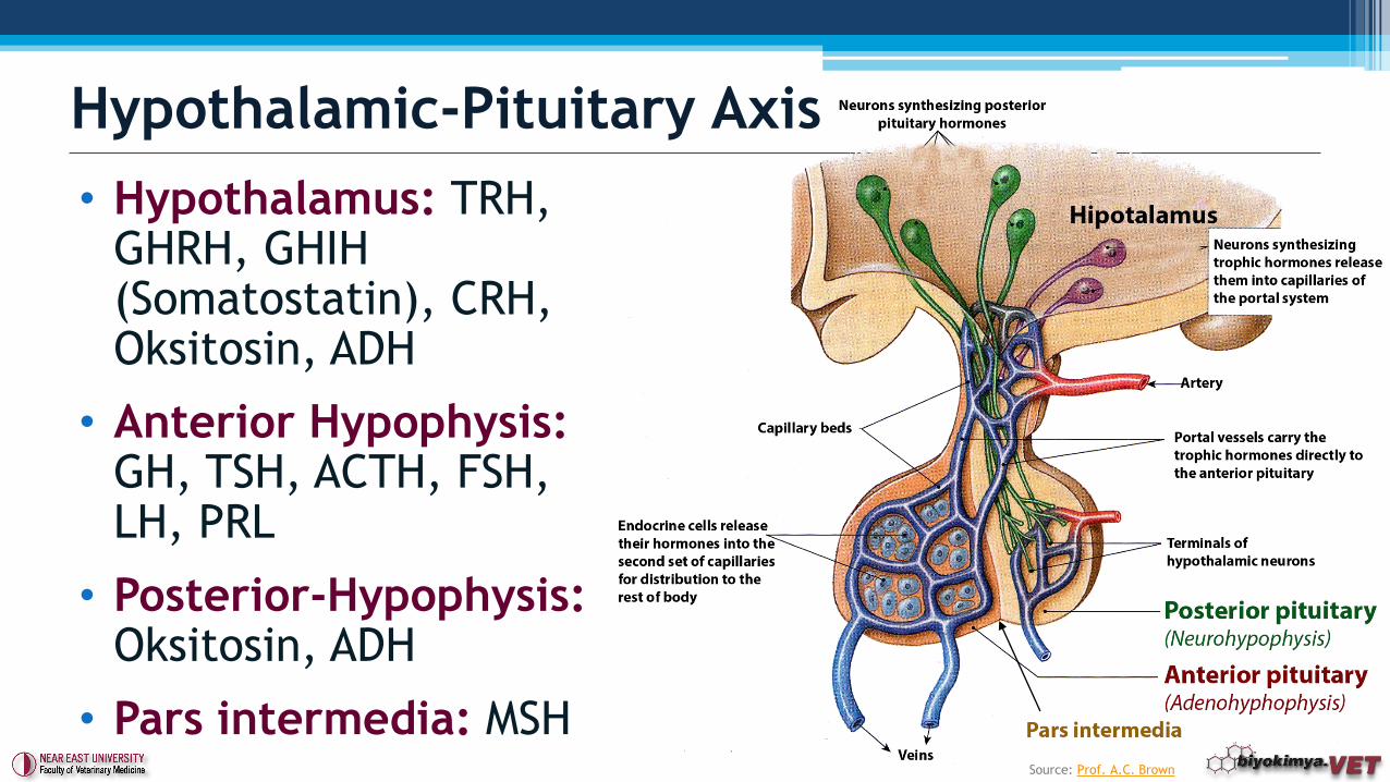
Hypothalamic-Pituitary Axis
• Hypothalamus: TRH, GHRH, GHIH (Somatostatin), CRH, Oksitosin, ADH
• Anterior Hypophysis:GH, TSH, ACTH, FSH, LH, PRL
• Posterior-Hypophysis: Oksitosin, ADH
• Pars intermedia: MSHSource: Prof. A.C. Brown
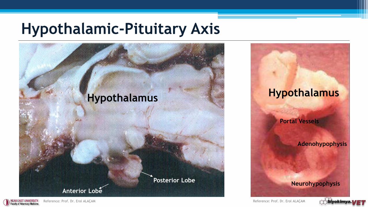
Hypothalamic-Pituitary Axis
Posterior Lobe
Anterior Lobe
Hypothalamus
Reference: Prof. Dr. Erol ALAÇAM
Hypothalamus
Adenohypophysis
Neurohypophysis
Portal Vessels
Reference: Prof. Dr. Erol ALAÇAM

Secondary Messengers
Intracellular messengers
c-AMP(Cyclic adenosine monophosphate)
Ca2+ Others
Calmodulin
Pathway
c-Kinase
Pathway
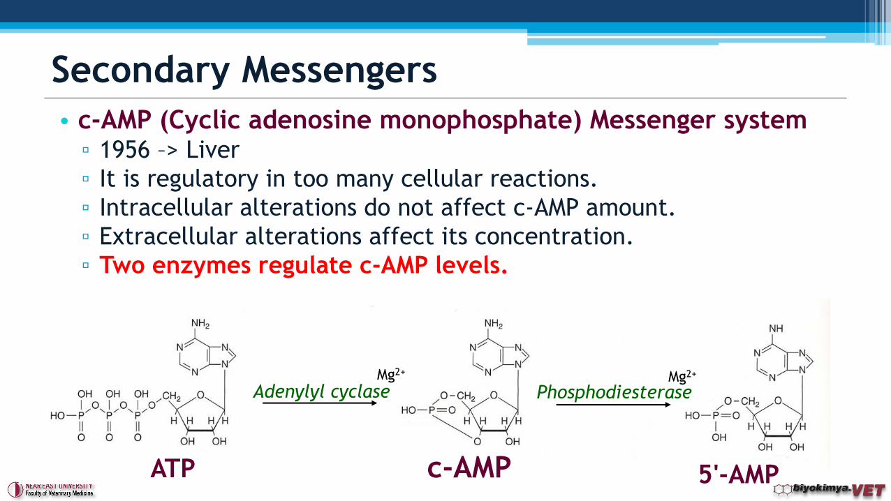
• c-AMP (Cyclic adenosine monophosphate) Messenger system▫ 1956 –> Liver
▫ It is regulatory in too many cellular reactions.
▫ Intracellular alterations do not affect c-AMP amount.
▫ Extracellular alterations affect its concentration.
▫ Two enzymes regulate c-AMP levels.
Secondary Messengers
ATP c-AMP 5'-AMP
Adenylyl cyclase PhosphodiesteraseMg2+ Mg2+
• c-AMP plays a role as an intermediary substance on the effect of
peptide hormone, exocrine and endocrine secretion, in the
secretion of neurotransmitters and the synapse for neuronal and
neuromuscular joints.
• Adenylyl cyclase is bound to cell membranes.
• Phosphodiesterase enzyme is found in the cytoplasm.
• When the cell is stimulated, c-AMP synthesis is increased by the
activation of Adenylyl cyclase.
• Protein kinase, phosphorylates many proteins in the cells.
• Ex.: Adrenalin in liver, PTH in renal tubulles.
Secondary Messengers
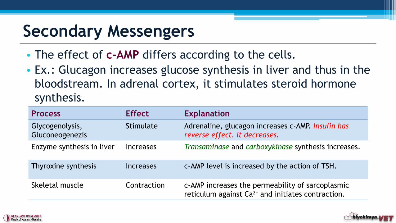
• The effect of c-AMP differs according to the cells.
• Ex.: Glucagon increases glucose synthesis in liver and thus in the
bloodstream. In adrenal cortex, it stimulates steroid hormone
synthesis.
Secondary Messengers
Process Effect Explanation
Glycogenolysis,
Gluconeogenezis
Stimulate Adrenaline, glucagon increases c-AMP. Insulin has
reverse effect. It decreases.
Enzyme synthesis in liver Increases Transaminase and carboxykinase synthesis increases.
Thyroxine synthesis Increases c-AMP level is increased by the action of TSH.
Skeletal muscle Contraction c-AMP increases the permeability of sarcoplasmic
reticulum against Ca2+ and initiates contraction.
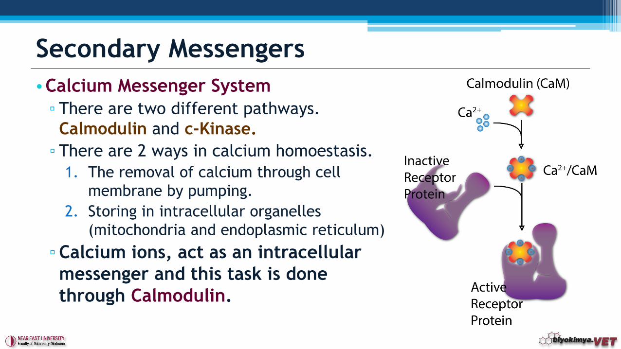
•Calcium Messenger System
▫ There are two different pathways.
Calmodulin and c-Kinase.
▫ There are 2 ways in calcium homoestasis.
1. The removal of calcium through cell
membrane by pumping.
2. Storing in intracellular organelles
(mitochondria and endoplasmic reticulum)
▫Calcium ions, act as an intracellular
messenger and this task is done
through Calmodulin.
Secondary Messengers
• Initiation of muscle contraction,
• Movement of chromosomes,
• Glycogen metabolism,
• Secretion and synthesis of neurotransmitters
• Regulating c-AMP levels,
• Role of calcium in secretion of insulin
• Calcium, activates calmodulin. This activates the inactive
protein enyzme.
▫ Ex. Phosphodiesterase.
Secondary Messengers
• c-Kinase (Protein kinase c/poliphosphoinositide messenger system)
▫ Activated directly by the calcium.
▫ It should have diaclyglycerol and phospholipids to be activated.
Therefore, enzyme is called phospholipid dependent protein kinase-c.
▫ Except brain, kidney and liver tissues, inactive protein kinase-c is found
freely.
▫ When Calcium messenger system is alerted by the hormones, membrane
diacylglycerol increases and causes protein kinase-c bound to inside of
the membrane.
▫ When required phospholipids are found in the membrane, calcium
dependent c-kinase is fully activated.
Secondary Messengers
• Many hormones are found in the vesicles which are bound tomembranes.
• Some of them are not taken into the vesicles, and found in themolecular form.
• Peptide and protein hormones are taken straight into vesicles in the ER. In here goes to Golgi complex and condenses into prepared vesicles. It is connected to the membrane and secreted outside the cell if necessary.
• Steroid hormones are directly passes through membrane.
• Hormones in the bloodstream is still attached to the proteinsand when they reach to the target tissue, they are removedfrom protein and become active.
Storage of Hormones
Source: Boundless
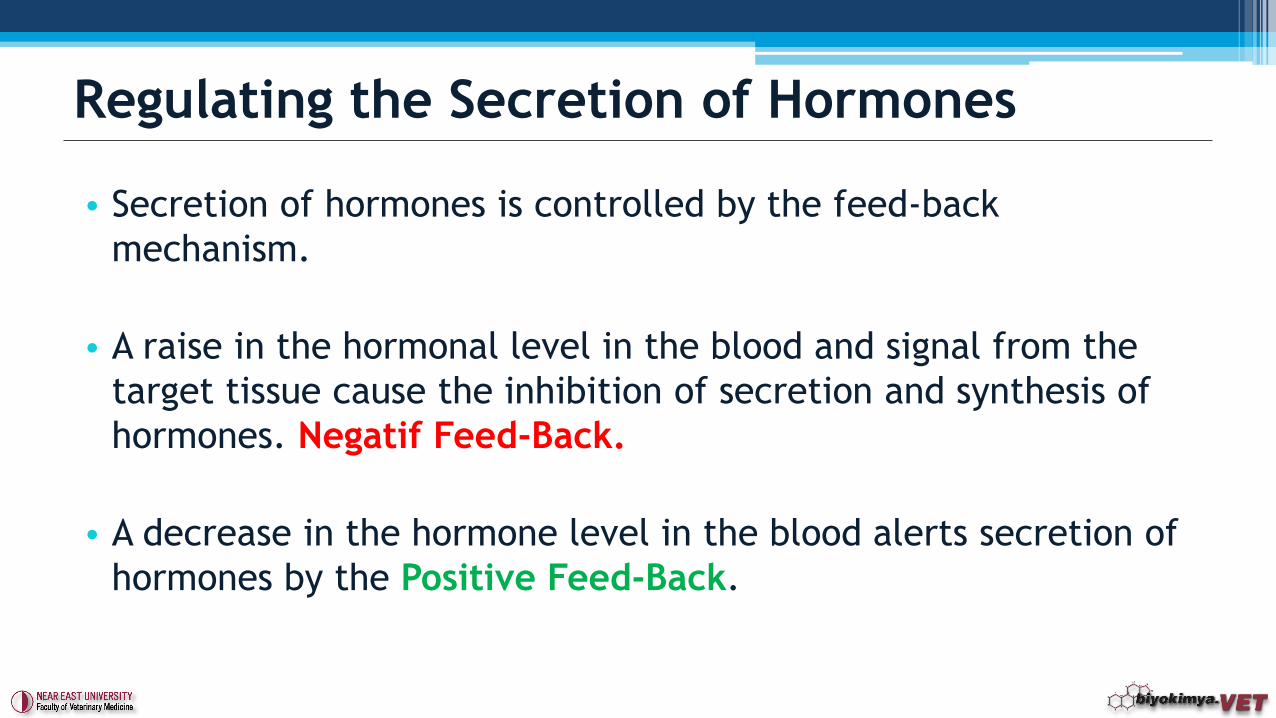
• Secretion of hormones is controlled by the feed-back
mechanism.
• A raise in the hormonal level in the blood and signal from the
target tissue cause the inhibition of secretion and synthesis of
hormones. Negatif Feed-Back.
• A decrease in the hormone level in the blood alerts secretion of
hormones by the Positive Feed-Back.
Regulating the Secretion of Hormones
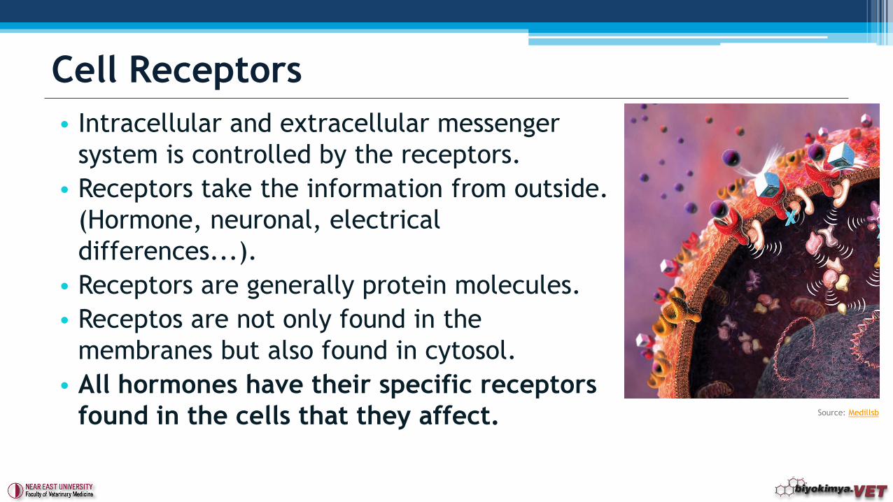
• Intracellular and extracellular messenger
system is controlled by the receptors.
• Receptors take the information from outside.
(Hormone, neuronal, electrical
differences...).
• Receptors are generally protein molecules.
• Receptos are not only found in the
membranes but also found in cytosol.
• All hormones have their specific receptors
found in the cells that they affect.
Cell Receptors
Source: Medillsb
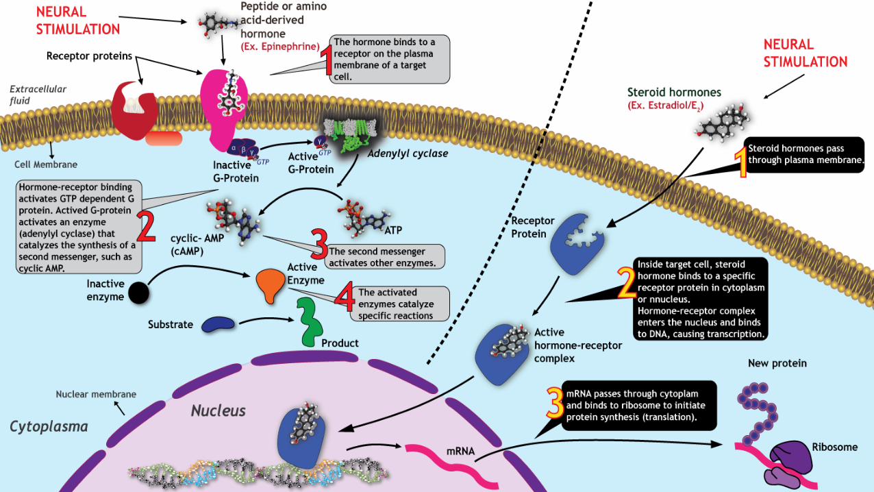
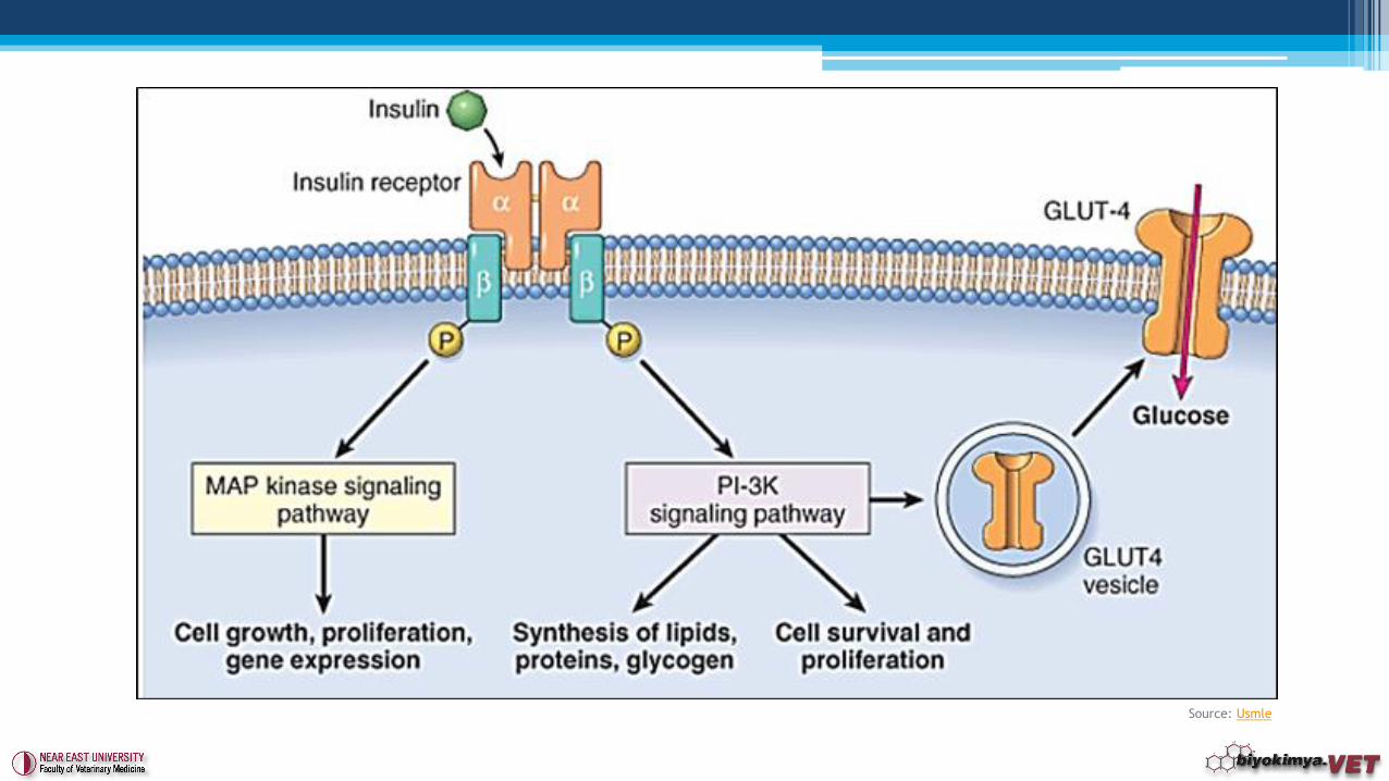
1. HORMONES WITH CELL SURFACE RECEPTORS (Adenylyl cyclase system)
• This sytem causes formation of intracellular cyclic-AMP (c-AMP)’ and also involves in the activity of many enzymes.
• Specially, anterior and posterior pituitary hormones, PTH, Glucagon, adrenaline and hormone releasing factors are influenced by this type of receptor systems.
• In this sytem, there is a specific receptor on the cell membrane according to each hormone. Hormone-receptor complex activates the to adenylyl cyclase. This enzymatic action converts ATP to c-AMP.
Mechanism of Hormone Action
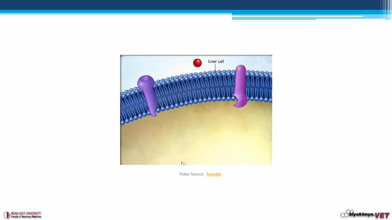
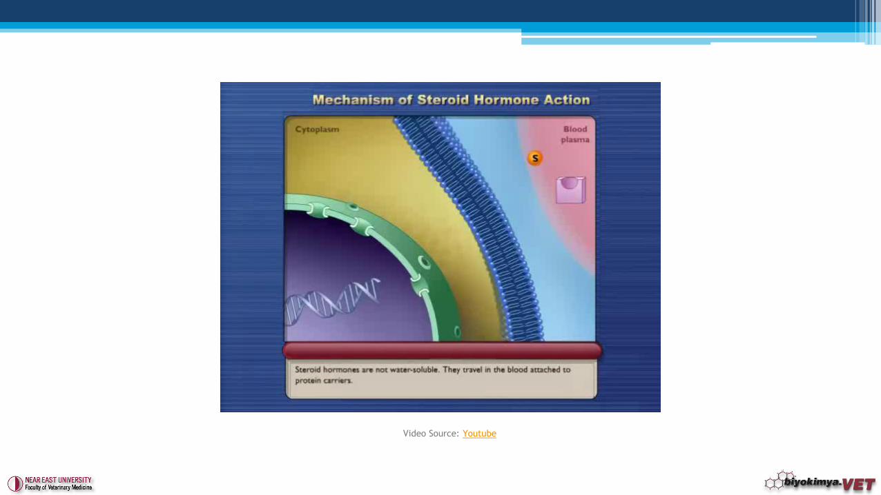
2. HORMONES WITH INTRACELLULAR RECEPTORS (Protein synthesis system)
• Receptors are located inside target cells, in the cytoplasm or nucleus.
• In this system, steroid hormones are more effective.
• The major consequence of activation (hormone-receptor complex) is that the receptor becomes competent to bind DNA.
• Activated receptors bind to "hormone response elements", which are short specific sequences of DNA which are located in promoters of hormone-responsive genes.
• Transcription from those genes to which the receptor is bound is affected. Most commonly, receptor binding stimulates transcription. The hormone-receptor complex thus functions as a transcription factor.
Mechanism of Hormone Action
Video Source: Youtube
Video Source: Youtube
Video Source: Youtube
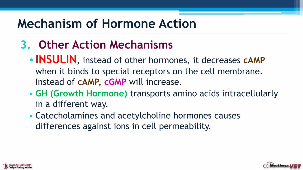
3. Other Action Mechanisms
• INSULIN, instead of other hormones, it decreases cAMP
when it binds to special receptors on the cell membrane.
Instead of cAMP, cGMP will increase.
• GH (Growth Hormone) transports amino acids intracellularly
in a different way.
• Catecholamines and acetylcholine hormones causes
differences against ions in cell permeability.
Mechanism of Hormone Action
Source: Usmle
Video Source: Youtube
• There are different types of classifications.
1. Glandular Hormones
2. Tissue Hormones
Classification of Hormones
1. Steroid Hormones
2. Amino acid derivative hormones
3. Peptid-Protein Structured hormones
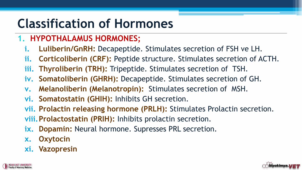
1. HYPOTHALAMUS HORMONES;
i. Luliberin/GnRH: Decapeptide. Stimulates secretion of FSH ve LH.
ii. Corticoliberin (CRF): Peptide structure. Stimulates secretion of ACTH.
iii. Thyroliberin (TRH): Tripeptide. Stimulates secretion of TSH.
iv. Somatoliberin (GHRH): Decapeptide. Stimulates secretion of GH.
v. Melanoliberin (Melanotropin): Stimulates secretion of MSH.
vi. Somatostatin (GHIH): Inhibits GH secretion.
vii. Prolactin releasing hormone (PRLH): Stimulates Prolactin secretion.
viii.Prolactostatin (PRIH): Inhibits prolactin secretion.
ix. Dopamin: Neural hormone. Supresses PRL secretion.
x. Oxytocin
xi. Vazopresin
Classification of Hormones
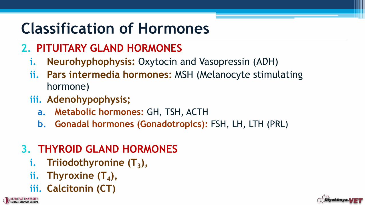
2. PITUITARY GLAND HORMONES
i. Neurohyphophysis: Oxytocin and Vasopressin (ADH)
ii. Pars intermedia hormones: MSH (Melanocyte stimulating
hormone)
iii. Adenohypophysis;
a. Metabolic hormones: GH, TSH, ACTH
b. Gonadal hormones (Gonadotropics): FSH, LH, LTH (PRL)
3. THYROID GLAND HORMONES
i. Triiodothyronine (T3),
ii. Thyroxine (T4),
iii. Calcitonin (CT)
Classification of Hormones
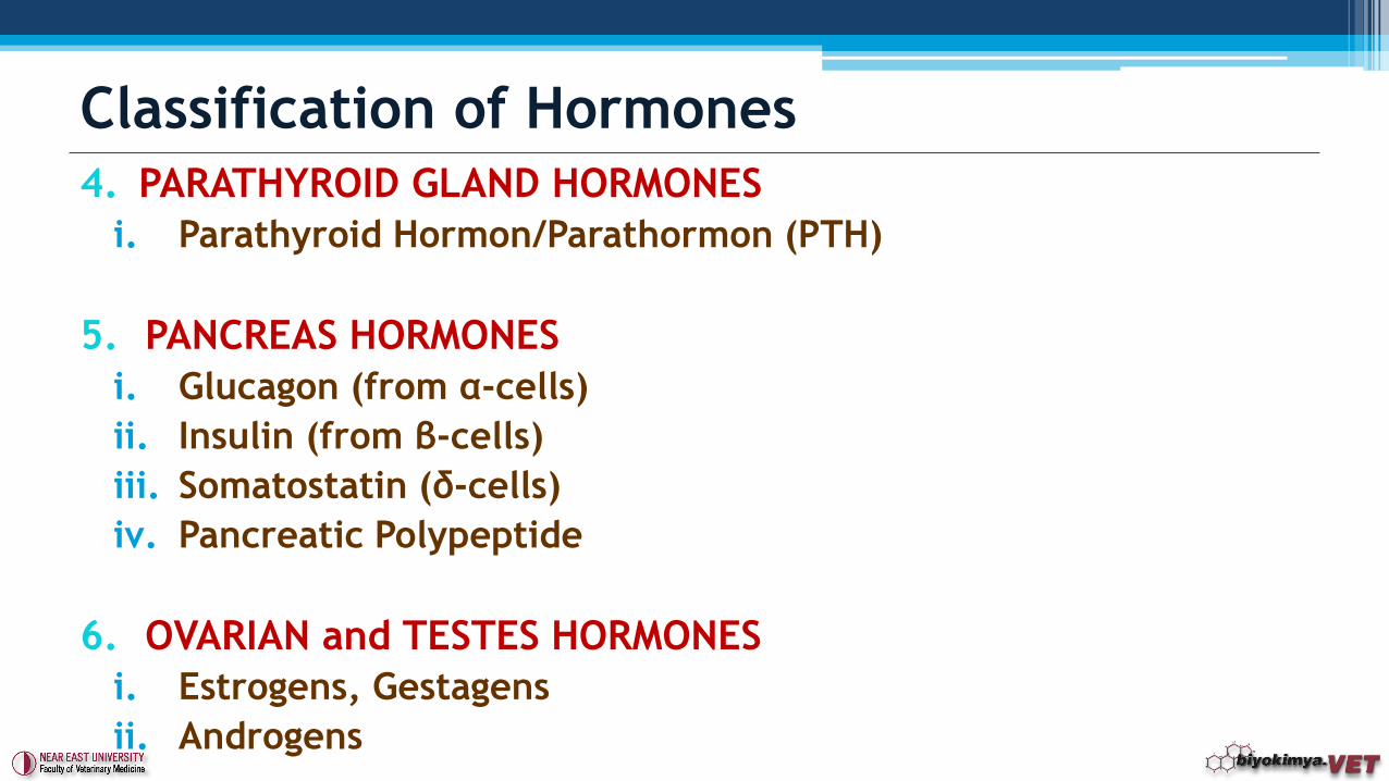
4. PARATHYROID GLAND HORMONES
i. Parathyroid Hormon/Parathormon (PTH)
5. PANCREAS HORMONES
i. Glucagon (from α-cells)
ii. Insulin (from β-cells)
iii. Somatostatin (δ-cells)
iv. Pancreatic Polypeptide
6. OVARIAN and TESTES HORMONES
i. Estrogens, Gestagens
ii. Androgens
Classification of Hormones
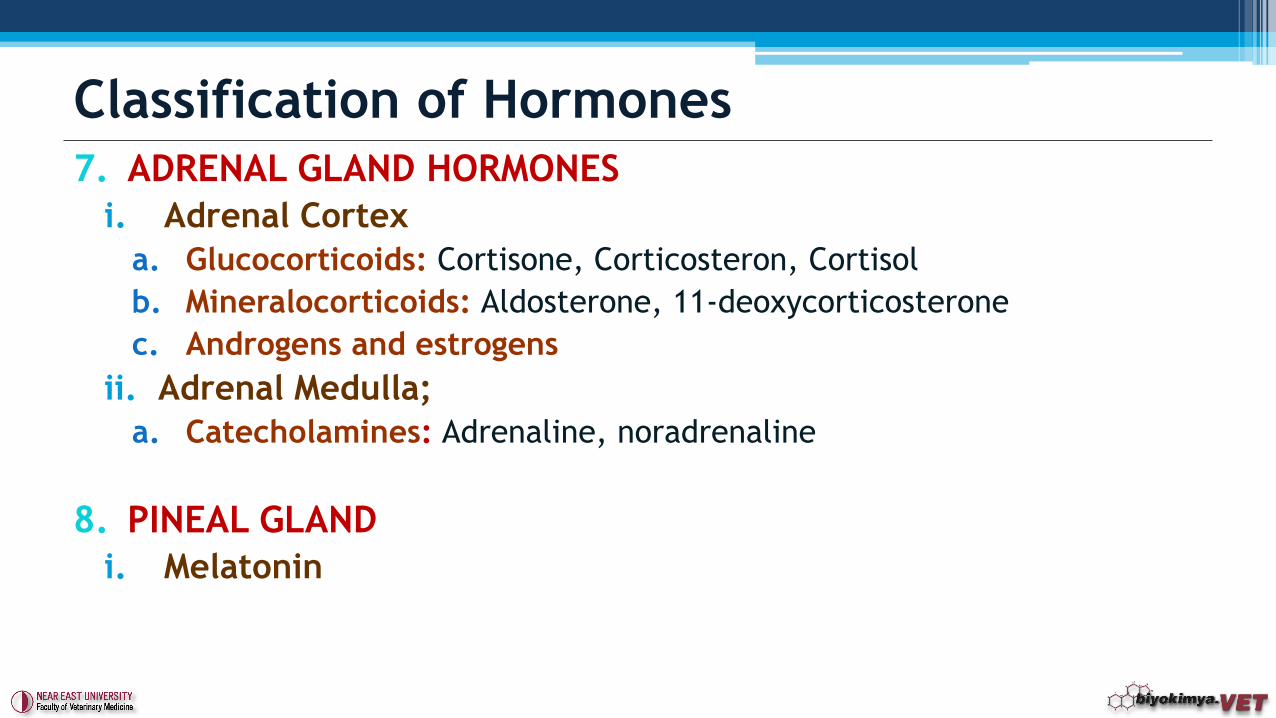
7. ADRENAL GLAND HORMONES
i. Adrenal Cortex
a. Glucocorticoids: Cortisone, Corticosteron, Cortisol
b. Mineralocorticoids: Aldosterone, 11-deoxycorticosterone
c. Androgens and estrogens
ii. Adrenal Medulla;
a. Catecholamines: Adrenaline, noradrenaline
8. PINEAL GLAND
i. Melatonin
Classification of Hormones
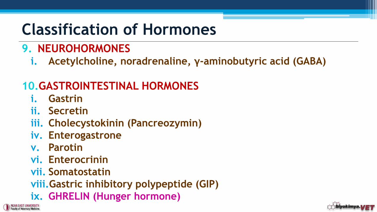
9. NEUROHORMONESi. Acetylcholine, noradrenaline, γ-aminobutyric acid (GABA)
10.GASTROINTESTINAL HORMONESi. Gastrin
ii. Secretin
iii. Cholecystokinin (Pancreozymin)
iv. Enterogastrone
v. Parotin
vi. Enterocrinin
vii. Somatostatin
viii.Gastric inhibitory polypeptide (GIP)
ix. GHRELIN (Hunger hormone)
Classification of Hormones
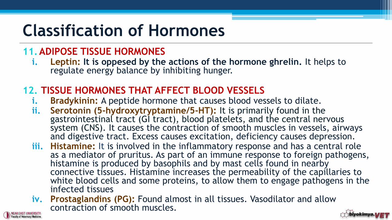
11. ADIPOSE TISSUE HORMONESi. Leptin: It is oppesed by the actions of the hormone ghrelin. It helps to
regulate energy balance by inhibiting hunger.
12. TISSUE HORMONES THAT AFFECT BLOOD VESSELSi. Bradykinin: A peptide hormone that causes blood vessels to dilate.ii. Serotonin (5-hydroxytryptamine/5-HT): It is primarily found in the
gastrointestinal tract (GI tract), blood platelets, and the central nervous system (CNS). It causes the contraction of smooth muscles in vessels, airways and digestive tract. Excess causes excitation, deficiency causes depression.
iii. Histamine: It is involved in the inflammatory response and has a central role as a mediator of pruritus. As part of an immune response to foreign pathogens, histamine is produced by basophils and by mast cells found in nearby connective tissues. Histamine increases the permeability of the capillaries to white blood cells and some proteins, to allow them to engage pathogens in the infected tissues
iv. Prostaglandins (PG): Found almost in all tissues. Vasodilator and allow contraction of smooth muscles.
Classification of Hormones
• Pituitary gland, shows the effect control over the entire endocrine system. It has a central role in the endocrine system. It manages many endocrine glands and tissues. It is also called as «the president gland».
• Releasing hormones or factore from hypothalamus affect onpituitary gland to cause hormone synthesis and secretion. Various centers are located in the hypothalamus participating the regulation of important functions.
• These are;▫ Centers that the occurrence of reproductive and sexual behavior is
regulated,▫ Centers that regulate metabolism,▫ Centers that regulate the intake of nutrients,▫ Centers for the compliance with various state organs,▫ Centers that regulate receiving and disposal of water.
Pituitary Gland Hormones
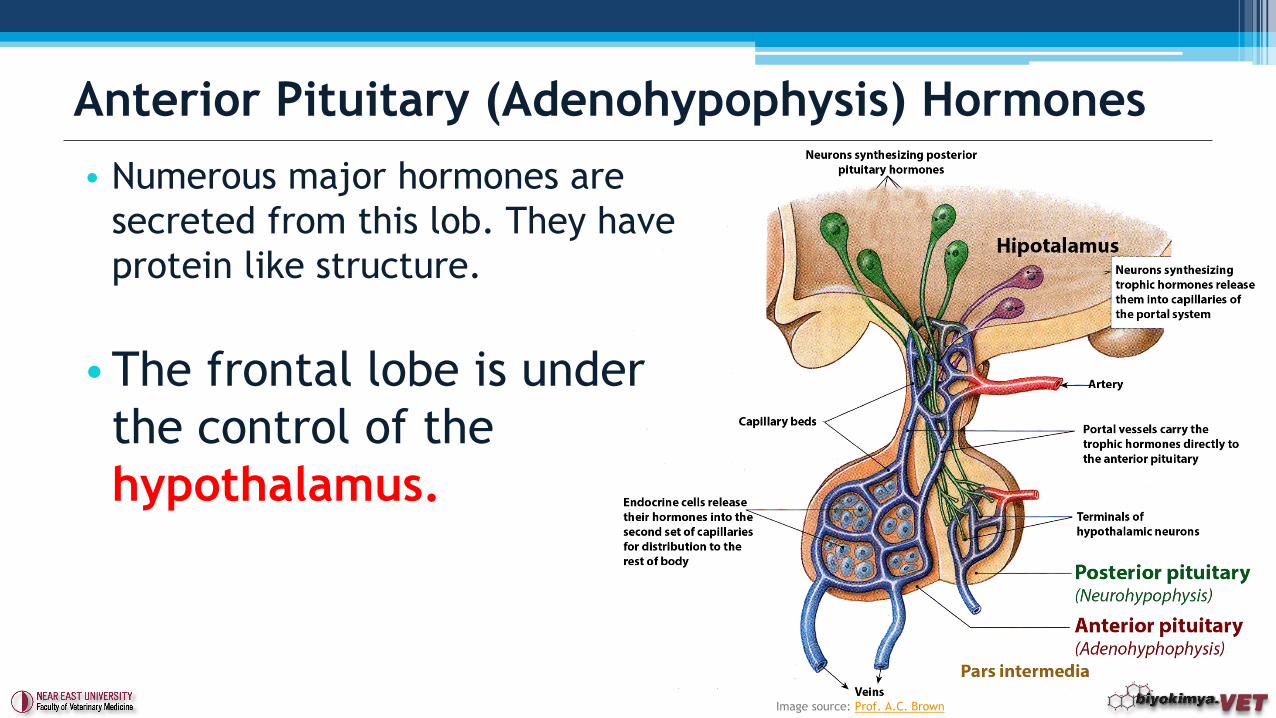
• Numerous major hormones are
secreted from this lob. They have
protein like structure.
•The frontal lobe is under
the control of the
hypothalamus.
Anterior Pituitary (Adenohypophysis) Hormones
Image source: Prof. A.C. Brown
1. ACTH (Adrenocorticotropic hormone): Its secretion is under
control of Corticotropin-releasing factor (CRF) which is
synthesized from hypothalamus. It consists of 39 amino acids in
the form of the prohormone. 23 amino acids required for the
effect.
▫Main effects;
Target organ is the adrenal cortex.
It increases steroid hormone synthesis.
Synthesis of glucocorticoids is increased.
Ascorbic acid amount in the adrenal glands is reduced.
It is involved in the acceleration of protein synthesis.
Anterior Pituitary (Adenohypophysis) Hormones
2. Somatotropin/Growth hormone (STH - Growth hormone /
GH): Polipeptide structure. Secretion is under control of
Growth hormon-Releasing Factor (GHRF) which is synthesized
by hypothalamus.
▫ The amino acid numbers it carries varies between animal species.
▫ For example; Human growth hormone 188, bovine growth hormone
369 and sheep growth hormone 191 amino acids.
▫ Human and monkey GH is composed of a single polypeptide chain,
however in cattle and sheep there are two-chain structure.
Anterior Pituitary (Adenohypophysis) Hormones
• Main effect;
▫ To increase protein synthesis in all cells present in the body.
▫ To increase the mobilization of fat and allow their use (lipolysis).
▫ Reducing carbohydrate utilization. It’s the antagonist of Insulin.
▫ It increases extracellular collagen and chondroitin sulfate synthesis.
▫ It is involved in water balance. While the tubular secretion is
increased, K, Na ve Cl retention occurs.
▫ GIGANTISM, ACROMEGALY, and DWARFISM.
Anterior Pituitary (Adenohypophysis) Hormones
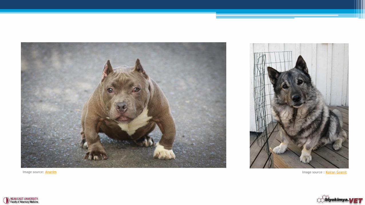
Image source: Anonim Image source : Koiran Geenit

Image source : Voorbij ve ark., 2011
Image source : AnimalEndocrinClinic
3. Thyroid Stimulating Hormone (TSH - Thyrotropin): It has a
Glycoprotein structure. It consists of 8% carbohydrates.
Secretion depends on Thyrotropin-Releasing Factor (TRH)
which is produced by Hypothalamus.
▫ Main effects;
It effects on thyroid gland therefore it stimulates thyroxine (T4) and
triiodothyronine (T3) hormones.
It accelerates the basal metabolism.
It accelerates the cardiac cycle.
It stimulates the nervous system function.
It decreases liver glycogen.
Anterior Pituitary (Adenohypophysis) Hormones
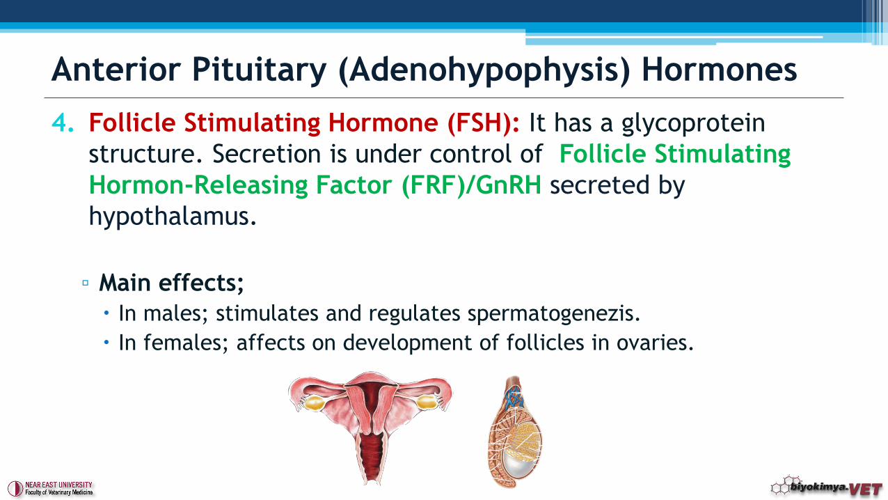
4. Follicle Stimulating Hormone (FSH): It has a glycoprotein
structure. Secretion is under control of Follicle Stimulating
Hormon-Releasing Factor (FRF)/GnRH secreted by
hypothalamus.
▫ Main effects;
In males; stimulates and regulates spermatogenezis.
In females; affects on development of follicles in ovaries.
Anterior Pituitary (Adenohypophysis) Hormones
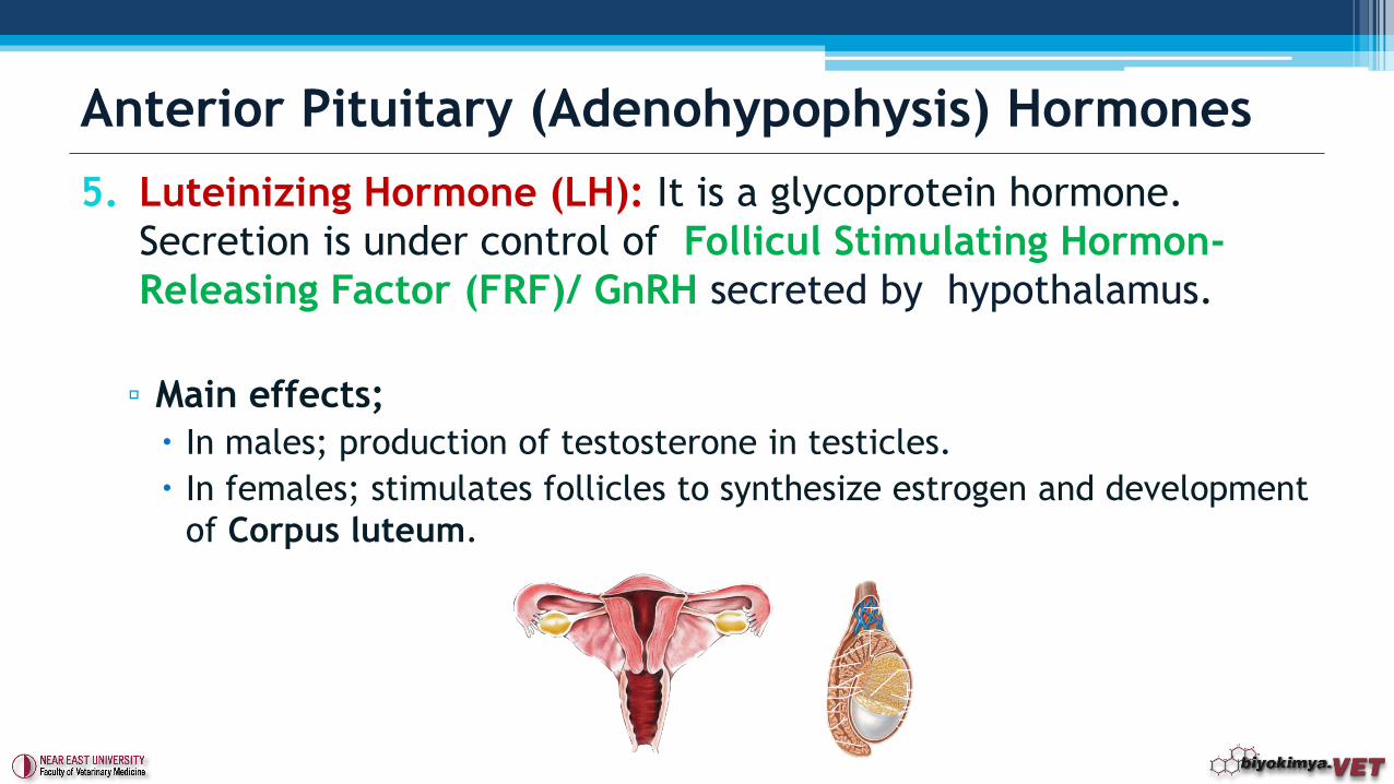
5. Luteinizing Hormone (LH): It is a glycoprotein hormone.
Secretion is under control of Follicul Stimulating Hormon-
Releasing Factor (FRF)/ GnRH secreted by hypothalamus.
▫ Main effects;
In males; production of testosterone in testicles.
In females; stimulates follicles to synthesize estrogen and development
of Corpus luteum.
Anterior Pituitary (Adenohypophysis) Hormones
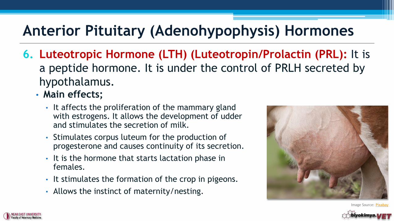
6. Luteotropic Hormone (LTH) (Luteotropin/Prolactin (PRL): It is
a peptide hormone. It is under the control of PRLH secreted by
hypothalamus.
Anterior Pituitary (Adenohypophysis) Hormones
• Main effects;
• It affects the proliferation of the mammary gland with estrogens. It allows the development of udderand stimulates the secretion of milk.
• Stimulates corpus luteum for the production of progesterone and causes continuity of its secretion.
• It is the hormone that starts lactation phase in females.
• It stimulates the formation of the crop in pigeons.
• Allows the instinct of maternity/nesting.
Image Source: Pixabay
• Melanocyte Stimulating Hormone (MSH/Melanotropins/Intermedins/ Melanophore Hormones): The structure of α-MSH is same for all species and consists of 13 amino acids. Pig β -MSH consists of 18 amino acid. But inhumans, N-terminal of hormone is longer than pigs (4 more amino acids).▫ Its secretion depends on light stimulation.
▫ Main effects; MSH is a hormone that affects the skin's pigmentation.
Fish, amphibians and reptiles, affects the distribution of pigment granules in pigment cells
Melanophore, iridophores. Melanocytes
Pars Intermedia (Intermediate pituitary) Hormones
• Posterior pituitary (neurohypophysis) includes pituicytes (Glial cells of posterior lobe). The posterior pituitary is not glandular as is the anterior pituitary. It is largely a collection of axonal projections from the hypothalamus that terminate behind the anterior pituitary, and is also a store for the later release of neurohypophysial hormones.
• Vasopressin (Anti-diuretic hormone/ADH): Vasopressin secretion siteis the region found in hypothalamus and called the supraoptic nucleus. It is a nonapeptide and an alkaline due to the presence of arginine in the structure.
• Main effects;▫ Inhibits urine. Enhance water reabsorption with mineralocorticoids.
▫ Raises the blood pressure (weaker than adrenalin but longer time).
▫ Controls Na concentration in the extracellular fluid.
Posterior Pituitary (Neurohyphophysis) Hormones
• Oxytocin: Its production location is the region called the
paraventricular nucleus of the hypothalamus. It has nonapeptid
structure. Amino acid type found at 8th. position determines the
species specificity.
▫ Main effects;
It provides the contractions of the uterus. This effect is called oxcitoxic
effect and it can compress uterine mucsle in pregnancy.
Uterus must enter a pre-estrogenic effect to show this effect during parturition.
It also causes the uterine contractions during sexual intercourse.
Oxytocin facilitates the flow of milk by contracting mammary gland muscles.
Triggers cells surrounding the alveoli and milk ducts (myoepitheliım cells) to
contract.
Reduces blood pressure.
Back Lop pituitary (neurohypophysis) hormones
1. Gastrin: It is a polypeptide. Secreted from pyloric mucous. It
causes HCl secretion from fundus cells.
2. Secretin: It is a polypeptide and secreted by the duodenum. It
controls the volume of pancreatic secretion and bicarbonate
levels.
3. Cholecystokinin (Pancreozymin): It is a polypeptide and
secreted by the duodenum. It has effects on gall bladder
(allows the discharge from ductus choledochus/common bile
duct) and on pancreas (enzyme secretion).
4. Enterogastrone: It is a polypeptide and secreted by the
intestine. Inhibits the prodution of gastric secretion and HCl.
Gastrointestinal Hormones
6. Parotin: It is a polypeptide. It is secreted by the salivary glands. Stimulate the calcification of teeth, lowers serum Ca levels, raises the serum P level.
7. Enterocrinin: Secreted from the intestinal mucosa as a protein hormone. It increases the secretion and excretion of jejunaland ileal discharge and the enzyme content.
8. Somatostatin: Inhibits gastrin ve secretin synthesis.
9. Gastric inhibitory polypeptide (GIP): Secreted by duedenumand jejenum. Stimulates Insulin secretion.
10.Ghrelin: Secreted mainly by stomach and duedenum. It is called as hunger hormone. It is involved in the regulation of energy balance.
Gastrointestinal Hormones
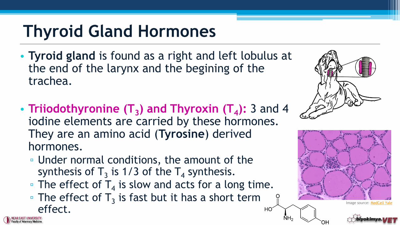
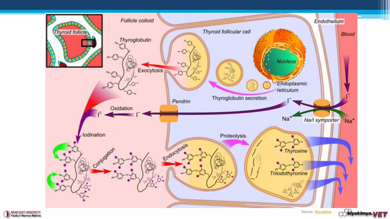
• Tyroid gland is found as a right and left lobulus at the end of the larynx and the begining of the trachea.
• Triiodothyronine (T3) and Thyroxin (T4): 3 and 4 iodine elements are carried by these hormones. They are an amino acid (Tyrosine) derived hormones. ▫ Under normal conditions, the amount of the
synthesis of T3 is 1/3 of the T4 synthesis.
▫ The effect of T4 is slow and acts for a long time.
▫ The effect of T3 is fast but it has a short term effect.
Thyroid Gland Hormones
Image source: MedCell Yale
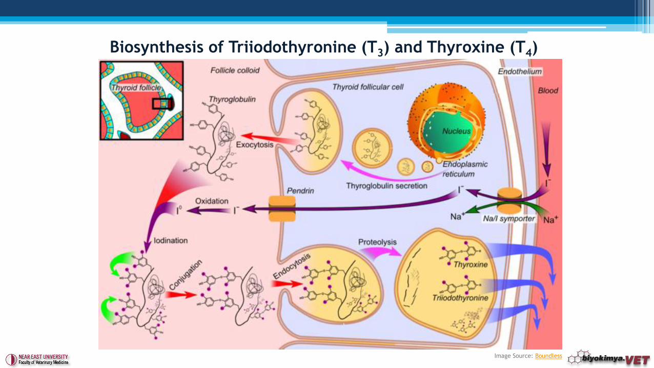
Image Source: Boundless
Biosynthesis of Triiodothyronine (T3) and Thyroxine (T4)
• Thyroid hormones circulate in the blood attached to the plasma
proteins.
• Thyroxine mostly passes through the blood. Enzymatic
reactions that takes place in either thyroid gland or other
tissues can seperated an iodine from thyroxine and forms
triiodothyronine.
• The actual metabolic impact, the real hormone effect, is
thought to be created by T3.
• The ways for catabolism is the deamination and
decarboxylation.
• Metabolites are removed by the kiney and bile.
Thyroid Gland Hormones
• Effects;
▫ Stimulates intake of substrates by mitochondrias, oxidation and ATP
production.
▫ To accelerate protein synthesis. This increase happens with the
raise of the synthesis of the enzymes at the same time. In protein
synthesis, thyroid hormones increases so this causes tissues to get
larger in size and also causes closure of epiphysis of the bones in a
very short time.
▫ It affects all the phases of carbohydrates metabolism. The usage of
glucose also increases.
Firstly, glycogenolysis increases.
Gluconeogenesis increases (relation with Diabetes mellitus ???).
Thyroid Gland Hormones
• Effects;
▫ It affects synthesis, mobilization and oxidation processes in the
lipid metabolism. This effect is madee through c-AMP. Decreases
blood cholesterol.
▫ Increases the synthesis of many enzymes. Depending on the
excessiveness of thyroid hormones, thiamine, riboflavin, cobalamin
and ascorbic acid need is increased.
▫ It is required for the synthesis of vitamin A from Carotene.
Thyroid Gland Hormones
• Calcitonin (CT);
▫ Polipeptide structure. Forming from 32 amino acids.
▫ It is synthesized from C-cells located between follicular cells
excepting in fish, amphibians, reptiles and birds.
▫ It is opposed by the actions of PTH on serum Ca levels.
▫ Calcitonin secretion by the thyroid gland is dependent on the
concentration of blood calcium.
▫ If calcium ions in the blood found within the physiological limits,
calcitonin secretion is very low.
▫ If blood calcium increases, calcitonin secretion significantly
increases to reduce calcium level.
Thyroid Gland Hormones
• Effects;▫ There is a quick impact on the Serum Ca levels, there will be a drop
for a short time.
▫ If the Ca content of the blood increases, the synthesis increases.
▫ Provides calcium increase in bones.
▫ Inhibits mobilization of Ca from bones.
▫ Prevents hypercalcemia.
• As the age gets older, the reaction skills of the bone tissue decreases against calcitonin.
• In poultry, calcitonin is less effective during nesting/laying an egg.
Thyroid Gland Hormones
• Parathyroid gland, located in the rear surface of the thyroid
gland. It has four small ovary shape.
• Parathyroid hormone/Parathormone (PTH);
▫ Consisting of a single chain peptide without cysteine.
▫ Amino acid sequence varies according to animal species.
▫ PTH regulates the Ca2+ metabolism and blood Ca2+ level control
the secretion of PTH.
▫ Affects calcium and phosphorus metabolism.
▫ Affects kidney, bone and gastrointestinal tract.
Parathyroid Gland Hormone
▫ PTH functions;
Effects on Kidney : Controls the removal of phosphorus, potassium,
calcium and hydrogen ions.
Effects on Bones : Affects calcium transport in bones. It causes the
removal or mobilization of calcium phosphate from the bones.
Effects on Gastrointestinal channel: Increases the Vitamin D synthesis
and facilitates calcium absorption in the intestine.
▫ The concentration of ionized calcium in the blood, controls the
secretion of PTH by negative feed-back.
Parathyroid Gland Hormones
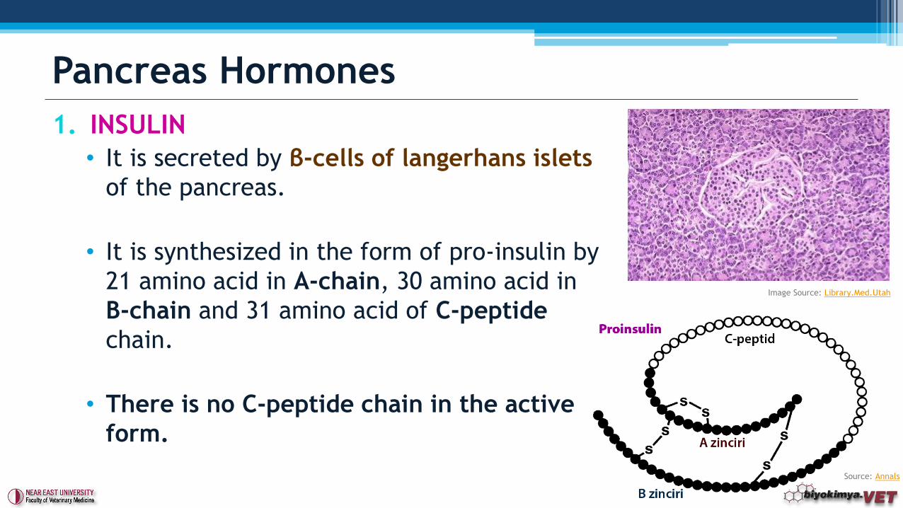
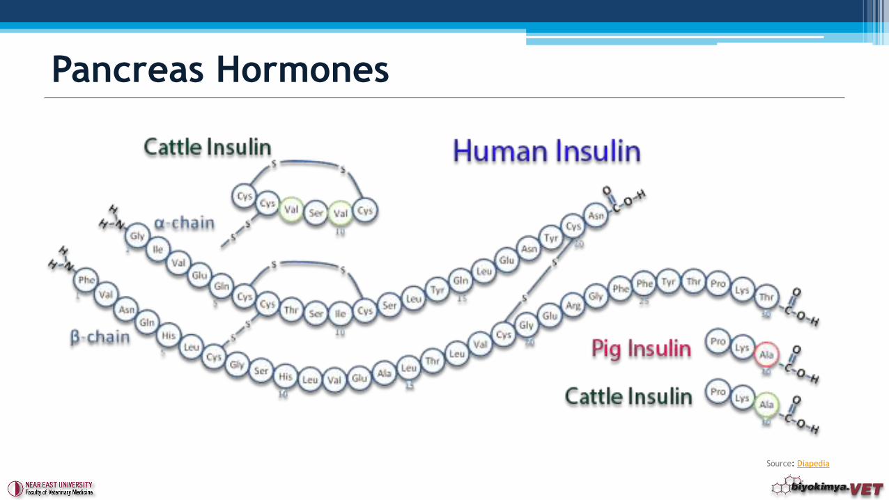
1. INSULIN
• It is secreted by β-cells of langerhans islets
of the pancreas.
• It is synthesized in the form of pro-insulin by
21 amino acid in A-chain, 30 amino acid in
B-chain and 31 amino acid of C-peptide
chain.
• There is no C-peptide chain in the active
form.
Pancreas Hormones
Image Source: Library.Med.Utah
Source: Annals
Pancreas Hormones
Source: Diapedia

Source: Usmle
Video Link: Youtube
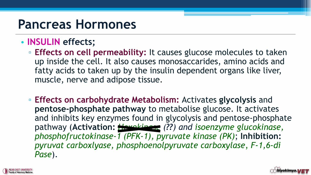
• INSULIN effects;▫ Effects on cell permeability: It causes glucose molecules to taken
up inside the cell. It also causes monosaccarides, amino acids and fatty acids to taken up by the insulin dependent organs like liver, muscle, nerve and adipose tissue.
▫ Effects on carbohydrate Metabolism: Activates glycolysis and pentose-phosphate pathway to metabolise glucose. It activates and inhibits key enzymes found in glycolysis and pentose-phosphate pathway (Activation: Hexokinase (??) and isoenzyme glucokinase, phosphofructokinase-1 (PFK-1), pyruvate kinase (PK); Inhibition: pyruvat carboxlyase, phosphoenolpyruvate carboxylase, F-1,6-di Pase).
Pancreas Hormones
• INSULIN effects;
▫ Effects on lipid metabolism: Increases fatty acid synthesis. Insulin
activates key enzymes and causes breakdown of glucose; the product
of this reaction, acetyl-CoA synthesis is increased. Therefore, this
speeds up the synthesis of fatty acids. Free fatty acids is stored as
triglycerides and by this way ketosis is prevented.
▫ Effects on protein metabolism: It occurs as a result of the increase
of the amino acids in the cells and also increase in permeability of
the cell membrane. Directly, causes increase in mRNA synthesis ve
increase in the entry of amino acids inside of cell proteins.
• Deficiency;
▫ Diabetes mellitus
Pancreas Hormones
2. GLUCAGON: It is secreted from α-cells of Langerhans islets. It
effects opposite way of Insulin. It increases blood glucose
levels. It is a hormone that is made up of 29 amino acids.
▫ Effects:
It activates key enzymes in gluconeogenesis (pyruvate carboxylase,
phosphoenolpyruvate carboxykinase, fructose-1,6-diphosphatase,
glucose-6-phosphatase).
The effect on glycogenolysis is done by activating adenylyl cyclase,
increasing the production of c-AMP, activating protein kinase and
lastly activating phosphorylase a and phosphorylase b.
Glucagon also stimulates the entry of fatty acids to mitochondrias and
oxidation of fatty acids.
Pancreas Hormones
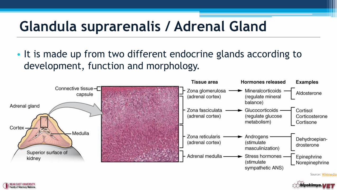
• It is made up from two different endocrine glands according to
development, function and morphology.
Glandula suprarenalis / Adrenal Gland
Source: Wikimedia
• The origin of the sympathetic nerve and adrenal medulla are the same and they worked together. Therefore it is called sympatico-adrenal system.
• The cells that secreting Catecholamines are the altered sympathetic neurons.
• Epinephrine are mainly secreted from medulla (80%) . Hormone that is secreted from sympathetic nerves is generelly norepinephrine.
• Precursor molecule for both hormone is ................
Adrenal Medulla
TYROSINE
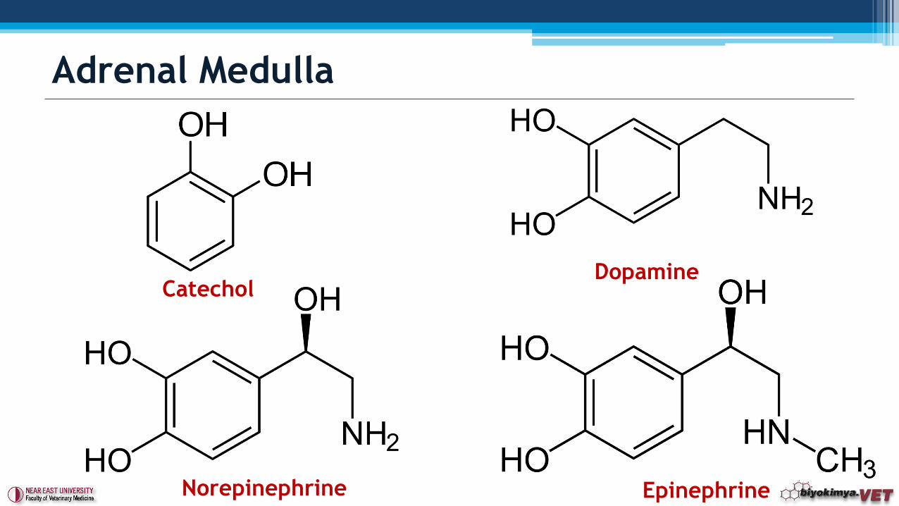
Adrenal Medulla
CatecholDopamine
Norepinephrine Epinephrine
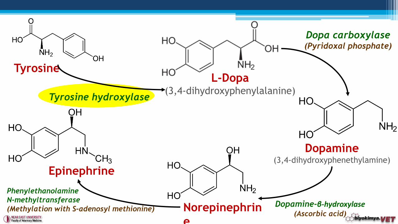
Tyrosine
Dopamine(3,4-dihydroxyphenethylamine)
Norepinephrin
e
Epinephrine
L-Dopa(3,4-dihydroxyphenylalanine)
Tyrosine hydroxylase
Dopa carboxylase(Pyridoxal phosphate)
Dopamine-β-hydroxylase(Ascorbic acid)
Phenylethanolamine
N-methyltransferase
(Methylation with S-adenosyl methionine)
• Control of the secretion of Catecholamines;▫ At rest they are very low in level. Activated sympathetic system increases
their synthesis and secretion.
▫ Receptors play a role in alerting the system. These receptors include glucose receptors, thermoreceptors, baroreseptors. Hormones are determined by the stimulated receptors.
▫ Fight or Flight Response
▫ Norepinephrine hormone is secreted when arteria carotis pressure receptors are stimulated.
▫ Sympathetic nerves and glucocorticoids arrange catecholamine synthesis. Tyrosine hydroxylase is affected by mainly nerves and less glucocorticoids.
Dopamine-β-hydroxylase activation is regulated by both nerves and glucocorticoids.
Phenylethanolamine N-methyltransferase activation is mostly regulated by glucocorticoids.
Adrenal Medulla
• Effects;▫ Sudden decrease in blood glucose causes increase in secretion of
adrenaline and this stimulates glycogenolysis. It’s an insulin antagonist. Noradrenalin has no importance on this effect.
▫ Adrenalin increases metabolic speed by ~ 30% . Noradrenalin has no importance on this effect.
▫ Increases cardiac cycle and also blood pressure. The pupils are dilated.
▫ Increases tones and contractions of smooth and skeletal muscles.
▫ Bronchials expand and this increases venting capacity of lungs.
▫ Blood circulation in skin increases and formation of sweat increases.
▫ Adrenalin and noradrenalin raises lipolysis. By this way heart muscle can take up energy from fatty acids.
Adrenal Medulla
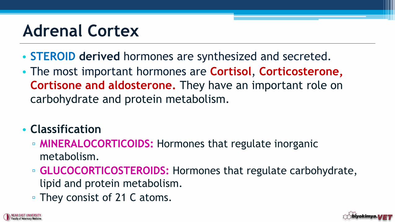
• STEROID derived hormones are synthesized and secreted.
• The most important hormones are Cortisol, Corticosterone,
Cortisone and aldosterone. They have an important role on
carbohydrate and protein metabolism.
• Classification
▫ MINERALOCORTICOIDS: Hormones that regulate inorganic
metabolism.
▫ GLUCOCORTICOSTEROIDS: Hormones that regulate carbohydrate,
lipid and protein metabolism.
▫ They consist of 21 C atoms.
Adrenal Cortex
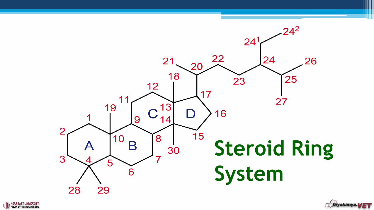
Steroid Ring
System
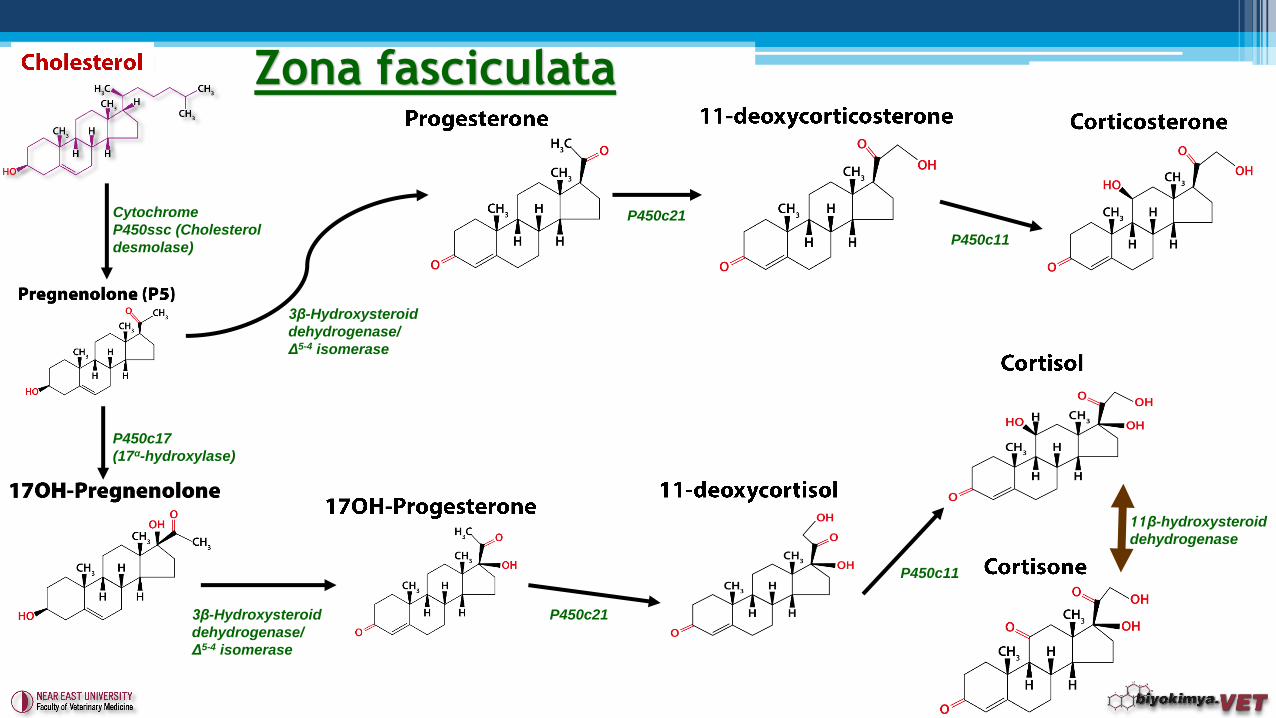
Zona fasciculata
3β-Hydroxysteroid
dehydrogenase/
Δ5-4 isomerase
Cytochrome
P450ssc (Cholesterol
desmolase)
P450c21
P450c11
P450c17
(17α-hydroxylase)
3β-Hydroxysteroid
dehydrogenase/
Δ5-4 isomerase
P450c21
P450c11
11β-hydroxysteroid
dehydrogenase
Zona glomerulosa
3β-Hydroxysteroid
dehydrogenase/
Δ5-4 isomerase
Cytochrome
P450ssc (Cholesterol
desmolase)
P450c21
P450c11
Aldosterone synthase
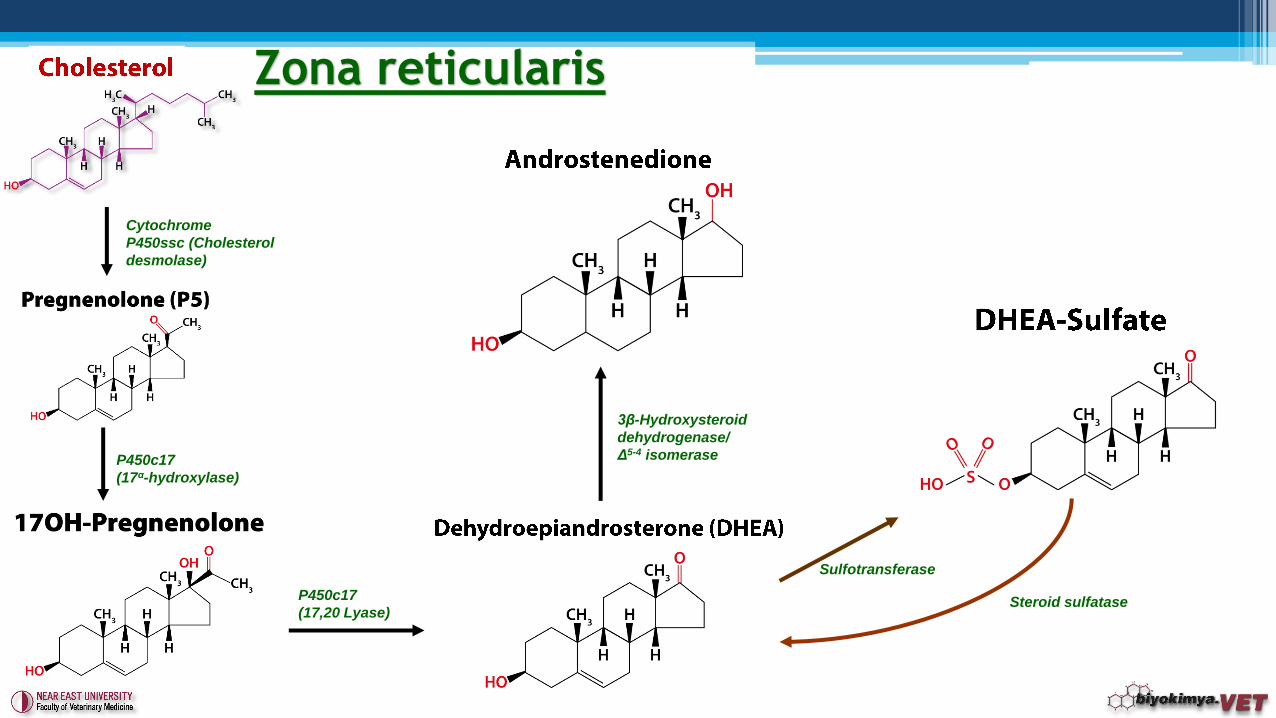
Zona reticularis
Cytochrome
P450ssc (Cholesterol
desmolase)
P450c17
(17α-hydroxylase)
P450c17
(17,20 Lyase)
Sulfotransferase
3β-Hydroxysteroid
dehydrogenase/
Δ5-4 isomerase
Steroid sulfatase
3β-Hydroxysteroid
dehydrogenase/
Δ5-4 isomerase
17β-hydroxysteroid
dehydrogenase
5α-Reductase
17β-hydroxysteroid
dehydrogenase
3β-Hydroxysteroid
dehydrogenase/
Δ5-4 isomerase
Aromatase
Aromatase
Fetal liver and
placenta
17β-hydroxysteroid
dehydrogenase
Androgen & Estrogen
Biosynthesis
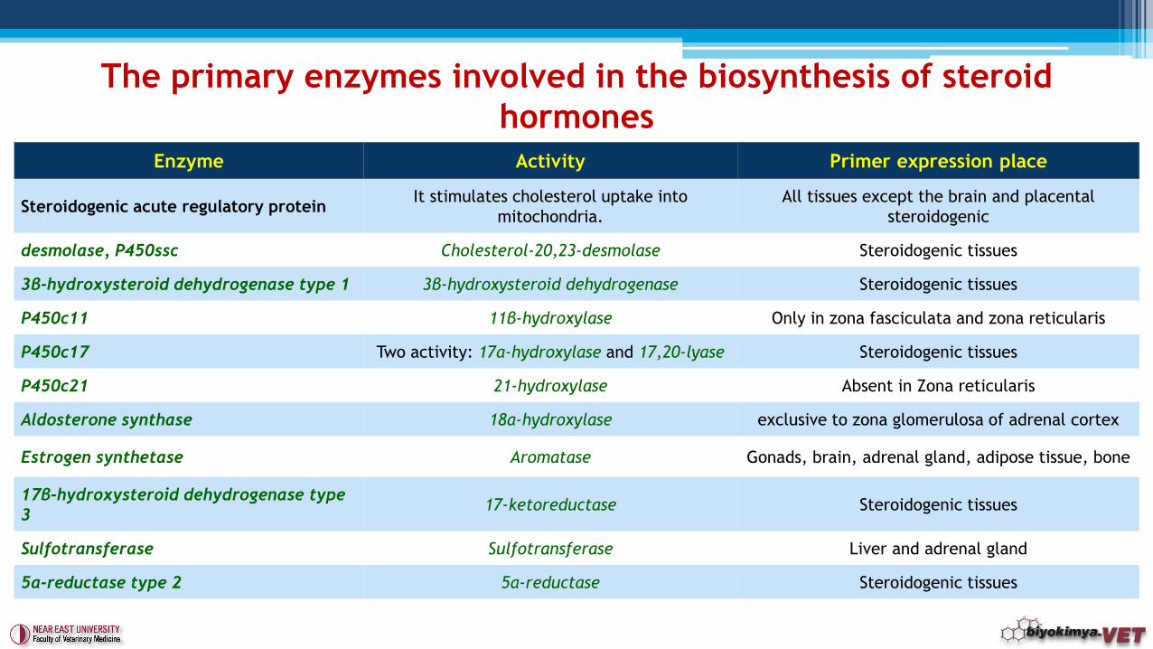
Enzyme Activity Primer expression place
Steroidogenic acute regulatory proteinIt stimulates cholesterol uptake into
mitochondria.
All tissues except the brain and placental
steroidogenic
desmolase, P450ssc Cholesterol-20,23-desmolase Steroidogenic tissues
3β-hydroxysteroid dehydrogenase type 1 3β-hydroxysteroid dehydrogenase Steroidogenic tissues
P450c11 11β-hydroxylase Only in zona fasciculata and zona reticularis
P450c17 Two activity: 17α-hydroxylase and 17,20-lyase Steroidogenic tissues
P450c21 21-hydroxylase Absent in Zona reticularis
Aldosterone synthase 18α-hydroxylase exclusive to zona glomerulosa of adrenal cortex
Estrogen synthetase Aromatase Gonads, brain, adrenal gland, adipose tissue, bone
17β-hydroxysteroid dehydrogenase type
317-ketoreductase Steroidogenic tissues
Sulfotransferase Sulfotransferase Liver and adrenal gland
5α-reductase type 2 5α-reductase Steroidogenic tissues
The primary enzymes involved in the biosynthesis of steroid
hormones
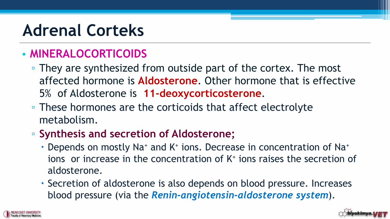
• MINERALOCORTICOIDS
▫ They are synthesized from outside part of the cortex. The most
affected hormone is Aldosterone. Other hormone that is effective
5% of Aldosterone is 11-deoxycorticosterone.
▫ These hormones are the corticoids that affect electrolyte
metabolism.
▫ Synthesis and secretion of Aldosterone;
Depends on mostly Na+ and K+ ions. Decrease in concentration of Na+
ions or increase in the concentration of K+ ions raises the secretion of
aldosterone.
Secretion of aldosterone is also depends on blood pressure. Increases
blood pressure (via the Renin-angiotensin-aldosterone system).
Adrenal Corteks
• Effects;
▫ Increases active transport of Sodium.
▫ Allows renal tubular retention of Na. This will cause increased
osmotic pressure. Water is reabsorbed at the same rate with Na to
prevent high osmotic pressure.
▫ To protect the electrochemical balance, K+ and H+ ions is given to
the tubule fluid simultaneously with Na reabsorption.
▫ Reduces to excrete Na+ ions from sweat glands, salivary glands and
intestines. Na + removal increases when deficiency occured.
▫ Aldosterone against heat are important. It is involved in the
prevention of excessive Na losses when an excessive sweating is
occured.
Adrenal Cortex
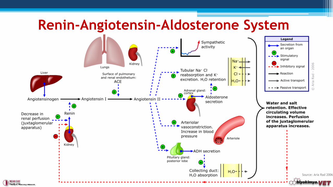
Source: Aria Rad 2006
Renin-Angiotensin-Aldosterone System
• GLUCOCORTICOIDS▫ They are secreted from the cortex of the middle and inner layers.
This name was given because they stimulate Gluconeogenesis.
▫ The most important representatives are; 17-hydroxycorticosterone/cortisol/hydrocortisone,
Corticosterone,
7-hydroxy-11-dehydrocorticosterone/cortisone.
▫ Effects; Carbohydrate Metabolism;
Stimulates the key enzymes in gluconeogenesis. Specially increases the usage of amino acids for gluconeogenesis and storage of the glycogen. It brings about a reduction in the use of peripherally glucose.
Adrenal Cortex
▫ Lipid Metabolism; Increases lipogenesis when taken in from outside in healthy animals.
▫ Protein Metabolism; Cortisol acts directly to increase protein synthesis in the liver. However, in
muscles, lymphoid and other tissues, inhibits the protein synthesis due to reduction in amino acid transport.
Promotes the destruction of proteins in skeletal muscles and in other tissues,
Increases the secretion of HCl and pepsinogen from the stomach and pancreatic trypsinogen.
▫ Anti-inflammatory effect and role in immune system; Decreases capillary permeability and stabilizes the membranes of lysosomes.
Thus, inflammation is inhibited.
They provide Ca mobilization in the bones.
The excessive amount of synthesis and long influence causes immunosuppression. Besides, RNA and protein synthesis in lymphocytes is reduced.
Adrenal CorteX
• Gonadal hormone production, especially made by interstitial
cells.
• The corpus luteum and placenta takes place as a temporary
place for the hormone synthesis in females.
• Sex hormones may also be synthesized by the adrenal cortex.
• The synthesis of sex hormones stimulated by gonadotropins.
• All of the sex hormones are STEROIDAL. They show a close
relationship with each other and adrenal cortex hormones. They
have common metabolic pathways in the body. Therefore, both
male and female sex hormones found in both males and females
together.
Gonadal hormones
• Progesterone has a key metabolite for the synthesis of all steroid hormones.
• Not only in the ovaries, it is located in both adrenal glands andthe testes. This is the intermediate metabolite in the body to other hormones.
• Androgens occur as intermediates during both degradation of progesteron and cortex hormones and also in the synthesis of estrogens.▫ Therefore, even if in small quantities, it is found in adrenal cortex
and ovaries.
• Androgens and estrogens, respectively, possess 19 and 18 carbon atoms.
Gonadal hormones
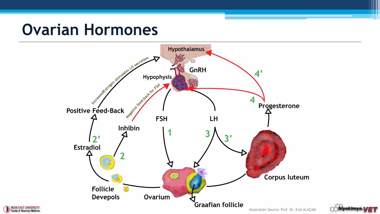
• Under the influence of FSH, estrogenic hormones is synthesizedin the ovary during follicular growth. Besides, progesteron is synthesized in Corpus luteum.
• ESTROGENS▫ Theca granulosa cells in ovarium synthesize estrogens.
▫ It is also synthesized in adrenal cortex, testis and placenta during pregnancy.
▫ Most effective form is estradiol-17β (E2). Others are, estrone (E1) and estriol (E3).
▫ Inactivation occurs in the liver. Excreted with urine and feces in the form of esters.
Ovarian Hormones
• ESTROGENS
▫ Estradiol is the active and available form.
▫ Estriol is mainly synthesized from placenta.
▫ Estrone is the very least form. Especially it is known as a
carcinogen. It is closely related to diseases and health status.
Estrone sulfate form has long life and kept as a reservoir for
conversion to estradiol. It is main form in postmenopausal women.
▫ Estrone sulfate is used to detect late pregnancy and also to
investigate the fetus viability in cows, horses, donkeys, pigs and
goats.
Ovarian Hormones
• Effects: ▫ Genital effects of estrogens are to stimulate RNA and protein
synthesis in order to induce the development of genital organs.
▫ Enables the development of the mammary gland combined with glucocorticoids, insulin, progesterone and prolactin.
▫ Act as an light anabolic on muscle, liver and kidneys.
▫ They allow the development secondary female characters. After castration in males, acts as an lipid anabolic.
▫ In fast-growing tissues of the male reproductive organs show an effect which prevents the mitotic event. Therefore it is used to treat prostate cancer.
▫ Increased blood levels reduces FSH secreting by affecting hypothalamus and besides, it increases the secretion of LH.
Ovarian Hormones
• GESTAGENS▫ The most important member is Progesterone. The diposal of gestagens
from the organism is done by the product Pregnandiol. It is removed by the urine.
▫ Progesteron is synthesized in Corpus luteum and it assures the continuitation of pregnancy.
▫ Otherwise it is synthesized by the adrenal glands. It is a precursor of androgens and estrogens .
▫ Effects; The first effect of progesterone is to ensure the continuation of the
pregnancy. Progesteron creates the environment for the settlement of the embryo in the
uterine mucosa which is prepared by estrogens. In case of insufficient progesterone secretion, uterine mucosa not fulfill the conditions for nutrition of the embryo.
The effect of follicle hormone (FSH) is inhibited by progesterone. Itssecretion is triggered by LH.
Ovarian Hormones
• RELAXIN▫ In females ovaries (corpus luteum) and placenta is the
locations that releases Relaxin.
▫ It is necessary for the continuity of the pregnancy.
▫ Sacroiliac joint and symphysis expands during pregnancy. It
facilitates the birth.
▫ In dogs, it is used to detect pregnancy.
▫ In males, it is secreted from prostate. It is important for the
sperm motility.
Ovarian Hormones
• INHIBIN▫ Polipeptide structure.
▫ Granulose cells in the ovary; sertoli cells in the testes are the
locations for the synthesis.
▫ It inhibits the secretion of FSH and activates paracrine function in
gonads.
▫ In mares, it is used in the diagnosis of Granulosa Cell Tumor.
Other Hormones
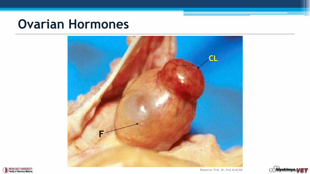
Ovarian Hormones
F
CL
Resource: Prof. Dr. Erol ALAÇAM
Ovarian Hormones
Illustration Source: Prof. Dr. Erol ALAÇAM
• ANTI-MULLERIAN HORMONE (AMH)
▫ It is a protein hormone.
▫ Granulose cells in the Ovary; sertoli cells in the testes are the
locations for the synthesis.
▫ It has low levels.
▫ In healthy mares, levels during the estrous cycle or pregnancy does
not change (<1 ng/ml) .
▫ In mares, it is used in the diagnosis of Granulosa Cell Tumor with
inhibin. Blood levels are increased (generally >25 ng/mL).
▫ Inhibin and testerones can give misleading results therefore, AMH is
more sensitive test.
Other Hormones
• ANDROGENS
▫ Androgens are the steroids that has 19 C atoms and they are
secreted from
Leydig interstitial cells in testicle.
▫ ‘Androgenic’ name is not only given for the biological activity of
the hormones, it is also given to point that they are steroids with
19 C atoms.
▫ Mainly effective examples of androgens are testosterone (17 β-
hydroxy-androst-4-en-3-one) and androsterone (androst-4-ene-
3,17-dione ) forms.
▫ These are found in Leydig cells and formed from 17-
hydroxyprogesterone.
Testes Hormones
• Effects;
▫ They allow the development of male sex organs.
▫ The extragenital effect is to stimulate the biosynthesis of proteins.
It is an anabolic hormone.
▫ They allow the development secondary male characters.
▫ Testosterone increases the protein synthesis within the cell.
Testosterone operates in the synthesis of RNA from DNA in the cell
nucleus. It increases the transcription.
Testes Hormonları
• Pineal gland affects many reproduction events and it is proved
that it can secrete many hormones at a time.
• This gland cells is the only place of melatonin synthesis and
secretion of the hormone melatonin.
• Synthesis occurs via serotonin and its amount is high in daylight
, on the other hand, is low at night.
• Light intensity and duration changes the amount of serotonin
in pineal gland.
• The amount of Melatonin is indirectly related to serotonin
rythm. Sytnhesis is less during daytime, however synthesis is
more at night .
Pineal Hormones (Pineal Gland)
• Effects:
▫ They stimulate mostly the accumulation of the melanin granules when it is compared to their distribution. They show a different effect other than they show in amphibians. They ensures that skin looks brighter.
▫ Changes in the synthesis of melatonin, provides the information to thehypothalamic-pituitary-gonadal axis about day length. It is a sensitive and complicated biological clock. It converts ambient light into neural activity.
▫ It has a role in reproduction system. In particular, monoestrous or polyestrous animals depending on the season.
▫ In mammalian organisms, Pineal gland is the only structure that melatonin is synthesized and the pituitary luteinizing hormone secretionis inhibited.
Pineal Hormones (Pineal Gland)
• INSULIN-LIKE GROWTH FACTOR ( IGF-1)
▫ It is similar to insulin according to its structure.
▫ Synthesized in the liver.
▫ Growth hormone (GH) stimulates its synthesis and secretion.
▫ Affects the development and growth in youngs.
▫ It has anabolic effects on adults.
▫ It is used in dogs and cats, as a indirectly evaluation for GH
secretion and activitiy.
▫ It is used in the diagnosis of Dwarfism and Agromegaly.
Other Hormones
• ERYTHROPOIETIN ( EPO)
▫ Excreted by the kidneys.
▫ Glycoprotein structure.
▫ Controls erythropoesis.
▫ It is a cytokine for erythrocytes in the bone marrow .
▫ Effects:
It increases the number of proerythroblasts in the bone marrow.
It stimulates RNA synthesis.
It stimulates iron for participation to hemoglobin in peripheral blood.
It stimulates angiogenesis.
Other Hormones
• PROSTAGLANDINS ( PG)▫ They are synthesized from arachidonic acid.
▫ At least 6 primary prostaglandins has being shown. These are PGE1 , PGE2 , PGE3 , PGF1α , PGF2α and PGF3α.
▫ Effects: They are potent vasodilators .
They assure the smooth muscle, especially uterine smooth muscle contractions.
Increase the efficiency of the heart muscle and cause the expansion of the bronchi.
They are antagonists against epinephrine, norepinephrine, glucagon and ACTH. They have impact on the migration of fatty acids from adipose tissue.
They have thermoregulatory effects .
Other Hormones
• PREGNANT MARE SERUM GONADOTROPIN (PMSG)
▫ It is also known as equine chorionic gonadotropin (eCG).
▫ It is produced in the chorion of pregnant mares
▫ It is synthesized in pregnant mares in between 40-130 days.
▫ It can be used as pregnancy diagnosis between these days.
Other Hormones
• Ası. T. 1999. Tablolarla Biyokimya, Cilt 2
• EclinPath. http://www.eclinpath.com/
• Nationwide Specialist Laboratories. http://thehormonelab.com/
• Pineda M, Dooley MP. McDonald's Veterinary Endocrinology & Reproduction 5th Ed. 2002.
Wiley-BlackWell
• Prof. Dr. Erol ALAÇAM. Ders Notları (Teşekkürlerimle).
• Sözbilir Bayşu N, Bayşu N. 2008. Biyokimya. Güneş Tıp Kitapevleri, Ankara
• Voorbij AM et al. A contracted DNA repeat in LHX3 intron 5 is associated with aberrant
splicing and pituitary dwarfism in German shepherd dogs. PLoS One. 2011;6(11):e27940.
• Special thanks to Tanju BORATAŞ, B.Sc. for English translation of this document.
References
• Which of the following hormone has iodine in its structure?
a. Estradiol
b. Insulin
c. Thyroxin
d. ACTH
e. Secretin
Question 1
Answer: c
• Which of the following hormone stimulates development of
mammary glands and the secretion of milk?
a. Testosterone
b. Prolactin
c. Somatotropin
d. IGF-1
e. Insulin
Question 2
Answer: b
• Which of the following hormone has a role in the calcium
metabolism?
a. Thyroxin
b. Secretin
c. PTH
d. Glucagon
e. Cortisol
Question 3
Answer: c

Any Questions?
Next Chapter;
Introduction to Metabolism
For more on
Biochemistry & Clinical Biochemistry
and the world of laboratories follow
www.biyokimya.vet
@biyokimya.vet