2016-2017 VOLUME 95, NUMBER 1 · 2019-12-10 · Journal of the PHILIPPINE MEDICAL ASSOCIATION 2016...
116
Transcript of 2016-2017 VOLUME 95, NUMBER 1 · 2019-12-10 · Journal of the PHILIPPINE MEDICAL ASSOCIATION 2016...


2016-2017 VOLUME 95, NUMBER 1

Journal of the
PHILIPPINE MEDICAL ASSOCIATION 2016 - 2017 VOLUME 95, NUMBER 1
TABLE OF CONTENTS
A Cross-sectional Study on the Impact of Acne Vulgaris 1
on the Quality of Life among High School Students in
Pasig City, Philippines
Vanessa Anne C. Bernal, MD, DPDS, Elizabeth V. Sanchez, MD, FPDS
The Appropriate Grading Tool to Assess Acne Severity 10
in Face-to-Face Consultation and Digital Skin Images Karen B. Mabilin-Prieto, MD, Elizabeth P. Prieto, MD, FPDS,
Jerlyn Maureen P. Servas, MD
A Clinical Trial on the Safety and Efficacy of Oil of Bergamot 23
30% and Sun Exposure combination versus Sun Exposure
Alone in the Management of Residual Hypomelanosis of
Pityriasis Versicolor Ma. Eleanor Cathryn DR. Salonga, MD, DPDS, Ma. Luisa Abad-Venida, MD, FPDS
Lillian Lopez-Villafuerte, MD, FPDS
A Randomized, Double-blind Placebo-controlled Clinical Trial on 40
the Efficacy and Safety of Adapalene 0.1% Gel on the Closure of
Neuropathic Ulcer among Leprosy Patients Ma. Cricelda M. Rescober, MD, Daisy Ismael-King, MD, FPDS
Association of Vitamin D Receptor Gene Polymorphisms in the 48
Occurrence and Spectrum of Leprosy in Filipino Patients seen
at Jose Reyes Memorial Medical Center
Donn M. Mendoza, MD, Zharlah Gulmatico-Flores, MD, FPDS
Patients of Different Body Mass Index Classification and Its Effect 56
on Tube Current Time Product, Scanner Radiation Output and
Image Quality as Applied in Multidetector Computed Tomography
of the Whole Abdomen - A Retrospective Study
Ginalynn Fincale, MD, Jackson Dy, MD
Acral Lentiginous Melanoma in Filipinos: Report of Two Cases 68 Luella Joy A. Escueta, MD, Ana Maria O. De la Serna-Mah, MD,
Johannes F. Dayrit, MD, FPDS
Cavernous Lymphangioma of the Thigh in a 4-Year Old Boy treated 72
by Excision: A Case Report
Josephine P. Nario-Manalo, MD, Elizabeth Amelia V. Tianco, MD
Submandibular Extraskeletal Osteosarcoma: A Case Report 77
Christian Joseph Z. Tagal, MD, Lorelei L. Chavez, MD, FPROS
Adverse Drug Reaction to Nevirapine in an Immuno-Compromised 82
Patient: A Case Report
Lei Anne Michelle R. Hernandez, MD, FPDS, Lonabel A. Encarnacion, MD, FPDS
Stage III Eumycetoma Successfully treated with Ketoconazole 87
and Surgical Debulking
Aileen I. Peña-Dumdum, MD, Geraldine O. Banate-Gulfan, MD
Therese Giannine V. Ledesma, MD, Ma. Teresita G. Gabriel, MD, FPDS
Leilani R. Senador, MD, FPDS
Case Report: X-linked Dystonia Parkinsonism - 91
Bridging Cultural Beliefs with Medical Practice Trisha Mae C. Mendoza, M.D.


MESSAGE
The issue of the Philippine Medical Association Journal this year,
contains case reports, research papers of Filipino physicians carefully selected
for publication by the Committee on Publications from more than three dozens
of original research papers and materials submitted from the different health
care institutions, specialty organizations, medical schools and universities.
Great transformations have been observed in recent years with the emergence of a
dynamically changing healthcare landscape with the dawning of the ASEAN Integration. For one,
the Continuing Professional Development Law or Republic Act 10912 had redefined professional
competency to include other aspects of professionalism. Research materials in this issue of the
PMA Journal , however, should maintain the professional and academic boundaries that serve
as our guidepost in the practice of the medical profession. It delights me to know that the
Committee on Publications and the specialty societies based in accredited training institutions
throughout the country are working hand in hand in contributing their share in this issue of the
Journal of the Philippine Medical Association.
IRINEO C. BERNARDO III, M.D.
President
EDITOR’S notes
Extending Frontiers
It has been said time and again that the surest way to advance in academic endeavours is to
do research. Adding to the knowledge base and contributing new data to the existing body of work
will undoubtedly add a modicum of respect to anyone‟s professional standing. The corresponding
curriculum vitae becomes all the more impressive if it is punctuated with these so-called scientific
papers and articles. But we do not stop there. One achieves greater relevance if the research finds
print, that is, gets published in a recognized journal, one which is worthy of note and with substantial
readership. This is exactly why the Journal of the Philippine Medical Association is in place.
Therein lies its reason for being. We offer another venue where expert and systematic inquiry
can be disseminated to a discerning audience. Again, this is easier said than done. Year in and year
out, there is a call for papers. Year in and year out, this is met by various challenges. Year in and year
out, we end up not being too ideal in our quest for distinction and excellence, tools that are neces-
sary to compete at a high level.
Yet, this does not mean that we give up, it does not mean that we adopt a cavalier attitude,
nor maintain a stand that is nothing short of optimistic. That is why we persist, that is why we
go against all odds. And we are proud to mention that we are seeing evident positive changes
amidst all these limitations. For one, there are more submissions. Secondly, the quality seems to be
improving. Call it an increased awareness, or a maturing mindset, or a growing acceptance of the
natural order of things. Globalization and international recognition admittedly have something to do
with this perceived transformation.
Hence, your medical journal will continue to work towards its mandate. We will proceed to do
our share and be your partner in generating information and enhancing progress in health and
medicine. For now, we select articles that are heterogeneous, not being restricted to one discipline;
and expansive, not representative of just one institution. With this issue, we have chosen both
original researches and stimulating case reports. It is our hope that with such a direction in mind, we
are able to reach a wider circulation. For the coming issues, we would be open to other genres,
especially those that emphasize the art of medicine. Experiences in clinical practice, reflections on
healing, and physician-patient-caregiver partnerships are more than welcome.
Ours is a time to move forward.
AJBAJ, Jan 2017
Journal of the
Philippine Medical Association Instruction for Authors
General Information
The Journal of the Philippine Medical
Association (JPMA) is the official publication of
the Philippine Medical Association (PMA).
The JPMA is published twice in a year at
the PMA Office, 2nd Floor, PMA Building, North
Avenue, Quezon City 1105, Philippines. It
publishes original scientific papers pertinent to
medicine and allied fields. It also considers for
republications of previously published articles,
either in their original or modified forms,
provided they are accompanied by written
permission from the publisher and principal
author.
Statements and opinions expressed the
articles and communications herein are those of
the author(s) and not necessarily those of the
Editor(s) or the publisher. The editor(s) and
publisher disclaim any responsibility or liability
for such materials and do not guarantee, warrant
or endorse any product or services advertised in
this publication.
No part of the art icles and
communications published in the JPMA may be
reproduced without the written permission of
the publisher.
Editorial Policies
The JPMA is a peer-review journal
designed to meet the continuing education
requirements of PMA members and the medical
community. It adheres to the guidelines
established by the International Community of
Medical Journal Editors (ICMJE); however, for
purposes of this issue, the previously circulated
JPMA Instructions for Authors, although with
some modifications, are still being followed.
Ethical Cosiderations
In the conduct and reporting of research,
the JPMA adheres to the ethical
considerations set forth by the ICMJE with
respect to authorship and and contributorship,
editorship, peer review, conflicts of interest, right
to privacy and confidentiality of patients, study
participants as well as authors and reviewers;
and, the protection of human subjects and
animals in research.
All financial or personal relationships that
could be viewed as presenting a potential
conflict of interest must be disclosed by the
author(s) and all participants in the review and
publication process.
In experiments involving human subjects,
authors must indicate in their reports whatever
procedures are complaint with the standards of
the responsible institutional and national
committee on human experimentation as well as
with the Helsinki Declaration of 1975, as revised
in 2000. In case of doubts as to the procedures,
authors mush show proof of approval of their
institutional review body or its equivalent.
In experiments involving animals, authors
must indicate in their reports compliance with
the institutional and national guide for
laboratory animal experimentation.
Manuscript Preparation
(This section is primarily based on the
previous and existing JPMA Instructions to
Contributors but with some modifications based
on the ICMJE recommendations. A completely
revised version based on the guidelines of the
ICMJE will be published in the next issue.
Accompanied by a cover letter from the
principal author, the manuscripts, figures, tables,
photographs, and references should be
submitted in duplicate (an original and a copy)
and typed double-space (including legends and
footnotes) on one side of a white bond paper,
8.5 and 11 inches properly numbered
consecutively on the upper right-hand corner of
each page beginning with the title page.
Illustrations must also be in duplicates. An
electronic copy of the articles in a CD must be
submitted.
The first page should contain the title,
subtitle (if any, all authorsí full names and
highest earned academic degrees, and hospital
or institutional affiliations. It must also include
disclaimer, if any.
For the original article, an abstract must
be type at the beginning of each paper after the
title. It must contain, in structured format, the
following: background or context of the study,
objectives, methods, results and conclusions of
the study, as appropriate. It must not be more
than 300 words. No footnotes/references must
be in the abstract. For other articles, an
unstructured abstract may be preferred. Below
the abstractr, identify three to ten keywords or
short phrases that will assist in indexers in cross-
indexing the article.
Abbreviations and nomenclatures: the
use of abbreviations should be minimized and
preferably confined to tables only; non-standard
abbreviations must be accompanied by legends.
Generic names of drugs are preferred.
Trade names may be given only once at the end
of the paper or in the acknowledgement and
should follow the generic name in parenthesis.
References are to be cited consecutively
in the text as superscripts numbers. At the end
of each article, references should be listed
consecutively in the numerical order as they
appeared in the text
Advertisement
Interested advertisers shall be sent
advertising rates and requirements upon
request.
The editors and publishers do not
guarantee, warrant or endorse any product or
service advertised in this publication. Neither do
they guarantee any claims made by the
manufacturer of such product or service.
Submission
Manuscripts, correspondence, and all
materials for review and publication should be
sent to the Editor-in-Chief of the Journal of
Philippine Medical Association at the Editorial
Office.
Subscription and advertisements,
including change of address should be sent to
the PMA Secretariat at 2nd floor PMA Building,
North Avenue, Quezon City, 1105 Philippines.

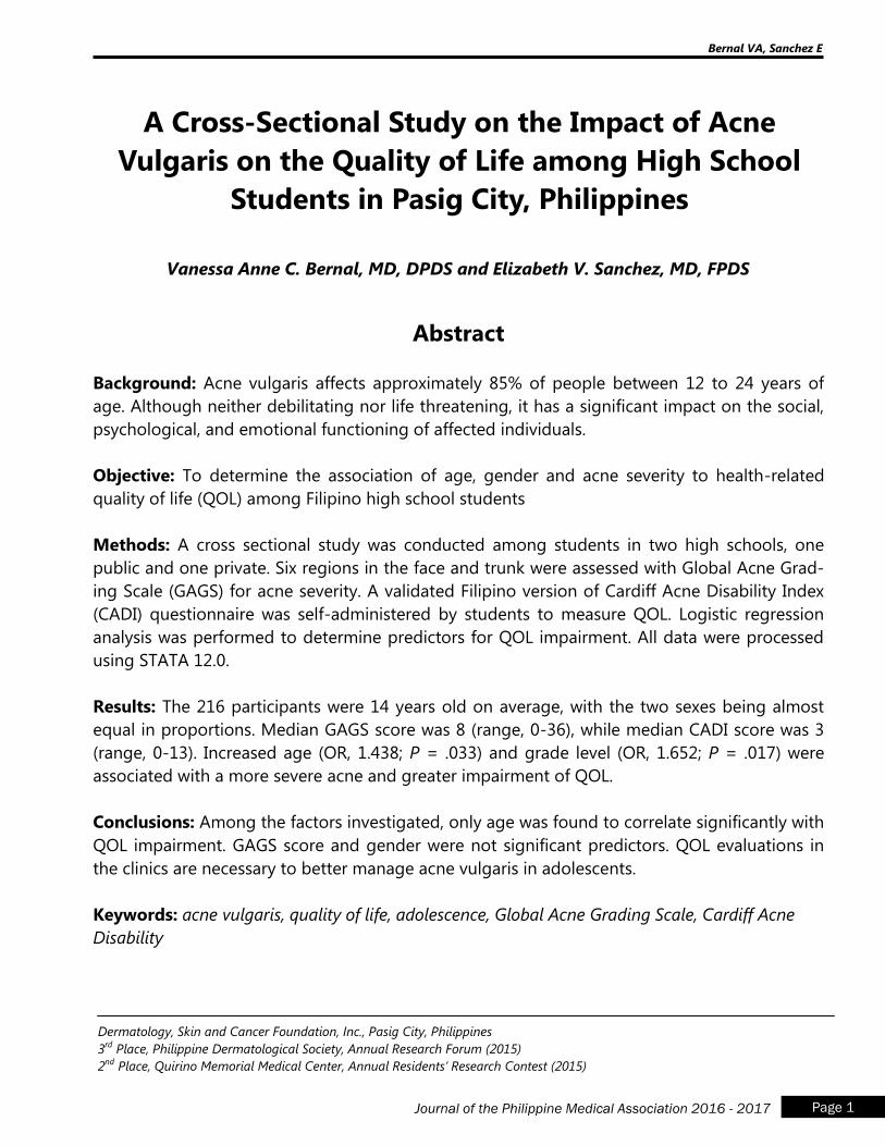
A Cross-Sectional Study on the Impact of Acne
Vulgaris on the Quality of Life among High School
Students in Pasig City, Philippines
Vanessa Anne C. Bernal, MD, DPDS and Elizabeth V. Sanchez, MD, FPDS
Abstract
Background: Acne vulgaris affects approximately 85% of people between 12 to 24 years of
age. Although neither debilitating nor life threatening, it has a significant impact on the social,
psychological, and emotional functioning of affected individuals.
Objective: To determine the association of age, gender and acne severity to health-related
quality of life (QOL) among Filipino high school students
Methods: A cross sectional study was conducted among students in two high schools, one
public and one private. Six regions in the face and trunk were assessed with Global Acne Grad-
ing Scale (GAGS) for acne severity. A validated Filipino version of Cardiff Acne Disability Index
(CADI) questionnaire was self-administered by students to measure QOL. Logistic regression
analysis was performed to determine predictors for QOL impairment. All data were processed
using STATA 12.0.
Results: The 216 participants were 14 years old on average, with the two sexes being almost
equal in proportions. Median GAGS score was 8 (range, 0-36), while median CADI score was 3
(range, 0-13). Increased age (OR, 1.438; P = .033) and grade level (OR, 1.652; P = .017) were
associated with a more severe acne and greater impairment of QOL.
Conclusions: Among the factors investigated, only age was found to correlate significantly with
QOL impairment. GAGS score and gender were not significant predictors. QOL evaluations in
the clinics are necessary to better manage acne vulgaris in adolescents.
Keywords: acne vulgaris, quality of life, adolescence, Global Acne Grading Scale, Cardiff Acne
Disability
Bernal VA, Sanchez E
Page 1 Journal of the Philippine Medical Association 2016 - 2017
Dermatology, Skin and Cancer Foundation, Inc., Pasig City, Philippines
3rd Place, Philippine Dermatological Society, Annual Research Forum (2015)
2nd Place, Quirino Memorial Medical Center, Annual Residents’ Research Contest (2015)
INTRODUCTION
Acne vulgaris is a common disorder of the
pilosebaceous unit that affects approximately 85% of
people between 12 and 24 years of age.1 Lesions
range from mild comedones to severe nodules or
cysts, with possible associated scarring, and are
found mainly on the face and upper trunk. Although
not a life-threatening disease, it can impart significant
impact upon an individual‟s social, psychological, and
emotional functioning.2
Acne prevalence peaks during adolescence, a
crucial stage in growth marked by accelerated, some-
times turbulent, physical, psychological, and social
development. Add this to the “disfigurement” caused
when located in exposed areas of the body, acne vul-
garis has been related to feelings of embarrassment,
reduced self-esteem, low self-assertiveness, poor self-
image, and self-consciousness. The negative feelings
are only exacerbated by taunting and perceptions of
judgment or scrutiny.3,4
Some young patients may
even be so adversely affected as to develop social
inhibition and phobia. Moreover, depression and sui-
cidal ideation were found to be significantly higher
among individuals with acne than among those with
other dermatological problems, such as alopecia
areata and atopic dermatitis, something surpassed
only in cases of severe psoriasis.5
With its considerable psychosocial impact,
recent studies have focused on the assessment of
quality of life (QOL) in patients with acne. Some used
validated instruments, like general health-related
QOL indexes and specific acne-related measures, and
were able to demonstrate a significant negative effect
of acne on QOL. This was noted to be comparable to
when having asthma, epilepsy, or diabetes.6 Results,
however, regarding the correlation of QOL to age,
gender, race, and acne severity have been inconsis-
tent.
In the Philippines, dermatologists, and even
lay people, are aware of the psychosocial and emo-
tional effects of acne vulgaris in the teenage popula-
tion. Yet, this aspect of the disease is seldom noted
or managed. There also exists a lack of quantitative
studies among Filipinos that measure the impact of
the condition in this age group. As such, we aimed to
assess the impact of acne vulgaris on QOL measures
among Filipino high school students, with the goal of
highlighting its psychosocial effects and, hopefully,
improve its current state of management. At the same
time, we provide a baseline study for future investiga-
tions focusing on acne-related QOL in adolescent stu-
dents.
REVIEW OF RELATED LITERATURE
Acne tends to exert negative effect on the
psychological well-being of individuals, as evidenced
by several studies. Uhlenhake et al. noted that the
prevalence of depression among these persons was
three to four times that in the general population.7
Depressive symptoms was also shown to have a posi-
tive correlation with acne severity of secondary school
students, with a higher frequency of suicidal thoughts
and attempts when there was perception of a
“problem acne.”8
A study by Uslu and colleagues
among high school students showed that there was a
direct correlation between the subjective, and not the
objective, severity of acne and symptoms of depres-
sion and anxiety.9 Symptoms of body dysmorphic dis-
order have been associated with acne patients, with
the risk doubled with the requirement of isotretinoin
therapy.10
The level of loneliness and anxiety among
those affected was comparable to that seen in serious
illnesses such as diabetes, cancer, epilepsy, and cystic
fibrosis.11
Effects were also noted to equal that of pa-
tients with asthma.6
Most of these non-dermatologic effects of
acne have been attributed to its prevalence in adoles-
cence, anatomical distribution of lesions, misconcep-
tion regarding etiology, and peer pressure.5 Adoles-
cence being a time of major changes in the physical,
emotional, and social dimensions of a person, this
stage is susceptible to issues concerning appearance,
self-image, and self-esteem. Acne lesions are easily
visible if they appear in areas not usually covered by
clothing, such as the face, upper trunk, and upper
arms. In a survey conducted by Tan et al., people
thought that acne develops from poor skin hygiene,
which may cause feelings of shame and guilt among
patients.12
A cross-sectional study on the impact of acne vulgaris on the quality of life among
high school students in Pasig City, Philippines
Page 2 Journal of the Philippine Medical Association 2016 - 2017
Bernal VA, Sanchez E
Because of the increasing body of evidence
relating acne vulgaris to psychosocial impairment,
recent studies have focused on quantifying the im-
pact of acne on quality of life of patients. According
to the World Health Organization, QOL is an
“individual‟s perception of their position in the con-
text of culture and value systems in which they live
and in relation to their goals, expectations, standards,
and concerns.”13
Measuring the QOL allows the clini-
cian to see the disease in the point of view of the pa-
tient. General health measures can be used to com-
pare skin diseases like acne with other disease enti-
ties that are not necessarily dermatologic. Dermatol-
ogy-specific measures, on the other hand, can be
used to compare the QOL of patients with different
skin diseases. Examples are the Dermatology Life
Quality Index (DLQI) and Skindex. Acne-specific
measures, to which the Cardiff Acne Disability Index
(CADI) belongs, are designed specifically for acne.6
Knowledge of the QOL of patients suffering
from acne allows a clinician to tailor his management
for enhanced patient compliance.6 A greater effect on
QOL has implications on self-esteem, body image,
and relationships with others.14
The psychological,
social, and emotional impact of acne on patients have
been demonstrated with the use of different vali-
dated QOL instruments. The degree of impact of acne
vulgaris varies among different countries, with India
having a lower magnitude of impact as compared to
others.
The correlation between acne severity and
QOL has been studied, yielding varying results.
Tasoula et al. noted that the impact of acne on the
QOL of adolescents in Greece was proportional to its
severity.14
Similarly, Pawin et al. observed a significant
correlation of perceived severity of acne and relation-
ships with friends, as well as with feelings of anger,
sadness, anxiousness, and shame.11
However, in an
investigation of university students in Turkey, no cor-
relation was found between acne severity and QOL.15
It was noted that acne, even of mild severity, can sig-
nificantly affect psychological status.16
The dispropor-
tionality between QOL and acne severity may also be
reflective of some disparity between the viewpoints
of patients and clinicians.17
Variations have been reported on the impact
of acne vulgaris on QOL in opposite sexes. In a study
on adolescents, acne was seen to have more impact
on the QOL of females.14
In contrast, Walker et al.
showed no significant difference between QOL mean
scores of the two sexes.18
As to age, studies have shown that older pa-
tients tend to be more affected by acne vulgaris than
younger patients. In a study by Lasek et al., it was
noted that older patients with acne had a lower QOL
compared to younger counterparts.19
The greater ef-
fect may be due to the prolonged duration of having
acne, poor response to tried treatments, and social
implications in the adult population, such as difficulty
in getting a job.5
OBJECTIVES
General objective
To determine the impact of acne vulgaris to health-
related quality of life among Filipino high school
students
Specific objectives
1. To measure the acne severity of high school stu-
dents by Global Acne Grading Scale (GAGS)
2. To assess quality of life of these students using
Cardiff Acne Disability Index (CADI)
3. To describe how age and grade level, gender, and
acne severity correlate with CADI scores
METHODS
This cross-sectional investigation took place in
two co-educational schools situated in Pasig City, Phil-
ippines. One was public and another was private, and
each had four year levels of high school. Permission to
conduct the study was initially sought from the princi-
pal of each school, as well as the Schools Division Su-
perintendent of Pasig City.
Page 3 Journal of the Philippine Medical Association 2016 - 2017
A standard data collection tool divided into
three sections was utilized in data collection. The first
part asked about the participant‟s gender, age, and
year level. The second portion consisted of the Global
Acne Grading Scale (GAGS). The third part was a vali-
dated Filipino version of the Cardiff Acne Disability
Index (CADI). The first two portions were filled up by
the primary investigator as the examination and in-
terview proper progressed.
Acne severity
For GAGS assessment, six facial and truncal
regions were examined and scored, namely: forehead,
right cheek, left cheek, nose, chin, and chest and up-
per back. A subscore was computed for each region
by multiplying the region-specific factor (2 for each
of forehead, right cheek, and left cheek; 1 for each of
nose and chin; and 3 for the chest and upper back)
with the area‟s most severe lesion (1 for comedone, 2
for papule, 3 for pustule, and 4 for nodule). Local
scores were added to obtain the global score. A total
GAGS score of 1‒18 is considered mild, 19‒30 is
moderate, 31‒38 is severe, and > 38 is very severe.
Quality of life
The CADI is a validated five-item question-
naire designed for use in teenagers and young
adults.21
The answer to every question corresponds to
a score (3, 2, 1, 0 for answers a, b, c, and d, respec-
tively). Individual item scores were summated to ob-
tain the CADI score. Any item left unanswered was
given a score of zero. If more than one item was left
unanswered, the respondent of the questionnaire was
dropped from the study. Indices may range from 0 to
15, with a higher score denoting greater QOL impair-
ment. For this study, a score of 1‒4 was considered
mild impairment, 5‒10 was moderate, and 11‒15 was
severe.
Statistical analysis
Continuous and categorical variables were
presented using descriptive statistics: median (range),
mean ± SD, and count (%). All valid data was included
in the analysis and no imputation was required.
Ordinal regression analysis was utilized to analyze the
association of age and year level, gender, and GAGS
score to the quality of life, as assessed by the CADI
score, among these students afflicted with acne
vulgaris. Data encoding and analysis were performed
using STATA 12.0.
Ethical considerations
The study was designed to be consistent with
the ethical principles contained within the Declaration
of Helsinki and the National Guidelines for Biomedi-
cal/Behavioral Research of the National Ethics Com-
mittee (NEC) of the Philippines. The Institutional Re-
view Board (IRB) approved the protocol before com-
mencement. No deviation from approved procedure
was committed during the study. All participants were
given clear and complete instructions about the
study‟s design and goal before eliciting assent. With-
drawal from the study for any reason was allowed at
any time. The investigators were available for any
questions or clarifications raised. Study participants
were not financially remunerated but rewarded with
medical attention.
Confidentiality of subjects was maintained.
Only the investigators or authorized representatives of
regulatory agencies were allowed to have direct access
to any of the information in the source documents.
Results
There were 216 high school participants re-
cruited, composed of 53% female and with a group
mean (±SD) age of 14.5 ± 1.4 years (Table 1). Grades 8
and 10 were slightly less represented than grades 7
and 9. Of the participants, 98.5% had acne. GAGS me-
dian score was 8 (range: 0‒36), while median CADI
score was 3 (range: 0‒13).
Page 4 Journal of the Philippine Medical Association 2016 - 2017
A cross-sectional study on the impact of acne vulgaris on the quality of life among
high school students in Pasig City, Philippines
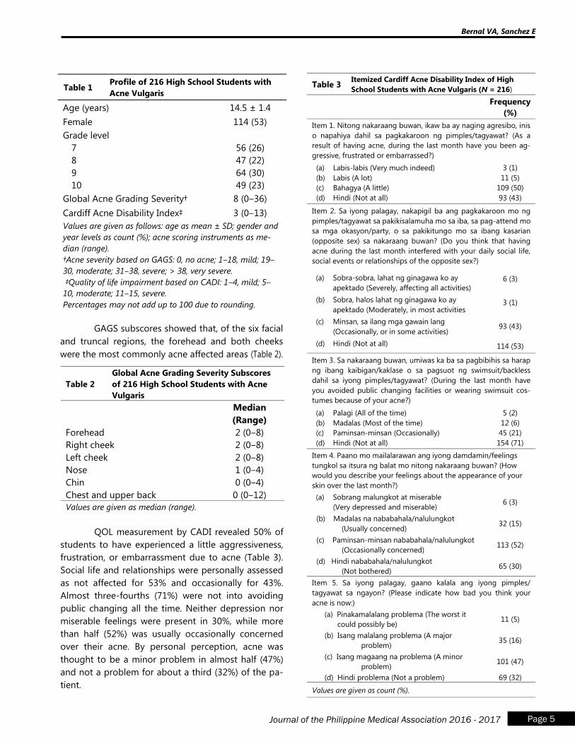
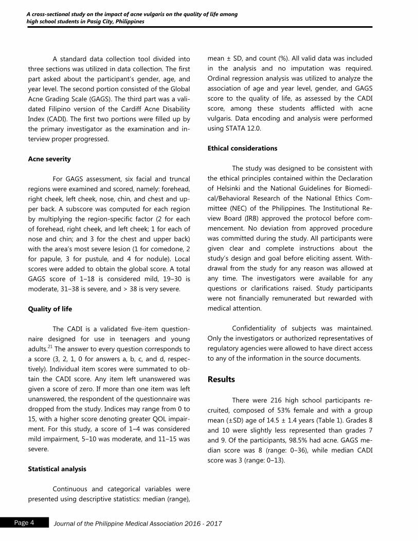
Table 1 Profile of 216 High School Students with
Acne Vulgaris
Age (years) 14.5 ± 1.4
Female 114 (53)
Grade level
7
8
9
10
56 (26)
47 (22)
64 (30)
49 (23)
Global Acne Grading Severity† 8 (0‒36)
Cardiff Acne Disability Index‡ 3 (0‒13)
Values are given as follows: age as mean ± SD; gender and
year levels as count (%); acne scoring instruments as me-
dian (range).
†Acne severity based on GAGS: 0, no acne; 1‒18, mild; 19‒
30, moderate; 31‒38, severe; > 38, very severe.
‡Quality of life impairment based on CADI: 1‒4, mild; 5‒
10, moderate; 11‒15, severe.
Percentages may not add up to 100 due to rounding.
GAGS subscores showed that, of the six facial
and truncal regions, the forehead and both cheeks
were the most commonly acne affected areas (Table 2).
Table 2
Global Acne Grading Severity Subscores
of 216 High School Students with Acne
Vulgaris
Median
(Range)
Forehead 2 (0‒8)
Right cheek 2 (0‒8)
Left cheek 2 (0‒8)
Nose 1 (0‒4)
Chin 0 (0‒4)
Chest and upper back 0 (0‒12)
Values are given as median (range).
QOL measurement by CADI revealed 50% of
students to have experienced a little aggressiveness,
frustration, or embarrassment due to acne (Table 3).
Social life and relationships were personally assessed
as not affected for 53% and occasionally for 43%.
Almost three-fourths (71%) were not into avoiding
public changing all the time. Neither depression nor
miserable feelings were present in 30%, while more
than half (52%) was usually occasionally concerned
over their acne. By personal perception, acne was
thought to be a minor problem in almost half (47%)
and not a problem for about a third (32%) of the pa-
tient.
Table 3 Itemized Cardiff Acne Disability Index of High
School Students with Acne Vulgaris (N = 216)
Frequency
(%)
Item 1. Nitong nakaraang buwan, ikaw ba ay naging agresibo, inis
o napahiya dahil sa pagkakaroon ng pimples/tagyawat? (As a
result of having acne, during the last month have you been ag-
gressive, frustrated or embarrassed?)
(a) Labis-labis (Very much indeed)
(b) Labis (A lot)
(c) Bahagya (A little)
(d) Hindi (Not at all)
3 (1)
11 (5)
109 (50)
93 (43)
Item 2. Sa iyong palagay, nakapigil ba ang pagkakaroon mo ng
pimples/tagyawat sa pakikisalamuha mo sa iba, sa pag-attend mo
sa mga okasyon/party, o sa pakikitungo mo sa ibang kasarian
(opposite sex) sa nakaraang buwan? (Do you think that having
acne during the last month interfered with your daily social life,
social events or relationships of the opposite sex?)
(a) Sobra-sobra, lahat ng ginagawa ko ay
apektado (Severely, affecting all activities)
(b) Sobra, halos lahat ng ginagawa ko ay
apektado (Moderately, in most activities
(c) Minsan, sa ilang mga gawain lang
(Occasionally, or in some activities)
(d) Hindi (Not at all)
6 (3)
3 (1)
93 (43)
114 (53)
Item 3. Sa nakaraang buwan, umiwas ka ba sa pagbibihis sa harap
ng ibang kaibigan/kaklase o sa pagsuot ng swimsuit/backless
dahil sa iyong pimples/tagyawat? (During the last month have
you avoided public changing facilities or wearing swimsuit cos-
tumes because of your acne?)
(a) Palagi (All of the time)
(b) Madalas (Most of the time)
(c) Paminsan-minsan (Occasionally)
(d) Hindi (Not at all)
5 (2)
12 (6)
45 (21)
154 (71)
Item 4. Paano mo mailalarawan ang iyong damdamin/feelings
tungkol sa itsura ng balat mo nitong nakaraang buwan? (How
would you describe your feelings about the appearance of your
skin over the last month?)
(a) Sobrang malungkot at miserable
(Very depressed and miserable) 6 (3)
(b) Madalas na nababahala/nalulungkot
(Usually concerned) 32 (15)
(c) Paminsan-minsan nababahala/nalulungkot
(Occasionally concerned) 113 (52)
(d) Hindi nababahala/nalulungkot
(Not bothered) 65 (30)
Item 5. Sa iyong palagay, gaano kalala ang iyong pimples/
tagyawat sa ngayon? (Please indicate how bad you think your
acne is now:)
(a) Pinakamalalang problema (The worst it
could possibly be) 11 (5)
(b) Isang malalang problema (A major
problem) 35 (16)
(c) Isang magaang na problema (A minor
problem) 101 (47)
(d) Hindi problema (Not a problem) 69 (32)
Values are given as count (%).
Page 5 Journal of the Philippine Medical Association 2016 - 2017
Bernal VA, Sanchez E
Medians of the total GAGS scores were all
within the category of mild acne (Table 4). They were
lowest in grade 7 and highest in grade 10. The ranges
also increased when comparing a grade level against
the one immediately below it (e.g., grade 8 vs. grade
7). The worst condition recorded in grade 7 was mod-
erate acne (GAGS score of 25), while it was severe
acne in grade 10 (GAGS score of 36). For the fore-
head, cheeks, and nose, median GAGS subscores
across all grade levels were 2, 2, 2, and 1, respectively.
Noticeable differences, however, were evident in the
maximum values registered, which generally tended
to increase with greater seniority. The chin displayed
more apparent differences in subscores, with recorded
medians of 0 (range, 0‒2) and 1 (range, 0‒4) for
grades 7 and 10, respectively. Overall GAGS subscore
for the chest and upper back became nonzero only in
the females of grade 10 (median, 3; range, 0‒9).
Median CADI scores increased almost consis-
tently with higher year levels. It was interpreted mild
(median, 1; range, 0-7) for grade 7 and moderate
(median, 5; range, 0-11) for grade 10.
Table 4 Acne Severity and Quality of Life Summarized by Year Level and Gender (N = 216)
Grade 7 Grade 8 Grade 9 Grade 10
Male Female Male Female Male Female Male Female
GAGS† 7 (0‒25) 5 (0‒21) 7.5 (0‒22) 7 (0‒19) 11 (0‒30) 7 (0‒22) 11 (2‒30) 12 (2‒36)
Forehead 2 (0‒6) 2 (0‒6) 2 (0‒4) 2 (0‒6) 2 (0‒6) 2 (0‒8) 2 (0‒8) 2 (0‒8)
Right cheek 2 (0‒6) 2 (0‒4) 2 (0‒4) 2 (0‒4) 2 (0‒8) 2 (0‒4) 2 (0‒8) 2 (0‒8)
Left cheek 2 (0‒6) 0 (0‒4) 2 (0‒6) 2 (0‒6) 2 (0‒6) 2 (0‒8) 2 (0‒6) 2 (0‒8)
Nose 1 (0‒1) 1 (0‒2) 1 (0‒4) 1 (0‒1) 1 (0‒4) 1 (0‒4) 1 (0‒4) 1 (0‒4)
Chin 0 (0‒1) 0 (0‒2) 0.5 (0‒2) 0 (0‒2) 1 (0‒2) 0 (0‒3) 1 (0‒4) 1 (0‒3)
Chest and
upper back 0 (0‒9) 0 (0‒6) 0 (0‒6) 0 (0‒6) 0 (0‒6) 0 (0‒6) 0 (0‒12) 3 (0‒9)
CADI‡ 1 (0‒4) 1 (0‒7) 2 (0‒6) 4 (0‒10) 4 (0‒10) 3.5 (0‒13) 5 (0‒13) 5 (2‒11)
Values are given as median (range).
†GAGS, Global Acne Grading Scale. Interpreted as: 0, no acne; 1‒18, mild; 19‒30, moderate; 31‒38, severe; > 38, very severe.
‡CADI, Cardiff Acne Disability Index. Interpreted as: 1‒4, mild; 5‒10, moderate; 11‒15, severe.
Multiple regression modeling was able to
confirm the trends observed. The relative odds of
having a worse CADI score was 1.438 if with greater
age (95% CI, 1.030‒2.007; P = .022) and 1.652 if with
higher grade level (95% CI, 1.093‒2.497; P = .017).
The odds of greater QOL impairment was increased
by 1.687 times in the female gender, but this did not
achieve statistical significance (Table 5).
Table 5 Regression Model for Cardiff Acne Disabil-
ity Index
Odds
Ratio 95% CI
P-
value
Age 1.438 1.030‒2.007 .033
Female 1.687 0.992‒2.868 .053
Year level 1.652 1.093‒2.497 .017
GAGS score interpretation
Mild vs. No acne 1.056 0.274‒4.077 .936
Moderate vs. No acne 0.920 0.196‒4.302 .915
Severe vs. No acne 3.171 0.048‒207.623 .589
Goodness of fit of the ordinal logistic regression model (r2)
was 11.07%, with P < .0001.
CI, confidence interval. GAGS, Global Acne Grading Scale.
Page 6 Journal of the Philippine Medical Association 2016 - 2017
A cross-sectional study on the impact of acne vulgaris on the quality of life among
high school students in Pasig City, Philippines
DISCUSSION
The CADI is composed of a set of questions
pertaining to different QOL facets. This study showed
that about 8 to 9 out of 10 students answered choice
(c) or (d) for almost all items in CADI, so that, overall,
the median CADI score obtained was 3. This corre-
sponds to mild QOL impairment, comparable to find-
ings obtained in similar studies from India and Malay-
sia.22,23
CADI scores have been found to be signifi-
cantly associated with age, with the older age group
having a higher degree of QOL impairment as com-
pared to the younger age group. This was found to
be similar to the results obtained by Jones-Caballero
et al. where Skindex-29, a QOL questionnaire, was
used in 1,878 patients under the care of 252 clinicians
in Spain.17
Such findings may be due to the noted
increased severity of acne vulgaris in the older age
group (Table 4), similar to findings in Malaysia.23
In a
study conducted by Ismail and Mohammed-Ali (2012)
in Erbil City, they found that older patients, age group
21-25 years, had QOL impairments ranging from
moderate to severe.26
This age-related difference may
be due to acne vulgaris being more prevalent during
adolescence, leading to better social acceptance of
their condition. It was also reported that negative
feelings towards acne vulgaris accumulate from the
self and the community as a person ages.26
Reports have been made of increased psy-
chological impairment with greater acne severity,6,14
but other studies found no correlation.14,15,24,25
Our
results also indicated no association, although me-
dian GAGS and CADI scores increased with higher
grade levels. It should be noted, however, that in this
study, students across grade levels generally had only
mild acne severity that may have positively affected
their CADI scores (Table 1). The lack of correlation,
however, shows that mild acne could have just as
much an effect on adolescents‟ psychology as severe
acne.16
Also, the goodness of the fit of the model
only came up to 11.07%, indicating that there other
factors, internal or external, likely at play in the QOL
of acne patients.
In this study, QOL impairment was more
associated with the female population, though the
difference between genders was not statistically
significant. This may be from women generally having
more concern over their appearances than men.26
However, the lack of significant difference might also
be reflecting how males are now becoming more
aware of their skin condition.23
The inverse phenome-
non was highlighted in a study conducted in Egypt,
wherein males were found to have more impairment in
QOL than females.27
One of the limitations of this study was that it
examined the impact of acne vulgaris on the QOL of
Filipino high school students at a single point in time.
This limitation may be overcome in future studies by
using a prospective study design. Also, increasing the
scope of the study will be able to uncover more fully
the dynamics involved between acne and QOL of teen-
agers.
CONCLUSION
Acne vulgaris was shown to impart a mild QOL
impairment among Filipino high school students, with
older students being significantly more affected than
their younger counterparts. Gender marginally ex-
ceeded significant correlation with QOL, perhaps
pointing to an increased awareness in males of their
skin condition. GAGS score was likewise not a signifi-
cant predictor for QOL impairment, indicating that
mildly afflicted individuals may be affected as much as
those with more severe conditions.
Our results demonstrate the importance of
QOL evaluation in managing adolescent patients with
acne; severity alone may not be enough to adequately
assess the impact of acne on an adolescent. In addi-
tion, it is recommended that future studies on acne
vulgaris, especially interventional ones, should include
quality of life measures as part of the parameters.
ACKNOWLEDGEMENTS
We extend our gratitude to the Philippine De-
rmatological Society for supporting this study. We also
thank our colleagues from the Skin and Cancer Foun-
dation, Inc., for giving helpful insight and assistance.
We would also like to thank Dr. Katrina Car-
mela Marie Belen for her assistance in data gathering
and Dr. Venus Oliva Cloma-Rosales for her help in data
analysis and for her comments that improved the
manuscript.
Page 7 Journal of the Philippine Medical Association 2016 - 2017
Bernal VA, Sanchez E
REFERENCES
1. Bolognia JL, Jorizzo JL, Schaffer JV. Dermatology.
3rd
ed. USA/UK: Elsevier; 2012.
2. Beresniak, A, de Linares Y, Krueger GG, Talarico S,
Tsutani K, Duru G, et al. Validation of a new inter-
national quality-of-life instrument specific to cos-
metics and physical appearance: BeautyqoL ques-
tionnaire. Arch Dermatol 2012;148(11):1275‒82.
3. Magin P, Adams J, Heading G, Pond D. Smith W.
Psychological sequelae of acne vulgaris: results of
a qualitative study. Can Fam Physician 2006
Aug;52:978‒9.
4. Jacobs DG, Deutsch NL, Brewer M. Suicide, de-
pression, and isotretinoin: is there a causal link? J
Am Acad Dermatol 2001 Nov;45(5):S168‒75.
5. Hanna S, Sharma J, Klotz J. Acne vulgaris: more
than skin deep. Dermatology Online Journal
2003;9(3):8.
6. Zip C. The impact of acne on quality of life. Skin
Therapy Letter 2007;12(10):7‒9.
7. Uhlenhake E, Yentzer BA, Feldman SR. Acne
vulgaris and depression: a retrospective examina-
tion. J Cosmetic Dermatol 2010;9:59‒63.
8. Purvis D, Robinson E, Merry S, Watson P. Acne,
anxiety, depression and suicide in teenagers: a
cross-sectional survey of New Zealand secondary
school students. J Paediatr Child Health 2006
Dec;42(12):793‒6.
9. Uslu G, Sendur N, Uslu M, Savk E, Karaman G,
Eskin M. Acne: prevalence, perceptions and
effects on psychological health among adoles-
cents in Aydin, Turkey. J Eur Acad Dermatol
Venereol 2008 Apr;22(4):462‒9.
10. Bowe WP, Leyden JJ, Crerand CE, Sarwer DB,
Margolis DJ. Body dysmorphic disorder symp-
toms among patients with acne vulgaris. J Am
Acad Dermatol 2007 Aug;57(2):222‒30.
11. Pawin H, Chivot M, Beylot C, Faure M, Poli F,
Revuz J, Dreno B. Living with acne: a study of
adolescents' personal experiences. Dermatology
2007;215(4):308‒14.
12. Tan JK, Vasey K, Fung KY. Beliefs and perceptions
of patients with acne. J Am Acad Dermatol
2001;44:439‒45.
13. The World Health Organization quality of life
assessment (WHOQOL): position paper from
the World Health Organization. Soc Sci Med
1995;41:1403‒9.
14. Tasoula E, Gregriou S, Chalikias J, Lazarou D, Da-
nopoulou I, Katsambas A, et al. The impact of
acne vulgaris on quality of life and psychic health
in young adolescents in Greece: results of popu-
lation survey. An Bras Dermatol 2012 Nov‒Dec;87
(6):862‒9.
15. Ilgen E, Derya A. There is no correlation between
acne severity and AQOLS/DLQI scrores. J Derma-
tol 2005 Sep;32(9):705‒10.
16. Dreno B, Alirezai M, Auffret N, Beylot C, Chivot M,
Daniel F, et al. [Clinical and psychological correla-
tion in acne: use of the ECLA and CADI scales].
Ann Dermatol Venereol 2007 May;134(5 Pt 1):451
‒5.
17. Jones-Caballero M, Chrenn MM, Soler B, Pedrosa
E, Penas PF. Quality of life in mild to moderate
acne: relationship to clinical severity and factors
influencing change with treatment. J Eur Acad
Dermatol Venereol 2007 Feb;21(2):219‒26.
18. Walker N, Lewis-Jones MS. Quality of life and
acne in Scottish adolescent schoolchildren: use of
the Children's Dermatology Life Quality Index
(CDLQI) and the Cardiff Acne Disability Index
(CADI). J Eur Acad Dermatol Venereol 2006 Jan;20
(1):45‒50.
19. Lasek RJ, Chren MM. Acne vulgaris and the qual-
ity of life of adult dermatology patients. Arch
Dermatol 1198;134(4):454‒8.
20. Peacock JL, Peacock PJ. Oxford Handbook of
Medical Statistics. 2011. NewYork: Oxford Uni-
versity Press.
21. Cardiff University Section of Dermatology. Quality
of life: Cardiff Acne Disability Index (CADI). Avail-
able from: http://www.dermatology.org.uk/
quality/cadi/quality-cadi.html. Accessibility veri-
fied May 24, 2013.
Page 8 Journal of the Philippine Medical Association 2016 - 2017
A cross-sectional study on the impact of acne vulgaris on the quality of life among
high school students in Pasig City, Philippines
22. Kubba R, Bajaj AK, Thappa DM, Sharma R, Veda-
murthy M, Dhar S, et al. Acne in India: guidelines
for management - IAA Consensus Document.
Indian J Dermatol Venereol Leprol 2009:75(Suppl
1):1‒62.
23. Hanisah A, Omar K, Shah SA. Prevalence of acne
and its impact on the quality of life in school-
aged adolescents in Malaysia. J Primary Health
Care 2009;1(1):20-25.
24. Mosam A, Vawda MB, Gordhan AH, Nkwayana N,
Aboobaker J. Quality of life issues for South Afri-
cans with acne vulgaris. Clin Exp Dermatol
2005;30(1):6-9.
25. Kokandi A. Evaluation of acne quality of life and
clinical severity in acne female adults. Dermatol
Res Pract 2010.
26. Ismail KH, Mohammed-Ali KB. Quality of life in
patients with acne in Erbil city. Health Qual Life
Outcomes 2012;10:60.
27. Abdel-Hafez K, Mahran M, Hofny ER, Mohammed
KA, Darweesh AM, Aal AA. The impact of acne
vulgaris on the quality of life and psychologic
status in patients from upper Egypt. Int J Derma-
tol 2009 Mar;48(3):280-5.
Page 9 Journal of the Philippine Medical Association 2016 - 2017
Bernal VA, Sanchez E
The Appropriate Grading Tool to Assess Acne
Severity in Face-to-face Consultation and Digital
Skin Images*
Karen B. Mabilin-Prieto, MD1
Elizabeth P. Prieto, MD, FPDS2
Jerlyn Maureen P. Servas, MD3
*Selected for Oral Paper Presentation, The 13th Asia Pacific Environmental and Occupational Dermatology Symposium and
The 38th Philippine Dermatological Society Annual Convention. November 4-6, 2015. EDSA Shangri-La Manila, Philippines 1Corresponding Author, Resident in training (during the study), Department of Dermatology, East Avenue Medical Center, Quezon
City, Philippines 2Co-Author, Training Officer, Department of Dermatology, East Avenue Medical Center, Quezon City, Philippines 3Co-Author, Resident in training (during the study), Department of Dermatology, East Avenue Medical Center, Quezon City,
Philippines
Abstract
Background: Acne vulgaris is a multifactorial disease of the pilosebaceous unit affecting
adolescents and young adults. This study examined whether acne assessment measures,
validated for face-to-face use, can be used to assess acne lesions captured from digital images.
The use of digital images is a useful and innovative way to continue delivery of follow-up care
to patients despite barriers such as distance and time.
Objective: To determine the most appropriate acne-grading tool to assess the acne severity
during face-to-face consultations and in digital skin images.
Method: A total of eighteen patients with facial acne vulgaris were included. Two dermatolo-
gists-in-training evaluated the acne-severity during face-to-face visit using validated acne
assessment measures: Total Inflammatory Lesion Count (TILC), LEEDS Technique and Investiga-
tor‟s Global Assessment (IGA). Digital images obtained during initial consult were presented to
the same raters during weeks 6 and 12 and graded accordingly. Cohen‟s kappa was used to
measure agreement between evaluations of two raters and acne grading tools.
The Appropriate Grading Tool to Assess Acne Severity in Face-to-face Consultation and Digital Skin Images
Page 10 Journal of the Philippine Medical Association 2016 - 2017
Results: Raters had significant moderate agreement in ratings using the three grading tools
during face-to-face visit but Investigator‟s Global Assessment had the highest measure of
concordance. Ratings on the digital images by each rater during week 6 and 12 had signifi-
cant substantial agreements based on Total Inflammatory Lesion Count. The inter rater reli-
ability had significant moderate agreement in ratings of digital images during week 12 using
Total Inflammatory Lesion Count and Investigator‟s Global Assessment. Total Inflammatory
Lesion Count had the highest intra rater agreement during ratings of face-to-face and digital
skin images, while LEEDS had the lowest measure of concordance.
Conclusion: All the three acne severity tools may be used for evaluating acne severity during
face-to-face consultation. The Investigator‟s global assessment may best be used for face-to-
face assessments while the total inflammatory lesion count may best be used in digital skin
images.
Commercial funding: N/A
Conflict of interest: N/A
Key words: acne assessment, acne grading tool, digital skin images
Mabilin-Prieto K, Prieto E, Servas JM
Page 11 Journal of the Philippine Medical Association 2016 - 2017
INTRODUCTION
Acne vulgaris is a common dermatological
condition primarily affecting the vast majority of
adolescents and young adults. It is one of the most
common dermatological problems encountered in
Out Patient Clinics of Dermatology. 1
Although easy to diagnose, the polymorphic
nature of acne vulgaris and its varied extent of
involvement do not permit simple evaluation of its
severity. Because the acne lesions may vary in num-
ber during the natural course of the disease, various
measurements have been developed, based on clini-
cal examination and photographic documentation, to
assess the clinical severity of acne vulgaris. 2
The Leeds Revised Acne Grading System,
published in 1998, provides a photographic standard
for acne grading of the face, back and chest. This
system is comprised of 12 facial grades.
In 1996, Lucky et al.6 assessed the reliability
of acne lesion counting. Acne counts were recorded
on a template divided into five facial segments: Right
and left sides of the forehead, right and left cheeks
and chin. The nose and the area around it were
excluded. Counts of each lesion type were recorded
within each segment of the template. Total lesion
count, along with total inflammatory lesions and
comedonal counts, were then calculated. They
concluded that reliability of acne lesion counting was
excellent when performed by the same trained rater
over time.
The grading scale for overall severity by Allen
and Smith Jr. has been the template for global
assessments in many acne trials as an Investigator‟s
Global Assessment (IGA) scale. It was based solely on
descriptive text, not on photographs, and also added
the dimension of increasing extent of facial involve-
ment. It further demonstrated the severity scale
correlated with inflammatory and non-inflammatory
lesion counts. 7
Technology offers new ways to deliver care
to dermatology patients. Dermatology is a specialty
long accustomed to using images in many aspects of
its practice, including education, surveillance, and
patient follow-up. High quality digital cameras are
now widely available. Although initially adopted by
patients as consumer devices, physicians and patients
have been looking to explore the healthcare applica-
tions for such devices. 3
Digital imaging in dermatology care has been
highlighted as a priority area for research for both the
American Telemedicine Association, and internation-
ally by the UK‟s National institute for Health and Clini-
cal Excellence. The application of this methodology
could provide dermatologists a tool for ongoing care,
allowing them to remotely monitor patient progress.3
Digital images and clinical information can be trans-
ferred from patient to clinician over the internet allow-
ing asynchronous, remote assessment.3 Acne patients
are well suited to this approach given that they are
generally technologically able, require ongoing follow-
up but have a non-malignant condition.
Although many acne-grading tools have been
validated, in our institution, the most appropriate acne
-grading tool has not been established when used in
face-to-face setting and in evaluating acne in digital
images.
This study was therefore designed to explore
on the acne assessment measure which, can be used
in the evaluation of acne observed from in person visit
(face-to-face consult) and in digital images.
The importance of this study is to have stan-
dardized system in the grading of acne vulgaris not
only during face-to-face consultation but also in digi-
tal images that we can be used consistently in clinical
practice and research. This is also important to achieve
uniformity in grading because this will further help in
better disease management and more efficacious
treatment. Lastly, the use of digital images is a useful
and innovative way to continue delivery of follow-up
care to patients despite barriers such as distance and
time by determining whether the existing acne assess-
ment measure can also be used in the evaluation of
acne observed from digital images.
The Appropriate Grading Tool to Assess Acne Severity in Face-to-face Consultation and Digital Skin Images
Page 12 Journal of the Philippine Medical Association 2016 - 2017
OBJECTIVES
General Objective
1. To determine the most appropriate acne grading
tool to evaluate the acne severity of patients dur-
ing face-to-face consultations and in digital skin
images at the Dermatology Out Patient Depart-
ment of a Tertiary Government Hospital?
Specific Objectives
1. To determine the demographic profile of the
patients diagnosed with acne vulgaris.
2. To determine the most appropriate acne grading
tool during face-to-face consultation of acne
vulgaris patients.
3. To determine the most appropriate acne grading
tool for digital skin images of acne vulgaris
patient.
4. To compare the grading based on most appro-
priate grading tool that is applicable for both
face-to-face consultation and digital skin images.
METHODS
This is a cross-sectional prospective study on
the appropriate acne-grading tool to assess acne se-
verity in face-to-face consultation and in digital skin
images.
A total of eighteen subjects clinically diag-
nosed with facial acne vulgaris were enrolled in the
study. Those who had drug-induced acneiform erup-
tions and with acne lesions on other body regions
(i.e. trunk, back) were not included in the study.
This study was conducted at the East Avenue
Medical Center Department of Dermatology – Out
Patient Department. Prior to conducting the study,
the research was submitted and approved by the In-
stitutional Ethics Review Board.
The primary investigator (PI), enrolled the
patient in the study, accomplished the data collection
form and have the informed consent forms signed.
The digital images of every enrolled subject were
captured by the primary investigator using a Canon
Ixus 16.1 megapixel digital camera in digital macro
mode without flash. This camera was chosen for its
ease of use and high quality image reproduction. The
light source was an overhead fluorescent with some
natural light through examining room windows.
In the first part of the study, the primary inves-
tigator presented the patient diagnosed with acne vul-
garis to two senior dermatology residents (Raters A
and B) and were graded using Total inflammatory le-
sion count, LEEDS acne assessment technique, and
Investigator‟s Global Assessment. The grading of two
raters were compared to determine the inter-rater
agreement in grading of acne severity during in-
person visits (face-to-face consultation).
In the second part of the study, digital skin
images captured during the initial consult of the pa-
tient were presented to Raters A and B at a later date
(during the 6th and 12th week from the time of re-
cruitment). Photos were evaluated using Total inflam-
matory lesion count, LEEDS acne assessment tech-
nique, and Investigator‟s Global Assessment. To evalu-
ate the inter rater agreement for the digital skin
images, the acne grading of Rater A and Rater B, 12
weeks after recruitment, were compared. To evaluate
the intra rater agreement for the digital images,
grading of the digital skin images by Rater A alone and
Rater B alone during the 6th
and 12th
week after re-
cruitment were compared. To evaluate the intra rater
agreement between face-to-face and digital im-
ages, the grading of Rater A alone and Rater B alone
at the time of recruitment (face-to-face consult) and
the grading of the digital images 12 weeks after were
compared (Figure 1).
For Total inflammatory lesion count (TILC),
raters were asked to count all non-inflammatory and
inflammatory lesions from the set of three facial digital
images and acne grading was based on a tabulated
severity score composed of Mild, Moderate and Se-
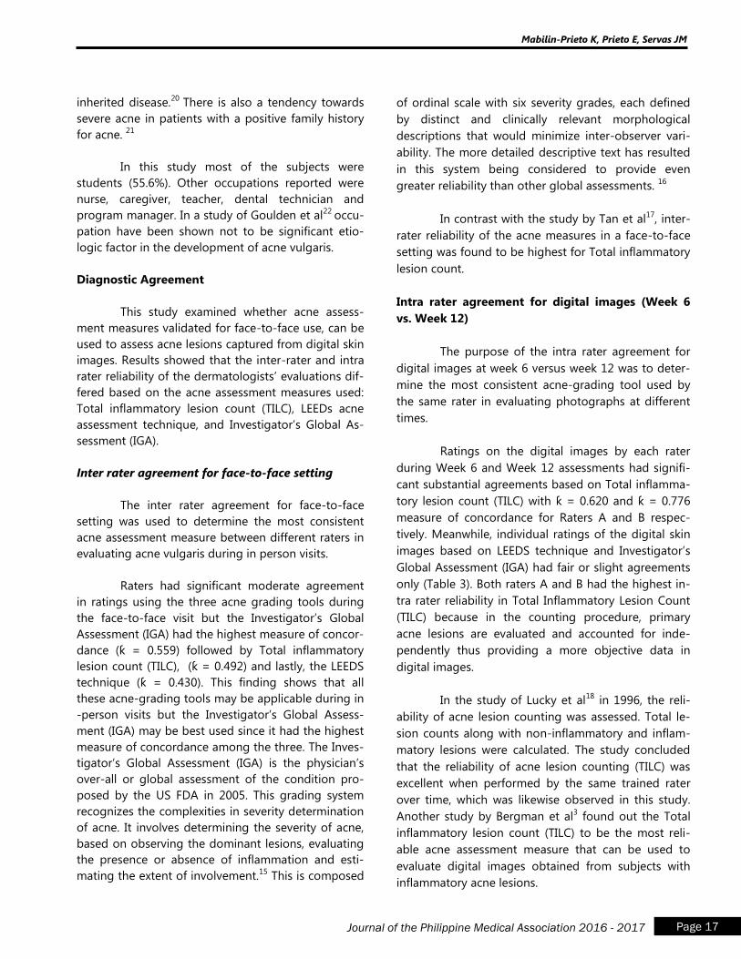
vere. The Leeds Technique was done by comparing the
patient‟s acne severity to a standard photographic
template and assigning a score from 0 to 12. The In-
vestigator‟s Global Assessment (IGA) used a validated
6-point rating scale ranging from clear to very severe.
Both raters were trained with sets of practice images
prior to carrying out their formal assessments.
(Appendix A)
Mabilin-Prieto K, Prieto E, Servas JM
Page 13 Journal of the Philippine Medical Association 2016 - 2017

The Appropriate Grading Tool to Assess Acne Severity in Face-to-face Consultation and Digital Skin Images
Page 14 Journal of the Philippine Medical Association 2016 - 2017
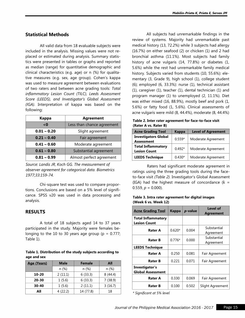
Statistical Methods
All valid data from 18 evaluable subjects were
included in the analysis. Missing values were not re-
placed or estimated during analysis. Summary statis-
tics were presented in tables or graphs and reported
as median (range) for quantitative demographic and
clinical characteristics (e.g. age) or n (%) for qualita-
tive measures (e.g. sex, age group). Cohen‟s kappa
was used to measure agreement between evaluations
of two raters and between acne grading tools: Total
inflammatory Lesion Count (TILC), Leeds Assessment
Score (LEEDS), and Investigator’s Global Assessment
(IGA). Interpretation of kappa was based on the
following:
All subjects had unremarkable findings in the
review of systems. Majority had unremarkable past
medical history (13, 72.2%) while 3 subjects had allergy
(16.7%) on either seafood (2) or chicken (1) and 2 had
bronchial asthma (11.1%). Most subjects had family
history of acne vulgaris (14, 77.8%) or diabetes (1,
5.6%) while the rest had unremarkable family medical
history. Subjects varied from students (10, 55.6%): ele-
mentary (3, Grade 9), high school (1), college student
(6); employed (6, 33.3%): nurse (1), technical assistant
(1), caregiver (1), teacher (1), dental technician (1) and
program manager (1) to unemployed (2, 11.1%). Diet
was either mixed (16, 88.9%), mostly beef and pork (1,
5.6%) or fatty food (1, 5.6%). Clinical assessments of
acne vulgaris were mild (8, 44.4%), moderate (8, 44.4%)
Kappa Agreement
<0 Less than chance agreement
0.01 – 0.20 Slight agreement
0.21 – 0.40 Fair agreement
0.41 – 0.60 Moderate agreement
0.61 – 0.80 Substantial agreement
0.81 – 0.99 Almost perfect agreement
Source: Landis JR, Koch GG. The measurement of
observer agreement for categorical data. Biometrics
1977;33:159-74.
Chi-square test was used to compare propor-
tions. Conclusions are based on a 5% level of signifi-
cance. SPSS v20 was used in data processing and
analysis.
RESULTS
A total of 18 subjects aged 14 to 37 years
participated in the study. Majority were females be-
longing to the 10 to 30 years age group (p = 0.777;
Table 1).
Age (Years) Male Female All
n (%) n (%) n (%)
10-20 2 (11.1) 6 (33.3) 8 (44.4)
20-30 1 (5.6) 6 (33.3) 7 (38.9)
30-40 1 (5.6) 2 (11.1) 3 (16.7)
All 4 (22.2) 14 (77.8) 18
Table 1. Distribution of the study subjects according to
age and sex
Acne Grading Tool Kappa Level of Agreement
Investigators Global
Assessment 0.559* Moderate Agreement
Total Inflammatory
Lesion Count 0.492* Moderate Agreement
LEEDS Technique 0.430* Moderate Agreement
Table 2. Inter rater agreement for face-to-face visit
(Rater A vs. Rater B)
Raters had significant moderate agreement in
ratings using the three grading tools during the face-
to-face visit (Table 2). Investigator‟s Global Assessment
(IGA) had the highest measure of concordance (ƙ =
0.559, p = 0.000).
Acne Grading Tool Kappa p-value Level of
Agreement
Total Inflammatory
Lesion Count
Rater A 0.620* 0.004 Substantial
Agreement
Rater B 0.776* 0.000 Substantial
Agreement
LEEDS Technique
Rater A 0.250 0.081 Fair Agreement
Rater B 0.221 0.071 Fair Agreement
Investigator’s
Global Assessment
Rater A 0.330 0.069 Fair Agreement
Rater B 0.100 0.502 Slight Agreement
Table 3. Intra rater agreement for digital images
(Week 6 vs. Week 12)
* Significant at 5% level
Mabilin-Prieto K, Prieto E, Servas JM
Page 15 Journal of the Philippine Medical Association 2016 - 2017
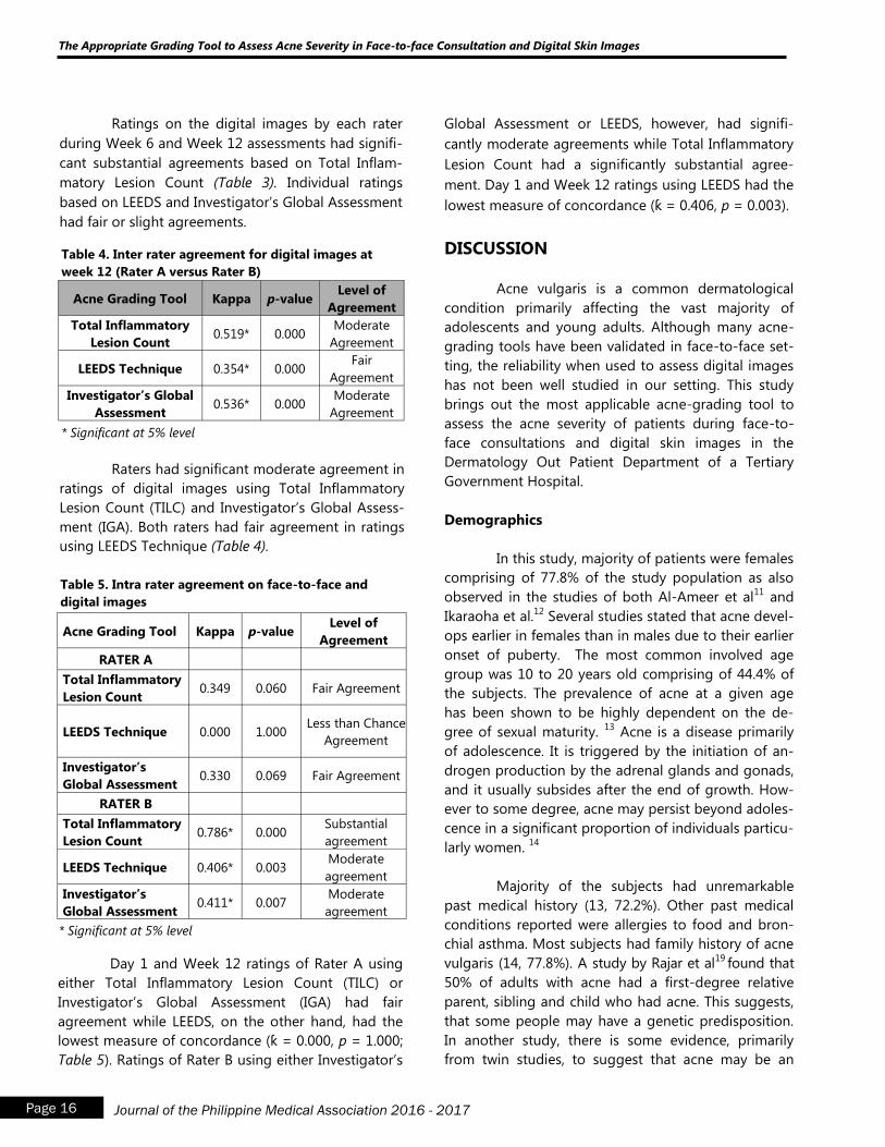
Ratings on the digital images by each rater
during Week 6 and Week 12 assessments had signifi-
cant substantial agreements based on Total Inflam-
matory Lesion Count (Table 3). Individual ratings
based on LEEDS and Investigator‟s Global Assessment
had fair or slight agreements.
Global Assessment or LEEDS, however, had signifi-
cantly moderate agreements while Total Inflammatory
Lesion Count had a significantly substantial agree-
ment. Day 1 and Week 12 ratings using LEEDS had the
lowest measure of concordance (ƙ = 0.406, p = 0.003).
DISCUSSION
Acne vulgaris is a common dermatological
condition primarily affecting the vast majority of
adolescents and young adults. Although many acne-
grading tools have been validated in face-to-face set-
ting, the reliability when used to assess digital images
has not been well studied in our setting. This study
brings out the most applicable acne-grading tool to
assess the acne severity of patients during face-to-
face consultations and digital skin images in the
Dermatology Out Patient Department of a Tertiary
Government Hospital.
Demographics
In this study, majority of patients were females
comprising of 77.8% of the study population as also
observed in the studies of both Al-Ameer et al11
and
Ikaraoha et al.12
Several studies stated that acne devel-
ops earlier in females than in males due to their earlier
onset of puberty. The most common involved age
group was 10 to 20 years old comprising of 44.4% of
the subjects. The prevalence of acne at a given age
has been shown to be highly dependent on the de-
gree of sexual maturity. 13
Acne is a disease primarily
of adolescence. It is triggered by the initiation of an-
drogen production by the adrenal glands and gonads,
and it usually subsides after the end of growth. How-
ever to some degree, acne may persist beyond adoles-
cence in a significant proportion of individuals particu-
larly women. 14
Majority of the subjects had unremarkable
past medical history (13, 72.2%). Other past medical
conditions reported were allergies to food and bron-
chial asthma. Most subjects had family history of acne
vulgaris (14, 77.8%). A study by Rajar et al19
found that
50% of adults with acne had a first-degree relative
parent, sibling and child who had acne. This suggests,
that some people may have a genetic predisposition.
In another study, there is some evidence, primarily
from twin studies, to suggest that acne may be an
Acne Grading Tool Kappa p-value Level of
Agreement
Total Inflammatory
Lesion Count 0.519* 0.000
Moderate
Agreement
LEEDS Technique 0.354* 0.000 Fair
Agreement
Investigator’s Global
Assessment 0.536* 0.000
Moderate
Agreement
Table 4. Inter rater agreement for digital images at
week 12 (Rater A versus Rater B)
* Significant at 5% level
Raters had significant moderate agreement in
ratings of digital images using Total Inflammatory
Lesion Count (TILC) and Investigator‟s Global Assess-
ment (IGA). Both raters had fair agreement in ratings
using LEEDS Technique (Table 4).
Acne Grading Tool Kappa p-value Level of
Agreement
RATER A
Total Inflammatory
Lesion Count 0.349 0.060 Fair Agreement
LEEDS Technique 0.000 1.000 Less than Chance
Agreement
Investigator’s
Global Assessment 0.330 0.069 Fair Agreement
RATER B
Total Inflammatory
Lesion Count 0.786* 0.000
Substantial
agreement
LEEDS Technique 0.406* 0.003 Moderate
agreement
Investigator’s
Global Assessment 0.411* 0.007
Moderate
agreement
Table 5. Intra rater agreement on face-to-face and
digital images
* Significant at 5% level
Day 1 and Week 12 ratings of Rater A using
either Total Inflammatory Lesion Count (TILC) or
Investigator‟s Global Assessment (IGA) had fair
agreement while LEEDS, on the other hand, had the
lowest measure of concordance (ƙ = 0.000, p = 1.000;
Table 5). Ratings of Rater B using either Investigator‟s
The Appropriate Grading Tool to Assess Acne Severity in Face-to-face Consultation and Digital Skin Images
Page 16 Journal of the Philippine Medical Association 2016 - 2017
inherited disease.20
There is also a tendency towards
severe acne in patients with a positive family history
for acne. 21
In this study most of the subjects were
students (55.6%). Other occupations reported were
nurse, caregiver, teacher, dental technician and
program manager. In a study of Goulden et al22
occu-
pation have been shown not to be significant etio-
logic factor in the development of acne vulgaris.
Diagnostic Agreement
This study examined whether acne assess-
ment measures validated for face-to-face use, can be
used to assess acne lesions captured from digital skin
images. Results showed that the inter-rater and intra
rater reliability of the dermatologists‟ evaluations dif-
fered based on the acne assessment measures used:
Total inflammatory lesion count (TILC), LEEDs acne
assessment technique, and Investigator‟s Global As-
sessment (IGA).
Inter rater agreement for face-to-face setting
The inter rater agreement for face-to-face
setting was used to determine the most consistent
acne assessment measure between different raters in
evaluating acne vulgaris during in person visits.
Raters had significant moderate agreement
in ratings using the three acne grading tools during
the face-to-face visit but the Investigator‟s Global
Assessment (IGA) had the highest measure of concor-
dance (ƙ = 0.559) followed by Total inflammatory
lesion count (TILC), (ƙ = 0.492) and lastly, the LEEDS
technique (ƙ = 0.430). This finding shows that all
these acne-grading tools may be applicable during in
-person visits but the Investigator‟s Global Assess-
ment (IGA) may be best used since it had the highest
measure of concordance among the three. The Inves-
tigator‟s Global Assessment (IGA) is the physician‟s
over-all or global assessment of the condition pro-
posed by the US FDA in 2005. This grading system
recognizes the complexities in severity determination
of acne. It involves determining the severity of acne,
based on observing the dominant lesions, evaluating
the presence or absence of inflammation and esti-
mating the extent of involvement.15
This is composed
of ordinal scale with six severity grades, each defined
by distinct and clinically relevant morphological
descriptions that would minimize inter-observer vari-
ability. The more detailed descriptive text has resulted
in this system being considered to provide even
greater reliability than other global assessments. 16
In contrast with the study by Tan et al17
, inter-
rater reliability of the acne measures in a face-to-face
setting was found to be highest for Total inflammatory
lesion count.
Intra rater agreement for digital images (Week 6
vs. Week 12)
The purpose of the intra rater agreement for
digital images at week 6 versus week 12 was to deter-
mine the most consistent acne-grading tool used by
the same rater in evaluating photographs at different
times.
Ratings on the digital images by each rater
during Week 6 and Week 12 assessments had signifi-
cant substantial agreements based on Total inflamma-
tory lesion count (TILC) with ƙ = 0.620 and ƙ = 0.776
measure of concordance for Raters A and B respec-
tively. Meanwhile, individual ratings of the digital skin
images based on LEEDS technique and Investigator‟s
Global Assessment (IGA) had fair or slight agreements
only (Table 3). Both raters A and B had the highest in-
tra rater reliability in Total Inflammatory Lesion Count
(TILC) because in the counting procedure, primary
acne lesions are evaluated and accounted for inde-
pendently thus providing a more objective data in
digital images.
In the study of Lucky et al18
in 1996, the reli-
ability of acne lesion counting was assessed. Total le-
sion counts along with non-inflammatory and inflam-
matory lesions were calculated. The study concluded
that the reliability of acne lesion counting (TILC) was
excellent when performed by the same trained rater
over time, which was likewise observed in this study.
Another study by Bergman et al3 found out the Total
inflammatory lesion count (TILC) to be the most reli-
able acne assessment measure that can be used to
evaluate digital images obtained from subjects with
inflammatory acne lesions.
Mabilin-Prieto K, Prieto E, Servas JM
Page 17 Journal of the Philippine Medical Association 2016 - 2017
Inter rater agreement for digital images at week
12 (Rater A vs. Rater B)
The inter rater agreement for digital images
between two different raters was used to assess
which acne-grading tool will have the highest meas-
ure of concordance in evaluating digital skin images.
Concordance between two dermatologists
receiving photographs alone at Week 12 had signifi-
cant moderate agreement in ratings of digital images
using both Total inflammatory lesion count (ƙ =
0.519) and Investigator‟s Global Assessment (ƙ =
0.536) but with only fair agreement in ratings of digi-
tal images using LEEDS (ƙ = 0.354) (Table 4). This
results showed that Total inflammatory lesion count
and Investigator‟s Global Assessment are applicable
for grading digital skin images but Total Inflamma-
tory Lesion Count may be best used for evaluating
photographs since it had the highest measure of intra
-rater agreement (Rater A : ƙ = 0.620, Rater B: ƙ =
0.776) compared to Investigator‟s Global Assessment
and LEEDS (Table 3).
It was observed in this study that the inter-
rater reliability (Table 3) was less than the intra-rater
reliability (Table 4) for each assessment tool in evalu-
ating digital skin images, reflecting a disparity in
scoring between raters. This underscores the impor-
tance of having the same rater evaluate a subject
throughout the course of a treatment period or
study. 3
Intra rater agreement on face-to-face and digital
images (Day 1 vs. Week 12)
The intra rater agreement for face-to-face
consult and digital images was used to evaluate
which acne grading tool will have a higher measure
of concordance when used by each individual rater
during face-to-face consult and digital skin images.
The ratings of Rater A at Day 1 (face-to-face
setting) and Week 12 (digital images) had the highest
measure of concordance for Total inflammatory le-
sion count (TILC) (ƙ = 0.349) while LEEDS had the
lowest measure of concordance (ƙ = 0.000, p =
1.000). Ratings of Rater B using Total inflammatory
lesion count (TILC) had a significantly substantial
agreement (ƙ = 0.786) however had moderate agree-
ments using either Investigator‟s Global Assessment
(IGA) or LEEDS. The ratings using LEEDS technique also
had the lowest measure of concordance (ƙ = 0.406, p
= 0.003). Individual raters had higher intra rater reli-
ability using TILC in face-to-face setting and digital
skin images.
Several studies have evaluated the use of digi-
tal images to make diagnoses and triage patient refer-
rals.8,9
A study by Bergman et al 3 in 2008, determined
whether a specific assessment tool designed to grade
acne during face-to-face visits could be applied to the
evaluation of digital images. It demonstrated that
digital images of inflammatory acne lesions could re-
liably be evaluated using certain clinical assessment
tools. In this study the Total Inflammatory Lesion
Count was found to be the most reliable acne assess-
ment measure for evaluating digital skin images,
which was consistent in the result of this study.
The LEEDS technique consistently had the
lowest measure of concordance among the other acne
-grading tools. In LEEDS technique each raters were
asked to compare the patient‟s acne severity to a stan-
dard photographic manual with an assigned acne se-
verity score. The study of Tan et al23
in 2013 stated
that the standard LEEDS image could be inadequate in
portraying facial acne grades and cannot accurately
represent the spectrum of the acne severity. There is a
current need for images that will correspond accu-
rately to a categorical acne scale.23
Thus, Total inflam-
matory lesion count (TILC) and Investigator‟s Global
Assessment (IGA) are more objective acne-grading
tools in evaluating acne severity.
CONCLUSION
Acne is a very common dermatologic condi-
tion that affects individuals around the world. The
grading system is one of the main issues because
there is no global standardized grading. Additionally,
grading is a subjective measure that varies from one
dermatologist to another (inter-rater reliability) and it
may also vary for the same dermatologist at different
times for the same patient (intra rater reliability). This
can be attributed to the tedious process of counting
lesions in various types of acne.
The Appropriate Grading Tool to Assess Acne Severity in Face-to-face Consultation and Digital Skin Images
Page 18 Journal of the Philippine Medical Association 2016 - 2017
All the three acne severity tools may be used
for evaluating acne severity during face-to-face con-
sultation. The Investigator‟s global assessment may
best be used for face-to-face assessments while the
total inflammatory lesion count may best be used for
evaluating acne severity in digital skin images.
RECOMMENDATION
This study recommends the use of a com-
bined acne assessment measure, the Investigator‟s
Global Assessment and Total Inflammatory Lesion
Count (Appendix B) that can allow for a more com-
prehensive approach to evaluate acne severity from a
clinical and investigational application. A uniform and
standard acne-grading tool is important to accurately
assess acne severity as this influences treatment se-
lection and assessment of response to therapy.
REFERENCES
1. Tan HH, Topical antibacterial treatments for acne
vulgaris: comparative review and guide to selec-
tion. Am J Clin Dermatol 2004; 5:79-84
2. Witkowski JA, Parish LC. The assessment of acne:
An evaluation of grading and lesion counting in
the measurement of acne. Clin Dermatol
2004;22:394-7.
3. Hagit Bergman, Kenneth Tsai, Su-Jean Seo, Jo-
seph Kvedar and Alice Watson. Remote Assess-
ment of Acne:The Use of Acne Grading Tools to
Evaluation Digital Skin Images. Journal of Tele-
medicin and E-health. 2009:426-430
4. Pochi PE, Shalita AR, Strauss JS, Webster SB. Re-
port of the consensus conference on acne classi-
fication. J. Am. Acad. Dermatol. 24, 495-500
(1991).
5. Burke BM, Cunliffe WJ. The assessment of acne
vulgaris: The Leeds technique. Br J Dermatol
1984;111:83-92.
6. Lucky AW, Barber BL, Girman CJ, Williams J, Rat-
terman J, Waldstreicher J. A multirater validation
study to assess the reliability of acne lesion
counting. J Am Acad Dermatol1996;35:559-65.
7. Jerry KL Tan. Current Measure for the evaluation of
Acne severity. Expert Rev Dermatol. 2008;3 (5):595-
603
8. Du Moulin MF, Bullens-Goessens YI, Henquet CJ et
al. The reliability of diagnosis using store-and-
foreward teledermatology. J Telemed Telecare
2003;9:249-252
9. High WA, Houston MS, Calobrisi SD et al. Assess-
ment of the accuracy of low-cost store-and-
foreward teledermatology consultation. J An Acad
Dermatol 2000: 42:776-783
10. Qureshi AA, Branding-Bennett HA, Giberti S, et al.
Evaluation of digital skin images submitted by pa-
tients who received practical training or an online
tutorial. J Telemed Telecare 2006;12:79-82
11. Al-Ameer AM, Al-Akloby OM. Demographic fea-
tures and seasonal variations in patients with acne
vuglaris in Saudi Arabia: a hospital study. Int J Der-
matol 2002; 41:870-1
12. Ikaraoha Cl, Taylor GOL, Anetor JI, et al. Demo-
graphic features, beliefs and socio psychological
impact of acne vulgaris among its sufferers in two
towns in Nigeria. Online J Health Allied Sc 2005; 4:
Unpaginated.
13. Stathakis V, Killkenny M, Marks R., Descriptive epi-
demiology of acne vulgaris in the community. Aus-
tralas J Dermatol. 1997 Aug; 38 (3): 115-23.
14. Goulden V, Stables GI, Cunliffe WJ. Prevalence of
facial acne in adults. J Am Acad Dermatol. 1999;
41; 577-580
15. Adityan B, Kumari R, Thappa DM. Scoring systems
in acne vulgaris. Indian J Dermatol Venereol Leprol
2009; 75:323-6.
16. Pochi PE, Shalita AR, Strauss JS, Webster SB,
Cunliffe WJ, Katz HI, et al. Report of the Consensus
Conference on Acne Classification. Washington,
D.C., March 24 and 25, 1990. J Am Acad Dermatol.
1991;24; 495-500.
17. Tan JK, Fung K, Bulger L. Reliability of dermatolo-
gists in acne lesion counts and global assessments.
J Cutan Med Surg 2006; 10:160-165.
Mabilin-Prieto K, Prieto E, Servas JM
Page 19 Journal of the Philippine Medical Association 2016 - 2017
18. Lucky AW, Barber BL, Girman CJ, Williams J, Rat-
terman J, Waldstreicher J. A multirater validation
study to assess the reliability of acne lesion
counting. J Am Acad Dermatol 1996; 35:559-65.
19. Rajar, UDM, Majeed R, Sheikh F, Sheikh I, Sid-
dique AA, Kumar S. Scarring in acne patients – A
study at Isra University Hyderabad. J Pak Med
Assoc. 2009 Aug; 59(8):525-7
20. Goulden V, McGeown CH, Cunliffe WJ. The
familial risk of adult acne: a comparison between
first-degree relatives of affected and unaffected
individuals. Br J Dermatolo. 1999 August; 141
(2):297-300. 1009 August; 59 (8) : 525-527
21. Dreno B, Poli F. Epidemiology of acne. Dermatol-
ogy. 2003;206; 7-10
22. Goulden V, Clark SM,Cunliffe WJ. Post-adolescent
acne: a review of clnical features. BR J Dermatol
1997;136:66-70
23. Tan JK, Zhang X, Jones E, Bulger L. Correlation of
photographic images from the Leeds revised
acne grading system with a six-category global
acne severity scale. Journal of the European
Academy of Dermatology and Venereology.
March 1, 2013; 27 (3); e414-9.
APPENDIX A ACNE GRADING TOOLS
I. Total Inflammatory Lesion Count (TILC)
Count all non-inflammatory and inflammatory lesions and grade based on the severity score below:
Severity Definition
Mild <20 comedones or
<15 inflammatory lesions or
<30 total lesions
Moderate 20 to 100 comedones, or
15-50 inflammatory lesions, or
30 to 125 total lesions
Severe >5 cysts, or
total comedone count >100, or
total inflammatory lesion count >50 or
>125 total lesions
II. LEEDS Technique
Compare the patient‟s acne severity from the digital images to the standard photographic manual by
assigning a score.
The Appropriate Grading Tool to Assess Acne Severity in Face-to-face Consultation and Digital Skin Images
Page 20 Journal of the Philippine Medical Association 2016 - 2017
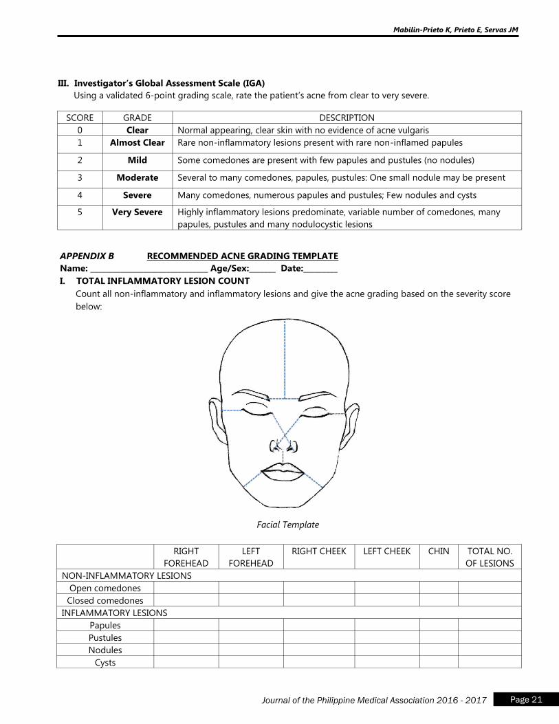
III. Investigator’s Global Assessment Scale (IGA)
Using a validated 6-point grading scale, rate the patient‟s acne from clear to very severe.
SCORE GRADE DESCRIPTION
0 Clear Normal appearing, clear skin with no evidence of acne vulgaris
1 Almost Clear Rare non-inflammatory lesions present with rare non-inflamed papules
2 Mild Some comedones are present with few papules and pustules (no nodules)
3 Moderate Several to many comedones, papules, pustules: One small nodule may be present
4 Severe Many comedones, numerous papules and pustules; Few nodules and cysts
5 Very Severe Highly inflammatory lesions predominate, variable number of comedones, many
papules, pustules and many nodulocystic lesions
APPENDIX B RECOMMENDED ACNE GRADING TEMPLATE
Name: _______________________________ Age/Sex:_______ Date:_________
I. TOTAL INFLAMMATORY LESION COUNT
Count all non-inflammatory and inflammatory lesions and give the acne grading based on the severity score
below:
Facial Template
RIGHT
FOREHEAD
LEFT
FOREHEAD
RIGHT CHEEK LEFT CHEEK CHIN TOTAL NO.
OF LESIONS
NON-INFLAMMATORY LESIONS
Open comedones
Closed comedones
INFLAMMATORY LESIONS
Papules
Pustules
Nodules
Cysts
Mabilin-Prieto K, Prieto E, Servas JM
Page 21 Journal of the Philippine Medical Association 2016 - 2017
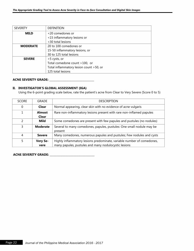
SEVERITY DEFINITION
MILD <20 comedones or
<15 inflammatory lesions or
<30 total lesions
MODERATE 20 to 100 comedones or
15-50 inflammatory lesions, or
30 to 125 total lesions
SEVERE >5 cysts, or
Total comedone count >100, or
Total inflammatory lesion count >50, or
125 total lesions
ACNE SEVERITY GRADE: __________________________________
II. INVESTIGATOR’S GLOBAL ASSESSMENT (IGA)
Using the 6-point grading scale below, rate the patient‟s acne from Clear to Very Severe (Score 0 to 5)
SCORE GRADE DESCRIPTION
0 Clear Normal appearing, clear skin with no evidence of acne vulgaris
1 Almost
Clear
Rare non-inflammatory lesions present with rare non-inflamed papules
2 Mild Some comedones are present with few papules and pustules (no nodules)
3 Moderate Several to many comedones, papules, pustules: One small nodule may be
present
4 Severe Many comedones, numerous papules and pustules; Few nodules and cysts
5 Very Se-
vere
Highly inflammatory lesions predominate, variable number of comedones,
many papules, pustules and many nodulocystic lesions
ACNE SEVERITY GRADE: __________________________________
The Appropriate Grading Tool to Assess Acne Severity in Face-to-face Consultation and Digital Skin Images
Page 22 Journal of the Philippine Medical Association 2016 - 2017
A Clinical Trial on the Safety and Efficacy of Oil of
Bergamot 30% and Sun Exposure combination
versus Sun Exposure alone in the Management of
Residual Hypomelanosis of Pityriasis Versicolor
Ma. Eleanor Cathryn DR. Salonga, M.D, DPDSa
Ma. Luisa Abad-Venida, M.D, FPDSb
Lillian Lopez-Villafuerte, M.D, FPDSb
a Primary author , Department of Dermatology, Jose R. Reyes Memorial Medical Center b Consultants, Department of Dermatology, Jose R. Reyes Memorial Medical Center
Abstract
This study compared the safety and efficacy of Oil of Bergamot with sun exposure, and
that of sun exposure alone in the management of the resulting hypomelanosis of Pityriasis
versicolor. There was indeed a difference in the melanin and erythema indices between the two
treatments with results favoring the former in terms of improvement of the condition of interest.
Introduction: Pityriasis versicolor is a skin problem in a tropical country like the Philippines. It is
characterized by pinkish to hypopigmented patches usually affecting trunk and extremities. The
residual hypomelanosis of this condition is a common concern for most patients. There is no
known treatment for it except to wait for its spontaneous resolution for a period of six months or
more, a solution which is unacceptable for many. Oil of Bergamot is an extract from the plant
Citrus bergamia containing the active ingredients of 5-methoxypsoralen and 8-genoxypsoralen,
members of psoralen compound which is currently being used along with UVA (PUVA) for
several dermatologic conditions such as Vitiligo and Progressive Macular Hypomelanosis.
It is a photosensitizing and phototoxic agent which aids in the turning back of melanocytes in
conditions characterized by reduced pigment cells.
Methods: This was a single-blind study among patients aged 11 to 65 years old diagnosed with
Pityriasis versicolor clinically and through a confirmatory KOH examination. There were 28
patients that were included in the study with no dropouts. Patients were instructed to split their
backs into halves, right and left, one side subjected to application of oil followed by 30-minute
sun exposure while the other was solely subjected to sun exposure, with the assignment of
treatment sides decided by a third party. The duration of treatment was four weeks with weekly
determination of melanin and erythema indices via a mexameter.
Page 23 Journal of the Philippine Medical Association 2016 - 2017
Salonga MEC, Abad-Venida ML, Lopez-Villafurete L
A Clinical Trial on the Safety and Efficacy of Oil of Bergamot 30% and Sun Exposure combination versus
Sun Exposure alone in the Management of Residual Hypomelanosis of Pityriasis Versicolor
Page 24 Journal of the Philippine Medical Association 2016 - 2017
Results: ANOVA of repeated measures was used to determine if there was a difference between
the two treatment groups for both the dependent variables melanin and erythema. With a p
value of <0.0001, the melanin and erythema significantly improved after the treatment of oil of
bergamot and sun exposure.
Conclusion: The combination of Oil of bergamot and 30-minute sun exposure significantly in-
creased the melanin and erythema levels of the hypomelanotic lesions brought about by Pityria-
sis versicolor as compared to sun exposure alone. Adverse reactions were limited and were of
mild intensity demonstrating that a short contact with the oil is sufficient enough to produce the
needed erythema but inadequate in yielding strong hypersensitivity reactions.
Keywords: Hypomelanosis; Pityriasis versicolor; Oil of bergamot; Mexameter; melanin index;
erythema index
INTRODUCTION
Skin color has always been an important issue
since time immemorial. Many individuals, particularly
during olden times, would look primarily on one‟s
complexion, unfortunately deducing judgments solely
from it. Though the latter seems no longer taking place
these days, the habit of giving so much value on a
person‟s skin appearance still holds true up to this
modern time. This is where the role of us, dermatolo-
gists, comes in. We are entrusted to take well care of
the largest organ system in a human‟s body.
Pigmentation whether in the form of hyper- or
hypomelanosis is a taxing dermatologic problem to
both the clinician and patient. It is a condition which on
the part of the former, is extremely difficult to manage
while for the latter, there is exhaustion on the waiting
process for its resolution. While there are several prod-
ucts and procedures available in the market to address
skin darkening, there is barely anything known to public
for its opposite. Several dermatoses bring about altera-
tion in skin color. For hyperpigmentation, causes vary
from sun damage to hypersensitivity reactions to
contact dermatitis. As for hypopigmentation, sources
include inflammatory dermatoses such as psoriasis and
seborrheic dermatitis, dermatophytoses, mycosis
fungoides, vitiligo and many others.
In the Philippines, being a tropical country, Sap-
rophyte infections are not infrequent, with Pityriasis
versicolor being one of the most commonly seen cases.
It is characterized by slightly scaling macules that can
be hypopigmented, pink or salmon-colored or hyper-
pigmented. This superficial mycosis which is common
among young adults is said to be caused by the yeast
Malasezzia sp., of which M. globosa and M. sympodialis
most frequently have been identified in scales of le-
sions.1 Current management for this infection includes
several topical antifungal preparations such as Selenium
sulfide, 2.5%, Ketoconazole, Ciclopirox etc. However,
these treatments while they can manage to relieve the
redness and pruritus, the residual hypopigmentation
remains to be unsolved.
Oil of Bergamot, an essential oil from Citrus
bergamia (Bergamot orange) which is being used
in various expensive fragrances, is proven to be an
effective treatment for Vitiligo. Bergaptene, also
known as 5-methoxypsoralen and Bergamotin, also
called as 5-Geranoxypsoralen, are the major furocou-
marin contents of this oil, and regarded as highly
photoreactive psoralens.2 Therefore, products contain-
ing Bergamot oil are said to be photosensitizing and
highly phototoxic causing erythema and eventual
repigmentation to occur in vitiligo patients.
On the other hand, Narrowband UVB light
therapy (NB-UVB) is a treatment which uses the
optimal part of the UVB light spectrum to cause
repigmentation of one‟s natural skin coloring.3 This
mechanism of action is currently taken into advantage
as treatment for hypopigmented dermatoses such as
vitiligo and progressive macular hypomelanosis.
Therefore, it is the aim of this study to work
around the strong points of these two treatments as a
means of finding an effective and safe treatment to
the challenging repercussions of Pityriasis versicolor
which is decreased skin pigmentation. However, with
the highly demanding requirement in the frequency of
hospital visits as well as the cost of Narrowband UVB
treatment, in this experimental research, sun exposure
will serve as its alternative.
REVIEW OF RELATED LITERATURE
Pityriasis versicolor (also known as Tinea versi-
color) is a common skin complaint characterized by
various pigmentary changes of the skin, usually affect-
ing the chest, back, neck and arms. The patches may
be pink, coppery brown or paler than surrounding skin,
which can often be associated with pruritus. The hy-
perpigmentation or hypopigmentation of skin de-
pends on the outcome of interactions between
Malassezia yeasts and the skin, such as lipoperoxida-
tion process, stimulus of inflammatory cell to melano-
cytes, and increased thickness of keratin layer.4
Salonga MEC, Abad-Venida ML, Lopez-Villafurete L
Page 25 Journal of the Philippine Medical Association 2016 - 2017
Confirmation of diagnosis is made in two ways:
The first one is via 10 % to 20% Potassium Hydroxide
(KOH) microscopy wherein hyphae and yeast cells are
seen resembling the so-called „spaghetti and meat-
balls‟. The second one is by means of Wood‟s light
(long wave Ultraviolet A) which shows a yellow-green
fluorescence over the affected areas among one out of
three patients.5
Pityriasis versicolor is more common in hot,
humid climates or in those who sweat heavily, so it may
recur each summer. It does not appear to predispose
affected areas to sunburn even when it causes pale
white marks.6 It is here where the cosmetic concern
comes in, the consequent hypopigmented patches over
the affected areas.
The oil of Bergamot, an essential oil extracted
from Citrus bergamia or Bergamot orange which is a
fragrant fruit the size of an orange, with a yellow color
similar to a lemon, is widely being used in the perfume
industry. The oil has the ability to combine with an
array of scents to form a bouquet of aromas which
complement each other. Approximately one third of all
men's and about half of women‟s perfumes contain
bergamot essential oil.7
Having 5-methoxypsoralen and 5-
geranoxypsoralen as its components, Bergamot oil is
both photosensitizing and phototoxic. Its photosensi-
tizing properties aid in turning back on melanocytes in
conditions with reduced amount of the said pigment
cells given the proper stimulation via ultraviolet light.8
Taking lead of this property the oil is being used as
one of the treatment for vitiligo, a skin condition
characterized by depigmented patches due to destruc-
tion of melanocytes as well as of other dermatologic
conditions characterized by hypopigmentation. In a
study done by Kinley et.al they made use of hairless
mice to assess the epidermal melanogenesis induction
of topical furocoumarins (active ingredient of Bergamot
oil) along with UVA radiation. Each subject was sub-
jected into a 12-day application of topical furocoumarin
followed by exposure to subphototoxic doses of UVA.
The results of which have shown evidences of
melanogenesis such as increased density and size of
melanocytes.9 In another experimental study of
Romano et.al the in vitro activity of Bergamot oil
against clinical isolates of dermatophytes was deter-
mined. The outcome of which suggested its potential
use against common species of dermatophytes.10
In a
similar study done by the same group of researchers,
they measured the Minimum Inhibitory Concentration
(MIC) of bergamot oil against clinical isolates of Can-
dida species from which the group have found a prob-
able usage of Citrus bergamia as a topical treatment
against Candidal infections.11
These studies therefore
make the Oil of Bergamot more appealing in the man-
agement of Pityriasis versicolor not only in terms of its
value in the restoration of skin color but as well as the
possibility of decreasing the growth and thus recur-
rence, of Malasezzia infection.
Interestingly, similar hypomelanotic conditions
such as vitiligo and progressive macular hypomelano-
sis can both be managed with either Narrowband UVB
or topical/oral Psoralen and UVA (PUVA). In a study
done by Wu CS et. al., they have proposed that PUVA
is best used in the active stages of vitiligo as it slows
down the destruction of melanocytes (MC) thereby
creating a favourable environment for MCs to survive.
On the other hand, NBUVB irradiation will best suit
stable vitiligo with its ability to stimulate both prolif-
eration and migration of MCs.12
Conversely, Progres-
sive Macular Hypomelanosis (PMH), a condition char-
acterized by ill-defined, nummular, hypopigmented
spots on the skin due to decreased pigmentation of
epidermis has been shown to be effectively and safely
treated using NBUVB. In a small uncontrolled and non
-blinded clinical trial done by Kim MB et.al, 9 out of 16
patients (56.2%) experienced more than 90% repig-
mentation while 13 of 16 patients (81.3%) had at least
50% repigmentation after several weekly sessions of
NBUVB.13
While in 2010, Duarte et al. reviewed pa-
tients seen in a phototherapy clinic between 1997 and
2008 to evaluate the therapeutic response to PUVA
photochemotherapy or Narrowbandband UVB. No
significant difference between these two treatments
after 16 sessions or three months of treatment has
been found with majority of the patients showing
more than 50% of skin repigmentation, with 65% clas-
sified as cured or much improved.14
A Clinical Trial on the Safety and Efficacy of Oil of Bergamot 30% and Sun Exposure combination versus
Sun Exposure alone in the Management of Residual Hypomelanosis of Pityriasis Versicolor
Page 26 Journal of the Philippine Medical Association 2016 - 2017
Page 27 Journal of the Philippine Medical Association 2016 - 2017
Whereas, the exact mechanism of hypopig-
mentation in pityriasis versicolor is not known with
recent discoveries pointing towards a role of trypto-
phan-derived metabolites of Malassezia furfur, simi-
larly it can take advantage of phototherapy relying on
its ability to induce maturation of existent melano-
cytes thus accelerating the repigmentation process.15
Both UVA and UVB comprised a certain por-
tion of sun‟s rays thereby making direct exposure to
sunlight, a good source of UV radiation. In particular,
UVB are the main rays which are said to cause sun-
burn. However, excessive exposure to sunlight can
increase an individual‟s predisposition to skin cancers
such as melanoma and squamous cell carcinoma. In
as much as UV radiation is warranted amongst pa-
tients with dermatologic conditions which can benefit
from exposure to phototherapy but with limited
ability to obtain such in a hospital facility due to lack
of time and/or for financial reasons making direct
sunlight exposure a good alternative, it is of impor-
tance still to advise avoidance of sun from 10 am to 4
pm when UVR intensity is at its peak in so doing,
eluding the harmful consequences of too much sun
exposure.16
Accordingly, proving the efficacy of these
different modes of treatment for pigmentary disor-
ders will be difficult if done in a subjective way.
Therefore, to minimize this point of error, the re-
searcher will be using a device used to measure skin
color that would provide a reproducible means and
sensitive means of quantifying small skin color differ-
ences. Park es et al. in 2006 have concluded that the
Mexameter is an objective pigment-measuring device
which can be used to achieve a more accurate diag-
nosis of hypopigmentary disorders, and that the rela-
tive melanin index which represents the relative pig-
ment levels, might be a more effective parameter
than the melanin index (MI) itself for comparing pig-
mentation differences.17
In conclusion, the researcher took advantage
of the strong points of these literatures with some
modifications in order to allow its feasibility in the
current setting of the trial with much consideration to
the study patients.
GENERAL OBJECTIVE
To compare the safety and efficacy of Oil of
Bergamot 30% and sun exposure combination versus
sun exposure alone in the restoration of the normal
skin color in post-hypopigmented lesions of Pityriasis-
versicolor.
SPECIFIC OBJECTIVES:
1. To measure the pigmentation level of the skin by
means of Mexameter at weeks 0,1,2,3, and 4 of
treatment.
2. To discern the relationship between Melanin Index
and the erythema measurement.
3. To determine the adverse effects associated with
the use of both treatments via patient‟s self--
assessment forms.
METHODOLOGY
STUDY DESIGN
A single-blinded clinical trial on the safety and
efficacy of oil of Bergamot 30% and sun exposure
combination versus sun exposure in the management
of the accompanying hypomelanosis of Pityriasis ver-
sicolor among normal healthy patients with Pityriasis
versicolor, who were diagnosed at the Dermatology
Out-Patient Department of Jose R. Reyes memorial
Medical Center.
STUDY POPULATION
Normal healthy male and female patients
aged 11 to 65 years old with Pityriasis versicolor over
back area. Diagnosis was made by means of clinical
history and examination and confirmed by means of
Potassium hydroxide (KOH) examination. Duration and
history of prior treatment was not considered. A total
of 28 patients were enrolled in this study.
Exclusion criteria include known hypersensitiv-
ity reaction to any form of fragrances (known to con-
tain Bergamot oil), and concomitant medical condi-
tions known to be exacerbated by Ultraviolet exposure
such as Lupus Erythematosus. Pregnant and breast-
feeding mothers were also excluded from the study.
Salonga MEC, Abad-Venida ML, Lopez-Villafurete L
MATERIALS
The oil of Bergamot 30% was purchased by the
researcher from a local supplier packaged in a plastic
opaque jar ensuring its stability. Sunblock gel from
another local company was also provided by the re-
searcher to warrant the skin protection of non-involved
skin areas of all study patients.
RANDOMIZATION, TREATMENT ALLOCATION,
AND BLINDING
The randomization of back areas of the sub-
jects was done by a third party via computer-
generated method. The oil was handed to the subject
by the said party who re-instructed the patient as to its
application and as to which side should it be placed. In
this experiment, the researcher as well as the physician
performed the mexameter readings were blinded.
STUDY INTERVENTION AND ASSESSMENT
Diagnosed patients were treated primarily with
topical and/or oral antifungal opted by the primary
clinician. A repeat KOH was performed to check for
clearance of dermatophyte infection.
On follow-up, the residual hypomelanosis was
managed. Each patient had the affected body area
divided into two areas. One area was subjected to the
application of Bergamot oil 30% 30 minutes prior to
sun exposure while the other side was directly exposed
to the sun without any other intervention. Both sides
had exposure between 6 am to 9 am to avoid the
intense burning heat past the given time. In addition,
patients were instructed to apply sunblock gel on
normal skin, 30 minutes prior to going out while the
involved areas with oil were washed off with water
after heat exposure prior to the application of sunblock
gel.
To address the possible result-altering external
factors, every patient was instructed to have avoidance
of sun exposure after the given time and to protect
their skin upon stepping outside via use of umbrella
and wearing of proper shirt at all times.
On doing the mexameter reading, two repre-
sentative lesions were chosen from each side by the
researcher and by the third party who handled the said
device. The representative lesions that were chosen
were based on which portion is the whitest or most
hypopigmented of all. Photos were taken on every
reading to mark the representative lesions.
CLINICAL ASSESSMENT
Baseline photos and mexameter readings were
taken prior to the initiation of therapy. Patients were
asked to follow-up on a weekly basis for a period of 4
weeks wherein a questionnaire regarding the adverse
effects was requested to be answered. Repeat photog-
raphy and Mexameter readings were done on every
follow-up.
Primary endpoint was the restoration of normal
skin color as per evidence of clinical photographs and
the increase in the level of pigmentation via the
mexameter. The grading were as follows: Grade 1 for <
50% of repigmentation or interpreted as slightly
improved; Grade 2 for 50% to 79% of repigmentation or
moderately improved; Grade 3 for 80% to 99% or much
improved; Grade 4 for 100% or cured. Secondary
endpoint was the number of weeks needed to achieve
at least 50% of re-pigmentation.
STOPPING GUIDELINES
A subject would have been withdrawn from
the study upon any signs of severe irritation such as
blistering, intense erythema and pruritus. Patients who
used other topical preparations or those who would
were unable to comply with the proper sun exposure as
well as the wearing of protective gears would have been
taken out of the study.
SAMPLE SIZE
Based on the 2013 census of the JRRMMC
Department of Dermatology, Out Patient Department,
237 cases of Pityriasis versicolor were seen out of
the total of 30,328 patients. The researcher set the
maximum permissible error at 5%, α at 5% and confi-
dence level at 95%. Open Epi results indicated that a
minimum of 28 patients was needed to achieve a 95%
A Clinical Trial on the Safety and Efficacy of Oil of Bergamot 30% and Sun Exposure combination versus
Sun Exposure alone in the Management of Residual Hypomelanosis of Pityriasis Versicolor
Page 28 Journal of the Philippine Medical Association 2016 - 2017
level of confidence, precision of 5% given an estimated
with outcome to be at 80%. The researcher needed
minimum of 28 patients to achieve a 95% level of
confidence, precision of 5% considering a 20% drop
outs.
RESULTS
DISCUSSION
There were 28 patients enrolled in the study.
Majority of which were males comprising 78.57% of the
study population. The mean age of patients was 23.3
years old. In the study, each subject‟s back area was
divided into two. One half of the back was subjected to
application of Oil of Bergamot followed by 30-minute
sun exposure while the other half was solely exposed to
sun for a similar duration as the former. The mean
percentage of melanin was 14.46% and erythema was
11.98% on baseline (see table 1).
ANOVA of repeated measures was used to
determine if there was a difference between the two
treatment groups for both the dependent variables
melanin and erythema. With a p value of <0.0001, it
can be concluded that the melanin and erythema
significantly improved after the treatment of oil of
bergamot and sun exposure (see table 2). A post hoc
comparison (tukey) test was used to determine at which
week showed marked improvement. With p-values of <
0.0001 for all the treatment weeks, it showed that using
the combination of oil of bergamot and sun exposure
significantly increased the percentage of erythema and
melanin.
Fundamentally, the increase in the melanin and
erythema values is projected amongst the areas sub-
jected into the combination of the oil and sun expo-
sure. As mentioned in the previous section, the aim of
this study is directed on taking advantage of the oil of
bergamot‟s photosensitizing and phototoxic properties.
With sufficient stimulation of ultraviolet radiation, the
melanocytes are expected to be stimulated following
the application of the oil on a hypopigmented spot and
thus, break open into a phototoxic reaction priming the
development of erythema over the area and eventually
leading to its repigmentation. As for the areas which
were merely exposed to the sun, the increase in their
melanin and erythema indices can be accounted by
the induction of maturation of the existing melano-
cytes by the adequate amount of ultraviolet radiation
received.
Conversely, adverse reactions were noted in a
number of subjects in areas applied with the Oil of
Bergamot. As mentioned, it is both photosensitizing
and phototoxic prompting the development of some
unwarranted symptoms among patients, particularly
those with strong personal and/or family history of
atopy. Reportedly, 5 out of 28 patients or 17.8% devel-
oped mild pruritus upon contact with the oil which
eventually resolved with rinsing of the area. There were
3 out of 28 or 10.7% who experienced sting or a burn-
ing sensation upon application of the oil which once
more disappeared after washing it off. There were
however, only 2 out of 28 patients or 7.1% who had
few papules over the area involved both of which
having resolution following the application of an
emollient cream for a few days. Fortunately, none of
the 28 subjects had experienced blister formation. All
of the symptoms mentioned were signs of a hypersen-
sitivity reaction to the Oil of bergamot and sun
exposure combination. On the other hand, sun
exposure alone showed no capacity of inducing such
reactions with none of the areas subjected to it had
the development of the said symptoms.
With regards to the grading of improvement
which has been set for this clinical trial, chi-square was
used to determine the difference between the two
groups and with a p value of 0.136, there was not
much of a difference visually between the two.
However, in terms of patient‟s satisfaction, majority of
them verbally expressed their satisfaction with the
usage of Bergamot oil along with the 30-min sun
exposure pushing them to continue with the manage-
ment even after the conclusion of their participation in
this trial.
In conclusion, the combination of Oil of berga-
mot and 30-minute sun exposure significantly
increased the melanin and erythema levels of the
hypomelanotic lesions brought about by Pityriasis ver-
sicolor as compared to sun exposure alone. Adverse
reactions were limited and were of mild intensity
demonstrating that a short contact with the oil is suffi-
cient enough to produce the needed erythema but in-
adequate in yielding strong hypersensitivity reactions.
Salonga MEC, Abad-Venida ML, Lopez-Villafurete L
Page 29 Journal of the Philippine Medical Association 2016 - 2017
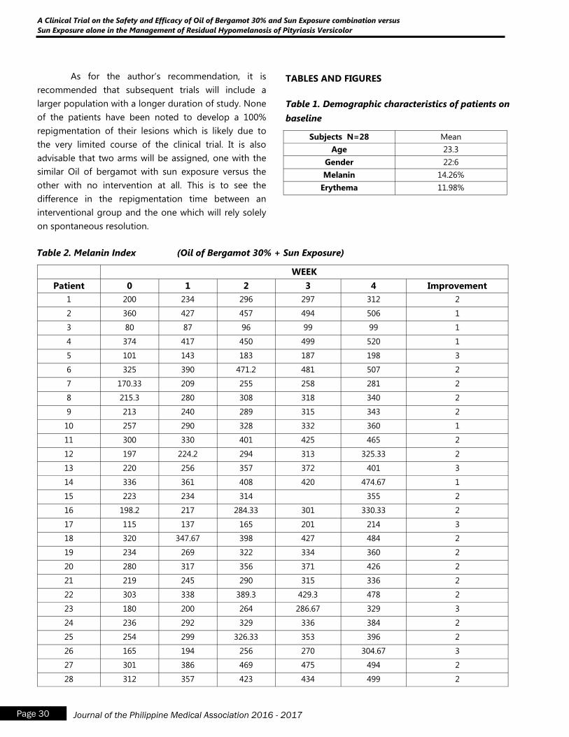
TABLES AND FIGURES
Table 1. Demographic characteristics of patients on
baseline
Subjects N=28 Mean
Age 23.3
Gender 22:6
Melanin 14.26%
Erythema 11.98%
As for the author‟s recommendation, it is
recommended that subsequent trials will include a
larger population with a longer duration of study. None
of the patients have been noted to develop a 100%
repigmentation of their lesions which is likely due to
the very limited course of the clinical trial. It is also
advisable that two arms will be assigned, one with the
similar Oil of bergamot with sun exposure versus the
other with no intervention at all. This is to see the
difference in the repigmentation time between an
interventional group and the one which will rely solely
on spontaneous resolution.
WEEK
Patient 0 1 2 3 4 Improvement
1 200 234 296 297 312 2
2 360 427 457 494 506 1
3 80 87 96 99 99 1
4 374 417 450 499 520 1
5 101 143 183 187 198 3
6 325 390 471.2 481 507 2
7 170.33 209 255 258 281 2
8 215.3 280 308 318 340 2
9 213 240 289 315 343 2
10 257 290 328 332 360 1
11 300 330 401 425 465 2
12 197 224.2 294 313 325.33 2
13 220 256 357 372 401 3
14 336 361 408 420 474.67 1
15 223 234 314 355 2
16 198.2 217 284.33 301 330.33 2
17 115 137 165 201 214 3
18 320 347.67 398 427 484 2
19 234 269 322 334 360 2
20 280 317 356 371 426 2
21 219 245 290 315 336 2
22 303 338 389.3 429.3 478 2
23 180 200 264 286.67 329 3
24 236 292 329 336 384 2
25 254 299 326.33 353 396 2
26 165 194 256 270 304.67 3
27 301 386 469 475 494 2
28 312 357 423 434 499 2
Table 2. Melanin Index (Oil of Bergamot 30% + Sun Exposure)
A Clinical Trial on the Safety and Efficacy of Oil of Bergamot 30% and Sun Exposure combination versus
Sun Exposure alone in the Management of Residual Hypomelanosis of Pityriasis Versicolor
Page 30 Journal of the Philippine Medical Association 2016 - 2017
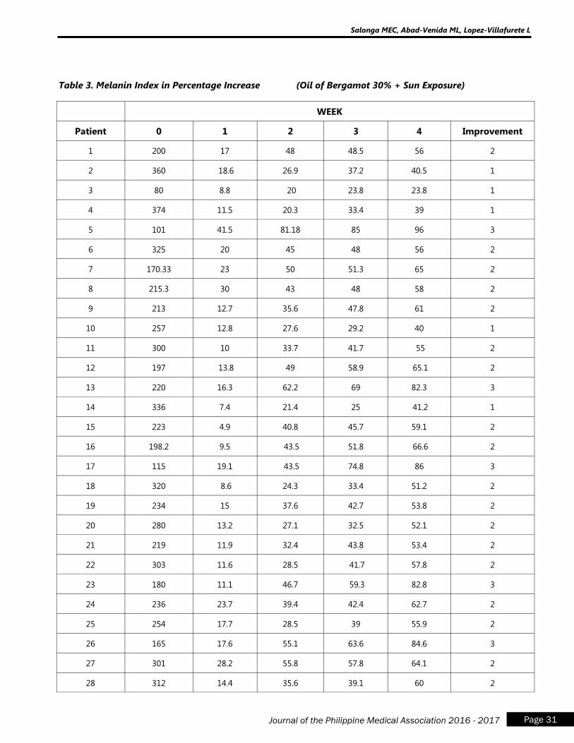
WEEK
Patient 0 1 2 3 4 Improvement
1 200 17 48 48.5 56 2
2 360 18.6 26.9 37.2 40.5 1
3 80 8.8 20 23.8 23.8 1
4 374 11.5 20.3 33.4 39 1
5 101 41.5 81.18 85 96 3
6 325 20 45 48 56 2
7 170.33 23 50 51.3 65 2
8 215.3 30 43 48 58 2
9 213 12.7 35.6 47.8 61 2
10 257 12.8 27.6 29.2 40 1
11 300 10 33.7 41.7 55 2
12 197 13.8 49 58.9 65.1 2
13 220 16.3 62.2 69 82.3 3
14 336 7.4 21.4 25 41.2 1
15 223 4.9 40.8 45.7 59.1 2
16 198.2 9.5 43.5 51.8 66.6 2
17 115 19.1 43.5 74.8 86 3
18 320 8.6 24.3 33.4 51.2 2
19 234 15 37.6 42.7 53.8 2
20 280 13.2 27.1 32.5 52.1 2
21 219 11.9 32.4 43.8 53.4 2
22 303 11.6 28.5 41.7 57.8 2
23 180 11.1 46.7 59.3 82.8 3
24 236 23.7 39.4 42.4 62.7 2
25 254 17.7 28.5 39 55.9 2
26 165 17.6 55.1 63.6 84.6 3
27 301 28.2 55.8 57.8 64.1 2
28 312 14.4 35.6 39.1 60 2
Table 3. Melanin Index in Percentage Increase (Oil of Bergamot 30% + Sun Exposure)
Salonga MEC, Abad-Venida ML, Lopez-Villafurete L
Page 31 Journal of the Philippine Medical Association 2016 - 2017
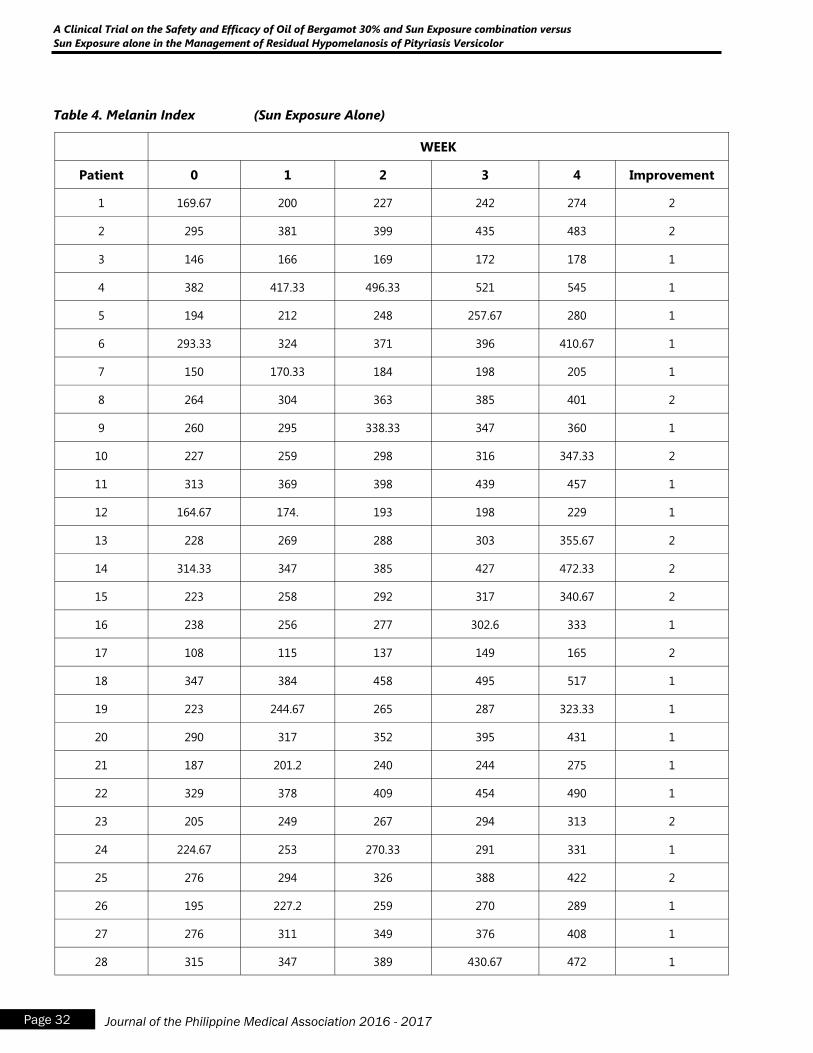
Table 4. Melanin Index (Sun Exposure Alone)
WEEK
Patient 0 1 2 3 4 Improvement
1 169.67 200 227 242 274 2
2 295 381 399 435 483 2
3 146 166 169 172 178 1
4 382 417.33 496.33 521 545 1
5 194 212 248 257.67 280 1
6 293.33 324 371 396 410.67 1
7 150 170.33 184 198 205 1
8 264 304 363 385 401 2
9 260 295 338.33 347 360 1
10 227 259 298 316 347.33 2
11 313 369 398 439 457 1
12 164.67 174. 193 198 229 1
13 228 269 288 303 355.67 2
14 314.33 347 385 427 472.33 2
15 223 258 292 317 340.67 2
16 238 256 277 302.6 333 1
17 108 115 137 149 165 2
18 347 384 458 495 517 1
19 223 244.67 265 287 323.33 1
20 290 317 352 395 431 1
21 187 201.2 240 244 275 1
22 329 378 409 454 490 1
23 205 249 267 294 313 2
24 224.67 253 270.33 291 331 1
25 276 294 326 388 422 2
26 195 227.2 259 270 289 1
27 276 311 349 376 408 1
28 315 347 389 430.67 472 1
A Clinical Trial on the Safety and Efficacy of Oil of Bergamot 30% and Sun Exposure combination versus
Sun Exposure alone in the Management of Residual Hypomelanosis of Pityriasis Versicolor
Page 32 Journal of the Philippine Medical Association 2016 - 2017
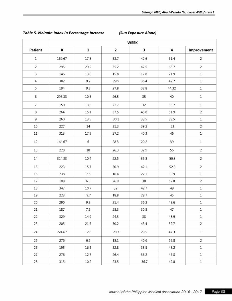
Table 5. Melanin Index in Percentage Increase (Sun Exposure Alone)
WEEK
Patient 0 1 2 3 4 Improvement
1 169.67 17.8 33.7 42.6 61.4 2
2 295 29.2 35.2 47.5 63.7 2
3 146 13.6 15.8 17.8 21.9 1
4 382 9.2 29.9 36.4 42.7 1
5 194 9.3 27.8 32.8 44.32 1
6 293.33 10.5 26.5 35 40 1
7 150 13.5 22.7 32 36.7 1
8 264 15.1 37.5 45.8 51.9 2
9 260 13.5 30.1 33.5 38.5 1
10 227 14 31.3 39.2 53 2
11 313 17.9 27.2 40.3 46 1
12 164.67 6 28.3 20.2 39 1
13 228 18 26.3 32.9 56 2
14 314.33 10.4 22.5 35.8 50.3 2
15 223 15.7 30.9 42.1 52.8 2
16 238 7.6 16.4 27.1 39.9 1
17 108 6.5 26.9 38 52.8 2
18 347 10.7 32 42.7 49 1
19 223 9.7 18.8 28.7 45 1
20 290 9.3 21.4 36.2 48.6 1
21 187 7.6 28.3 30.5 47 1
22 329 14.9 24.3 38 48.9 1
23 205 21.5 30.2 43.4 52.7 2
24 224.67 12.6 20.3 29.5 47.3 1
25 276 6.5 18.1 40.6 52.8 2
26 195 16.5 32.8 38.5 48.2 1
27 276 12.7 26.4 36.2 47.8 1
28 315 10.2 23.5 36.7 49.8 1
Salonga MEC, Abad-Venida ML, Lopez-Villafurete L
Page 33 Journal of the Philippine Medical Association 2016 - 2017
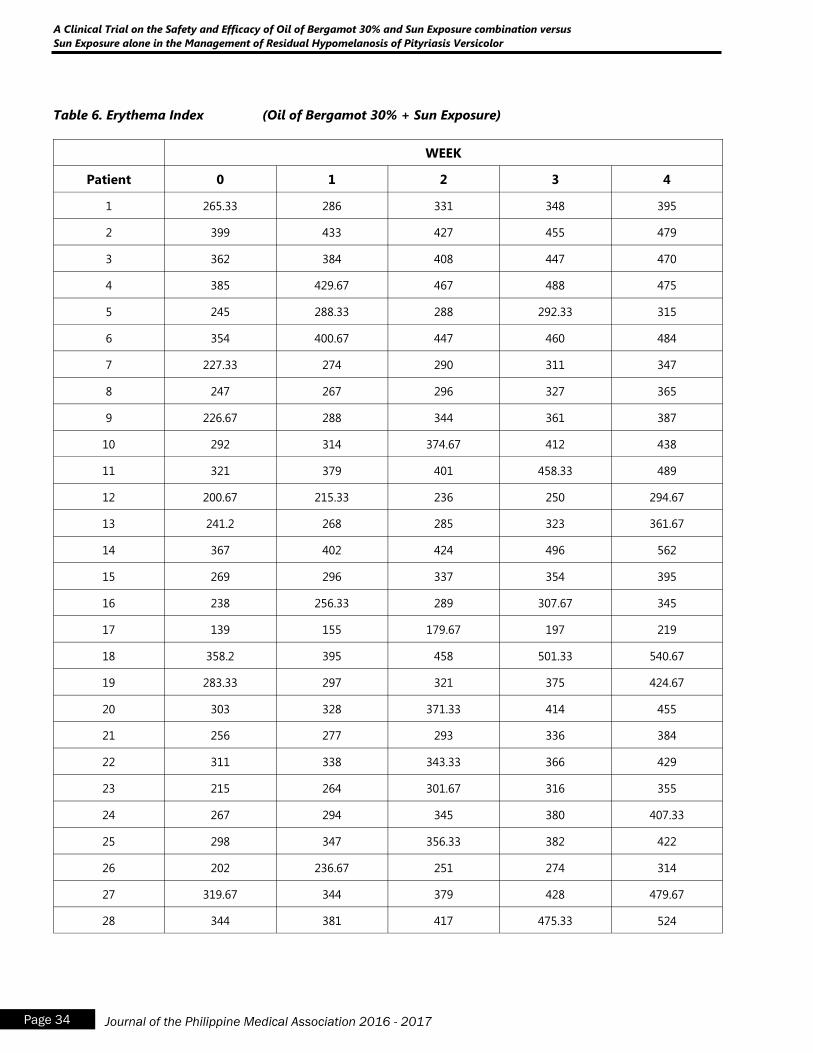
Table 6. Erythema Index (Oil of Bergamot 30% + Sun Exposure)
WEEK
Patient 0 1 2 3 4
1 265.33 286 331 348 395
2 399 433 427 455 479
3 362 384 408 447 470
4 385 429.67 467 488 475
5 245 288.33 288 292.33 315
6 354 400.67 447 460 484
7 227.33 274 290 311 347
8 247 267 296 327 365
9 226.67 288 344 361 387
10 292 314 374.67 412 438
11 321 379 401 458.33 489
12 200.67 215.33 236 250 294.67
13 241.2 268 285 323 361.67
14 367 402 424 496 562
15 269 296 337 354 395
16 238 256.33 289 307.67 345
17 139 155 179.67 197 219
18 358.2 395 458 501.33 540.67
19 283.33 297 321 375 424.67
20 303 328 371.33 414 455
21 256 277 293 336 384
22 311 338 343.33 366 429
23 215 264 301.67 316 355
24 267 294 345 380 407.33
25 298 347 356.33 382 422
26 202 236.67 251 274 314
27 319.67 344 379 428 479.67
28 344 381 417 475.33 524
A Clinical Trial on the Safety and Efficacy of Oil of Bergamot 30% and Sun Exposure combination versus
Sun Exposure alone in the Management of Residual Hypomelanosis of Pityriasis Versicolor
Page 34 Journal of the Philippine Medical Association 2016 - 2017

Table 7. Erythema Index in Percentage Increase (Oil of Bergamot 30% + Sun Exposure)
WEEK
Patient 0 1 2 3 4
1 265.33 7.79 24.8 31.2 50.8
2 399 8.5 7.01 14 20
3 362 6.07 12.7 23.5 29.8
4 385 11.6 21.3 26.7 23.4
5 245 17.7 17.6 19.3 28.6
6 354 13.2 26.3 30 36.7
7 227.33 20.5 27.6 36.8 52.6
8 247 8.1 19.8 32.4 47.7
9 226.67 27 51.8 59.3 70.7
10 292 7.53 28.3 41.1 50
11 321 18.1 37.3 42.8 52.3
12 200.67 7.3 17.6 24.6 47.1
13 241.2 11.1 18.2 3.9 50
14 367 9.5 15.5 35.1 53.1
15 269 10 25.3 31.6 46.8
16 238 7.7 21.4 29.2 45
17 139 11.5 29.3 41.7 57.6
18 358.2 10.3 27.9 40 51
19 283.33 4.8 13.3 32.4 49.9
20 303 8.25 22.6 36.6 50.2
21 256 8.2 14.5 31.3 50
22 311 8.7 10.4 17.6 37.9
23 215 22.8 40.3 47 65
24 267 10.1 29.2 42.3 52.5
25 298 16.4 19.6 28.2 41.6
26 202 17.2 24.3 35.6 55.4
27 319.67 7.61 18.6 33.9 50
28 344 10.8 21.2 38.2 52.3
Salonga MEC, Abad-Venida ML, Lopez-Villafurete L
Page 35 Journal of the Philippine Medical Association 2016 - 2017
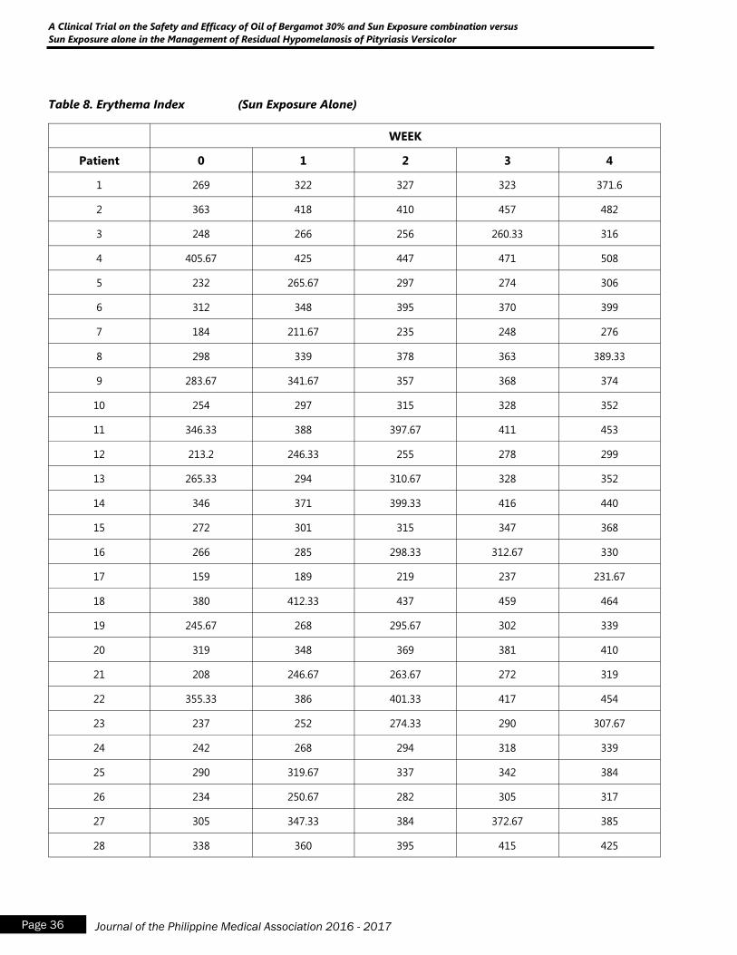
Table 8. Erythema Index (Sun Exposure Alone)
WEEK
Patient 0 1 2 3 4
1 269 322 327 323 371.6
2 363 418 410 457 482
3 248 266 256 260.33 316
4 405.67 425 447 471 508
5 232 265.67 297 274 306
6 312 348 395 370 399
7 184 211.67 235 248 276
8 298 339 378 363 389.33
9 283.67 341.67 357 368 374
10 254 297 315 328 352
11 346.33 388 397.67 411 453
12 213.2 246.33 255 278 299
13 265.33 294 310.67 328 352
14 346 371 399.33 416 440
15 272 301 315 347 368
16 266 285 298.33 312.67 330
17 159 189 219 237 231.67
18 380 412.33 437 459 464
19 245.67 268 295.67 302 339
20 319 348 369 381 410
21 208 246.67 263.67 272 319
22 355.33 386 401.33 417 454
23 237 252 274.33 290 307.67
24 242 268 294 318 339
25 290 319.67 337 342 384
26 234 250.67 282 305 317
27 305 347.33 384 372.67 385
28 338 360 395 415 425
A Clinical Trial on the Safety and Efficacy of Oil of Bergamot 30% and Sun Exposure combination versus
Sun Exposure alone in the Management of Residual Hypomelanosis of Pityriasis Versicolor
Page 36 Journal of the Philippine Medical Association 2016 - 2017
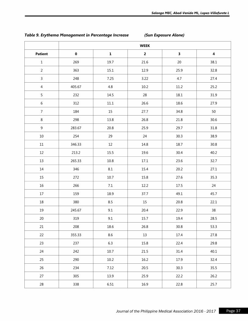
Table 9. Erythema Management in Percentage Increase (Sun Exposure Alone)
WEEK
Patient 0 1 2 3 4
1 269 19.7 21.6 20 38.1
2 363 15.1 12.9 25.9 32.8
3 248 7.25 3.22 4.7 27.4
4 405.67 4.8 10.2 11.2 25.2
5 232 14.5 28 18.1 31.9
6 312 11.1 26.6 18.6 27.9
7 184 15 27.7 34.8 50
8 298 13.8 26.8 21.8 30.6
9 283.67 20.8 25.9 29.7 31.8
10 254 29 24 30.3 38.9
11 346.33 12 14.8 18.7 30.8
12 213.2 15.5 19.6 30.4 40.2
13 265.33 10.8 17.1 23.6 32.7
14 346 8.1 15.4 20.2 27.1
15 272 10.7 15.8 27.6 35.3
16 266 7.1 12.2 17.5 24
17 159 18.9 37.7 49.1 45.7
18 380 8.5 15 20.8 22.1
19 245.67 9.1 20.4 22.9 38
20 319 9.1 15.7 19.4 28.5
21 208 18.6 26.8 30.8 53.3
22 355.33 8.6 13 17.4 27.8
23 237 6.3 15.8 22.4 29.8
24 242 10.7 21.5 31.4 40.1
25 290 10.2 16.2 17.9 32.4
26 234 7.12 20.5 30.3 35.5
27 305 13.9 25.9 22.2 26.2
28 338 6.51 16.9 22.8 25.7
Salonga MEC, Abad-Venida ML, Lopez-Villafurete L
Page 37 Journal of the Philippine Medical Association 2016 - 2017
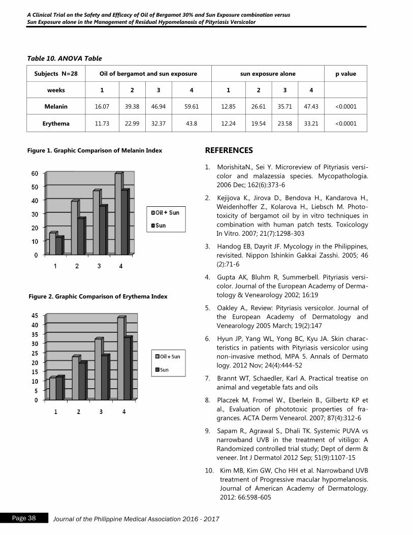
Table 10. ANOVA Table
Subjects N=28 Oil of bergamot and sun exposure sun exposure alone p value
weeks 1 2 3 4 1 2 3 4
Melanin 16.07 39.38 46.94 59.61 12.85 26.61 35.71 47.43 <0.0001
Erythema 11.73 22.99 32.37 43.8 12.24 19.54 23.58 33.21 <0.0001
Figure 1. Graphic Comparison of Melanin Index
Figure 2. Graphic Comparison of Erythema Index
REFERENCES
1. MorishitaN., Sei Y. Microreview of Pityriasis versi-
color and malazessia species. Mycopathologia.
2006 Dec; 162(6):373-6
2. Kejijova K., Jirova D., Bendova H., Kandarova H.,
Weidenhoffer Z., Kolarova H., Liebsch M. Photo-
toxicity of bergamot oil by in vitro techniques in
combination with human patch tests. Toxicology
In Vitro. 2007; 21(7):1298-303
3. Handog EB, Dayrit JF. Mycology in the Philippines,
revisited. Nippon Ishinkin Gakkai Zasshi. 2005; 46
(2):71-6
4. Gupta AK, Bluhm R, Summerbell. Pityriasis versi-
color. Journal of the European Academy of Derma-
tology & Venearology 2002; 16:19
5. Oakley A., Review: Pityriasis versicolor. Journal of
the European Academy of Dermatology and
Venearology 2005 March; 19(2):147
6. Hyun JP, Yang WL, Yong BC, Kyu JA. Skin charac-
teristics in patients with Pityriasis versicolor using
non-invasive method, MPA 5. Annals of Dermato
logy. 2012 Nov; 24(4):444-52
7. Brannt WT, Schaedler, Karl A. Practical treatise on
animal and vegetable fats and oils
8. Placzek M, Fromel W., Eberlein B., Gilbertz KP et
al., Evaluation of phototoxic properties of fra-
grances. ACTA Derm Venearol. 2007; 87(4):312-6
9. Sapam R., Agrawal S., Dhali TK. Systemic PUVA vs
narrowband UVB in the treatment of vitiligo: A
Randomized controlled trial study; Dept of derm &
veneer. Int J Dermatol 2012 Sep; 51(9):1107-15
10. Kim MB, Kim GW, Cho HH et al. Narrowband UVB
treatment of Progressive macular hypomelanosis.
Journal of American Academy of Dermatology.
2012: 66:598-605
A Clinical Trial on the Safety and Efficacy of Oil of Bergamot 30% and Sun Exposure combination versus
Sun Exposure alone in the Management of Residual Hypomelanosis of Pityriasis Versicolor
Page 38 Journal of the Philippine Medical Association 2016 - 2017
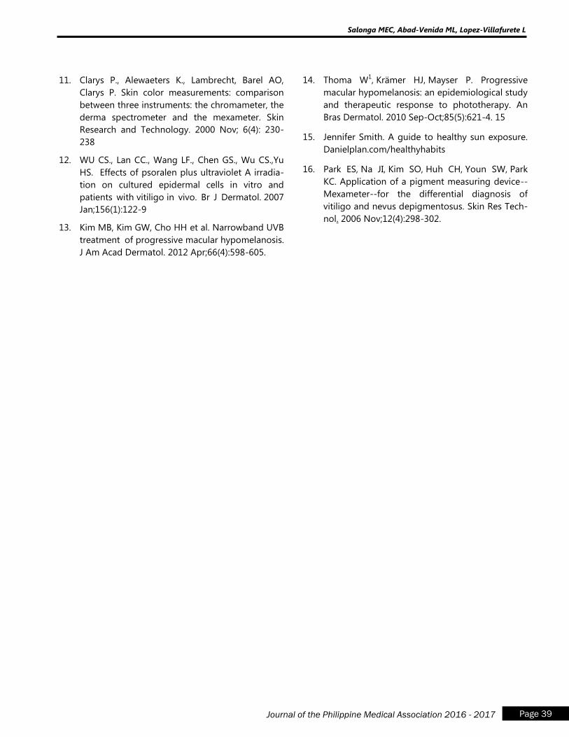
11. Clarys P., Alewaeters K., Lambrecht, Barel AO,
Clarys P. Skin color measurements: comparison
between three instruments: the chromameter, the
derma spectrometer and the mexameter. Skin
Research and Technology. 2000 Nov; 6(4): 230-
238
12. WU CS., Lan CC., Wang LF., Chen GS., Wu CS.,Yu
HS. Effects of psoralen plus ultraviolet A irradia-
tion on cultured epidermal cells in vitro and
patients with vitiligo in vivo. Br J Dermatol. 2007
Jan;156(1):122-9
13. Kim MB, Kim GW, Cho HH et al. Narrowband UVB
treatment of progressive macular hypomelanosis.
J Am Acad Dermatol. 2012 Apr;66(4):598-605.
Salonga MEC, Abad-Venida ML, Lopez-Villafurete L
Page 39 Journal of the Philippine Medical Association 2016 - 2017
14. Thoma W
1, Krämer HJ, Mayser P. Progressive
macular hypomelanosis: an epidemiological study
and therapeutic response to phototherapy. An
Bras Dermatol. 2010 Sep-Oct;85(5):621-4. 15
15. Jennifer Smith. A guide to healthy sun exposure.
Danielplan.com/healthyhabits
16. Park ES, Na JI, Kim SO, Huh CH, Youn SW, Park
KC. Application of a pigment measuring device--
Mexameter--for the differential diagnosis of
vitiligo and nevus depigmentosus. Skin Res Tech-
nol. 2006 Nov;12(4):298-302.
Page 40 Journal of the Philippine Medical Association 2016 - 2017
A Randomized, Double-blind Placebo-controlled Clinical Trial on the Efficacy and Safety of Adapalene 0.1%
Gel on the Closure of Neuropathic Ulcer among Leprosy Patients
A Randomized, Double-blind Placebo-controlled
Clinical Trial on the Efficacy and Safety of
Adapalene 0.1% Gel on the Closure of
Neuropathic Ulcer among Leprosy Patients
Ma. Cricelda M. Rescober, M.D.a
Daisy Ismael-King, M.D., F.P.D.Sb
a Resident, Department of Dermatology, Jose R. Reyes Memorial Medical Center b Consultant Staff, Department of Dermatology, Jose R. Reyes Memorial Medical Center
INTRODUCTION
Leprosy is a chronic infectious disease princi-
pally affecting the skin, eyes, and the peripheral
nerves. It is caused by the acid-fast, rod-shaped bacil-
lus Mycobacterium leprae.1 According to the World
Health Organization, three million persons are suffer-
ing from the burden of the disease.2 Damage to pe-
ripheral nerves is a key feature of leprosy and the
motor and sensory loss that follow the disease is the
basis of developing wounds, fissures, ulcerations,
clawed hand, foot drop, and lagophthalmos. Neuro-
pathic ulcers (NU) are one of the most common com-
plications of leprosy seen in 10% of patients.3 NU oc-
cur due to impairment of the sensory, motor, and
autonomic nerve functions leading to lowered
threshold for tissue damage.4 They may arise in any
part of the body and are divided into 2 groups: plan-
tar and extra-plantar. Plantar ulcers, also called tro-
phic ulcers or malum perforans pedis, are more com-
mon, comprising 70% of all NU. Ulcers occurring in
other denervated sites, such as the dorsum and lat-
eral malleolar region of the foot and hands are de-
scribed under extra-plantar ulcers.3 Aside from the
loss of protective sensation on the foot, other factors
such as repetitive moderate stress, callus formation,
direct trauma, pressure, burns, and infection contrib-
ute to the development of NU.5
Despite recent and emerging advances in
combating leprosy, management of NU in leprosy
has taken a backseat. A recent systematic review of
randomized controlled trials involving anyone with
leprosy and damage to peripheral nerves treated with
any measures designed to prevent damage with the
aim of healing existing ulcers and preventing develop-
ment of new ulcers identified eight trials with a total of
557 participants.6 The quality of the trials was generally
poor and interventions and outcome measures were
diverse. Strategies in the approach to these frustrating
ulcers have mainly been supportive including antibiot-
ics, various wound dressings, and use of corrective
shoes. Clinical trials exist in ulcer closure but these are
few and deemed lacking.
The pathophysiology of chronic non-healing
ulcers such as NU is marked by excessive infiltration of
neutrophils that release significant amounts of en-
zymes such as collagenase or matrix metalloproteinase
-8 (MMP-8) that is responsible for the breakdown of
connective tissue matrix, and elastase that is capable
of destroying essential healing factors such as platelet-
derived growth factor (PDGF) and transforming growth
factor-beta (TGF-ß). Another marker of these chronic
ulcers is an environment containing excessive reactive
oxygen species that further damage the cells and heal-
ing tissues.7
The collagen molecule is characterized by
repeating sequences of proline and hydroxyproline
(HP). Collagen is important in all stages of wound
healing and is critical in regaining tissue integrity and
Page 41 Journal of the Philippine Medical Association 2016 - 2017
Rescober MC, Ismael-King D
strength. In particular, hydroxyproline is important
because it gives the collagen molecule its stable heli-
cal configuration.7 The initial cleavage of collagen by
MMP-8 represents the rate-limiting step in the deg-
radation of this extracellular matrix protein. In non-
healing ulcers, there is increased collagenolytic activ-
ity, reflected by elevated levels of MMP-8, decreased
HP, and lower levels of tissue inhibitor of matrix met-
alloproteinase-1 (TIMP-1). These findings suggest
that overexpression and activation of MMP-8 may be
involved in the pathogenesis of non-healing
wounds.8
Retinoids have broad uses in dermatology
and their role in wound healing has been widely
studied. Preoperative use of retinoids is believed to
enhance wound healing. However, there are mixed
reviews regarding its efficacy in fresh and healing
wounds. Vitamin A is an essential fat-soluble vitamin
that plays an integral role in the maintenance of nor-
mal epidermis by promoting desquamation and
maturation through a decrease in the production of
keratin, keratohyalin granules, and desmosomes.
Moreover, vitamin A also enhances would healing by
stimulating angiogeneseis, collagen synthesis, and
epithelialization.9 Retinoids are synthetic and natural
derivatives of vitamin A. They exert their activity on
keratinocytes by binding to nuclear receptors,
thereby regulating gene expression. .
Topical tretinoin (all-trans-retinoic acid) has
been found to have positive effects on wound heal-
ing.10
The use of tretinoin on a poorly granulating
wound bed increases epidermal thickness and cell
turnover, leading to faster reepithelialization and
granulation tissue formation.11
It also stimulates an-
giogenesis in the superficial dermis which allows effi-
cient delivery of oxygen and nutrients to the tissues.
Endothelial cells secrete biologically active substances
that include growth factors.10
Moreover, tretinoin also
increases granulation tissue formation by increasing
collagen synthesis by way of downregulating MMP-
8.12
A randomized, double-blind, clinical trial by
Tom et al. assessed the efficacy of short-contact
tretinoin versus placebo.10
The study showed that
applying tretinoin 0.0.5% solution for 10 minutes on
diabetic foot ulcers once a day for 4 weeks was supe-
rior to placebo saline solution. A similar technique was
used in a clinical trial by Chua et al. where short-
contact (10 minutes) tretinoin 0.05% cream improved
the healing of foot ulcers in leprosy patients versus the
placebo cream.13
However, irritation caused by topical
tretinoin is often a concern. Short-contact application
of tretinoin may cause mild to moderate pain on the
surrounding skin and ulcer itself.10
Also, tretinoin is
relatively more expensive compared to other wound
care medications limiting its use.
Adapalene is a new naphthoic acid derivative
with strong retinoid agonistic properties and belongs
to the third-generation retinoids. It is also known to
behave similarly to tretinoin.14
Animal studies suggest
that adapalene contributes to the wound healing
process resulting in enhancement of collagen produc-
tion, angiogenesis, and granulation tissue formation.14-
15 It is also noted that adapalene is more stable and is
less irritating than tretinoin because of its unique re-
ceptor specificity.15
Ulceration of the feet is the single most com-
mon cause of disability and dehabilitation of patients
with leprosy. Because of its impact in the patient‟s
physical, emotional, and social health, it is of utmost
importance that there be continuing search for a solu-
tion. A simple and well-tolerated treatment that af-
fords closure of ulcer will prevent further ulceration,
susceptibilty to infection, risk of amputation, and ulti-
mately, disability. Currently, there are no existing stud-
ies about the use of topical adapalene on neuropathic
ulcers in leprosy patients. A randomized controlled
trial is therefore needed to determine the efficacy and
safety of a retinoid derivative such as adapalene that is
similar to tretinoin in pharmacologic action but is
cheaper, more stable, and with less adverse effects.
OBJECTIVES
The general objective of the study is to deter-
mine the efficacy and safety of adapalene 0.1% gel
versus placebo in the closure of neuropathic ulcer
among leprosy patients.
The specific objectives are to determine if
patients treated with adapalene 0.1% gel compared to
Page 42 Journal of the Philippine Medical Association 2016 - 2017
A Randomized, Double-blind Placebo-controlled Clinical Trial on the Efficacy and Safety of Adapalene 0.1%
Gel on the Closure of Neuropathic Ulcer among Leprosy Patients
those given placebo gel were significantly different in
terms of 1.) Percent change of ulcer surface area from
baseline to post-treatment, and 2.) Number of weeks
for the ulcer to close, and 3.) Incidence of adverse
events.
MATERIALS AND METHODS
Patients and Study Design
This is a prospective, randomized, double-
blind, placebo-controlled clinical trial on leprosy
patients who present with neuropathic ulcer, plantar
or extra-plantar, at the Dermatology Outpatient De-
partment of Jose R. Reyes Memorial Medical Center.
Patients recruited included male or female
patients both newly or previously diagnosed with
leprosy who presented with neuropathic ulcer, plan-
tar or extra-plantar, treated or currently under mul-
tidrug therapy (paucibacillary or multibacillary) with
at least two weeks washout period from any oral anti-
biotics or topical medications before the study enroll-
ment period.
Excluded from the study were patients un-
able to give informed consent, with known bleeding
disorders, pregnant or breastfeeding at the time of
enrollment, who have infected ulcers or nearby tis-
sues at time of enrollment, who have other systemic
disorders that will impede wound healing such as
diabetes mellitus, with clinical and radiological signs
of osteomyelitis, and who have a known or docu-
mented allergy to adapalene and other retinoid de-
rivatives.
A certificate of approval from the Institutional
Review Board of Jose R. Reyes Memorial Medical
Center was obtained prior to starting the clinical trial.
Written informed consent was secured before initiat-
ing treatment.
Materials
Both adapalene 0.1% gel and placebo gel
were obtained from local companies. Both were re-
packaged and put in a plastic opaque jar and labeled
by numbers. Placebo gel was specially made so that it
would be identical to the treatment in terms of color,
texture, and odor.
Randomization, treatment allocation, and blinding
Randomization was performed by an unin-
volved third party who did a computer-generated
random sequence to balance the participants of the 2
treatment groups. The study was double-blind in such
a way that all dispensed jars were similar in appear-
ance and that neither the patient nor the investigator
was aware to which the patient was assigned until after
the completion of the study.
Study Intervention
The patients were instructed to clean the
wound bed with cotton swab moistened with normal
saline solution before applying adapalene 0.1% gel or
placebo gel. They would then apply the assigned gel
directly on the wound and leave it on for 10 minutes
with special instructions to avoid surrounding normal
skin. After 10 minutes, patients would rinse it off with
normal saline solution. This procedure was done once
a day for 4 weeks. The participants were allowed to
receive routine wound care such as protection of the
ulcerated area with appropriate dressings. Patients
were also advised to wear open-toed soft sandals as
standard protective footwear. Accepted footwear was
defined as having a soft insole about 1 cm thick, a
hard sole that cannot be pierced by sharp objects such
as stones and thorns, and an upper that fits well and
has straps or laces over the forefoot that can be loos-
ened. These were provided by the investigator to pre-
serve uniformity.
Clinical Assessment
Photographs of the ulcers were taken to evalu-
ate and document initial size and appearance. The
photographs were taken using Nikon Coolpix P310
under standardized lighting and positioning of the
patient. Photographs and measurement of ulcer size
and volume were taken every 2 weeks for a maximum
of 16 weeks after the start of the study or until com-
plete healing occurs, whichever occurred first. Manual
planimetry, wherein the ulcer is traced on acetate with
grids, was used to measure surface area. The number
of grids that fit within the traced circumference was
counted and multiplied by area in square centimeters
to yield the surface area.18
All partial grids divided by
the tracing line by more than half were likewise
included.
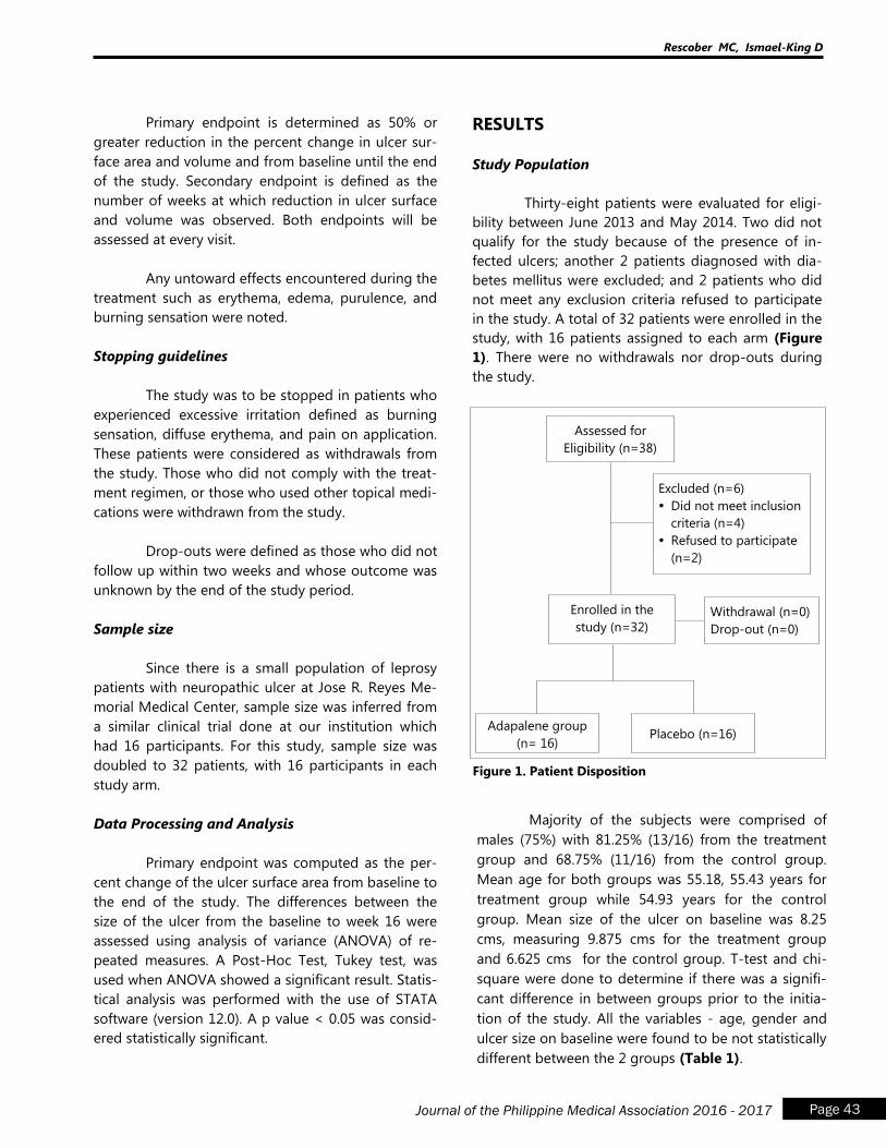
Page 43 Journal of the Philippine Medical Association 2016 - 2017
Rescober MC, Ismael-King D
Primary endpoint is determined as 50% or
greater reduction in the percent change in ulcer sur-
face area and volume and from baseline until the end
of the study. Secondary endpoint is defined as the
number of weeks at which reduction in ulcer surface
and volume was observed. Both endpoints will be
assessed at every visit.
Any untoward effects encountered during the
treatment such as erythema, edema, purulence, and
burning sensation were noted.
Stopping guidelines
The study was to be stopped in patients who
experienced excessive irritation defined as burning
sensation, diffuse erythema, and pain on application.
These patients were considered as withdrawals from
the study. Those who did not comply with the treat-
ment regimen, or those who used other topical medi-
cations were withdrawn from the study.
Drop-outs were defined as those who did not
follow up within two weeks and whose outcome was
unknown by the end of the study period.
Sample size
Since there is a small population of leprosy
patients with neuropathic ulcer at Jose R. Reyes Me-
morial Medical Center, sample size was inferred from
a similar clinical trial done at our institution which
had 16 participants. For this study, sample size was
doubled to 32 patients, with 16 participants in each
study arm.
Data Processing and Analysis
Primary endpoint was computed as the per-
cent change of the ulcer surface area from baseline to
the end of the study. The differences between the
size of the ulcer from the baseline to week 16 were
assessed using analysis of variance (ANOVA) of re-
peated measures. A Post-Hoc Test, Tukey test, was
used when ANOVA showed a significant result. Statis-
tical analysis was performed with the use of STATA
software (version 12.0). A p value < 0.05 was consid-
ered statistically significant.
RESULTS
Study Population
Thirty-eight patients were evaluated for eligi-
bility between June 2013 and May 2014. Two did not
qualify for the study because of the presence of in-
fected ulcers; another 2 patients diagnosed with dia-
betes mellitus were excluded; and 2 patients who did
not meet any exclusion criteria refused to participate
in the study. A total of 32 patients were enrolled in the
study, with 16 patients assigned to each arm (Figure
1). There were no withdrawals nor drop-outs during
the study.
Assessed for
Eligibility (n=38)
Enrolled in the
study (n=32)
Withdrawal (n=0)
Drop-out (n=0)
Excluded (n=6)
Did not meet inclusion
criteria (n=4)
Refused to participate
(n=2)
Adapalene group
(n= 16) Placebo (n=16)
Figure 1. Patient Disposition
Majority of the subjects were comprised of
males (75%) with 81.25% (13/16) from the treatment
group and 68.75% (11/16) from the control group.
Mean age for both groups was 55.18, 55.43 years for
treatment group while 54.93 years for the control
group. Mean size of the ulcer on baseline was 8.25
cms, measuring 9.875 cms for the treatment group
and 6.625 cms for the control group. T-test and chi-
square were done to determine if there was a signifi-
cant difference in between groups prior to the initia-
tion of the study. All the variables - age, gender and
ulcer size on baseline were found to be not statistically
different between the 2 groups (Table 1).
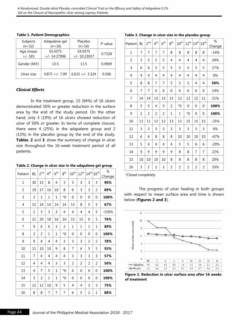
Page 44 Journal of the Philippine Medical Association 2016 - 2017
A Randomized, Double-blind Placebo-controlled Clinical Trial on the Efficacy and Safety of Adapalene 0.1%
Gel on the Closure of Neuropathic Ulcer among Leprosy Patients
Subjects
(n=32)
Adapalene gel
(n=16)
Placebo
(n=16) P-value
Age (mean
+/- SD)
55.4375
+/- 14.27096
54.9375
+/- 10.23037 0.7328
Gender (M:F) 13:3 11:5 0.4909
Ulcer size 9.875 +/- 7.99 6.625 +/- 3.324 0.500
Table 1. Patient Demographics
Clinical Effects
In the treatment group, 15 (94%) of 16 ulcers
demonstrated 50% or greater reduction in the surface
area by the end of the study period. On the other
hand, only 3 (19%) of 16 ulcers showed reduction of
ulcer of 50% or greater. In terms of complete closure,
there were 4 (25%) in the adapalene group and 2
(13%) in the placebo group by the end of the study.
Tables 2 and 3 show the summary of change in ulcer
size throughout the 16-week treatment period of all
patients.
Patient BL 2nd 4th 6th 8th 10th 12th 14th 16th %
Change
1 30 15 8 4 3 3 3 3 3 90%
2 19 17 16 10 8 6 3 3 2 89%
3 2 1 1 1 *0 0 0 0 0 100%
4 15 14 14 14 14 13 8 5 5 67%
5 2 3 3 3 4 4 4 4 5 -150%
6 21 20 18 16 16 15 15 6 5 76%
7 9 6 6 3 2 1 1 1 1 89%
8 2 2 1 1 *0 0 0 0 0 100%
9 9 4 4 4 3 3 3 2 2 78%
10 11 10 10 9 8 7 6 5 5 55%
11 7 6 4 4 4 3 3 3 3 57%
12 4 4 4 3 3 2 2 2 2 50%
13 4 7 5 1 *0 0 0 0 0 100%
14 3 2 1 1 *0 0 0 0 0 100%
15 12 12 10 9 5 4 4 3 3 75%
16 8 8 7 7 7 6 5 2 1 88%
Table 2. Change in ulcer size in the adapalene gel group
Table 3. Change in ulcer size in the placebo group
Patient BL 2nd 4th 6th 8th 10th 12th 14th 16th %
Change
1 7 7 7 7 8 8 8 8 8 -14%
2 5 5 5 5 4 4 4 4 4 20%
3 6 6 5 5 5 5 5 5 5 17%
4 4 4 4 4 4 4 4 4 4 0%
5 8 8 7 7 5 5 5 4 4 50%
6 7 7 6 6 6 6 6 6 6 14%
7 14 14 13 13 13 12 12 12 11 21%
8 5 5 4 3 2 *0 0 0 0 100%
9 2 2 2 2 1 1 *0 0 0 100%
10 12 11 12 12 13 13 15 15 15 -25%
11 3 3 3 3 3 3 3 3 3 0%
12 6 6 8 8 8 10 10 10 10 -67%
13 5 4 4 4 4 5 5 6 6 -20%
14 9 9 9 9 9 8 8 7 7 22%
15 10 10 10 10 8 8 8 8 8 20%
16 3 2 2 2 2 2 2 2 2 33%
*Closed completely
The progress of ulcer healing in both groups
with respect to mean surface area and time is shown
below (Figures 2 and 3).
Figure 2. Reduction in ulcer surface area after 16 weeks
of treatment
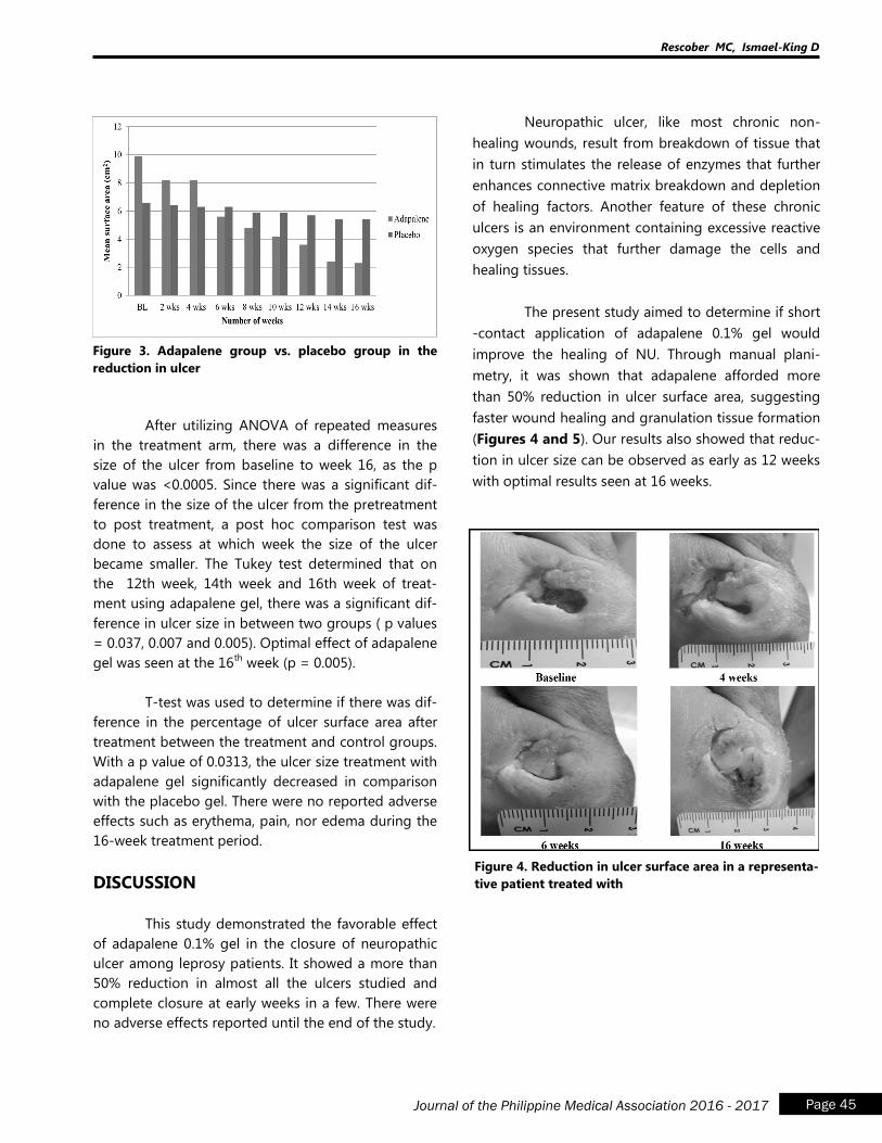
Page 45 Journal of the Philippine Medical Association 2016 - 2017
Rescober MC, Ismael-King D
Figure 3. Adapalene group vs. placebo group in the
reduction in ulcer
After utilizing ANOVA of repeated measures
in the treatment arm, there was a difference in the
size of the ulcer from baseline to week 16, as the p
value was <0.0005. Since there was a significant dif-
ference in the size of the ulcer from the pretreatment
to post treatment, a post hoc comparison test was
done to assess at which week the size of the ulcer
became smaller. The Tukey test determined that on
the 12th week, 14th week and 16th week of treat-
ment using adapalene gel, there was a significant dif-
ference in ulcer size in between two groups ( p values
= 0.037, 0.007 and 0.005). Optimal effect of adapalene
gel was seen at the 16th
week (p = 0.005).
T-test was used to determine if there was dif-
ference in the percentage of ulcer surface area after
treatment between the treatment and control groups.
With a p value of 0.0313, the ulcer size treatment with
adapalene gel significantly decreased in comparison
with the placebo gel. There were no reported adverse
effects such as erythema, pain, nor edema during the
16-week treatment period.
DISCUSSION
This study demonstrated the favorable effect
of adapalene 0.1% gel in the closure of neuropathic
ulcer among leprosy patients. It showed a more than
50% reduction in almost all the ulcers studied and
complete closure at early weeks in a few. There were
no adverse effects reported until the end of the study.
Neuropathic ulcer, like most chronic non-
healing wounds, result from breakdown of tissue that
in turn stimulates the release of enzymes that further
enhances connective matrix breakdown and depletion
of healing factors. Another feature of these chronic
ulcers is an environment containing excessive reactive
oxygen species that further damage the cells and
healing tissues.
The present study aimed to determine if short
-contact application of adapalene 0.1% gel would
improve the healing of NU. Through manual plani-
metry, it was shown that adapalene afforded more
than 50% reduction in ulcer surface area, suggesting
faster wound healing and granulation tissue formation
(Figures 4 and 5). Our results also showed that reduc-
tion in ulcer size can be observed as early as 12 weeks
with optimal results seen at 16 weeks.
Figure 4. Reduction in ulcer surface area in a representa-
tive patient treated with
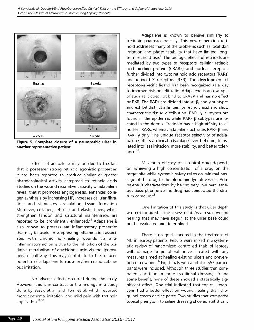
Page 46 Journal of the Philippine Medical Association 2016 - 2017
A Randomized, Double-blind Placebo-controlled Clinical Trial on the Efficacy and Safety of Adapalene 0.1%
Gel on the Closure of Neuropathic Ulcer among Leprosy Patients
Effects of adapalene may be due to the fact
that it possesses strong retinoid agonistic properties.
It has been reported to produce similar or greater
pharmacological activity compared to retinoic acids.
Studies on the wound reparative capacity of adapalene
reveal that it promotes angiogenesis, enhances colla-
gen synthesis by increasing HP, increases cellular filtra-
tion, and stimulates granulation tissue formation.
Moreover, collagen, reticular and elastic fibers, which
strengthen tension and structural maintenance, are
reported to be prominently enhanced.14
Adapalene is
also known to possess anti-inflammatory properties
that may be useful in suppressing inflammation associ-
ated with chronic non-healing wounds. Its anti-
inflammatory action is due to the inhibition of the oxi-
dative metabolism of arachidonic acid via the lipooxy-
genase pathway. This may contribute to the reduced
potential of adapalene to cause erythema and cutane-
ous irritation.
No adverse effects occurred during the study.
However, this is in contrast to the findings in a study
done by Basak et al. and Tom et al. which reported
more erythema, irritation, and mild pain with tretinoin
application.15,10
Adapalene is known to behave similarly to
tretinoin pharmacologically. This new-generation reti-
noid addresses many of the problems such as local skin
irritation and photoinstability that have limited long-
term retinoid use.17
The biologic effects of retinoids are
mediated by two types of receptors: cellular retinoic
acid binding protein (CRABP) and nuclear receptors
further divided into two: retinoid acid receptors (RARs)
and retinoid X receptors (RXR). The development of
receptor-specific ligand has been recognized as a way
to improve risk-benefit ratio. Adapalene is an example
of such as it does not bind to CRABP and has no effect
or RXR. The RARs are divided into α, β, and γ subtypes
and exhibit distinct affinities for retinoic acid and show
characteristic tissue distribution. RAR- γ subtypes are
found in the epidermis while RAR- β subtypes are lo-
cated in the dermis. Tretinoin has a high affinity to all
nuclear RARs, whereas adapalene activates RAR- β and
RAR- γ only. The unique receptor selectivity of adala-
palene offers a clinical advantage over tretinoin, trans-
lated into less irritation, more stability, and better toler-
ance.18
Maximum efficacy of a topical drug depends
on achieving a high concentration of a drug on the
target site while systemic safety relies on minimal pas-
sage of the drug to the blood and lymph vessels. Ada-
palene is characterized by having very low percutane-
ous absorption once the drug has penetrated the stra-
tum corneum.18
One limitation of this study is that ulcer depth
was not included in the assessment. As a result, wound
healing that may have begun at the ulcer base could
not be evaluated and determined.
There is no gold standard in the treatment of
NU in leprosy patients. Results were mixed in a system-
atic review of randomized controlled trials of leprosy
with damage to peripheral nerves treated with any
measures aimed at healing existing ulcers and preven-
tion of new ones.6 Eight trials with a total of 557 partici-
pants were included. Although three studies that com-
pared zinc tape to more traditional dressings found
some benefit, none of these showed a statistically sig-
nificant effect. One trial indicated that topical ketan-
serin had a better effect on wound healing than clio-
quinol cream or zinc paste. Two studies that compared
topical phenytoin to saline dressing showed statistically
Figure 5. Complete closure of a neuropathic ulcer in
another representative patient
Page 47 Journal of the Philippine Medical Association 2016 - 2017
Rescober MC, Ismael-King D
significant effects in favor of phenytoin for
healing of ulcer. Canvas shoes were not much better
than polyvinyl chloride (PVC)-boots, and double
rocker shoes did not promote healing much more
than below-knee plasters.6 There is
an obvious lack of
high quality research in the field of ulcer prevention
and treatment in leprosy. The success of tretinoin in
closure of NU among leprosy patients and diabetic
ulcer serves as the basis of selecting adapalene which
has similar properties but is more stable, with less
adverse effects, less expensive, and better tolerated –
features that make it a good alternative in the treat-
ment of foot ulcers in leprosy patients.
CONCLUSION
This randomized controlled study found that
compared to placebo, short-contact adapalene 0.1%
gel is more effective and generally safe to use in the
reduction of surface area of neuropathic ulcer among
leprosy patients. It suggests that adapalene may be a
novel treatment option for treating neuropathic
ulcers.
REFERENCES
1. Jopling WH, McDougall AC. Definition, epidemiol-
ogy and world distribution. In: Handbook of Lep-
rosy. 5th ed. Oxford: Heinemann. Medical Books;
1996. p. 1–9.
2. Nordeen SK, Lopez B, Sundaresan TK. Estimated
number of leprosy cases in the world. Bull World
Health Organ. 1992;70(1):7-10.
3. Srinivasan H. Management of ulcers in neurologi-
cally impaired feet in leprosy-affected persons. In:
Schwarz R, Brandsma JW (eds). Offset Press pvt.
Ltd, Kathmandu, Nepal, 2004; pp. 193–226.
4. Vieira R, Felicissimo P. Surgical treatment of three
cases of plantar foot ulceration in leprosy. Lepr
Rev. 2008 Sep; 79(3): 325-30.
5. Nollet F. Evaluation of biomechanical risk factors
for plantar foot ulceration in leprosy patients with
peripheral neuropathy. Royal Netherlands Acad-
emy of Arts and Sciences.2008.
6. Reinar LM, Forsetlund L, Bjørndal A, Lockwood D.
Interventions for skin changes caused by nerve
damage in leprosy. Cochrane Database of System-
atic Reviews 2008, Issue 3. Art. No.:CD004833. DOI:
10.1002/14651858.CD004833.pub3.
7. Tom W, Peng D, Allaei A, et al. The Effect of Short-
Contact Topical Tretinoin Therapy for Foot Ulcers in
Patients with Diabetes. Arch Dermatol. 2005 Nov;
141(11): 1373-77.
8. Elson M.L. The Role of Retinoids in Wound Healing.
J Am Acad Dermatol. 1998 Aug;39(2 Pt 3):79-81.
9. Chua C, Yaptinchay-Castillo V, Barcelona-Tan J.
Short-Contact Tretinoin 0.05% Cream for Foot Ul-
cers in Leprosy Patients – A Randomized Trial.2010.
10. Ceyhan AM, Akkaya VB, Sutcu R. The Effects of
Topical Application of Adapalene and Tretinoin on
Tissue Hydroxyproline Content in Wound Model
Impaired by Corticosteroid. J Turk Acad Dermatol.
2011;5(11): 1151a1.
11. Basak P, Eroglu E, Altuntas I et al. Comparison of
the effects of tretinoin, adapalene and collagenase
in an experimental model of wound healing. Euro-
pean Journal of Dermatology. 2002 Mar-Apr;12
(2):145-8.
12. Diegelmann R, Evans M. Wound Healing: An Over-
view of Acute, Fibrotic and Delayed Healing. Fron-
tiers in Bioscience. 2004 Jan;9:283-89.
13. Nwomeh BC, Liang HX, Cohen IK, et al. MMP-8 is
the predominant collagenase in healing wounds
and nonhealing ulcers. J Surg Res. 1999 Feb;81
(2):189-95.
14. Abdelmalek M, Spencer. Retinoids and wound heal-
ing. J.Dermatol Surg. 2006 Oct; 32(10):1219-30.
15. Uchida G, Yoshimura K, Okazaki M, et al. Tretinoin
reverses upregulation of matrix metalloproteinase-
13 in human keloid-derived fibroblasts. Experimen-
tal Dermatology 2003;12 (2): 35–42.
16. Michel S, Jomard A, Demarchez M. Pharamacology
of Adapalene. British Journal of Dermatology.1998;
139 (52):3-7.
17. Millikan L. Adapalene: An update on newer com-
parative studies between the various retinoids. In-
ternational Journal of Dermatology.2000;39:784-88.
18. Oien RF, Hakansson A, Hansen BU et al. Measuring
the size of ulcers by planimetry: a useful method in
the clinical setting. J Wound Care. 2002 May; 11
(5):165-8.
19. Wright C. Exploring alternative methods for
measuring wounds in a nursing home. Nursing
Times.2003 Oct; 99 (42):66.
Page 48 Journal of the Philippine Medical Association 2016 - 2017
Association of Vitamin D Receptor Gene Polymorphisms in the Occurrence and Spectrum
of Leprosy in Filipino Patients Seen at Jose R. Reyes Memorial Medical Center
Association of Vitamin D Receptor Gene
Polymorphisms in the Occurrence and Spectrum
of Leprosy in Filipino Patients Seen at
Jose R. Reyes Memorial Medical Center
Donn M. Mendoza, M.D.1
Zharlah Gulmatico-Flores, M.D., FPDS2
1Resident, Department of Dermatology, Jose R. Reyes Memorial Medical Center 2Consultant, Department of Dermatology, Jose R. Reyes Memorial Medical Center
Abstract
Introduction / Background: Host genetic factors including major histocompatibility complex
(MHC) polymorphisms influence both susceptibility to leprosy and also to leprosy type. Recent
studies have implicated variation in the vitamin D receptor (VDR) gene in susceptibility to sev-
eral diseases, including osteoporosis and pulmonary tuberculosis. Putative polymorphisms at
the VDR gene, which potentially modifies VDR mRNA stability and/or activity, have been impli-
cated in susceptibility to intracellular pathogens.
Objectives: The general objectives of the study are to determine the effects of Vitamin D recep-
tor gene polymorphism on the occurrence and spectrum of leprosy among Filipino patients
seen at Jose R. Reyes Memorial Medical Center, Department of Dermatology Hospital.
Methods: Five (5) cc of whole blood samples obtained from patients diagnosed with Hansen‟s
disease by smear and histological confirmation as well as healthy controls seen in the outpatient
department. DNA was isolated from white blood cells using the phenol/chloroform method and
was used for PCR amplification. The primers 5‟-CAGAGCATGGACAGGGAGCA-3‟ and 5‟-
GGTGGCGGCAGCGGATGTACGT-3‟ yielding a product of 352 base pairs in the 61675 and 62026
positions of the VDR gene was used (Goulart et al., 2005). The PCR cycle conditions would be
94oC for 20 seconds, 60
oC for 30 seconds, and 72
oC for 30 seconds (35 cycles), using 2 mM
MgCl2, 0.2 mM dNTPs, 0.009 mM of each primer, 100 ng of DNA, and 1 U of Taq polymerase in
a 25-μL reaction (Roy et al., 1999).
Page 49 Journal of the Philippine Medical Association 2016 - 2017
Mendoza D, Gulmatico-Flores Z
Results: Sixty (60) subjects were included in the study. A total of 43 (71.67%) patients who
were diagnosed case of Leprosy with a spectrum of tuberbuloid and lepromatous type. Fifteen
patients (25%) were classified as tubercuoid type and 28 (46.67%) patients were of leproma-
tous type. Seventeen 17 (28.33%) patients in the control group were composed of healthy vol-
unteers unrelated to patients. The distribution of VDR genotypes at codon 352 in exon 9 was
highly significant in between the control and lepromatous, tuberculoid and lepromatous type
of Hansen‟s disease. Heterozygous type of VDR (Tt) were found to be highly significant among
controls at 82.35% while 66.67% of tuberculoid has homozygous recessive type (tt) of VDR and
96.43% of lepromatous type has homozygous dominant type VDR (TT) . Significant difference
were observed between the HD group (tuberculoid and lepromatous) and control. Fstat =
31.32, (p <0.001). Post-hoc analysis of genotypes between the clinical groups showed that tu-
berculoid vs lepromatous had significantly different genotypes (p=< 0.001) and lepromatous
type of HD vs control had significantly different genotypes (p=< 0.001).
Conclusion: The polymorphism of VDR genotypes at codon 352 in exon 9 was highly signifi-
cant in between the control, lepromatous, and tuberculoid type of Hansen‟s disease. Homozy-
gous TT was highly associated with lepromatous type (odds ratio = 16, p=< 0.001), and homo-
zygous tt is highly related to the tuberculoid type (odds ratio = 5.8, p=< 0.001).
Page 50 Journal of the Philippine Medical Association 2016 - 2017
Association of Vitamin D Receptor Gene Polymorphisms in the Occurrence and Spectrum
of Leprosy in Filipino Patients Seen at Jose R. Reyes Memorial Medical Center
INTRODUCTION
Leprosy is a chronic, infectious, and slowly pro-
gressive disease caused by the bacillus Mycobacterium
leprae characterized by granulomas and neurotropism
with a predilection for the skin and peripheral nerves.
The diagnosis of the disease is often delayed and ag-
gravated by subclinical infection, presence of healthy
carriers and inconsistent evidence of reduced transmis-
sion following multidrug therapy.1
Once worldwide in distribution, leprosy is now
seen primarily in tropical and subtropical regions of
Asia, Africa, and Central and South America. In en-
demic countries, the vast majority of new cases are in
children and young adults who have close relatives
with contagious forms of the disease. Elimination of
leprosy as a public health problem is defined as a
prevalence rate of less than one case per 10 000 per-
sons. Efforts currently focus on eliminating leprosy at a
national level in the remaining endemic countries and
at a sub-national level from the others. The number of
new cases detected in 2008 was 240,007 globally, and
has fallen by 9126 (4% decrease) compared with 2007.
However, there was a slight increase at the end of 2009
to 244,796.
The National Leprosy Control Program (NLCP)
of the Philippines was established in 1986 under the
supervision of the National Center for Disease Preven-
tion and Control (NCDPC) of the Department of Health
(DOH). Leprosy Control Program envisions to eliminate
leprosy as a human disease by 2020. The program
thrust is towards finding hidden cases of leprosy and
putting them on MDT, emphasizing the completion of
treatment within the WHO-prescribed duration.28
In
1986, there were 38,570 registered patients with lep-
rosy in the Philippines, with a prevalence rate of 7.2 per
10,000 Filipinos. There was a dramatic decrease in
prevalence by the end of 1998, to 0.90 per 10,000
population, and leprosy was no longer considered to
be a public health problem of the country. In 2004, the
prevalence was further reduced to 0.38 per 10,000
population, translating to a total decline of 3,146 from
7,005 in 1998. In 2006, the prevalence of leprosy in
the Philippines was more than 3000 cases (0.42%),
while the number of new cases detected was 2,517.
There was a decrease in the number of patients in
2007. However, an upward trend was observed in 2008
to 3,338 patients (0.35%).
The three requirements for the spread of lep-
rosy are: a contagious patient, a susceptible person,
and close or intimate contact. The organism is spread
predominantly via nasal and oral droplets from the ba-
cilliferous patient and much less often from eroded
skin and even after 1 to 7 days, the bacillus is still viable
in dried secretions. Inoculation is via the nasal mucosa
or, less commonly, through breaks in the skin barrier.
Transmission also depends on the infectivity of the
contagious patient.
Leprosy is mainly a clinical diagnosis. Labora-
tory techniques may serve as adjuncts in the diagnosis.
AFB microscopy is still being used in the Philippines;
samples for bacilloscopy may be obtained from the
earlobes, forehead, chin, extensor forearms, and dorsal
fingers, as well as buttocks and trunk. The smear is usu-
ally stained by the Fite (or Ziehl-Neelsen) method and a
search is made for red rods (against a blue back-
ground) at 100x with oil immersion. Routine skin punch
biopsy with hematoxylin-eosin stain and Fite-Faraco
are also being requested in training institutions in the
Philippines but not routinely done in leprosy control
programs and rural health centers. In subclinical cases
seen in clinics, a clinicopathological correlation may
deem necessary. A biopsy specimen of the skin lesions
should be obtained, especially in patients with sus-
pected tuberculoid leprosy. There are three basic histo-
pathologic patterns observed in leprosy: lepromatous,
tuberculoid and borderline.
Clinical manifestations depend on the relation-
ship of the bacillus and the host immune response.
Therefore, patients present a spectrum of clinical and
histological characteristics, ranging from multibacillary
form, which corresponds to lepromatous leprosy with a
high bacillus load in tissues, to the paucibacillary form
or tuberculoid leprosy, in which the bacillus is rarely
found in skin smears.2
Host genetic factors including major histocom-
patibility complex (MHC) polymorphisms influence
both susceptibility to leprosy and also to leprosy type.
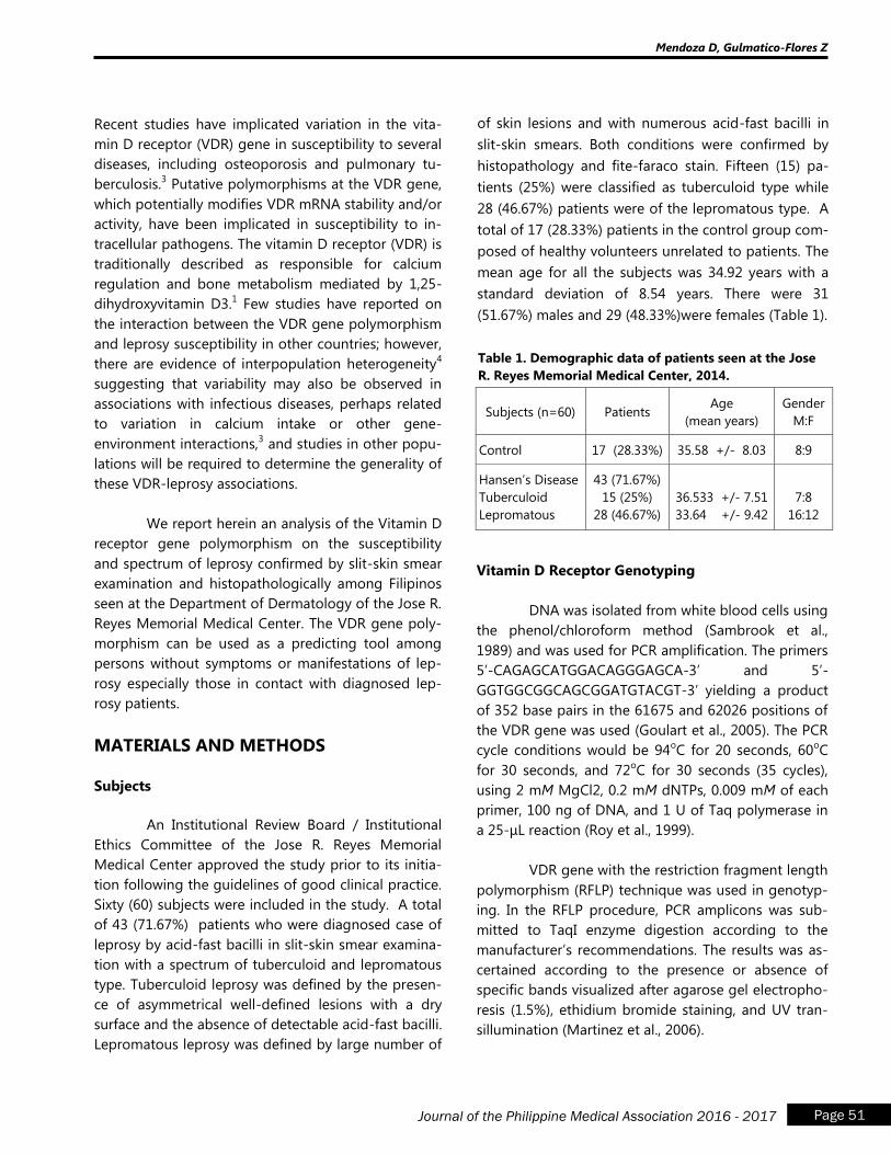
Page 51 Journal of the Philippine Medical Association 2016 - 2017
Mendoza D, Gulmatico-Flores Z
Recent studies have implicated variation in the vita-
min D receptor (VDR) gene in susceptibility to several
diseases, including osteoporosis and pulmonary tu-
berculosis.3 Putative polymorphisms at the VDR gene,
which potentially modifies VDR mRNA stability and/or
activity, have been implicated in susceptibility to in-
tracellular pathogens. The vitamin D receptor (VDR) is
traditionally described as responsible for calcium
regulation and bone metabolism mediated by 1,25-
dihydroxyvitamin D3.1 Few studies have reported on
the interaction between the VDR gene polymorphism
and leprosy susceptibility in other countries; however,
there are evidence of interpopulation heterogeneity4
suggesting that variability may also be observed in
associations with infectious diseases, perhaps related
to variation in calcium intake or other gene-
environment interactions,3 and studies in other popu-
lations will be required to determine the generality of
these VDR-leprosy associations.
We report herein an analysis of the Vitamin D
receptor gene polymorphism on the susceptibility
and spectrum of leprosy confirmed by slit-skin smear
examination and histopathologically among Filipinos
seen at the Department of Dermatology of the Jose R.
Reyes Memorial Medical Center. The VDR gene poly-
morphism can be used as a predicting tool among
persons without symptoms or manifestations of lep-
rosy especially those in contact with diagnosed lep-
rosy patients.
MATERIALS AND METHODS
Subjects
An Institutional Review Board / Institutional
Ethics Committee of the Jose R. Reyes Memorial
Medical Center approved the study prior to its initia-
tion following the guidelines of good clinical practice.
Sixty (60) subjects were included in the study. A total
of 43 (71.67%) patients who were diagnosed case of
leprosy by acid-fast bacilli in slit-skin smear examina-
tion with a spectrum of tuberculoid and lepromatous
type. Tuberculoid leprosy was defined by the presen-
ce of asymmetrical well-defined lesions with a dry
surface and the absence of detectable acid-fast bacilli.
Lepromatous leprosy was defined by large number of
of skin lesions and with numerous acid-fast bacilli in
slit-skin smears. Both conditions were confirmed by
histopathology and fite-faraco stain. Fifteen (15) pa-
tients (25%) were classified as tuberculoid type while
28 (46.67%) patients were of the lepromatous type. A
total of 17 (28.33%) patients in the control group com-
posed of healthy volunteers unrelated to patients. The
mean age for all the subjects was 34.92 years with a
standard deviation of 8.54 years. There were 31
(51.67%) males and 29 (48.33%)were females (Table 1).
Subjects (n=60) Patients Age
(mean years)
Gender
M:F
Control 17 (28.33%) 35.58 +/- 8.03 8:9
Hansen‟s Disease
Tuberculoid
Lepromatous
43 (71.67%)
15 (25%)
28 (46.67%)
36.533 +/- 7.51
33.64 +/- 9.42
7:8
16:12
Table 1. Demographic data of patients seen at the Jose
R. Reyes Memorial Medical Center, 2014.
Vitamin D Receptor Genotyping
DNA was isolated from white blood cells using
the phenol/chloroform method (Sambrook et al.,
1989) and was used for PCR amplification. The primers
5‟-CAGAGCATGGACAGGGAGCA-3‟ and 5‟-
GGTGGCGGCAGCGGATGTACGT-3‟ yielding a product
of 352 base pairs in the 61675 and 62026 positions of
the VDR gene was used (Goulart et al., 2005). The PCR
cycle conditions would be 94oC for 20 seconds, 60
oC
for 30 seconds, and 72oC for 30 seconds (35 cycles),
using 2 mM MgCl2, 0.2 mM dNTPs, 0.009 mM of each
primer, 100 ng of DNA, and 1 U of Taq polymerase in
a 25-μL reaction (Roy et al., 1999).
VDR gene with the restriction fragment length
polymorphism (RFLP) technique was used in genotyp-
ing. In the RFLP procedure, PCR amplicons was sub-
mitted to TaqI enzyme digestion according to the
manufacturer‟s recommendations. The results was as-
certained according to the presence or absence of
specific bands visualized after agarose gel electropho-
resis (1.5%), ethidium bromide staining, and UV tran-
sillumination (Martinez et al., 2006).
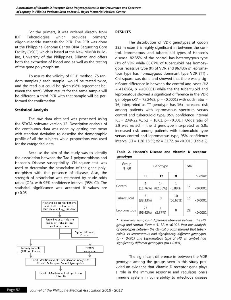
Page 52 Journal of the Philippine Medical Association 2016 - 2017
Association of Vitamin D Receptor Gene Polymorphisms in the Occurrence and Spectrum
of Leprosy in Filipino Patients Seen at Jose R. Reyes Memorial Medical Center
For the primers, it was ordered directly from
IDT Tehcnologies which provides primers/
oligonucleotide synthesis for PCR. The PCR was done
at the Philippine Genome Center DNA Sequecing Core
Facility (DSCF) which is based at the New NIMBB Build-
ing, University of the Philippines, Diliman and offers
both the extraction of blood and as well as the testing
of the gene polymorphism.
To assure the validity of RFLP method, 75 ran-
dom samples / each sample would be tested twice,
and the read-out could be given (98% agreement be-
tween the tests). When results for the same sample will
be different, a third PCR with that sample will be per-
formed for confirmation.
Statistical Analysis
The raw data obtained was processed using
the STATA software version 12. Descriptive analysis of
the continuous data was done by getting the mean
with standard deviation to describe the demographic
profile of all the subjects while proportions was used
for the categorical data.
Because the aim of the study was to identify
the association between the Taq 1 polymorphisms and
Hansen‟s Disease susceptibility, Chi-square test was
used to determine the association of the gene poly-
morphism with the presence of disease. Also, the
strength of association was estimated by crude odds
ratios (OR), with 95% confidence interval (95% CI). The
statistical significance was accepted if values are
p<0.05.
RESULTS
The distribution of VDR genotypes at codon
352 in exon 9 is highly significant in between the con-
trol, lepromatous, and tuberculoid types of Hansen‟s
disease. 82.35% of the control has heterozygous type
(Tt) of VDR while 66.67% of tuberculoid has homozy-
gous recessive type (tt) of VDR and 96.43% of leproma-
tous type has homozygous dominant type VDR (TT) .
Chi-square was done and showed that there was a sig-
nificant difference in between the control and cases (X2
= 41.6564, p =<0.0001) while the the tuberculoid and
lepromatous showed a significant difference in the VDR
genotype (X2 = 72.2468, p =<0.0001) with odds ratio =
16, interpreted as TT genotype has 16x increased risk
among patients with lepromatous spectrum versus
control and tuberculoid type, 95% confidence interval
[CI = 2.48-22.76; x2 = 10.61, p=<0.001.] Odds ratio of
5.8 was noted in the tt genotype interpreted as 5.8x
increased risk among patients with tuberculoid type
versus control and lepromatous type, 95% confidence
interval [CI = 1.26-18.55; x2 = 21.72, p=<0.001.] (Table 2)
Group
N=60 Genotype Total
TT Tt tt p-value
Control 2
(11.76%)
14
(82.35%)
1
(5.88%) 17
<0.0001
Tuberculoid 5
(33.33%) 0
10
(66.67%) 15
<0.0001
Lepromatous 27
(96.43%)
1
(3.57%) 0 28
<0.0001
Table 2. Hansen’s Disease and Vitamin D receptor
genotype
* There was significant difference observed between the HD
group and control. Fstat = 31.32, p <0.001. Post hoc analysis
of genotypes between the clinical groups showed that tuber-
culoid vs lepromatous had significantly different genotypes
(p=< 0.001) and Lepromatous type of HD vs control had
significantly different genotypes (p=< 0.001).
The significant difference in between the VDR
genotype among the groups seen in this study pro-
vided an evidence that Vitamin D receptor gene plays
a role in the immune response and regulates one‟s
immune system in vulnerability to infectious disease
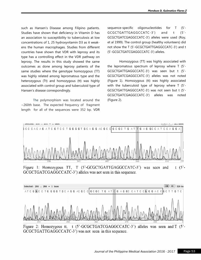
Page 53 Journal of the Philippine Medical Association 2016 - 2017
Mendoza D, Gulmatico-Flores Z
such as Hansen‟s Disease among Filipino patients.
Studies have shown that deficiency in Vitamin D has
an association to susceptibility to tuberculosis at low
concentrations of 1, 25 hydroxyvitamin D3 as it weak-
ens the human macrophages. Studies from different
countries have shown that VDR with leprosy and its
type has a controlling effect in the VDR pathway on
leprosy. The results in this study showed the same
outcomes as done among leprosy patients of the
same studies where the genotype homozygous (TT)
was highly related among lepromatous type and the
heterozygous (Tt) and homozygous (tt) was highly
associated with control group and tuberculoid type of
Hansen‟s disease correspondingly.
The polymorphism was located around the
~260th base. The expected frequency of fragment
length for all of the sequences were 352 bp. VDR
sequence-specific oligonucleotides for T (5‟-
GCGCTGATTGAGGCCATC-3‟) and t (5‟-
GCGCTGATCGAGGCCATC-3‟) alleles were used (Roy,
et al 1999). The control group (healthy volunteers) did
not show the T (5‟-GCGCTGATTGAGGCCATC-3‟) and t
(5‟-GCGCTGATCGAGGCCATC-3‟) alleles.
Homozygous (TT) was highly associated with
the lepromatous spectrum of leprosy where T (5‟-
GCGCTGATTGAGGCCATC-3‟) was seen but t (5‟-
GCGCTGATCGAGGCCATC-3‟) alleles was not noted
(Figure 1). Homozygous (tt) was highly associated
with the tuberculoid type of leprosy where T (5‟-
GCGCTGATTGAGGCCATC-3‟) was not seen but t (5‟-
GCGCTGATCGAGGCCATC-3‟) alleles was noted
(Figure 2).
Page 54 Journal of the Philippine Medical Association 2016 - 2017
Association of Vitamin D Receptor Gene Polymorphisms in the Occurrence and Spectrum
of Leprosy in Filipino Patients Seen at Jose R. Reyes Memorial Medical Center
DISCUSSION
The association between VDR genotype and
susceptibility to leprosy observed in this study pro-
vides further evidence for the proposal that the vitamin
D receptor may be an immune response gene regulat-
ing susceptibility to infectious disease in humans.
There is epidemiologic evidence linking vitamin D defi-
ciency and susceptibility to tuberculosis and in vitro
data indicating an effect of the active metabolite of
vitamin D (1,25 D3), on mycobacterial growth.3 Addi-
tion of physiologic concentrations of 1,25 D3 impairs
growth of Mycobacterium tuberculosis in human
macrophages and monocytic cell lines.5 Such in vitro
studies are not possible for Mycobacterium leprae, but
early studies of the treatment of leprosy with medica-
tions containing vitamin D are consistent with a possi-
ble immunomodulatory effect on this bacterium.6
This case-control study identifying associations
of VDR with leprosy and leprosy type in Filipino pa-
tients provides evidence for a modulatory effect of the
VDR pathway on leprosy as well as further data sup-
porting the functional relevance of variation in or near
the VDR gene. The differential association of VDR
genotype with leprosy type is of particular interest in
relation to the genetic control of cellular and humoral
immune responses.7 Leprosy provides a model for un-
derstanding human immune responses to infection in
that the disease presents as a spectrum in which the
clinical manifestations correlate with the level of cell-
mediated immunity to the bacterial pathogen.1, 3
Tu-
berculoid spectrum presents with strong cellular im-
mune response with skin lesions that contain well-
organized granulomas with very few bacilli. In contrast,
lepromatous leprosy patients have a stronger humoral
immune response but a weak cellular response with
bacilli-laden macrophages.8
Increasing evidence indicates that tuberculoid
leprosy is associated with a predominantly Th1-type
pattern of cytokine production in T cells from skin and
peripheral blood and conversely, lepromatous leprosy
is associated with a more Th2-shifted pattern of cyto-
kine production.9, 10
There is considerable interest in
identifying the genetic and other factors that deter-
mine whether a Th1- or Th2-type response is made to
an antigen or foreign pathogens, as this often corre-
lates with resistance or susceptibility. These leprosy
data suggest that one of the genetic factors influencing
this Th1-Th2 shift in humans may be VDR genotype,
with tt homozygotes tending to produce a TH1-type
immune response and TT homozygotes producing a
Th2-type response. This possibility is compatible with
the observation that tt homozygotes have been found
to be strongly correlated with the tuberculoid spectrum
of leprosy and TT homozygotes were related to the
lepromatous type, as seen in this study. Of interest, in
leprosy, the tt genotype is associated with tuberculoid
type characterized by a strong cellular response but
not with resistance to leprosy per se, which was associ-
ated with heterozygosity for VDR genotype.
The role of the VDR gene in regulating the im-
mune response may have a potential application with
regard to alternative diagnostics and therapeutics that
could solve the public health problem that leprosy still
represents. Moreover, the genetic association study of
the VDR gene polymorphism with infectious diseases
like leprosy and tuberculosis can be a starting point for
a better understanding of these diseases.
CONCLUSION
The polymorphism of VDR genotypes at
codon 352 in exon 9 was highly significant in between
the control, lepromatous, and tuberculoid type of Han-
sen‟s disease. Homozygous TT was highly associated
with lepromatous type (odds ratio = 16, p=< 0.001),
and homozygous tt is highly related to the tuberculoid
type (odds ratio = 5.8, p=< 0.001).
These data suggesting that VDR genotype may
influence the Th1-Th2 pattern of immune response in
leprosy encourage assessment of the role of this ge-
netic locus in a wide variety of human infectious and
autoimmune diseases.
ACKNOWLEDGEMENTS
We would like to thank the first year medicine
students of Our Lady of Fatima University SY 2014-
2015 who helped us make this study possible in
coordinating with PCR reading and in bringing the
specimens to the Philippine Genome Sequencing.
Page 55 Journal of the Philippine Medical Association 2016 - 2017
Mendoza D, Gulmatico-Flores Z
REFERENCES
1. Goulart et al. Interaction of Taq1 polymorphism
at exon 9 of the vitamin D receptor gene with the
negative lepromin response may favor the occur-
rence of leprosy. FEMS Immunol Med Microbiol
2006; 48. 91-98
2. Ridley DS & Jopling WH. Classification of leprosy
according to immunity: a five groups system. Int J
Leprosy 1966; 34: 255–273.
3. Roy S, Frodsham A, Saha B et al. Association of
vitamin D receptor genotype with leprosy type. J
Infect Dis 1999; 179: 187–191.
4. Eisman JA. Vitamin D receptor gene variants: im-
plications for therapy. Curr Opin Genet Develop
1996; 6:361–5.
5. Rook GA, Steele J, Fraher L, et al. Vitamin D3,
gamma interferon, and control of proliferation of
Mycobacterium tuberculosis by human monocytes.
Immunology 1986; 57:159–63.
6. Rogers L. Chaulmoogra oil in leprosy and tuber-
culosis. Lancet 1921; 1: 1178–80.
7. RogerM, LeveeG, ChanteauS, GicquelB, SchurrE.
No evidence for linkage between leprosy suscepti-
bility and the human natural resistance-associated
macrophage protein 1 (NRAMP1) gene in French
Polynesia. Int J Lepr Other Mycobact Dis 1997;
65:197–202.
8. Goulart IMB, Penna GO & Cunha G. Immunopa-
thology of leprosy: the complexity of the mecha-
nisms of host immune response to Mycobacterium
leprae. Rev Soc Bras Med Trop 2002; 35: 365–375.
9. Misra N, Murtaza A, Walker B, et al. Cytokine
profile of circulating T cells in leprosy patients
reflects both indiscriminate and polarized T-helper
subsets. Immunology 1995; 86:97–103.
10. Yamamura M, Uyemura K, Deans RJ, et al. Defining
protective responses to pathogens: cytokine pro-
files in leprosy lesions. Science 1991; 254:277–9.

Patients of Different Body Mass Index
Classification and its Effect on Tube Current Time
Product, Scanner Radiation Output and Image
Quality as Applied in Multidetector Computed
Tomography of the Whole Abdomen –
A Retrospective Study
Ginalynn Fincale, M.D.a, and Jackson Dy, M.D.
b
Abstract
OBJECTIVE: This study aimed to retrospectively determine if there is significant difference in
the tube current-time product (mAs) used and radiation output in patients of different BMI
classification undergoing CT study, and how this would affect image quality.
METHODS: This study included 144 adult patients who underwent CT of abdomen. Patients
were classified according to their BMI. The mAs is set by the CT machine, adjusted according to
patient cross section. Radiation output parameters were collected after each study. Qualitative
evaluation of images was done based on image noise and effect on diagnostic accuracy.
RESULTS: Correlation Analysis showed significant increase in mAs and radiation output in
groups of patient with higher BMI. There is however no significant difference in image quality
between BMI groups, such that there is no significant improvement in image quality when mAs
is increased.
CONCLUSION: This study reiterates the need for body size-adapted CT protocol in imaging
patients since the radiation dose required in patients of higher BMI is increased to maintain di-
agnostic quality of images.
Patients of Different Body Mass Index Classification and its Effect on Tube Current Time Product, Scanner Radiation Output
and Image Quality as Applied in Multidetector Computed Tomography of the Whole Abdomen – A Retrospective Study
Page 56 Journal of the Philippine Medical Association 2016 - 2017
aCT-MRI Fellow-in-Training, Department of Radiology, Makati Medical Center bChairman, Research Adviser, Department of Radiology, Makati Medical Center
INTRODUCTION and REVIEW OF RELATED
LITERATURE
According to the International Atomic Energy
Agency (IAEA), there has been a steady increase in
the number of computed tomography (CT) studies
performed that on an average, it now contributes 60
to 70% of patient dose for any given center. This is
despite the fact that CT only makes up about 15% of
all x-ray based imaging studies (16). CT imaging has
revolutionized the way clinicians are able to manage
their patient that it is not surprising that the number
of CT studies is continuously increasing since the in-
ception of the first CT machines in the early 1970s.
However, the benefits of CT studies come with risks
as expected for an imaging modality using radiation.
The American College of Radiology has released a
White Paper stating that “medical exposure might be
responsible for approximately 1% of the cancer in the
United States” (1). The upside with these events is
that it has forced the medical community to re-
evaluate the risk-benefit of ordering a CT study.
The steady and expected continuous increase
in the utilization of CT poses a challenge to the ra-
diologic community to uphold the principle of ALARA
(as low as reasonably achievable). A topic associated
in CT dose reduction and management is quantifying
CT radiation dose. The scanner radiation output is
most commonly represented by volume averaged CT
dose index (CTDl), a measure of the amount of radia-
tion delivered from a series of contiguous irradiations
to standardized acrylic phantoms (10). Thus CTDI is
not a reflection of dose for an individual patient,
rather represents the dose within the scan volume
from a particular scan protocol. The SI unit is milligray
(mGy). Dose-Length Product (DLP) on the other hand,
is an indicator of the integrated radiation dose of an
entire CT examination (8). It is the product of CTDI
and scan length (z-extent), and is given in units of
milligray-centimeters. DLP better represents the over-
all energy delivered by a given scan protocol (10).
The effective dose (E), is not actually a measurement
of dose, but rather reflects the stochastic risk from an
exposure to ionizing radiation (14). What it does is
it allows an approximate comparison of radiation
induced risk among different types of examinations
(18). It takes into account the tissue that has
absorbed the radiation, and attempts to approximate
whole-body radiation, and thus is a weighted average
of organ doses (13). Be wary that effective dose
should not be used to predict absolute risk for an
actual individual patient. Effective dose is measured
in millisieverts (mSv). DLP values can be converted
from mGy-cm to mSv using a weighting factor (k)
coefficient (in mSv/[mGy-cm]) (9, 10, 13).
The benefit of an appropriately indicated CT
scan exceeds the associated risk, and it falls on the
radiologist to make an accurate diagnosis vis-à-vis
using the minimal amount of radiation required to
obtain images adequate for evaluating the patient‟s
condition. Reasonably, what radiologists do not want
sacrificed in the goal of reducing patient dose is im-
age diagnostic quality. The determination of optimal
image quality is a complicated task and is assessed
by both objective and subjective measures, the latter
affected by inter-observer variation regarding the
acceptable image noise. Two of the most commonly
used image quality parameters are spatial resolution
and low contrast resolution. The former is determined
by the number of rays and spacing of the detectors in
the CT machine for a projection. Contrast resolution
on the other hand is determined solely by noise (6).
Noise describes the variation in CT numbers in a
physically uniform region. (18). The scan time, tube
current and peak kilovoltage, pitch, and slice thick-
ness all affect noise. More importantly, patient size is
the lone parameter that cannot be controlled (9).
The tube current-time product with SI unit of
milliamperage per second (mAs) is a measure of the
photon flux and is the product of the tube current
and exposure time. Radiation dose is directly propor-
tional to mAs if all other factors are held constant.
One can reduce tube current-time product by either
lowering the tube current or increasing the x-ray tube
rotation speed (decreasing the rotation time). X-rays
are attenuated by soft tissue in an exponential fash-
ion. To maintain a consistent noise level from thin to
obese patient, the mAs setting would have to in-
crease exponentially (9).
The relationship between kilovolt peak (kVp)
and noise is less direct. Tube potential is defined
as the x ray beam energy (5). Radiation dose is
Fincale G, Dy J
Page 57 Journal of the Philippine Medical Association 2016 - 2017
Patients of Different Body Mass Index Classification and its Effect on Tube Current Time Product, Scanner Radiation Output
and Image Quality as Applied in Multidetector Computed Tomography of the Whole Abdomen – A Retrospective Study
Page 58 Journal of the Philippine Medical Association 2016 - 2017
is proportional to kVp raised to an exponential nth
power, with n ranging from 2.49 to 3.12 depending on
patient size (9, 17). For example, in a typical abdominal
CT phantom, decreasing the peak voltage from 120 to
100 kVp reduces the dose by 28%-40%, and as much
as 65% if kVp is further reduced to 80 kVp. The down-
side of decreasing kVp is an increase in image noise
due to reduced photon flux and photon energy (9).
The use of iodinated contrast media may further im-
prove image quality, since the attenuation coefficient
of iodine increase as photon energy decrease toward
the k-edge energy of 33 keV. Thus pathologies that
take up contrast are more conspicuous at lower kVp
(12). The benefit of lower-kV for non-contrast CT ex-
ams is still in question since soft tissue contrast is not
highly dependent on tube potential.
Automatic Exposure Control (AEC) systems are
now installed in almost all major scanners manufac-
tured in recent years, with the aim of adjusting radia-
tion dose according to the patient‟s attenuation. It as-
sesses the size of the patient cross-section being
scanned and adjusts tube current relative to the refer-
ence effective mAs. In turn, the reference mAs is based
on an “average size” patient, which is 70-80 kg for
adults and 20 kg for pediatric patients. Pre-set peak
kVp settings of 80, 100, 120 and 140 are found in the
system. With this, image quality is maintained using
the minimum required radiation exposure and can re-
duce dose by as much as 20-44% (8). Even with an ex-
isting AEC, the CT user must still be familiar with the
parameters employed to ensure correct use. Any type
of AEC system is not perfect, since it may not be tech-
nically feasible to maintain constant image noise over
all patient sizes since it cannot achieve such extremely
low and high tube current-time product values (10).
STUDY OBJECTIVES
1. To determine if there is significant difference in the
value of the tube current-time product (mAs)
among patients of different body mass index (BMI)
classification.
2. To determine if the radiation output, measured as
CT dose index (CTDl), Dose-Length Product (DLP)
and effective dose (E), in patients of higher BMI
classification is significantly increased due to in-
creased tube current-time product (mAs).
3. To determine if the difference in tube current-
time product (mAs) among patients of different
BMI classification would significantly affect image
quality.
Research hypothesis:
Null Hypothesis: Significant increase in tube current-
time product (mAs) and thus radiation output is
needed in patients of higher BMI classification to
maintain image quality.
Alternate Hypothesis: Significant increase in tube
current-time product (mAs) and thus radiation output
is not needed in patients of higher BMI classification
to maintain image quality.
METHODOLOGY
A. Justification of Study Design
A retrospective, cross sectional study was
conducted. This was a cross sectional study because
of the following factors: body mass index (BMI), tube
current-time product (mAs), CT dose index (CTDl),
Dose-Length Product (DLP) and effective does (E)
were ascertained simultaneously within a defined
time period.
B. Study Duration and Location.
The study was conducted in the Department
of Radiology, Makati Medical Center. Included data
were of patients who underwent CT of the abdomen
following the inclusion criteria from April 2014 to July
2014.
C. Inclusion and Exclusion Criteria
Inclusion criteria:
1. Male and female adult patients ages 18 years
old and above.
2. Studies requested for out-patients only.
3. CT whole abdomen with intravenous (IV)
contrast which will include:
a. CT whole abdomen with intravenous (IV)
contrast.
b. CT chest and whole abdomen with IV
contrast.
c. CT whole abdomen with intravenous (IV)
contrast, oral and/or rectal contrast.
d. CT chest and whole abdomen with intra-
venous (IV) contrast, oral and/or rectal
contrast.
Exclusion criteria:
1. Pediatric patients/patients below 18 years
old.
2. Patients without available height measure-
ment needed for BMI measurement (e.g.
wheelchair bound patients).
3. CT whole abdomen without IV contrast to
eliminate discrepancy in image quality as-
sessment between plain and contrast en-
hanced studies.
D. CT system and scanning protocol
The scanner used in this study was the Sie-
mens Somatom Definition 128 MDCT machine. Pa-
tients were scanned using the available online tube
current modulation software. The CT scanner Sie-
mens Somatom has a reference milliamperage-based
AEC system called CARE Dose. In this system, the
peak kilovoltage setting in the CT console is set auto-
matically to 120 kVp. Tube current-time product
(mAs) is then set by Care Dose based on the patient
cross-section measured from the initial tomogram
scanning. The parameters set by the AEC system for
each scanned patient including kVP and mAs, are
seen in the CT scanner console and sent in the PACS
file of each patient for reference. (Appendix 1).
The other components of the scanning pro-
tocol were the same as per usual in the department:
single breath hold, 0.5 second rotation time, 0.6 pitch,
individually adapted field of view, and a B20f smooth
software reconstruction kernel. Images were recon-
structed as 2 mm. thick sections with an increment of
2 mm. The contrast enhanced CT whole abdominal
protocol in the department has three phases; arterial,
portal venous and delayed. The amount of intrave-
nous iodinated contrast (370 mg iodine/ml) given is
115 to 120 mL., administered at a rate of 2.5 to 3.0
mL/sec, depending on patient weight and catheter
size.
E. Data Collection
Patient:
All out patients undergoing a diagnostic
exam in the department have their gender, age,
height (cm2) and weight (kg) obtained and recorded
as part of completion of patient chart. Access to pa-
tient records was facilitated by the CT unit manager
Joel Molina, RRT (Registered Radiologic Technolo-
gists) who is in charge of safe keeping of said re-
cords. Permission to access these records were given
by the department chair, Jackson Dy MD. The pa-
tients were categorized according to their BMI based
on the World Health Organization BMI classification:
underweight (< 18.5 kg/m2), normal (18.5 – 24. 99),
overweight (25 – 29.99), obese class I (30 – 34.99),
obese class II (35 – 39.99) and obese class III (>/= 40).
Dose measurement:
Dose values based on the CT dose index
(CTDl), dose length product (DLP) computed by the
CT system were collected as seen in the CT scanner
console and sent in the PACS file of each patient. The
DLP values were then converted to effective dose (E)
using the region specific coefficient for the abdomen
(k = 0.015 mSc/mGy x cm.) as set by the International
Commission on Radiation Protection (ICRP 60).
The acquired patient information and dose
measurements were tabulated on a standard data
collection template.
F. Image Quality Evaluation
Qualitative evaluation of images was done by
two CT consultants from the Department of Radiol-
ogy. Both were blinded as to the BMI of each patient.
The images only from the portal venous phase of the
Fincale G, Dy J
Page 59 Journal of the Philippine Medical Association 2016 - 2017
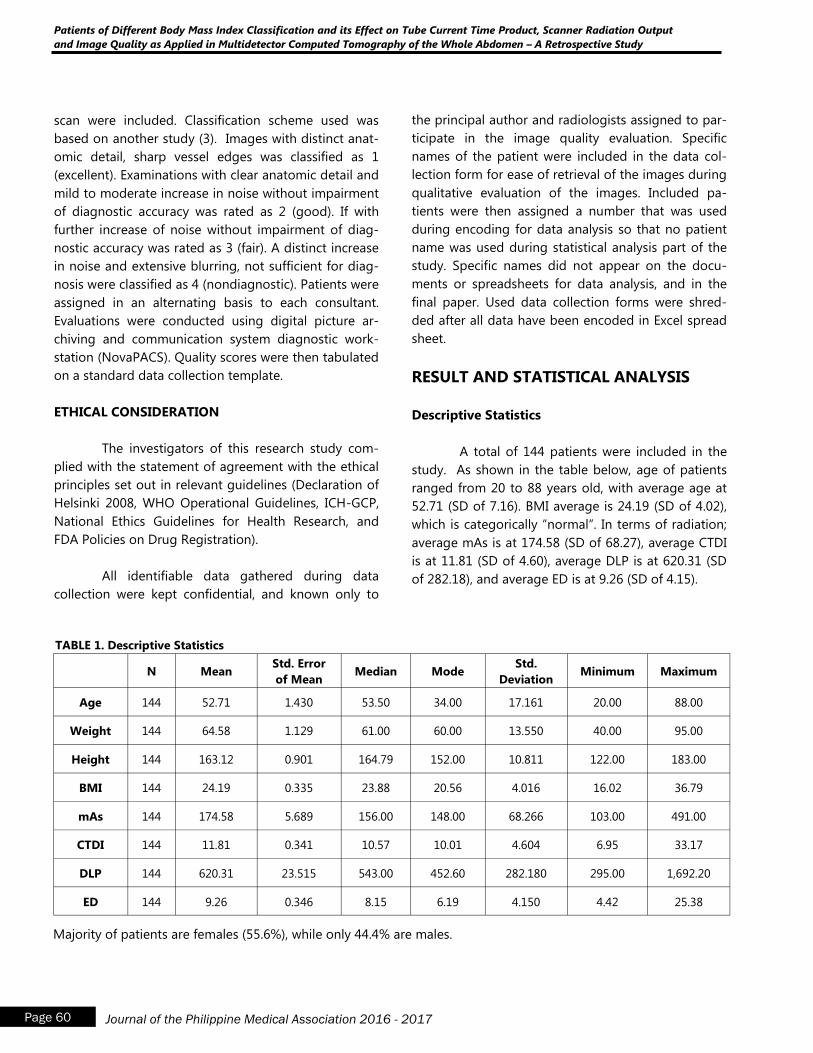
Patients of Different Body Mass Index Classification and its Effect on Tube Current Time Product, Scanner Radiation Output
and Image Quality as Applied in Multidetector Computed Tomography of the Whole Abdomen – A Retrospective Study
Page 60 Journal of the Philippine Medical Association 2016 - 2017
scan were included. Classification scheme used was
based on another study (3). Images with distinct anat-
omic detail, sharp vessel edges was classified as 1
(excellent). Examinations with clear anatomic detail and
mild to moderate increase in noise without impairment
of diagnostic accuracy was rated as 2 (good). If with
further increase of noise without impairment of diag-
nostic accuracy was rated as 3 (fair). A distinct increase
in noise and extensive blurring, not sufficient for diag-
nosis were classified as 4 (nondiagnostic). Patients were
assigned in an alternating basis to each consultant.
Evaluations were conducted using digital picture ar-
chiving and communication system diagnostic work-
station (NovaPACS). Quality scores were then tabulated
on a standard data collection template.
ETHICAL CONSIDERATION
The investigators of this research study com-
plied with the statement of agreement with the ethical
principles set out in relevant guidelines (Declaration of
Helsinki 2008, WHO Operational Guidelines, ICH-GCP,
National Ethics Guidelines for Health Research, and
FDA Policies on Drug Registration).
All identifiable data gathered during data
collection were kept confidential, and known only to
the principal author and radiologists assigned to par-
ticipate in the image quality evaluation. Specific
names of the patient were included in the data col-
lection form for ease of retrieval of the images during
qualitative evaluation of the images. Included pa-
tients were then assigned a number that was used
during encoding for data analysis so that no patient
name was used during statistical analysis part of the
study. Specific names did not appear on the docu-
ments or spreadsheets for data analysis, and in the
final paper. Used data collection forms were shred-
ded after all data have been encoded in Excel spread
sheet.
RESULT AND STATISTICAL ANALYSIS
Descriptive Statistics
A total of 144 patients were included in the
study. As shown in the table below, age of patients
ranged from 20 to 88 years old, with average age at
52.71 (SD of 7.16). BMI average is 24.19 (SD of 4.02),
which is categorically “normal”. In terms of radiation;
average mAs is at 174.58 (SD of 68.27), average CTDI
is at 11.81 (SD of 4.60), average DLP is at 620.31 (SD
of 282.18), and average ED is at 9.26 (SD of 4.15).
N Mean Std. Error
of Mean Median Mode
Std.
Deviation Minimum Maximum
Age 144 52.71 1.430 53.50 34.00 17.161 20.00 88.00
Weight 144 64.58 1.129 61.00 60.00 13.550 40.00 95.00
Height 144 163.12 0.901 164.79 152.00 10.811 122.00 183.00
BMI 144 24.19 0.335 23.88 20.56 4.016 16.02 36.79
mAs 144 174.58 5.689 156.00 148.00 68.266 103.00 491.00
CTDI 144 11.81 0.341 10.57 10.01 4.604 6.95 33.17
DLP 144 620.31 23.515 543.00 452.60 282.180 295.00 1,692.20
ED 144 9.26 0.346 8.15 6.19 4.150 4.42 25.38
TABLE 1. Descriptive Statistics
Majority of patients are females (55.6%), while only 44.4% are males.
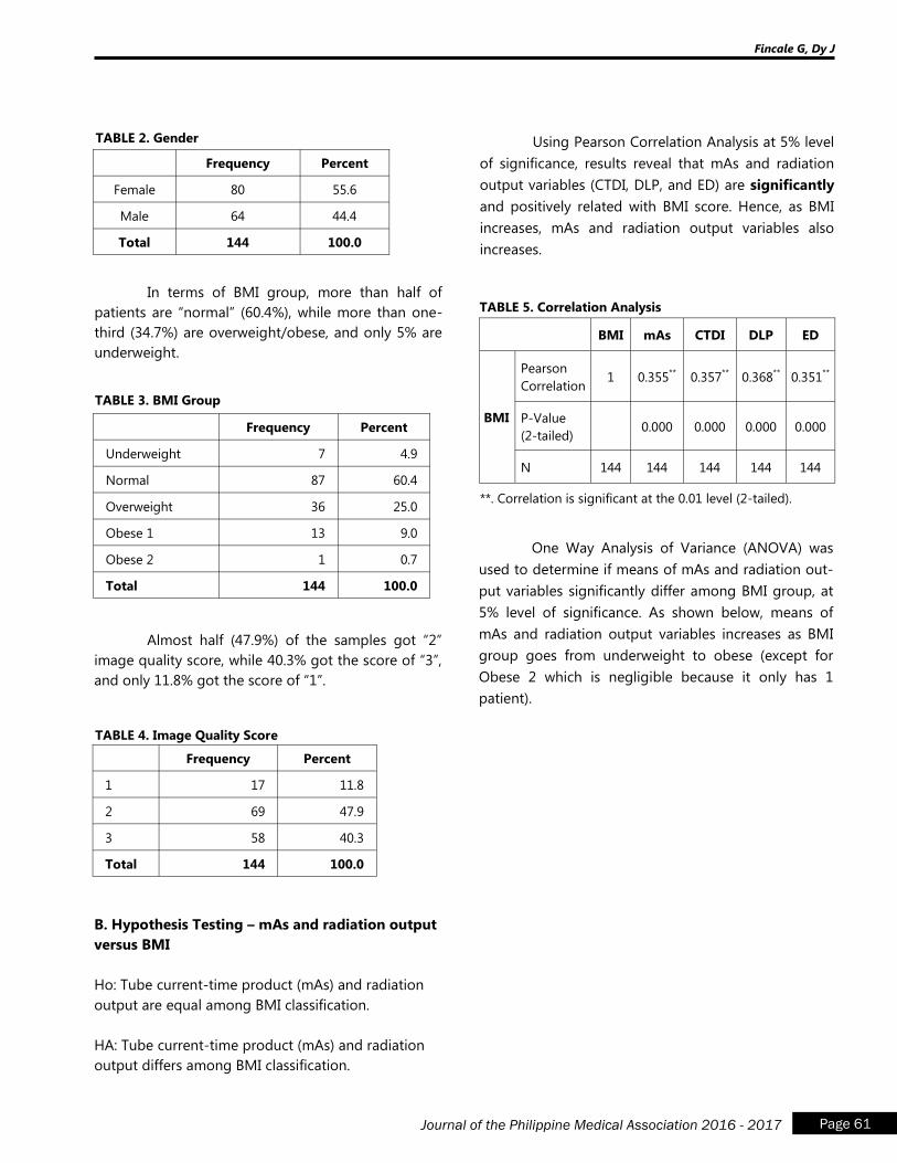
Fincale G, Dy J
Page 61 Journal of the Philippine Medical Association 2016 - 2017
Frequency Percent
Female 80 55.6
Male 64 44.4
Total 144 100.0
TABLE 2. Gender
In terms of BMI group, more than half of
patients are “normal” (60.4%), while more than one-
third (34.7%) are overweight/obese, and only 5% are
underweight.
Frequency Percent
Underweight 7 4.9
Normal 87 60.4
Overweight 36 25.0
Obese 1 13 9.0
Obese 2 1 0.7
Total 144 100.0
TABLE 3. BMI Group
Almost half (47.9%) of the samples got “2”
image quality score, while 40.3% got the score of “3”,
and only 11.8% got the score of “1”.
Frequency Percent
1 17 11.8
2 69 47.9
3 58 40.3
Total 144 100.0
TABLE 4. Image Quality Score
B. Hypothesis Testing – mAs and radiation output
versus BMI
Ho: Tube current-time product (mAs) and radiation
output are equal among BMI classification.
HA: Tube current-time product (mAs) and radiation
output differs among BMI classification.
Using Pearson Correlation Analysis at 5% level
of significance, results reveal that mAs and radiation
output variables (CTDI, DLP, and ED) are significantly
and positively related with BMI score. Hence, as BMI
increases, mAs and radiation output variables also
increases.
BMI mAs CTDI DLP ED
BMI
Pearson
Correlation 1 0.355
** 0.357
** 0.368
** 0.351
**
P-Value
(2-tailed) 0.000 0.000 0.000 0.000
N 144 144 144 144 144
TABLE 5. Correlation Analysis
**. Correlation is significant at the 0.01 level (2-tailed).
One Way Analysis of Variance (ANOVA) was
used to determine if means of mAs and radiation out-
put variables significantly differ among BMI group, at
5% level of significance. As shown below, means of
mAs and radiation output variables increases as BMI
group goes from underweight to obese (except for
Obese 2 which is negligible because it only has 1
patient).
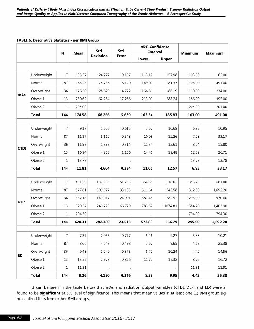
N Mean Std.
Deviation
Std.
Error
95% Confidence
Interval Minimum Maximum
Lower Upper
mAs
Underweight 7 135.57 24.227 9.157 113.17 157.98 103.00 162.00
Normal 87 165.23 75.736 8.120 149.09 181.37 105.00 491.00
Overweight 36 176.50 28.629 4.772 166.81 186.19 119.00 234.00
Obese 1 13 250.62 62.254 17.266 213.00 288.24 186.00 395.00
Obese 2 1 204.00 . . . . 204.00 204.00
Total 144 174.58 68.266 5.689 163.34 185.83 103.00 491.00
CTDI
Underweight 7 9.17 1.626 0.615 7.67 10.68 6.95 10.95
Normal 87 11.17 5.112 0.548 10.08 12.26 7.08 33.17
Overweight 36 11.98 1.883 0.314 11.34 12.61 8.04 15.80
Obese 1 13 16.94 4.203 1.166 14.41 19.48 12.59 26.71
Obese 2 1 13.78 . . . . 13.78 13.78
Total 144 11.81 4.604 0.384 11.05 12.57 6.95 33.17
DLP
Underweight 7 491.29 137.030 51.793 364.55 618.02 355.70 681.00
Normal 87 577.61 309.527 33.185 511.64 643.58 312.30 1,692.20
Overweight 36 632.18 149.947 24.991 581.45 682.92 295.00 970.60
Obese 1 13 929.32 240.775 66.779 783.82 1074.81 584.20 1,403.90
Obese 2 1 794.30 . . . . 794.30 794.30
Total 144 620.31 282.180 23.515 573.83 666.79 295.00 1,692.20
ED
Underweight 7 7.37 2.055 0.777 5.46 9.27 5.33 10.21
Normal 87 8.66 4.643 0.498 7.67 9.65 4.68 25.38
Overweight 36 9.48 2.249 0.375 8.72 10.24 4.42 14.56
Obese 1 13 13.52 2.978 0.826 11.72 15.32 8.76 16.72
Obese 2 1 11.91 . . . . 11.91 11.91
Total 144 9.26 4.150 0.346 8.58 9.95 4.42 25.38
Patients of Different Body Mass Index Classification and its Effect on Tube Current Time Product, Scanner Radiation Output
and Image Quality as Applied in Multidetector Computed Tomography of the Whole Abdomen – A Retrospective Study
Page 62 Journal of the Philippine Medical Association 2016 - 2017
TABLE 6. Descriptive Statistics - per BMI Group
It can be seen in the table below that mAs and radiation output variables (CTDI, DLP, and ED) were all
found to be significant at 5% level of significance. This means that mean values in at least one (1) BMI group sig-
nificantly differs from other BMI groups.
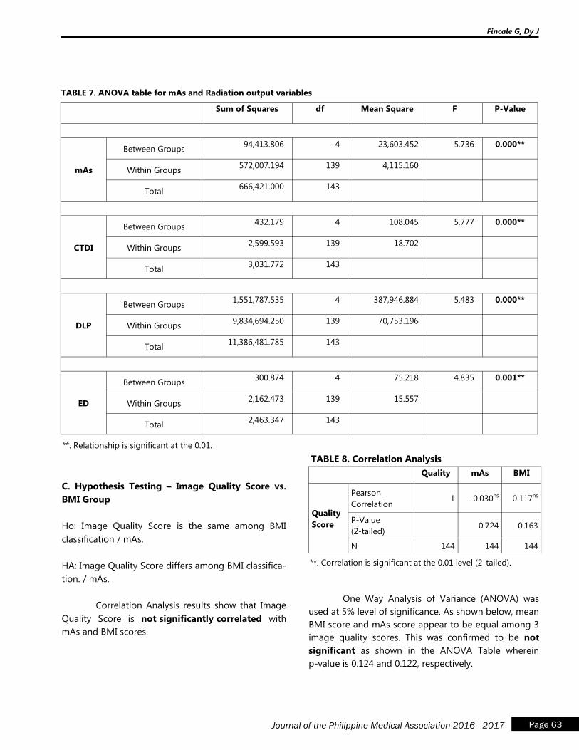
Fincale G, Dy J
Page 63 Journal of the Philippine Medical Association 2016 - 2017
Sum of Squares df Mean Square F P-Value
mAs
Between Groups 94,413.806 4 23,603.452 5.736 0.000**
Within Groups 572,007.194 139 4,115.160
Total 666,421.000 143
CTDI
Between Groups 432.179 4 108.045 5.777 0.000**
Within Groups 2,599.593 139 18.702
Total 3,031.772 143
DLP
Between Groups 1,551,787.535 4 387,946.884 5.483 0.000**
Within Groups 9,834,694.250 139 70,753.196
Total 11,386,481.785 143
ED
Between Groups 300.874 4 75.218 4.835 0.001**
Within Groups 2,162.473 139 15.557
Total 2,463.347 143
TABLE 7. ANOVA table for mAs and Radiation output variables
**. Relationship is significant at the 0.01.
C. Hypothesis Testing – Image Quality Score vs.
BMI Group
Ho: Image Quality Score is the same among BMI
classification / mAs.
HA: Image Quality Score differs among BMI classifica-
tion. / mAs.
Correlation Analysis results show that Image
Quality Score is not significantly correlated with
mAs and BMI scores.
Quality mAs BMI
Quality
Score
Pearson
Correlation 1 -0.030ns 0.117ns
P-Value
(2-tailed) 0.724 0.163
N 144 144 144
TABLE 8. Correlation Analysis
**. Correlation is significant at the 0.01 level (2-tailed).
One Way Analysis of Variance (ANOVA) was
used at 5% level of significance. As shown below, mean
BMI score and mAs score appear to be equal among 3
image quality scores. This was confirmed to be not
significant as shown in the ANOVA Table wherein
p-value is 0.124 and 0.122, respectively.
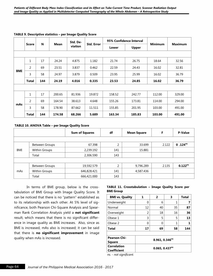
Patients of Different Body Mass Index Classification and its Effect on Tube Current Time Product, Scanner Radiation Output
and Image Quality as Applied in Multidetector Computed Tomography of the Whole Abdomen – A Retrospective Study
Page 64 Journal of the Philippine Medical Association 2016 - 2017
TABLE 9. Descriptive statistics – per Image Quality Score
In terms of BMI group, below is the cross-
tabulation of BMI Group with Image Quality Score. It
can be noticed that there is no “pattern” established as
to its relationship with each other. At 5% level of sig-
nificance, both Pearson Chi-Square Analysis and Spear-
man Rank Correlation Analysis yield a not significant
result, which means that there is no significant differ-
ence in image quality as BMI increases. Also, since as
BMI is increased, mAs also is increased; it can be said
that there is no significant improvement in image
quality when mAs is increased.
Score N Mean Std. De-
viation Std. Error
95% Confidence Interval Minimum Maximum
Lower Upper
BMI
1 17 24.24 4.875 1.182 21.74 26.75 18.64 32.56
2 69 23.51 3.837 0.462 22.59 24.43 16.02 32.81
3 58 24.97 3.879 0.509 23.95 25.99 16.02 36.79
Total 144 24.19 4.016 0.335 23.53 24.85 16.02 36.79
mAs
1 17 200.65 81.936 19.872 158.52 242.77 112.00 329.00
2 69 164.54 38.613 4.648 155.26 173.81 114.00 294.00
3 58 178.90 87.662 11.511 155.85 201.95 103.00 491.00
Total 144 174.58 68.266 5.689 163.34 185.83 103.00 491.00
Sum of Squares df Mean Square F P-Value
BMI
Between Groups 67.398 2 33.699 2.122 0 .124ns
Within Groups 2,239.192 141 15.881
Total 2,306.590 143
mAs
Between Groups 19,592.579 2 9,796.289 2.135 0.122ns
Within Groups 646,828.421 141 4,587.436
Total 666,421.000 143
TABLE 10. ANOVA Table – per Image Quality Score
BMI vs. Quality 1 2 3 Total
Underweight 0 6 1 7
Normal 12 40 35 87
Overweight 2 18 16 36
Obese 1 3 5 5 13
Obese 2 0 0 1 1
Total 17 69 58 144
Pearson Chi-
Square 8.961, 0.346ns
Correlation
Coefficient 0.065, 0.437ns
TABLE 11. Crosstabulation – Image Quality Score per
BMI Group
ns. - not significant.
Fincale G, Dy J
Page 65 Journal of the Philippine Medical Association 2016 - 2017
DISCUSSION and RECOMMENDATION
This research aimed to determine whether
there is significant difference in tube current-time
product (mAs) and radiation output in patients of
different BMI classification. Pearson Correlation
Analysis and ANOVA showed there is indeed signifi-
cant increase in mAs as BMI increased (Table 5 and
6). As expected, there is corresponding increase in all
radiation dose parameters, namely CTDI, DLP and
effective dose (E), as BMI increased (Table 6 and 7).
An increase in radiation dose with increasing BMI was
observed even if the same peak kilovoltage (kVp =
120) was used in all BMI groups. This is due to the
use of the Automatic Exposure Control (AEC) system
in place in the CT scanner which adjusts the mAs to
the size of the patient based on the initial topogram.
Another question asked by this study was
whether there is a difference in the image quality of
CT images of patients of different BMI classification
due to differences in mAs. The most frequent score
given across all BMI groups was 2 or “good”, were in
the images obtained had “clear anatomic detail with
mild to moderate increase in noise but without im-
pairment of diagnostic accuracy” (3). No score of 4 or
a non-diagnostic image was obtained during the
study. Correlation analysis showed that there is no
significant correlation of image quality score with
mAs and BMI (Table 8). Using ANOVA at 5% level of
significance, the mean of the BMI and mAs among
the three quality scores given – namely excellent (1),
good (2) and fair (3), did not differ significantly (Table
9 and 10). This indicates that there is no significant
difference in image quality with increasing mAs and
BMI. In underweight, normal and overweight BMI
groups, the most frequent quality score were all 2,
while in the Obese 1 group, there is equal number of
patients with scores of 2 and 3 (Table 10).
This study reiterates the need for body size-
adapted CT protocol in imaging patients since the
minimal radiation dose required would be varied
even at the same diagnostic task. Several strategies
have been proposed to modulate radiation dose.
One modulation technique is to lower kilovolt-
age (kV) that allows reduction of radiation dose (8).
However, an ideal balance between lowering the kV
with corresponding increase in tube current time prod-
uct (mAs) should be achieved since lowering the mAs
simultaneously would increase contrast to noise ratio,
and thus reduce image quality. Scan range should also
be carefully planned considering the patient‟s body
habitus to avoid exposure of any regions of the body
that are not necessary for diagnosis (18). In this study,
there is maintenance of diagnostic image quality even
with increasing BMI due to increase in mAs. As reiter-
ated by another study, this is the correct strategy since
higher tube current is used at lower tube potential to
compensate for the expected increase in noise (5). In
particular in larger-sized patients, the use of lower
tube potentials tends to increase noise due to higher
absorption of lower-energy photons by the patient
(17). However, even with an AEC system in place to
limit radiation dose, the AEC system would still have to
increase exponentially the mAs to achieve ideal con-
trast to noise ratio. The limitation of an AEC system is
highlighted in this study since there is significant in-
crease in radiation dose with higher BMI groups.
The author recommends future studies that
will evaluate several protocols that may reduce radia-
tion dose used in CT studies not just of the abdomen,
but in evaluation of other body parts. This may be
done by manipulating several scanning parameters
such as kilovoltage, tube current, scan time and range.
A more statistically sound method of evaluating quality
is to compare quality between a control group and a
group undergoing a reduced dose protocol. Patient
safety without compromising image diagnostic quality
should always be the primary aim of these studies.
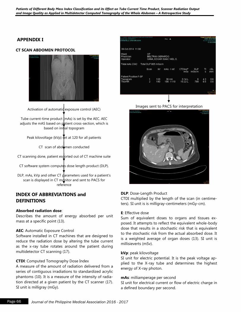
Patients of Different Body Mass Index Classification and its Effect on Tube Current Time Product, Scanner Radiation Output
and Image Quality as Applied in Multidetector Computed Tomography of the Whole Abdomen – A Retrospective Study
Page 66 Journal of the Philippine Medical Association 2016 - 2017
APPENDIX I
CT SCAN ABDOMEN PROTOCOL
Activation of automatic exposure control (AEC)
Tube current-time product (mAs) is set by the AEC. AEC
adjusts the mAS based on patient cross-section, which is
based on initial topogram
Peak kilovoltage (kVp) set at 120 for all patients
CT scan of abdomen conducted
CT scanning done, patient escorted out of CT machine suite
CT software system computes dose length product (DLP).
DLP, mAs, kVp and other CT parameters used for a patient‟s
scan is displayed in CT monitor and sent to PACS for
reference
Images sent to PACS for interpretation
INDEX OF ABBREVIATIONS and
DEFINITIONS
Absorbed radiation dose:
Describes the amount of energy absorbed per unit
mass at a specific point (13).
AEC: Automatic Exposure Control
Software installed in CT machines that are designed to
reduce the radiation dose by altering the tube current
as the x-ray tube rotates around the patient during
multidetector CT scanning (17).
CTDl: Computed Tomography Dose Index
A measure of the amount of radiation delivered from a
series of contiguous irradiations to standardized acrylic
phantoms (10). It is a measure of the intensity of radia-
tion directed at a given patient by the CT scanner (17).
SI unit is milligray (mGy).
DLP: Dose-Length Product
CTDI multiplied by the length of the scan (in centime-
ters). SI unit is is milligray-centimeters (mGy-cm).
E: Effective dose
Sum of equivalent doses to organs and tissues ex-
posed. It attempts to reflect the equivalent whole-body
dose that results in a stochastic risk that is equivalent
to the stochastic risk from the actual absorbed dose. It
is a weighted average of organ doses (13). SI unit is
millisieverts (mSv).
kVp: peak kilovoltage
SI unit for electric potential. It is the peak voltage ap-
plied to the X-ray tube and determines the highest
energy of X-ray photon.
mAs: milliamperage per second
SI unit for electrical current or flow of electric charge in
a defined boundary per second.
Fincale G, Dy J
Page 67 Journal of the Philippine Medical Association 2016 - 2017
mGy: milligray
SI unit for absorbed dose. 1 Gray is equivalent to 1
Joule per kilogram (1 Gy = 1 J/kg).
mSv: millisieverts
SI unit for effective dose. 1 mSv is the dose produced
by exposure to 1 milligray (mG) of radiation.
MDCT: Multi-Detector Computed Tomography
A form of CT technology that uses multiple arrays of
detector which enables to acquire multiple slices si-
multaneously and greatly increases speed of image
acquisition.
REFERENCES
1. American College of Radiology White Paper on
Radiation Dose in Medicine. Journal of the Ameri-
can College of Radiology. Vol. 4. No. 5 May 2007.
2. Antypass E.J., et al. A Comprehensive Approach
to CT Radiation Dose Reduction: One Institu-
tion’s Experience. American Journal of Roent-
genology. 2011; 197:935-940.
3. Das M., et al. Individually Adapted Examination
Protocols for Reduction of Radiation Exposure for
16-MDCT Chest Examinations. American Journal
of Roentgenology. 2005; 184:1437-1443.
4. Goldman A.R. and Maldjian P.D. Reducing Radia-
tion Dose in Body CT: A Practical Approach to
Optimizing CT Protocols. American Journal of
Roentgenology. 2013; 200:748-754.
5. Goo H.W. CT Radiation Dose Optimization and
Estimation: an Update for Radiologists. Korean
Journal of Roentgenology. 2012; 13(1):1-11.
6. International Atomic Energy Agency. Dose Re-
duction in CT while Maintaining Diagnostic Confi-
dence: A Feasibility/Demonstration Study. IAEA,
Vienna, 2009.
7. Kalra M.K., et al. Clinical Comparison of Standard
Dose and 50% Reduced-Dose Abdominal CT: Ef-
fect on Image Quality. American Journal of Ro-
entgenology. 2002; 179:1101-1106.
8. Lee C.H., Goo J.M., Lee H.J., et al. Radiation Dose
Modulation Techniques in the Multidetector CT
Era: From Basics to Practice. RadioGraphics. 2008;
28:1451-1459.
9. Maldjian P.D. and Goldman A.R. Reducing Radia-
tion Dose in Body CT: A Primer on Dose Metrics
and Key CT Technical Parameters. American Jour-
nal of Roentgenology. 2013; 200:741-747.
10. McCollough C., Bruesewitz M., and Kofler J. CT
dose reduction and dose management tools: Over-
view of available options. RadioGraphics. 2006; 26:
503-512.
11. McCollough C.H., Christner J.A. and Kofler J.M.
How Effective is Effective Dose as a Predictor of
Radiation Risk? American Journal of Roentgenol-
ogy. 2010; 194:890-896.
12. McCollough C., et al. Strategies for Reducing Ra-
diation Dose in CT. Radiologic Clinic North Amer-
ica. 2009 January; 47 (1): 27-40.
13. McNitt-Gray M. AAPM/RSNA Physics Tutorial for
Residents: Topics in CT. RadioGraphics. 2002; 22:
1541-1553.
14. Nakayama Y., et al. Abdominal CT with Low Tube
Voltage: Preliminary Observations about Radiation
Dose, Contrast Enhancement, Image Quality and
Noise.
15. Prasad SR., et al. Standard Dose and 50% Reduced-
Dose Chest CT: Comparing the Effect on Image
Quality. American Journal of Roentgenology. 2002;
179:461-465.
16. Tack D. et al. Dose Reduction in Multidetector CT
Using Attenuation-Based Online Tube Current
Modulation. American Journal of Roentgenology.
2003; 181: 331-334.
17. Tamm E., et al. CT Radiation Dose Reduction: How
to Implement Change without Sacrificing Diagnos-
tic Quality. RadioGraphics. 2011; 31:1823-1832.
18. Yu L., et al. Radiation dose reduction in computed
tomography: techniques and future perspective.
Imaging med. 2009 October; 1(1): 65-84.
Acral Lentiginous Melanoma in Filipinos: Report of Two Cases
Page 68 Journal of the Philippine Medical Association 2016 - 2017
Acral Lentiginous Melanoma in Filipinos:
Report of Two Cases
Luella Joy A. Escueta, MD
Ana Maria O. de la Serna-Mah, MD, DPDS
Johannes F. Dayrit, MD, FPDS
Abstract
Introduction: Acral Lentiginous Melanoma is a clinicopathologic subtype of malignant
melanoma. It is the most common expression among the four variants in Asian population. Its
prognosis is generally poorer than the other subtypes. Hence, early diagnosis and surgical
excision is the key in management.
Case Summaries:
A 65-year-old male presented with a 10-year history of patch on the second digit of the right
hand. There was history of trauma to the finger where subungual hematoma developed. Five
years after the injury, a black macule was noted on the previously bruised area. The lesion in-
creased in size involving the periungual area with destruction of the nail plate. Biopsy showed
acanthosis and basal layer hyperpigmentation. Intra-epidermal and dermal vacuolated
melanocytes were seen. The patient was referred to an oncologist and orthopedic surgeon for
management. An 88-year-old male had a 2-year history of black patch on the interdigital area
between the 4th and 5th digits of the left foot. No history of trauma was reported prior to the
appearance of the lesion. Biopsy showed lentiginous proliferation of atypical melanocytes in
the epidermis. Referral to an oncologist and orthopedic surgeon for treatment was made.
Conclusion:
Clinical management of ALM begins with an accurate diagnosis. Therefore it is important to
remain vigilant and where there is clinical suspicion, patients should be referred for a derma-
tological opinion. A typical patient profile, which includes age of onset, location of the lesion,
prior trauma, and its relatively high incidence in the Asian population must be borne in mind.
Department of Dermatology, Research Institute for Tropical Medicine
Escueta LJ, de la Serna-Mah AM, Dayrit J
Page 69 Journal of the Philippine Medical Association 2016 - 2017
INTRODUCTION
Cutaneous malignant melanoma is the most
common cause of mortality from skin cancers in
Caucasian populations. The incidence rate continues
to rise in the UK and Australia. Currently there are
around 8,500 new cases annually in the UK. In 2003,
there were 9,534 new cases of melanoma reported in
Australia.1 In contrast, a significantly lower incidence
rate has been reported in Asian populations with
rates of 0.65-1/100,000.2
There are four distinct categories of mela-
noma based on histological features. These are
superficial spreading (SSM), nodular (NM), lentigo
maligna (LMM), and acral lentiginous (ALM).1-4
The
incidence rates of SSM, NM, LMM and ALM In the
United States are approximately 70%, 15%, 13%
and 2-3% respectively. However, ALM is the most
common expression in Asian and Black populations,
the rate of ALM is 41% in Japan, 65% in Korea and
62% in the American Blacks.2
The prognosis of ALM is generally poorer
than the other subtypes. Patients likely overlook the
lesion, especially on soles and nail beds. Moreover,
ALM may mimic benign morphologies. Hence early
diagnosis and surgical excision is the key in manage-
ment. We report herein two cases of ALM seen in our
institution.
Case 1
A 65-year-old male presented with a 10-year
history of a black patch on the second digit of the
right hand. There was history of trauma to the finger
where subungual hematoma developed. The hema-
toma spontaneously resolved. Five years after the
injury, a solitary black macule was noted on the pre-
viously bruised area. The lesion increased in size in-
volving the periungual area with destruction of the
nail plate. (Figure 1A) Biopsy showed acanthosis and
and basal layer hyperpigmentation due to increased
melanocytic activity. There was elongation and
broadening of the rete pegs. Intra-epidermal and
dermal singly scattered vacuolated melanocytes
that form intraepithelial theques were seen. There
was also pigmented parakeratosis accompanied by
moderate numbers of lymphocytic infiltrates. (Figure
1B) Diagnosis of acral lentiginous melanoma was
made. The patient was then referred to an oncologist
and orthopedic surgeon for management.
Case 2
An 88-year-old male had a 2-year history of
black patch on the interdigital area between the 4th
and 5th digits and medial aspect of the 5th digit of
the left foot. (Figure 2A) No history of trauma was
reported prior to the appearance of the lesion. Skin
biopsy confirmed the diagnosis of acral lentiginous
melanoma. Marked acanthosis, elongation of rete
ridges and lentiginous proliferation of atypical
melanocytes in the epidermis were present. (Figures
2B-D) Referral to an oncologist and orthopedic sur-
geon for treatment was made.
DISCUSSION
Acral Lentiginous Melanoma accounts for 2-
3% of all melanomas.5 Overall incidence of melanoma
is less in dark skinned individuals. However, ALM
makes up a much higher proportion in Asian and
Black populations.2-3
Furthermore, ALM has a very
poor prognosis. In Reed‟s study, patients with ALM
had a mean 3-year survival rate of 11%.6 The poor
survival rate of these patients may have been due in
part to delay in diagnosis. Metzger et al also found
that ALM had a high probability of being clinically
misdiagnosed as benign melanocytic lesion, which
defers the initiation of treatment.7
The mean age at diagnosis for ALM was 62.8
years. Our patients were diagnosed at age 65 and 88
years. Bradford et al conducted a study, which stated
that the lower extremity was most commonly in-
volved than the upper extremity.6 Similarly, Kai and
Fujiwara reported that ALM occurs more frequently
on lower extremities. The soles of the feet are the
most frequent sites of ALM, where 56% of ALM on
lower extremities occurred. In contrast, most frequent
sites on upper extremities are fingernails, where 45%
of ALM on upper extremities appeared2, which is the
case in our first patient.
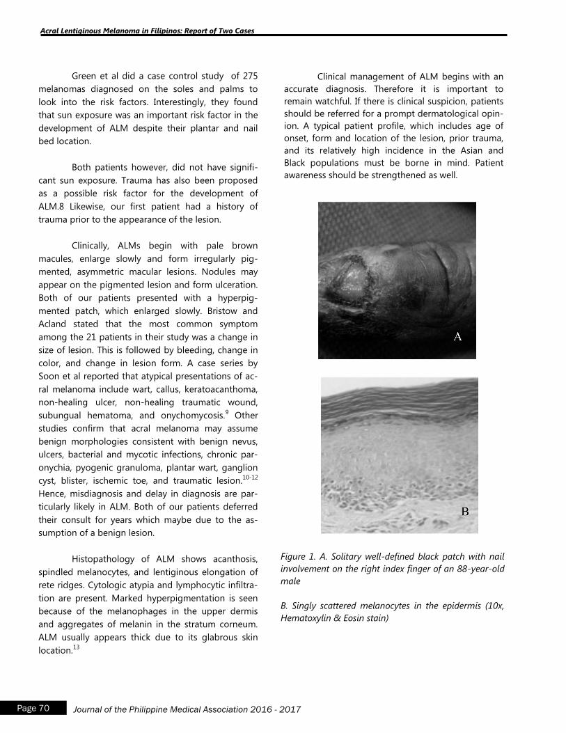
Green et al did a case control study of 275
melanomas diagnosed on the soles and palms to
look into the risk factors. Interestingly, they found
that sun exposure was an important risk factor in the
development of ALM despite their plantar and nail
bed location.
Both patients however, did not have signifi-
cant sun exposure. Trauma has also been proposed
as a possible risk factor for the development of
ALM.8 Likewise, our first patient had a history of
trauma prior to the appearance of the lesion.
Clinically, ALMs begin with pale brown
macules, enlarge slowly and form irregularly pig-
mented, asymmetric macular lesions. Nodules may
appear on the pigmented lesion and form ulceration.
Both of our patients presented with a hyperpig-
mented patch, which enlarged slowly. Bristow and
Acland stated that the most common symptom
among the 21 patients in their study was a change in
size of lesion. This is followed by bleeding, change in
color, and change in lesion form. A case series by
Soon et al reported that atypical presentations of ac-
ral melanoma include wart, callus, keratoacanthoma,
non-healing ulcer, non-healing traumatic wound,
subungual hematoma, and onychomycosis.9 Other
studies confirm that acral melanoma may assume
benign morphologies consistent with benign nevus,
ulcers, bacterial and mycotic infections, chronic par-
onychia, pyogenic granuloma, plantar wart, ganglion
cyst, blister, ischemic toe, and traumatic lesion.10-12
Hence, misdiagnosis and delay in diagnosis are par-
ticularly likely in ALM. Both of our patients deferred
their consult for years which maybe due to the as-
sumption of a benign lesion.
Histopathology of ALM shows acanthosis,
spindled melanocytes, and lentiginous elongation of
rete ridges. Cytologic atypia and lymphocytic infiltra-
tion are present. Marked hyperpigmentation is seen
because of the melanophages in the upper dermis
and aggregates of melanin in the stratum corneum.
ALM usually appears thick due to its glabrous skin
location.13
Clinical management of ALM begins with an
accurate diagnosis. Therefore it is important to
remain watchful. If there is clinical suspicion, patients
should be referred for a prompt dermatological opin-
ion. A typical patient profile, which includes age of
onset, form and location of the lesion, prior trauma,
and its relatively high incidence in the Asian and
Black populations must be borne in mind. Patient
awareness should be strengthened as well.
Figure 1. A. Solitary well-defined black patch with nail
involvement on the right index finger of an 88-year-old
male
B. Singly scattered melanocytes in the epidermis (10x,
Hematoxylin & Eosin stain)
Acral Lentiginous Melanoma in Filipinos: Report of Two Cases
Page 70 Journal of the Philippine Medical Association 2016 - 2017

Escueta LJ, de la Serna-Mah AM, Dayrit J
Page 71 Journal of the Philippine Medical Association 2016 - 2017
4. Kosmidis C, Efthimiadis C, Anthimidis G, Grigoriou
M, Vasiliadou K, Ioannidou G et al., Acral Lentigi-
nous Melanoma: A Case Control Study and
Guidelines Update. Case Rep Med. 2011 Apr 6.
2011;2011:670581. doi: 10.1155/2011/670581.
PMID: 21541184; [PubMed] PMCID: PMC3085478.
5. Bradford P, Goldstein A, McMaster M, Tucker M.
Acral Lentiginous Melanoma: Incidence and
Survival Patterns in the United States, 1986-2005.
Arch Dermatol. 2009 April; 145(4): 427-434.
6. Reed, RJ. New Concepts in Surgical Pathology of
the Skin, New York, NY: John Wiley & Sons,
1976:89-90.
7. Metzger S, Ellwanger U, Stroebel W, Schiebel U,
Rassner G, Fierlbeck G. Extent and consequences
of physician delay in the diagnosis of acral mela-
noma. Melanoma Res 1998 Apr;8(2):181-86.
8. Green A, McCredie M, MacKie R, Giles G, Young
P, Morton C, et al. A casecontrol study of melano-
mas of the soles and palms (Australia and Scot-
land). CancerCauses Control. 1999 Feb;10(1):21-5.
9. Soon SL, Solomon AR, Papadopoulous D, Murray
DR, McAlpine B, Washington C. Acral lentiginous
melanoma mimicking benign disease: The Emory
experience. J Am Acad Dermatol 2003 Feb;48:183
-8.
10. Bennett D, Wasson D, MacArthur J, McMillen M.
The effect of misdiagnosis and delay in diagnosis
on clinical outcome in melanoma of the foot. J
Am Coll Surg. 1994 Sep;179(3)
11. Fortin P, Freiberg A, Rees R, Sondak V, Johnson T.
Malignant melanoma of the foot and ankle. J
Bone Joint Surg Am. 1995 Sep;77(9):1396-403.
12. McBurney EI, Herron CB. Melanoma mimicking
plantar wart. J Am Acad Dermatol. 1979 Aug;1
(2):144-6
13. Weyers W, Euler M, Diaz-Cascajo C, Schill WB,
Bonczkowitz, M. Classification of Cutaneous
Malignant Melanoma: A Reassessment of Histo-
pathologic Criteria for the Distinction of Different
Types. Cancer.1999 July 15;86(2):288-98
References
1. Bristow IR, Acland K. Acral Lentiginous melanoma
of the foot and ankle: A case series and review of
literature. J Foot Ankle Res. 2008 Sept 15:1-11.
2. Yoshitaka Kai and Sakuhei Fujiwara (2013).
Management of Acral Lentiginous Melanoma,
Melanoma - From Early Detection to Treatment,
Dr. Ht Duc (Ed.), ISBN: 978-953-51-0961-7, In-
Tech, DOI: 10.5772/54266. Available from: http:/
www.intechopen.com/books/melanoma-from
earlydetection-to-treatment/management-of-
acral-lentiginous-melanoma
3. Tambe Swagata A, Ghate Smita S, Ashiwini G,
Ganesh A, Jerajani Hemangi R. Acral lentiginous
melanoma: report on three cases. EDOJ. 2011
Nov30;7(2):1-11.
Figure 2. A. Solitary well-defined black patch with
areas of depigmentation on the medial aspect of the
5th digit and interdigital area between the 4th
and 5th
digits of the left foot of an 88-year-old male
B. Elongation of rete ridges, lentiginous proliferation,
and melanophages (10x, S100 stain)
C. Upward migration of melanocytes (40x, S100 stain)
D. Melanocytic nests at the tip of rete ridges, pigment
incontinence and plasma cells (40x, S100 stain)
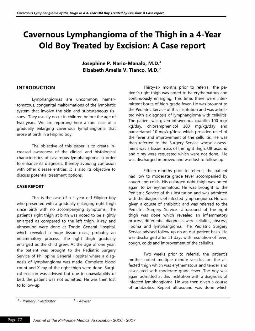
Cavernous Lymphangioma of the Thigh in a 4-Year
Old Boy Treated by Excision: A Case report
Josephine P. Nario-Manalo, M.D.a
Elizabeth Amelia V. Tianco, M.D.b
INTRODUCTION
Lymphangiomas are uncommon, hamar-
tomatous, congenital malformations of the lymphatic
system that involve the skin and subcutaneous tis-
sues. They usually occur in children before the age of
two years. We are reporting here a rare case of a
gradually enlarging cavernous lymphangioma that
arose at birth in a Filipino boy.
The objective of this paper is to create in-
creased awareness of the clinical and histological
characteristics of cavernous lymphangioma in order
to enhance its diagnosis, thereby avoiding confusion
with other disease entities. It is also its objective to
discuss potential treatment options.
CASE REPORT
This is the case of a 4-year-old Filipino boy
who presented with a gradually enlarging right thigh
since birth with no accompanying symptoms. The
patient‟s right thigh at birth was noted to be slightly
enlarged as compared to the left thigh. X-ray and
ultrasound were done at Tondo General Hospital,
which revealed a huge tissue mass, probably an
inflammatory process. The right thigh gradually
enlarged as the child grew. At the age of one year,
the patient was brought to the Pediatric Surgery
Service of Philippine General Hospital where a diag-
nosis of lymphangioma was made. Complete blood
count and X-ray of the right thigh were done. Surgi-
cal excision was advised but due to unavailability of
bed, the patient was not admitted. He was then lost
to follow-up.
Thirty-six months prior to referral, the pa-
tient‟s right thigh was noted to be erythematous and
continuously enlarging. This time, there were inter-
mittent bouts of high-grade fever. He was brought to
the Pediatric Service of this institution and was admit-
ted with a diagnosis of lymphangioma with cellulitis.
The patient was given intravenous oxacillin 100 mg/
kg/day, chloramphenicol 100 mg/kg/day and
paracetamol 10 mg/kg/dose which provided relief of
the fever and improvement of the cellulitis. He was
then referred to the Surgery Service whose assess-
ment was a tissue mass of the right thigh. Ultrasound
and x-ray were requested which were not done. He
was discharged improved and was lost to follow-up.
Fifteen months prior to referral, the patient
had low to moderate grade fever accompanied by
cough and colds. His enlarged right thigh was noted
again to be erythematous. He was brought to the
Pediatric Service of this institution and was admitted
with the diagnosis of infected lymphangioma. He was
given a course of antibiotic and was referred to the
Pediatric Surgery Service. Ultrasound of the right
thigh was done which revealed an inflammatory
process; differential diagnoses were cellulitis, abscess,
lipoma and lymphangioma. The Pediatric Surgery
Service advised follow-up on an out-patient basis. He
was discharged after 11 days with resolution of fever,
cough, colds and improvement of the cellulitis.
Two weeks prior to referral, the patient‟s
mother noted multiple minute vesicles on the af-
fected thigh which was erythematous and tender and
associated with moderate grade fever. The boy was
again admitted at this institution with a diagnosis of
infected lymphangioma. He was then given a course
of antibiotics. Repeat ultrasound was done which
Cavernous Lymphangioma of the Thigh in a 4-Year Old Boy Treated by Excision: A Case report
Page 72 Journal of the Philippine Medical Association 2016 - 2017
a - Primary Investigator b - Adviser
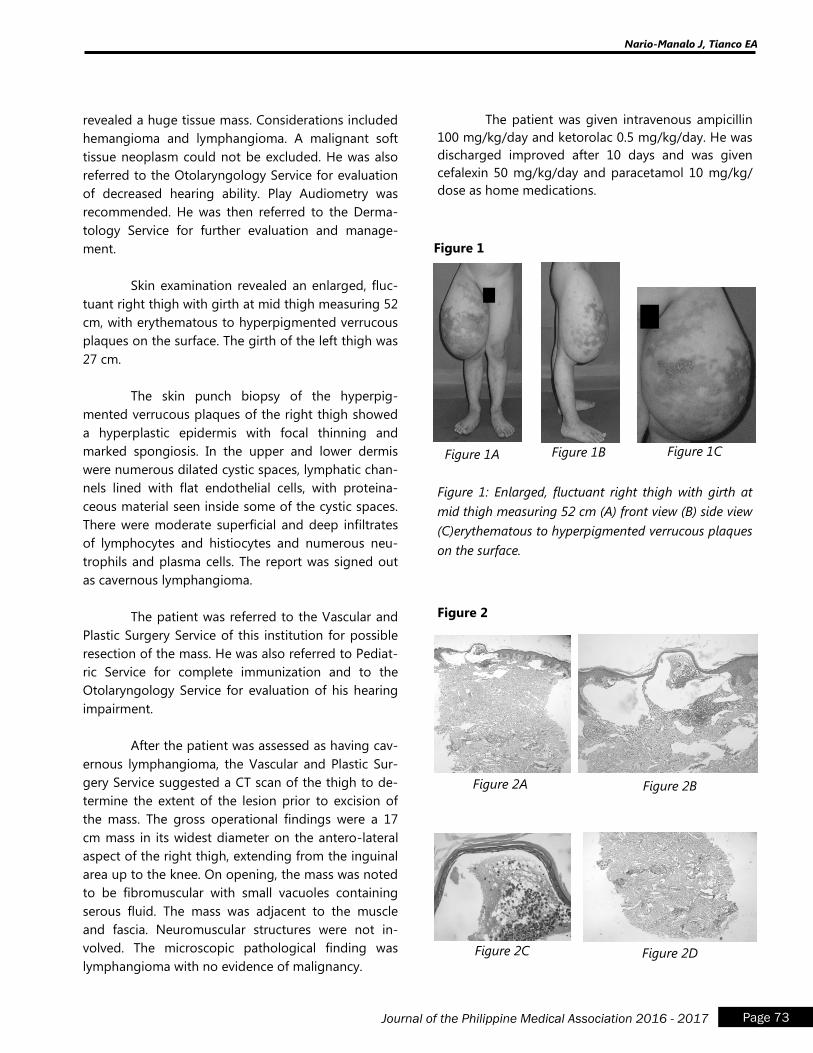
Nario-Manalo J, Tianco EA
Page 73 Journal of the Philippine Medical Association 2016 - 2017
revealed a huge tissue mass. Considerations included
hemangioma and lymphangioma. A malignant soft
tissue neoplasm could not be excluded. He was also
referred to the Otolaryngology Service for evaluation
of decreased hearing ability. Play Audiometry was
recommended. He was then referred to the Derma-
tology Service for further evaluation and manage-
ment.
Skin examination revealed an enlarged, fluc-
tuant right thigh with girth at mid thigh measuring 52
cm, with erythematous to hyperpigmented verrucous
plaques on the surface. The girth of the left thigh was
27 cm.
The skin punch biopsy of the hyperpig-
mented verrucous plaques of the right thigh showed
a hyperplastic epidermis with focal thinning and
marked spongiosis. In the upper and lower dermis
were numerous dilated cystic spaces, lymphatic chan-
nels lined with flat endothelial cells, with proteina-
ceous material seen inside some of the cystic spaces.
There were moderate superficial and deep infiltrates
of lymphocytes and histiocytes and numerous neu-
trophils and plasma cells. The report was signed out
as cavernous lymphangioma.
The patient was referred to the Vascular and
Plastic Surgery Service of this institution for possible
resection of the mass. He was also referred to Pediat-
ric Service for complete immunization and to the
Otolaryngology Service for evaluation of his hearing
impairment.
After the patient was assessed as having cav-
ernous lymphangioma, the Vascular and Plastic Sur-
gery Service suggested a CT scan of the thigh to de-
termine the extent of the lesion prior to excision of
the mass. The gross operational findings were a 17
cm mass in its widest diameter on the antero-lateral
aspect of the right thigh, extending from the inguinal
area up to the knee. On opening, the mass was noted
to be fibromuscular with small vacuoles containing
serous fluid. The mass was adjacent to the muscle
and fascia. Neuromuscular structures were not in-
volved. The microscopic pathological finding was
lymphangioma with no evidence of malignancy.
The patient was given intravenous ampicillin
100 mg/kg/day and ketorolac 0.5 mg/kg/day. He was
discharged improved after 10 days and was given
cefalexin 50 mg/kg/day and paracetamol 10 mg/kg/
dose as home medications.
Figure 1
Figure 1A Figure 1B Figure 1C
Figure 1: Enlarged, fluctuant right thigh with girth at
mid thigh measuring 52 cm (A) front view (B) side view
(C)erythematous to hyperpigmented verrucous plaques
on the surface.
Figure 2
Figure 2A Figure 2B
Figure 2C Figure 2D
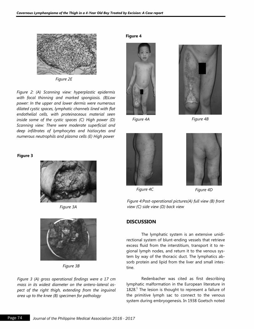
Figure 2E
Figure 2: (A) Scanning view: hyperplastic epidermis
with focal thinning and marked spongiosis. (B)Low
power: In the upper and lower dermis were numerous
dilated cystic spaces, lymphatic channels lined with flat
endothelial cells, with proteinaceous material seen
inside some of the cystic spaces (C) High power (D)
Scanning view: There were moderate superficial and
deep infiltrates of lymphocytes and histiocytes and
numerous neutrophils and plasma cells (E) High power
Figure 3
Figure 3A
Figure 3B
Figure 3 (A) gross operational findings were a 17 cm
mass in its widest diameter on the antero-lateral as-
pect of the right thigh, extending from the inguinal
area up to the knee (B) specimen for pathology
Figure 4
Figure 4A Figure 4B
Figure 4C Figure 4D
Figure 4:Post-operational pictures(A) full view (B) front
view (C) side view (D) back view
DISCUSSION
The lymphatic system is an extensive unidi-
rectional system of blunt-ending vessels that retrieve
excess fluid from the interstitium, transport it to re-
gional lymph nodes, and return it to the venous sys-
tem by way of the thoracic duct. The lymphatics ab-
sorb protein and lipid from the liver and small intes-
tine.
Redenbacher was cited as first describing
lymphatic malformation in the European literature in
1828.1 The lesion is thought to represent a failure of
the primitive lymph sac to connect to the venous
system during embryogenesis. In 1938 Goetsch noted
Cavernous Lymphangioma of the Thigh in a 4-Year Old Boy Treated by Excision: A Case report
Page 74 Journal of the Philippine Medical Association 2016 - 2017
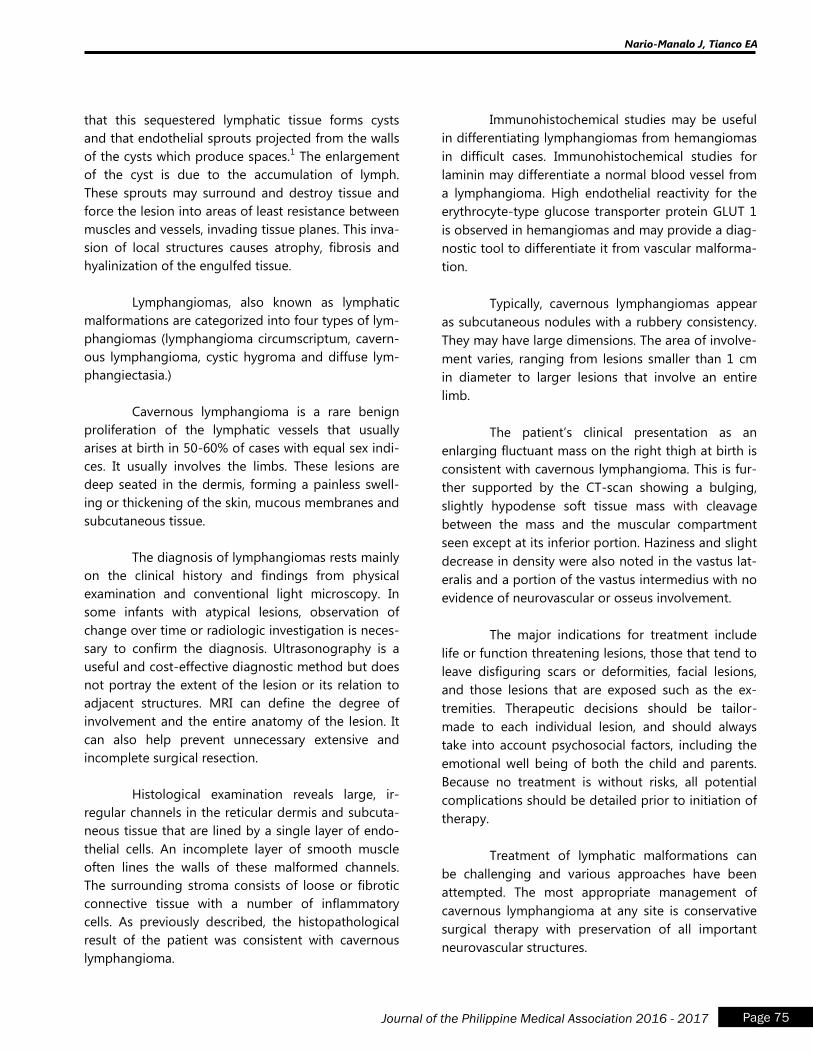
that this sequestered lymphatic tissue forms cysts
and that endothelial sprouts projected from the walls
of the cysts which produce spaces.1 The enlargement
of the cyst is due to the accumulation of lymph.
These sprouts may surround and destroy tissue and
force the lesion into areas of least resistance between
muscles and vessels, invading tissue planes. This inva-
sion of local structures causes atrophy, fibrosis and
hyalinization of the engulfed tissue.
Lymphangiomas, also known as lymphatic
malformations are categorized into four types of lym-
phangiomas (lymphangioma circumscriptum, cavern-
ous lymphangioma, cystic hygroma and diffuse lym-
phangiectasia.)
Cavernous lymphangioma is a rare benign
proliferation of the lymphatic vessels that usually
arises at birth in 50-60% of cases with equal sex indi-
ces. It usually involves the limbs. These lesions are
deep seated in the dermis, forming a painless swell-
ing or thickening of the skin, mucous membranes and
subcutaneous tissue.
The diagnosis of lymphangiomas rests mainly
on the clinical history and findings from physical
examination and conventional light microscopy. In
some infants with atypical lesions, observation of
change over time or radiologic investigation is neces-
sary to confirm the diagnosis. Ultrasonography is a
useful and cost-effective diagnostic method but does
not portray the extent of the lesion or its relation to
adjacent structures. MRI can define the degree of
involvement and the entire anatomy of the lesion. It
can also help prevent unnecessary extensive and
incomplete surgical resection.
Histological examination reveals large, ir-
regular channels in the reticular dermis and subcuta-
neous tissue that are lined by a single layer of endo-
thelial cells. An incomplete layer of smooth muscle
often lines the walls of these malformed channels.
The surrounding stroma consists of loose or fibrotic
connective tissue with a number of inflammatory
cells. As previously described, the histopathological
result of the patient was consistent with cavernous
lymphangioma.
Immunohistochemical studies may be useful
in differentiating lymphangiomas from hemangiomas
in difficult cases. Immunohistochemical studies for
laminin may differentiate a normal blood vessel from
a lymphangioma. High endothelial reactivity for the
erythrocyte-type glucose transporter protein GLUT 1
is observed in hemangiomas and may provide a diag-
nostic tool to differentiate it from vascular malforma-
tion.
Typically, cavernous lymphangiomas appear
as subcutaneous nodules with a rubbery consistency.
They may have large dimensions. The area of involve-
ment varies, ranging from lesions smaller than 1 cm
in diameter to larger lesions that involve an entire
limb.
The patient‟s clinical presentation as an
enlarging fluctuant mass on the right thigh at birth is
consistent with cavernous lymphangioma. This is fur-
ther supported by the CT-scan showing a bulging,
slightly hypodense soft tissue mass with cleavage
between the mass and the muscular compartment
seen except at its inferior portion. Haziness and slight
decrease in density were also noted in the vastus lat-
eralis and a portion of the vastus intermedius with no
evidence of neurovascular or osseus involvement.
The major indications for treatment include
life or function threatening lesions, those that tend to
leave disfiguring scars or deformities, facial lesions,
and those lesions that are exposed such as the ex-
tremities. Therapeutic decisions should be tailor-
made to each individual lesion, and should always
take into account psychosocial factors, including the
emotional well being of both the child and parents.
Because no treatment is without risks, all potential
complications should be detailed prior to initiation of
therapy.
Treatment of lymphatic malformations can
be challenging and various approaches have been
attempted. The most appropriate management of
cavernous lymphangioma at any site is conservative
surgical therapy with preservation of all important
neurovascular structures.
Nario-Manalo J, Tianco EA
Page 75 Journal of the Philippine Medical Association 2016 - 2017
Another option for cavernous lymphangioma
is injection of sclerosing agents such as alcohol,
steroids, bleomycin sulfate, tetracycline and OK-432
(picibanil).
Qin et al2 described 200 patients with various
types of head and neck lymphangiomas who were
treated by inratumorous injection of bleomycin-A5.
All 200 cases were followed from 2 to 10 years; 95
cases were followed for more than 5 years. The effec-
tive rate was 97% and the curative rate was 86.5%.
No pulmofibrosis or other serious complications were
found.
In a study made by Kobayashi et al3, three
patients with cavernous lymphangioma of the legs
were treated. The suggested guidelines for treatment
are extensive surgical resection of the mass and
approximately two weeks after the operation, aspira-
tion of serous fluid which has accumulated at the
operation site. This is then followed by injection of
OK-432 at the resected area.
Side effects include pyrexia and focal inflam-
mation for several days, but the accumulation of
serous fluid disappeared after the injection. There
were no complications with any recurrence, joint
contracture, or pain during a mean follow-up of 48
months.
Recurrence of larger lymphomas after surgi-
cal excision is common because the tumor closely
involves important nerves and vessels. Saijo et al4
reported that tumor resection recurred in one third of
operations that were only partially successful. Patients
who have grossly or microscopically infiltrative le-
sions should be counseled about the increased likeli-
hood of recurrence, even if resection is thought to be
complete. In our patient‟s case, the neurovascular
structures were not involved and the likehood of
recurrence is less.
The prognosis of cavernous lymphangioma
heavily depends on early diagnosis with definitive
surgical treatment. Full recovery is possible in pa-
tients whose lesions are completely resected.
CONCLUSION
Cavernous lymphangioma is a rare clinical
entity that is defined as a congenital malformation
of the lymphatic system that involves the skin and
subcutaneous tissues.
A keen awareness of the clinical and histo-
logical features of cavernous lymphangioma can alert
physicians to early detection and proper manage-
ment. Several treatment options are available but the
patient should be aware of the risk of recurrence with
this disease.
REFERENCES
1. Fliegelman LJ. Lymphatic malformation: predictive
factors for recurrence. Otolar, Head & Neck Surg
2000; 23:76-80.
2. Qin Z.P. Long-term results of intratumourous
bleomycin-A5 injection of head and neck
lymphangioma. J Oral Surg, Oral Med, Oral Patho,
Oral Radio 1998; 86:139-44.
3. Kabayashi D et al. Cavernous lymphangioma of
the leg in children treated by the injection of
OK-432 after resection. J Bone Joint Surg 2003;
85:91-94.
4. Martinez M.T. Lymphangiomas: a 10 year review.
J Amer Acad Dermatol 2004; 50:159-64.
5. Kennedy T.L. Cystic hygroma/lymphangioma: a
rational approach to management. Laryngoscope
2001; 11:1929-37.
6. Giguere C.M. Treatment of lymphangioma with
OK-432 (picibanil) sclerotherapy. Archives of
Otolar Head & Neck Surg 2002; 128:1137-44.
7. Devaney J. Skeletal-extraskeletal angiomatoses. J
Bone Joint Surg 1994; 76:878-91.
8. Santiago I. Sclerotherapy for lymphangioma in
children. Int J Pediatr Olorlaryngol 2003; 67:795-
800.
Cavernous Lymphangioma of the Thigh in a 4-Year Old Boy Treated by Excision: A Case report
Page 76 Journal of the Philippine Medical Association 2016 - 2017
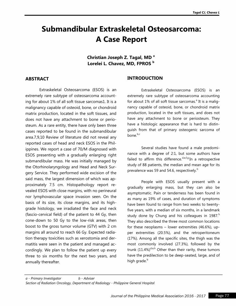
Submandibular Extraskeletal Osteosarcoma:
A Case Report
Christian Joseph Z. Tagal, MD a
Lorelei L. Chavez, MD, FPROS b
ABSTRACT
Extraskeletal Osteosarcoma (ESOS) is an
extremely rare subtype of osteosarcoma account-
ing for about 1% of all soft tissue sarcomas1. It is a
malignancy capable of osteoid, bone, or chondroid
matrix production, located in the soft tissues, and
does not have any attachment to bone or perio-
steum. As a rare entity, there have only been three
cases reported to be found in the submandibular
area.7,9,10 Review of literature did not reveal any
reported cases of head and neck ESOS in the Phil-
ippines. We report a case of 70/M diagnosed with
ESOS presenting with a gradually enlarging right
submandibular mass. He was initially managed by
the Otorhinolaryngology and Head and Neck Sur-
gery Service. They performed wide excision of the
said mass, the largest dimension of which was ap-
proximately 7.5 cm. Histopathology report re-
vealed ESOS with close margins, with no perineural
nor lymphovascular space invasion seen. On the
basis of its size, its close margins, and its high-
grade histology, we irradiated the face and neck
(fascio-cervical field) of the patient to 44 Gy, then
cone-down to 50 Gy to the low-risk areas, then
boost to the gross tumor volume (GTV) with 2 cm
margins all around to reach 66 Gy. Expected radia-
tion therapy toxicities such as xerostomia and der-
matitis were seen in the patient and managed ac-
cordingly. We plan to follow the patient up every
three to six months for the next two years, and
annually thereafter.
INTRODUCTION
Extraskeletal Osteosarcoma (ESOS) is an
extremely rare subtype of osteosarcoma accounting
for about 1% of all soft tissue sarcomas.a It is a malig-
nancy capable of osteoid, bone, or chondroid matrix
production, located in the soft tissues, and does not
have any attachment to bone or periosteum. They
have a histologic appearance that is hard to distin-
guish from that of primary osteogenic sarcoma of
bone.b,c
Several studies have found a male predomi-
nance with a degree of 2:1, but some authors have
failed to affirm this difference.d,e,f,g
In a retrospective
study of 88 patients, the median and mean age for its
prevalence was 59 and 54.6, respectively.h
People with ESOS usually present with a
gradually enlarging mass, but they can also be
asymptomatic. Pain or tenderness has been found in
as many as 19% of cases, and duration of symptoms
have been found to range from two weeks to twenty-
five years, with a median of six months, in a landmark
study done by Chung and his colleagues in 1987.h
They also described the three most common locations
for these neoplasms – lower extremities (46.6%), up-
per extremities (20.5%), and the retroperitoneum
(17%). Among all the specific sites, the thigh was the
most commonly involved (27.3%), followed by the
trunk (11.4%)d,e,h
Other than their rarity, these tumors
have the predilection to be deep-seated, large, and of
high grade.b
Tagal CJ, Chavez L
Page 77 Journal of the Philippine Medical Association 2016 - 2017
a - Primary Investigator b - Adviser
Section of Radiation Oncology, Department of Radiology - Philippine General Hospital
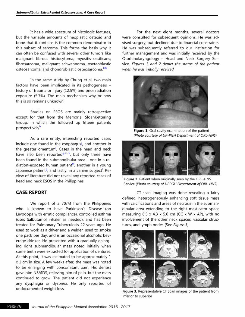
It has a wide spectrum of histologic features,
but the variable amounts of neoplastic osteoid and
bone that it contains is the common denominator in
this subset of sarcoma. This forms the basis why it
can often be confused with several other tumors like
malignant fibrous histiocytoma, myositis ossificans,
fibrosarcoma, malignant schwannoma, oseteoblastic
osteosarcoma, and chondroblastic osteosarcoma.b,h
In the same study by Chung et al, two main
factors have been implicated in its pathogenesis –
history of trauma or injury (12.5%) and prior radiation
exposure (5.7%). The main mechanism why or how
this is so remains unknown.
Studies on ESOS are mainly retrospective
except for that from the Memorial SloanKettering
Group, in which the followed up fifteen patients
prospectivelyb.
As a rare entity, interesting reported cases
include one found in the esophagusi, and another in
the greater omentumj. Cases in the head and neck
have also been reportedg,k,l,m
, but only three have
been found in the submandibular area - one in a ra-
diation-exposed human patientm
, another in a young
Japanese patientg, and lastly, in a canine subject
l. Re-
view of literature did not reveal any reported cases of
head and neck ESOS in the Philippines.
CASE REPORT
We report of a 70/M from the Philippines
who is known to have Parkinson‟s Disease (on
Levodopa with erratic compliance), controlled asthma
(uses Salbutamol inhaler as needed), and has been
treated for Pulmonary Tuberculosis 22 years ago. He
used to work as a driver and a welder, used to smoke
one pack per day, and is an occasional alcoholic bev-
erage drinker. He presented with a gradually enlarg-
ing right submandibular mass noted initially when
some teeth were extracted for application of dentures.
At this point, it was estimated to be approximately 1
x 1 cm in size. A few weeks after, the mass was noted
to be enlarging with concomitant pain. His dentist
gave him NSAIDS, relieving him of pain, but the mass
continued to grow. The patient did not experience
any dysphagia or dyspnea. He only reported of
undocumented weight loss.
For the next eight months, several doctors
were consulted for subsequent opinions. He was ad-
vised surgery, but declined due to financial constraints.
He was subsequently referred to our institution for
further management and was initially received by the
Otorhinolaryngology – Head and Neck Surgery Ser-
vice. Figures 1 and 2 depict the status of the patient
when he was initially received.
Figure 1. Oral cavity examination of the patient
(Photo courtesy of UP-PGH Department of ORL-HNS)
Figure 2. Patient when originally seen by the ORL-HNS
Service (Photo courtesy of UPPGH Department of ORL-HNS)
CT-scan imaging was done revealing a fairly
defined, heterogeneously enhancing soft tissue mass
with calcifications and areas of necrosis in the subman-
dibular area extending to the right masticator space
measuring 6.5 x 4.3 x 5.6 cm (CC x W x AP), with no
involvement of the other neck spaces, vascular struc-
tures, and lymph nodes (See Figure 3).
Figure 3. Representative CT Scan images of the patient from
inferior to superior
Submandibular Extraskeletal Osteosarcoma: A Case Report
Page 78 Journal of the Philippine Medical Association 2016 - 2017
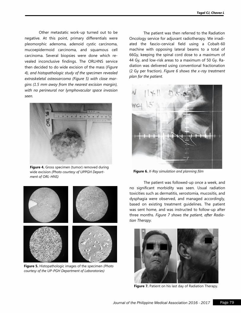
Other metastatic work-up turned out to be
negative. At this point, primary differentials were
pleomorphic adenoma, adenoid cystic carcinoma,
mucoepidermoid carcinoma, and squamous cell
carcinoma. Several biopsies were done which re-
vealed inconclusive findings. The ORLHNS service
then decided to do wide excision of the mass (Figure
4), and histopathologic study of the specimen revealed
extraskeletal osteosarcoma (Figure 5) with close mar-
gins (1.5 mm away from the nearest excision margin),
with no perineural nor lymphovacular space invasion
seen.
The patient was then referred to the Radiation
Oncology service for adjuvant radiotherapy. We irradi-
ated the fascio-cervical field using a Cobalt-60
machine with opposing lateral beams to a total of
66Gy, keeping the spinal cord dose to a maximum of
44 Gy, and low-risk areas to a maximum of 50 Gy. Ra-
diation was delivered using conventional fractionation
(2 Gy per fraction). Figure 6 shows the x-ray treatment
plan for the patient.
Figure 4. Gross specimen (tumor) removed during
wide excision (Photo courtesy of UPPGH Depart-
ment of ORL-HNS)
Figure 5. Histopathologic images of the specimen (Photo
courtesy of the UP-PGH Department of Laboratories)
Figure 6. X-Ray simulation and planning film
The patient was followed-up once a week, and
no significant morbidity was seen. Usual radiation
toxicities such as dermatitis, xerostomia, mucositis, and
dysphagia were observed, and managed accordingly,
based on existing treatment guidelines. The patient
was sent home, and was instructed to follow-up after
three months. Figure 7 shows the patient, after Radia-
tion Therapy.
Figure 7. Patient on his last day of Radiation Therapy.
Tagal CJ, Chavez L
Page 79 Journal of the Philippine Medical Association 2016 - 2017
DISCUSSION
Treatment
Researchers from the Memorial Sloan-
Kettering Cancer Center have recommended that
treatment of ESOS should adhere to currently estab-
lished guidelines in the treatment of soft tissue sarco-
mas.b The use of adjuvant therapy (chemotherapy or
radiation therapy) should be individualized, based on
independent risk factors, such as incomplete resec-
tions, large tumors (>5cm), or high-grade lesions.
Chemotherapy and RT did not appear to affect or
influence survival, but this may be attributed to the
low number of patients in their study (fifteen pathol-
ogically-confirmed ESOS in a 17-year period from
1982-1999).
Ahmad and his colleagues from MD Ander-
son Cancer Center have supported this claim on the
value of adjuvant therapy.n In their institutional
experience, an overall response rate of 19% (two
complete and three partial responses) was seen for
Doxorubicin-based systemic therapy, which they
deemed as a low response rate. They then insisted
that ESOS should be viewed as clinically and thera-
peutically distinct from osseous osteosarcoma.
Though no clear survival or local control
benefit when giving adjuvant radiotherapy have been
established for ESOS as of the moment, the patient
was irradiated on the basis of its very close margins
on histopathology (1.5mm), and its size (>5 cm) pre-
operatively. Upon consultation with the Medical
Oncology service, adjuvant chemotherapy is not
warranted since there are no adverse features seen,
but close follow-up was advised.
Prognosis
ESOS has an exceptionally poor prognosis
which can partially be attributed to the limited clinical
information about its natural course.c,d,e,o
Researchers
have set the estimated mortality to be between 70 to
80%, and overall disease-specific survival at 5 years at
50% (with a median follow-up of 35 months).b
The MD Anderson experience cited earliern
revealed their statistics to be 82% (95% CI, 70-98), 64%
(95% CI, 43-93%), 47% (95% CI, 30-70%), and 46%
(95% CI, 26-80%) for the 5-year actuarial local recur-
rence-free survival, distant recurrence-free survival,
event-free survival, and disease-specific survival,
respectively.
Of the 88 patients in the landmark study by
Chungh, 65 had follow-up data. Twentyfive (38.5%)
were alive, eight of whom were well, while 17 had
recurrence or metastasis. The remaining 40 (61.5%)
died, 4 of whom attributable to other causes not con-
nected to the malignancy. Median follow-up for this
study was 2.4 years, ranging from one to 22 years.
We plan to follow the patient up every three
to six months for two to three years, then every six
months for the next two years, then annually thereaf-
ter. Though limited data has shown poor prognosis for
this kind of malignancy, compliance to follow-up and
other medications was advised and reiterated, as he
has a really good performance status at baseline.
REFERENCES
a. Allan CJ, Soule EH. Osteogenic sarcoma of the
somatic soft tissues. Clinicopathologic study of 26
cases and review of literature. Cancer 1971;27:1121
-33.
b. McCarter MD, Lewis JL, et al. Extraskeletal Os-
teosarcoma: Analysis of Outcome of a rare neo-
plasm. Sarcoma (2000) 4, 119-123
c. Enzinger, FM, Weiss SW. Soft Tissue Sarcomas. St.
Louis: Mosby-Year Book, 1995
d. Sordillo PP, Hajdu SI, Magill GB, Golbey RB.
Extraosseous osteogenic sarcoma. A review of 48
patients. Cancer 1983;51:727-34.
e. Lidang Jensen M, Schumacher B, Myhre Jensen O
et al. Extraskeletal osteosarcomas: a clinicopa-
thologic study of 25 cases. Am J Surg Pathol
1998;22:588-94.
f. Lee JS, Fetsch JF, Wasdhal DA et al. A review of 40
patients with extraskeletal osteosarcoma. Cancer
1995;76:2253-9.
Submandibular Extraskeletal Osteosarcoma: A Case Report
Page 80 Journal of the Philippine Medical Association 2016 - 2017
g. Saito Y, Miyajima C, et al. Highly malignant sub-
mandibular extraskeletal osteosarcoma in a
young patient. Auris Nasus Larynx, International
Journal of ORL and HNS. Dec 2008, Vol 35, Issue
4;576-578
h. Chung EB, Enzinger FM. Extraskeletal Osteosar-
coma. Cancer 60:1132-1142, 1987
i. Wegner N, McGrath K, et al. Extraosseous Os-
teosarcoma of the Esophagus: A Case Report.
Sarcoma. Hindawi Publishing Corporation, 2010.
Article ID 907127
j. Wu Z, Chu X, et al. An abdominal extraskeletal
osteosarcoma: A case report. Oncology Letters 6:
990- 992, 2013
k. Luna-Ortiz K, Ortega-Gutierrez C, et al. Extraosse-
ous osteosarcoma in the neck. Auris Nasus Lar-
ynx, International Journal of ORL and HNS. June
2012, Vol 39 Issue 3:333-336
l. Thomsen BV, Myers RK. Extraskeletal osteosar-
coma of the mandibular salivary gland in a dog.
Vet Pathol, 1999 Jan;36(1):71-3
m. Ayad W, Walendzik F, Freitag P, Wierich W. Ra-
diation-induced submandibular extra-osseous
osteogenic sarcoma. Mund-, Kiefer- und Ge-
sichtschirurgie. March 1999, Volume 3, Issue
2:106-108
n. Ahmad SA, Patel SR, et al. Extraosseous osteosar-
coma: response to treatment and long-term out-
come. J Clin Oncol. 2002 Jan 15;20(2):521-7
o. Lane JM, Healey JH. Extraskeletal malignant tu-
mors of bone. In: Raaf JH, ed. Soft tissue sarco-
mas: diagnosis and treatment. St Louis: Mosby-
Year Book, 1993:261-9.
Tagal CJ, Chavez L
Page 81 Journal of the Philippine Medical Association 2016 - 2017
Page 82 Journal of the Philippine Medical Association 2016 - 2017
Adverse Drug Reaction to Nevirapine in an Immuno-Compromised Patient: A Case Report
Adverse Drug Reaction to Nevirapine in
an Immuno-Compromised Patient:
A Case Report
Lei Anne Michelle R. Hernandez MD, DPDS
Lonabel A. Encarnacion MD,FPDS
Abstract
Adverse drug reaction in an immuno-compromised patient is a common condition
brought about by poly-pharmacy, and notably due to the use of antiretroviral medications. We
present a case of a 35 year old male, who initially presented with a morbilliform rash.. The pa-
tient was treated initially as a case of drug hypersensitivity reaction who did not respond to
regular course of intravenous corticosteroid. After revealing his sero-positive human immuno-
deficiency virus (HIV) status, corticosteroid was discontinued and the patient responded with
conservative management.
Key words: nevirapine, drug hypersensitivity reaction, adverse drug reaction
Department of Dermatology, St. Luke’s Medical Center, Quezon City
Page 83 Journal of the Philippine Medical Association 2016 - 2017
Hernandez LAM, Encarnacion L
INTRODUCTION
Cutaneous adverse drug reactions are quite
common especially among patients taking multiple
medication. Among immuno-compromised patients,
the initiation of anti-retroviral medications and con-
comitant treatment of their associated diseases may
compound and increase their risk of developing hy-
persensitivity reaction. Hence, complete history taking
and adequate physical examination are important
steps in the correct diagnosis and management of
such patients.
CASE REPORT
A 35-year old Filipino male presented with
morbilliform rash of 2 day duration. Eight weeks prior
to consult, he was diagnosed to be seropositive with
HIV with no medications started. At 6 weeks prior to
consult, he had concomittant ill-defined, dry, slightly
scaly plaques on the forehead and malar area ac-
companied by mild pruritus, nonspecific symptoms of
cough, colds and malaise. He was started on Cotri-
moxazole 800mg tab OD, Isoniazid 400mg tab OD
and Clarithromycin 500mg tab BID on follow up when
his CD4 count was noted to be 23cells/mm3. There
were no noted lesions after starting these medica-
tions. He noted gradual progression of scaling and
erythema over the next 6 weeks but claims no associ-
ated skin lesions.
At 4 weeks prior to consult, he was subse-
quently started on anti-retrovirals: Stavudine 40mg
+Lamivudine 150mg tab twice daily and Nevirapine
200mg tab once daily for the first 2 weeks. The dose
of nevirapine was then increased to twice daily on
the succeeding 2 weeks. During these times, there
were no noted skin lesions.
At 2 days prior to consult, the patient still had
persistence of erythematous scaly plaques on the
face but he claimed gradual onset of multiple and
generalized, erythematous, blanching, pruritic mor-
billiform rash on the chest, back, arms, legs and palms
and soles. This was associated with odynophagia, de-
scribed as difficulty swallowing solid food, and bilat-
eral periorbital edema. He also claimed development
of undocumented fever. This was accompanied by
oral ulcers. The patient had malaise, fever, associated
undocumented weight-loss, weakness and odyno-
phagia. He also claims easy fatiguability. The patient is
a known diabetic since 2010 maintained on met-
formin. He also withheld at the time of interview that
he had 2 male sexual partners in the last 5 years and
practices unprotected sex. He is in a male monoga-
mous relationship.
On examination, the patient had multiple,
scattered, blanching, erythematous macules, papules
and plaques on the trunk, arms and legs. Very promi-
nent localized, ill-defined, dry, moderately erythema-
tous patches with adherent scales on the forehead,
nose, malar areas and ears were seen. He also had
mild bilateral periorbital edema, with conjunctival
suffusion accompanied by labial swelling and tender
ulcers on the buccal mucosa (Figure 1A-C).
Upon admission, skin punch biopsy showed
interface dermatitis, most probably drug hypersensi-
tivity reaction or early erythema multiforme (Figure2).
He was started on intravenous hydrocortisone 100 mg
every 6 hours. We started him on a mouthwash con-
sisting of sucralfate, diphenhydramine and aluminum
chloride and twice daily 50mg intravenous diphenhy-
dramine. After four doses of 100mg intravenous hy-
drocortisone were given, there was progression of
lesions. It was only during this time that the patient
revealed that he was previously diagnosed with HIV
hence, intravenous hydrocortisone was discontinued.
The patient was shifted to Levocetirizine 5mg twice
daily due to persistent pruritus. Oral thrush was
treated with Nystatin 5ml twice daily. Treatment of
seborrheic dermatitis with ketoconazole cream and
desonide cream twice daily was started. During in-
terim, there was gradual improvement of the skin le-
sions. On the 6th hospital day, the patient was dis-
charged with improvement of seborrheic dermatitis
and mostly hyperpigmented patches on the body.
DISCUSSION
Adverse drug reaction (ADR) was defined by
the world health organization (WHO) as “a response
to a drug that is noxious and unintended and occurs
at doses normally used in man for prophylaxis, diag-
noses or therapy of disease, or for the modification of
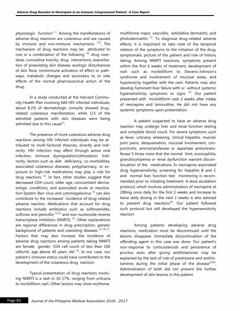
Page 84 Journal of the Philippine Medical Association 2016 - 2017
Adverse Drug Reaction to Nevirapine in an Immuno-Compromised Patient: A Case Report
physiologic function” 1. Among the manifestations of
adverse drug reactions are cutaneous and are caused
by immune and non-immune mechanisms 2-9
. The
mechanism of drug reactions may be attributed to
one or a combination of the following 10
: drug over-
dose, cumulative toxicity, drug interactions, exacerba-
tion of preexisting skin disease, ecologic disturbances
of skin flora, nonimmune activation of effect or path-
ways, metabolic changes and secondary to or side
effects of the normal pharmaceutical action of the
drug .
In a study conducted at the Harvard Commu-
nity Health Plan involving 684 HIV infected individuals,
about 8.2% of dermatologic consults showed drug-
related cutaneous manifestation, while 1/3 of the
admitted patients with skin diseases were being
admitted due to this cause11
.
The presence of more cutaneous adverse drug
reactions among HIV infected individuals may be at-
tributed to multi-factorial theories, directly and indi-
rectly. HIV infection may affect through active viral
infection, immune dysregulation/stimulation. Indi-
rectly, factors such as diet deficiency, co-morbidities,
associated cutaneous diseases, polypharmacy, or ex-
posure to high-risk medications may play a role for
drug reactions 12
. In fact, other studies suggest that
decreased CD4 count, older age, concomitant derma-
tologic conditions, and associated acute or reactiva-
tion Epstein-Barr virus and cytomegalovirus 13
can also
contribute to the increased incidence of drug-related
adverse reaction. Medications that account for drug
reactions include antibiotics such as sulfonamides,
sulfones and penicillin 11,13
and non-nucleoside reverse
transcriptase inhibitors (NNRTI) 14
. Other explanations
are regional differences in drug prescription, genetic
background of patients and coexisting diseases 15, 16, 17
.
Factors that may also increase the incidence of
adverse drug reactions among patients taking NNRTI
are female gender, CD4 cell count of less than 100
cells/ml, age above 40 years old 14
. In our case, our
patient‟s immune status could have contributed to the
development of the cutaneous drug reaction.
Typical presentation of drug reactions involv-
ing NNRTI is a rash in 10-17%, ranging from urticaria
to morbillifom rash. Other lesions may show erythema
multiforme major, vasculitis, exfoliative dermatitis, and
photodermatitis 11
. To diagnose drug-related adverse
effects, it is important to take note of the temporal
relation of the symptoms to the initiation of the drug,
symptomatic picture of the patient and role of history
taking. Among NNRTI reactions, symptoms present
within the first 6 weeks of treatment, development of
rash such as morbilliform to Stevens-Johnson‟s
syndrome and involvement of mucosal areas, and
hypotoxicity together with the rash. Patients may also
develop fulminant liver failure with or without systemic
hypersensitivity symptoms or signs 18
. Our patient
presented with morbilliform rash 2 weeks after intake
of nevirapine and lamivudine. He did not have any
systemic symptoms upon presentation.
A patient suspected to have an adverse drug
reaction may undergo liver and renal function testing
and complete blood count. For severe symptoms such
as fever, urticaria, wheezing, clinical hepatitis, muscle/
joint pains, desquamation, mucosal involvement, con-
junctivitis, aminotransferase or aspartate aminotrans-
ferase 5 times more that the normal limit, eosinophilia,
granulocytopenia or renal dysfunction warrant discon-
tinuation of the medications. In nevirapine-associated
drug hypersensitivity, screening for hepatitis B and C
and normal liver function test monitoring is recom-
mended prior to initiating treatment. A dose escalation
protocol, which involves administration of nevirapine at
200mg once daily for the first 2 weeks and increase to
twice daily dosing in the next 2 weeks is also advised
to prevent drug reactions18
. Our patient followed
such protocol but still developed the hypersensitivity
reaction.
Among patients developing adverse drug
reactions, medication must be discontinued until the
lesions disappear. Immediate discontinuation of the
offending agent in this case was done. Our patient‟s
non-response to corticosteroids and persistence of
pruritus even after giving antihistamines may be
explained by the lack of role of prednisone and antihis-
tamines during the initial phase of the disease19,20
.
Administration of both did not prevent the further
development of skin lesions in this patient.
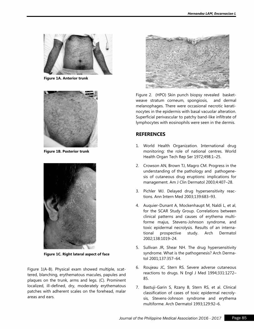
Page 85 Journal of the Philippine Medical Association 2016 - 2017
Hernandez LAM, Encarnacion L
Figure 1A. Anterior trunk
Figure 1B. Posterior trunk
Figure 1C. Right lateral aspect of face
Figure 1(A-B). Physical exam showed multiple, scat-
tered, blanching, erythematous macules, papules and
plaques on the trunk, arms and legs. (C). Prominent
localized, ill-defined, dry, moderately erythematous
patches with adherent scales on the forehead, malar
areas and ears.
Figure 2. (HPO) Skin punch biopsy revealed basket-
weave stratum corneum, spongiosis, and dermal
melanophages. There were occasional necrotic kerati-
nocytes in the epidermis with basal vacuolar alteration.
Superficial perivascular to patchy band-like infiltrate of
lymphocytes with eosinophils were seen in the dermis.
REFERENCES
1. World Health Organization. International drug
monitoring: the role of national centres. World
Health Organ Tech Rep Ser 1972;498:1–25.
2. Crowson AN, Brown TJ, Magro CM. Progress in the
understanding of the pathology and pathogene-
sis of cutaneous drug eruptions: implications for
management. Am J Clin Dermatol 2003;4:407–28.
3. Pichler WJ. Delayed drug hypersensitivity reac-
tions. Ann Intern Med 2003;139:683–93.
4. Auquier-Dunant A, Mockenhaupt M, Naldi L, et al,
for the SCAR Study Group. Correlations between
clinical patterns and causes of erythema multi-
forme majus, Stevens-Johnson syndrome, and
toxic epidermal necrolysis. Results of an interna-
tional prospective study. Arch Dermatol
2002;138:1019–24.
5. Sullivan JR, Shear NH. The drug hypersensitivity
syndrome. What is the pathogenesis? Arch Derma-
tol 2001;137:357–64.
6. Roujeau JC, Stern RS. Severe adverse cutaneous
reactions to drugs. N Engl J Med 1994;331:1272–
85.
7. Bastuji-Garin S, Rzany B, Stern RS, et al. Clinical
classification of cases of toxic epidermal necroly-
sis, Stevens-Johnson syndrome and erythema
multiforme. Arch Dermatol 1993;129:92–6.
Page 86 Journal of the Philippine Medical Association 2016 - 2017
Adverse Drug Reaction to Nevirapine in an Immuno-Compromised Patient: A Case Report
8. Wintroub BU, Stern R. Cutaneous drug reactions:
pathogenesis and clinical classification. J Am Acad
Dermatol 1985;13:167–79.
9. Kauppinen K. Cutaneous reactions to drugs with
special reference to severe bullous mucocutane-
ous eruptions and sulphonamides. Acta Derm
Venereol 1972;68:1–89.
10. Wintroub BU, Stern R. Cutaneous drug reactions:
pathogenesis and clinical classification. J Am Acad
Dermatol 985;13:167–79.
11. Coopman SA, Johnson RA, Platt R, et al. Cutane-
ous disease and drug reactions in HIV infection. N
Engl J Med 1993;328:1670–4.
12. Todd G Adverse Cutaneous Drug Eruptions and
HIV:a Clinician‟s Global Perspective Dermatol Clin
2006;24:459–472.
13. Smith KJ, Skelton HG, Yeager J, et al. Increased
drug reactions in HIV-1 positive patients: a possi-
ble explanation based on patterns of immune dys-
regulation seen in HIV-1 disease. The Military
Medical Consortium for the Advancement of Ret-
roviral Research (MMCARR). Clin Exp Dermatol
1997;22: 118–23
14. Carr A, Cooper DA. Adverse effects of antiretrovi-
ral therapy. Lancet 2000;356(9239): 1423–30.
15. Aguiar D, Pazo R, Duran I, et al. Toxic epidermal
necrolysis in patients receiving anticonvulsants and
cranial irradiation: a risk to consider. J Neurooncol
2004;66: 345.
16. Aydin F, Cokluk C, Senturk N, et al. Stevens-
Johnson syndrome in two patients treated with
cranial irradiation and phenytoin. J Eur Acad Der-
matol Venereol 2006;20:588.
17. Harr T and French L, MD Severe Cutaneous Adverse
Reactions: Acute Generalized Exanthematous Pus-
tulosis, Toxic Epidermal Necrolysis and Stevens-
Johnson Syndrome Med Clin N Am 2010;94:727–
742.
18. Hoffman R and Currier J, Management of Antiret-
roviral Treatment–Related Complications Infect Dis
Clin N Am 2007;21: 103–132.
19. Launay O, Roudiere L, Boukli N, et al. Assessment
of cetirizine, an antihistamine, to preventcutaneous
reactions to nevirapine therapy: results of the Vira-
mune-Zyrtec doubleblind, placebocontrolled trial.
Clin Infect Dis 2004;38(8):e66–72.
20. Knobel H, Miro JM, Domingo P, et al. Failure of a
short-term prednisone regimen to preventnevirap-
ine-associated rash: a double-blind placebo-
controlled trial: the GESIDA 09/ 99 study. J Acquir
Immune Defic Syndr 2001;28(1):14–8.
Page 87 Journal of the Philippine Medical Association 2016 - 2017
Peña-Dumbum A, Banate-Gulfan G, Ledesma TG, Gabriel MT, Senador L
Stage III Eumycetoma successfully treated with
Ketoconazole and Surgical Debulking
Aileene I. Peña-Dumdum, MD
Geraldine O. Banate-Gulfan, MD
Therese Giannine V. Ledesma, MD
Ma. Teresita G. Gabriel, MD, FPDS
Leilani R. Senador, MD, FPDS
Abstract
Mycetoma is a chronic, debilitating, granulomatous disease affecting the subcutaneous
tissue, fascia, muscle, bone and adjacent organs characterised by triad of tumefaction, draining
sinus, and grains. Ten-year incidence at our institution is 3/81,015. We present a 33-year old
male with a 9-year history of painless nodules with draining sinuses on the left foot unrespon-
sive to oral antibiotics and topical antifungals. Biopsy of the nodule was consistent with
mycetoma. Fungal culture revealed Madurella mycetomatis growth. Xray of the left foot
showed poorly marginated lucencies on the calcaneus. Ultrasound of the left foot revealed
mixed hyper reflective echoes and multiple small cavities. Diagnosis was Stage III Eumyce-
toma. Ketoconazole 200mg twice daily was given for 9 months achieving 50-60% decrease in
lesion size. Surgical debunking was done and Ketoconazole continued for 9 months. There
was good granulation tissue formation and no appearance of new lesions.
Research Institute for Tropical Medicine
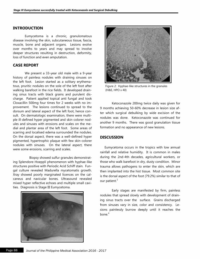
Page 88 Journal of the Philippine Medical Association 2016 - 2017
Stage III Eumycetoma successfully treated with Ketoconazole and Surgical Debulking
INTRODUCTION
Eumycetoma is a chronic, granulomatous
disease involving the skin, subcutaneous tissue, fascia,
muscle, bone and adjacent organs. Lesions evolve
over months to years and may spread to involve
deeper structures resulting in destruction, deformity,
loss of function and even amputation.
CASE REPORT
We present a 33-year old male with a 9-year
history of painless nodules with draining sinuses on
the left foot. Lesion started as a solitary erythema-
tous, pruritic nodules on the sole of the left foot after
walking barefoot in the rice fields. It developed drain-
ing sinus tracts with black grains and purulent dis-
charge. Patient applied topical anti fungal and took
Cloxacillin 500mg four times for 2 weeks with no im-
provement. The lesions continued to spread to the
dorsum and lateral aspect of the left foot, hence con-
sult. On dermatologic examination, there were multi-
ple ill-defined hyper pigmented and skin-colorer nod-
ules and sinuses with erosions and scales on the me-
dial and plantar area of the left foot. Some areas of
scarring and localised edema surrounded the nodules.
On the dorsal aspect, there was a well-defined hyper
pigmented, hypertrophic plaque with few skin-colorer
nodules with sinuses. On the lateral aspect, there
were some erosions, scarring and scales.
Biopsy showed sulfur granules demonstrat-
ing Splendore-Hoeppli phenomenon with hyphae-like
structures positive with Periodic Acid Schiff stain. Fun-
gal culture revealed Madurella mycetomatis growth.
Xray showed poorly marginated licences on the cal-
caneus and navicular bones. Ultrasound revealed
mixed hyper reflective echoes and multiple small cavi-
ties. Diagnosis is Stage III Eumycetoma.
Ketoconazole 200mg twice daily was given for
9 months achieving 50-60% decrease in lesion size af-
ter which surgical debulking by wide excision of the
nodules was done. Ketoconazole was continued for
another 9 months. There was good granulation tissue
formation and no appearance of new lesions.
DISCUSSION
Eumycetoma occurs in the tropics with low annual
rainfall and relative humidity. It is common in males
during the 2nd-4th decades, agricultural workers, or
those who walk barefoot in dry, dusty condition. Minor
trauma allows pathogens to enter the skin, which are
then implanted into the hist tissue. Most common site
is the dorsal aspect of the foot (79.2%) similar to that of
our patient.2
Early stages are manifested by firm, painless
nodules that spread slowly with development of drain-
ing sinus tracts over the surface. Grains discharged
from sinuses vary in size, color and consistency. Le-
sions painlessly burrow deeply until it reaches the
bone.4
Figure 2: Hyphae-like structures in the granules
(H&E, HPO x 40)
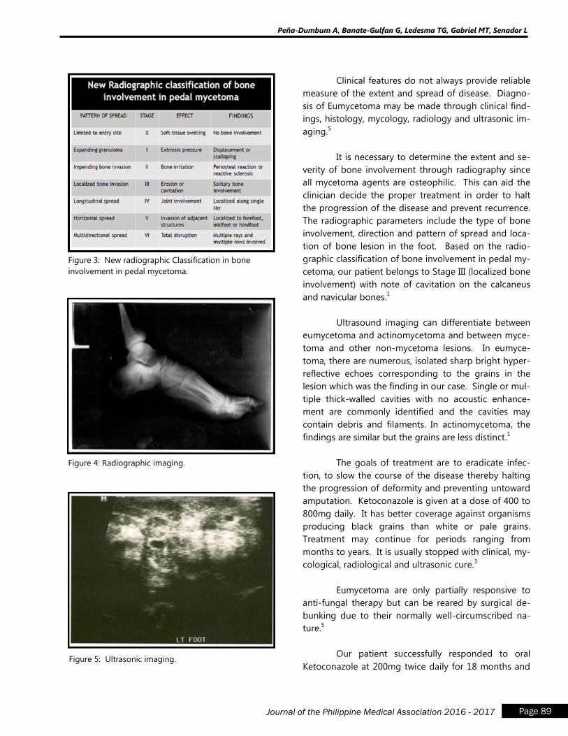
Page 89 Journal of the Philippine Medical Association 2016 - 2017
Peña-Dumbum A, Banate-Gulfan G, Ledesma TG, Gabriel MT, Senador L
Figure 3: New radiographic Classification in bone
involvement in pedal mycetoma.
Figure 4: Radiographic imaging.
Figure 5: Ultrasonic imaging.
Clinical features do not always provide reliable
measure of the extent and spread of disease. Diagno-
sis of Eumycetoma may be made through clinical find-
ings, histology, mycology, radiology and ultrasonic im-
aging.5
It is necessary to determine the extent and se-
verity of bone involvement through radiography since
all mycetoma agents are osteophilic. This can aid the
clinician decide the proper treatment in order to halt
the progression of the disease and prevent recurrence.
The radiographic parameters include the type of bone
involvement, direction and pattern of spread and loca-
tion of bone lesion in the foot. Based on the radio-
graphic classification of bone involvement in pedal my-
cetoma, our patient belongs to Stage III (localized bone
involvement) with note of cavitation on the calcaneus
and navicular bones.1
Ultrasound imaging can differentiate between
eumycetoma and actinomycetoma and between myce-
toma and other non-mycetoma lesions. In eumyce-
toma, there are numerous, isolated sharp bright hyper-
reflective echoes corresponding to the grains in the
lesion which was the finding in our case. Single or mul-
tiple thick-walled cavities with no acoustic enhance-
ment are commonly identified and the cavities may
contain debris and filaments. In actinomycetoma, the
findings are similar but the grains are less distinct.1
The goals of treatment are to eradicate infec-
tion, to slow the course of the disease thereby halting
the progression of deformity and preventing untoward
amputation. Ketoconazole is given at a dose of 400 to
800mg daily. It has better coverage against organisms
producing black grains than white or pale grains.
Treatment may continue for periods ranging from
months to years. It is usually stopped with clinical, my-
cological, radiological and ultrasonic cure.3
Eumycetoma are only partially responsive to
anti-fungal therapy but can be reared by surgical de-
bunking due to their normally well-circumscribed na-
ture.5
Our patient successfully responded to oral
Ketoconazole at 200mg twice daily for 18 months and
Page 90 Journal of the Philippine Medical Association 2016 - 2017
Stage III Eumycetoma successfully treated with Ketoconazole and Surgical Debulking
surgical debulking. There was no appearance of new
lesions. Repeat culture was negative. Repeat X-ray of
the left foot showed on the calcaneus and navicular
bones and ultrasound revealed absence of hyperre-
flective echoes and cavities.
CONCLUSION
Our patient is a confirmed case of Stage III
Eumycetoma through clinical findings, histology, my-
cology, radiology and ultrasonic imaging. Eumycetoma
must be considered when confronted with persistent
nodules on the foot and if not treated appropriately
can lead to unwanted deformities and even amputa-
tion. Institution of medical treatment and surgical de-
bunking in this case was curative and has improved our
patient‟s quality of life.
REFERENCE
1. Abd El Bagi. New Radiographic Classification of
Bone Involvement in Pedal Mycetoma. American
Journal of Radiology 2003;180:665-668
2. Lupi et al. Tropical Dermatology: Fungal Tropical
Diseases. J Am Acad Dermatol. 2005;53:931-51
3. Evidence Based-Guidelines for the Management of
Mycetoma Patients. Mycetoma Research Center.
Khartoum-Sudan. October 2002.
4. Kiran et al. Histological Diagnosis of Madura Foot
(Mycetoma): A Must for Definitive Treatment. Jour
of Global Infectious Diseases. 2009(I):64-67
5. Klaus Wolff et al. Fitzpatrick‟s Dermatology in Gen-
eral Medicine 7th ed. 2008; 190:1831-1844
Figure 6: Immediately after surgical debulking.
Figure 7: Two months after surgical debulking.
Case Report: X-linked Dystonia Parkinsonism -
Bridging Cultural Beliefs with Medical Practice
Trisha Mae C. Mendoza, M.D.
a
Abstract
Dystonia is often ascribed by common folk to supernatural causes rather than to a treatable
medical condition. Such is what happened in this case of a 24 year-old male who presented with
involuntary opening and closing of the mouth and writhing movements of the extremities, and
who was eventually diagnosed with X-linked Dystonia Parkinsonism, endemic in males from
Panay Island in the Philippines. A biopsychosocial approach on the management of the patient
included symptom relief with an atypical antipsychotic, neuroleptics, and botulinum toxin injec-
tion, CEA, and risk assessment evaluation for prevention of long term complications and death.
Page 91 Journal of the Philippine Medical Association 2016 - 2017
aResident Physician, Department of Family and Community Medicine, UP-PGH Medical Center
Paper selected for presentation, Interesting Case Competition, Philippine Academy of Family Physicians - Manila Chapter
Mendoza TM
Page 92 Journal of the Philippine Medical Association 2016 - 2017
Case Report: X-linked Dystonia Parkinsonism - Bridging Cultural Beliefs with Medical Practice
INTRODUCTION
Dystonia is often ascribed by common folk to
supernatural causes rather than to a treatable medical
condition, especially in our country that is rich in cul-
ture and folklore. X-linked Dystonia Parkinsonism
(XDP), a progressive, debilitating, adult-onset move-
ment disorder first described in Filipino males from
Panay Island is highlighted in this rare case. The num-
ber of existing cases of primary dystonia in the popu-
lation is not precisely known, but the condition is
probably much more frequent than reported. The
prevalence of Primary Dystonia is at 16.43/100,000
while as of January 2010, 505 cases of XDP (500 males,
5 females) have been identified in the updated XDP
Philippine registry.
CASE DISCUSSION
The patient is a 24 year-old male, single, Fili-
pino, Roman Catholic, right-handed, born and raised
in Cajidiocan, Romblon. He was previously well, with
no known previous hospitalization or surgeries, and
no history of encephalitis or trauma. He first exhibited
jaw stiffening, accompanied by involuntary opening
and closing of the mouth and temporomandibular
joint pain, 1 year prior to consult. There was no history
of animal bite, excessive drooling, dysphagia, restless-
ness or hydrophobia. Consult was done with a private
doctor and he was prescribed a muscle relaxant and
analgesics which afforded temporary relief. Nine
months prior to consult, he started having involuntary
writhing movements of the right upper and lower ex-
tremity. Contractions would persist throughout the
day accompanied by periods of relaxation. The patient
was still able to perform activities of daily living until 7
months prior to consult, when the involuntary writhing
movements progressed to the abdomen and left up-
per and lower extremities. There was noted difficulty
in ambulation. Spasms would cease when patient was
asleep. There was no fever, pain, dyspnea, dysphagia,
upward rolling of eyeballs, loss of consciousness,
numbness, weakness or bladder and bowel changes.
In the interim, there was progression in severity of
patient‟s symptoms. The sustained contractions would
occur more often and the intervals of relaxation were
shorter. A consult to a faith healer was done and he
he was made to drink a bitter concoction which did not
afford any relief. Persistence of symptoms prompted
consult at a tertiary hospital in Manila, and the patient
was subsequently decked to Family Medicine at the
Emergency Room.
Review of Systems
There was noted weight loss, from 55kg to 51
kg. Otherwise, the review of systems was unremarkable.
Past Medical History.
There were no known co-morbidities, allergies,
previous hospitalizations, surgeries or blood transfu-
sions. The patient had completed the recommended
Expanded Program of Immunization vaccines. He has
also received a tetanus booster in 2012.
Family History.
The maternal great grandfather also had symp-
toms similar to the patient‟s. (Figure 1. Genogram).
There was a family history of pulmonary tuberculosis
and hepatitis B in the maternal side; while there was
arthritis in the paternal side of the family.
Personal and Social History.
Patient had a 1.25 pack-years smoking history.
He was an occasional alcohol beverage drinker and he
denied illicit drug use. The patient also denies sexual
activity. At that time, was residing in a dormitory close
to school and used purified water as water source. He
was taking a BS Customs Administration course and
was on his last year when the involuntary movements
began as a result of which he had to stop his studies.
He was born and raised in Romblon but moved to
Subic in 2008 to study.
Physical Examination.
The patient was wheel-chair borne, well-kempt,
conscious, coherent, cooperative with stable vital signs.
His weighed 51 kg (ideal body weight should have
been 72 kg), and had a height of 5 feet and 8 inches,
with a calculated BMI of 16.28 kg/m2. There was no
Page 93 Journal of the Philippine Medical Association 2016 - 2017
and non-rhythmic, resulting from a continuous random
flow of muscle contractions. Dystonia is an involuntary
abnormal co-contraction of antagonistic muscles,
which may cause sustained abnormal postures or twist-
ing repetitive movements. Myoclonus is a sudden,
brief, shock-like involuntary movement, which is usually
caused by muscle contraction but can sometimes be
due to inhibition of muscular tone.
The patient presented with intermittent flexion
of abdominal muscles, writhing movements noted on
legs, and involuntary lifting of hip while lying down,
with tremors at rest on the right hand, intermittent,
involuntary contraction of the muscles of the right arm,
and both legs, as well as involuntary extension of
feet and toes. These symptoms were characteristic of
dystonia.
Dystonia is a movement disorder characterized
by sustained or intermittent muscle contractions caus-
ing abnormal, often repetitive, movements, postures or
both. It is typically patterned, twisting, and may be
tremulous, often initiated or worsened by voluntary
action and associated with overflow muscle activation.
The underlying neurochemistry of dystonia is not
known, but dopaminergic and cholinergic neurotrans-
mitters may be involved.
Dystonia may be classified by clinical charac-
teristics and by etiology. By clinical characteristics, the
classification may be further divided into age of onset,
body distribution, temporal pattern and associated fea-
tures. (Appendix A)
The patient manifested generalized, persistent
and progressive dystonia in his early adulthood, with
associated Parkinsonism. The laboratories done so far,
were all unremarkable. (Appendix B) Ideally, genetic
testing should be done to confirm the diagnosis how-
ever, due to financial constraints, the patient opted not
to have it done immediately.
Along with a family history of a similar symp-
tom in the maternal great grandfather, our final diag-
nosis for the patient was Dystonia, probably genetic in
origin, probably X-linked Dystonia Parkinsonism,
Underweight.
noted temporomandibular joint tenderness or stiff-
ness. The abdomen was flat with no visible pulsations.
There was 2cm x 3cm birthmark on the left lower
quadrant of the abdomen. There were intermittent
involuntary contractions of abdominal muscles. There
was no organomegaly of costovertebral angle tender-
ness. Direct rectal examination revealed no skin tags,
and intact rectal vault, tight sphincter tone, non-
bloody brown stools on the examining finger. Pulses
were full and equal. There was no edema, cyanosis, or
tenderness. Neurologic findings revealed an awake,
conversant patient who was oriented to person, place
and time, and was able to follow commands. The
pupils were 2 to 3 mm in size and were equally and
briskly reactive to light. There were no visual field
deficits. There was no weakness of the extra-ocular
muscles. There were no sensory deficits on the face.
There was no weakness of the temporalis and masse-
ter muscles, there was no facial asymmetry, gross
hearing was intact, the tongue was midline, and the
gag reflex was good. The spatula test was negative,
there was good shoulder shrug, no resistance on turn-
ing the head. There was no dysmetria or dysdiado-
chokinesia, and the patient maintained good balance
with eyes closed. There was no nuchal rigidity, and
Kernig‟s and Brudzinski‟s signs were absent. There was
good muscle bulk and tone, and motor strength was
5/5 on all extremities. Writhing movements were
noted on legs and there was involuntary lifting of the
hip while lying down. Pinprick, light touch, position
and vibration senses were intact. Reflexes were sym-
metric and there was no Babinski‟s sign or clonus.
There were no joint deformities and there was good
range of motion in the joints of the hands, wrists, el-
bows, shoulders, spine, hips, knees, ankles. There were
tremors at rest on the right hand. There were intermit-
tent, involuntary contractions of the muscles of the
right arm, and both legs with intermittent involuntary
extension of feet and toes. Gait was normal. The other
systems were unremarkable.
Movement disorders commonly encountered
at the clinic may be essential tremors, chorea, dysto-
nia, myoclonus or a combination of any of these. Es-
sential tremors are involuntary, rhythmic alternating
movements of one or more body parts. Chorea are
involuntary movements that are abrupt, unpredictable
Mendoza TM
Page 94 Journal of the Philippine Medical Association 2016 - 2017
Dubbed as the Panay dystonia, or Lubag in
the native tongue which means “musically out of
sync”, X-linked dystonia parkinsonism was popularly
known as the “aswang of Panay”. In 1975, the exis-
tence of a severe and progressive movement disorder
called XDP was discovered, which is a form of Parkin-
son‟s with dystonic features. In 1968, an independent
study by the Roxas Memorial Provincial Hospital,
pointed to a physical aberration that is believed to
explain the aswang myth and the reason why the
province of Capiz is singled out in all the folklores. The
affected gene is the DYT3, mapped to a 350 kb locus
in the DXS 7117-DX 559 region. Females are rarely
affected, most of them are carriers, and the disease
may hide in families until there is an affected male.
Symptoms begin as focal dystonia, often becoming
generalized by 4 years, commonly manifesting as
twisting or dragging a foot, repeated jaw opening and
closing, abnormal turning of the neck. In the next 7-15
years of illness, dystonia and Parkinsonism manifest
together until after 15 years, when Parkinsonism pre-
dominates. By then, the writhing movements are very
minimal. Neuropathology studies revealed marked
atrophy of the caudate and putamen while the cere-
bral cortices, thalamus, subthalamic nuclei, substancia
nigra and pons were unremarkable.
The diagnosis of dystonia is clinical, based on
the recognition of specific features. It is highly relevant
for providing appropriate management, prognostic
information and genetic counselling. Certain labora-
tory examinations are requested depending on the
etiology that the clinician has in mind. As for the
patient in this case, a XDP gene test was requested.
Treatment involves symptom relief of dystonia
and parkinsonism with anticholinergics, benzodiazepi-
nes or neuroleptics. In this case, Clonazepam 2mg/tab
½- ½ - 1xxx and Baclofen 10mg/tab ½ tab TID were
prescribed. Also, Botulinum toxin injections are the
first line treatment for dystonia affecting the face and
neck, provided the patient does not have pre-existing
dysphagia. However, for symptomatic improvement
in individuals with XDP with advanced disease and medically
refractory dystonia may be best treated with bilateral
pallidal stimulation according to a study by Wadia et
al 2010, Aguilar et al 2011, Patel et al 2014.
To prevent disease complications, diet modifi-
cation and other techniques to reduce the risk of aspi-
ration by doing a swallow evaluation is suggested.
Physical and occupational therapy may also be done to
improve mobility and assistive devices. This may pre-
vent the formation of contractures and delay bed-
bound state. Prolonged sitting or lying down is dis-
couraged to avoid pressure sores.
In dealing with these cases, the family physi-
cian must understand the cultural context of the pa-
tient and his family. In this case, the patient was born
and raised in a small town in Panay and their family
and ancestors have their own health beliefs, one of
them being that these symptoms were a “sumpa” in
the family spread through generations. This explains
why the patient went home when his disease couldn‟t
be treated with the medicines prescribed to him. Faith
healers were consulted, but because their interventions
were not helpful, one of their relatives suggested that a
consult to a tertiary hospital must be done to put an
end to their belief that the patient had indeed become
a victim of the “sumpa”.
Helping the patient and his family understand
the true biomedical context of this chronic, familial dis-
ease will help them come to an acceptance of the dis-
ease so that the patient will agree to the major thera-
peutic efforts that the physician will suggest.
Because dystonia influences various aspects of
quality of life, particularly those related to physical and
social functioning, it is important that the patient is
educated about his disease. Functional disability, body
concept and depression are important predictors of
quality of life in dystonia. In a study conducted by
Jamora et al, Suicidality among patients with sex-linked
Dystonia Parkinsonism, about 10.8% deaths in XDP are
attributed to suicide, with hanging as the number one
method used, followed by starvation organophosphate
poisoning non-accidental and self-mutilation. Mean
age at suicide was between 32 – 75 years of age, at an
average of 5.8 years from diagnosis. Majority were in
the generalized stage of the disease. This may be due
Case Report: X-linked Dystonia Parkinsonism - Bridging Cultural Beliefs with Medical Practice
Page 95 Journal of the Philippine Medical Association 2016 - 2017
End Notes: 1Lee LV, Rivera, Teleg, Dantes, Pasco, Jamora, Arancillo, Villareal-Jordan, Rosales, Demaisip, Maranon, Peralta, Borres, Tolentino,
Monding, Sarcia et al. The Unique Phenomenology of Sex-linked Dystonia Parkinsonism (XDP, DYT3, “Lubag”). Published Nov 2010 2Steeves, Day, Dykeman, Jette, Pringsheim. The Prevalence of Primary Dystonia: A systematic review and meta-analysis. Published
October 2012 3Lee, LV, Munoz, Tan, Teyes. Sex linked recessive dystonia parkinsonism of Panay, Philippines (XDP). March 2001 4Jamora, Cenina, Teleg, Lee. Suicidality among Patients with Sex-linked Dystonia Parkinsonism (XDP). Acta Medica Philippina,
The National Health Science Journal.
REFERENCES
1. Lee, Rivera, Teleg, Dantes, Pasco, Jamora, Arancillo,
Villareal-Jordan, Rosales, Demaisip, Maranon,
Peralta, Borres, Tolentino, Monding, Sarcia. The
unique phenomenology of sex-linked dystonia
parkinsonism (XDP, DYT3, "Lubag"). Nov 2010
2. Steeves, Day, Dykeman, Jette, Pringsheim. The
Prevalence of Primary Dystonia: A systematic
review and meta-analysis. Published October 2012
3. Lee, Munoz, Tan, Reyes. Sex linked recessive dysto-
nia parkinsonism of Panay, Philippines (XDP).
March 2001
4. Lee, Maranon, Demaisip, Peralta, Borres-Icasiano,
Arancillo, Rivera, Munoz, Tan, Reyes. The natural
history of sex-linked recessive dystonia parkinson-
ism of Panay, Philippines (XDP)q. December 2012
5. Guidelines for the diagnosis and treatment of pri-
mary (idiopathic) dystonia Report by an EFNS MDS
-ES Task Force
6. Dystonia: A clinical approach Article in Acta neu-
rologica Taiwanica · January 2009
7. Phenomenology and classification of dystonia: a
consensus update. Mov Disord. 2013 Jun;28(7):863-
73. Epub 2013 May 6.
8. Evidente. X-linked Dystonia-Parkinsonism.Apr 2015
9. Jamora, Cenina, Teleg, Lee. Suicidality among Pa-
tients with Sex-linked Dystonia Parkinsonism (XDP).
Acta Medica Philippina, The National Health Sci-
ence Journal.
Mendoza TM
to poor functional capacity, disfigurement and
perhaps, abandonment. Screening for depression is
highly recommended for patients with XDP, as well as
counselling of their caregiver to promote better care
for the patient, prevent abandonment, as well as, care-
giver strain.
In this case, risk assessment was done at the
end of the consult. The service recommended genetic
screening, depression screening, as well as lifestyle
modifications. the service also suggested, if finances
allowed, that the patient be vaccinated for Hepatitis B,
pneumococcal polysaccharide vaccine, annual influ-
enza vaccine, HPV, Hepatitis A vaccine and meningo-
coccal conjugate vaccine. Anticipatory guidelines
for the avoidance of tobacco, alcohol and other
substances of abuse were also given. Rehabilitation
was suggested and education about the prevention of
fals and aspiration was done.
There is no known definitive treatment for
X-linked Dystonia Parkinsonism to date. However, in
the Filipino community, it is imperative that a rare
disease like this be recognized, especially since it
is hypothesized to be endemic in the Panay Island
in the Philippines. In addition, it is important to be
recognized in the primary care setting as the dystonia
is associated with the aswang myth, a part of the
Philippine folklore and has been passed on from
generations to generations.
The patients who suffer from X-linked dysto-
nia-parkinsonism are people who need care, and are
not supposed to be feared. Perhaps, if the disease be-
comes common knowledge, the fear will be turned
into understanding, the stigma would be abolished,
and the patients afflicted with the disease will be
given the appropriate management and be given
hope.
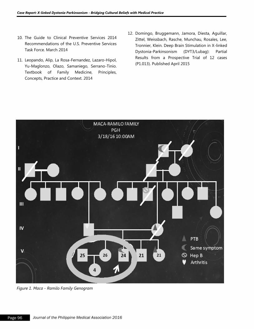
Page 96 Journal of the Philippine Medical Association 2016
Figure 1. Maca - Ramilo Family Genogram
10. The Guide to Clinical Preventive Services 2014
Recommendations of the U.S. Preventive Services
Task Force. March 2014
11. Leopando, Alip, La Rosa-Fernandez, Lazaro-Hipol,
Yu-Maglonzo, Olazo, Samaniego, Serrano-Tinio.
Textbook of Family Medicine, Principles,
Concepts, Practice and Context. 2014
12. Domingo, Bruggemann, Jamora, Diesta, Aguillar,
Zittel, Weissbach, Rasche, Munchau, Rosales, Lee,
Tronnier, Klein. Deep Brain Stimulation in X-linked
Dystonia-Parkinsonism (DYT3/Lubag): Partial
Results from a Prospective Trial of 12 cases
(P1.013). Published April 2015
Case Report: X-linked Dystonia Parkinsonism - Bridging Cultural Beliefs with Medical Practice
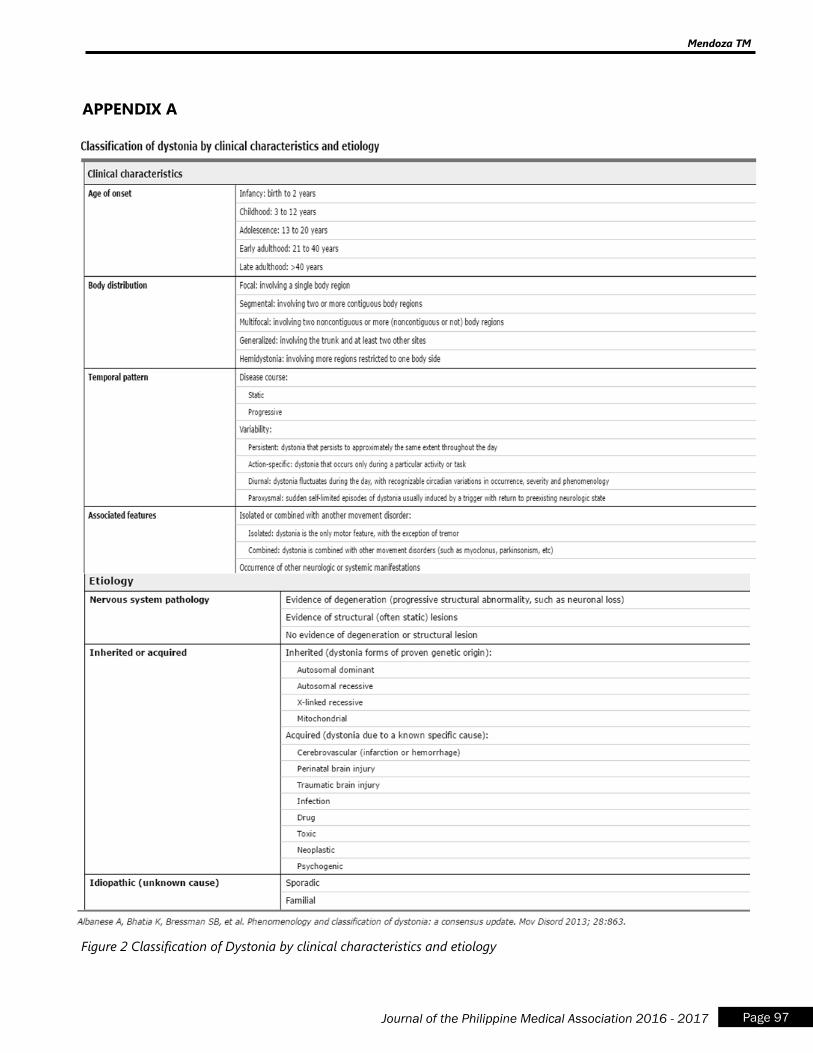
Page 97 Journal of the Philippine Medical Association 2016 - 2017
APPENDIX A
Figure 2 Classification of Dystonia by clinical characteristics and etiology
Mendoza TM
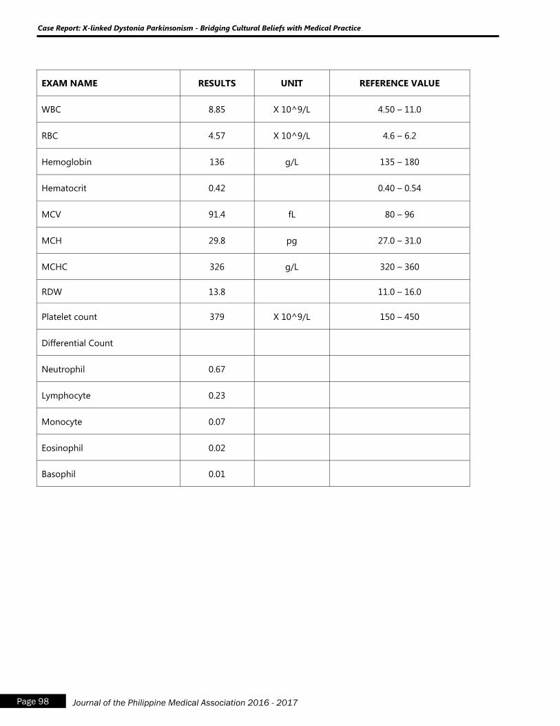
Page 98 Journal of the Philippine Medical Association 2016 - 2017
EXAM NAME RESULTS UNIT REFERENCE VALUE
WBC 8.85 X 10^9/L 4.50 – 11.0
RBC 4.57 X 10^9/L 4.6 – 6.2
Hemoglobin 136 g/L 135 – 180
Hematocrit 0.42 0.40 – 0.54
MCV 91.4 fL 80 – 96
MCH 29.8 pg 27.0 – 31.0
MCHC 326 g/L 320 – 360
RDW 13.8 11.0 – 16.0
Platelet count 379 X 10^9/L 150 – 450
Differential Count
Neutrophil 0.67
Lymphocyte 0.23
Monocyte 0.07
Eosinophil 0.02
Basophil 0.01
Case Report: X-linked Dystonia Parkinsonism - Bridging Cultural Beliefs with Medical Practice
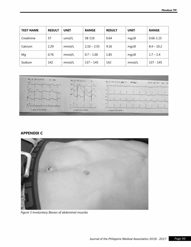
Page 99 Journal of the Philippine Medical Association 2016 - 2017
APPENDIX C
TEST NAME RESULT UNIT RANGE RESULT UNIT RANGE
Creatinine 57 umol/L 58-110 0.64 mg/dl 0.66-1.15
Calcium 2.29 mmol/L 2.10 – 2.55 9.16 mg/dl 8.4 – 10.2
Mg 0.76 mmol/L 0.7 – 1.00 1.85 mg/dl 1.7 – 2.4
Sodium 142 mmol/L 137 – 145 142 mmol/L 137 - 145
Figure 3 Involuntary flexion of abdominal muscles
Mendoza TM
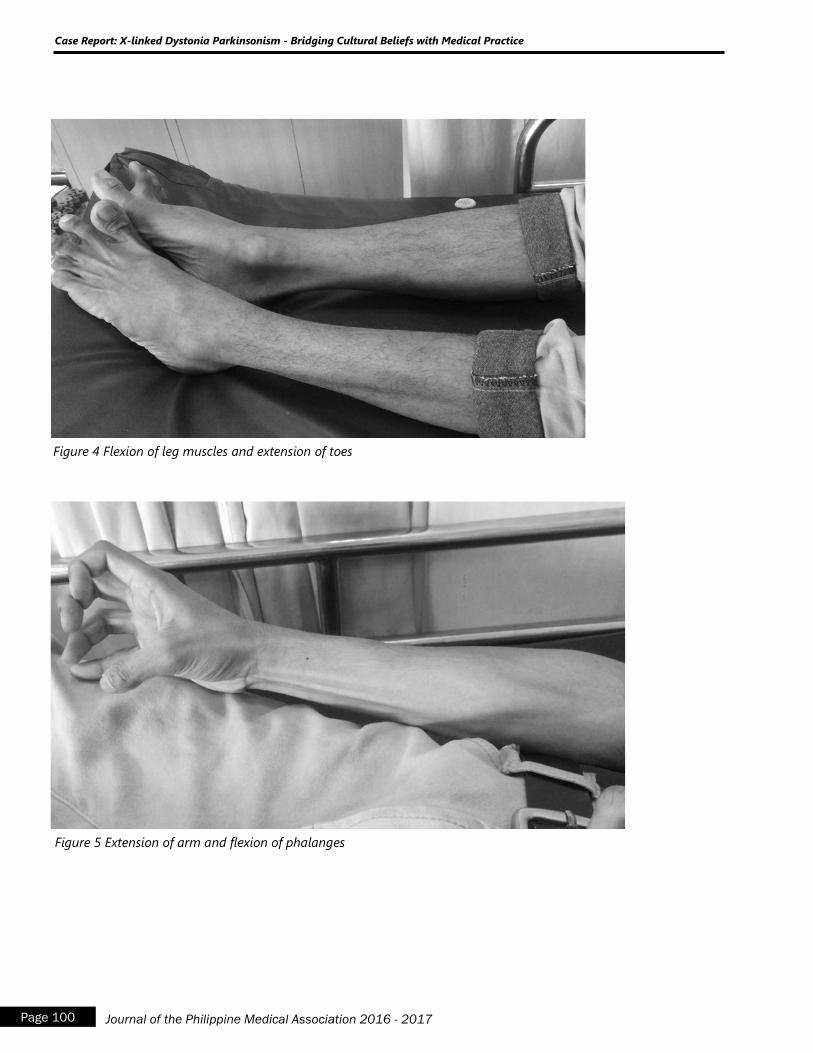
Page 100 Journal of the Philippine Medical Association 2016 - 2017
Figure 5 Extension of arm and flexion of phalanges
Case Report: X-linked Dystonia Parkinsonism - Bridging Cultural Beliefs with Medical Practice
Figure 4 Flexion of leg muscles and extension of toes
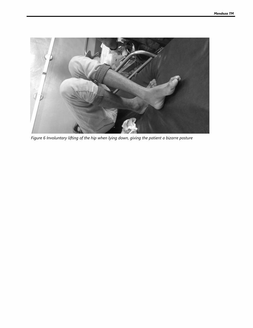
Figure 6 Involuntary lifting of the hip when lying down, giving the patient a bizarre posture
Mendoza TM