©2015 Jeana Louise Drake ALL RIGHTS RESERVED
163
©2015 Jeana Louise Drake ALL RIGHTS RESERVED
Transcript of ©2015 Jeana Louise Drake ALL RIGHTS RESERVED

©2015
Jeana Louise Drake
ALL RIGHTS RESERVED

THE SKELETAL PROTEOME AND PRODUCTION OF CALCIFYING PROTEINS
IN THE STONY CORAL STYLOPHORA PISTILLATA
by
JEANA LOUISE DRAKE
A dissertation submitted to the
Graduate School – New Brunswick
Rutgers, The State University of New Jersey
in partial fulfillment of the requirements
For the degree of
Doctor of Philosophy
Graduate Program in Oceanography
Written under the direction of
Paul G. Falkowski
and approved by
New Brunswick, New Jersey
OCTOBER, 2015

ii
Abstract of the Dissertation
The skeletal proteome and production of calcifying proteins in the stony coral Stylophora
pistillata
by JEANA LOUISE DRAKE
Dissertation Director:
Paul G. Falkowski
Coral biomineralization is important at the organismal, ecosystem, and global scales, yet
the biological component has not been well understood. In particular, identities, roles,
and environmental susceptibility of the proteins retained in coral skeleton were
previously unknown. To address this, my thesis sequenced the first coral skeletal
proteome, generated the first application of coral cell cultures to understanding the effects
of ocean acidification on coral calcification at the cellular and molecular levels, and made
the first use of NanoSIMS to test the co-localization of aspartic acid and newly formed
aragonite in corals.
To determine the proteins directly involved in the coral biomineralization process,
I used LC-MS/MS sequencing and a novel genome to sequence the skeletal proteome of
the stony coral, Stylophora pistillata. It contains an assemblage of adhesion and
structural proteins as well as two highly acidic proteins that constitute a novel coral SOM
protein sub-family. I next used cluster analysis to compare mineralizing genes known
from coral skeleton, mollusk shell, and sea urchin spines and tests. The analysis suggests
that there are few sequence similarities across all three phyla, supporting the independent

iii
evolution of biomineralization. However, there are core sets of conserved motifs in all
three phyla examined, including acidic proteins that appear to be responsible for the
nucleation reaction as well as inhibition; structural and adhesion proteins that determine
spatial patterning; and signaling proteins that modify enzymatic activities.
With the guidance of the sequenced coral skeletal proteome and the cluster
analysis, I chose four proteins to focus on for their expression response to increased CO2,
and their potential control on calcification, in cell cultures. The results suggest that
compensatory molecular adjustments to deal with ocean acidification are successful only
up to a point, beyond which these mechanisms cannot compete with local chemical
conditions unfavorable to biomineralization. Finally, I used NanoSIMS and S. pistillata
cell cultures to develop a method to co-localize highly acidic proteins and newly formed
calcium carbonate. Initial results point to both intra- and extracellular roles for these
proteins in transporting Ca to the calcification site and adhering cells to each other,
substrate, and new mineral.

iv
Prior Publications
Several sections of this dissertation have been published, or are under review for
publication, elsewhere. Chapter 2 was published prior to the data re-analysis and has the
following citation: Proteomic analysis of skeletal organic matrix from the stony coral
Stylophora pistillata. Proceedings of the National Academy of Sciences, 110, 3788-3793
(2013). Chapter 3 was published in its entirety and has the following citation: The
evolution and future of carbonate precipitation in marine invertebrates: Witnessing
extinction or documenting resilience in the Anthropocene? Elementa: Science of the
Anthropocene, 2, 000026 (2014). Chapter 4 has been formatted for submission to Global
Change Biology and was in preparation with co-authors at the time of dissertation
submission.

v
Acknowledgements
First and foremost, I am grateful to my committee for their guidance throughout this
dissertation. My advisor, Paul Falkowski, pushed me to search deeper and think harder
than I thought I could, and in exchange opened doors for me where doors didn’t
previously exist. I am most grateful that he took me on as his student. Debashish
Bhattacharya and his lab group provided rigor to my attempts at bioinformatics;
Debashish also showed me a true joy for research. Yair Rosenthal was a patient teacher
of geochemical methods and, along with his lab, taught me a whole new definition of
‘clean’. Both Yair and Rob Sherrell – who was not on my committee – were central to
developing the Sr-based calcification rate method used in Chapter 4. Oscar Schofield
encouraged me to think about the ecology of the biomineralization process. Throughout
my dissertation, Steve Weiner has pushed me to consider how understanding coral
biomineralization helps not just the coral crowd, but also the broader biomineralization
community. In the end, I have probably received more from that community than I gave
to them, and I am grateful to those biologists, geochemists, physical chemists, and
modelers for their insights and discussions.
Work for this dissertation was conducted as part of many fruitful collaborations.
Co-authors of Chapter 2 are Tali Mass, Liti Haramaty, Ehud Zelzion, Debashish
Bhattacharya, and Paul Falkowski. This chapter benefited from the services and support
of the Rutgers CABM Biological Mass Spectrometry Facility, Joseph Yaiullo of the Long
Island Aquarium, and Frank Natale of IMCS. N. Kröger for helpful comments on an
early draft of the published manuscript. Tali Mass and Udi Zelzion performed the

vi
genetic analysis of Stylophora pistillata versus Seriatopora sp. from the IMCS aquaria
for the data re-analysis.
Co-authors of Chapter 3 are Tali Mass and Paul Falkowski. Athena Fu and Mary
Battle contributed to sequence data collection and drawings, respectively, for Chapter 3.
Morgan Schaller and Ehud Zelzion contributed constructive discussions to the
development of the manuscript for this chapter.
Co-authors of the manuscript drawing heavily on Chapter 4 are Morgan Schaller,
Tali Mass, Athena Fu, Rob Sherrell, Yair Rosenthal, and Paul Falkowski. Ryan Bu and
Max Gorbunov kindly contributed technical advice, while Athena Fu, Liti Haramaty,
Thor Jensen, Christine Lee, and Frank Natale assisted in the laboratory and coral
aquarium.
Chapter 5 was a collaborative effort with Jess Adkins of the California Institute of
Technology. Yunbin Guan, at the CalTech Microanalysis Center, was a patient and
knowledgeable teacher as I produced samples appropriate for NanoSIMS analysis.
Athena Fu, Valentin Starovoytov, Nicolas Van Oostende, and Bess Ward provided
technical assistance. I am also grateful to Beatrice Birrer and Charlene Glascock for
sorting out the instrument User Agreement (and just generally for everything).
On a personal level, I am grateful to IMCS faculty and staff for indulging my
apparent attempt to use every instrument in this building. The students of the Graduate
Program in Oceanography and IMCS post-docs provided a supportive community for
‘nerding out’ as well as having fun. Specifically, Orly Levitan, Tali Mass, Mansha Seth-
Pasricha, Kim Thamatrakoln have been a crucial part of my ‘how do I be both a scientist
and a real human being’ pursuits.

vii
The most supportive people, I am lucky to say, have been my family. My Mom
and Dad have encouraged all of my scientific endeavors, no matter how far from Iowa
they take me. My grandmother is an inspiration in finding what I love to do and then
figuring out how to get paid for it. And to my husband and daughter, there is too much to
say so I simply say thank you.
Finally, adequate financial support during graduate work is rare, so I feel
incredibly lucky for and am indebted to IMCS and my advisor, committee members, and
colleagues for providing an environment where I (1) didn’t have to worry about paying
my rent and (2) had access to fabulous research instrumentation and opportunities. IMCS
provided me with a living wage and health insurance for the majority of my dissertation.
Research funding for this work came from National Science Foundation grant
EF1041143 to Paul Falkowski, Oscar Schofield, Rob Sherrell, and Yair Rosenthal, grant
EF-1416785 to Debashish Bhattacharya, Paul Falkowski, and Tali Mass, and a Sigma Xi
Grant-in-Aid-of-Research to me. Conference and workshop travel support was provided
on multiple occasions by the Rutgers Graduate School – New Brunswick.

viii
Dedication
For Luke and Clara Drake.
And for my parents, Sue and Don Gerrald, and Jim Goddard and Jackie Thompson, and
my grandmother, Mary Lou Hinrichsen.

ix
Table of Contents
Abstract of the Dissertation ............................................................................................. ii Prior Publications ............................................................................................................ iv
Acknowledgements ........................................................................................................... v
Dedication ....................................................................................................................... viii List of Tables .................................................................................................................... xi List of Figures .................................................................................................................. xii Chapter 1: Thesis Introduction ....................................................................................... 1
Chapter 2: Proteomic analysis of the skeletal organic matrix from the stony coral Stylophora pistillata ........................................................................................................... 9
2.1 Introduction ..................................................................................................................................... 9 2.2 Results and Discussion ............................................................................................................... 12
2.2.1 Structural Proteins .......................................................................................................................... 13 2.2.2 CARP Sub-Family ............................................................................................................................. 17
2.3 Methods ......................................................................................................................................... 20 2.3.1 Model organism .................................................................................................................................. 20 2.3.2 Protein separation and characterization .................................................................................... 21 2.3.3 Proteomics ............................................................................................................................................ 21 2.3.4 Gene Confirmation ............................................................................................................................ 22 2.3.5 Bioinformatics ..................................................................................................................................... 22
2.4 Supplementary Information ..................................................................................................... 28 2.5 Data Re-analysis with additional gene models ..................................................................... 29
Chapter 3: The evolution and future of carbonate precipitation in marine invertebrates: Witnessing extinction or documenting resilience in the Anthropocene? ................................................................................................................ 41
3.1 Introduction .................................................................................................................................. 42 3.2 Background and context for biomineralization in the three invertebrate phyla ......... 44
3.2.1 Body plan and calcifying tissues ................................................................................................... 45 3.2.2 Biomineralizing Proteins ................................................................................................................. 48
3.3 Comparison of carbonate organic matrix proteins across marine invertebrate phyla ................................................................................................................................................................ 50
3.3.1 Evolution of the acidic proteins ..................................................................................................... 50 3.3.2 Conserved non-acidic proteins across phyla ............................................................................. 54 3.3.3 Signaling and Regulation ................................................................................................................ 55 3.3.4 Summary of Conservation of Biomineralizing Proteins ......................................................... 57
3.4 Future of marine invertebrate biomineralizers ................................................................... 58 3.5 Supplementary Material ........................................................................................................... 66
Chapter 4: The influence of CO2 on calcification in coral cell cultures ..................... 67 4.1 Introduction .................................................................................................................................. 68 4.2 Methods ......................................................................................................................................... 70
4.2.1 Experimental Setup and Conditions ............................................................................................. 70 4.2.2 Sample Collection .............................................................................................................................. 72 4.2.3 SOM Protein Expression.................................................................................................................. 73
x
4.2.4 Calcification Rate............................................................................................................................... 74 4.2.5 Variable Fluorescence ...................................................................................................................... 75 4.2.6 Statistical Analysis ............................................................................................................................. 76
4.3 Results ............................................................................................................................................ 76 4.4 Discussion ...................................................................................................................................... 78 4.5 Conclusions ................................................................................................................................... 86
Chapter 5: A NanoSIMS study on the mechanisms of calcification in coral cells ... 92 5.1 Introduction .................................................................................................................................. 92 5.2 Methods ......................................................................................................................................... 95
5.2.1 Cell Culture Preparation ................................................................................................................. 95 5.2.2 Choosing Sampling Timepoints ..................................................................................................... 96 5.2.3 15N-Asp Additions ............................................................................................................................... 98 5.2.4 Sample Fixation .................................................................................................................................. 98 5.2.5 Protopolyp Imaging ........................................................................................................................... 99 5.2.6 NanoSIMS .......................................................................................................................................... 100 5.2.7 NanoSIMS Image Processing ...................................................................................................... 100 5.3.8 CARP4 Transmembrane Prediction .......................................................................................... 101
5.3 Results .......................................................................................................................................... 101 5.4 Discussion .................................................................................................................................... 104 5.5 Conclusions ................................................................................................................................. 107
Chapter 6: Conclusions ................................................................................................ 116
Appendix 1: Chapter 4 Supplementary Tables ......................................................... 119
Appendix 2: Additional NanoSIMS images ............................................................... 120
References ...................................................................................................................... 129
xi
List of Tables
2.1 S. pistillata SOM proteins…………………………………………………...24
2.2 Potential contaminant SOM proteins…………………………………..……35
2.3 Additional potential SOM proteins………………………………………….35
2.4 Final list of 32 S. pistillata SOM proteins…………………………………..36
3.1 Proteins from coral, mollusk, and sea urchin carbonate organic matrices…..61
3.2 Proteins of shared function, but not shared sequence……………………….62
4.1 Artificial seawater and growth medium chemical parameters………………87
5.1 NanoSIMS masses detected………………………………………………..108
5.2 Cation EDS peak area ratio ranges……………………………………..….108
5.3 NanoSIMS counting statistics…………………………………………..…109
5.4 NanoSIMS co-localization counts……………………………………..….110
A1 Coral cell cultures growth media carbonate parameters…………………..119
A2 S. pistillata qPCR primer sequences………………………………...……119

xii
List of Figures
1.1 Phylogeny of eukaryotic biomineralization……………………………………7
1.2 Seawater carbonate parameters………………………………………………..8
2.1 Predicted structure of P12…………………………………………………....26
2.2 CARP4 sub-family general pattern…………………………………………..27
2.3 Corals used for SOM proteome sequencing………………………………....38
2.4 Phylogenetic trees of (A) COI-1 and (B) ITS…………………………….….39
3.1 Schematic drawings of three biomineralizing marine invertebrates …….…..63
3.2 Cellular component ontology of COM proteins………………………….….64
3.3 Multiple sequence alignment of CARP4 homologues…………………..…..65
4.1 Light micrographs of S. pistillata cell cultures…………………….………..88
4.2 Expression fold-change of skeletal organic matrix genes……….…….…….89
4.3 Cadherin protein expression quantified by western blot…….………………90
4.4 Calcification rate of S. pistillata cell cultures…………………….…………90
4.5 Variable fluorescence of S. pistillata cell cultures……………………..……91
5.1 Expression of CARP4………………………………………………...……111
5.2 Formation of S. pistillata protopolyps……………………………….…….112
5.3 Intracellular and extracellular Ca in S. pistillata protopolyps………….….113
5.4 5d-5 sample ROI……………………………………………………….…..114
5.5 12d-12 sample ROI………………………………………………………...115 A1-A8 Additional NanoSIMS images…..………………………………..120-128

1
Chapter 1: Thesis Introduction
Biomineralization is the process by which a living organism directly or indirectly causes
a mineral to be produced (Lowenstam et al. 1989). This ranges from the sulfide minerals
precipitated when H2S released by sulfate reducing bacteria reacts with high
concentration metals in the surrounding medium (e.g.;Baas Becking et al. 1961, Fortin et
al. 1996, Labrenz et al. 2000) to the highly controlled production and dissolution of
hydroxyl-apatite into bones and teeth by our bodies (e.g.;Kawasaki et al. 2009). The
roles for biologically controlled biominerals include adhesion to a substrate, protection,
cation storage, light modulation, buoyancy, and magnetic perception among others
(Lowenstam et al. 1989, Mann 2001).
Biomineralization occurs across the tree of life and is prominent within
eukaryotes (Figure 1.1); however, the mechanism(s) remain enigmatic. The questions that
address these mechanisms for each organism cross traditional disciplinary boundaries:
What biomolecules are involved? And at what time? If an organism possesses multiple
cell types, which biomolecules come from which cell? What is the role of each of these
biomolecules in forming the mineral, from making a concentration of relevant anions and
cations through to terminating any crystallization? How do the functions of these
biomolecules result in the ‘vital effects’ that we observe as a difference in element and
isotope ratios between the source medium and the formed mineral? Answering these
questions requires insights from molecular biology, bioinformatics, biochemistry,
physical chemistry, and geology.

2
Marine calcification has been of interest over the past decade or so, as the
formation and persistence of these carbonates is susceptible to ocean acidification (Feely
et al. 2004, Orr et al. 2005). The geochemical perspective proposes less marine
calcification due to the lowering CaCO3 saturation state of seawater (Zeebe et al. 2001).
Dissolved CO2 reacts with seawater and dissociates to form bicarbonate and carbonate,
releasing a proton (and lowering the pH) with each dissociation per the reaction:
𝐶𝐶𝐶𝐶2 + 𝐻𝐻2𝐶𝐶 ↔ 𝐻𝐻2𝐶𝐶𝐶𝐶3 ↔ 𝐻𝐻𝐶𝐶𝐶𝐶3− + 𝐻𝐻+ ↔ 𝐶𝐶𝐶𝐶32− + 𝐻𝐻+
The concentration of each carbonate species in seawater is a function of the pH, with the
amount of bicarbonate nearly an order of magnitude higher than that of carbonate at
current ocean pH (Figure 1.2). Calcium carbonate saturation state describes the observed
concentrations of calcium and carbonate relative to those values at saturation, a
relationship driven more by the carbonate component than by calcium:
M = (𝐶𝐶𝑎𝑎2+)𝑜𝑜𝑜𝑜𝑜𝑜∗ (𝐶𝐶𝑂𝑂32−)𝑜𝑜𝑜𝑜𝑜𝑜
𝐾𝐾𝑜𝑜𝑠𝑠∗
where
𝐾𝐾𝑠𝑠𝑠𝑠∗ = (𝐶𝐶𝑎𝑎2+)𝑠𝑠𝑎𝑎𝑠𝑠 + (𝐶𝐶𝐶𝐶32−)𝑠𝑠𝑎𝑎𝑠𝑠
Hence, increased atmospheric CO2 leads to decreased ocean pH and carbonate anion, and
in turn a lower carbonate saturation state, making it harder for inorganic calcium
carbonate to be formed or maintained.
In contrast, the biological view of marine mineralization suggests that, like other
biominerals, marine bio-carbonates are very different from inorganic carbonates. Not
only are biomolecules located on and within individual crystals (e.g.;Suzuki et al. 2009,
Mass et al. 2014), but interim stabilized amorphous phases exist (Gotliv et al. 2003, Politi
et al. 2008) and a nano-structure to the crystals is apparent (Cuif et al. 2008, Falini et al.

3
2013). Additionally, physiological and ecological experiments highlight metabolic costs
that are predominantly pH-based (Hofmann et al. 2010) and suggest that the
biomineralization process allows for adaptation and plasticity (Kelly et al. 2013, Munday
et al. 2013). Hence, it is becoming increasingly clear that marine biomineralization is
much more complex than simple inorganic mineral formation and that mineralizing
organisms do not all respond in the same way to ocean acidification (Ries et al. 2009,
Kroeker et al. 2010), suggesting that the ‘bio’ component of marine biomineralization
requires better understanding.
A biological component of marine biominerals has been known for over a century
(Silliman 1846, Wainwright 1963). Specifically in corals, biomolecules make up 0.1-5%
of the carbonate mineral (Wainwright 1963, Cuif et al. 2004, Falini et al. 2013). The
biomolecules are comprised of polysaccharides, lipids, proteins, with the protein
component biased towards the two acidic amino acids aspartic and glutamic acid (Young
et al. 1971, Mass et al. 2012). Although the coral skeletal organic matrix (SOM) protein
complex has been immunolocalized to the calicoblastic epithelium (Puverel et al. 2005),
until recently, the makeup of this complex was unknown. Using gene models (Mass et al.
2013) for the stony coral, Seriatopora sp., a member of a clade within Stylophora
pistillata (Keshavmurthy et al. 2013), I sequenced the S. pistillata skeletal proteome
(Chapter 2) by liquid chromatography coupled to tandem mass spectrometry (LC-
MS/MS). Both the original work as well as a re-analysis of the LC-MS/MS data against
two additional Stylophora pistillata and Seriatopora hystrix transcriptomes and a revised
Seriatopora sp. genome reveal that coral SOM is composed largely of structural and
4
adhesion proteins such as cadherin, collagen, and vitellogenin, while also incorporating
enzymes such as carbonic anhydrase and the low pI coral acid rich protein 4 (CARP4).
During the oral portion of my qualifying exams, my advisor, Dr. Paul Falkowski,
asked me to draw a phylogenetic tree of biomineralization proteins. I told him I didn’t
think an analysis had been done by others to support my reproduction of such a tree on
the white board. Additionally, phylogenetic trees require gene similarity between
organisms. Although some vertebrates have sufficiently similar biomineralization genes
– such as between Tyrannosaurus rex and chickens, frogs, and mice (Asara et al. 2007) –
the similarities, and hence shared evolutionary histories, of marine invertebrate
biomineralizers was unexamined. To address this, I performed a cluster analysis of over
1500 coral, mollusk, and sea urchin mineral-associated proteins (Chapter 3). For each
phylum, the complexes of these proteins could be considered a ‘toolkit’, a suite of
functional proteins from which organisms can draw to build their skeletons, shells, and
spines. Several structural, signaling, and protein reworking proteins are conserved across
phyla. In contrast, the highly acidic proteins are novel to each phylum suggesting that
they evolved through convergent evolution. Additionally, other structural, adhesion, and
enzymatic proteins are conserved in function but not sequence across the phyla
suggesting that, for the most part, the biomineralization ‘toolkits’ for each group of
invertebrates evolved independently but converged toward the optimal re-assignment of
preexisting proteins.
Stony corals are ecologically and economically important members of coastal
habitat and are thought to be highly susceptible to current anthropogenic processes
including climate change and ocean acidification (White et al. 2000, Cesar et al. 2003,

5
Hoegh-Guldberg et al. 2007). Therefore, in addition to detailing and understanding the
function of coral SOM proteins, it is important to determine how increased CO2atm affects
production of both SOM proteins and mineral. Gene expression by corals treated with
increased CO2atm has been examined (Moya et al. 2012), but without the benefit of a list
of confirmed SOM proteins (Drake et al. 2013, Ramos-Silva et al. 2013). Hence, it was
clear that that the effects of ocean acidification at the cellular and molecular levels
required further examination.
The study of biomineralization has benefited for the past 30 years from the
development of model organisms such as sea urchin larvae and mice (Benson et al. 1986,
Rosati et al. 1994). A comparable system exists for corals in the form of cell cultures
(Domart-Coulon et al. 2001, Helman et al. 2008, Mass et al. 2012). Although they have
not been maintained for longer than one month, coral cell cultures form proto-polyps
similar in structure to intact tissue, produce an extracellular matrix, and precipitate
aragonite like their mother colony (Mass et al. 2012). These cell cultures can be used to
study processes at the cellular and molecular levels on timescales from hours to days.
Therefore, I subjected cell cultures of S. pistillata to four CO2 concentrations, from 400
to 2000 ppm, and measured photosynthetic capability, SOM protein gene expression and
protein production, and calcification rate from four hours to 1.5 weeks (Chapter 4). S.
pistillata cells responded dramatically to CO2 treatments. Protopolyps formed at low and
moderate, but not at high and very high, CO2. SOM genes were up-regulated in low to
high CO2, but most were down-regulated at very high CO2. And calcification was not
detectable at 1000 ppm CO2 and beyond. These results highlight the molecular

6
machinery corals possess to maintain calcification under increased CO2, but this
machinery fails above a threshold.
A group of SOM proteins clearly important to the biomineralization process is the
CARPs. Found in SOM complexes across coral families (Puverel et al. 2005, Drake et al.
2013, Ramos-Silva et al. 2013, Mass et al. 2014), the CARPs contain a high (>20%)
proportion of aspartic and glutamic acids, giving them a net-negative charge and low
isoelectric points (pIs). The low pIs allow them to precipitate CaCO3 in vitro from
seawater at both pH 8.2 and 7.6 (Mass et al. 2013). They have been immunolocalized not
only to precise locations, including early mineralization zones in (e.g.) S. pistillata
skeleton, but also within individual crystals (Mass et al. 2014). Therefore, the CARPs
could act as nucleation sites (Mass et al. 2013) or inhibitors (Shiraga et al. 1992, Treccani
et al. 2006), to extend existing crystals, to terminate crystal growth (Gerbaud et al. 2000),
or as stabilizers of a pre-nucleation amorphous phase (Ma et al. 2007, Politi et al. 2007),
nano-particle accretion (Gal et al. 2015), or liquid precursor (Olszta et al. 2003). To
address the role of highly acidic proteins in the biomineralization process, I used nano-
scale secondary ion mass spectrometry (NanoSIMS), with a resolution of ~60 nm, to co-
localize newly formed calcium carbonate and 15N-labeled aspartic acid incorporated by S.
pistillata cell cultures (Chapter 5). Preliminary analyses reveal roles for these proteins as
intracellular Ca concentrators and extracellular adhesives for cell-cell connections and
secure newly formed calcium carbonate to cells and to substrate. This expands the
potential role for these proteins in biomineralization.

7
Figure 1.1 Phylogeny of eukaryotic biomineralization focusing on calcium carbonates (C), calcium phosphates (P), and silicates (S). Reprinted from Reviews in Mineralogy and Geochemistry, Volume 54, Andrew Knoll, Biomineralization and Evolutionary History, p329-356, Figure 2, copyright 2003, with permission of Mineralogical Society of America.

8
Figure 1.2. Seawater carbonate parameters. Concentrations of CO2, HCO3-, and CO3
2- as a function of pH, including the effects of temperature and salinity. Reprinted from CO2 in Seawater: Equilibrium, Kinetics, Isotopes, Richard E. Zeebe and Dieter Wolf-Gladrow, Chapter 1 Equilibrium, p10, Figure 1.1.4, Copyright 2001, with permission from Elsevier.
pH

9
Chapter 2: Proteomic analysis of the skeletal organic matrix from the stony coral Stylophora pistillata Abstract
It has long been recognized that a suite of proteins exists in coral skeletons that is critical
for the oriented precipitation of calcium carbonate crystals, yet these proteins remain
poorly characterized. Using liquid chromatography-tandem mass spectrometry analysis
of proteins extracted from the cell free skeleton of the hermatypic coral, Stylophora
pistillata, combined with a draft genome assembly from the cnidarian host cells of the
same species, we identified 36 coral skeletal organic matrix (SOM) proteins. The
proteome of the coral skeleton contains an assemblage of adhesion and structural proteins
as well as two highly acidic proteins that may constitute a novel coral SOM protein sub-
family. We compared the 36 SOM protein sequences to genome and transcriptome data
from three other corals, three additional invertebrates, one vertebrate, and three single-
celled organisms. This work represents the first extensive proteomic analysis of
biomineralization-related proteins in corals from which we identify a biomineralization
“toolkit” - an organic scaffold upon which aragonite crystals can be deposited in specific
orientations to form a phenotypically identifiable structure.
Keywords: biomineralization, coral acid-rich proteins, collagen
2.1 Introduction
Biomineralizing organisms are found in all biological kingdoms and incorporate a variety
of metals, from sodium to lead, as major components of minerals whose nucleation and
growth are under a range of biological control (Lowenstam et al. 1989, Dove et al. 2003,
Dobbs et al. 2004, Boorungsiman et al. 2012, Drescher et al. 2012). The skeletal organic

10
matrix (SOM), occluded in the mineral, has been implicated as a source of the increased
strength of biominerals over comparable geominerals and has long been hypothesized to
aid in the stabilization, nucleation, growth, and spatial orientation of biominerals
(Wainwright 1963, Weiner et al. 1975, Addadi et al. 2006, Beniash 2011, Asenath-Smith
et al. 2012). However, the mechanism(s) for the role of the SOM in coral biomineral
formation remain to be elucidated, primarily because the organic molecules have yet to
be characterized.
At present, the best-characterized SOM is that contained in mammalian bones and
teeth and is divided into structural and highly acidic protein categories. Collagen, a well-
characterized fibrillar (i.e., framework) protein, plays a structural role in mammal SOM
(review in (Beniash 2011)), and has been investigated in the structural components of
several coelenterates (Marks et al. 1949, Goldberg 1974), of which Order Scleractinia
(i.e., stony corals) is a member. However, the greatest emphasis for nucleation proteins
rests on the highly acidic proteins such as bone sialoproteins and dentin matrix proteins in
teeth in vertebrates (Hunter et al. 1993, He et al. 2003). Highly acidic proteins also have
been identified or hypothesized in mineralizing invertebrates, and several have recently
been described, including the Asprich family in the pen shell, Atrina rigida (Gotliv et al.
2005), Pif and Aspein in the pearl oyster, Pinctada fucata (Takeuchi et al. 2008, Suzuki
et al. 2009), the Adi-SAPs (highly acidic proteins proposed to be soluble and/or secreted)
in the stony coral Acropora digitifera (Shinzato et al. 2011), and the CARPs in
Stylophora pistillata (Mass et al. 2013). Sequence-based homologs of each have been
found in a variety of other invertebrates, yet these proteins, and their consequent
biomineralization reactions, appear to have originated several times independently (Knoll

11
2003, Murdock et al. 2011), and their sequence similarity is most likely explained by
convergent evolution. Proteins containing acidic amino acids (Asp and Glu) or
phosphorylation sites (on Ser residues) are thought to be used at various stages of
aragonite and calcite mineralization to temporarily stabilize amorphous calcium
carbonate or nucleate the mineral under appropriate conditions (review in (Marin 2007)).
Stony corals (Class Anthozoa) are early-branching metazoans composed of four
cell layers (Veron 1986). Whereas the oral endodermal cells host endosymbiotic
photosynthetic algae of the genus Symbiodinium, the aboral ectodermal cells, or
calicoblastic cells, are the sites of biomineralization. Calicoblastic cells are thought to
secrete SOM that adheres the cells to recently formed extracellular skeleton, and are also
considered to be intimately involved in nucleation and growth of aragonite crystals
(Clode et al. 2002, Goffredo et al. 2011). SOM is retained in the coral skeleton and the
amino acid composition of the protein fraction indicates that the SOM is distinct from
cellular or mucus protein (Young 1971, Ducklow et al. 1979). To date, only one coral
SOM protein, galaxin, has been fully sequenced and its role in the biomineralization
process is not well understood because it does not bind calcium (Watanabe et al. 2003).
Other proteins hypothesized to play a role in the mineralizing space between the
calicoblastic cells and the skeleton include carbonic anhydrases (Moya et al. 2012),
collagen (Goldberg 1974), ion transporters (Kaniewska et al. 2012), cysteine-rich
proteins (Sunagawa et al. 2009), von Willebrand factor type A domain-containing
proteins and zona pellucidas (Hayward et al. 2011), and secreted acidic proteins (SAPs)
(Sarashina et al. 2006, Shinzato et al. 2011). Together, these proteins may represent a
‘biomineralization toolkit’ of calcifying proteins in corals, but most remain to be

12
independently confirmed in coral skeleton, described as complete genes, or characterized
with respect to function.
Here we use a proteomics approach to describe the SOM proteins in the widely
distributed, Pocilloporid coral, Stylophora pistillata. Using liquid chromatography-
tandem mass spectrometry (LC-MS/MS) protein sequencing and a draft genome from S.
pistillata (Mass et al. 2013), we identify partial and complete sequences of proteins in the
S. pistillata aragonite skeleton. Comparison of our results with genome and
transcriptome data from other mineralizers suggests that a coral skeleton contains a
complex group of proteins that guide the biomineralization process to form specific,
genetically determined structures. This, the first proteome from a coral skeleton,
identifies a ‘biomineralization toolkit’ in these key, ecologically critical organisms.
2.2 Results and Discussion
Thirty-six proteins from the genes predicted from the draft S. pistillata genome assembly
were detected by three LC-MS/MS analyses of SOM proteins (Table 2.1). This is similar
in number, but not sequence identity, to the 33 shell matrix proteins recently identified by
LC-MS/MS from mollusk nacre (aragonite) (Marie et al. 2012). Twenty-five of the coral
SOM protein candidates were only observed after deglycosylation (SI T1). 31 could be
observed with tryptic digestion while the remaining five were observed only after
proteinase K digestion (SI T1).
A major problem in isolating and identifying specific proteins from corals is their
post-translational modification, primarily by glycosylation (Sarashina et al. 2006).
Although we consistently observed five distinct protein bands in size-fractionated SOM
proteins, there was always a strong background smear on silver stained polyacrylamide

13
gels (SI F1). Periodic acid-Schiff staining confirmed the abundance of glycans in these
samples (SI F1). Hence, the results presented here are almost certainly a conservative
estimate of the total number of proteins in the skeletal matrix.
Nearly all proteins detected by LC-MS/MS show similarities to proteins known to
play roles in the structure and adhesion of cells (Table 2.1). We detected multiple
proteins with hits (e-value ≤ 10-5) to precursor cadherins (P1, P9, P10, and P23) and von
Willebrand factor (P3, P5, P13, P14, P18, P32) domains. Blast hits for other proteins
include two collagens (P14, P18), three actins (P6, P7, and P11), and a carbonic
anhydrase (P35), among others. Five candidate proteins exhibit poor (e-value > 10-5) or
no blast hits in NCBI (P2, P12, P15, P16, and P22). Although amino acid analyses of
SOM from a variety of corals suggest that highly acidic proteins are compositionally
important (Young 1971, Mass et al. 2012), we only identified two such predicted proteins
by LC-MS/MS.
2.2.1 Structural Proteins
The highest scoring predicted protein, P1, is an incomplete protocadherin fat 1-
like protein that contains a von Willebrand factor type A domain. We identified three
separate PCR amplicons that map to this gene in S. pistillata cDNA, validating its
expression. These transcripts encompass 11 of the 15 MS-sequenced peptides for this
candidate protein (SI F2). Comparison with a very similar predicted protein from A.
digitifera and Favia sp. (Table 2.1) suggests that the S. pistillata protein is incomplete
and can be extended ~750 residues towards the N-terminus, likely containing a secretory
signal, and over 1000 residues toward the C-terminus (SI T1; (Shinzato et al. 2011, Mehr

14
et al. 2013)). Two additional proteins among the top 10 detected also show cadherin
precursor-like qualities (P9, P10; Table 2.1).
Cadherins are calcium-dependent cell-cell adhesion molecules with specificity for
certain cell types such as neurons or osteoblasts (Derycke et al. 2006). Apoptotic
osteoblasts and intact retinal cells can cleave the extracellular portion of their respective
N-cadherins to produce a 90 kDa soluble protein that may have roles in cell survival and
protein expression. In addition, they have been implicated in the aggregation of cells into
which they have been cloned (Nose et al. 1988), and could help explain the proto-polyps
recently observed in S. pistillata cell cultures (Mass et al. 2012).
Of the six detected proteins that contain von Willebrand factor type A (vWFa, an
adhesion glycoprotein), four (P3, P5, P14, and P18) show strong sequence similarity
between the predicted sequences from S. pistillata and those from A. digitifera, Favia sp.,
and P. damicornis (Table 2.1; SIT1; (Shinzato et al. 2011, Traylor-Knowles et al. 2011,
Mehr et al. 2013)). We compared these four predicted proteins from S. pistillata with
pre- and post-settlement genes in A. millepora larvae as described by Hayward et al.
(Hayward et al. 2011). A. millepora genes A9, A90, and A102 are ≥40% similar to the
P5 protein found in this study, with blastp e-values ranging from 10-15 - 10-23. All three
proteins in the A. millepora expression study were up-regulated in pre-settlement
planulae relative to the post-settlement stage and two, A9 and A90 were localized to the
aboral region post-settlement (Hayward et al. 2011). This suggests that A9, A90, and
A102 – and therefore, possibly P5 – are involved in adhesion of calicoblastic cells to the
skeleton. The highly acidic mollusk nacre protein, Pif, also contains a von Willebrand

15
factor type A, although there is no sequence similarity between Pif and the coral proteins
described here (Suzuki et al. 2009).
Collagen and chitin share similar roles in biomineralizing organisms (Ehrlich
2010), although chitin would not be detectable by the procedures we used here. Two
alpha collagen-like proteins were detected both before and after deglycosylation (P14 and
P18, SI T1). We have confirmed the transcription of a portion of P14 by PCR
amplification of S. pistillata cDNA, (SI F2). P14 shows strong similarities to two
predicted sequences from A. digitifera (e-value 10-16) and Favia sp. (e-value 10-41) (Table
2.1; (Shinzato et al. 2011, Mehr et al. 2013)). Collagen, a fibrillar protein common in
extracellular matrix, is generally considered as a place for non-collagenous proteins to
bind and nucleate the mineral (review in (Beniash 2011)), but may also form sites of
nucleation in ‘holes’ of packed collagen molecules (Silver et al. 2011). Although
collagen has been confirmed in sea pen axial stalks (Marks et al. 1949) and gorgonian
skeletons (Goldberg 1974), little work has been conducted on its presence in scleractinian
skeletons. The extracellular matrix portion of coral cell cultures has been shown to
positively stain for collagen (Helman et al. 2008), and so it seems highly likely that
collagen-like molecules are a component of S. pistillata SOM.
Strong similarities were observed for a carbonic anhydrase (P35), which was
found only in deglycosylated samples, and gene sequences from P. damicornis, Favia
sp., A. digitifera, P. maxima, and E. huxleyii (Table 2.1; SI T1; (Jackson et al. 2010,
Shinzato et al. 2011, Traylor-Knowles et al. 2011, Mehr et al. 2013)). The complete
sequence of P35 has previously been determined, and was named S. pistillata carbonic
anhydrase 2 (STPCA2; accession number ACE95141.1) by (Bertucci et al. 2011); it has

16
been immunolocalized to the cytosol of endo- and ectodermal cells of S. pistillata tissue
slides and has a very high enzyme efficiency for interconverting HCO3- and metabolic
CO2 (Bertucci et al. 2011). Although SPTCA1 and STPCA2 show 35% sequence
identity, peptides we detected by LC-MS/MS were unique to STPCA2. Whereas only
STPCA1 has previously been localized in S. pistillata skeleton (Moya et al. 2008), our
results strongly suggest that STPCA2 is also present in the calicoblastic space and is
retained in the skeleton after mineralization. Presence of both STPCA1 and 2 in the
calcifying space likely appears to be an enzymatic “bet-hedging”, a strategy that permits
integral pH and bicarbonate control for both coral skeleton (Venn et al. 2012) and
mollusk shell (Miyamoto et al. 1996) formation.
The final structural protein of interest is a 184-amino acid, 21 kDa protein with a
theoretical pI of 5.06 (P12, Table 2.1). This gene contains a secretory signal at the N-
terminus, ends with a stop codon, shows significant sequence identity to a predicted
protein from Favia sp. (Mehr et al. 2013), contains no known domains, and is predicted
to have an N-linked glycosylation site at Asn-79 (Fig 2.1). Lastly, it does not contain
disproportionate amounts of acidic, basic, or sulfur-bearing residues. We used PCR
amplification of S. pistillata cDNA to confirm the correct transcription of an internal
portion of P12 that contains one of the LC-MS/MS sequences (Fig. S2).
Consensus structure predictions of P12 in I-TASSER and Phyre2 (Fig 2.1),
suggest that it may be related to the secreted protein noggin that binds to and inhibits the
function of some bone morphogenic proteins (BMPs), of which one, a BMP2/4 ortholog,
is known to be present in coral calicoblastic cells (Zoccola et al. 2009). The predicted
structure (I-TASSER : Tm-score of 0.74, root mean square deviation (RMSD) of 3.5 Å,

17
and query sequence coverage of 94.6%,) exhibits a -sheet portion and cystine-knot
cytokine fold found in the noggin protein family (98.9% confidence by Phyre2). In the
complete structure, disulfide bonds could occur between Cys107 and Cys144, and Cys
137 and Cys181 (Fig 2.1). The P12 sequence contains CXGC and CXC and the last Cys
in the knot is immediately followed by a stop codon, which is standard for this type of
fold (Vitt et al. 2001). These structure predictions suggest that P12 may have a role in
inhibiting skeleton formation by blocking the coral BMP2/4 ortholog’s receptor binding
regions.
2.2.2 CARP Sub-Family
We have recently described a family of coral acid-rich proteins (CARPs) that
were identified in the S. pistillata gene models (Mass et al. 2013). Two of these, CARP4
and CARP5 were sequenced by LC-MS/MS and we propose that they belong to a highly
acidic sub-family of proteins that is well conserved across Order Scleractinia but appears
to be absent from other known biomineralizers. Six internal peptides of CARP4 (P2) and
4 internal peptides of CARP5 (P15) were sequenced by LC-MS/MS with best sequencing
following proteinase K digestion (SI F2). CARP4 and CARP5 have predicted pIs of 4.02
and 4.04, respectively. Two sites of glycosylation, Asn-98 and Asn-126, are predicted
for CARP4, whereas only one, Asn-133, is predicted for CARP5 (Fig S3). Detection of
CARP5 and the C-terminal end of CARP4 only in deglycosylated samples supports this
prediction. Glycosylation of CARP4 is also suggested by the difference between its
predicted size and that of a 55 kDa protein that was partially analyzed by Puverel et al.
(Puverel et al. 2005) and which contains two internal peptides matching CARP4 (Fig S3).
Additionally, anomalous migration of highly charged proteins in SDS-PAGE systems has

18
previously been observed for Aspein, a highly acidic molluscan protein (Takeuchi et al.
2008).
CARPs 4 and 5 exhibit significant sequence identity (31 to 85%) with predicted
genes from three other stony corals (Fig S3). However, similar sequences are absent
outside of Order Scleractinia (Table 2.1). Multiple sequence alignment of CARP4- and
CARP5-like proteins from P. damicornis, A. digitifera, and Favia sp. reveals several
highly conserved regions of this novel coral protein sub-family (Shinzato et al. 2011,
Traylor-Knowles et al. 2011, Mehr et al. 2013). The general pattern appears to be a
variable N-terminus followed by one highly acidic region plus a highly conserved non-
acidic region; this combination of acidic-plus-non-acidic regions is then repeated before
ending in a variable C-terminus (Fig 3). The duplication and then variation of the general
gene pattern within each species examined could allow for redundancy in supporting the
activity of the protein sub-family. A similar pattern of redundancy, but not sequence
similarity, in a highly acidic protein sub-family is observed in the Asprich proteins of the
mollusk, Atrina rigida, (Gotliv et al. 2005).
Like the Asprich protein family described by Gotliv et al (Gotliv et al. 2005) in
Atrina rigida, we propose that the two acidic regions of the CARP sub-family in corals
are templates on which calcium carbonate nucleation or growth could occur (Weiner et
al. 1975). Unlike the Asprich sub-family, these CARPs contain two non-acidic, yet
highly conserved regions that we propose to represent potential protein-protein
interaction sites based on their degree of conservation (Fig 2.2). Binding to structural
proteins described above would allow these highly acidic proteins to be arrayed in an

19
ordered fashion in the calcifying space for a tighter control by corals over
biomineralization.
Additional, highly acidic proteins will almost certainly be found in coral skeleton
by other methods. The lack of peptide sequence variability in these proteins makes them
poor candidates for identification by LC-MS/MS. Hence, CARPs 4 and 5 should be
considered the first, but likely not the only, acidic proteins involved in coral
biomineralization.
In summary, the proteomics approach used here identified 36 proteins in S.
pistillata SOM. Our results suggest that the in vivo coral SOM protein complex is laid
out as follows: cadherins, integrins, contactin, and similar adhesion proteins play a dual
role to: (a) constitute an extracellular matrix that adheres to newly formed skeleton and,
(b) attach calicoblastic cells to this skeleton-blanketing matrix. Actins and tubulins allow
for flexibility of the calicoblastic space’s size and shape during aragonite crystal growth.
Collagens provide a structural support within the calicoblastic space to which CARP sub-
family and analogous proteins can bind as sites of mineral nucleation and growth, while
at least two carbonic anhydrases mediate the subsequent carbonate chemistry effects.
Mineral re-working proteins, although not found in this study, are also likely present and
P12 may modulate their activity. We suggest that together, these 36 proteins constitute
part of the ‘biomineralization toolkit’ of Order Scleractinia; some proteins may be part of
the general toolkit of all calcium carbonate mineralizers (e.g., recent proteomic analyses
by (Mann et al. 2010) and (Marie et al. 2012)), while others, particularly the CARP sub-
family described here, are clearly limited to the aragonite precipitating corals. Whereas
almost certainly more proteins will be discovered in the SOM, this initial set provides a

20
basis for understanding the spatial relationships between the major components within
the skeleton and how their relative expression influences rates of calcification. Future
expression studies of the effects of disturbance on coral biomineralization will be guided
by pinpointing the spatial and temporal arrangement and function of these 36 SOM
proteins in the calicoblastic space.
2.3 Methods
2.3.1 Model organism
Stylophora pistillata, a common hermatypic coral found throughout the Pacific and
Indian oceans (Veron 2000), has been well studied both in situ and in laboratory settings.
We grew coral nubbins at 28° C in an 800-L flow through system, as previously
described (Mass et al. 2012).
SOM extraction
S. pistillata skeletons were soaked for four hours in 3% sodium hypochlorite,
copiously rinsed in deionized water, and dried overnight at 60oC. Dried skeletons were
ground to a fine powder with an agate mortar and pestle and again bleached, rinsed, and
dried. The skeletal powder was decalcified in 1 N HCl at room temperature while
shaking. HCl was added gradually so that the solution reached neutral pH within 30
minutes of acid addition; more HCl was only added if skeleton powder remained after 30
minutes. pH of the decalcification solution was brought to neutral with 1 M NaOH.
Water-soluble and -insoluble organic fractions were separated by centrifugation and
analyzed separately. Trichloroacetic acid (TCA)-acetone precipitations were used to
clean and precipitate proteins from the decalcification solution (Jiang et al. 2004).
Briefly, one volume of 60% TCA was added to five volumes soluble SOM samples while

21
1 ml 60% TCA was added to insoluble SOM pellets. Both fractions were incubated at 4°
C overnight, centrifuged at 10,000 g at 4° C for 30 min, washed twice with ice-cold 90%
acetone at 4° C for 15 min, and centrifuged at 10,000 g at 4° C for 30 min. Additionally,
SOM proteins were enzymatically deglycosylated with O-glycosidase, N-glycosidase F,
sialidase, B1-4 galactosidase, and B-N-acetylglucosaminidase in a deglycosylation mix
per manufacturer instructions (New England BioLabs Inc.).
2.3.2 Protein separation and characterization
SOM proteins were separated by SDS-PAGE and bands were visualized by silver
staining (Pierce silver stain for mass spectrometry) and Periodic acid-Schiff staining
(Pierce glycoprotein staining kit). Smearing of proteins in gels precluded extraction of
individual bands for sequencing.
2.3.3 Proteomics
SOM complexes were digested either by trypsin or proteinase K, and masses and charges
of the digested peptides were analyzed on a Thermo LTQ-Orbitrap-Velos ETD mass
spectrometer with Dionex U-3000 Rapid Separation nano LC system. The LC-MS/MS
data were searched using predicted gene models from S. pistillata (Mass et al. 2013) by
X! Tandem using an in-house version of the Global Proteome Machine (GPM USB,
Beavis Informatics Ltd, Winnipeg, Canada) with carbamidomethyl on cysteine as a fixed
modification and oxidation of methionine and tryptophan as a variable modification
(Craig et al. 2004). Spectra were also analyzed against a suite of potential microbial
genomes to exclude possible microbial contamination of the dry skeleton. Data for LC-
MS/MS sequenced proteins have been deposited in GenBank (Table 2.1).

22
2.3.4 Gene Confirmation
Internal sequences of predicted genes were confirmed in DNA and cDNA by PCR using
gene-specific primers (SI T2). Holobiont DNA and cDNA were prepared as previously
described from S. pistillata colonies maintained in in-house aquaria (Mass et al. 2012).
All PCR tubes contained 0.25 µg template, 0.2 mM dNTPs, 1X High Fidelity reaction
buffer, 0.5 M of each primer, and 0.04 units µl-1 of Phusion® polymerase (New
England BioLabs) in a 25 µL reaction volume. Amplifications were performed in a
Veriti Thermal Cycler (Applied Biosystems) at 35 cycles of 98 oC for 10 s, primer-
specific annealing temperature for 30 s, and 72 oC for 30-180 s. PCR products were
sequenced by GENEWIZ, Inc. (New Jersey, USA).
2.3.5 Bioinformatics
LC-MS/MS results were filtered to remove hits from standard contamination (common
Repository of Adventitious Proteins, or cRAP, database). A non-redundant list of all
proteins detected with e-values ≤10-10 was used for blast analysis against NCBI and to
query a database we created that contains translated sequences from Homo sapiens (Levy
et al. 2007), Thalassiosira pseudonana (diatom (Armbrust et al. 2004)), Nematostella
vectensis (anemone (Sullivan et al.)), Strongylocentrotus purpuratus (urchin (Sea Urchin
Genome Sequencing Consortium et al. 2006)), Emiliania huxleyi CCMP1516
(coccolithophore; draft genome), and Acropora digitifera (hard coral (Shinzato et al.
2011)) genomes; a transcriptome from Pocillopora damicornis (hard coral (Traylor-
Knowles et al. 2011)); and expressed sequence tag (EST) libraries from Favia sp. (hard
coral, (Mehr et al. 2013)), Reticulomyxa filosa (foraminiferan (Burki et al. 2006)), and
Pinctada maxima (oyster, (Jackson et al. 2010)). N. vectensis and R. filosa do not

23
biomineralize; all other comparison species produce calcium- or silica-based minerals.
Predicted proteins from the comparison species with similarities greater than 35% and e-
values ≤10-10 were retained for further analysis.
For CARP sub-family homologs, predicted proteins in comparison species were
combined if they closely mimicked matched S. pistillata CARPs. These combinations
are noted in protein names when they are presented in the multiple sequence alignment.
Residues whose conservation suggests a functional role were predicted in ConSurf
(Ashkenazy et al. 2010) using CARP4 as the query sequence.
Structures of selected proteins were predicted using both I-TASSER (Roy et al.
2010) and Phyre2 (Kelley et al. 2009). We used these two programs to obtain a
consensus in structure matching, particularly in the case of one S. pistillata protein that
showed no similarity to proteins in NCBI and contained no known domains. Images of
predicted structures were generated in MacPyMOL v1.3r1 (Schrödinger LLC).

24
Table 2.1. S. pistillata SOM proteins. Thirty-six predicted proteins in S. pistillata SOM samples detected by LC-MS/MS and their bioinformatics analysis. Returned sequences with e-values ≤10-10 are presented in order of decreasing e-value. “Protein name” is the best BLAST hit in NCBI. “Gene” is the code number in our S. pistillata gene prediction model. The “+” and “-“ represent presence and absence, respectively, of similar sequences in comparison species; 1 indicates sequence similarity is greater than 70%; 2 indicates most similar sequence by bit score; 3 indicates export signal. Protein Gene Accession
No. Name P.
damicornis A.
digitifera Favia
sp. N.
vectensis P.
maxima S.
purpuratus E.
huxleyii R.
filosa H.
sapiens T.
pseudonana
P1 g11108 KC509947 Protocadherin fat-
like - + +1,2,3 + + - - - - -
P2 g11187 KC493647 CARP4 + +3 +2,3 - - - - - - - P3 g12510 KC342189 Thrombospondin - + +2 + - - - - - -
P4 g9861 KC342190 Viral inclusion
protein +1 + +1,2 + - - - - - -
P5 g11674 KC150884 Hemicentin + +2 +3 +3 + + - - +3 - P6 g11666 KC149520 Actin +1,2 +1 +1 +1 +1 +1 +1 +1 +1 +1
P7 g4601 KC342191 Actin +1 +1,3 +1 +1,2 +1 +1 +1 +1 +1 +1
P8 g9654 KC342192 Major yolk protein +3 + +2,3 - + - - - - -
P9 g10811 KC000002 Protocadherin fat-
like - +3 +2,3 +3 - - - - - -
P10 g11107 KC509948 Cadherin +1 + +2,3 + - - - - - - P11 g13727 KC342193 Actin +1 +1,2 +1 +1 +1 +1 +1 +1 +1 +1
P123 g2385 JX891654 - - - +2,3 - - - - - - -
P13 g6918 KC342194 Sushi domain-
containing + +2 + - - - - - - -
P14 g9951 KC342195 Collagen - alpha - + +2 - - - - - - - P15 g1532 KC493648 CARP5 - +3 +2,3 - - - - - - - P16 g11702 KC342196 - - +2 +1 + - - - - - -
P17 g12472 KC149521
Glyceraldehyde 3-phosphatase
dehydrogenase
+1 +1,2 +1 +1 + + + + +1 +
P18 g810 KC342197 Collagen - alpha - + +2 + - - - - - -
P19 g20041 KC342198 Contactin-
associated protein - + +2,3 + - - - - - -
P20 g6066 KC342199 MAM domain anchor protein
+ +2,3 +1 + - - + - +3 -
P21 g18277 KC479163 Zona pellucida +1,2,3 + +1,3 + - - - - - - P22 g19762 KC493649 - - - - - - - - - - - P23 g1057 KC000004 Protocadherin + + +1 +2 + + - - - - P24 g15888 KC479164 Vitellogenin - + +1,2,3 - - - - - - - P25 g11220 KC479165 Ubiquitin +1 +1,3 +1 +1,2 +1 +1 +1 +1 +1 +1

25
P26 g1441 KC479166 Vitellogenin + - +2,3 - - - - - - - P27 g18472 KC479167 Integrin - alpha +1 + +2,3 - - - - - - -
P28 g11651 KC149519 Late embryogenesis
protein +2 - - - - - - - - -
P29 g13377 KC479168 Tubulin - beta +1 +1 +1,2 +1 +1 +1 +1 +1 +1 +
P30 g11056 KC000003 Myosin regulatory
light chain +1 + +2,3 - + - - - - -
P31 g20420 KC479169 Neurexin - + +2,3 - - - - - - - P32 g5540 KC479170 Kielin/chordin llke +1,2 +3 +3 - - - - - +3 -
P33 g8985 KC479171 Flagellar associated
protein +1,2 +1 +1 + - - - - - -
P34 g1714 KC479172
MAM/LDL receptor domain containing
protein
+ +3 +1,2 + - + + - + -
P35 g7349 EU532164.1 Carbonic anhydrase
(STPCA2) +1,2 +3 + - + - - - + -
P36 g13890 KC479173 Zonadhesion-like
precursor + +1,2,3 + +1 - + - - - -

26
Figure 2.1. Predicted structure of P12, a potential BMP inhibitor. The amino acid sequence (above) and predicted secondary structure (below) of a novel SOM protein after cleavage of the export signal peptide. For the sequence, peptides sequenced by LC-MS/MS are colored orange; translated sequence confirmed from PCR amplification of S. pistillata cDNA is underlined; STOP codon is marked with *; the predicted glycosylation site, Asn79 is indicated by a ‘+’. In the structure, the N-terminal region corresponding to a potential binding site with a bone morphogenic protein is shown in blue, Cys potentially involved in a cystine knot fold disulfide bonds between Cys107 and Cys144, and Cys 137 and Cys181 are colored red.

27
Figure 2.2. CARP4 sub-family general pattern. (a) Conservation of residues in the CARP sub-family as predicted by ConSurf, with CARP4 as the query. Warmer (more red) and cooler (more blue) colors represent conserved and variable amino acid positions, respectively. Residues are predicted to be exposed (e), buried (b), functional (i.e., highly conserved and exposed; f), or structural (i.e., highly conserved and buried, s). Numbers indicate residue number of CARP4. (b) Schematic of the CARP sub-family of SOM proteins. An N-terminal variable region is followed by a repeat of a highly acidic region plus a highly conserved non-acidic region; the C-terminus is variable.

28
2.4 Supplementary Information Supplementary information can be obtained from the publisher at http://www.pnas.org/content/suppl/2013/02/19/1301419110.DCSupplemental. Supplementary Figure 1. SOM proteins separated by SDS-PAGE. (a) Silver staining and (b) Periodic acid-Schiff (PAS) staining of SOM proteins from decalcified S. pistillata skeleton. Silver staining (a) was performed on glycosylated soluble SOM (lane 2). PAS staining (b) was performed on glycosylated soluble (lane 2) and insoluble (lane 3) SOM, and deglycosylated soluble (lane 4) and insoluble (lane 5) SOM. Lane 1 of each gel contains molecular weight standards; numbers indicate kDa. Arrows indicate protein bands. Supplementary Figure 2. Predicted amino acid sequences of 36 S. pistillata proteins. Peptides detected by LC-MS/MS after tryptic digestion are in bold and after Proteinase K digestion are underlined. Translations of internal sequences confirmed by PCR amplification of S. pistillata cDNA using gene-specific primers are highlighted. Discrepancies between the predicted sequence and that determined by translation of PCR product are in red. The secretion signal peptide of P12, STPnv1, is crossed out over the portion that is predicted to be cleaved prior to secretion. Supplementary Figure 3. Multiple sequence alignment. Aligned sequences of CARP4 and CARP5, two highly acidic predicted proteins detected by LC-MS/MS analysis of deglycosylated S. pistillata SOM, and similar proteins from an A, digitifera genome, a Favia sp. EST library, and a P. damicornis transcriptome. Identical amino acids are highlighted in gray. Dashes represent gaps. Yellow highlight residues were previously determined by N-terminal sequencing by (Puverel et al. 2005). Blue stars denote predicted glycosylation sites of CARP4. Supplementary Table 1. Putative homologous proteins from other mineralizers or related organisms. The most similar predicted protein sequence from each comparison organism is given. Lack of a similar protein sequence for a given species is noted as “-“. Supplementary Table 2. SOM protein primer sets. Gene specific primers used to confirm the DNA and cDNA sequences of selected SOM proteins.
29
2.5 Data Re-analysis with additional gene models
Introduction
New gene model information for our organism of interest, Stylophora pistillata, allows
for a reanalysis of the S. pistillata skeletal proteome. This reanalysis was undertaken for
several reasons. First, the gene models used in the original skeletal organic matrix
(SOM) proteome analysis were sequenced from Seriatopora sp., rather than Stylophora
pistillata. Determination of coral species is typically done morphologically, and the
candidate nubbin for the original genome sequencing clearly appears to be S. pistillata
rather than Seriatopora sp. (Figure 2.3). However, Cytochrome Oxidase I and Internal
Transcribed Spacer PCR amplification and sequencing places our genome source within
Seriatopora sp. but the coral skeleton, likely sourced from an Australian parent colony,
within S. pistillata Clade 1 (Figure 2.4). The gene models are still appropriate for the
original analysis of S. pistillata proteomes, however, as it has been shown that the genus
Seriatopora is actually a clade within Stylophora pistillata (Keshavmurthy et al. 2013).
Second, the transcriptome used to sequence the S. pistillata SOM proteins was
incomplete. For instance, the highly abundant SOM protein, protocadherin, was
predicted in the original Seriatopora sp. transcriptome as at least two separate genes
(g11107 and g11108) that exhibited strong sequence similarity to the same gene in other
corals (Drake et al. 2013 SI Table1). Third, all but one of the predicted genes from the
original Seriatopora sp. genome lacked N-termini and stop codons. Fourth, a clear
homologue for galaxin, the first SOM protein sequenced from a stony coral (Fukuda et al.
2003), and one of the SOM proteins sequenced from Acropora millepora skeleton
(Ramos-Silva et al. 2013), was lacking from not just the S. pistillata skeletal proteome

30
but from the entire Seriatopora sp. genome. Hence, when two similar
Stylophora/Seriatopora transcriptomes became available, all LC-MS/MS data were re-
analyzed against these three novel gene models.
Methods
All eight LC-MS/MS output datasets were re-analyzed against the original Seriatopora
sp. genome (Mass et al. 2013), and against Stylophora pistillata (Liew et al. 2014), and
Seriatopora hystrix ((http://people.oregonstate.edu/~meyere/data.html) transcriptome-
based gene models by X!Tandem on an in-house version of the Global Proteome
Machine (GPM USB; Beavis Informatics) with the same parameters as the original in
silico analyses (Drake et al. 2013). Gene models were refined from their source datasets
to remove sequences <300 bp, protein duplicates, and likely Symbiodinium spp.
contaminants (Bhattacharya et al. in review), with the output obtainable from
http://comparative.reefgenomics.org/datasets.html. X!Tandem parameters included
carbamidomethyl on cysteine as a fixed modification and oxidation of methionine and
tryptophan as a variable modification. This returned a list of 84 proteins. Redundant
potential SOM proteins from across the three transcriptomes were removed after a blast
all-versus-all analysis, leaving 60 proteins.
The list of non-redundant proteins was then screened for potential human
contaminants. Proteins were blasted against the NCBI Homo sapiens nr database (taxid:
9606); any sequence with both >50% coverage and >50% sequence identity between the
proposed coral protein and the H. sapiens BLAST hit were removed. These proteins
included ATP synthase, actin, and ubiquitin among others (Table 2.2). 49 proteins
remained as potential SOM genes.

31
Finally, a conservative screening was applied to remove proteins that may have
resulted from cellular contamination (see discussion below). All potential human
contaminants resulted from three of the eight samples submitted for LC-MS/MS
sequencing. Because several of these proteins are clearly intracellular, for example
cytochrome c and ATP synthase, they could also represent cellular contamination. It was
then assumed that other proteins in the three analyses that generated the contaminant list
may also be cellular contaminants, but that proteins sequenced from the other five
analyses are not cellular contaminants. Therefore, any protein that was only sequenced
from one or more of the three potentially cellular protein-contaminated analyses may also
be cellular proteins and were removed from the final list of SOM proteins (Table 2.3),
leaving 32 likely SOM candidates. This screening probably also removes valid SOM
proteins.
Results
The extended transcriptome search and refined processing to remove contaminants
returned 32 likely SOM proteins (Table 2.4). Of the original list of S. pistillata SOM
proteins (Drake et al. 2013), 25 SOM proteins appear robust after this reanalysis.
Additionally, 14 of these proteins have homologues also sequenced from A. millepora
skeleton (Ramos-Silva et al. 2013). However, this analysis removed three actins and a
ubiquitin as potential H. sapiens contamination, and multiple adhesion proteins as
potential cellular contamination.
Discussion
Reanalysis of the S. pistillata skeletal proteome LC-MS/MS data against three novel S.
pistillata and genus-Seriatopora gene models supports the majority of the original SOM

32
proteins while clarifying proteins that were originally predicted as separate partial
sequences. Protocadherin Fat 4-like protein was found in both HCl-soluble and –
insoluble fractions with and without deglycosylation treatment; hence, this very large
predicated protein is likely glycosylated and may be present in multiple cleavage states
(Paradies et al. 1993) that affect its solubility. Post-translational cleavage modification
may also explain why this protein is found in skeletal crystals of differing solubilities
produced at different times over the day (Mass et al. 2014).
A structural-adhesive complex remains an integral component of the SOM under
this conservative approach. Transmembrane proteins such as integrins and cadherins
likely interact with collagens, hemicentin, and LDL receptor among others to form and
modulate the calcifying region (Mass et al. 2014). Additionally, functional proteins such
as CARP4 and carbonic anhydrase are clearly important at various stages of the
biomineralization process.
The main differences between the original analysis of S. pistillata SOM proteins
and the current reanalysis are two-fold. First, 12 SOM protein candidates were added.
These include a cubulin-like protein, neural cell adhesion molecule, ependymin-like
precursor, and a neuroglian-like protein. These represent additional extracellular and
membrane-associated proteins known to have roles in cell adhesion.
Secondly, controversial proteins such as actin and ubiquitin (Ramos-Silva et al.
2013) were removed from the final SOM protein list as potential contaminants from
human contact with the samples. Their removal stems from the high sequence
conservation for these proteins with those from H. sapiens. This does not mean,
however, that these proteins have no reason to be included as SOM protein candidates.

33
Reduced SOM incorporation into coral skeleton has been noted when cytoskeletal (i.e.,
actin) polymerization is inhibited (Allemand et al. 1998), and biomineralization in other
organisms does appear to utilize actin (Hildebrand et al. 2008.) In fact, recent analysis of
the brachiopod Magellania venosa shell proteome detected very high amounts of actin by
mass spectrometry sequencing, convincing the authors that the actin is indeed derived
from the mollusk rather than from human contamination (Jackson et al. 2015).
Additionally, ubiquitylation of biomineralization proteins is known in mollusks (Fang et
al. 2012) and diatoms (Hazelaar et al. 2003) as a mechanism to remove proteinaceous
structural supports.
Other proteins that could be considered cellular contamination were also removed
as potential human contaminants based on strong sequence homology between corals and
H. sapiens. These include the mitochondrial proteins cytochrome c, succinyl-coA
synthetase, and ATP synthase. Non-conserved proteins such as nuclear histones and
demethylases are absent from this re-analysis, supporting the supposition that these
contaminating proteins are human-derived rather than coral cell-derived.
Multiple viable SOM protein candidates were removed after they were only
detected in LC-MS/MS analyses that also returned likely H. sapiens contaminants (Table
2.3). The only potential cellular contaminant in this list is a deoxyribunclease. However,
extracellular deoxyribonucleases are known from fungi (Cazin et al. 1969, Desai et al.
2000). In fact, fungal extracellular nuclease activity has been correlated with urease
activity (Cazin et al. 1969) and urease activity is known in corals (Barnes et al. 1976).
Therefore, there is no clear indication of coral cellular contamination from these analyses.
Conclusions

34
After an in silico re-analysis of the S. pistillata skeletal proteome against three
Stylophora/Seriatopora gene models, the majority of the original list of SOM proteins
remains robust. Many of the predicted protein sequences have been extended by
inclusion of the additional transcriptome data. It is clear from both the original analysis
as well as this re-examination that structural and adhesion proteins dominate the SOM
protein complex, while enzymes such as CARP4 and STPCA2 remain important players
in the biomineralization process.

35
Table 2.2. Potential contaminant SOM proteins removed from LC-MS/MS protein sequencing results based on high sequence conservation with similar genes in H. sapiens.
Name Reference Species Reference Gene ID
peptidyl-prolyl cis-trans isomerase Stylophora pistillata 20350
cytochrome c Seriatopora sp. 3633
succinyl-coA synthetase Seriatopora sp. 32134
ATP synthase Seriatopora sp. 28608
ATP synthase Seriatopora sp. 28606
ATP synthase Seriatopora sp. 24911 acetylglucosanyl asparaginase Seriatopora hystrix 29635
actin Seriatopora hystrix 4601
ubiquitin Seriatopora hystrix 7482 Table 2.3. Additional potential SOM proteins removed from LC-MS/MS protein sequencing results because they were only sequenced from samples also containing likely H. sapiens contamination.
Name Reference Species Reference Gene ID
Previous Seriatopora sp. Gene ID
A. millepora Gene ID
deoxyribonuclease I Stylophora pistillata 2778 - -
protocadherin 16-like Stylophora pistillata 13174 g1057 JT011093 plexin domain-containing protein Stylophora pistillata 192 - - polycystic kidney disease protein Stylophora pistillata 2480 - JR991141
protein sidekick Stylophora pistillata 14133 - - disintigrin/metalloproteinase domain-containing protein Stylophora pistillata 21790 - -
nidogen-2/collagen alpha Stylophora pistillata 8994 - - MAM and LDL-receptor protein Seriatopora sp. 3633 g11651
JT011118/JR994474
late embryogenesis protein Seriatopora sp. 29675 g11651 -
molecular chaperone GroEL Seriatopora sp. 37848 - -
B-cell receptor CD22 Seriatopora hystrix 8775 - -
calsyntenin-1-like Seriatopora hystrix 41581 - -
uncharacterized protein Seriatopora hystrix 54301 g9654 - major yolk protein/melanotransferrin Seriatopora hystrix 12216 - -
hypothetical protein Seriatopora hystrix 83695 g8985 -

36
Table 2.4. Final list of 32 S. pistillata SOM proteins, after re-analysis of LC-MS/MS spectra against Stylophora pistillata, Seriatopora sp., and Seriatopora hystrix gene models, and with stringent culling of potential contaminants.
Protein # Name
Reference Species
Reference Gene ID
Previous Seriatopora sp.
Gene ID
A. millepora Gene ID
Signal Peptide?
1 protocadherin Fat 4 isoform
Stylophora pistillata 975
g11108/g11107/g10811/9861 JT011093 Y
2 hypothetical protein Stylophora pistillata 8932
g12510/g9951/ g810/g11674 JT016638
no N terminus
3 hypothetical protein Stylophora pistillata 17591 g18472 JR972076
no N terminus
4 mucin-like Stylophora pistillata 20241 g6918 JR987773
no N terminus
5 hypothetical protein Stylophora pistillata 15729 g2385 - Y
6 collagen alpha Stylophora pistillata 13704 g810 JT016638
no N terminus
7 integrin alpha Stylophora pistillata 7948 - -
no N terminus
8 CARP4/CARP5 Stylophora pistillata 16791 g11187/g1532
JT001945/ JR991407
no N terminus
9 cubulin-like Stylophora pistillata 5078 - -
no N terminus
10
MAM and LDL-receptor class A domain-containing protein
Stylophora pistillata 8833
g1714/g13890/ g6066
JT011118/ JR994474
no N terminus
11 hypothetical protein Stylophora pistillata 16744 - - N
12 otoancorin-like Stylophora pistillata 1774 g11702 - N
13 neural cell adhesion molecule
Stylophora pistillata 19889 - - N
14 complement component 3
Stylophora pistillata 5083 - - Y
15 pancreatic lipase-related protein
Stylophora pistillata 5106 - -
no N terminus
16 hemicentin-1 Stylophora pistillata 3888 - JT016638 Y
17 hypothetical protein Stylophora pistillata 16743 - - Y
18
sporulation-specific protein/viarl A-type inclusion protein
Stylophora pistillata 4520 g9861 - N
19 otoancorin Stylophora pistillata 1773 g11702 - Y
20
low-density lipoprotein receptor-related protein
Stylophora pistillata 18934 - -
no N terminus
21 predicted protein Stylophora pistillata 8073 - JT016638
no N terminus
22 protein kinase C-binding protein
Stylophora pistillata 15964 g5540 - Y
23 collagen alpha Stylophora pistillata 8774 - JT016638
no N terminus
24
major yolk protein/melanotransferrin
Seriatopora sp. 10942 g9654 - N

37
25 vitellogenin-like Seriatopora sp. 11941 g15888/g1441 - N
26 contactin-associated/neurexin
Seriatopora hystrix 11636 g20420/g20041 JR980881 Y
27 STPCA2 Seriatopora hystrix 50892 g7349 JR998014 Y
28 ependymin-like precursor
Seriatopora hystrix 52650 - -
no N terminus
29
CUB and zona pellucida-like domain
Seriatopora hystrix 47732 g18277 JN631095 Y
30 neuroglian isoform Seriatopora hystrix 96486 - JR993827
no N terminus
31 - Seriatopora hystrix 19619 - -
no N terminus

38
Figure 2.3. Corals used for SOM proteome sequencing. (A) Stylophora pistillata parent colony from which skeletons were obtained for SOM protein sequencing. (B & C) Seriatopora sp. parent colonies from which DNA was obtained for genome sequencing; nubbins were grown in separate systems as backup, hence the variable coloring and morphology. (D) Seriatopora hystrix colony. Note how, morphologically, Seriatopora sp. parent colony resembles S. pistillata rather than Seriatopora hystrix.
A
B
C
D
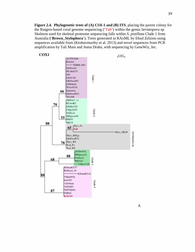
39
Figure 2.4. Phylogenetic trees of (A) COI-1 and (B) ITS, placing the parent colony for the Rutgers-based coral genome sequencing (‘Tali’) within the genus Seriatopora sp. Skeleton used for skeletal proteome sequencing falls within S. pistillata Clade 1 from Australia (‘Brown_Stylophora’). Trees generated in RAxML by Ehud Zelzion using sequences available from (Keshavmurthy et al. 2013) and novel sequences from PCR amplification by Tali Mass and Jeana Drake, with sequencing by GeneWiz, Inc.
A

40
B

41
Chapter 3: The evolution and future of carbonate precipitation in marine invertebrates: Witnessing extinction or documenting resilience in the Anthropocene?
Abstract
Morphological and phylogenetic analyses suggest that the ability to precipitate carbonates
evolved several times in marine invertebrates in the past 600 million years. Over the past
decade, there has been a profusion of genomic, transcriptomic, and proteomic analyses of
calcifying representatives from three metazoan phyla: Cnidaria, Echinodermata, and
Mollusca. Based on this information, we compared proteins intimately associated with
precipitated calcium carbonate in these three phyla. Specifically, we used a cluster
analysis and gene ontology approach to compare ~1500 proteins, from over 100 studies,
extracted from calcium carbonates in stony corals, in bivalve and gastropod mollusks,
and in adult and larval sea urchins to identify common motifs and differences. Our
analysis suggests that there are few sequence similarities across all three phyla,
supporting the independent evolution of biomineralization. However, there are core sets
of conserved motifs in all three phyla we examined. These motifs include acidic proteins
that appear to be responsible for the nucleation reaction as well as inhibition; structural
and adhesion proteins that determine spatial patterning; and signaling proteins that
modify enzymatic activities. Based on this analysis and the fossil record, we propose that
biomineralization is an extremely robust and highly controlled process in metazoans that
can withstand extremes in pH predicted for the coming century, similar to their
persistence through the Paleocene-Eocene Thermal Maximum (~55 Mya).
Keywords: biomineralization, metazoan, ocean acidification

42
3.1 Introduction The cumulative and continuing emissions of anthropogenic emissions of CO2 since the
beginning of the industrial revolution are projected to decrease the pH of the surface
oceans between 0.24 and 0.55 pH units by the year 2100 (2007), leading to a nearly 50%
drop in carbonate ion concentration (Feely et al. 2009). It has also been suggested that
the average pH decrease and overall diel pH variability could be even more pronounced
in specific areas, such as coastal seas (Jury et al. 2013, Melzner et al. 2013). The change
in carbonate chemistry potentially could result in a decreased ability for marine
organisms to precipitate CaCO3 (Doney et al. 2009), and has been experimentally
correlated with reduced calcification rates as well as changes in ecosystem structure
(Gazeau et al. 2007, Anthony et al. 2008, Cohen et al. 2009, Sheppard Brennand et al.
2010). However, the effects of ocean acidification on calcifying marine invertebrates are
not consistent and suggest a degree of physiological acclimation and potential genetic
adaptation (Fine et al. 2007, Ries et al. 2009, Kelly et al. 2013, Shamberger et al. 2014).
One suggested mechanism for the retention of biological calcification is low-pCO2
refugia (Tittensor et al. 2010, Manzello et al. 2012). While processes responsible for
carbonate precipitation in marine invertebrates are not completely understood, we suggest
that a specific suite of proteins allows biomineralization to persist in these organisms
even under low pH conditions.
Although biomineralization is a common phenomenon across the tree of life, it is
difficult to reduce the process to simple, general principles (Lowenstam et al. 1989, Dove
et al. 2003, Müller 2011). By far, the most common biominerals produced in the
contemporary ocean are calcium carbonates, especially aragonite and calcite. Although
the two ingredients required to produce these mineral forms, calcium and bicarbonate

43
ions, are present in seawater in excess of 10 and 2 mM, respectively, CaCO3 does not
spontaneously precipitate in the contemporary ocean. The precipitation reaction requires
catalysis by living organisms to overcome kinetic barriers.
The carbonate structures formed by metazoans are often well preserved in the
fossil record. Their elemental and isotopic compositions have been extremely helpful in
reconstructing the thermal and chemical history of the ocean. Carbonates themselves
have been examined at different stages of the biomineralization process in marine
invertebrates. For more than 50 years, it has been generally understood that the
precipitation and organization of biominerals is modulated by specific biomolecules,
especially proteins (Müller 2011). However, how the proteins function to facilitate the
precipitation of minerals in marine invertebrates is poorly understood.
Over the past decade there has been a proliferation in the number of genomes and
transcriptomes of mineralizing invertebrates (Jackson et al. 2006, Sea Urchin Genome
Sequencing Consortium et al. 2006, Joubert et al. 2010, Shinzato et al. 2011, Traylor-
Knowles et al. 2011, Takeuchi et al. 2012). These data sets have allowed numerous
studies of the proteomes of sea urchin teeth and spicules, mollusk shells, and most
recently coral skeletons (e.g., (Mann et al. 2008, Mann et al. 2008, Mann et al. 2010,
Marie et al. 2010, Marie et al. 2011, Marie et al. 2011, Zhang et al. 2012, Drake et al.
2013, Marin et al. 2013, Ramos-Silva et al. 2013)). We are poised to compare the
evolutionary history of biomineralizers in the fossil record (Knoll 2003) with this
developing biochemical and molecular biological understanding of how the minerals are
formed by the animals.
44
Here we review over 100 studies in a search for similarities between marine
invertebrate carbonate organic matrix (COM) proteins that have been identified through
genomics, transcriptomics, and proteomics. We focus on stony corals (Cnidaria), bivalve
and gastropod mollusks (Mollusca), and sea urchins (Echinodermata), thereby covering
representatives from three widely divergent metazoan phyla. We first outline the
physiology and known biomineralization processes in each phylum, then detail the COM
protein groups conserved, if not in sequence, then in function. Finally, we discuss the
effects of impending ocean acidification on both the function of some of these COM
proteins as well as the continuation of these organisms.
3.2 Background and context for biomineralization in the three invertebrate phyla
Despite their shared use of CaCO3, evolutionary evidence suggests that biomineralization
evolved independently several times across the tree of life (Knoll 2003, Murdock et al.
2011). Over the ~700 million years during which Cnidaria, Mollusca, and
Echinodermata diverged from shared ancestors (Erwin et al. 2011), biomineralization was
selected to support several functions. The first is as a substrate for growth. In modern
stony corals, precipitation of aragonite allows for growth of the thin film of animal on a
hard, continuous substrate. The process begins when larvae settle and colonies adhere to
a benthic matrix. In contrast, bivalve and gastropod mollusks precipitate aragonite and/or
calcite shells as a protective wall surrounding soft tissues. These structures are very
effective in defending against predators. In sea urchins, calcitic spines are also a
deterrent to predators, but being relatively light, they do not interfere with motility of the
animal.

45
3.2.1 Body plan and calcifying tissues Coral
Stony corals in the phylum Cnidaria are some of the earliest known metazoans to
precipitate calcium carbonate (Knoll 2003). Cnidaria also contain Order Alcyonacea, or
soft corals, which, unlike stony corals, produce small internal sclerites, or ~ mm-sized
calcite plates. In Paleozoic time, stony coral structures appear to have been formed
primarily from the precipitation of calcite, whereas over the past ~ 250 Ma, with the
appearance of Scleractinian corals, these organisms have evolved a de novo aragonite
precipitation process (Porter 2010). This period also broadly corresponds to their
transition as host animals to symbiotic photosynthetic dinoflagellates of the genus
Symbiodinium (Stanley et al. 2009, Tchernov et al. 2012). While some corals that lack
symbionts can produce CaCO3 skeletons, the builders of the relatively rapidly accreting
massive reefs observable from space are hermatypic, or symbiont-containing.
Up to 90% of the carbon requirement of the coral host is translocated in the form
of glycerol and/or glucose from Symbiodinium spp. (Falkowski et al. 1984, Burriesci et
al. 2012); additionally, corals can obtain nitrogen and phosphorus by feeding on
dissolved and particulate matter from the overlying water supply (reviewed
byHoulbrèque et al. 2009). Some of the energy in the translocated carbon is suggested to
support biomineralization, a phenomenon termed ‘light enhanced calcification’
(Kawaguti et al. 1948, Goreau 1959, Rinkevich et al. 1984)– although the mechanism(s)
remain unclear (reviewed byTambutté et al. 2011).
Stony corals precipitate an extracellular skeleton in the form of aragonite under
their calicoblastic ectodermal layer (Figures 3.1A & B). Recently, there have been
suggestions of an amorphous phase through atomic force microscopy and synchrotron-

46
based XANES (Cuif et al. 2008, Falini et al. 2013). Planulae begin to precipitate
aragonite only upon settling. Proteins related to adhesion have been localized toward the
aboral end immediately prior to settlement (Hayward et al. 2011). Growth rate depends
strongly on environmental factors, including irradiance and temperature, with some
corals showing gross precipitation as high as 10 kg m–2 yr–1 (Milliman 1993). Several N-
terminal sequences have been obtained from proteins associated with soft coral calcitic
sclerites, but the vast majority of research on COM proteins from corals has focused on
the tropical symbiotic stony corals.
Mollusks
Mollusks, the oldest extant biomineralizing bilaterians (reviewed byKnoll 2003),
encompass a far more advanced and varied body plan than stony corals. Generally, the
interior of the body with defined digestive organs is protected from the exterior
environment by one or two CaCO3 shells (although in some classes this protection is
much reduced or absent) (Marin et al. 2012). The shell is generally covered with a thin
organic layer, the periostracum, which not only separates the shell from the overlying
water, but also plays a role in the biomineralization process. While some gastropod and
bivalve mollusks contain endosymbiotic Symbiodinium spp., the majority are filter
feeders, particularly when sessile, or scrape off algae from surfaces when motile (Klumpp
et al. 1992).
In larval gastropods and bivalves, a protein-rich layer, the periostracum, is
secreted from a gland in the shell prior to onset of biomineralization (Figure 3.1).
Ancestral mollusks have been proposed as solely aragonite producers (Porter 2010), but
in modern species the mineral is often amorphous calcium carbonate (ACC), though

47
occasionally is calcite or aragonite (Weiss et al. 2002). In adults, the periostracum is
secreted by cells in the mantle folds and is maintained throughout the life of the mollusk,
thus providing a barrier between the mineral CaCO3 and the environment (Marin et al.
2012) (Figure 3.1C & D). Additionally, the periostracum serves as a preliminary scaffold
on which the organism lays down CaCO3 (Checa 2000). Together the periostracum and
mantle provide a distinct area, the extrapallial space, in which calcification occurs in the
presence of COM composed of chitin and proteins (Nudelman et al. 2006, Marin et al.
2012) (Figure 3.1D). The COM proteins, polysaccharides, and lipids secreted by the
mantle guide nucleation, elongation, and finally termination of aragonite and or calcite
crystals in well-defined locations and morphologies (reviewed byMarin et al. 2004)).
Additionally, specific COM proteins, such as Asprich, stabilize the ACC (Politi et al.
2007) and influence the mechanical properties of the biomineral (reviewed byWeiner et
al. 2011). Vesicles containing mineral precursors are also exported from specific cells
within the animal and aid in facilitating biomineralization in some mollusks (reviewed
byAddadi et al. 2006)).
Echinoderms
Although echinoderms display radial symmetry as adults, they evolved from a bilaterian
ancestor shared with mollusks and display bilateral symmetry as embryos (Hyman 1955).
Echinoderms have a defined digestive tract consisting of mouth, pharynx, esophagus,
stomach, intestine, and anus. While some echinoderms are carnivorous, sea urchins –
currently the only echinoderm with a sequenced genome (Sea Urchin Genome
Sequencing Consortium et al. 2006) – are predominately herbivores. They use calcified

48
spicules for motility, protection, and to preserve body shape, and five polycrystalline
magnesium calcite teeth for foraging (Ma et al. 2008).
Larval planktonic sea urchins secrete CaCO3 spicules and juveniles produce a
calcified test and spines, while adults construct these features plus magnesium-enriched
calcite teeth, pedicellariae, and ossicles in their tube feet, all in close proximity to
mineralizing cells (Killian et al. 2008). Larval sea urchins begin to create CaCO3, guided
by COM from primary mesenchyme cells, prior to settling (Figure 3.1E & F). As is often
the case with bivalve and gastropod mollusks, it appears that this CaCO3 precipitation
begins in the form of amorphous calcium carbonate (ACC) but within days changes to
calcite (Politi et al. 2008). Conversion of ACC to calcite is also known in adult urchins
(Politi et al. 2004, Killian et al. 2009), and has been suggested as a general tactic in
biomineralization, to have mineral precursors ready for crystallization (reviewed
byGilbert et al. 2011).
3.2.2 Biomineralizing Proteins The concept of biologically controlled mineralization is widely accepted (Mann 2001).
In stony corals, the “biomineralome” contains an organic component that is 0.1 to 5% by
weight with respect to that of the total biomineral and contains a high preponderance of
acidic residues in the skeletal proteins (Young 1971, Cuif et al. 2004, Mass et al. 2012).
The proteome of coral COM contains an assemblage of adhesion and structural proteins
as well as highly acidic proteins (Drake et al. 2013, Ramos-Silva et al. 2013). These
highly acidic proteins exhibit pKas of < 5 and a high relative composition (≥ 28%) of the
acidic amino acids, aspartic (Asp) and/or glutamic (Glu) acid. Four coral-acid rich
proteins (CARPs), one of which was found in both coral skeleton proteomic studies
(Drake et al. 2013, Ramos-Silva et al. 2013), were individually cloned and purified. All

49
four proteins precipitate calcium carbonate in vitro in artificial seawater at ambient
carbonate concentrations at pH 8.2 and 7.6 (Mass et al. 2013). These results have led to
the proposal of a nucleation mechanism in corals in which clusters of Asp and/or Glu
residues coordinately bind Ca2+ ions, potentially leading to a Lewis acid displacement of
protons in bicarbonate anions (Mass et al. 2013). The resulting reaction alters the
mineralizing environment below the calicoblastic ectoderm such that the pKa of the local
environment on the protein surface decreases and carbonate crystals are precipitated on
the protein matrix, even at relatively low ambient pH (Mass et al. 2013).
As with stony corals, the organic component of the bivalve and gastropod mollusk
shell minerals generally comprises between 0.01 to 5% by weight, with many of the
(glyco)proteins containing a high proportion of Asp residues and/or post-translational
decorations of sulfate groups (Lowenstam et al. 1989). As in corals, these proteins are
highly acidic. Initially much of the focus on biomineralizing proteins in mollusks
focused on these highly acidic proteins and their possible template-specific roles in
nucleating precipitation, with distances between acidic residues in -sheet conformation
corresponding to distances between Ca2+ ions in both calcite and aragonite (Weiner et al.
1975). More recently, the role of highly acidic proteins in mollusk and echinoderm
mineralization has grown to encompass the inclusion of these proteins in a hypothesized
organic gel, the potential stabilization of a transitional amorphous calcium carbonate step,
and the binding and inhibition of growing crystal growth faces (reviewed byMarin
2007)). Additionally, a number of proteomic comparisons have shown that COM
proteins include many of unknown function as well as those specific to aragonite versus
calcite (e.g., (Jackson et al. 2006, Marie et al. 2010, Marie et al. 2012, Marin et al.

50
2013)). Of the proteins with known function, many are involved in maintenance of the
extrapallial environment and in reworking of calcium carbonate.
Analyses of the proteins in COM from spicules, tests, and teeth of sea urchins
have yielded a non-redundant list of over 200 individual molecules (Mann et al. 2008,
Mann et al. 2008, Mann et al. 2010). While some of these proteins appear to be specific
to one type of mineral or structure (spines versus teeth, etc.), many overlaps are observed
(reviewed byGilbert et al. 2011)). Some of these proteins have been localized to larval
primary mesenchyme cells, but other cells, potentially secondary mesenchyme cells, are
more likely responsible for biomineralization in adult animals (Killian et al. 2008).
Although highly acidic proteins are also found in sea urchin biominerals (George et al.
1991, Illies et al. 2002, Mann et al. 2008), non-acidic proteins that control either the
mineralizing environment or activity of other proteins are also appreciated as essential
(example is SM30 family (e.g.;Wilt et al. 2013)).
3.3 Comparison of carbonate organic matrix proteins across marine invertebrate phyla
3.3.1 Evolution of the acidic proteins The term ‘acidic’ protein has a variety of definitions, but ultimately describes a protein
that possesses a net negative charge on some or all of the primary sequence or through
post-translational modification. In an attempt at clarity, we compiled a list of the ‘acidic’
proteins and their gene accession numbers in Table 3.1. It is clear from a phylogenetic
analysis that these proteins arose independently several times in the evolution of animals
through convergent evolution, with gene fusion potentially conferring additional roles to
these peptides (Miyamoto et al. 1996, Mass et al. 2013). These proteins appear to be
essential for biomineralization. We conducted a protein sequence similarity analysis,

51
using > 1500 non-redundant coral, mollusk, and sea urchin mineral-derived proteins
reported in the literature. Through these analyses, we found only one case of cross-phyla
sequence similarity among any of the acidic biomineralization proteins for which
sequences are available. Prismalins, shematrins, and some shell matrix proteins from
mollusks show 30 to 40% similarity with several collagens from echinoderms and corals
(Figure 3.2, SI Table 3.1); this is likely due to the high glycine content of shematrins
(Yano et al. 2006) similar to the primary structure of collagens.
Within corals, several acidic proteins show sequence similarity across Orders
Scleractinia and Alcyonacea. These include stony coral CARPs 4 and 5 (Drake et al.
2013), SAARPs 1 and 2, SOMP, and Amil-SAP2 (Moya et al. 2012, Ramos-Silva et al.
2013) and the soft coral protein ECMP-67 (Rahman et al. 2011) (SI Table 2). Although
not yet analyzed in Acropora digitifera and Pocillopora damicornis skeleton, CARP4
homologues are found in transcriptomes of these species as well (Shinzato et al. 2011,
Vidal-Dupiol et al. 2013). The CARP4 sub-family has been shown to be both highly
conserved across and limited to stony corals (Drake et al. 2013), and now the N-terminal
sequence from the soft coral, Lobophytum crassum, may be assigned to this family based
on sequence alignment (Figure 3.3).
Stony corals precipitate aragonite whereas soft corals form calcite, suggesting that
the CARP4 sub-family arose in Anthozoa prior to divergence between Scleractinia and
Alcyonacea. However, because Actiniarians, or sea anemones, which lack the CARP4
subfamily, diverged from Scleractinia after Alcyonacea (Berntson et al. 1999), they may
have lost the gene. It has been proposed that ECMP-67 encourages the precipitation of
only calcite (Rahman et al. 2011), despite its similarity to the aragonite-associated

52
CARP4 and SAARP1 (Figure 3.3). Interestingly, several mollusk proteins have also
been suggested to function in both aragonite and calcite precipitation (MS17; (Feng et al.
2009)) or in polymorph control (Aspein; (Takeuchi et al. 2008)). Additionally, Aspein,
Asprich, and Pif, which all are components of molluscan shells, share 30% sequence
similarity (SI Table 2; (Tsukamoto et al. 2004, Gotliv et al. 2005, Suzuki et al. 2009).
Thus although the acidic proteins are independently derived, this sequence comparison
suggests a common mechanism of CaCO3 precipitation by acidic proteins in the three
marine invertebrate phyla.
Some of the biomineralizing proteins appear to have dual functions. For example,
perlwapin, prismalin, molluscan shell prism nacre protein, shematrins, and several
collagens, are all implicated in organization and structural support of the bioinorganic
matrix. These are called ‘other’ gene ontology (GO) terms in Figure 3.2 due to equal
distribution of collagen, membrane, and extracellular GO assignments. These proteins
may also serve a dual role as terminators of biomineralization and are not the only COM
proteins, both acidic and non-acidic alike, shown to have crystallization inhibition
activities (e.g.;Ma et al. 2007, Politi et al. 2007, Feng et al. 2009). A dual nature of a
biomineralizing protein is also observed for the mollusk protein, nacrein, which exhibits
both internal acidic regions and carbonic anhydrase activity (Miyamoto et al. 1996). In
sea urchins, SpP16 is an acidic protein with a transmembrane region, suggesting that it is
anchored to the primary mesenchyme cells to which it has been localized; while its
precise mechanism in calcitic spicule formation remains to be determined, it shows some
similarities to other calcium binding proteins to regulate calcium transport, again
suggesting dual function (Illies et al. 2002).

53
Carbonic anhydrases, which interconvert CO2 and HCO3–, are integral within
carbonate-based biominerals (Bertucci et al. 2013). Their use in biomineralization has
been suggested to date back to the earliest calcifying metazoans (Jackson et al. 2007).
Gene duplication events and further mutations since the Paleozoic have resulted in a wide
variety of the enzyme, many of which are localized within the bioinorganic matrix. Our
proteomic analysis revealed representatives of divergent carbonic anhydrases in the
biomineral matrix in each of the three invertebrate phyla (Table 3.2). These proteins
reinforce their functional necessity in the evolution of the precipitation of extracellular
calcium carbonate in each of the three metazoan phyla. Indeed, multiple carbonic
anhydrases are found in COM of each phylum, suggesting a level of redundancy for the
biomineralization process.
Just as bicarbonate is required for the biomineralization mechanism, so is Ca2+.
In animals, Ca2+ is transported into and out of cells by plasma membrane-bound Ca-
dependent ATPases. These enzymes are widely distributed in mollusks, sea urchins, and
corals (Zoccola et al. 2004, Wang et al. 2008, Todgham et al. 2009). However, of the
more than 100 reports of COM, only one identified a Ca-ATPase in a ‘biomineralome’
(Joubert et al. 2010). An additional mechanism for Ca2+ transport in corals is the
translocation of calcium from the bulk water to the site of mineralization (Gagnon et al.
2012) by a so-called “paracellular” pathway (Tambutté et al. 2012). However, without a
chelating or trapping molecule, this mechanism would not concentrate calcium ions.
Finally, proton pumps not associated with mitochondrial ATPases, an additional method
to modify the saturation state of the mineralizing environment (Corstjens et al. 2001),
were not observed in COM by the authors of any of the studies used in our bioinformatics

54
analysis. Additionally, none of the 295 non-redundant proteins conserved across phyla
returned molecular function GO terms associated with non-mitochondrial proton pumps.
3.3.2 Conserved non-acidic proteins across phyla A number of non-acidic COM proteins are either conserved by sequence (Figure 3.2) or
function (Table 3.2). For heuristic purposes, we have aggregated these proteins by
function into major groups: (i) proteins that participate in adhesion of cells to their
substrates and/or provide a scaffold for organizing the biomineral, and (ii) proteins that
signal and regulate biomineralization. Our list is not exhaustive (see SI Table 3.2).
However, it is useful in providing a framework for comparing similarities and differences
in COM proteins across the three phyla.
Adhesion and Microstructure
Actins and beta-tubulin, cytoskeletal proteins that work as polymerization molecular
motors, are present in COM of stony corals and in sea urchins (Figure 3.2). Although
they could be contaminants from the animal component (i.e., not truly part of the COM;
(Mann et al. 2008), physiological and genomic data suggest they are part of the
biomineralization “toolkits” for both corals and sea urchins. Inhibition of these proteins
significantly reduces organic matrix synthesis and calcification in corals (Allemand et al.
1998). While similar studies have not been conducted on sea urchin biomineralization,
actin is crucial to the primary mesenchyme cell ingression stage just prior to onset of
embryonic biomineralization, with actin inhibition resulting in loss of ingression (Wu et
al. 2007). Secondly, biomineralization in paralogue organisms such as diatoms, which
precipitate amorphous silicate, involves actins and tubulins (Hildebrand et al. 2008).
A number of proteins that adhere to or interact with actin and tubulin are found in
COM across marine invertebrate phyla, but are not conserved by amino acid sequence,

55
and therefore are assumed to be independently evolved (Table 3.2). For example,
laminins, a family of glycoproteins, along with fibronectins and integrins, serve as
structural support in mollusks, echinoderms, and corals, respectively (Table 3.2).
Peroxidasins and thrombospondins (in corals and sea urchins) may also serve as
extracellular structural and adhesive proteins (Table 3.2; (Péterfi et al. 2009).
Finally, collagens, which provide a flexible framework and confer structural
support for biominerals, appear to be widely distributed in corals and sea urchins (Figure
3.2, Table 3.2). It is unclear whether collagens are actually found in mollusks (Blank et
al. 2003, Xuan Ri et al. 2007). Alternatively, chitin, a potential structural replacement for
collagen is found in mollusks (reviewed by (reviewed byEhrlich 2010). In this case,
chitin, like collagen, can provide a highly structured scaffold on which COM proteins can
bind (reviewed by (Falini et al. 2004). It has been suggested to have a more direct role in
polymorph control (Falini et al. 1996). Chitin synthase (Joubert et al. 2010) and a chitin-
binding protein (Mann et al. 2012) have been detected in mollusk shell proteomes; they
do not show cross-phyla sequence similarities in our analysis (SI Table 2).
3.3.3 Signaling and Regulation Low-density lipoprotein (LDL) receptors are ancient proteins that aid in signal
transduction across cell membranes and are responsible for cell specification and axis
patterning in cnidarians and sea urchins (Wikramanayake et al. 2004, Lee et al. 2006, Lee
et al. 2007). Notch proteins, which contain an epidermal growth factor domain (EGF),
are calcium-binding transmembrane signal transduction pathway proteins that interact
with LDL receptors (Baron 2003). In sea urchins, they direct secondary mesenchyme
cell development and spiculogenesis (Suyemitsu et al. 1990, McClay et al. 2000).

56
Several of these proteins, with ‘membrane’ GO terms assigned, are reported in sea urchin
and coral COM (Figure 3.2).
Bone morphogenic proteins (BMPs) are responsible for signal gene regulation in
apatite producing cells (reviewed byCanalis et al. 2003) and up-regulate Notch signaling
genes in osteoblast precursor cells (de Jong et al. 2004). BMPs are reported in COM
from all three phyla (Table 3.2) but, as ‘extracellular’ GO terms, are only conserved
between mollusks and sea urchins (Figure 3.2). As signal transduction pathway proteins,
BMPs from corals and mollusks induce mesenchyme cell differentiation and osteogenesis
(Zoccola et al. 2009, Takami et al. 2013) and are involved in sea urchin cell specification
and axis patterning (Angerer et al. 2000). They have also been immunolocalized to
calicoblastic ectodermal cells in corals (Zoccola et al. 2009) and a novel protein that
shows structural (from structure prediction) but not sequence similarity to the BMP
inhibitor Noggin was recently observed in coral COM (Drake et al. 2013).
In addition to cell signaling, reworking of individual COM proteins is important
to the function of the complex. Matrix metalloproteinases (MMPs) are specific
extracellular matrix degradation enzymes that require cations for their catalytic activity.
COM MMPs, as extracellular proteins, show sequence similarity between corals and sea
urchins (Figure 3.2). MMPs affect sea urchin spicule elongation but not nucleation
(Ingersoll et al. 1998) and regulate extracellular matrix in humans (reviewed byWoessner
1991, Birkedal-Hansen et al. 1993). They are found in mollusk hemolymph and an MMP
inhibitor has been extracted from mussel pearls (Mannello et al. 2001, Jian-Ping et al.
2010), suggesting that they may be present in mollusk COM as well.

57
Ubiquitin is a protein that attaches to and signals other proteins for degradation or
relocation, or inhibits protein binding or activity. It is highly conserved in COM proteins
across all three phyla and is generally assigned a ‘membrane’ cellular component GO
term (Figure 3.2). Ubiquitylated proteins are reported from the prismatic layer of
mollusks; removal of ubiquitin decreased the ability of these proteins to inhibit calcium
carbonate mineralization (Fang et al. 2012). Hazelaar et al. (Hazelaar et al. 2003)
proposed that, in diatoms, ubiquitin may signal templating proteins for degradation as the
cell walls develop pores; a similar role in invertebrate calcium mineralization has yet to
be examined.
3.3.4 Summary of Conservation of Biomineralizing Proteins Morphological, phylogenetic, and now proteomic evidence indicates that marine CaCO3
precipitation is a convergent evolutionary process. Coral, mollusks, and sea urchins have
all evolved their own suites of acidic proteins to catalyze the nucleation of CaCO3 (Illies
et al. 2002, Gotliv et al. 2005, Drake et al. 2013) and co-opted a number of adhesion,
structural, signaling, and regulation proteins to provide structure and environmental
stability to the calcifying regions (Marin et al. 2004, Zhang et al. 2006, Allemand et al.
2011). This convergence suggests that there are general roles for biomolecules in the
biomineralization process and that, although biomineralization toolkits may not be
conserved across phyla, the requirements to have these biological roles fulfilled is.
Further, the precise macroscopic morphologies of skeleton, shell, and spicule/teeth, etc.
may then be driven by those proteins or other biomolecules that are functionally
individual not only to each phylum, but also often to specific minerals. Examples of
these functionally individual proteins, which in some cases have been reviewed
elsewhere, are lysine-rich matrix proteins and amorphous calcium carbonate binding

58
proteins in mollusks (Zhang et al. 2006, Ma et al. 2007), galaxins in corals (Fukuda et al.
2003), and the mesenchyme specific cell surface glycoproteins in sea urchins (Leaf et al.
1987, Killian et al. 2008).
3.4 Future of marine invertebrate biomineralizers
Estimates of CO2 emissions and the projected effects on upper ocean ecosystems over the
next 100 years are dire, especially for marine calcifiers that will face the combined issues
of warming and ocean acidification (Raven 2005, Feely et al. 2009, Ries et al. 2009,
Hoegh-Guldberg et al. 2010, Pandolfi et al. 2011). Models of the average calcite
saturation state (Ωcalcite) in the surface ocean projects a decrease from 5 to 2 in the next
100 years (Doney et al. 2009, Norris et al. 2013). Studies of biomineralizing marine
invertebrates indicate a variety of potential responses to ocean acidification, from
potential decreased calcification rates (Ries et al. 2009, Holcomb et al. 2010, Kroeker et
al. 2010) to ecosystem shifts (Anthony et al. 2008, Crook et al. 2012). Additionally, each
of the marine invertebrate mineralizers discussed here have representatives that naturally
persist in waters with low calcium carbonate saturation (i.e., > 1000 m depth or in regions
of upwelling, high respiration rates, or rainwater runoff; e.g., (Anagnostou et al. 2011,
Shamberger et al. 2014)). However, in addition to examining the responses of current
marine biomineralizers, it is useful to consider both the micro- and macroscopic effects of
increased atmospheric CO2, particularly as it pertains to ocean acidification.
As detailed above, several highly acidic coral proteins function at ambient and
low pH (Mass et al. 2013). This capacity suggests that while crystal growth rate and
morphology can be dependent of the saturation state of the medium (Holcomb et al.
2009), the pKas of these and likely other highly acidic proteins are three orders of

59
magnitude lower than seawater. Thus, in spite of the fact that pH may decline by up to
0.5 pH units in the coming century, the reactivity of these proteins in the precipitation of
carbonates will remain virtually unchanged. This built-in molecular resilience will likely
allow the nucleation reaction to continue. Although this resilience does not address the
physiological response of marine biomineralizers to a surrounding medium whose pH is
stressful to processes beyond the nucleation of minerals (Lannig et al. 2010, Stumpp et al.
2011, Moya et al. 2012), these organisms are not static and there is evidence of genetic
and transcriptional adaptation to low pH conditions (Moya et al. 2012, Hüning et al.
2013, Kelly et al. 2013).
Given the inevitable acidification of the contemporary ocean in the coming
decades it is potentially also instructive to examine the geological record of analogues.
One possible analogue is the Paleocene Eocene Thermal Maximum (PETM) (Norris et al.
2013). This period, 55.5 to 55.9 million years ago, was characterized by a 4 to 5o C
increase in sea surface temperatures in the tropics (Zachos et al. 2003) and nearly 1000
ppm increase in CO2 in the atmosphere (Beerling et al. 2011). While the PETM
corresponds with a dramatic reduction in CaCO3 in the geologic record (Zachos et al.
2005) and a decrease in the coverage of coral reefs (reviewed byNorris et al. 2013)), all
marine invertebrate taxa discussed here survived through this extraordinary period.
Indeed, some experienced rapid radiation and diversification in the Eocene (e.g.;Stanley
2003)).
The survival of the major carbonate-precipitating marine invertebrates in the face
of ocean acidification is likely because (i) the site of biomineralization is removed from
the surrounding medium, (ii) the proteins responsible for the precipitation of carbonates

60
function at low pH, and (iii) genetic adaptation allows the organisms to persist, albeit
perhaps not optimally, in their new environments. Additionally, recent research suggests
that these organisms are able to modify their mineralizing environment in response to pH
stress (Yuen et al. 2006, Venn et al. 2013). This ability suggests that the
biomineralization processes will almost certainly persist throughout the Anthropocene. It
appears that these organisms are far more resilient than is often acknowledged. However,
from a molecular biology perspective, significant research remains toward understanding
1) the expression rates of known COM proteins under variable environments, as well as
the biochemical cascades responsible for any changes; 2) the interactions of the COM
proteins with each other to regulate the initiation, elongation, and termination steps of
mineralization; and 3) how signals of COM protein evolution, which also guided
biomineralization in the past, may be retained in fossil minerals.
Data Accessibility Statement: All data were taken from publically available datasets.
Original sources and accession numbers of all sequences are noted in Table S2.

61
Table 3.1. Proteins from coral, mollusk, and sea urchin carbonate organic matrices that have been characterized as ‘acidic’. The gene accession number is given along with the primary reference to the work describing the proteins. Characteristic Coral Mollusk Sea urchin Aspartic acid-rich regions
CARP4 (KC493647)
and CARP5 (KC493648) (Drake et al.
2013) SAARP1 (JT001945), SAARP2 (JT991407), Amil-SAP2 (JR983041) (Ramos-Silva et al. 2013) EMCP-67 (no accession
#) (Rahman et al. 2011)
Aspein (AB094512) (Tsukamoto et al. 2004) Asprich (AAU04807-15) (Gotliv et al. 2005) Pif (AB236929) (Suzuki et
al. 2009) Prismalin (AB159512) (Suzuki et al. 2004) Nacrein (Q27908) (Miyamoto et al. 1996) MSI60 (O02402) (Sudo et
al. 1997) AP7 (AF225916) (Michenfelder et al. 2003)
Low pI, serine-rich regions
Mucoperlin (AF145215) (Marin et al. 2000)
P16 (AF519415) (Illies et
al. 2002) Glutamic acid-rich regions
MSI31 (O02401) (Sudo et
al. 1997) P19 (AF519413) (Illies et al.
2002) Tyrosine-rich regions
N14 (Q9NL39) (Kono et al.
2000) Pearlin (O97048) (Miyashita et al. 2000) N16 (AB023067) (Samata
et al. 1999)
Aspartic and glutamic acid-rich regions
AP24 (AF225915) (Michenfelder et al. 2003)
Serine- and aspartic acid-rich regions
MSP-1 (Q95YF6) (Sarashina et al. 2001)
Phosphorylated 34 phosphoproteins (*) (Mann et al. 2010)
* See reference cited for the available 34 sequence ID numbers and to download sequences from publisher.

62
Table 3.2. Proteins of shared function, but not shared sequence, found in carbonate organic matrices across the three marine invertebrate phyla. Protein classifications follow those found in their original publications unless they grouped with another classification during similarity clustering. The gene accession numbers are provided. Protein Name Coral Mollusk Sea urchin Bone morphogenic protein *
Acropora sp. (EU785982) (Zoccola et al. 2009)
Stylophora pistillata (EU78981) (Zoccola et al. 2009)
Pinctada margaritifera
(90c_1652_1) (Joubert et al.
2010)
Strongylocentrotus purpuratus (NP_999820)
(Mann et al. 2010)
C-type lectin - P. margaritifera (90c_3116_2,
298867_2192_0444_6) (Joubert
et al. 2010)
S. purpuratus (XP_003726084,
XP_003726239, XP_793473, AF519418) (Illies et al. 2002,
Mann et al. 2008, Mann et al.
2010) Carbonic anhydrase
S. pistillata (EU532164, ACE95141, ACA53457) (Moya et
al. 2008, Bertucci et al. 2011, Drake et
al. 2013) Acropora millepora
(JR995761, JR973601, JR998380, JT002659, JT014542, JR989434, JT018935, JR979146, JR996464, JR990087, JR998014) (Moya et al.
2012, Ramos-Silva et al. 2013)
P. margaritifera (90c_299_1) (Joubert et al. 2010)
Crassostrea gigas (EKC19847.1) (Zhang et al.
2012)
Lottia gigantea (lotgi66515, lotgi205401)
(Mann et al. 2012)
S. purpuratus (XP784796, XP_003726289,
XP_784328_ABE27963) (Mann et al. 2008, Mann et al.
2010, Stumpp et al. 2011)
Collagen * A. millepora (JR991083,) (Ramos-Silva et al. 2013)
S. pistillata (KC479166, KC479163, KC342195,
KC342197) (Drake et al. 2013)
Haliotis asinina (GT274423) (Marie et al. 2010)
C. gigas (EKC3725.1) (Zhang et al. 2012)
L. gigantea (lotgi123902) (Mann et al. 2012)
S. purpuratus (SPU009076, SPU015708, SPU003768, SPU022116,
NP_999676, (Todgham et al.
2009, Mann et al. 2010)
Fibronectin * Mytilus californianus (P86861) (Marie et al. 2011)
C. gigas (EKC41461.1 – EKC41463.1) (Zhang et al.
2012)
S. purpuratus (XP_003729283) (Mann et al.
2010)
Hemicentin/ Thrombospondin *
A. millepora (JT016638, JR989905) (Moya et al. 2012, Ramos-
Silva et al. 2013) S. pistillata (KC342189, KC150884) (Drake et al. 2013)
- S. purpuratus (XP_794971, XP003727369, XP003728681, XP784935,
XP_780466, XP001198520, XP_786756) (Mann et al. 2008,
Mann et al. 2010) Integrin S. pistillata (KC479167) (Drake
et al. 2013) S. purpuratus
(XP_794080) (Mann et al. 2010) Laminin/ Neurexin A. millepora (JR980881)
(Ramos-Silva et al. 2013) S. pistillata (KC342198, KC479169) (Drake et al. 2013)
(receptor) P. margaritifera
(90c_665_4) (Joubert et al. 2010) L. gigantea (lotgi203487)
(Mann et al. 2012)
S. purpuratus (XP781951, XP_003726434,
XP_780725.3, XP_001197577.1) (Mann et al.
2008, Mann et al. 2010)
• Indicates that some sequences show sequence similarity across phyla. See SI Table 1.

63
Figure 3.1. Schematic drawings of three biomineralizing marine invertebrates. (A–B) Four cell layers separate the external aragonite skeleton from overlying seawater (after;Allemand et al. 2004). (C–D) Mollusk aragonite, calcite, and/or ACC are precipitated in the extrapallial space between the periostracum and mantle (after;Marin et al. 2004). (E–F) Primary mesenchyme cells precipitate larval sea urchin ACC and/or calcite (after;Urry et al. 2000).

64
Figure 3.2. Cellular component ontology of COM proteins conserved across phyla. Sequences from over 100 biomineral proteome studies were grouped by hierarchical clustering using the CD-HIT suite web server (Li et al. 2006, Huang et al. 2010) (http://weizhong-lab.ucsd.edu/cd-hit/) and assigned GO terms using Blast2Go software (Conesa et al. 2005). Although 1531 proteins reduced to 1051 clusters at 30% similarity, only 64 clusters showed sequence similarity across phyla. Studies published from the 1990s through June 2013, using N-terminal and mass spectrometry COM sequencing, RT-PCR, or GO and KEGG annotation of genomic and transcriptomic data sets are included. Mass spectrometry sequences were excluded if the experimental data were compared with gene models from a different species.

65
Figure 3.3. Multiple sequence alignment of CARP4 sub-family homologues. Alignment of homologues from four stony corals places a previously un-aligned soft coral N-terminal sequence into this family. The CARP4 subfamily is not found in sea anemones even though anemones diverged from stony corals after the soft coral divergence.

66
3.5 Supplementary Material Supplementary material is available from the publisher at http://dx.doi.org/10.12952%2Fjournal.elementa.000026. Table S1. Protein sequences with >=30% sequence similarity between phyla. Cross-phyla clustering of non-redundant carbonate organic matrix proteins from N-terminal and mass spectrometry COM sequencing, RT-PCR, or GO and KEGG annotation of genomic and transcriptomic data sets (from over 100 studies) grouped by hierarchical clustering using the CD-HIT suite web server (http://weizhong-lab.ucsd.edu/cd-hit/). The gene accession numbers are included. 1531 proteins reduced to 1051 clusters at 30% or greater similarity, although only 64 clusters showed sequence similarity across phyla. Studies published from the 1990s through June 2013 are included. Clusters with the same name have been combined. Note: Mass spectrometry sequences were excluded if the experimental data were compared with gene models from a different species. * indicates that non-homologous proteins with similar function were also observed. See Table 2 in main document. Table S2. Proteins from coral, mollusk, and sea urchin COM sequencing, RT-PCR, or GO and KEGG annotation. 1076 proteins, including redundancy when noted by multiple sources, reduced to 1031 non-redundant sequences.

67
Chapter 4: The influence of CO2 on calcification in coral cell cultures
Abstract
Understanding the cellular and molecular responses of stony corals to ocean acidification
is key to predicting their ability to maintain calcification under high CO2 conditions.
Biomineralization toolkit proteins have been proposed for several corals and elucidating
the connection between expression of these genes and calcification will allow a better
understanding of their roles in the mineralization process. We used cell cultures of the
Indo-Pacific stony coral, Stylophora pistillata to test the effects of increased CO2 on the
calcification process at the cellular and molecular levels. After rearing cell cultures for
up to nine days in four CO2 conditions and two glucose treatments, we analyzed samples
by microscopy, qPCR, western blot, FIRe, and mass spectrometry. S. pistillata grown at
low (400 ppm) and moderate (700 ppm) CO2 re-aggregate into proto-polyps and
precipitate CaCO3. When grown at high and very high CO2, (1000 and 2000 ppm,
respectively) S. pistillata cells up-regulate several highly acidic genes as well as a
carbonic anhydrase, but down-regulate long-term cadherin production and minimize
proto-polyp formation; this coincides with cessation of measurable CaCO3 precipitation.
These results suggest that compensatory molecular adjustments allowing cells to deal
with ocean acidification are successful only up to a point, beyond which these
mechanisms cannot compete with local chemical conditions unfavorable to
biomineralization.

68
4.1 Introduction As precipitators of an aragonitic skeleton, stony corals are thought to be under
considerable stress from increasing atmospheric pCO2 (e.g.; Smith et al. 1992, Leclercq
et al. 2002, Pandolfi et al. 2011). A variety of studies have shown that calcification rate
(e.g.; Anthony et al. 2008, Drenkard et al. 2013, Strahl et al. 2015) as well as
photosynthetic (Anthony et al. 2008) and other metabolic processes (e.g.; Edmunds et al.
2014) are depressed in experimentally increased CO2 treatments for some corals.
However, the effects of increased CO2 on coral calcification are not negative for all
corals (Ries et al. 2009, Comeau et al. 2013, Strahl et al. 2015), and some taxa persist in
naturally high CO2 environments such as near volcanic or karst vents (Fabricius et al.
2011, Crook et al. 2012) or in bays that experience low aragonite saturation due to high
calcification and respiration (Shamberger et al. 2014). They also exhibit differential
responses to increased pCO2 when fed (e.g.; Drenkard et al. 2013, Houlbrèque et al.
2015).
Elemental and isotopic information from fossil coral skeletons have been used as
proxies for paleo-environmental conditions. In particular, boron isotopes are used to
calculate pH of ancient seawater (Honisch et al. 2004). A number of assumptions must
be made for these proxies including that the precipitation reaction proceeds with no or
minimal biological control. However, corals have a strong direct control over both the
mineralization process and the chemical make-up of the mineralizing interface
(Lowenstam et al. 1989, Falini et al. 2013, Venn et al. 2013, Holcomb et al. 2014). For
these reasons, it is necessary to understand how coral biomineralization happens at the
cellular level.

69
For many years, model organisms have been used to understand biomineral
formation at the cellular level; one such marine example is sea urchin larval studies (e.g.;
Beniash et al. 1999). Although a similar model system does not exist for stony corals,
there has been success in maintaining coral cell bundles (Domart-Coulon et al. 2001) or
re-aggregated cells as proto-polyps for up to one month (Helman et al. 2008, Mass et al.
2012). In these systems, coral cells secrete an extracellular matrix that adheres them to
the substrate and precipitate extracellular aragonite crystals on ‘proto-polyps’. However,
rate of calcification or of skeletal organic matrix (SOM) protein expression in these
systems have not been examined to date. Neither have they been used to study the
cellular-level effects of environmental stressors on corals.
SOM proteins retained in coral skeleton from corals in the families Pocilloporidae
and Acroporidae have recently been sequenced (Drake et al. 2013, Ramos-Silva et al.
2013). These include proteins of known function such as the adhesive cadherin and a
carbonic anhydrase, as well as uncharacterized and highly acidic proteins. Several of
these SOM proteins have been immunolocalized to the calicoblastic cell layer and at the
nano-scale within skeleton (Mass et al. 2014).
Marine invertebrate calcifiers share a variety of related SOM proteins of known
functions including membrane-associated and extracellular proteins (Drake et al. 2014).
However, the highly acidic proteins evolved independently. In stony corals, soluble
acidic proteins (SAPs) have only been found in Acroporid skeletons, while the coral acid
rich proteins (CARPs) or secreted acidic Asp-rich proteins (SAARPs) have been found in
both Acroporid (Ramos-Silva et al. 2013) and Pocilloporid (Drake et al. 2013, Mass et al.
2014) aragonitic skeleton and in soft coral calcitic sclerites (Rahman et al. 2011).

70
Additionally, several CARPs/SAARPs have been shown to precipitate calcium carbonate
from seawater, potentially by a Lewis acid mechanism (Mass et al. 2013).
In this study, we grew Stylophora pistillata cell cultures in growth media
manipulated from 400 to 2000 ppm pCO2 and in 0.1 and 0.2 mM glucose to examine the
effects of ocean acidification and nutrition, respectively, on both mineral and SOM
protein production. Previous studies have examined the effects of pCO2 and
heterotrophy on coral spat (Drenkard et al. 2013) and nubbins (Edmunds 2011) and show
that corals recover some degree of mineralization ability under heterotrophic conditions.
In contrast to other studies using nubbins, our cultures allow us to focus on cellular rather
than organismal processes and produce new CaCO3 uncontaminated by parent mineral.
Here, we highlight the cellular response to ocean acidification to detail, through
molecular, biochemical, and elemental analysis, the direct control of coral cells on the
biomineralization process.
4.2 Methods
4.2.1 Experimental Setup and Conditions Experimental growth medium was prepared at 400, 700, 1000, and 2000 (±30) ppm CO2
in an incubator (Percival Scientific) at 26oC. 1 L artificial seawater (ASW) was mixed to
achieve a salinity of 40; this salinity was chosen because the ASW was eventually diluted
in growth medium to a final salinity of 34 (Appendix Table 4.1). ASW was sterile-
filtered to 45 µm and bubbled in a standard algae bubbling system consisting of an
autoclaved plastic Erlenmeyer flask, stopper, and tubing plus sterile serotological pipette
and sterile 0.45 µm one-time-use syringe filters. This bubbling system was opened only
in a UV hood next to a flame. The above precautions were employed to eliminate
external bacterial contamination of the cultures. Sealed sterile ASW was bubbled with

71
air ambient to the incubator in the dark until TOC and pH measurements stabilized.
Initial and stabilized samples were also taken for alkalinity titrations.
At each CO2 level, ASW was mixed with Dulbecco’s Modified Eagle Medium
(DMEM, Sigma D5030) plus additives to achieve the following: 12.5% DMEM (at 8.3
g/L containing 0.578 g/L L-glutamine, 0.05 g/L taurine), 20 mg/L aspartic acid, 50mg/L
ascorbic acid, 2% heat-inactivated fetal bovine serum (Invitrogen), 0.1 mM glucose, and
1% anti-biotics/mycotics cocktail (GIBCO). Growth medium was filtered and bubbled as
above until TOC and pH measurements stabilized; alkalinity titrations were also
performed.
DIC was measured on a TOC analyzer (Shimadzu) with a bicarbonate standard
curve, and certified reference material (CRM; Batch 132) from the Dickson lab at UC
San Diego as an internal standard. Alkalinity titrations of initial ASW mixtures were
conducted on a Metrohm Titrando 888 against NIST pH standards and using the CRM
noted above as an internal standard. CO2 was maintained in the incubator using CO2 gas
(Airgas; bone dry) and soda lime; manufacturer specifications state that output accuracy
is ±20 ppm + 2% of the reading which tended to fluctuate ±20 ppm of the set CO2
concentration. pH of equilibrated seawater was calculated from incubator CO2 setting,
DIC, and alkalinity in CO2sys (Pierrot et al. 2006). pH of equilibrated DMEM+ASW
was measured on a Fisher Scientific Accumet pH meter against NIST standards.
Chemical parameters of growth medium are given in Table 4.1.
Cell cultures of the zooxanthellate Indo-Pacific coral, Stylophora pistillata, were
produced from nubbins contained in an in-house 800-L aquaria as previously described
with the following modifications (Mass et al. 2012). Initial 4-hr incubations in calcium-

72
free seawater plus 3% anti-biotics/mycotics (GIBCO) and 20 µg/L chloramphenicol,
during which cell adhesion is disrupted, were conducted at ambient CO2 and room
temperature for nubbins used in all treatments. Secondary incubations, during which
cells spontaneously dissociate from the skeleton, were conducted in sterile DMEM+ASW
described as above but made with ASW stabilized at ambient CO2. These secondary
incubations were maintained for 1.5 days at room temperature in a non-sterile humidified
incubation chamber at ambient light:dark cycle. Dissociated S. pistillata cells and tissue
were gently pipetted away from the skeleton and centrifuged at 3000 rpm for 10 minutes
at room temperature. The pellet was re-suspended in medium bubbled to the appropriate
CO2, as described above, and filtered on 20 µm nylon mesh into Primaria culture dishes
to a total volume of 3 ml bubbled medium. An additional volume of glucose was added
to half of the cultures to bring their final glucose concentration to 0.2 mM. Cultures were
maintained in a sealed, ethanol sterilized, humidified incubator for up to 9 days. The
incubator was opened only to remove culture wells for sample collection. Medium was
replaced in 9 d cultures after 5 d. We have previously shown that CaCO3 does not
precipitate in cultures that (1) contain medium but no cells, and (2) contain cells killed
with sodium azide (Mass et al. 2012).
4.2.2 Sample Collection S. pistillata cell cultures were cultivated in triplicate immediately upon set-up and at 4 hr,
1 d, 5 d, and 9 d. All wells were examined on an inverted IX71 epifluorescent
microscope (Olympus) to monitor proto-polyp formation. Due to incompatibility of
methods of processing, each of the analyses below received its own triplicate of samples
at each time point.

73
4.2.3 SOM Protein Expression qPCR
Triplicate samples for qPCR were cultivated at each time point by scraping each well
with a rubber policeman and centrifuging the contents at 13,000 g for 3 minutes; pellets
were stored in 0.55 ml TRI Reagent (Life Technologies) at -80oC. RNA was extracted
per the manufacturer’s protocol and treated with TURBO DNase (Ambion) to remove
DNA. Within 48 hours of extraction, RNA was converted to cDNA using SuperScript III
(Invitrogen) by manufacturer’s methods. All samples were analyzed by PCR with a
primer set for CARP1 that amplifies a 160 bp product from cDNA and a ~300 bp product
from DNA (Appendix Table A2) to ensure that only genomic DNA-free cDNA was used
for qPCR.
S. pistillata cell culture cDNA was amplified on a Stratagene MX300P qPCR
thermal cycler. Primers specific to CARPs 3 and 4 and STPCA2 were used (Appendix
Table A2 (Bertucci et al. 2011)). Before use, efficiency of primers was confirmed to be
100% ±10% and amplicon sequences were confirmed (Genewiz Sequencing Service).
The housekeeping gene was the S. pistillata-specific 18S (Appendix Table A2 (Kvitt et
al. 2011)). All reactions contained 0.1-04 µg template, 0.5 µM of each primer, and 1X
Power SYBR Green PCR Master Mix (Life Technologies). Thermal profiles comprised:
initial denaturing at 94oC for 10 minutes; then 40 cycles of 94oC for 30 seconds, primer-
specific annealing temperature for 30 seconds, and 72oC for 30 seconds; followed by
final extension at 72oC for 5 minutes with a 37-cycle dissociation curve. As per standard
∆∆CT protocol, CTs of genes of interest were standardized to those of the HKG and then
to those of 400 ppm CO2 T0 samples before natural log transformation.
Western Blot

74
Total protein was cultivated at each time point as described above; cell pellets were
stored at -80oC. Protein was resuspended in phosphate buffered saline (PBS) and
quantified by bichronoic acid assay (Thermo Scientific) with bovine serum albumin as a
standard. Equal total protein amounts were loaded onto TGX Stain-Free precast gels
(Bio-Rad); known amounts of a peptide from the S. pistillata SOM cadherin (accession
number AGG36361.1) were also loaded on each gel. Separated proteins were transferred
to PVDF membrane using the Trans-Blot Turbo Transfer System (Bio-Rad). After
blocking with 4% bovine serum albumin in 0.05% Tween-20 in PBS, blots were
incubated for one hour in a S. pistillata cadherin-specific primary antibody at room
temperature (Mass et al. 2014), rinsed copiously, incubated in goat-anti-rabbit-HRP
(Santa Cruz) secondary antibody, and rinsed again. Blots were imaged on a Chemi-Doc
(Bio-Rad) using Lumigen TMA-100. Images were analyzed in PHOTOSHOP; intensity
of cadherin bands in sample lanes were quantified against band intensity in standard
lanes.
4.2.4 Calcification Rate We developed a novel method to approximate calcification rate in S. pistillata cell
cultures using Sr. Radiogenic Ca can be useful to measure calcification on coral nubbins,
where inorganic Ca greatly exceeds Ca bound to contaminating organic material adhered
to skeleton (Goreau 1959, Moya et al. 2006). However, in our system, the majority of the
solid contents of cell culture wells is organic with a large pool of bio-bound Ca. In
contrast, Sr is minimally bioactive (Pors Nielsen 2004).
Media was removed from each well by pipetting, and wells were rinsed three
times with 1 ml NH4OH-infused MilliQ water for one minute with minimal agitation to
remove residual media without dissolving CaCO3. Rinses were removed by pipetting

75
with a fresh, water-rinsed tip and discarded. Wells were then incubated in 0.5 ml 0.05 M
acetic acid (Optima, Trace Metal clean) for 30 minutes at room temperature, after which
the acid was pipetted from the well using water- and acid-rinsed tips into acid-leached
eppendorf tubes. Acid samples were centrifuged at 13,000 g for 3 minutes and 0.4 ml of
the supernatant was transferred by water- and acid-rinsed tip to fresh acid-leached
eppendorf tubes. Samples were stored at 4oC until further processing.
Samples were analyzed for Sr content on a Finnigan Element XR analyzer against
an in-house-made seawater standard optimized for coral skeleton samples. Matrix
standards were included in each run and all samples included In and Sc spikes as a
secondary test of matrix effects and instrument drift. Data were blank- and then drift-
corrected. Calcification rates were calculated by first converting 86Sr in low resolution
detection mode to CaCO3 assuming a Sr/Ca partitioning coefficient of 8.81 * 10-3 at 26oC
(Ferrier-Pages et al. 2002), then standardizing to total protein content of cultures
cultivated for quantitative cadherin Western blotting.
4.2.5 Variable Fluorescence Variable fluorescence of S. pistillata cell cultures was measured using a custom-built
fluorescence induction and relaxation (FIRe) system with external probe (Gorbunov et al.
2004). Only single turnover flash data were collected. Briefly, blue light emitting diodes
(450 nm with 30 nm bandwidth) induce red fluorescence (measured at 680 nm with 20
nm bandwidth) from chlorophyll a caused by reduction of the plastoquinone (PQ) pool on
the acceptor side of Photosystem II (Kuzminov et al. 2013). A fully oxidized PQ pool
yields Fo, or minimal fluorescence while a fully reduced PQ pool yields Fm, or maximum
fluorescence. After dark adaption, the variable fluorescence, (Fm-Fo)/Fo, is the maximum

76
quantum yield of photochemistry; however, S. pistillata cell cultures were not dark
adapted in these experiments.
4.2.6 Statistical Analysis Statistical analyses were performed in IBM SPSS Statistics. Fv/Fm, calcification rate, and
cadherin production rate data were analyzed by Independent Samples t Test performed
pairwise between timepoints, CO2 treatments, and glucose treatments. p < 0.05 was
chosen as the significance cutoff.
4.3 Results Stylophora pistillata cell cultures formed proto-polyps as in previous experiments (Mass
et al. 2012), but size was affected by CO2 treatment. At 400 and 700 ppm CO2 (hereafter
referred to as low and moderate, respectively), large proto-polyps greater than 10 cells in
diameter formed; however, at 1000 and 2000 ppm CO2 (hereafter referred to as high and
very high, respectively), proto-polyps were generally less than 10 cells across (Figure
4.1). Additionally, at the two higher CO2 treatments many cells remained suspended in
the medium rather than adhering to the growth plate. No difference in proto-polyp size,
cell adhesion, or occurrence of bacteria was observed between glucose treatments.
We followed the expression, through time, of four skeletal organic matrix proteins
– two CARPs, a carbonic anhydrase, and a cadherin (Drake et al. 2013, Ramos-Silva et
al. 2013, Mass et al. 2014). CARPs 3 and 4 were chosen for their ability to precipitate
CaCO3 in vitro (Mass et al. 2013) and their localization to less-soluble growth layers of
intact skeleton (Mass et al. 2014). STPCA-2 is not only a highly active form of carbonic
anhydrase (Bertucci et al. 2011), but is also currently the only carbonic anhydrase
sequenced from coral skeleton (Drake et al. 2013, Ramos-Silva et al. 2013). The final
protein of interest, a protocadherin fat-like protein, was chosen due to its ubiquitous

77
immunolocalization throughout the skeleton as well its consistently high e-value in S.
pistillata skeletal proteome sequencing, suggesting that it is relatively abundant (Drake et
al. 2013, Mass et al. 2014).
Expression of SOM genes examined in this study was affected by CO2 treatment,
glucose addition, and age of cultures (Figure 4.2). Both CARPs 3 and 4 were up-
regulated under increased CO2, in 0.2 mM glucose, and in older cell cultures. CARP3
expression persisted at a >2.5 fold increase even at very high CO2, regardless of glucose
treatment (Figure 4.2a). In contrast, CARP4 was down-regulated at very high CO2, and
with added glucose early in the experiment (Figure 4.2b). However, added glucose
resulted in increased CARP4 expression at low, moderate, and high CO2 later in the
experiment.
Transcription of the highly active carbonic anhydrase, STPCA-2 (Bertucci et al.
2011), was upregulated later in the experiment, under increased CO2, and with glucose
addition (Figure 4.2c). Cultures ≤1 day old exhibited severely down-regulated STPCA-2
expression, but transcription recovered by 5 days. It is noteworthy that STPCA-2
expression in the highest CO2 treatment decreased by an order of magnitude relative to
initial samples, but showed a trend toward recovery when additional glucose was added.
Cadherin protein production was negatively impacted by CO2 treatment, but
positively impacted by glucose addition (Figure 4.2d). In fact, no cadherin was detected
by western blotting later in the experiment at high CO2 or at any time point for samples
grown at very high CO2 (Figure 4.3). In contrast, early in the experiment at high CO2,
cadherin production was significantly higher after 4 hours than initially (p < 0.05), and

78
had recovered to initial levels by 9 days at low and moderate CO2, with no effect of
glucose addition.
CaCO3 precipitation by coral cells in culture was detected only in low and
moderate CO2 treatments, particularly after 5 days (Figure 4.4). After 5 days, there was
no difference in CaCO3 precipitation between glucose or CO2 treatments. However, at 9
days, cultures reared at moderate CO2 precipitated CaCO3 at a nearly 20x higher rate than
those reared at low CO2 (average 130 ng CaCO3-1 µg protein-1 d-1 versus 7 ng CaCO3
-1 µg
protein-1 d-1), with no significant difference between glucose addition treatments. In fact,
glucose addition did not significantly affect calcification rate, positively or negatively, in
coral cell cultures at any point in the experiment.
Photosynthesis and calcification were decoupled in our cell culture system. After
a delay likely induced by preparation of the culture, symbiotic dinoflagellates’ variable
fluorescence (Fv/Fm) was optimized at moderate and high CO2 (Figure 4.5). In fact,
recovery of Fv/Fm was strongest after 5 days in cultures grown at high CO2, which was
significantly higher than any other CO2 treatments (p < 0.05), but was not significantly
different between glucose treatments. At 9 days, cultures grown at moderate and high
CO2 recovered an Fv/Fm of 0.5 – 0.7. At this time point, there was no difference between
glucose treatments. However, Fv/Fm was significantly and nearly significantly higher in
moderate versus high pCO2 treatments at 0.1 and 0.2 mM glucose (p = 0.02 and 0.09,
respectively). In contrast, when grown at low or very high CO2, variable fluorescence did
not recover over the course of the experiment.
4.4 Discussion This study was designed to test the effect of increased CO2 on biomineralization-related
gene expression and calcification rates of coral cell cultures, and potential amelioration of
79
negative responses by increased nutrition. CO2 effects on calcification by juvenile and
adult stony corals has been studied in both field and laboratory experiments, often over
the course of weeks, and with varying organismal responses (e.g.; Drenkard et al. 2013,
Rodolfo-Metalpa et al. 2015, Strahl et al. 2015). Additionally, differential expression of
biomineralization-related genes has been studied in juvenile and adult corals subject to a
variety of stressors (e.g.; Moya et al. 2012,Moya, 2014 #1020, Carreiro-Silva et al. 2014,
Maor-Landaw et al. 2014). In order to elucidate the more immediate effects of increased
CO2 on a stony coral, the current experiments addressed short-term responses of
Stylophora pistillata, specifically at the cellular level. In these experiments, we focused
on expression of two coral-specific highly acidic genes, a highly active carbonic
anhydrase, and an abundant cadherin. By using coral cell cultures, we show how
separated cells of the S. pistillata holobiont respond within hours to days to altered CO2.
One advantage of our cell culturing system over intact nubbins is that in the
culture system, there is no difference in chemical composition, specifically CO2, between
the calicoblastic interface of tissue and skeleton and the overlying medium because the 3-
dimensional structure of the organism has been removed. In previous studies, micro-
electrodes and dies have been used to show that intact nubbins can alter the chemistry of
the calicoblastic interface (Al-Horani et al. 2003 , Venn et al. 2013, Holcomb et al. 2014).
Using the present cell culture method, we are able to assess more directly the cellular
responses to altered CO2. Specifically, we are able to examine the direct effects of
specific CO2 concentrations on biomineralization gene expression and calcification or
dissolution without the organism moderating CO2 in a separated calcifying interface

80
(McCulloch et al. 2012, Vidal-Dupiol et al. 2013). Additionally, the cell culture system
generates new CaCO3 uncontaminated by parent skeletal material (Mass et al. 2012).
Total SOM protein production has recently been shown to increase under
acidified medium conditions (Tambutte et al. 2015). Additionally, in newly settled
Acropora millepora polyps reared for three days at 380, 750, and 1000 ppm CO2,
approximately 40% of examined SAPs are up-regulated at moderate and high CO2;
(Moya et al. 2012). In contrast, expression of the acidic proteins ‘secreted protein acidic
and rich in cysteine’ and aspein in mussels is not affected or is down-regulated,
respectively, by increased CO2 (Liu et al. 2012, Hüning et al. 2013). Like the coral
SAPs, S. pistillata CARPs 3 and 4 in the current experiments were up-regulated as CO2
increased in the low glucose treatment. These two proteins are able to precipitate CaCO3
from unamended seawater (Mass et al. 2013), and appear to be incorporated into the less-
soluble growth skeletal layers as in-fillers of a mold (Mass et al. 2014). Hence, our
results suggest that S. pistillata cells will retain a molecular ability to grow their aragonite
crystals as ocean pH decreases because CARPs should remain deprotonated and therefore
functional at pHs below those predicted for the ocean. However, the effect of increased
CO2 on production of nucleation-specific proteins and on preservation of the mineral
remain to be determined.
By catalyzing the reversible conversion of CO2 to HCO3-, carbonic anhydrases
(CAs) are proposed to be integral to the biomineralization process not only in corals, but
in other carbonate producers as well (Heathfield 1970, Medakovic 2000) (reviewed
byBertucci et al. 2013). While many CAs have intracellular roles related to homeostasis,
several representatives have been found in mineralizing marine invertebrate shell, test,

81
and skeleton (Mann et al. 2008, Zhang et al. 2012, Drake et al. 2013, Ramos-Silva et al.
2013). However, although the role of these proteins is conserved across phyla, the
sequences are not (Drake et al. 2014). Interestingly, STPCA-2, the only CA found to
date in coral skeleton is an α-CA that both was originally immunolocalized to the cytosol
of S. pistillata and clusters at the base of a group of intracellular CAs (Bertucci et al.
2011, Drake et al. 2013, Ramos-Silva et al. 2013). In contrast, STPCA, an α-CA
immunolocalized to the calicoblastic epithelium has not been detected in coral skeleton
(Moya et al. 2008). The Kcat of STPCA-2 is nearly twice that of STPCA and is higher
than many comparable human CAs (Bertucci et al. 2011). Therefore, as a part of the
biomineralization toolkit, it is expected that STPCA-2 should be quite effective at dealing
with an increase in H+ associated with increased CO2 or resulting from the production of
CaCO3.
In the present study, STPCA-2 transcription increased as a function of culture age,
CO2 concentration, and glucose addition. Up-regulation of α-CAs thought to be involved
in coral biomineralization have been noted in a cold-water coral (Carreiro-Silva et al.
2014) and a mussel (Hüning et al. 2013) in response to induced ocean acidification,
whereas in warm-water corals, CA expression is differentially expressed under similar
treatments depending on the length of exposure, the species, and the CA of interest
(Moya et al. 2012, Ogawa et al. 2013). While it should be noted that STPCA-2 was not
included in these previous studies of the effects of altered pH on CA expression, there are
likely multiple CAs involved in the coral biomineralization process. The localization of
the original STPCA specifically to the calicoblastic epithelium in S. pistillata suggests
that it is responsible for transporting DIC to the site of calcification, even if it is not

82
directly involved in at the site of calcification and therefore is not retained in the skeleton
(Moya et al. 2008). Additionally, broad-spectrum CA inhibitors induce a general
decrease in calcification (Tambutte et al. 1996, Furla et al. 2000). Therefore, although
STPCA-2 is the only CA presently known in coral skeleton, it is likely that many CAs
have a role in biomineralization.
Expression of cadherin, a cell-cell and cell-substrate adhesion protein, may be
linked to later phases of proto-polyp formation, although its production does not appear
to be a successful compensatory mechanism to continue such formation under high CO2
(Figure 4.1). For example, cadherin expression at high CO2 was initially strongly up-
regulated; however, this was insufficient to maintain proto-polyps adhered to substrate.
This suggests that despite the localization of the protein at cell junctions in nubbin
epithelia (Mass et al.), production of this particular cadherin is not a guarantee of cell-cell
or cell-substrate adhesion in S. pistillata cell cultures stressed by increased CO2.
Disintegration of the coenosarc and separation of polyps has been observed in a related
Pocilloporid coral, Pocillopora damicornis, in response to acidified growth medium
(Kvitt et al. 2015). Therefore, reduction in cell or tissue adhesion appears to be a stress
response to increased CO2 and may be mediated by cadherin production at moderate CO2,
but not at high CO2. This may correspond to reduced calcification. Indeed, knockdown
of genes involved in cell adhesion pathways has been shown to lead to reduced
calcification in sea urchins and mice (Barron et al. 2008, Rho et al. 2011).
Strong up-regulation of cadherin after one day of exposure to increased CO2
suggests that this protein may be a good indicator of initial CO2 stress. In contrast,
cadherin expression was strongly down-regulated at very high CO2; in fact, no cadherin
83
protein was detected by western blotting for any timepoints of cultures grown at 2000
ppm CO2. This pattern is similar to decreased cell bundle formation coincident with
decreased E-cadherin expression in malignant tumor cells grown at 8000 ppm CO2 and
higher (Ma et al. 2009).
Links between cadherin and mineral production have previously been shown in
other systems. Cadherin knockdown results in decreased vesicle adhesion and otolith
assembly in zebrafish (Clendenon et al. 2009). Similarly, cadherin-11 null mutant mice
cell cultures exhibit reduced calcification (Kawaguchi et al. 2001). In our S. pistillata
cultures, severely down-regulated cadherin expression from Day 5 onward was observed
coincident with loss of CaCO3 production or preservation at high (1000 ppm) and very
high (2000 ppm) CO2. Although this work does not resolve the precise role for cadherin
specific to coral biomineralization, it does show the integral nature of this protein.
Symbiodinium spp. in S. pistillata cells lost the ability to photosynthesize in our
culture system, as determined by variable fluorescence, Fv/Fm, at ‘modern’ (400 ppm) and
very high (1000 ppm) CO2 , but recovered this ability at moderate and high CO2. With
seasonal variation, S. pistillata Fv/Fm tends to range from 0.55 to 0.7 (Winters et al.
2006), which was obtained by our cell cultures at 5 days when grown at high CO2 (1000
ppm), and 9 days when grown at moderate CO2 (700 ppm). A significant increase in net
photosynthesis has been observed in some corals grown in naturally high CO2
environments (Strahl et al. 2015) and under experimental conditions (Anthony et al.
2008). In contrast, S. pistillata and Seriatopora caliendrum have shown no difference in
net photosynthesis or Fv/Fm from low to very high CO2 (Houlbrèque et al. 2012, Wall et
al. 2014). In our culture system, within each glucose treatment, there was no difference

84
in Fv/Fm at moderate versus high CO2 (700 versus 1000 ppm, respectively). It is
unknown at this time why Fv/Fm did not recover at low or very high CO2 (400 versus
2000 ppm, respectively).
Stylophora pistillata calcification rates, when examined on the order of weeks,
have been shown to be maintained at the CO2 concentrations similar to the low,
moderate, and high treatments in the present study (Marubini et al. 2008, Venn et al.
2013). Other Pocilloporid (Comeau et al. 2013, Strahl et al. 2015) and temperate (Ries et
al. 2009) corals have shown the same pattern of maintenance of calcification up to 1000
ppm CO2, while some coral families such as Acroporidae, Agariciidae, and Favidae
(Comeau et al. 2013, Drenkard et al. 2013, Strahl et al. 2015) appear to show sensitivity
to increased CO2 at lower concentrations than do the Pocilloporid. It should be noted that
because no calicoblastic interface exists in our culture system, there can be no delineated
zone of pH up-regulation or Ca supersaturation observed for intact nubbins (Al-Horani et
al. 2003 , Venn et al. 2013). Hence, calcification response by S. pistillata cells in culture
is not directly translatable to calcification by intact corals. Instead, our data show that
coral cells maintain calcification up to 700 ppm CO2, but not at higher concentrations.
In the case of release by nutrition of CO2 effects on calcification, it is thought that
the added energy of heterotrophy contributes in multiple ways. When delivered as
zooplankton, feeding may provide nutrients to Symbiodinium spp. photosynthesis that
may use some of the additional DIC (Holcomb et al. 2010), or may support an increase in
biomass, although how this allows to corals to maintain calcification is not understood
(Edmunds 2011). Additionally, in S. pistillata, feeding allows increased energy storage
as lipid under temperature stress (Borell et al. 2008). In contrast, a degree of recovery in

85
calcification is observed in Favia sp. under heterotrophic conditions without a coincident
increase in lipid content (Drenkard et al. 2013). In our system, the growth medium is
already rich in inorganic nutrients in excess of that which allows recovery of calcification
under moderate CO2 concentrations (Holcomb et al. 2010), replicating well-fed
conditions.
In our study, a doubling of glucose resulted in increased transcription and
production of genes of interest, but this did not translate to increased calcification, even
when photosynthesis did not occur at low CO2. This reinforces the important role of a
fixed carbon source in allowing SOM protein production, whose synthesis may be
perturbed under increased CO2 (Edmunds et al. 2014). However, previous studies have
shown that addition of fixed carbon as glucose or glycerol leads to increased calcification
(Holcomb et al. 2014); this was not observed in our cell cultures. Clearly, additional
processes are at play in that SOM protein production did not translate to increased
calcification.
Net accumulation of CaCO3 requires not just precipitation but also preservation.
Because of a potential cellular diffusion layer of ~1.5 µm (Karlsson et al. 1991), any
mineral that extended more than this distance from the cultured cells would no longer
receive the micro-environment changing benefits of enzymes such as carbonic anhydrase,
reducing that mineral’s preservation under raised CO2. Therefore, the coral cell cultures
may have continued to nucleate and extend CaCO3 that was subsequently dissolved in the
growth medium >1.5 µm away from the cell, in a process that likely does not occur in an
intact coral’s calicoblastic interface. Future experiments using, among other things, a
more sensitive method to detect calcification may help to resolve this.
86
4.5 Conclusions Coral skeleton formation is under direct biological control suggesting a mechanism for
maintenance of mineral production under moderately acidified ocean conditions. Our
results suggest that cell adhesion to form protopolyps, mediated by cadherin, is integral to
the calcification process. Once proto-polyps are formed, a localized mineralizing milieu
can be biochemically modified by proteins such as STPCA-2 to create conditions
favorable for mineralization to commence under low and moderate CO2 concentrations,
and CaCO3 framing can be filled in by CARPs and other proteins. However, this
biological control is insufficient to overcome the role of carbonate chemistry of the
calcifying medium at high CO2 (1000 ppm), or is spatially restricted to a zone near the
coral cells and is unable to provide mineral preservation, and apparent calcification drops
off precipitously. Therefore, we propose that biological controls allow coral cells to
continue to calcify in simulated ocean acidification conditions proposed for the remainder
of the 21st century. However, the ability of cells to precipitate and preserve CaCO3
appears to be detrimentally affected when the calcifying cells encounter ≥1000 ppm CO2.

87
Table 4.1. Artificial seawater and growth medium chemical parameters.
400 ppm CO2
700 ppm CO2
1000 ppm CO2
2000 ppm CO2
ASW Calculated pH 8.06 7.89 7.72 7.45 ASW Equilibration DIC (mM) 2.387 2.432 2.368 2.375
ASW Alkalinity (µmol/kg) 2597 2800 2537 2559
Medium Equilibrated pH 7.95 7.87 7.61 7.3 Medium Equilibration DIC (mM) 1.287 1.545 1.468 1.611
Medium Alkalinity (µmol/kg) 3067 3461 3370 3231
ASW Salinity 40 Medium Salinity 34 All Temperature (oC) 26 All Light (µmol quanta/m2/s) (L/D) 100 (12:12)

88
Figure 4.1. Light micrographs of S. pistillata cell cultures reared at 400 ppm (A) and 1000 ppm (B) CO2. Formation of proto-polyps greater than 10 cells in diameter was observed in cultures reared at low and moderate CO2, but no proto-polyps greater than 5 cells in diameter were observed at high or very high CO2. No feeding effect was observed in formation of proto-polyps. Images acquired at 200x magnification.
A
B

89
Figure 4.2. Expression fold change of skeletal organic matrix genes by S. pistillata cell cultures through time at four CO2 and two feeding treatments. CARP3 (A), CARP4 (B), and STPCA-2 (C) gene expression was determined by qPCR by the ∆∆CT method. Cadherin (D) protein expression was quantified by Western blot. At all time points, fold change is relative to expression at 400 ppm CO2 at T0. All samples were collected in triplicate. Brighter green and red correspond to greater up- and down-regulation, respectively.

90
Figure 4.3. Cadherin protein expression quantified by western blot.
Figure 4.4. Calcification rate of S. pistillata cell cultures at 400 and 700 ppm CO2 based on Sr incorporation. No appreciable CaCO3 was detected at 1000 or 2000 ppm CO2.

91
Figure 4.5. Variable fluorescence of S. pistillata cell cultures through time at four CO2 and two feeding treatments. Solid symbols and lines denote 0.1 mM glucose treatments; empty symbols and dashed lines denote 0.2 mM treatments. All readings were collected on four individual wells.

92
Chapter 5: A NanoSIMS study on the mechanisms of calcification in coral cells
Abstract
Localization of biomineralization proteins to tissues and mineral has greatly expanded
our understanding of the process. However, the roles of these proteins remain elusive,
particularly because the spatial relationship, at the nano-scale, between the functional
domains of these proteins and the growing mineral is unknown. Cell cultures of the stony
coral, Stylophora pistillata, grown in the presence of 15N-aspartic acid, were examined by
electron microscopy and NanoSIMS to develop these tools for understanding coral
calcification. Our findings to date show that proteins containing a high proportion of this
amino acid, such as the coral acid rich proteins (CARPs), have both intra- and
extracellular roles in mineral formation. Intracellularly, CARPs appear to concentrate
Ca, potentially for export from the cell to sites of calcification. Extracellularly, CARPs
adhere cells to their neighbors and secure newly formed CaCO3 nano-particles to cells
and the substrate. These results show that combining cell cultures and NanoSIMS will be
a very useful tool to understand the roles of highly acidic proteins in coral calcification.
5.1 Introduction
Coral biomineralization is a highly controlled process, yet it remains poorly understood.
Recent advances have been made in the areas of skeletal proteomics (Drake et al. 2013,
Ramos-Silva et al. 2013), transcriptome analysis (Moya et al. 2012, Mass et al. 2013,
Vidal-Dupiol et al. 2013, Mass et al. in review), and cellular physiology (Zoccola et al.
2004, Moya et al. 2008, Zoccola et al. 2009, Bertucci et al. 2011, Laurent et al. 2013),
and suggest that multiple types of proteins are required for specific steps of the

93
calcification mechanism. Many of these proteins are transmembrane or secreted proteins
with catalytic function, such as K+-dependent Na+/Ca2+ exchangers, calcium ATPases
(Zoccola et al. 2004), and carbonic anhydrases (Bertucci et al. 2011). Additionally, a
group of coral-specific proteins, termed coral acid rich proteins (CARPs), with unique
sequence structure and CaCO3-precipitating ability has recently been described (Mass et
al. 2013). Soluble acidic proteins (SAPs) and secreted acidic Asp-rich proteins (SAARPs)
have also been identified by genome (Shinzato et al. 2011) and skeletal proteome
(Ramos-Silva et al. 2013) studies although their functions are unknown.
The presence of highly acidic proteins is well-established in biominerals
precipitated by many organisms (Lowenstam et al. 1989, Dove et al. 2003). Although
some proteins are made acidic by post-translational modifications such as
phosphorylation (Mann et al. 2010), many of the proteins contain a domain with a high
proportion of aspartic or glutamic acids. At physiological pH, the carboxyl group on both
of these amino acids is de-protonated, giving them a net negative charge. It was
discovered that if separated by a single amino acid, these negative charges could produce
an environment with negative charges at distances comparable to the distances between
calcium atoms in CaCO3 crystals (Weiner et al. 1975). Recently however, polymer-
induced liquid-precursor (PILP) phases (Olszta et al. 2003), amorphous particle accretion
processes (Gal et al. 2014), and a Lewis acid reaction involving specifically CARPs
(Mass et al. 2013) have been proposed as more likely mechanisms for CaCO3
precipitation.
CARPs and SAARPs are coral-specific proteins that contain >30% and >20%,
respectively, aspartic and/or glutamic acids (Mass et al. 2013, Ramos-Silva et al. 2013).

94
They possess N-terminal signal peptides and, along with Amil-SAP-1 and -2, have been
localized to the skeleton by both proteomic (Drake et al. 2013, Ramos-Silva et al. 2013)
and immunohistochemical (Mass et al. 2014) analyses. The CARP-based CaCO3
precipitation reaction has been proposed to be a Lewis acid process whereby CARP
carboxylate-localized Ca ions lead to a micro-environmental pKa decrease, whereupon
Ca is released from the carboxylates in favor of bicarbonate which is deprotonated by the
acidic amino acid (Mass et al. 2013). The CARPs (and SAPs and SAARPs) ultimately
remain trapped within the mineral they have induced, leading to an amino acid signature
biased heavily in favor of aspartate/asparagine and glutamate/glutamine (Young 1971,
Mass et al. 2012).
CARP4 is an interesting highly acidic coral protein. It was the first such protein
sequenced, by Edman degradation, from coral skeleton (Puverel et al. 2005) and is one of
few sequenced by mass spectrometry (Drake et al. 2013, Ramos-Silva et al. 2013). It has
been immunolocalized to early mineralization zones (Mass et al. 2014) suggesting that it
is involved in the initial nucleation reaction (Cuif et al. 1998). However, it is also found
embedded in crystals thought to infill a skeletal mold during the day (Mass et al. 2014).
In primary structure, the protein is comprised of a repeated pattern of an acidic region
followed by a phylogenetically conserved non-acidic region (Drake et al. 2013). The first
highly acidic region contains a string of >20 consecutive aspartic acid residues. Like
other CARPs, CARP4 precipitates CaCO3 from unamended seawater in vitro (Mass et al.
2013).
Nanoscale Secondary Ion Mass Spectrometry (NanoSIMS) is now a well-
established technology used to determine major and trace element and isotope

95
distributions in both biological (Clode et al. 2007, Kopp et al. 2013) and mineral
(Domart-Coulon et al. 2014, Gothmann et al. 2015, Rollion-Bard et al. 2015) samples, as
well as interfaces of the two in non-coral systems (e.g.;Mueller et al. 2013). Element and
ratio information is obtained with a resolution of 1 µm or less (Meibom et al. 2008).
However, a limiting factor to NanoSIMS’ usefulness is instrumental constraints against
obtaining positive and negative ion information simultaneously. For instance, 15N/14N is
typically determined as 15N/12C- and 14N12C- using a Cs+ beam while Ca abundance is
obtained as a cation using an O- beam. This issue can be circumvented by using the
anion forms of alkali and alkaline earth metals, such as 24Mg16O-, 40Ca16O- (Paris et al.
2014), and potentially 40Ca-.
Here I analyzed 15N-aspartic acid (hereafter ‘15N-Asp’) labeled cell cultures of the
stony coral, Stylophora pistillata, by SEM and NanoSIMS to identify regions of potential
co-localization of CARPs and calcium carbonate. Results indicate at least two roles for
CARPs: concentrating intracellular Ca stores and adhesion of cells both to other cells and
to extracellular CaCO3. These roles may explain why some CARPs, notably CARP4, are
found ubiquitously in coral skeleton.
5.2 Methods
5.2.1 Cell Culture Preparation
The Indo-Pacific stony coral, Stylophora pistillata, was chosen for these experiments due
to its frequent use in ecological and physiological studies of hermatypic corals
(Keshavmurthy et al. 2013), and because several recent analyses focusing on coral SOM
proteins have employed this species (Drake et al. 2013, Mass et al. 2014) (Chapter 3).
Cell cultures were generated from S. pistillata nubbins obtained from an in-house

96
aquarium facility according to previously a published protocol (Mass et al. 2012).
Briefly, nubbins were incubated for four hours in calcium-free seawater containing 3%
antibiotics/mycotics cocktail (GIBCO) and 20 mg/L chloramphenicol while gently
rocking at room temperature to disrupt cell-cell adhesion. Nubbins were then transferred
to a nutrient-rich growth medium prepared as follows. Dulbecco’s Modified Eagle
Medium (Gibco11966-025; at 8.3 g/L with added 0.578 g/L L-glutamine, 0.05 g/L
taurine, and 25 mM HEPES buffer) was diluted to 12.5% in Instant Ocean with a 20 µg/L
aspartic acid, 50 µg/L ascorbic acid, 2% heat-inactivated fetal bovine serum (Invitrogen),
0.1 mM glucose, and 1% antibiotics/mycotics cocktail. Final salinity of the growth
medium was 34. The medium was brought to a pH of 8.2 with sodium hydroxide and
then filtered on a 0.22 µm PES membrane (Nalgene). Nubbins in diluted growth medium
were gently rocked overnight at room temperature during which time cells spontaneously
dissociated from each other and from the skeleton. Dissociated cells were pelleted at
3000 rpm for 10 minutes, gently resuspended in fresh growth medium, and filtered
through a 20 µm nylon mesh into Primary 6-well culture plates containing 10 mm glass
slides to a total volume of 3 ml per well.
5.2.2 Choosing Sampling Timepoints
S. pistillata cell cultures transcribe increasing amounts of CARP 3 and 4 genes from
initial culture creation to 9 days (Chapter 3). However, 15N-Asp additions should begin
only once both CaCO3 precipitation has commenced and CARP4 production significantly
exceeds background levels. I focused on CARP4 because it is the only CARP family to
date sequenced from S. pistillata and Acropora millepora skeleton (Drake et al. 2013,
Ramos-Silva et al. 2013) and because CARP4 contains a region of >20 consecutive Asp

97
residues (S. pistillata CARP4 accession number AGG36357.1). S. pistillata cell cultures
were generated as described above (without glass slides) and pelleted in triplicate at 1, 3,
5, 6, 9, and 10 days. Pellets were stored in TRI Reagent (Life Technologies) at -80oC
until RNA extraction per the manufacturer’s protocol. RNA was treated with TURBO
DNase (Ambion) to remove DNA and, within 48 hours of extraction, converted to cDNA
using SuperScript III (Invitrogen) by manufacturer’s methods. All samples were
analyzed by PCR with a primer set for CARP1 (F-AGGTCATTTCTCTGATGATG; R-
CTCCGTCATTGTTGGATTAT; Tanneal = 57oC), that amplifies a short CARP1 intron, to
ensure that only genomic DNA-free cDNA was used for qPCR.
cDNA was analyzed by qPCR on a Stratagene MX300P thermal cycler using
gene-specific primers for the CARP4 (F-AGACCCATTTGTGAAGAC; R-
CGGTAACCACTCCTTTATT; Tanneal = 50oC) and 18S (F-
AACGATGCCAACTAGGGATCA; R-GGTTTCCCATAAGGTGCCAAA; Tanneal =
56oC (Kvitt et al. 2011)) genes as previously described (Chapter 4). Thermal profiles
were initial denaturing at 94oC for 10 minutes; then 40 cycles of 94oC for 30 seconds,
primer-specific annealing temperature for 30 seconds, and 72oC for 30 seconds; followed
by final extension at 72oC for 5 minutes with a 37-cycle dissociation curve. Relative
gene expression of CARP4 was determined by the standard ∆∆CT method; CTs of the
gene of interest (CARP4) were standardized to those of the housekeeping gene (18S),
then to those of Day-1 samples, before conversion to ‘fold change’ as 2-∆∆Ct. Fold-
changes were determined to be statistically different from zero by Student’s t-test.

98
5.2.3 15N-Asp Additions
12.5% DMEM was prepared as described above except that the unlabeled aspartic acid
was replaced with 15N-Asp (Aldrich). 1-ml of this labeled growth medium was
exchanged for the unlabeled medium at 5, 9, and 12 days. Cultures were allowed to grow
for 30 minutes in this labeled medium; this length of time was chosen as corals show
minimum catabolism of aspartic acid (Allemand et al. 1998, Swanson et al. 1998, Roberts
et al. 1999, Kopp et al. 2013) coincident with incorporation of the amino acid into protein
in this time window (Paulsson et al. 1983, Allemand et al. 1998). After 30 minutes,
labeled growth medium was discarded and cultures were rinsed with unlabeled medium
three times for three minutes each. Cultures were then allowed to grow either for an
additional hour in unlabeled medium before termination and fixation, or until the next
time point.
5.2.4 Sample Fixation
After 1-hour incubations in culture medium to clear un-incorporated 15N-Asp samples
were collected and fixed at 5, 9, and 12 days. Glass slides in the wells were gently
transferred from the growth medium to 2% glutaraldehyde in 0.22 µm-filtered phosphate
buffered saline (PBS) adjusted to pH 8.2 with sodium hydroxide. Cells were fixed in
glutaraldehyde for four hours without rocking so as not to dislodge cells from the slides.
Glutaraldehyde was the gently replaced with ethanol in a 5-step dehydration series from
50-100% molecular grade ethanol in MilliQ, with stationary incubations at each step for
20 minutes and a second 100% ethanol step. Cells on glass slides were then stored at -
20oC in 100% ethanol for ≤10 days before critical point drying with CO2liq. Dried
samples were immediately coated in 10 nm Au.

99
15N enrichment was confirmed for cell pellets by Elemental Analysis – Isotope
Ratio Mass Spectrometry (EA-IRMS) in the laboratory of Bess Ward at Princeton
University. Cells adhered to the culture dish were dislodged with a rubber policeman,
transferred to eppendorf tubes, pelleted at 10,000 g for 3 minutes, rinsed with filtered
PBS, and stored at -80oC. Pellets were resuspended in PBS to determine protein
concentration by bichronoic acid assay (Thermo Scientific) against bovine serum
albumin, then analyzed by EA-IRMS against a 3-point urea standard (Sigma) checked
against high purity N2 gas with signal calibration performed regularly using L-glutamic
acid reference material (USGS40), with tin cup and PBS blanks were included in the
analysis. As sample sizes were very small and did not allow replication for EA-IRMS,
data were interpreted as positive- or no-signal and were not meant to be quantitative.
Additionally, the control sample ‘dropped’ with a labeled sample so that we could not
confirm by EA-IRMS that the control was 15N -unlabeled.
5.2.5 Protopolyp Imaging
It has previously been shown that S. pistillata protopolyps produce extracellular CaCO3
after 10 days (Mass et al. 2012). To confirm that the present protopolyps were indeed
producing extracellular calcium carbonate, Au-coated glass slides were imaged on a
Phenom Pro X with energy dispersive spectroscopy (EDS). Protopolyps >3 cells in
diameter were point-analyzed by energy dispersive spectroscopy (EDS) on cells,
aggregations of nano-particles, and background to detect regions containing Ca. Of
interest was the Ca Kα peak at 3.6-3.8 keV, and the Mg and Na Kα peaks at 1.15-1.35
and 0.91-1.13 keV, respectively. EDS spectra were blank corrected to the signal at 5 keV
and standardized to the signal at 3.9 keV to visualize small Ca peaks.

100
5.2.6 NanoSIMS
S. pistillata protopolyps were analyzed on the California Institute of Technology
Microanalysis Center’s Cameca NanoSIMS 50-L. Samples were coated with an
additional 30 nm gold to increase conductivity. Protopolyp samples exhibited strong
charging; hence the electron gun was employed negating acquisition of secondary ion
images. Select protopolyp regions of interest (ROIs) were pre-sputtered with a positive
primary beam (Cs+; ~1 pA) beam for five minutes and then 14 or 27.5 ms/pixel dwell
times for sample analysis. Each ROI was analyzed as up to 7 layers for secondary ions of
16O-, 12C2-, 14N12C-, 15N/12C-, 32S-, 40Ca-, and 40Ca16O- (Table 5.1). Because the
ionization of Ca to negative ions is low, the 40Ca- peak was checked against a calcite
standard. ROIs were 8x8 to 18x18 µm and were analyzed as a 256x256-point grid,
resulting in spot sizes of ~30-70 nm. Following SIMS analysis, each sample was re-
imaged by SEM with elemental point-analysis by EDS.
5.2.7 NanoSIMS Image Processing
Pixel counts for 15N/12C-, 14N12C-, and 40Ca- were analyzed using Poisson counting
statistics. Standard deviation, σx, of all non-zero counts for each mass were computed as
𝜎𝜎𝑥𝑥 = √𝜇𝜇𝑥𝑥
where µx is the mean of all non-zero counts within an assigned mass window, x. As this
is also the Poisson noise, 2σx was used as the cutoff for significant total counts per pixel.
Isotope and element ratios for 15N/12C-/14N12C- and 40Ca-/14N12C-were generated using
OpenMIMS open-source software in ImageJ at these cutoffs for individual relevant
masses and for each plane of analysis. Because enrichment of 15N is used here as a proxy
for presence of highly acidic proteins, a cutoff of 0.0036 15N/12C-/14N12C-, or δ15N natural

101
abundance, was applied to the 15N /14N ratio images. 40Ca-/14N12C- was also calculated to
distinguish non-cellular inorganic Ca (high 40Ca/14N12C-) versus cellular Ca (low
40Ca/14N12C-).
5.3.8 CARP4 Transmembrane Prediction To aid in understanding the localization of 15N/14N enrichment, the potential for CARP4
anchoring in cell membranes was examined. Consensus transmembrane predictions for
CARP4 (Genbank Accession No. Agg36357.1) were generated in TMpred, TMHMM,
and DAS TMfilter servers (Hofmann et al. 1993, Krogh et al. 2001, Cserzo et al. 2002).
5.3 Results
CARP4 expression, as fold change, increased from cell culture creation to 10-days’
growth, and is described equally well by log10 and ln functions (Figure 5.1A, B).
Expression statistically exceeded a fold-change of zero after 5 days of S. pistillata cell
culture incubation. This also coincides with detectable calcification (as Sr incorporation
into the acetic acid-soluble phase; see Chapter 4) and previous observations of cell
cultures. Hence, 5-day incubations were chosen as the first timepoint for 15N-Asp
additions.
Nano-balls of apparently amorphous CaCO3 were observed by SEM on
protopolyps at all timepoints (Figure 5.2). These nano-balls contained Ca and were
depleted in Na/Mg relative to cellular or background locations (Table 5.2). They were
only observed on or near S. pistillata cells. No aragonite crystals were observed on
protopolyps in any of the samples.
Ca, in both inorganic and cellular forms, was detected by NanoSIMS in samples
aged 5- and 12-days (‘5d-12’ and ‘12d-12’, respectively; Figure 5.3A-D). Average non-

102
zero 40Ca- relative to non-zero 14N12C- locations of detection within each ROI was
marginally higher in 12-d samples compared with 5-d samples (Table 5.3), suggesting
that protopolyps were calcifying over the 12-day incubations. However, 14N12C-
counts/pixel were nearly an order of magnitude lower in 5-day samples compared with
12-d samples, so all further comparisons using these samples must be made with great
care.
Absolute values of EA-IRMS analysis of δ15N should be used with caution as
total N content of cell pellets was very low and samples were not analyzed in replicate.
Instead, EA-IRMS values should be considered as indication of whether enrichment was
achieved. With this caveat, 9-day and 12-day samples appear to be labeled with 15N, but
5-day samples were minimally labeled (Table 5.3). As noted above, the sample to which
15N-Asp was added and the samples collected both on the fifth day, accidentally
‘dropped’ with the Control sample during EA-IRMS analysis; therefore, the extent of
actual labeling is unknown. Additionally, the samples for which 15N-Asp was added only
on the fifth day, but were allowed to grow until 12 days, appeared by EA-IRMS to not be
15N -enriched (Table 5.3).
Minimal 15N/12C-/14N12C- >0.0036 and few total 15N/12C- counts above the Poisson
noise cutoffs suggest that 5-day samples were not 15N -labeled effectively for NanoSIMS
detection (Table 5.3; Figure 5.4). NanoSIMS count data show that while 0.3% 14N12C—
containing pixels exhibited 15N/12C-/14N12C- >0.01 in Control samples, 5-day samples
displayed 15N/12C-/14N12C- >0.01 in <0.05% of 14N12C--containing pixels. This supports
the EA-IRMS results (Table 5.3). In contrast, 12-day samples exhibited 15N/12C-/14N12C-
>0.01 in 3.1% of 14N12C—containing pixels.

103
Although the samples to which 15N-Asp was added on both day 5 and day 9 (‘9d-
12’ and ‘9d-9’) appear to be enriched in 15N (Table 5.3), detection of 15N12C- and 40Ca-
was minimal. In fact, maximum counts per pixel were 3 and 5, respectively for 15N12C-
and 40Ca-. This was likely a function of both shorter dwell time compared with the
Control and 12-day samples (14 ms versus 27.5 ms), and that the NanoSIMS was set up
to run both a 5-day and a 9-day sample overnight with auto-optimization. Therefore, 9-
day ROIs cannot be used in the current 15N /14N-versus-Ca analysis.
S. pistillata cultured protopolyps, to which 15N-Asp was added on days 5, 9, and
12, (‘12d-12’) exhibit sufficient labeling (Table 5.3) and experienced manually optimized
NanoSIMS time so as to acquire a relatively large number of ion counts per pixel within
each ROI (Figure 5.5). Although a background of low-level enrichment (15N /14N <
0.008) is observed ubiquitously on and in cells in all 12-day ROIs, aggregation of 15N
labeling is evident on the edges of cells and at cell junctions, as well as on non-cellular
material (Figure 5.5).
40Ca- matches well with material that, by SEM, appears to be non-cellular,
although it occurs both extra- and intracellularly (Figure 5.3 C and F). SEM resolution
does not allow for distinguishing different morphological features of areas that
correspond only to 40Ca-/14N12C- versus areas that correspond to 40Ca-. However, it is
clear from NanoSIMS and from post-SIMS SEM images that organically tied Ca and
inorganic Ca occur as both intra- and extracellular forms.
Co-localization of enrichment of 15N and 40Ca- in 12d-12 samples is minimal, but
present, as seen in composite images of 15N12C-/14N12C- (green) and 40Ca- or 40Ca-/14N12C-
(blue). On each ROI of this sample, there are several areas at the cells’ surfaces where

104
blue and bright green are observed in adjacent or shared pixels. 15N enrichment, when
co-localized with 40Ca-/14N12C-, is typically seen at the edges of the 40Ca-/14N12C-.
Although, in these cases, 15N/14N enrichment is not significantly different from that
which is not associated with 40Ca-/14N12C-, intracellular 40Ca- counts are ~5 times higher
(Table 5.4). Because the samples are topographically complex and contain multiple
sources of matrix effects as compared to a polished flat calcite standard, it is not possible
at this time to determine the total Ca concentration of these regions.
CARP4 is predicted to have a transmembrane region at its C-terminus from amino
acids 292 to 311. This anchor is predicted by all three servers queried.
5.4 Discussion
Although CARP4 transcription was relatively high by day 5, incorporation of 15N-Asp
during 30-minute incubations was insufficient to produce isotopic enrichment. However,
samples that received 15N-Asp additions at days 5 and 9, and were cultivated at day 9
were enriched above background. Therefore, it appears that transcription >500 times the
initial rate of the cell cultures is necessary for 15N-Asp to be incorporated into protein at a
sufficient level for NanoSIMS detection. A 20-minute lag in 14C-Asp incorporation into
coral skeletal organic matrix has previously been observed (Allemand et al. 1998).
However, after this lag, 14C-Asp incorporation quickly reaches a plateau. The current
experiment sought to avoid metabolism of aspartic acid (Swanson et al. 1998, Kopp et al.
2013), hence the choice of 30-minute incubations. Additionally, without the three-
dimensional structure of an intact nubbin, the protopolyps may incorporate the labeled
amino acid more quickly than previously observed. Therefore, 30-minute incubations

105
seem appropriate for this type of work, and the limiting factor is simply translation of a
sufficient amount of aspartic acid-rich protein.
Acidic proteins have been implicated in cell-cell adhesion, both in terms of
enhancement (Liaw et al. 1994) and limitation (Girard et al. 1996). Much of the higher
enrichment of 15N-Asp in 12-day S. pistillata cell cultures that received three pulses of
label was observed at the edges of cells and at cell junctions. CARPs 1, 3, and 4 have
been immunolocalized to ectodermal cell junctions in S. pistillata tissue sections (Mass et
al. 2014). More specifically, CARP4 antibodies bind strongly to the skeletal side of
desmocytes, specialized coral cells for attachment to newly formed skeleton (Muscatine
et al. 1997). Hence, the localization of 15N-Asp to edges of cells and cell junctions
provides support for minimal metabolism of the amino acid, suggesting that 15N detection
is indeed specific to aspartic acid. Along with the C-terminus transmembrane prediction
for CARP4, it also reinforces a role in cell-cell adhesion for CARPs.
CARPs and other acidic proteins have been shown to precipitate CaCO3 and
control the mineral polymorph in unamended seawater and supersaturated solution,
respectively (Politi et al. 2007, Takeuchi et al. 2008, Rahman et al. 2011, Mass et al.
2013). Additionally, acidic proteins have been immunolocalized to various polymorphs
and grow layers of biogenic CaCO3 (Alvares et al. 2009, Suzuki et al. 2009, Mass et al.
2014). Hence, it was anticipated that there would be strong co-localization of 15N-Asp
with Ca in S. pistillata cell cultures. However, this was not the case.
Inorganic Ca (as 40Ca-/14N12C-) was observed intra-, trans-, and extracellularly in
S. pistillata cell culture protopolyps after 12 days of growth. When co-localized, 15N-Asp
most frequently occurred with or adjacent to intracellular inorganic Ca. Intracellular

106
aragonite crystal precursors have been suggested in coral desmocytes for the past four
decades (Hayes et al. 1977), although there is disagreement as to whether some of the
reported membrane-associated biomineralization vesicles may actually be a product of
chemical fixation (Clode et al. 2002). Overall, however, there is agreement that the
calicoblastic epithelium contains small pockets of high calcium concentration (Clode et
al. 2003, Marshall et al. 2007). Therefore, the present experiment supports the hypothesis
of intracellularly concentrated Ca, in this case with Ca concentrations likely ~5 times
higher than the background cellular, mediated by organic molecules such as CARPs, to
be used for extracellular mineral production. Additionally, although the CARPs can
precipitate CaCO3 from seawater in vitro (Mass et al. 2013), the current results suggest a
second in vivo role to concentrate and transport intracellular Ca to extracellular sites of
calcification.
A third role for CARPs, again as adhesive, is also suggested by the NanoSIMS
data. Figure 5.3 clearly shows extracellular inorganic Ca, which is, in some cases,
spatially separated from 15N-Asp. This inorganic Ca also appears to have extended away
from the cell, but still retains some contact with the glass slide. High sequence
conservation of non-acidic regions of CARP4 has led to the suggestion that these
domains are involved in protein-protein interactions (Fraser et al. 2002, Aytuna et al.
2005, Mass et al. 2012 554). Hence, aspartic acid-rich CARPs may act as part of the
adhesion network observed between calicoblastic cells and newly formed skeleton (Clode
et al. 2003). This process is known in other biomineralization systems, with acidic
regions of proteins able to bind mineral while non-acidic regions bind other proteins such

107
as integrin (Smith et al. 1996, Gilbert et al. 2000, Goldberg et al. 2001), which is also
observed in the coral skeletal organic matrix protein complex (Drake et al. 2013).
Overall, NanoSIMS is a highly useful tool to study the interface of biomolecules
and biominerals. Protopolyps at least nine days old incorporate sufficient 15N-Asp so
that, with sufficient ROI acquisition optimization and dwell times of ~30 seconds,
reanalysis of these samples may yield interesting insights into the temporal progression of
protopolyp calcification. Future experiments using coral cell cultures should incorporate
both single- and multiple-exposure pulses of 15N-Asp to clarify that minimal aspartic acid
catabolism occurs over the incubations. However, it is clear from the present experiment
that 15N-Asp and 40Ca- co-localization is possible in this system and yields fruitful
information about the spatial relationship between CARPs and newly formed CaCO3.
5.5 Conclusions
S. pistillata protopolyps >9 days old incorporate 15N-Asp labeling in sufficient amounts
for NanoSIMS analysis. NanoSIMS confirmed the intracellular co-localization of highly
acidic proteins and CaCO3. This reveals that CARPs likely have a role in concentrating
intracellular Ca that can be used for external calcification. CARPs also appear to support
adhesion of cells to each other and to extracellular CaCO3. Hence, there appear to be
multiple roles for CARPs in the biomineralization process.

108
Table 5.1. NanoSIMS masses detected after sputtering with a Cs+ beam. Atomic Mass Unit Element(s) 15.985 16O- 23.960 12C2
- 25.924 14N12C- 27.002 15N12C- 31.806 32S- 39.798 40Ca- 55.787 40Ca16O- Table 5.2. Cation EDS peak area ratio ranges after baseline correction and standardization to the 4 keV baseline. Sample Type Auto-detected Ca
Peak Na1/Mg2 Peak Area Ratio
Mg2/Ca3 Peak Area Ratio
Nano-balls adhered to or near cells
Yes 1.1-2.7 1.6-3.2
Cell No 1.4-5.8 2.0-4.7 Glass slide background
No 4.5-6.9 1.6-2.9
1 Na peak area was integrated for the Kα peak from 0.91 to 1.13 keV. 2 Mg peak area was integrated for the Kαpeak from 1.15 to 1.35 keV. 3 Ca peak area was integrated for the Kα peak from 3.6 to 3.8 keV.

10
Table 5.3. NanoSIMS counting statistics of ROIs on S. pistillata protopolyps. Sample Dwell
Time (ms)
Average Maximum 14N12C- counts per pixel
Average Maximum 15N12C- counts per pixel
Average Maximum 40Ca- counts per pixel
Average # non-zero 14N12C- counts-containing pixels
Average # non-zero 15N12C- counts-containing pixels
Average # non-zero 40Ca- counts-containing pixels
14N12C- 2 σ cutoff
15N12C- 2 σ cutoff
40Ca- 2σ cutoff
δ15N
Control 27.5 600.7 9.0 9.0 61,757 218 5,674 19 3 3 -#
5d-5 14 335.3 3.4 6.4 9529 389 772 5 2 3 8.25#
5d-12+ -1.26 9d-9* 14 84.2 4.7 3.0 11427 543 213 4 2 2 19.04 9d-12 5.13 12d-12+ 27.5 4288.6 27.9 18.5 51909.9 1637.3 9343.2 39 5 3 12.86 * Only one ROI considered for 9d-9 sample. # Control and 5d-5 samples ‘dropped’ together during EA-IRMS analysis; hence no δ15N data were obtained for the Control sample, and enrichment of 5d-5 sample is diluted to an unknown degree by the Control. + Sample not analyzed by NanoSIMS.

110
Table 5.4. NanoSIMS counts of co-localized 40Ca- and 15N12C-/14N12C- for 12-day-old S. pistillata protopolyp regions of interest (ROI). Three spots of each type were analyzed for each ROI.
ROI 40Ca- and 15N12C-/14N12C- co-localized
40Ca- and 15N12C-/14N12C- not co-localized
Average 40Ca- Counts
Average 15N12C-
/14N12C- Average 40Ca-
Counts Average 15N12C-
/14N12C- 12d_12_3 4.9 (±2.81)1 0.01363
(±0.02355) 0.6 7(±1.11) 0.03094
(±0.10279) 12d_12_4 4.49 (±2.64)1 0.00315
(±0.00186) 0.78 (±0.95)1 0.00355
(±0.00046) 12d_12_5 4.56 (±2.55)1 0.00227
(±0.00156) 0.98 (±1.25)1 0.00405
(±0.00096) 1 Indicates significant difference between co-localized and non-co-localized spots, p<0.001.

111
Figure 5.1. Expression of CARP4 throughout the 10-day growth of S. pistillata cell cultures is described equally well by log10 (A) and ln (B) functions.
A
B

112
Figure 5.2. Formation of S. pistillata protopolyps and production of extracellular CaCO3 nano-particles after 5- (A), 9- (B), and 12-days (C) of growth.
A
B
C

113
Figure 5.3. Intracellular and extracellular Ca in S. pistillata protopolyps. Highly organic-associated Ca, as 40Ca- (A,D); inorganic Ca (as 40Ca-/14N12C- (B,E), and post-SIMS SEM-detected nano-particles (C,F) after 5-days’ (A,B,C) and 12-days (D,E,F) growth. SIMS orientation is a mirror of SEM orientation.
A D
B E
C F

114
Figure 5.4. 5d-5 sample ROI. (A) 15N-enrichment (green; as 15N12C-/14N12C- by NanoSIMS) by 5d-5 samples was insufficient for NanoSIMS analysis, despite apparent enrichment detected by EA-IRMS. (B) Post-SIMS SEM image of the same location. SIMS orientation is a mirror of SEM orientation.
A
B
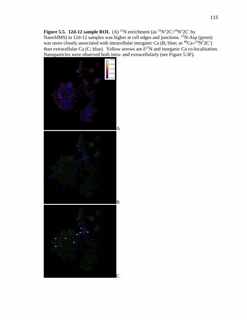
115
Figure 5.5. 12d-12 sample ROI. (A) 15N enrichment (as 15N12C-/14N12C- by NanoSIMS) in 12d-12 samples was higher at cell edges and junctions. 15N-Asp (green) was more closely associated with intracellular inorganic Ca (B; blue; as 40Ca-/14N12C-) than extracellular Ca (C; blue). Yellow arrows are δ15N and inorganic Ca co-localization. Nanoparticles were observed both intra- and extracellularly (see Figure 5.3F).
A
B
C

116
Chapter 6: Conclusions
This thesis constitutes the first coral skeletal proteome (Chapter 2), the first application of
coral cell cultures for understanding the effects of ocean acidification on coral
calcification at the cellular and molecular levels (Chapter 4), and the first use of
NanoSIMS to test the co-localization of aspartic acid and newly formed aragonite in
corals (Chapter 5). These findings detail the skeletal protein production response of
corals to environmental perturbations and provide additional evidence for potential roles
of highly acidic proteins in corals’ biomineralization process. Additionally, the work in
this dissertation prompts numerous areas of future research.
Corals, like mollusks, likely produce family-specific biomineral-associated
protein complexes (Marin et al. 2013). In fact, while there is significant overlap in SOM
proteins detected in S. pistillata and A. digitifera (Drake et al. 2013, Ramos-Silva et al.
2013), there are clearly proteins from skeleton of each species not found in the other by
LC-MS/MS sequencing. An example is galaxin, a potential collagen-binding protein
(Bhattacharya et al. in review) observed in A. digitifera, but not S. pistillata, skeleton.
Recent sequence analysis suggests that galaxin is polyphyletic within corals
(Bhattacharya et al. in review), and as such may have a diminished role in
biomineralization of Pocilloporids (S. pistillata) compared with Acroporids (A.
digitifera). This is not surprising given the confusion surrounding coral phylogenetics
(Budd et al. 2010). Further comparative skeletal proteome analyses will be useful to
resolve the evolution of the stony coral biomineralization toolkit(s).
An issue that came to light during this dissertation is the problem of coral cellular
versus human contamination of SOM protein samples (Ramos-Silva et al. 2013).

117
Contamination of carbonate organic matrix proteins by highly conserved proteins of
potential human origin are not unknown in proteomic studies (Chapter 3 and (Drake et al.
2014)). While I am confident that coral cellular contamination is not present in the S.
pistillata SOM proteome (Drake et al. 2013), it would be useful to determine if highly
conserved proteins, such as actin and ubiquitin, are derived from the coral or from a
human source. Immunological localization of some of these proteins in coral skeleton
thin sections is a step in the right direction (Mass et al. 2014), but does not conclusively
resolve the issue.
Now that two independent coral skeletal proteomes exist (Chapter 1 and (Drake et
al. 2013, Ramos-Silva et al. 2013)), future transcriptomic studies should utilize these
targets to assess the ability of corals to maintain calcification during dramatic global
change. Several recent studies using intact coral nubbins have missed this opportunity
(Carreiro-Silva et al. 2014, Rosic et al. 2014) while others have examined expression of a
few of these genes (ex; (Maor-Landaw et al. 2014). Because the three-dimensional
structure of intact nubbins affects the actual pH experienced by calicoblastic cells, it will
be interesting to compare CARP and cadherin expression profiles of whole corals
compared with cell cultures.
Finally, the NanoSIMS analysis of S. pistillata cell cultures is preliminary and
several ‘kinks’ remain to be resolved in the method. These include establishing
minimum effective dwell times and marking mounts so that ROIs chosen before SIMS
analysis can successfully be found in the SIMS without the aid of secondary ion images.
Additionally, efforts to support the identity of SIMS-based Ca signals as CaCO3,
including co-localization with S (as 32S-) or Mg (as 24Mg16O-) did not come to fruition.

118
32S- co-localized with 14N12C- and the mass window chosen for 24Mg16O- turned out to fit
40Ca- better. Mass windows for P (31P-) have been used as negative ions in previous mass
spectrometry studies of coral skeletons (LaVigne et al. 2010), may not be useful because
of the element’s biological roles in DNA and cell membranes. However, B (11B12C-,
11B14N-, 11B16O-)(Valle et al. 2011), Fe (56Fe16O- or 57Fe16O-)(Byrne et al. 2010), and
negatively ionized Ba and Sr should be examined for their applicability as tracers of
carbonate-based, rather than cellular, Ca.

119
Appendix 1: Chapter 4 Supplementary Tables Table A1. Carbonate parameters of S. pistillata culture medium bubbled to four CO2 levels. 400 ppm
CO2
700 ppm CO2
1000 ppm CO2
2000 ppm CO2
ASW Equilibration pH
8.18 8.04 7.76 7.64
ASW Equilibration DIC (mM)
2.39 2.43 2.37 2.38
Equilibration pH 7.95 7.87 7.61 7.30 Equilibration DIC (mM)
1.29 1.55 1.47 1.61
All Salinity 34 All Temperature (oC) 26 All Light (µmol quanta/m2/s) (L/D)
100 (12:12)
All Carbonate Alkalinity (µmol/kg)
2612.3 (±125.1)
All Total Medium Alkalinity (µmol/kg)
3276.9 (±139.7)
Table A2. S. pistillata qPCR primer information. Primer Sequence Amplicon
size (bp) qPCR Tanneal (oC)
Reference
CARP1 F-AGGTCATTTCTCTGATGATG R-CTCCGTCATTCTTGGATTAT
160 57 This work
CARP3 F-GTAGCTGAACCATCTGAAG R-CGTTCTCGTCAACACTATC
166 56 This work
CARP4 F-AGACCCATTTGTGAAGAC R-CGGTAACCACTCCTTTATT
142 50 This work
STPCA2 F-GCAAAGAAACTGACAAAGG R-TCTACAGCTTCTGGATCA
158 50 (Bertucci et al. 2011)
18S F-AACGATGCCAACTAGGGATCA R-GGTTTCCCATAAGGTGCCAAA
- 56 (Kvitt et al. 2011)

120
Appendix 2: Additional NanoSIMS images Figure A1. Control sample total Ca (40Ca-;A & E), δ15N (15N12C-/14N12C-) >0.0036 (B & F), δ15N >0.0036 relative to total Ca (C & G), and δ15N >0.0036 relative to inorganic Ca (40Ca-/14N12C-; D & H). Post-SIMS SEM image of the same location (I). A-D are the first ~0.5 µm-thick plane of the sample; E-H are the second ~0.5 µm-thick plane. ROI size was 12x12 µm. The sample was incubated only in un-enriched Asp and was cultivated on day 12.
A E I
B F
C G
D H

121
Figure A2. 5d-5 sample ROI2 total Ca (40Ca-;A, B, C), δ15N (15N12C-/14N12C-) >0.0036 (D, E, F). A & D, B & E, and C & F are the first, third, and fifth (of seven) ~0.5 µm-thick planes of the sample, respectively. ROI size was 15x15 µm. The sample was both incubated in 15N-Asp and cultivated on day 5.
A D
B E
C F

122
Figure A3. 5d-5 sample ROI3 total Ca (40Ca-;A, B, C), δ15N (15N12C-/14N12C-) >0.0036 (D, E, F), δ15N >0.0036 relative to total Ca (G, H, I), and δ15N >0.0036 relative to inorganic Ca (40Ca-/14N12C-; J, K, L). Post-SIMS SEM image of the same location (M). A/D/G/J; B/E/H/K, and C/F/I/L are the first, fourth, and sixth (of seven) ~0.5 µm-thick planes through the sample, respectively. ROI size was 15x15 µm. The sample was both incubated in 15N-Asp and cultivated on day 5.
A D
B E
C F

123
G J
H K
I L
M

124
Figure A4. 5d-5 sample ROI4 total Ca (40Ca-;A, B, C), δ15N (15N12C-/14N12C-) >0.0036 (D, E, F). A & D, B & E, and C & F are the first, third, and fifth (of seven) ~0.5 µm-thick planes of the sample, respectively. ROI size was 15x15 µm. Sample 5d-5 was both incubated in 15N-Asp and cultivated on day 5.
A D
B E
C F

125
Figure A5. 12d-12 sample ROI2 total Ca (40Ca-;A & E), δ15N (15N12C-/14N12C-) >0.0036 (B & F), δ15N >0.0036 relative to total Ca (C & G), and δ15N >0.0036 relative to inorganic Ca (40Ca-/14N12C-; D & H). Post-SIMS SEM image of the same location (I). A-D are the first ~0.5 µm-thick plane of the sample; E-H are the second ~0.5 µm-thick plane. ROI size is 8x8 µm. Arrows in D and H indicate areas of δ15N and inorganic Ca co-localization. The sample was incubated in 15N-Asp on days 5, 9, and 12, and cultivated on day 12.
A E I
B F
C G
D H

126
Figure A6. 12d-12 sample ROI3 total Ca (40Ca-;A & E), δ15N (15N12C-/14N12C-) >0.0036 (B & F), δ15N >0.0036 relative to total Ca (C & G), and δ15N >0.0036 relative to inorganic Ca (40Ca-/14N12C-; D & H). Post-SIMS SEM image of the same location (I). A-D are the first ~0.5 µm-thick plane of the sample; E-H are the second ~0.5 µm-thick plane. ROI size is 18x18 µm. Arrows in D and H indicate areas of δ15N and inorganic Ca co-localization. Sample 12d-12 was incubated in 15N-Asp on days 5, 9, and 12, and cultivated on day 12.
A E I
B F
C G
D

127
Figure A7. 12d-12 sample ROI4 total Ca (40Ca-;A & E), δ15N (15N12C-/14N12C-) >0.0036 (B & F), δ15N >0.0036 relative to total Ca (C & G), and δ15N >0.0036 relative to inorganic Ca (40Ca-/14N12C-; D & H). Post-SIMS SEM image of the same location (I). A-D are the second ~0.5 µm-thick plane of the sample; E-H are the fourth ~0.5 µm-thick plane. ROI size is 11x11 µm. Arrows in D and H indicate areas of δ15N and inorganic Ca co-localization. Sample 12d-12 was incubated in 15N-Asp on days 5, 9, and 12, and cultivated on day 12.
A E I
B F
C G
D H
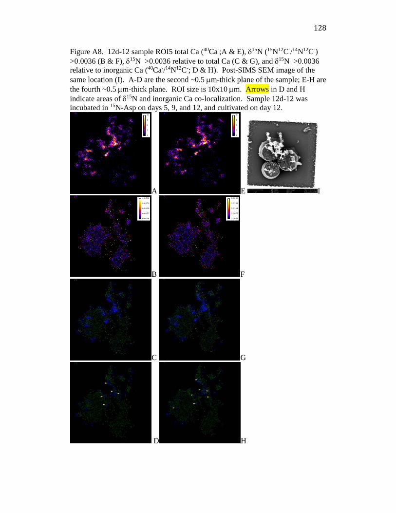
128
Figure A8. 12d-12 sample ROI5 total Ca (40Ca-;A & E), δ15N (15N12C-/14N12C-) >0.0036 (B & F), δ15N >0.0036 relative to total Ca (C & G), and δ15N >0.0036 relative to inorganic Ca (40Ca-/14N12C-; D & H). Post-SIMS SEM image of the same location (I). A-D are the second ~0.5 µm-thick plane of the sample; E-H are the fourth ~0.5 µm-thick plane. ROI size is 10x10 µm. Arrows in D and H indicate areas of δ15N and inorganic Ca co-localization. Sample 12d-12 was incubated in 15N-Asp on days 5, 9, and 12, and cultivated on day 12.
A E I
B F
C G
D H

129
References Addadi, L., D. Joester, F. Nudelman and S. Weiner (2006). "Mollusk shell formation: a source of new concepts for understanding biomineralization process." Chemical European Journal 12: 980-987. Al-Horani, F. A., S. M. Al-Moghrabi and D. de Beer (2003 ). "The mechanism of calcification and its relation to photosynthesis and respiration in the scleractinian coral Galaxea fascicularis." Marine Biology 142(3): 419-426. Allemand, D., C. Ferrier-Pagès, P. Furla, F. Houlbrèque, S. Puverel, et al. (2004). "Biomineralisation in reef-building corals: from molecular mechanisms to environmental control." Comptes Rendus Palevol 3(6–7): 453-467. Allemand, D., E. Tambutte, J.-P. Girard and J. Jaubert (1998). "Organic matrix synthesis in the scleractinian coral Stylophora pistillata: Role in biomineralization and potential target of the organotin tributyltin." Journal of Experimental Biology 201: 2001-2009. Allemand, D., E. TambuttE, J. Girard and J. Jaubert (1998). "Organic matrix synthesis in the scleractinian coral Stylophora pistillata: Role in biomineralization and potential target of the organotin tributyltin." Journal of Experimental Biology 201(13): 2001-2009. Allemand, D., É. Tambutté, D. Zoccola and S. Tambutté (2011). Coral calcification, cells to reefs. Coral reefs: an ecosystem in transition, Springer: 119-150. Alvares, K., S. N. Dixit, E. Lux and A. Veis (2009). "Echinoderm phosphorylated matrix proteins UTMP16 and UTMP19 have different functions in sea urchin tooth mineralization." Journal of Biological Chemistry 284(38): 26149-26160. Anagnostou, E., R. M. Sherrell, A. Gagnon, M. LaVigne, M. P. Field, et al. (2011). "Seawater nutrient and carbonate ion concentrations recorded as P/Ca, Ba/Ca, and U/Ca in the deep-sea coral Desmophyllum dianthus." Geochimica et Cosmochimica Acta 75(9): 2529-2543. Angerer, L. M., D. W. Oleksyn, C. Y. Logan, D. R. McClay, L. Dale, et al. (2000). "A BMP pathway regulates cell fate allocation along the sea urchin animal-vegetal embryonic axis." Development 127(5): 1105-1114. Anthony, K. R. N., D. I. Kline, G. Diaz-Pulido, S. Dove and O. Hoegh-Guldberg (2008). "Ocean acidification causes bleaching and productivity loss in coral reef builders." Proceedings of the National Academy of Sciences 105(45): 17442-17446. Armbrust, E. V., J. A. Berges, C. Bowler, B. R. Green, D. Martinez, et al. (2004). "The genome of the diatom Thalassiosira pseudonana: ecology, evolution, and metabolism." Science 306(5693): 79-86. Asara, J. M., M. H. Schweitzer, L. M. Freimark, M. Phillips and L. C. Cantley (2007). "Protein sequences from mastodon and Tyrannosaurus rex revealed by mass spectrometry." Science 316(5822): 280-285. Asenath-Smith, E., H. Li, E. C. Keene, Z. W. Seh and L. A. Estroff (2012). "Crystal growth of calcium carbonate in hydrogels as a model of biomineralization." Advanced Functional Materials 22(14): 2891-2914.

130
Ashkenazy, H., E. Erez, E. Martz, T. Pupko and N. Ben-Tal (2010). "ConSurf 2010: calculating evolutionary conservation in sequence and structure of proteins and nucleic acids." Nucleic Acids Research 38(suppl 2): W529-W533. Aytuna, A. S., A. Gursoy and O. Keskin (2005). "Prediction of protein–protein interactions by combining structure and sequence conservation in protein interfaces." Bioinformatics 21(12): 2850-2855. Baas Becking, L. G. M. and D. Moore (1961). "Biogenic sulfides." Economic Geology 56(2): 259-272. Barnes, D. and C. Crossland (1976). "Urease activity in the staghorn coral, Acropora acuminata." Comparative Biochemistry and Physiology Part B: Comparative Biochemistry 55(3): 371-376. Baron, M. (2003). "An overview of the Notch signalling pathway." Seminars in Cell & Developmental Biology 14(2): 113-119. Barron, M. J., S. J. Brookes, C. E. Draper, D. Garrod, J. Kirkham, et al. (2008). "The cell adhesion molecule nectin-1 is critical for normal enamel formation in mice." Human Molecular Genetics 17(22): 3509-3520. Beerling, D. J. and D. L. Royer (2011). "Convergent Cenozoic CO2 history." Nature Geosci 4(7): 418-420. Beniash, E. (2011). "Biominerals—hierarchical nanocomposites: the example of bone." Wiley Interdisciplinary Reviews: Nanomedicine and Nanobiotechnology 3(1): 47-69. Beniash, E., L. Addadi and S. Weiner (1999). "Cellular control over spicule formation in sea urchin embryos: a structural approach." Journal of Structural Biology 125(1): 50-62. Benson, S. C., N. C. Benson and F. Wilt (1986). "The organic matrix of the skeletal spicule of sea urchin embryos." The Journal of Cell Biology 102(5): 1878-1886. Berntson, E. A., S. C. France and L. S. Mullineaux (1999). "Phylogenetic relationships within the class Anthozoa (phylum Cnidaria) based on nuclear 18S rDNA sequences." Molecular Phylogenetics and Evolution 13(2): 417-433. Bertucci, A., A. Moya, S. Tambutté, D. Allemand, C. T. Supuran, et al. (2013). "Carbonic anhydrases in anthozoan corals—A review." Bioorganic & Medicinal Chemistry 21(6): 1437-1450. Bertucci, A., S. Tambutté, C. Supuran, D. Allemand and D. Zoccola (2011). "A new coral carbonic anhydrase in Stylophora pistillata." Marine Biotechnology 13(5): 992-1002. Bhattacharya, D., S. Agrawal, M. Aranda, S. Baumgarten, M. Belcaid, et al. (in review). "Basis for ecological success of reef-forming corals elucidated using comparative genomics." Science. Birkedal-Hansen, H., W. G. I. Moore, M. K. Bodden, L. J. Windsor, B. Birkedal-Hansen, et al. (1993). "Matrix metalloproteinases: A review." Critical Reviews in Oral Biology & Medicine 4(2): 197-250. Blank, S., M. Arnoldi, S. Khoshnavaz, L. Treccani, M. Kuntz, et al. (2003). "The nacre protein perlucin nucleates growth of calcium carbonate crystals." Journal of Microscopy 212(3): 280-291.

131
Boorungsiman, S., E. Gentleman, R. Carzaniga, N. D. Evans, D. W. McComb, et al. (2012). "The role of intracellular calcium phosphate in osteoblast-mediated bone apatite formation." Proceedings of the National Academy of Sciences 109(35): 14170-14175. Borell, E. M., A. R. Yuliantri, K. Bischof and C. Richter (2008). "The effect of heterotrophy on photosynthesis and tissue composition of two scleractinian corals under elevated temperature." Journal of Experimental Marine Biology and Ecology 364(2): 116-123. Budd, A. F., S. L. Romano, N. D. Smith and M. S. Barbeitos (2010). "Rethinking the phylogeny of Scleractinian corals: a review of morphological and molecular data." Integrative and Comparative Biology 50(3): 411-427. Burki, F., S. I. Nikolaev, I. Bolivar, J. Guiard and J. Pawlowski (2006). "Analysis of expressed sequence tags from a naked foramineran Reticulomyxa filosa." Genome 49(8): 882-887. Burriesci, M. S., T. K. Raab and J. R. Pringle (2012). "Evidence that glucose is the major transferred metabolite in dinoflagellate–cnidarian symbiosis." Journal of Experimental Biology 215(19): 3467-3477. Byrne, M. E., D. A. Ball, J.-L. Guerquin-Kern, I. Rouiller, T.-D. Wu, et al. (2010). "Desulfovibrio magneticus RS-1 contains an iron- and phosphorus-rich organelle distinct from its bullet-shaped magnetosomes." Proceedings of the National Academy of Sciences 107(27): 12263-12268. Canalis, E., A. N. Economides and E. Gazzerro (2003). "Bone morphogenetic proteins, their antagonists, and the skeleton." Endocrine Reviews 24(2): 218-235. Carreiro-Silva, M., T. Cerqueira, A. Godinho, M. Caetano, R. S. Santos, et al. (2014). "Molecular mechanisms underlying the physiological responses of the cold-water coral Desmophyllum dianthus to ocean acidification." Coral Reefs: 1-12. Cazin, J., T. R. Kozel, D. M. Lupan and W. R. Burt (1969). "Extracellular deoxyribonuclease production by yeasts." Journal of Bacteriology 100(2): 760-762. Cesar, H., L. Burke and L. Pet-Soede (2003). The economics of worldwide coral reef degredation. Checa, A. (2000). "A new model for periostracum and shell formation in Unionidae (Bivalvia, Mollusca)." Tissue and Cell 32(5): 405-416. Clendenon, S. G., B. Shah, C. A. Miller, G. Schmeisser, A. Walter, et al. (2009). "Cadherin-11 controls otolith assembly: Evidence for extracellular cadherin activity." Developmental Dynamics 238(8): 1909-1922. Clode, P. L. and A. T. Marshall (2002). "Low temperature FESEM of the calcifying interface of a scleractinian coral." Tissue and Cell 34(3): 187-198. Clode, P. L. and A. T. Marshall (2003). "Calcium associated with a fibrillar organic matrix in the scleractinian coral Galaxea fascicularis." Protoplasma 220(3): 153-161. Clode, P. L., R. A. Stern and A. T. Marshall (2007). "Subcellular imaging of isotopically labeled carbon compounds in a biological sample by ion microprobe (NanoSIMS)." Microscopy Research and Technique 70(3): 220-229.

132
Cohen, A. L., D. C. McCorkle, S. d. Putron, G. A. Gaetani and K. A. Rose (2009). "Morphological and compositional changes in the skeletons of new coral recruits reared in acidified seawater : Insights into the biomineralization response to ocean acidification." Geochemistry Geophysics Geosystems 10: 1-12. Comeau, S., P. J. Edmunds, N. B. Spindel and R. C. Carpenter (2013). "The responses of eight coral reef calcifiers to increasing partial pressure of CO2 do not exhibit a tipping point." Limnology and Oceanography 58(1): 388-398. Conesa, A., S. Götz, J. M. García-Gómez, J. Terol, M. Talón, et al. (2005). "Blast2GO: a universal tool for annotation, visualization and analysis in functional genomics research." Bioinformatics 21(18): 3674-3676. Corstjens, P. L. A. M., Y. Araki and E. L. González (2001). "A coccolithophorid calcifying vesicle with a vacuolar-type ATPase proton pump: cloning and immunolocalization of the V0 subunit c." Journal of Phycology 37(1): 71-78. Craig, R. and R. C.Beavis (2004). "TANDEM: matching proteins with tandem mass spectra." Bioinformatics 20: 1466-1467. Crook, E., D. Potts, M. Rebolledo-Vieyra, L. Hernandez and A. Paytan (2012). "Calcifying coral abundance near low-pH springs: Implications for future ocean acidification." Coral Reefs 31(1): 239-245. Cserzo, M., F. Eisenhaber, B. Eisenhaber and I. Simon (2002). "On filtering false positive transmembrane protein predictions." Protein Engineering 15: 745-752. Cuif, J.-P. and Y. Dauphin (1998). "Microstructural and physico-chemical characterization of ‘centers of calcification’ in septa of some recent scleractinian corals." Paläontologische Zeitschrift 72(3-4): 257-269. Cuif, J.-P., Y. Dauphin, P. Berthet and J. Jegoudez (2004). "Associated water and organic compounds in coral skeletons: Quantitative thermogravimetry coupled to infrared absorption spectrometry." Geochemistry Geophysics Geosystems 5(11): Q11011. Cuif, J. P., Y. Dauphin, B. Farre, G. Nehrke, J. Nouet, et al. (2008). "Distribution of sulphated polysaccharides within calcareous biominerals suggests a widely shared two-step crystallization process for the microstructural growth units." Mineralogical Magazine 72(1): 233-237. de Jong, D. S., W. T. Steegenga, J. M. A. Hendriks, E. J. J. van Zoelen, W. Olijve, et al. (2004). "Regulation of Notch signaling genes during BMP2-induced differentiation of osteoblast precursor cells." Biochemical and Biophysical Research Communications 320(1): 100-107. Derycke, L., L. Morbidelli, M. Ziche, O. De Wever, M. Bracke, et al. (2006). "Soluble N-cadherin fragment promotes angiogenesis." Clinical and Experimental Metastasis 23(3): 187-201. Desai, N. A. and V. Shankar (2000). "Purification and characterization of the single‐strand‐specific and guanylic‐acid‐preferential deoxyribonuclease activity of the extracellular nuclease from Basidiobolus haptosporus." European Journal of Biochemistry 267(16): 5123-5135. Dobbs, F. C., R. C. Zimmerman and L. A. Drake (2004). "Occurrence of intracellular crystals in leaves of Thalassia testudinum." Aquatic Botany 80(1): 23-28.

133
Domart-Coulon, I., J. Stolarski, C. Brahmi, E. Gutner-Hoch, K. Janiszewska, et al. (2014). "Simultaneous extension of both basic microstructural components in scleractinian coral skeleton during night and daytime, visualized by in situ 86Sr pulse labeling." Journal of Structural Biology 185(1): 79-88. Domart-Coulon, I. J., D. C. Elbert, E. P. Scully, P. S. Calimlim and G. K. Ostrander (2001). "Aragonite crystallization in primary cell cultures of multicellular isolates from a hard coral, Pocillopora damicornis." Proceedings of the National Academy of Sciences 98(21): 11885-11890. Doney, S. C., V. J. Fabry, R. A. Feely and J. A. Kleypas (2009). "Ocean acidification: The other CO2 problem." Annual Review of Marine Science 1(1): 169-192. Dove, P. M., J. J. D. Yoreo and S. Weiner, Eds. (2003). Reviews in Mineralogy and Geochemistry: Biomineralizaiton. Washington, D.C., Mineralogical Society of America. Drake, J. L., T. Mass and P. G. Falkowski (2014). "The evolution and future of carbonate precipitation in marine invertebrates: Witnessing extinction or documenting resilience in the Anthropocene?" Elementa Science of the Anthropocene 2(1): 000026. Drake, J. L., T. Mass, L. Haramaty, E. Zelzion, D. Bhattacharya, et al. (2013). "Proteomic analysis of skeletal organic matrix from the stony coral Stylophora pistillata." Proceedings of the National Academy of Sciences 110(10): 3788-3793. Drake, J. L., T. Mass, L. Haramaty, E. Zelzion, D. Bhattacharya, et al. (2013). "Reply to Ramos-Silva et al.: Regarding coral skeletal proteome." Proceedings of the National Academy of Sciences 110(24): E2147-E2148. Drenkard, E. J., A. L. Cohen, D. C. McCorkle, S. J. Putron, V. R. Starczak, et al. (2013). "Calcification by juvenile corals under heterotrophy and elevated CO2." Coral Reefs: 1-9. Drescher, B., R. M. Dillaman and A. R. Taylor (2012). "Coccolithogenesis In Scyphosphaera apsteinii (Prymnesiophyceae)." Journal of Phycology 48(6): 1343-1361. Ducklow, H. W. and R. Mitchell (1979). "Composition of mucus released by coral reef coelenterates." Limnology and Oceanography 24(4): 706-714. Edmunds, P. J. (2011). "Zooplanktivory ameliorates the effects of ocean acidification on the reef coral Porites spp." Limnology and Oceanography 56(6): 2402-2410. Edmunds, P. J. and C. B. Wall (2014). "Evidence that high pCO2 affects protein metabolism in tropical reef corals." The Biological Bulletin 227(1): 68-77. Ehrlich, H. (2010). "Chitin and collagen as universal and alternative templates in biomineralization." International Geology Review 52(7-8): 661+699. Erwin, D. H., M. Laflamme, S. M. Tweedt, E. A. Sperling, D. Pisani, et al. (2011). "The Cambrian conundrum: Early divergence and later ecological success in the early history of animals." Science 334(6059): 1091-1097. Fabricius, K. E., C. Langdon, S. Uthicke, C. Humphrey, S. Noonan, et al. (2011). "Losers and winners in coral reefs acclimatized to elevated carbon dioxide concentrations." Nature Climate Change 1: 165-169.
134
Falini, G., S. Albeck, S. Weiner and L. Addadi (1996). "Control of aragonite or calcite precipitation by mollusk shell macromolecules." Science 271: 67-69. Falini, G. and S. Fermani (2004). "Chitin mineralization." Tissue engineering 10(1-2): 1-6. Falini, G., M. Reggi, S. Fermani, F. Sparla, S. Goffredo, et al. (2013). "Control of aragonite deposition in colonial corals by intra-skeletal macromolecules." Journal of Structural Biology 183(2): 226-238. Falkowski, P. G., Z. Dubinsky, L. Muscatine and J. W. Porter (1984). "Light and the bioenergetics of a symbiotic coral." Bioscience 34(11): 705-709. Fang, D., C. Pan, H. Lin, Y. Lin, G. Xu, et al. (2012). "Ubiquitylation functions in the calcium carbonate biomineralization in the extracellular matrix." PLoS ONE 7(4): e35715. Feely, R. A., S. C. Doney and S. R. Cooley (2009). "Ocean acidification: Present conditions and future changes in a high-CO2 world." Oceanography 22(4): 36-47. Feely, R. A., C. L. Sabine, K. Lee, W. Berelson, J. Kleypas, et al. (2004). "Impact of anthropogenic CO2 on the CaCO3 system in the oceans." Science 305(5682): 362-366. Feng, Q., Z. Fang, Z. Yan, R. Xing, L. Xie, et al. (2009). "The structure–function relationship of MSI7, a matrix protein from pearl oyster Pinctada fucata." Acta Biochimica et Biophysica Sinica 41(11): 955-962. Ferrier-Pages, C., F. Boisson, D. Allemand and E. Tambutte (2002). "Kinetics of strontium uptake in the scleractinian coral Stylophora pistillata." Marine Ecology Progress Series 245: 8. Fine, M. and D. Tchernov (2007). "Scleractinian coral species survive and recover from decalcification." Science 315(5820): 1811. Fortin, D., B. Davis and T. Beveridge (1996). "Role of Thiobacillus and sulfate-reducing bacteria in iron biocycling in oxic and acidic mine tailings." FEMS Microbiology Ecology 21(1): 11-24. Fraser, H. B., A. E. Hirsh, L. M. Steinmetz, C. Scharfe and M. W. Feldman (2002). "Evolutionary rate in the protein interaction network." Science 296(5568): 750-752. Fukuda, I., S. Ooki, T. Fujita, E. Murayama, H. Nagasawa, et al. (2003). "Molecular cloning of a cDNA encoding a soluble protein in the coral exoskeleton." Biochemical and Biophysical Research Communications 304(1): 11-17. Furla, P., I. Galgani, E. Durand and D. Allemand (2000). "Sources and mechanisms of inorganic carbon transport for coral calcification and photosynthesis." Journal of Experimental Biology 203: 3445-3457. Gagnon, A. C., J. F. Adkins and J. Erez (2012). "Seawater transport during coral biomineralization." Earth and Planetary Science Letters 329–330(0): 150-161. Gal, A., K. Kahil, N. Vidavsky, R. T. DeVol, P. U. P. A. Gilbert, et al. (2014). "Particle accretion mechanism underlies biological crystal growth from an amorphous precursor phase." Advanced Functional Materials 24(34): 5420-5426. Gal, A., S. Weiner and L. Addadi (2015). "A perspective on underlying crystal growth mechanisms in biomineralization: solution mediated growth versus nanosphere particle accretion." CrystEngComm.

135
Gazeau, F., C. Quiblier, J. M. Jansen, J.-P. Gattuso, J. J. Middelburg, et al. (2007). "Impact of elevated CO2 on shellfish calcification." Geophysical Research letters 34(7): L07603. George, N. C., C. E. Killian and F. H. Wilt (1991). "Characterization and expression of a gene encoding a 30.6-kDa Strongylocentrotus purpuratus spicule matrix protein." Developmental Biology 147(2): 334-342. Gerbaud, V., D. Pignol, E. Loret, J. A. Bertrand, Y. Berland, et al. (2000). "Mechanism of calcite crystal growth inhibition by the N-terminal undecapeptide of lithostathine." Journal of Biological Chemistry 275(2): 1057-1064. Gilbert, M., W. J. Shaw, J. R. Long, K. Nelson, G. P. Drobny, et al. (2000). "Chimeric peptides of statherin and osteopontin that bind hydroxyapatite and mediate cell adhesion." Journal of Biological Chemistry 275(21): 16213-16218. Gilbert, P. U. P. A. and F. Wilt (2011). Molecular aspects of biomineralization of the echinoderm endoskeleton. Molecular Biomineralization. W. E. G. Müller, Springer Berlin Heidelberg: 199-223. Girard, J.-P. and T. A. Springer (1996). "Modulation of endothelial cell adhesion by hevin, an acidic protein associated with high endothelial venules." Journal of Biological Chemistry 271(8): 4511-4517. Goffredo, S., P. Vergni, M. Reggi, E. Caroselli, F. Sparla, et al. (2011). "The skeletal organic matrix from mediterranean coral Balanophyllia europaea influences calcium carbonate precipitation." PLoS ONE 6(7). Goldberg, H. A., K. J. Warner, M. C. Li and G. K. Hunter (2001). "Binding of bone sialoprotein, osteopontin and synthetic polypeptides to hydroxyapatite." Connective Tissue Research 42(1): 25-37. Goldberg, W. M. (1974). "Evidence of a sclerotized collagen from the skeleton of a gorgonian coral." Comparative Biochemistry and Physiology Part B: Comparative Biochemistry 49(3): 525-526. Gorbunov, M. and P. Falkowski (2004). Fluorescence Induction and Relaxation (FIRe) technique and instrumentation for monitoring photosynthetic processes and primary production in aquatic ecosystems. Photosynthesis: Fundamental Aspects to Global Perspectives. A. v. d. Est and D. Bruce: 1029-1031. Goreau, T. F. (1959). "The physiology of skeleton formation in corals. I. A method for measuring the rate of calcium deposition by corals under different conditions." Biological Bulletin 116: 59-75. Gothmann, A. M., J. Stolarski, J. F. Adkins, B. Schoene, K. J. Dennis, et al. (2015). "Fossil corals as an archive of secular variations in seawater chemistry since the Mesozoic." Geochimica et Cosmochimica Acta 160(0): 188-208. Gotliv, B.-A., L. Addadi and S. Weiner (2003). "Mollusk shell acidic proteins: in search of individual functions." ChemBioChem 4(6): 522-529. Gotliv, B.-A., N. Kessler, J. L. Sumerel, D. E. Morse, N. Tuross, et al. (2005). "Asprich: A novel aspartic acid-rich protein family from the prismatic shell matrix of the bivalve Atrina rigida." ChemBioChem 6(2): 304-314. Hayes, R. L. and N. I. Goreau (1977). "Intracellular crystal-bearing vesicles in the epidermis of Scleractinian corals, Astrangia danae (Agassiz) and Porites porites (Pallas)." The Biological Bulletin 152(1): 26-40.

136
Hayward, D. C., S. Hetherington, C. A. Behm, L. C. Grasso, S. Foret, et al. (2011). "Differential gene expression at coral settlement and metamorphosis - a subtractive hybridization study." PLoS ONE 6(10): e26411. Hazelaar, S., H. J. van der Strate, W. W. C. Gieskes and E. G. Vrieling (2003). "Possible role of ubiquitin in silica biomineralization in diatoms: Identification of a homologue with high silica affinity." Biomolecular Engineering 20(4–6): 163-169. He, G., T. Dahl, A. Veis and A. George (2003). "Nucleation of apatite crystals in vitro by self-assembled dentin matrix protein 1." Nature Materials 2(8): 552-558. Heathfield, B. M. (1970). "Calcification in echinoderms: effects of temperature and diamox on incorporation of calcium-45 in vitro by regenerating spines of Strongylocentrotus purpuratus." The Biological Bulletin 139(1): 151-163. Helman, Y., F. Natale, R. M. Sherrell, M. LaVigne, V. Starovoytov, et al. (2008). "Extracellular matrix production and calcium carbonate precipitation by coral cells in vitro." Proceedings of the National Academy of Sciences 105(1): 54-58. Hildebrand, M., M. J. Doktycz and D. P. Allison (2008). "Application of AFM in understanding biomineral formation in diatoms." Pflügers Archiv - European Journal of Physiology 456: 127-137. Hoegh-Guldberg, O. and J. F. Bruno (2010). "The impact of climate change on the world’s marine ecosystems." Science 328(5985): 1523-1528. Hoegh-Guldberg, O., P. J. Mumby, A. J. Hooten, R. S. Steneck, P. Greenfield, et al. (2007). "Coral reefs under rapid climate change and ocean acidification." Science 318(5857): 1737-1742. Hofmann, G. E., J. P. Barry, P. J. Edmunds, R. D. Gates, D. A. Hutchins, et al. (2010). "The effect of ocean acidification on calcifying organisms in marine ecosystems: an organism-to-ecosystem perspective." Annual Review of Ecology, Evolution, and Systematics 41(1): 127-147. Hofmann, K. and W. Stoffel (1993). "TMbase - A database of membrane spanning proteins segments." Biological Chemistry Hoppe-Seyler 374: 166. Holcomb, M., A. L. Cohen, R. I. Gabitov and J. L. Hutter (2009). "Compositional and morphological features of aragonite precipitated experimentally from seawater and biogenically by corals." Geochimica et Cosmochimica Acta 73(14): 4166-4179. Holcomb, M., D. C. McCorkle and A. L. Cohen (2010). "Long-term effects of nutrient and CO2 enrichment on the temperate coral Astrangia poculata (Ellis and Solander, 1786)." Journal of Experimental Marine Biology and Ecology 386(1–2): 27-33. Holcomb, M., E. Tambutté, D. Allemand and S. Tambutté (2014). "Light enhanced calcification in Stylophora pistillata: effects of glucose, glycerol and oxygen." PeerJ 2: e375. Holcomb, M., A. A. Venn, E. Tambutte, S. Tambutte, D. Allemand, et al. (2014). "Coral calcifying fluid pH dictates response to ocean acidification." Nature Scientific Reports 4. Honisch, B., N. G. Hemming, A. G. Grottoli, A. .Amat, G. N. Hanson, et al. (2004). "Assessing scleractinian corals as recorders for paleo-pH: Empirical

137
calibration and vital effects." Geochimica et Cosmochimica Acta 68(18): 3675-3685. Houlbrèque, F. and C. Ferrier-Pagès (2009). "Heterotrophy in tropical scleractinian corals." Biological Reviews 84(1): 1-17. Houlbrèque, F., S. Reynaud, C. Godinot, F. Oberhänsli, R. Rodolfo-Metalpa, et al. (2015). "Ocean acidification reduces feeding rates in the scleractinian coral Stylophora pistillata." Limnology and Oceanography 60(1): 89-99. Houlbrèque, F., R. Rodolfo-Metalpa, R. Jeffree, F. Oberhänsli, J. L. Teyssié, et al. (2012). "Effects of increased pCO2 on zinc uptake and calcification in the tropical coral Stylophora pistillata." Coral Reefs 31(1): 101-109. http://people.oregonstate.edu/~meyere/data.html. Huang, Y., B. Niu, Y. Gao, L. Fu and W. Li (2010). "CD-HIT Suite: A web server for clustering and comparing biological sequences." Bioinformatics. Hüning, A., F. Melzner, J. Thomsen, M. Gutowska, L. Krämer, et al. (2013). "Impacts of seawater acidification on mantle gene expression patterns of the Baltic Sea blue mussel: implications for shell formation and energy metabolism." Marine Biology 160(8): 1845-1861. Hunter, G. K. and H. A. Goldberg (1993). "Nucleation of hydroxyapatite by bone sialoprotein." Proceedings of the National Academy of Sciences 90(18): 8562-8565. Hyman, L. H. (1955). The Invertebrates. New York, McGraw-Hill. Illies, M., M. Peeler, A. Dechtiaruk and C. Ettensohn (2002). "Identification and developmental expression of new biomineralization proteins in the sea urchin Strongylocentrotus purpuratus." Development Genes and Evolution 212(9): 419-431. Ingersoll, E. P. and F. H. Wilt (1998). "Matrix metalloproteinase inhibitors disrupt spicule formation by primary mesenchyme cells in the sea urchin embryo." Developmental Biology 196(1): 95-106. IPCC (2007). Contribution of Working Group I to the Fourth Assessment Report of the Intergovernmental Panel on Climate Change. S. Solomon, D. Qin, M. Manning et al. New York, NY, Cambridge University Press. Jackson, D., C. McDougall, K. Green, F. Simpson, G. Worheide, et al. (2006). "A rapidly evolving secretome builds and patterns a sea shell." BMC Biology 4(1): 40. Jackson, D. J., L. Macis, J. Reitner, B. M. Degnan and G. Wörheide (2007). "Sponge paleogenomics reveals an ancient role for carbonic anhydrase in skeletogenesis." Science 316(5833): 1893-1895. Jackson, D. J., K. Mann, V. Häussermann, M. B. Schilhabel, C. Lüter, et al. (2015). "The Magellania venosa biomineralizing proteome: A window into brachiopod shell evolution." Genome Biology and Evolution 7(5): 1349-1362. Jackson, D. J., C. McDougall, B. Woodcroft, P. Moase, R. A. Rose, et al. (2010). "Parallel evolution of nacre building gene sets in molluscs." Mol Biol Evol 27(3): 591-608. Jian-Ping, D., C. Jun, B. Yi-Fei, H. Bang-Xing, G. Shang-Bin, et al. (2010). "Effects of pearl powder extract and its fractions on fibroblast function relevant to wound repair." Pharmaceutical Biology 48(2): 122-127.

138
Jiang, L., L. He and M. Fountoulakis (2004). "Comparison of protein precipitation methods for sample preparation prior to proteomic analysis." Journal of Chromatography A 1023: 317-320. Joubert, C., D. Piquemal, B. Marie, L. Manchon, F. Pierrat, et al. (2010). "Transcriptome and proteome analysis of Pinctada margaritifera calcifying mantle and shell: Focus on biomineralization." BMC Genomics 11(1): 613. Jury, C., F. Thomas, M. Atkinson and R. Toonen (2013). "Buffer capacity, ecosystem feedbacks, and seawater chemistry under global change." Water 5(3): 1303-1325. Kaniewska, P., P. R. Campbell, D. I. Kline, M. Rodriguez-Lanetty, D. J. Miller, et al. (2012). "Major cellular and physiological impacts of ocean acidification on a reef building coral." PLoS ONE 7(4): e34659. Karlsson, J. and P. Artursson (1991). "A method for the determination of cellular permeability coefficients and aqueous boundary layer thickness in monolayers of intestinal epithelial ( Caco-2) cells grown in permeable filter chambers." International Journal of Pharmaceutics 71(1): 55-64. Kawaguchi, J., Y. Azuma, K. Hoshi, I. Kii, S. Takeshita, et al. (2001). "Targeted disruption of cadherin-11 leads to a reduction in bone density in calvaria and long bone metaphyses." Journal of Bone and Mineral Research 16(7): 1265-1271. Kawaguti, S. and D. Sakumoto (1948). "The effect of light on the calcium deposition of corals." Bulletin of the Oceanograpaical Institute of Taiwan 4: 65-70. Kawasaki, K., A. V. Buchanan and K. M. Weiss (2009). "Biomineralization in humans: making the hard choices in life." Annual Review of Genetics 43(1): 119-142. Kelley, L. A. and M. J. Sternberg (2009). "Protein structure prediction on the Web: a case study using the Phyre server." Nature Protocols 4(3): 363-371. Kelly, M. W., J. L. Padilla-Gamiño and G. E. Hofmann (2013). "Natural variation and the capacity to adapt to ocean acidification in the keystone sea urchin Strongylocentrotus purpuratus." Global Change Biology 19(8): 2536-2546. Keshavmurthy, S., S.-Y. Yang, A. Alamaru, Y.-Y. Chuang, M. Pichon, et al. (2013). "DNA barcoding reveals the coral “laboratory-rat”, Stylophora pistillata encompasses multiple identities." Sci. Rep. 3. Killian, C. E., R. A. Metzler, Y. U. T. Gong, I. C. Olson, J. Aizenberg, et al. (2009). "Mechanism of calcite co-orientation in the sea urchin tooth." Journal of the American Chemical Society 131(51): 18404-18409. Killian, C. E. and F. H. Wilt (2008). "Molecular aspects of biomineralization of the echinoderm endoskeleton." Chemical reviews 108(11): 4463-4474. Klumpp, D. W., B. L. Bayne and A. J. S. Hawkins (1992). "Nutrition of the giant clam Tridacna gigas (L.) I. Contribution of filter feeding and photosynthates to respiration and growth." Journal of Experimental Marine Biology and Ecology 155(1): 105-122. Knoll, A. H. (2003). "Biomineralization and evolutionary history." Reviews in Mineralogy and Geochemistry 54(1): 329-356.

139
Kono, M., N. Hayashi and T. Samata (2000). "Molecular mechanism of the nacreous layer formation in Pinctada maxima." Biochemical and Biophysical Research Communications 269(1): 213-218. Kopp, C., M. Pernice, I. Domart-Coulon, C. Djediat, J. E. Spangenberg, et al. (2013). "Highly dynamic cellular-level response of symbiotic coral to a sudden increase in environmental nitrogen." mBio 4(3). Kroeker, K. J., R. L. Kordas, R. N. Crim and G. G. Singh (2010). "Meta-analysis reveals negative yet variable effects of ocean acidification on marine organisms." Ecology Letters 13(11): 1419-1434. Krogh, A., B. Larsson, G. v. Heijne and E. L. Sonnhammer (2001). "Predicting transmembrane protein topology with a hidden Markov model: application to complete genomes." Journal of Molecular Biology 305(3): 567-580. Kuzminov, F. I., C. M. Brown, V. V. Fadeev and M. Y. Gorbunov (2013). "Effects of metal toxicity on photosynthetic processes in coral symbionts, Symbiodinium spp." Journal of Experimental Marine Biology and Ecology 446(0): 216-227. Kvitt, H., E. Kramarsky-Winter, K. Maor-Landaw, K. Zandbank, A. Kushmaro, et al. (2015). "Breakdown of coral colonial form under reduced pH conditions is initiated in polyps and mediated through apoptosis." Proceedings of the National Academy of Sciences 112(7): 2082-2086. Kvitt, H., H. Rosenfeld, K. Zandbank and D. Tchernov (2011). "Regulation of apoptotic pathways by Stylophora pistillata (Anthozoa, Pocilloporidae) to survive thermal stress and bleaching." PLoS ONE 6(12): e28665. Labrenz, M., G. K. Druschel, T. Thomsen-Ebert, B. Gilbert, S. A. Welch, et al. (2000). "Formation of sphalerite (ZnS) deposits in natural biofilms of sulfate-reducing bacteria." Science 290(5497): 1744-1747. Lannig, G., S. Eilers, H. O. Pörtner, I. M. Sokolova and C. Bock (2010). "Impact of ocean acidification on energy metabolism of oyster, Crassostrea gigas—changes in metabolic pathways and thermal response." Marine drugs 8(8): 2318-2339. Laurent, J., A. Venn, É. Tambutté, P. Ganot, D. Allemand, et al. (2013). "Regulation of intracellular pH in cnidarians: response to acidosis in Anemonia viridis." FEBS Journal 281(3): 683-695. LaVigne, M., K. A. Matthews, A. G. Grottoli, K. M. Cobb, E. Anagnostou, et al. (2010). "Coral skeleton P/Ca proxy for seawater phosphate: Multi-colony calibration with a contemporaneous seawater phosphate record." Geochimica et Cosmochimica Acta 74(4): 1282-1293. Leaf, D. S., J. A. Anstrom, J. E. Chin, M. A. Harkey, R. M. Showman, et al. (1987). "Antibodies to a fusion protein identify a cDNA clone encoding MSP130, a primary mesenchyme-specific cell surface protein of the sea urchin embryo." Developmental Biology 121(1): 29-40. Leclercq, N., J.-P. Gattuso and J. Jaubert (2002). "Primary production, respiration, and calcification of a coral reef mesocosm under increased CO2 partial pressure." Limnology and Oceanography 47(2): 558-564. Lee, P. N., S. Kumburegama, H. Q. Marlow, M. Q. Martindale and A. H. Wikramanayake (2007). "Asymmetric developmental potential along the animal–

140
vegetal axis in the anthozoan cnidarian, Nematostella vectensis, is mediated by Dishevelled." Developmental Biology 310(1): 169-186. Lee, P. N., K. Pang, D. Q. Matus and M. Q. Martindale (2006). "A WNT of things to come: evolution of Wnt signaling and polarity in cnidarians." Seminars in Cell & Developmental Biology 17(2): 157-167. Levy, S., G. Sutton, P. C. Ng, L. Feuk, A. L. Halpern, et al. (2007). "The diploid genome sequence of an individual human." PLoS Biol 5(10): e254. Li, W. and A. Godzik (2006). "CD-Hit: a fast program for clustering and comparing large sets of protein or nucleotide sequences." Bioinformatics 22(13): 1658-1659. Liaw, L., M. Almeida, C. E. Hart, S. M. Schwartz and C. M. Giachelli (1994). "Osteopontin promotes vascular cell adhesion and spreading and is chemotactic for smooth muscle cells in vitro." Circulation Research 74(2): 214-224. Liew, Y. J., M. Aranda, A. Carr, S. Baumgarten, D. Zoccola, et al. (2014). "Identification of microRNAs in the coral Stylophora pistillata." PLoS ONE 9(3): e91101. Liu, W., X. Huang, J. Lin and M. He (2012). "Seawater acidification and elevated temperature affect gene expression patterns of the pearl oyster Pinctada fucata." PLoS ONE 7(3): e33679. Lowenstam, H. A. and S. Weiner (1989). On Biomineralization. New York, New York, Oxford University Press. Ma, J.-J., B. Feng, Y. Zhang, J.-W. Li, A.-G. Lu, et al. (2009). "Higher CO2-insufflation pressure inhibits the expression of adhesion molecules and the invasion potential of colon cancer cells." World Journal of Gastroenterology : WJG 15(22): 2714-2722. Ma, Y., S. R. Cohen, L. Addadi and S. Weiner (2008). "Sea urchin tooth design: An “all-calcite” polycrystalline reinforced fiber composite for grinding rocks." Advanced Materials 20(8): 1555-1559. Ma, Z., J. Huang, J. Sun, G. Wang, C. Li, et al. (2007). "A novel extrapallial fluid protein controls the morphology of nacre lamellae in the pearl oyster, Pinctada fucata." Journal of Biological Chemistry 282(32): 23253-23263. Mann, K., E. Edsinger-Gonzales and M. Mann (2012). "In-depth proteomic analysis of a mollusc shell: acid-soluble and acid-insoluble matrix of the limpet Lottia gigantea." Proteome science 10(1): 1-18. Mann, K., A. Poustka and M. Mann (2008). "In-depth, high-accuracy proteomics of sea urchin tooth organic matrix." Proteome science 6(1): 33. Mann, K., A. Poustka and M. Mann (2008). "The sea urchin (Strongylocentrotus purpuratus) test and spine proteomes." Proteome science 6(1): 22. Mann, K., A. J. Poustka and M. Mann (2010). "Phosphoproteomes of Strongylocentrotus purpuratus shell and tooth matrix: identification of a major acidic sea urchin tooth phosphoprotein, phosphodontin." Proteome Sci 8(6). Mann, K., F. H. Wilt and A. J. Poustka (2010). "Proteomic analysis of sea urchin (Strongylocentrotus purpuratus) spicule matrix." Proteome Science 8: 33. Mann, S. (2001). Biomineralization: Principles and Concepts in Bioinorganic Materials Chemistry. New York, Oxford University Press.
141
Mannello, F., L. Canesi, G. Gazzanelli and G. Gallo (2001). "Biochemical properties of metalloproteinases from the hemolymph of the mussel Mytilus galloprovincialis Lam." Comparative Biochemistry and Physiology Part B: Biochemistry and Molecular Biology 128(3): 507-515. Manzello, D. P., I. C. Enochs, N. Melo, D. K. Gledhill and E. M. Johns (2012). "Ocean acidification refugia of the Florida Reef Tract." PLoS ONE 7(7): e41715. Maor-Landaw, K., S. Karako-Lampert, H. W. Ben-Asher, S. Goffredo, G. Falini, et al. (2014). "Gene expression profiles during short-term heat stress in the red sea coral Stylophora pistillata." Global Change Biology 20(10): 3026-3035. Marie, B., C. Joubert, A. Tayalé, I. Zanella-Cléon, C. Belliard, et al. (2012). "Different secretory repertoires control the biomineralization processes of prism and nacre deposition of the pearl oyster shell." Proceedings of the National Academy of Sciences 109(51): 20986-20991. Marie, B., A. Marie, D. Jackson, L. Dubost, B. Degnan, et al. (2010). "Proteomic analysis of the organic matrix of the abalone Haliotis asinina calcified shell." Proteome science 8(1): 54. Marie, B., N. Roy, I. Zanella-Cléon, M. Becchi and F. Marin (2011). "Molecular evolution of mollusc shell proteins: Insights from proteomic analysis of the edible mussel Mytilus." Journal of Molecular Evolution 72(5-6): 531-546. Marie, B., N. Trinkler, I. Zanella-Cleon, N. Guichard, M. Becchi, et al. (2011). "Proteomic identification of novel proteins from the calcifying shell matrix of the Manila clam Venerupis philippinarum." Marine Biotechnology 13(5): 955-962. Marin, F. (2007). Unusually Acidic Proteins in Biomineralization. Handbook of Biomineralization. E. Baurlein and J. Picket-Heaps. Max Planck Institute for Biochemistry, Max Planck Institute for Biochemistry. Marin, F., P. Corstjens, B. de Gaulejac, E. de Vrind-De Jong and P. Westbroek (2000). "Mucins and molluscan calcification: molecular characterization of mucoperlin, a novel mucin-like protein from the nacreous shell layer of the fan mussel Pinna nobilis (Bivalvia, Pteriomorphia)." Journal of Biological Chemistry 275(27): 20667-20675. Marin, F., N. Le Roy and B. Marie (2012). "The formation and mineralization of mollusc shell." Frontiers in Bioscience 4: 1099-1125. Marin, F. and G. Luquet (2004). "Molluscan shell proteins." Comptes Rendus Palevol 3(6–7): 469-492. Marin, F., B. Marie, S. B. Hamada, P. Ramos-Silva, N. Le Roy, et al. (2013). "'Shellome': Proteins involved in mollusk shell biomineralization-diversity, functions." Recent Advances in Pearl Research: 149-166. Marks, M. H., R. S. Bear and C. H. Blake (1949). "X-ray diffraction evidence of collagen-type protein fibers in the Echinodermata, Coelenterata and Porifera." Journal of Experimental Zoology 111: 55-78. Marshall, A. T., P. L. Clode, R. Russell, K. Prince and R. Stern (2007). "Electron and ion microprobe analysis of calcium distribution and transport in coral tissues." Journal of Experimental Biology 210(14): 2453-2463. Marubini, F., C. Ferrier-Pagès, P. Furla and D. Allemand (2008). "Coral calcification responds to seawater acidification: a working hypothesis towards a physiological mechanism." Coral Reefs 27(3): 491-499.

142
Mass, T., Jeana L. Drake, L. Haramaty, J. D. Kim, E. Zelzion, et al. (2013). "Cloning and characterization of four novel coral acid-rich proteins that precipitate carbonates in vitro." Current Biology 23(12): 1126-1131. Mass, T., J. L. Drake, L. Haramaty, Y. Rosenthal, O. M. E. Schofield, et al. (2012). "Aragonite precipitation by “proto-polyps” in coral cell cultures." PLoS ONE 7(4): e35049. Mass, T., J. L. Drake, E. C. Peters, W. Jiang and P. G. Falkowski (2014). "Immunolocalization of skeletal matrix proteins in tissue and mineral of the coral Stylophora pistillata." Proceedings of the National Academy of Sciences 111(35): 12728-12733. Mass, T., J. L. Drake, H. M. Putnam, E. Zelzion, R. D. Gates, et al. (in review). "Temporal expression of biomineralization proteins during early development in the stony coral Pocillopora damicornis." McClay, D. R., R. E. Peterson, R. C. Range, A. M. Winter-Vann and M. J. Ferkowicz (2000). "A micromere induction signal is activated by beta-catenin and acts through Notch to initiate specification of secondary mesenchyme cells in the sea urchin embryo." Development 127(23): 5113-5122. McCulloch, M., J. Falter, J. Trotter and P. Montagna (2012). "Coral resilience to ocean acidification and global warming through pH up-regulation." Nature Clim. Change 2: 623-627. Medakovic, D. (2000). "Carbonic anhydrase activity and biomineralization process in embryos, larvae, and adult blue mussels Mytilus edulis L." Helgoland Marine Research 54: 1-6. Mehr, S. F., R. DeSalle, H.-T. Kao, A. Narechania, Z. Han, et al. (2013). "Transcriptome deep-sequencing and clustering of expressed isoforms from Favia corals." BMC Genomics 14: 546. Meibom, A., J.-P. Cuif, F. Houlbreque, S. Mostefaoui, Y. Dauphin, et al. (2008). "Compositional variations at ultra-structure length scales in coral skeleton." Geochimica et Cosmochimica Acta 72(6): 1555-1569. Melzner, F., J. Thomsen, W. Koeve, A. Oschlies, M. Gutowska, et al. (2013). "Future ocean acidification will be amplified by hypoxia in coastal habitats." Marine Biology 160(8): 1875-1888. Michenfelder, M., G. Fu, C. Lawrence, J. C. Weaver, B. A. Wustman, et al. (2003). "Characterization of two molluscan crystal-modulating biomineralization proteins and identification of putative mineral binding domains." Biopolymers 70(4): 522-533. Milliman, J. D. (1993). "Production and accumulation of calcium carbonate in the ocean: Budget of a nonsteady state." Global Biogeochemical Cycles 7(4): 927-957. Miyamoto, H., T. Miyashita, M. Okushima, S. Nakano, T. Morita, et al. (1996). "A carbonic anhydrase from the nacreous layer in oyster pearls." Proceedings of the National Academy of Sciences 93(18): 9657-9660. Miyashita, T., R. Takagi, M. Okushima, S. Nakano, H. Miyamoto, et al. (2000). "Complementary DNA cloning and characterization of pearlin, a new class of matrix protein in the nacreous layer of oyster pearls." Marine Biotechnology 2(5): 409-418.

143
Moya, A., L. Huisman, E. E. Ball, D. C. Hayward, L. C. Grasso, et al. (2012). "Whole transcriptome analysis of the coral Acropora millepora reveals complex responses to CO2-driven acidification during the initiation of calcification." Molecular Ecology 21(10): 2440-2454. Moya, A., S. Tambutté, A. Bertucci, E. Tambutté, S. Lotto, et al. (2008). "Carbonic anhydrase in the scleractinian coral Stylophora pistillata." Journal of Biological Chemistry 283(37): 25475-25484. Moya, A., S. Tambutté, E. Tambutté, D. Zoccola, N. Caminiti, et al. (2006). "Study of calcification during a daily cycle of the coral Stylophora pistillata: implications for `light-enhanced calcification'." Journal of Experimental Biology 209(17): 3413-3419. Mueller, C. W., P. K. Weber, M. R. Kilburn, C. Hoeschen, M. Kleber, et al. (2013). "Advances in the analysis of biogeochemical interfaces: NanoSIMS to investigate soil microenvironments." Adv. Agron 121: 1-46. Müller, W. E. G. (2011). Molecular Biomineralization: Aquatic Organisms Forming Extraordinary Materials. New York, NY, Springer. Munday, P. L., R. R. Warner, K. Monro, J. M. Pandolfi and D. J. Marshall (2013). "Predicting evolutionary responses to climate change in the sea." Ecology Letters 16(12): 1488-1500. Murdock, D. J. E. and P. C. J. Donoghue (2011). "Evolutionary origins of animal skeletal biomineralization." Cells Tissues Organs 194(2-4): 98-102. Muscatine, L., E. Tambutte and D. Allemand (1997). "Morphology of coral desmocytes, cells that anchor the calicoblastic epithelium to the skeleton." Coral Reefs 16(4): 205-213. Norris, R. D., S. K. Turner, P. M. Hull and A. Ridgwell (2013). "Marine ecosystem responses to Cenozoic global change." Science 341(6145): 492-498. Nose, A., A. Nagafuchi and M. Takeichi (1988). "Expressed recombinant cadherins mediate cell sorting in model systems." Cell 54(7): 993-1001. Nudelman, F., B. A. Gotliv, L. Addadi and S. Weiner (2006). "Mollusk shell formation: Mapping the distribution of organic matrix components underlying a single aragonitic tablet in nacre." Journal of Structural Biology 153(2): 176-187. Ogawa, D., T. Bobeszko, T. Ainsworth and W. Leggat (2013). "The combined effects of temperature and CO2 lead to altered gene expression in Acropora aspera." Coral Reefs: 1-13. Olszta, M. J., D. J. Odom, E. P. Douglas and L. B. Gower (2003). "A new paradigm for biomineral formation: mineralization via an amorphous liquid-phase precursor." Connective Tissue Research 44 Suppl 1: 326-334. Orr, J. C., V. J. Fabry, O. Aumont, L. Bopp, S. C. Doney, et al. (2005). "Anthropogenic ocean acidification over the twenty-first century and its impact on calcifying organisms." Nature 437(7059): 681-686. Pandolfi, J. M., S. R. Connolly, D. J. Marshall and A. L. Cohen (2011). "Projecting coral reef futures under global warming and ocean acidification." Science 333(6041): 418-422. Paradies, N. E. and G. B. Grunwald (1993). "Purification and characterization of NCAD90, a soluble endogenous form of N-cadherin, which is generated by

144
proteolysis during retinal development and retains adhesive and neurite-promoting function." Journal of Neuroscience Research 36(1): 33-45. Paris, G., J. S. Fehrenbacher, A. L. Sessions, H. J. Spero and J. F. Adkins (2014). "Experimental determination of carbonate‐associated sulfate δ34S in planktonic foraminifera shells." Geochemistry, Geophysics, Geosystems 15(4): 1452-1461. Paulsson, M., Y. Sommarin and D. Heinegard (1983). "Metabolism of cartilage proteins in cultured tissue sections." Biochemistry Journal 212: 659-667. Péterfi, Z., Á. Donkó, A. Orient, A. Sum, Á. Prókai, et al. (2009). "Peroxidasin is secreted and incorporated into the extracellular matrix of myofibroblasts and fibrotic kidney." The American Journal of Pathology 175(2): 725-735. Pierrot, D. E., E. Lewis and D. W. R. Wallas (2006). "MS Excel program developed for CO2 system calculations." ORNL/CDIAC-105a. Carbon dioxide information analysis center, Oak Ridge National Laboratory, U.S. Department of Energy, OakRidge, Tennessee. Politi, Y., T. Arad, E. Klein, S. Weiner and L. Addadi (2004). "Sea urchin spine calcite forms via a transient amorphous calcium carbonate phase." Science 306(5699): 1161-1164. Politi, Y., J. Mahamid, H. Goldberg, S. Weiner and L. Addadi (2007). "Asprich mollusk shell protein: in vitro experiments aimed at elucidating function in CaCO3 crystallization." CrystEngComm 9(12): 1171-1177. Politi, Y., R. A. Metzler, M. Abrecht, B. Gilbert, F. H. Wilt, et al. (2008). "Transformation mechanism of amorphous calcium carbonate into calcite in the sea urchin larval spicule." Proceedings of the National Academy of Sciences 105(45): 17362-17366. Pors Nielsen, S. (2004). "The biological role of strontium." Bone 35(3): 583-588. Porter, S. M. (2010). "Calcite and aragonite seas and the de novo acquisition of carbonate skeletons." Geobiology 8(4): 256-277. Puverel, S., E. Tambutte, L. Pereira-Mouries, D. Zoccola, D. Allemand, et al. (2005). "Soluble organic matrix of two Scleractinian corals: partial and comparative analysis." Comparative Biochemistry and Physiology Part B 141: 480-487. Puverel, S., E. Tambutte, D. Zocoola, I. Dumart-Coulon, A. Bouchot, et al. (2005). "Antibodies against the organic matrix in scleractinians: a new tool to study coral biomineralization." Coral Reefs 24: 149-156. Rahman, A., T. Oomori and G. Woerheide (2011). "Calcite formation in soft coral sclerites is determined by a single reactive extracellular protein." Journal of Biological Chemistry. Rahman, M. A., T. Oomori and G. Wörheide (2011). "Calcite formation in soft coral sclerites is determined by a single reactive extracellular protein." Journal of Biological Chemistry 286(36): 31638-31649. Ramos-Silva, P., J. Kaandorp, L. Huisman, B. Marie, I. Zanella-Cleon, et al. (2013). "The skeletal proteome of the coral Acropora millepora: The evolution of calcification by cooption and domain shuffling." Molecular Biology and Evolution. Ramos-Silva, P., F. Marin, J. Kaandorp and B. Marie (2013). LETTER: Biomineralization toolkit: the importance of sample cleaning prior to the

145
characterization of biomineral proteomes. Proceedings of the National Academy of Sciences. Raven, J. (2005). Acidification due to increasing carbon dioxide. Policy Document 12/05. London, The Royal Society. Rho, H. K. and D. R. McClay (2011). "The control of foxN2/3 expression in sea urchin embryos and its function in the skeletogenic gene regulatory network." Development 138(5): 937-945. Ries, J. B., A. L. Cohen and D. C. McCorkle (2009). "Marine calcifiers exhibit mixed responses to CO2-induced ocean acidification." Geology 37(12): 1131-1134. Rinkevich, B. and Y. Loya (1984). "Does light enhance calcification in hermatypic corals?" Marine Biology 80(1): 1-6. Roberts, J. M., P. S. Davies, L. M. Fixter and T. Preston (1999). "Primary site and initial products of ammonium assimilation in the symbiotic sea anemone Anemonia viridis." Marine Biology 135(2): 223-236. Rodolfo-Metalpa, R., P. Montagna, S. Aliani, M. Borghini, S. Canese, et al. (2015). "Calcification is not the Achilles’ heel of cold-water corals in an acidifying ocean." Global Change Biology 21(6): 2238-2248. Rollion-Bard, C. and D. Blamart (2015). "Possible controls on Li, Na, and Mg incorporation into aragonite coral skeletons." Chemical Geology 396(0): 98-111. Rosati, R., G. S. B. Horan, G. J. Pinero, S. Garofalo, D. R. Keene, et al. (1994). "Normal long-bone growth and development in type-X collagen null mice." Nature Genetics 8(2): 129-135. Rosic, N., P. Kaniewska, C.-K. Chan, E. Ling, D. Edwards, et al. (2014). "Early transcriptional changes in the reef-building coral Acropora aspera in response to thermal and nutrient stress." BMC Genomics 15(1): 1052. Roy, A., A. Kucukural and Y. Zhang (2010). "I-TASSER: a unified platform for automated protein structure and function prediction." Nat. Protocols 5(4): 725-738. Samata, T., N. Hayashi, M. Kono, K. Hasegawa, C. Horita, et al. (1999). "A new matrix protein family related to the nacreous layer formation of Pinctada fucata." FEBS Letters 462(1–2): 225-229. Sarashina, I. and K. Endo (2001). "The complete primary structure of molluscan shell protein 1 (MSP-1), an acidic glycoprotein in the shell matrix of the scallop Patinopecten yessoensis." Marine Biotechnology 3(4): 362-369. Sarashina, I. and K. Endo (2006). "Skeletal matrix proteins of invertebrate animals: comparative analysis of their amino acid sequences." Paleontological Research 10(4): 311-336. Sea Urchin Genome Sequencing Consortium, E. Sodergren, G. M. Weinstock, E. H. Davidson, R. A. Cameron, et al. (2006). "The genome of the sea urchin Strongylocentrotus purpuratus." Science 314(5801): 941-952. Shamberger, K. E. F., A. L. Cohen, Y. Golbuu, D. C. McCorkle, S. J. Lentz, et al. (2014). "Diverse coral communities in naturally acidified waters of a Western Pacific reef." Geophysical Research letters 41(2): 499-504.

146
Sheppard Brennand, H., N. Soars, S. A. Dworjanyn, A. R. Davis and M. Byrne (2010). "Impact of ocean warming and ocean acidification on larval development and calcification in the sea rchin Tripneustes gratilla." PLoS ONE 5(6): e11372. Shinzato, C., E. Shoguchi, T. Kawashima, M. Hamada, K. Hisata, et al. (2011). "Using the Acropora digitifera genome to understand coral responses to environmental change." Nature 476(7360): 320-323. Shiraga, H., W. Min, W. J. VanDusen, M. D. Clayman, D. Miner, et al. (1992). "Inhibition of calcium oxalate crystal growth in vitro by uropontin: another member of the aspartic acid-rich protein superfamily." Proceedings of the National Academy of Sciences 89(1): 426-430. Silliman, B. (1846). "On the chemical composition of the calcareous corals." Am J Sci 1: 189-199. Silver, F. H. and W. J. Landis (2011). "Deposition of apatite in mineralizing vertebrate extracellular matrices: A model of possible nucleation sites on type I collagen." Connective Tissue Research 52(3): 242-254. Smith, L. L., H.-K. Cheung, L. E. Ling, J. Chen, D. Sheppard, et al. (1996). "Osteopontin N-terminal domain contains a cryptic adhesive sequence recognized by α9β1 integrin." Journal of Biological Chemistry 271(45): 28485-28491. Smith, S. V. and R. W. Buddemeier (1992). "Global Change and Coral Reef Ecosystems." Annual Review of Ecology and Systematics 23: 89-118. Stanley, G. D. (2003). "The evolution of modern corals and their early history." Earth-Science Reviews 60(3–4): 195-225. Stanley, G. D., Jr. and B. Schootbrugge (2009). The evolution of the coral–algal symbiosis. Coral Bleaching. M. H. Oppen and J. Lough, Springer Berlin Heidelberg. 205: 7-19. Strahl, J., I. Stolz, S. Uthicke, N. Vogel, S. H. C. Noonan, et al. (2015). "Physiological and ecological performance differs in four coral taxa at a volcanic carbon dioxide seep." Comparative Biochemistry and Physiology Part A: Molecular & Integrative Physiology 184(0): 179-186. Stumpp, M., S. Dupont, M. C. Thorndyke and F. Melzner (2011). "CO2 induced seawater acidification impacts sea urchin larval development II: Gene expression patterns in pluteus larvae." Comparative Biochemistry and Physiology Part A: Molecular & Integrative Physiology 160(3): 320-330. Sudo, S., T. Fujikawa, T. Nagakura, T. Ohkubo, K. Sakaguchi, et al. (1997). "Structures of mollusc shell framework proteins." Nature 387(6633): 563-564. Sullivan, J. C., J. F. Ryan, J. A. Watson, J. Webb, J. C. Mullikin, et al. "StellaBase: The Nematostella vectensis genomics database." Nucleic Acids Research 34(suppl 1): D495-D499. Sunagawa, S., M. K. DeSalvo, C. R. Voolstra, A. Reyes-Bermudez and M. Medina (2009). "Identification and gene expression analysis of a taxonomically restricted cysteine-rich protein family in reef-building corals." PLoS ONE 4(3): e4865. Suyemitsu, T., Y. Tonegawa and K. Ishihara (1990). "Similarities between the primary structures of exogastrula-inducing peptides and peptide B purified from embryos of the Sea Urchin, Anthocidaris crassipina." 7(5).

147
Suzuki, M., E. Murayama, H. Inoue, N. Ozaki, H. Tohse, et al. (2004). "Characterization of Prismalin-14, a novel matrix protein from the prismatic layer of the Japanese pearl oyster (Pinctada fucata)." Biochem. J. 382(1): 205-213. Suzuki, M., K. Saruwatari, T. Kogure, Y. Yamamoto, T. Nishimura, et al. (2009). "An acidic matrix protein, Pif, Is a key macromolecule for nacre formation." Science 325(5946): 1388-1390. Swanson, R. and O. Hoegh-Guldberg (1998). "Amino acid synthesis in the symbiotic sea anemone Aiptasia pulchella." Marine Biology 131(1): 83-93. Takami, A., H. Kato, R. Takagi and T. Miyashita (2013). "Studies on the Pinctada fucata BMP-2 gene: Structural similarity and functional conservation of its osteogenic potential within the animal kingdom." International Journal of Zoology 2013: 9. Takeuchi, T., T. Kawashima, R. Koyanagi, F. Gyoja, M. Tanaka, et al. (2012). "Draft genome of the pearl oyster Pinctada fucata: A platform for understanding bivalve biology." DNA Research 19(2): 117-130. Takeuchi, T., I. Sarashina, M. Iijima and K. Endo (2008). "In vitro regulation of CaCO3 crystal polymorphism by the highly acidic molluscan shell protein Aspein." FEBS Letters 582(5): 591-596. Tambutte, D. Allemand, E. Mueller and J. Jaubert (1996). "A compartmental approach to the mechanism of calcification in hermatypic corals." Journal of Experimental Biology 199(5): 1029-1041. Tambutté, E., S. Tambutté, N. Segonds, D. Zoccola, A. Venn, et al. (2012). "Calcein labelling and electrophysiology: Insights on coral tissue permeability and calcification." Proceedings of the Royal Society B: Biological Sciences 279(1726): 19-27. Tambutte, E., A. A. Venn, M. Holcomb, N. Segonds, N. Techer, et al. (2015). "Morphological plasticity of the coral skeleton under CO2-driven seawater acidification." Nat Commun 6. Tambutté, S., M. Holcomb, C. Ferrier-Pagès, S. Reynaud, É. Tambutté, et al. (2011). "Coral biomineralization: From the gene to the environment." Journal of Experimental Marine Biology and Ecology 408(1–2): 58-78. Tchernov, D., T. Mass and D. F. Gruber (2012). "Symbiotic transition of algae–coral triggered by paleoclimatic events?" Trends in ecology & evolution 27(4): 194-195. Tittensor, D. P., A. R. Baco, J. M. Hall-Spencer, J. C. Orr and A. D. Rogers (2010). "Seamounts as refugia from ocean acidification for cold-water stony corals." Marine Ecology 31: 212-225. Todgham, A. E. and G. E. Hofmann (2009). "Transcriptomic response of sea urchin larvae Strongylocentrotus purpuratus to CO2-driven seawater acidification." Journal of Experimental Biology 212(16): 2579-2594. Traylor-Knowles, N., B. Granger, T. Lubinski, J. Parikh, S. Garamszegi, et al. (2011). "Production of a reference transcriptome and a transcriptomic database (PocilloporaBase) for the cauliflower coral, Pocillopora damicornis." BMC Genomics 12(1): 585. Treccani, L., K. Mann, F. Heinemann and M. Fritz (2006). "Perlwapin, an abalone nacre protein with three four-Ddsulfide core (whey acidic protein) domains,

148
inhibits the growth of calcium carbonate crystals." Biophysical journal 91(7): 2601-2608. Tsukamoto, D., I. Sarashina and K. Endo (2004). "Structure and expression of an unusually acidic matrix protein of pearl oyster shells." Biochemical and Biophysical Research Communications 320(4): 1175-1180. Urry, L. A., P. C. Hamilton, C. E. Killian and F. H. Wilt (2000). "Expression of spicule matrix proteins in the sea urchin embryo during normal and experimentally altered spiculogenesis." Developmental Biology 225(1): 201-213. Valle, N., J. Drillet, A. Pic and H. N. Migeon (2011). "Nano-SIMS investigation of boron distribution in steels." Surface and Interface Analysis 43(1-2): 573-575. Venn, A. A., E. Tambutte, M. Holcomb, J. Laurent, D. Allemand, et al. (2012). "Impact of seawater acidification on pH at the tissue–skeleton interface and calcification in reef corals." Proceedings of the National Academy of Sciences early edition: 1-6. Venn, A. A., E. Tambutté, M. Holcomb, J. Laurent, D. Allemand, et al. (2013). "Impact of seawater acidification on pH at the tissue–skeleton interface and calcification in reef corals." Proceedings of the National Academy of Sciences 110(5): 1634-1639. Veron, J. (1986). Corals of Australia and the Indo-Pacific. Honolulu, HI, University of Hawaii Press. Veron, J. E. N. (2000). Corals of the World. Monterey, CA, Sea Challengers. Vidal-Dupiol, J., D. Zoccola, E. Tambutté, C. Grunau, C. Cosseau, et al. (2013). "Genes related to ion-transport and energy production are upregulated in response to CO2-driven pH decrease in corals: New insights from transcriptome analysis." PLoS ONE 8(3): e58652. Vitt, U. A., S. Y. Hsu and A. J. W. Hsueh (2001). "Evolution and classification of cystine knot-containing hormones and related extracellular signaling molecules." Molecular Endocrinology 15(5): 681-694. Wainwright, S. A. (1963). "Skeletal organization in the coral, Pocillopora damicornis." Quarterly Journal of Microscopical Science s3-104(66): 169-183. Wall, C. B., T. Y. Fan and P. J. Edmunds (2014). "Ocean acidification has no effect on thermal bleaching in the coral Seriatopora caliendrum." Coral Reefs 33(1): 119-130. Wang, X., W. Fan, L. Xie and R. Zhang (2008). "Molecular cloning and distribution of a plasma membrane calcium ATPase homolog from the pearl oyster Pinctada fucata." Tsinghua Science & Technology 13(4): 439-446. Watanabe, T., I. Fukuda, K. China and Y. Isa (2003). "Molecular analyses of protein components of the organic matrix in the exoskeleton of two scleractinian coral species." Comparative Biochemistry and Physiology Part B: Biochemistry and Molecular Biology 136(4): 767-774. Weiner, S. and L. Addadi (2011). "Crystallization pathways in biomineralization." Annual Review of Materials Research 41: 21-40. Weiner, S. and L. Hood (1975). "Soluble protein of the organic matrix of mollusk shells: a potential template for shell formation." Science 190(4218): 987-989.

149
Weiss, I. M., N. Tuross, L. Addadi and S. Weiner (2002). "Mollusc larval shell formation: amorphous calcium carbonate is a precursor phase for aragonite." Journal of Experimental Zoology 293(5): 478-491. White, A. T., H. P. Vogt and T. Arin (2000). "Philippine coral reefs under threat: the economic losses caused by reef destruction." Marine Pollution Bulletin 40(7): 598-605. Wikramanayake, A. H., R. Peterson, J. Chen, L. Huang, J. M. Bince, et al. (2004). "Nuclear β-catenin-dependent Wnt8 signaling in vegetal cells of the early sea urchin embryo regulates gastrulation and differentiation of endoderm and mesodermal cell lineages." genesis 39(3): 194-205. Wilt, F., C. E. Killian, L. Croker and P. Hamilton (2013). "SM30 protein function during sea urchin larval spicule formation." Journal of Structural Biology 183(2): 199-204. Winters, G., Y. Loya and S. Beer (2006). "In situ measured seasonal variations in Fv/Fm of two common Red Sea corals." Coral Reefs 25(4): 593-598. Woessner, J. F. (1991). "Matrix metalloproteinases and their inhibitors in connective tissue remodeling." The FASEB Journal 5(8): 2145-2154. Wu, S.-Y., M. Ferkowicz and D. R. McClay (2007). "Ingression of primary mesenchyme cells of the sea urchin embryo: A precisely timed epithelial mesenchymal transition." Birth Defects Research Part C: Embryo Today: Reviews 81(4): 241-252. Xuan Ri, S., K. Hideyuki and T. Koretaro (2007). "Characterization of molecular species of collagen in scallop mantle." Food Chemistry 102(4): 1187-1191. Yano, M., K. Nagai, K. Morimoto and H. Miyamoto (2006). "Shematrin: A family of glycine-rich structural proteins in the shell of the pearl oyster Pinctada fucata." Comparative Biochemistry and Physiology Part B: Biochemistry and Molecular Biology 144(2): 254-262. Young, S. D. (1971). "Organic material from scleractinian coral skeletons - I. Variation in composition between several species." Comparitive Biochemistry and Physiology 40B: 113-120. Young, S. D., J. D. O'Connor and L. Muscatine (1971). "Organic material from scleractinian coral skeletons--II. Incorporation of 14C into protein, chitin and lipid." Comparative Biochemistry and Physiology Part B: Comparative Biochemistry 40(4): 945-958. Yuen, K. I., Ai M. Loong, Kum C. Hiong, Wai P. Wong, Shit F. Chew, et al. (2006). "Light induces an increase in the pH of and a decrease in the ammonia concentration in the extrapallial luid of the giant clam Tridacna squamosa." Physiological and Biochemical Zoology 79(3): 656-664. Zachos, J. C., U. Röhl, S. A. Schellenberg, A. Sluijs, D. A. Hodell, et al. (2005). "Rapid acidification of the ocean during the Paleocene-Eocene Thermal Maximum." Science 308(5728): 1611-1615. Zachos, J. C., M. W. Wara, S. Bohaty, M. L. Delaney, M. R. Petrizzo, et al. (2003). "A transient rise in tropical sea surface temperature during the Paleocene-Eocene Thermal Maximum." Science 302(5650): 1551-1554. Zeebe, R. E. and D. Wolf-Gladrow (2001). CO2 in Seawater: Equilibrium, Kinetics, Isotopes. New York, New York, Elsevier.

150
Zhang, C., L. Xie, J. Huang, X. Liu and R. Zhang (2006). "A novel matrix protein family participating in the prismatic layer framework formation of pearl oyster, Pinctada fucata." Biochemical and Biophysical Research Communications 344(3): 735-740. Zhang, C. and R. Zhang (2006). "Matrix proteins in the outer shells of molluscs." Marine Biotechnology 8(6): 572-586. Zhang, G., X. Fang, X. Guo, L. Li, R. Luo, et al. (2012). "The oyster genome reveals stress adaptation and complexity of shell formation." Nature 490(7418): 49-54. Zoccola, D., A. Moya, G. Béranger, E. Tambutté, D. Allemand, et al. (2009). "Specific expression of BMP2/4 ortholog in biomineralizing tissues of corals and action on mouse BMP receptor." Marine Biotechnology 11(2): 260-269. Zoccola, D., E. Tambutté, E. Kulhanek, S. Puverel, J.-C. Scimeca, et al. (2004). "Molecular cloning and localization of a PMCA P-type calcium ATPase from the coral Stylophora pistillata." Biochimica et Biophysica Acta (BBA) - Biomembranes 1663(1–2): 117-126.