2015 ACR/EULAR Gout Classification Criteria · • To identify, in a standardized manner, a...
53
2015 ACR/EULAR Gout Classification Criteria
Transcript of 2015 ACR/EULAR Gout Classification Criteria · • To identify, in a standardized manner, a...
2015 ACR/EULAR Gout Classification Criteria
Published Simultaneously in the October 2015 Issues of A&R and ARD
Intent of Gout Classification Criteria
• To identify, in a standardized manner, a relatively homogeneous group of individuals with gout for enrollment into clinical studies
• Gout classification criteria focus on key features of the disease intended to capture the majority of patients with gout – Classification criteria cannot capture all possible
presentations of a disease nor avoid capturing presentations of other diseases

Overall Project Structure
Delphi Exercise
Crystal Identification Certification
SUGAR Study (MSU+ cases vs. controls)
Paper Patient Cases
Imaging Systematic Review
In-person Consensus Meeting
Final Criteria
Delphi Exercise
Prowse, et al. J Rheumatol. 2013;40:498-505

Delphi Results: Item Generation Items rated as definitely discriminatory by physicians and/or patients with gout:
Imaging Systematic Literature Review
Ogdie, et al. ARD. 2014. Online first 10-JUN-14. doi:10.1136/annrheumdis-2014-205431

Imaging Review Results • 10 studies met inclusion/exclusion criteria
– Studies in which diagnosis was confirmed by MSU identification
– Literature reviewed to March 2013, plus abstracts from ACR + EULAR 2007-2013

Two Key Ultrasound Features
Ultrasound – tophus Sensitivity: 0.65 Specificity: 0.80
Ultrasound – double contour sign Sensitivity: 0.80 Specificity: 0.76

Dual Energy CT (DECT)
DECT-evidence of urate deposition Sensitivity: 0.87 Specificity: 0.76

SUGAR: Study for Updated Gout Classification Criteria
Taylor, et al. AC&R. 2015. Online first 16-MAR-15. doi:10.1002/acr.22585
SUGAR • International, multicenter cross-sectional
study of patients with possibility of gout – 25 centers from 16 countries and 4 continents – 983 subjects; 653 used as development sample
• Aim: to identify key features that differentiate
MSU-crystal proven gout from MSU-negative conditions in patients presenting with joint pain and/or swelling

SUGAR • All patients underwent synovial fluid or
tophus aspirate with polarizing microscopy by a certified* observer to ascertain MSU status, irrespective of clinical diagnosis
*All participating investigators passed a web-based crystal certification examination and examination of a reference set of synovial fluid samples
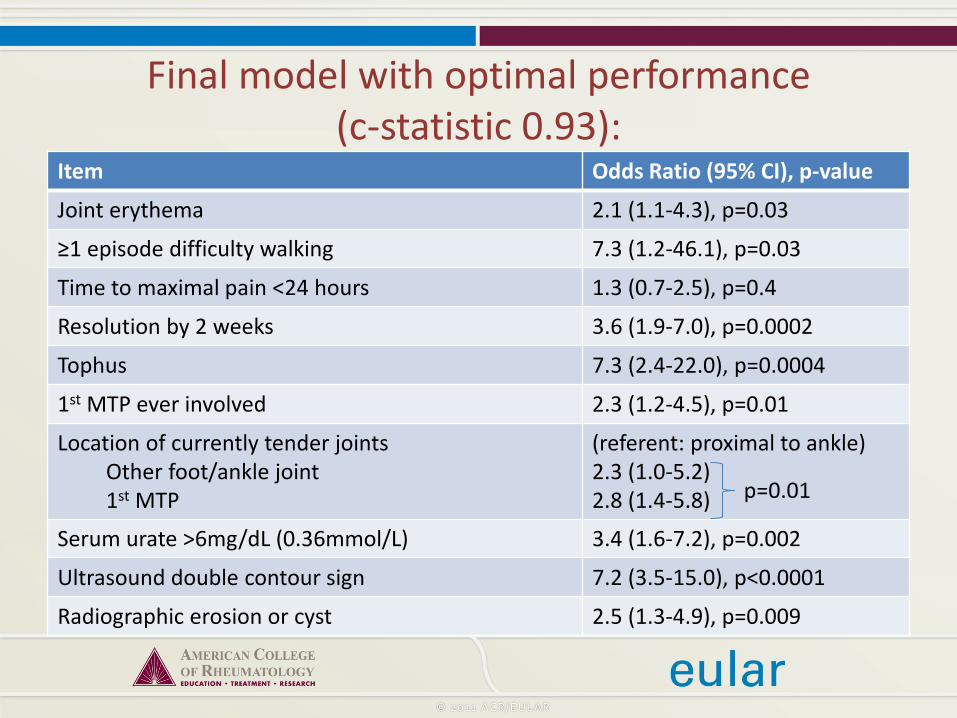
Final model with optimal performance (c-statistic 0.93):
Item Odds Ratio (95% CI), p-value
Joint erythema 2.1 (1.1-4.3), p=0.03
≥1 episode difficulty walking 7.3 (1.2-46.1), p=0.03
Time to maximal pain <24 hours 1.3 (0.7-2.5), p=0.4
Resolution by 2 weeks 3.6 (1.9-7.0), p=0.0002
Tophus 7.3 (2.4-22.0), p=0.0004
1st MTP ever involved 2.3 (1.2-4.5), p=0.01
Location of currently tender joints Other foot/ankle joint 1st MTP
(referent: proximal to ankle) 2.3 (1.0-5.2) 2.8 (1.4-5.8)
Serum urate >6mg/dL (0.36mmol/L) 3.4 (1.6-7.2), p=0.002
Ultrasound double contour sign 7.2 (3.5-15.0), p<0.0001
Radiographic erosion or cyst 2.5 (1.3-4.9), p=0.009
p=0.01

Distribution of Joint Involvement
Gout subjects Non-gout subjects Joint pattern Prevalence (%) Joint pattern Prevalence (%) MTP1 39.4 Knee only 56.8
Knee/ankle 37.1 Any lower extremity joint 30.1
Elbow/wrist/hand 14.9 Wrist/hand + knee 8.0
Polyarticular 8.6 Polyarticular 5.1 Latent class analysis-derived clusters of joint involvement determined from currently tender or swollen joints within each patient group.

Consensus Meeting using Multicriterion Decision Analysis
Methodology
Rationale for Final Phase • SUGAR
– Requirement for MSU determination could lead to potential for selection bias (larger joints, more severe disease, tophaceous disease)
• Imaging systematic literature review – Sufficient data may not be present in existing
literature • How to “value” and “weight” each item of
information?
Complementary Phase • To consider a broader spectrum of clinical
gout through patient paper cases • Use multicriterion decision analysis
methodology (conjoint analysis) to derive weights for final criteria – Similar process to RA and SSc classification criteria

Overview of Approach Used Rank order patient paper cases: based
on probability of gout
SUGAR data
Specify Entry, Sufficient, and
Exclusion Criteria
Identify Domains and Categories
Derive weights: Multicriterion
decision analysis
Final Gout Classification
Criteria
Determine Threshold for
classifying as gout
Imaging Review
Identify pertinent positive and
negative factors
Threshold exercise
Assess face validity of scoring system
and threshold

Entry and Sufficient Criteria • Entry: at least one episode of swelling, pain, or
tenderness in a peripheral joint or bursa – If not fulfilled, do not apply criteria
• Sufficient: Presence of MSU crystals in symptomatic joint or bursa (i.e., in synovial fluid) or tophus – If present, can classify as gout without further
assessment of criteria – Microscopy should be performed by competent
examiner
Exclusion Criteria • There are no exclusion criteria
– Gout can coexist with other conditions

Domains • Clinical (4):
– Pattern of joint/bursa involvement, characteristics of episodes, time-course of episodes, clinical evidence of tophus
• Laboratory (2): – MSU crystals, serum urate
• Imaging (2): – Evidence of urate deposition (ultrasound double
contour sign or DECT), evidence of gout-related joint damage (radiographic gouty erosion)

Clinical Domains

1. Pattern of Joint/Bursa Involvement
• During symptomatic episode(s) ever: – Involvement of 1st MTP joint as part of
monoarticular or oligoarticular episode – Involvement of ankle OR midfoot as part of
monoarticular or oligoarticular episode without involvement of the 1st MTP
– Any other pattern, including 1st MTP/ankle/midfoot as part of polyarticular presentation
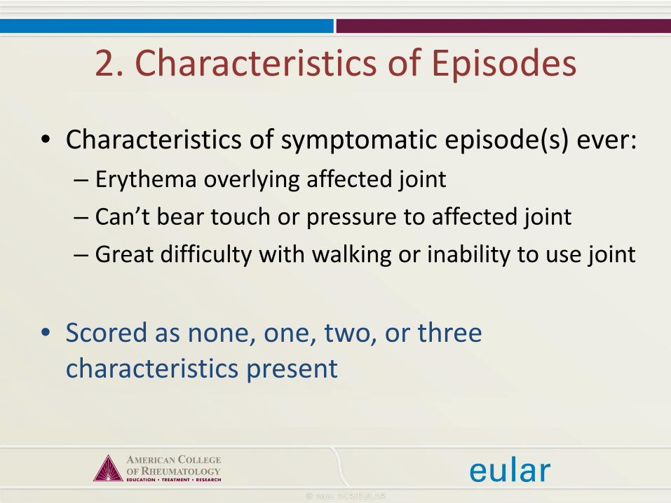
2. Characteristics of Episodes
• Characteristics of symptomatic episode(s) ever: – Erythema overlying affected joint – Can’t bear touch or pressure to affected joint – Great difficulty with walking or inability to use joint
• Scored as none, one, two, or three characteristics present
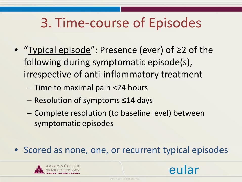
3. Time-course of Episodes
• “Typical episode”: Presence (ever) of ≥2 of the following during symptomatic episode(s), irrespective of anti-inflammatory treatment – Time to maximal pain <24 hours – Resolution of symptoms ≤14 days – Complete resolution (to baseline level) between
symptomatic episodes
• Scored as none, one, or recurrent typical episodes
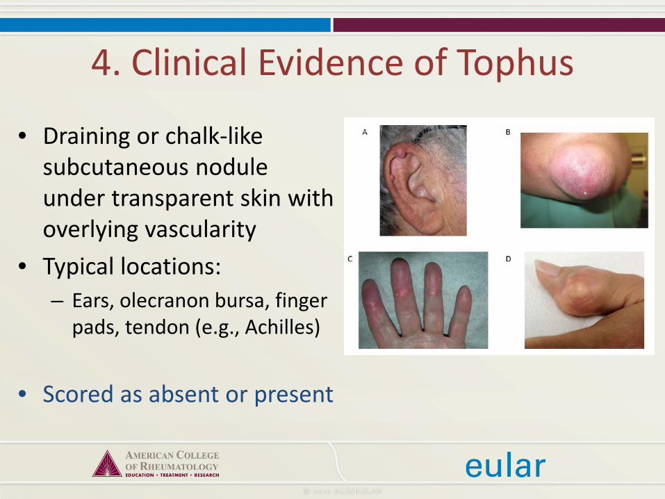
4. Clinical Evidence of Tophus
• Draining or chalk-like subcutaneous nodule under transparent skin with overlying vascularity
• Typical locations: – Ears, olecranon bursa, finger
pads, tendon (e.g., Achilles)
• Scored as absent or present
Laboratory Domains

5. Serum Urate
• Measured by uricase method • Ideally, should be scored at a time when
patient was not taking ULT, and patient was beyond 4 weeks of start of an episode (i.e., during intercritical period)
• If practicable, retest under those conditions • Highest value, irrespective of timing, should be
scored

5. Serum Urate (continued)
• Scored based on SUA value: – <4 mg/dL (<0.24 mmol/L) – 4-<6 mg/dL (0.24-<0.36 mmol/L) – 6-<8 mg/dL (0.36-<0.48 mmol/L) – 8-<10 mg/dL (0.48-<0.60 mmol/L) – ≥10 mg/dL (≥0.60 mmol/L)
6. Synovial Fluid Analysis
• Synovial fluid analysis of a symptomatic (ever) joint or bursa assessed by a trained observer*
• Scored as negative or not done
• *If it was positive, then the subject would have met the sufficient criterion and would not need to be further assessed with the criteria scoring

Imaging Domains
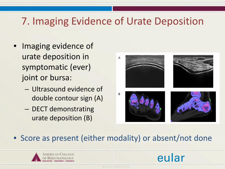
7. Imaging Evidence of Urate Deposition
• Imaging evidence of urate deposition in symptomatic (ever) joint or bursa: – Ultrasound evidence of
double contour sign (A) – DECT demonstrating
urate deposition (B)
• Score as present (either modality) or absent/not done

8. Imaging Evidence of Gout-related Joint Damage
• Conventional radiography of hands and/or feet demonstrate at least one erosion – Erosion: cortical break with
sclerotic margin and overhanging edge
– Excludes: DIP joints, gull wing appearance (to exclude OA-related findings)
• Score as present or absent/not done

Threshold for Classifying Gout
• The total possible score is 23 • The threshold for classifying as gout is ≥8
– Balance of Sensitivity and Specificity – Performance tested in independent dataset
(N=330): • Sensitivity: 0.92 • Specificity: 0.89 • AUC: 0.95
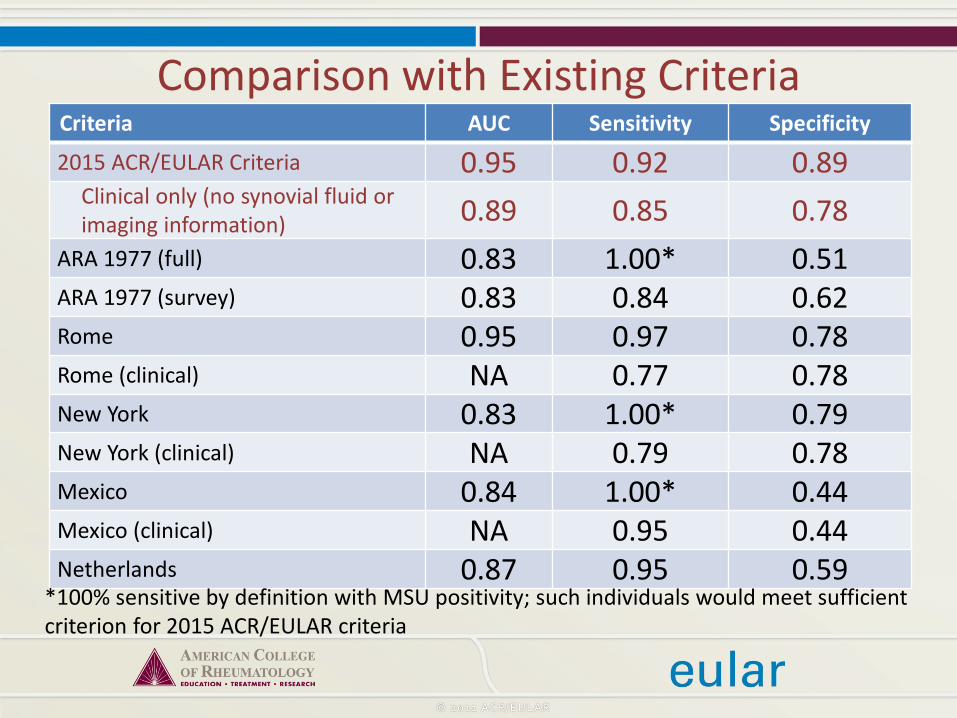
Comparison with Existing Criteria Criteria AUC Sensitivity Specificity
2015 ACR/EULAR Criteria 0.95 0.92 0.89 Clinical only (no synovial fluid or imaging information) 0.89 0.85 0.78
ARA 1977 (full) 0.83 1.00* 0.51 ARA 1977 (survey) 0.83 0.84 0.62 Rome 0.95 0.97 0.78 Rome (clinical) NA 0.77 0.78 New York 0.83 1.00* 0.79 New York (clinical) NA 0.79 0.78 Mexico 0.84 1.00* 0.44 Mexico (clinical) NA 0.95 0.44 Netherlands 0.87 0.95 0.59
*100% sensitive by definition with MSU positivity; such individuals would meet sufficient criterion for 2015 ACR/EULAR criteria

ACR-EULAR 2015 Gout Classification Criteria
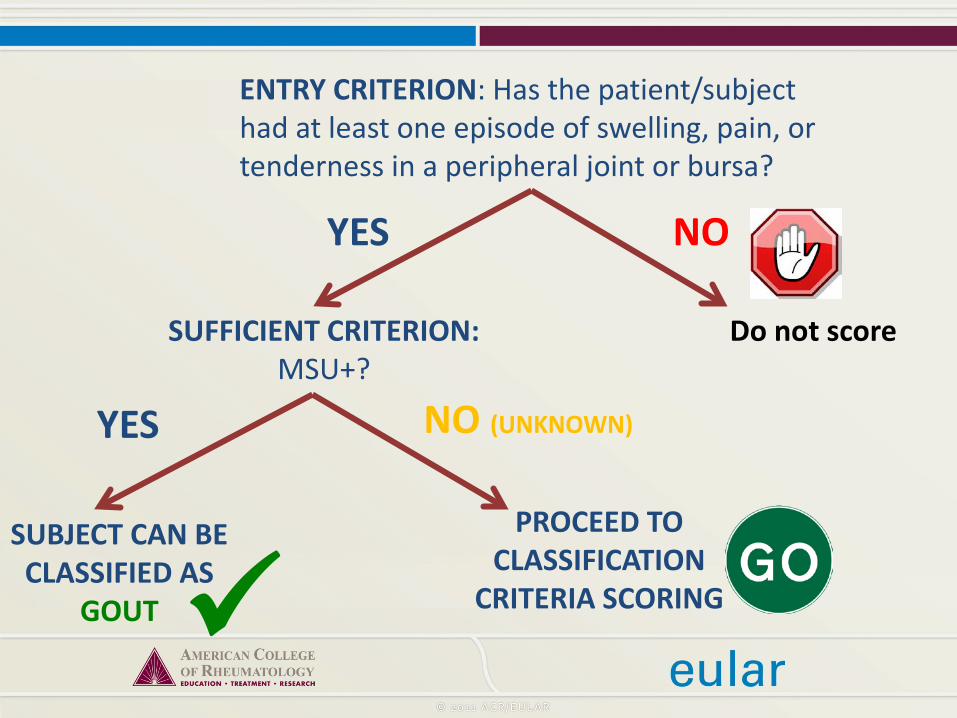
ENTRY CRITERION: Has the patient/subject had at least one episode of swelling, pain, or tenderness in a peripheral joint or bursa?
SUFFICIENT CRITERION: MSU+?
Do not score
YES NO
YES
SUBJECT CAN BE CLASSIFIED AS
GOUT
NO (UNKNOWN)
PROCEED TO CLASSIFICATION
CRITERIA SCORING
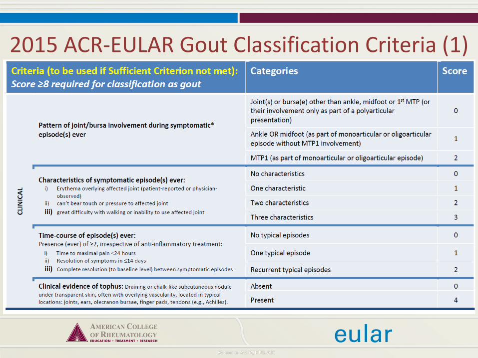
2015 ACR-EULAR Gout Classification Criteria (1)

2015 ACR-EULAR Gout Classification Criteria (2)
Maximum score is 23. Threshold to classify as gout is ≥8.
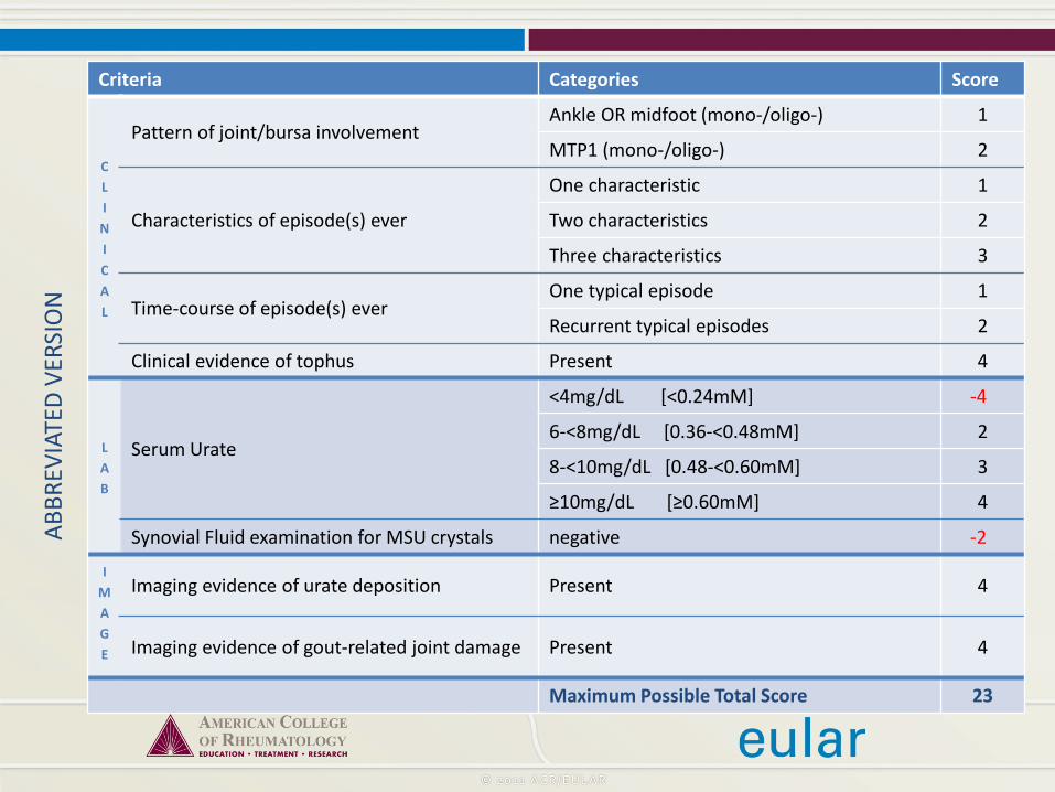
Criteria Categories Score
CLINICAL
Pattern of joint/bursa involvement Ankle OR midfoot (mono-/oligo-) 1
MTP1 (mono-/oligo-) 2
Characteristics of episode(s) ever
One characteristic 1
Two characteristics 2
Three characteristics 3
Time-course of episode(s) ever One typical episode 1
Recurrent typical episodes 2
Clinical evidence of tophus Present 4
LAB
Serum Urate
<4mg/dL [<0.24mM] -4
6-<8mg/dL [0.36-<0.48mM] 2
8-<10mg/dL [0.48-<0.60mM] 3
≥10mg/dL [≥0.60mM] 4
Synovial Fluid examination for MSU crystals negative -2
IMAGE
Imaging evidence of urate deposition Present 4
Imaging evidence of gout-related joint damage Present 4
Maximum Possible Total Score 23
ABBR
EVIA
TED
VERS
ION

A web-based calculator can be accessed at:
http://goutclassificationcalculator.auckland.ac.nz

Illustrative Examples

Case Examples – #1
• 68 y.o. F • MTP1 – ≥1 monoarticular episode
(plus other patterns) • Erythema, can’t bear pressure,
great difficulty • Recurrent ‘typical’ episodes
– Maximal pain within 12 hrs – Resolution within 7 days – Complete resolution between
episodes • Clinical tophus (pinnae of ears) • SUA 0.71mM (~11.8mg/dL) • No SF/tophus aspiration • U/S: +double contour sign • No x-ray performed
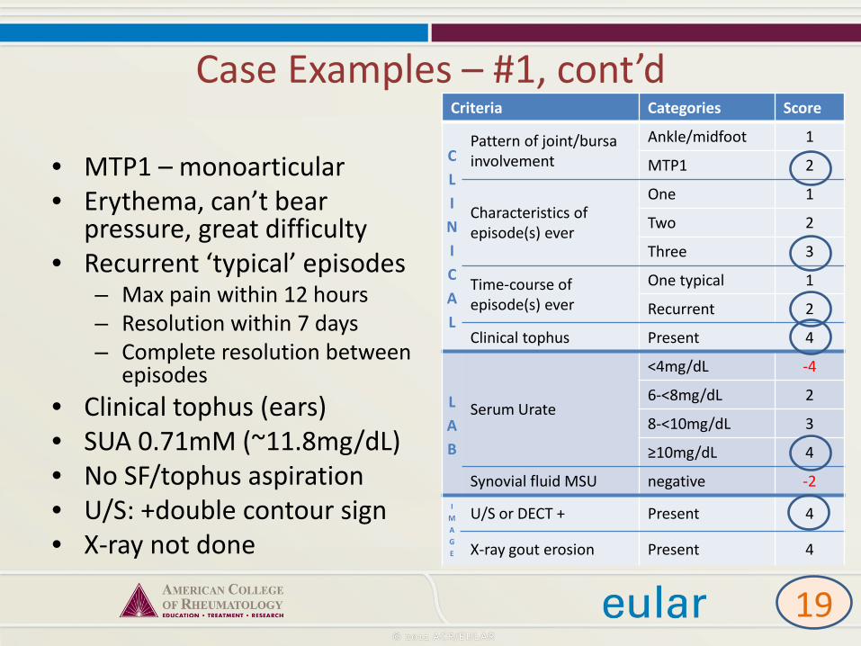
Case Examples – #1, cont’d
• MTP1 – monoarticular • Erythema, can’t bear
pressure, great difficulty • Recurrent ‘typical’ episodes
– Max pain within 12 hours – Resolution within 7 days – Complete resolution between
episodes • Clinical tophus (ears) • SUA 0.71mM (~11.8mg/dL) • No SF/tophus aspiration • U/S: +double contour sign • X-ray not done
Criteria Categories Score
CLINICAL
Pattern of joint/bursa involvement
Ankle/midfoot 1
MTP1 2
Characteristics of episode(s) ever
One 1
Two 2
Three 3
Time-course of episode(s) ever
One typical 1
Recurrent 2
Clinical tophus Present 4
LAB
Serum Urate
<4mg/dL -4
6-<8mg/dL 2
8-<10mg/dL 3
≥10mg/dL 4
Synovial fluid MSU negative -2 I
MAGE
U/S or DECT + Present 4
X-ray gout erosion Present 4
19

Case Examples – #2 • 25 y.o. M, multiple episodes • MTP1 – monoarticular • Characteristics: can’t bear
touch, great difficulty walking
• Time-course: maximal pain within 12 hrs; resolves by 14 days; never complete resolution to baseline
• No tophus • SUA: 0.49 mM (~8.2 mg/dL) • MSU: negative • U/S: + DCS • X-ray: negative
11
Criteria Categories Score
CLINICAL
Pattern of joint/bursa involvement
Ankle/midfoot 1
MTP1 2
Characteristics of episode(s) ever
One 1
Two 2
Three 3
Time-course of episode(s) ever
One typical 1
Recurrent 2
Clinical tophus Present 4
LAB
Serum Urate
<4mg/dL -4
6-<8mg/dL 2
8-<10mg/dL 3
≥10mg/dL 4
Synovial fluid MSU negative -2 I
MAGE
U/S or DECT + Present 4
X-ray gout erosion Present 4

Case Examples – #3 • 46 y.o. M, multiple episodes • Ankle/midfoot – mono (no
MTP1 monoarticular episodes) • Characteristics: erythema,
can’t bear touch, great difficulty walking
• Time-course: maximal pain within 12 hrs; no resolution by 14 days; complete resolution to baseline between episodes
• No tophus • SUA: 0.43mM (~7.2 mg/dL) • MSU: not done • U/S, DECT: not done • X-ray: negative
8
Criteria Categories Score
CLINICAL
Pattern of joint/bursa involvement
Ankle/midfoot 1
MTP1 2
Characteristics of episode(s) ever
One 1
Two 2
Three 3
Time-course of episode(s) ever
One typical 1
Recurrent 2
Clinical tophus Present 4
LAB
Serum Urate
<4mg/dL -4
6-<8mg/dL 2
8-<10mg/dL 3
≥10mg/dL 4
Synovial fluid MSU negative -2 I
MAGE
U/S or DECT + Present 4
X-ray gout erosion Present 4

Case Examples – #4 • 47 y.o. M, multiple episodes • Oligoarticular MTPs (other) • Characteristics: can’t bear
touch, great difficulty walking, erythema
• Time-course: maximal pain within 12 hrs; no resolution by 14 days; never complete resolution to baseline
• No tophus • SUA: 0.43 mM (~7.2 mg/dL) • MSU: negative • U/S: - DCS, DECT not done • X-ray: +erosion
Criteria Categories Score
CLINICAL
Pattern of joint/bursa involvement
Ankle/midfoot 1
MTP1 2
Characteristics of episode(s) ever
One 1
Two 2
Three 3
Time-course of episode(s) ever
One typical 1
Recurrent 2
Clinical tophus Present 4
LAB
Serum Urate
<4mg/dL -4
6-<8mg/dL 2
8-<10mg/dL 3
≥10mg/dL 4
Synovial fluid MSU negative -2 I
MAGE
U/S or DECT + Present 4
X-ray gout erosion Present 4
7
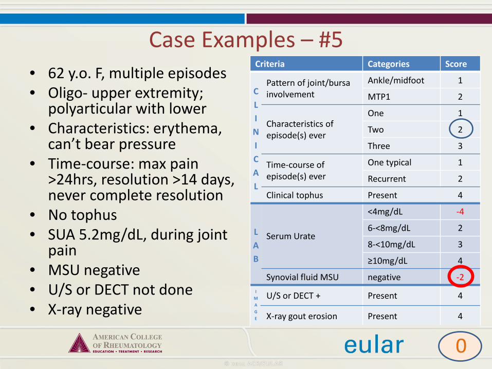
Case Examples – #5 • 62 y.o. F, multiple episodes • Oligo- upper extremity;
polyarticular with lower • Characteristics: erythema,
can’t bear pressure • Time-course: max pain
>24hrs, resolution >14 days, never complete resolution
• No tophus • SUA 5.2mg/dL, during joint
pain • MSU negative • U/S or DECT not done • X-ray negative
Criteria Categories Score
CLINICAL
Pattern of joint/bursa involvement
Ankle/midfoot 1
MTP1 2
Characteristics of episode(s) ever
One 1
Two 2
Three 3
Time-course of episode(s) ever
One typical 1
Recurrent 2
Clinical tophus Present 4
LAB
Serum Urate
<4mg/dL -4
6-<8mg/dL 2
8-<10mg/dL 3
≥10mg/dL 4
Synovial fluid MSU negative -2 I
MAGE
U/S or DECT + Present 4
X-ray gout erosion Present 4
0
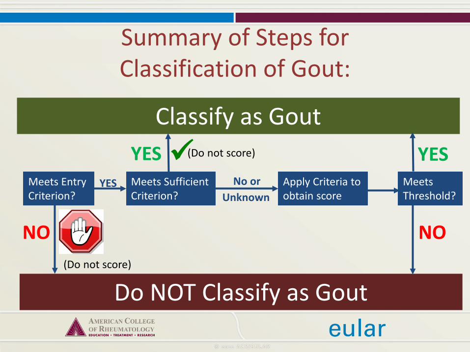
Summary of Steps for Classification of Gout:
Meets Entry Criterion?
Classify as Gout
Do NOT Classify as Gout
NO (Do not score)
Meets Sufficient Criterion?
YES (Do not score)
YES Apply Criteria to obtain score
No or Unknown
Meets Threshold?
YES
NO

Summary
• New classification criteria for gout have been developed and validated through an international collaborative effort
Acknowledgements (1) Steering committee: Nicola Dalbeth, Jaap Fransen, Tim L. Jansen (EULAR PI), Tuhina Neogi (ACR PI), H. Ralph Schumacher, William Taylor
Fellows: Dianne Berendsen (EULAR), Alexis Ogdie (ACR)
SUGAR investigators: Melanie Brown, Worawit Louthrenoo, Janitzia Vazquez-Mellado, Maxim Eliseev, Geraldine McCarthy, Lisa K. Stamp, Fernando Perez-Ruiz, Francisca Sivera, Hang-Korng Ea , Martijn Gerritsen, Carlo Scire, Lorenzo Cavagna, Chingtsai Lin, Yin-Yi Chou, Anne-Kathrin Tausche, Ana Beatriz Vargas dos Santos, Matthijs Janssen, Jiunn-Horng Chen, Ole Slot, Marco Cimmino, Till Uhlig
We gratefully acknowledge the help of Eduardo Aranda-Arreola, Dianne Berendsen, Giovanni Cagnotto, Su-Ting Chang, Jiunn-Horng Chen, Yi-Hsing Chen, Yin-Yi Chou, Viktoria Fana, Angelo Gaffo, Chien-Chung Huang, Po-Hao Huang, Kanon Jatuworapruk, Fatima Kudaeva, Femke Lamers-Karnebeek, Joung-Liang Lan, Juris Lazovskis, Panomkorn Lhakum, Hui-Ju Lin, Anne Madigan, Olivier Peyr, Geraldo da Rocha Castelar-Pinheiro, Alain Sanchez-Rodríguez, and Douglas White with data collection, crystal examination or patient referral for SUGAR. We are also grateful to Eliseo Pascual (Alicante, Spain) for help with MSU observer certification. Delphi study: Rebecca Prowse was supported by a Summer Student Scholarship from Arthritis New Zealand. Imaging systematic review: Alexis Ogdie performed the study, Mark Weatherall helped with the analysis, Janet Joyce for performing the literature search and Yihui Connie Jiang for administrative support.

Acknowledgements (2) Consensus meeting panel: Hyon Choi, Nicola Dalbeth, N. Lawrence Edwards, Jaap Fransen, Tim Jansen, Heins Janssens, Frederic Liote, Tuhina Neogi, George Nuki, Fernando Perez-Ruiz, Kenneth Saag, H. Ralph Schumacher, Jasvinder Singh, John Sundy, Anne-Kathrin Tausche, William Taylor, Janitzia Vazquez-Mellado, Steven Yarrows; Fellow members: Dianne Berendson, Alexis Ogdie; Facilitators: Melanie Brown, Ray Naden We would like to thank Thomas Bardin for participating in ranking of the paper cases. We would like to acknowledge and thank Esperanza Naredo for her advice regarding standardization of the ultrasound definition of double-contour sign.
We are grateful to the following investigators for contributing additional paper patient cases: Everardo Alvarez H, Ruben Burgos, Geraldo Castelar, Marco Cimmino, Tony Dowell, Angelo Gaffo, Rebecca Grainger, Leslie Harrold, Phillip Helliwell, Changtsai Lin, Worawit Louthrenoo, Claudia Schainberg, Naomi Schlesinger, Carlos Scire, Ole Slot, Lisa Stamp, Robert Terkeltaub, Harald Vonkeman, Zeng Xuejun We would like to acknowledge and thank Ian Sayer (Application Specialist, Information Services, Faculty of Medical and Health Sciences, University of Auckland) for his work on developing the gout classification calculator webpage. Funding sources: • ACR & EULAR • Arthritis New Zealand, Association
Rhumatisme et Travail, Asociación de Reumatólogos del Hospital de Cruces.