
20060503053 - apps.dtic.mil · (CBIs) because the co-activator proteins can no longer bind; thus,...
53
AD Award Number: W81XWH-04-1-0784 TITLE: Computational Characterization and Predication of Estrogen Receptor Coactivator Binding Site Inhibitors PRINCIPAL INVESTIGATOR: Brian J. Bennion, Ph.D. Kris Kulp Monique Cosman Felice Lightstone CONTRACTING ORGANIZATION: The University of California Livermore, CA 94551 REPORT DATE: September 2005 TYPE OF REPORT: Final PREPARED FOR: U.S. Army Medical Research and Materiel Command Fort Detrick, Maryland 21702-5012 DISTRIBUTION STATEMENT: Approved for Public Release; Distribution Unlimited The views, opinions and/or findings contained in this report are those of the author(s) and should not be construed as an official Department of the Army position, policy or decision unless so designated by other documentation. 20060503053
-
Upload
trinhtuong -
Category
Documents
-
view
215 -
download
0
Transcript of 20060503053 - apps.dtic.mil · (CBIs) because the co-activator proteins can no longer bind; thus,...

AD
Award Number: W81XWH-04-1-0784
TITLE: Computational Characterization and Predication of Estrogen ReceptorCoactivator Binding Site Inhibitors
PRINCIPAL INVESTIGATOR: Brian J. Bennion, Ph.D.Kris KulpMonique CosmanFelice Lightstone
CONTRACTING ORGANIZATION: The University of CaliforniaLivermore, CA 94551
REPORT DATE: September 2005
TYPE OF REPORT: Final
PREPARED FOR: U.S. Army Medical Research and Materiel CommandFort Detrick, Maryland 21702-5012
DISTRIBUTION STATEMENT: Approved for Public Release;Distribution Unlimited
The views, opinions and/or findings contained in this report are those of the author(s) andshould not be construed as an official Department of the Army position, policy or decisionunless so designated by other documentation.
20060503053

Form ApprovedREPORT DOCUMENTATION PAGE OMB No. 0704-0188
Public reporting burden for this collection of information is estimated to average 1 hour per response, including the time for reviewing instructions, searching existing data sources, gathering and maintaining thedata needed, and completing and reviewing this collection of information. Send comments regarding this burden estimate or any other aspect of this collection of information, including suggestions for reducingthis burden to Department of Defense, Washington Headquarters Services, Directorate for Information Operations and Reports (0704-0188), 1215 Jefferson Davis Highway, Suite 1204, Arlington, VA 22202-4302. Respondents should be aware that notwithstanding any other provision of law, no person shall be subject to any penalty for failing to comply with a collection of information if it does not display a currentlyvalid OMB control number. PLEASE DO NOT RETURN YOUR FORM TO THE ABOVE ADDRESS.
1. REPORT DATE (DD-MM-YYYY) 2. REPORT TYPE 3. DATES COVERED (From - To)
01-09-2005 Final 2 Aug 2004 - 1 Aug 20054. TITLE AND SUBTITLE 5a. CONTRACT NUMBER
Computational Characterization and Predication of Estrogen ReceptorCoactivator Binding Site Inhibitors 5b. GRANT NUMBER
W81XWH-04-1-07845c. PROGRAM ELEMENT NUMBER
6. AUTHOR(S) 5d. PROJECT NUMBER
Brian J. Bennion, Ph.D.
Kris Kulp 5e. TASK NUMBER
Monique CosmanFelice Lightstone 5f. WORK UNIT NUMBERE-mail: bennionl~cllnl.qov7. PERFORMING ORGANIZATION NAME(S) AND ADDRESS(ES) 8. PERFORMING ORGANIZATION REPORT
NUMBER
The University of CaliforniaLivermore, CA 94551
9. SPONSORING / MONITORING AGENCY NAME(S) AND ADDRESS(ES) 10. SPONSOR/MONITOR'S ACRONYM(S)
U.S. Army Medical Research and Materiel CommandFort Detrick, Maryland 21702-5012
11. SPONSOR/MONITOR'S REPORTNUMBER(S)
12. DISTRIBUTION / AVAILABILITY STATEMENTApproved for Public Release; Distribution Unlimited
13. SUPPLEMENTARY NOTES
14. ABSTRACT Many carcinogens have been shown to cause tissue specific tumors in animal models. The mechanism for thisspecificity has not been fully elucidated and is usually attributed to differences in organ metabolism. For heterocyclic amines,potent carcinogens that are formed in well-done meat, the ability to either bind to the estrogen receptor and activate or inhibitan estrogenic response will have a major impact on carcinogenicity. Here we describe our work with the human estrogenreceptor alpha (hERa) and the mutagenic/carcinogenic heterocyclic amines PhIP, MelQx, IFP, and the hydroxylatedmetabolite of PhIP, N2-hydroxy-PhIP. We found that PhIP, in contrast to the other heterocyclic amines, increased cell-proliferation in MCF-7 human breast cancer cells and activated the hERa receptor. We show mechanistic data supporting thisactivation both computationally by homology modeling and docking, and by NMR confirmation that PhIP binds with the ligandbinding domain (LBD). This binding competes with estradiol (E2) in the native E2 binding cavity of the receptor. We also findthat other heterocyclic amines and N2-hydroxy-PhIP inhibit ER activation presumably by binding into another cavity on theLBD. Moreover, molecular dynamics simulations of inhibitory heterocyclic amines reveal a disruption of the surface of thereceptor protein involved with co-activator proteins thereby inhibiting protein-protein signaling. We therefore propose that themechanism for the tissue specific carcinogenicity seen in the rat breast tumors and the presumptive human breast cancerassociated with the consumption of well-done meat maybe mediated by this receptor activation.15. SUBJECT TERMSEstrogen receptor, ligand, xenoestrogens, co-activator binding inhibitor, breast cancer, carcinogen
16. SECURITY CLASSIFICATION OF: 17. LIMITATION 18. NUMBER 19a. NAME OF RESPONSIBLE PERSON
OF ABSTRACT OF PAGES USAMRMC
a. REPORT b. ABSTRACT c. THIS PAGE 19b. TELEPHONE NUMBER (include area
U U U UU 53 code)
Standard Form 298 (Rev. 8-98)Prescribed by ANSI Std. Z39.18

"Table of Contents
C over ................................................................................................. 1
SF 298 ............................................................................................ 2
Table of Contents ............................................................................. 3
Introduction ..................................................................................... 4
Body ................................................................................................ 4
Key Research Accomplishments ............................................................ 15
Reportable Outcomes ....................................................................... 15
Conclusions .................................................................................... 15
References ...................................................................................... 17
Appendices ...................................................................................... 19
IntroductionIn an effort to treat breast cancer, selective estrogen receptor modulators (SERMs)have been used to modulate the estrogen-signaling pathway with mixed results [1]. Aclassic example of a SERM is tamoxifen. When used as a therapeutic for a limited time,tamoxifen is effective in disrupting the estrogen-signaling pathway. Unfortunately, withprolonged use, breast tumor cells become resistant to tamoxifen and are able to use thebioactivated metabolite of tamoxifen to interact with co-activators that activate theestrogen-signaling pathway, reversing its original role [1]. An alternative therapeuticapproach is to target the binding site of the co-activator protein. Recent studies haveshown that some small molecules may bind in sites (e.g. co-activator site) other thanthe estradiol binding site [2] and still disrupt the estrogen-signaling pathway. By bindingin the co-activator site while estradiol is bound in the estrogen receptor (ER) ligandbinding domain (LBD), these small molecules act as co-activator binding inhibitors(CBIs) because the co-activator proteins can no longer bind; thus, gene transcription isinhibited. Potentially, these CBIs can act as a new therapeutics against environmentalor natural agonists of ERa. Quantitative structure-activity relationship (QSAR) studieshave been used to develop therapeutics that will compete and bind in the estradiolbinding site of the ERa LBD [3-5]. Because these studies have focused on the estradiolbinding site, new potential ER disruptors that bind in the co-activator site have beenmissed. Our proposal focuses on developing a new computational approach to predicttherapeutically useful ERa disruptors by investigating CBIs binding to the co-activatorsite in conjunction with estrogenic compounds bound in the estradiol site
BodyTask 1: Computationally predict the relative binding of CBIs in the co-activator site andthe CBI binding properties.
Three estrogenic compounds were chosen as positive controls, estradiol (E2), 1-methyl-2-amino-6-phenylimadazo[4,5b]pyridine (PhIP), and diethylstilbestrol (DES).These three compounds have been shown to be capable of activating the estrogenreceptor and producing an estrogen specific response (see Appendix A and more [6]).Other compounds that have been shown to inhibit the function of ERa (Appendix A)were chosen as possible CBI's. These compounds are aromatic heterocyclic aminesand known mutagens [7, 8].
The entire ERa-LDB was used in the docking calculations by including it inall the atom interaction grids. As shown in appendix A and figure 1, E2 dockingrecovered the crystallographic binding mode with a probability of binding in the nativebinding cavity of -99%. DES also showed a very high probability for binding in the E2site (94%). Striking, however, was the discovery of a second binding site in the interiorof the protein (Task 1.1). This site, which has only been discussed twice before in theliterature, was found because the whole protein was included in the dockingcalculations. The paper that described the original crystal structure defined ths region asa solvent channel [9]. A second mention of this cavity in the literature was an attempt toexplain unusual kinetics experiments [10]. At this writing, no other evidence for smallmolecule binding in the solvent cavity has been published. E2 and DES showed lessthan 1% probability of binding in this solvent cavity.
4

In our ranking, we found that compounds, which inhibited the activation of theER, showed a preference in binding to the alternative site (Figure 2 and activiationfigure) (Task 1.2).
Boltzman Binding Distributions
0.9-7
.6- EST~JPhiPO5 2NOH-PhIP
2N3M-PhIP!*3M5-PhIP
0.'2- -
01--
E 2 B ack
Figure 1: The Boltzman binding distribution of E2 and other heterocyclic amines on the ERa-LDB at thenative binding cavity and the back cavity. Details of the calculations and compounds are described inappendix A.
The end of the alternative binding site adjoins residues important for binding theco-activator protein (Figure 3). We hypothesized that compounds bound in thisalternate cavity could affect the dynamics of residues on the surface of the protein.Molecular dynamics simulations of highest ranking CBI (3M5-PhIP) showed thatsignificant distortion of the protein surface was possible when the alternate cavitycontains small molecules (Task 1.3). In our simulations of 3M5-PhIP bound to ERa-LDB we noticed that Met 357 was very dynamic; the side chain would move significantlyrelative to surrounding residues during the 2ns simulation. This side chain motionaffected the surface of the protein, especially in the region required for co-activatorbinding (Figure 4). Two separate simulations had similar results. Control simulationsof E2 and the Tif-2 peptide from the co-activator protein are in progress. Comparison ofthe control simulations against simulations of the CBI bound in the alternate cavity willallow for better quantification of the surface distortions.
5

EstradiolS•:;:::!, > Binding 4e'• '•
Cavity(E2)
/0
"• ' ~~Helix 1 2ý.....
!L • ` Binding,• 3•M5 •
•,)• : •1} Site P l
Figure 2: ERa-LDB shown with the backbone in ribbons and binding cavities in surface representationcolored by residue type (red, negative charge; blue, positive charge; green, polar; white non-polar). Theprotein is rotated 1800 to show the opposite positions of the cavities on the surface. E2 is shown in stickrepresentation inside the crystallographic binding site, while the PhIP congener 3-methyl-5phenyl-PhIP isshown in its predicted "alternate binding cavity". The blue surface on the protein at the left defines theregion of residues critical for binding the co-activator protein.
6
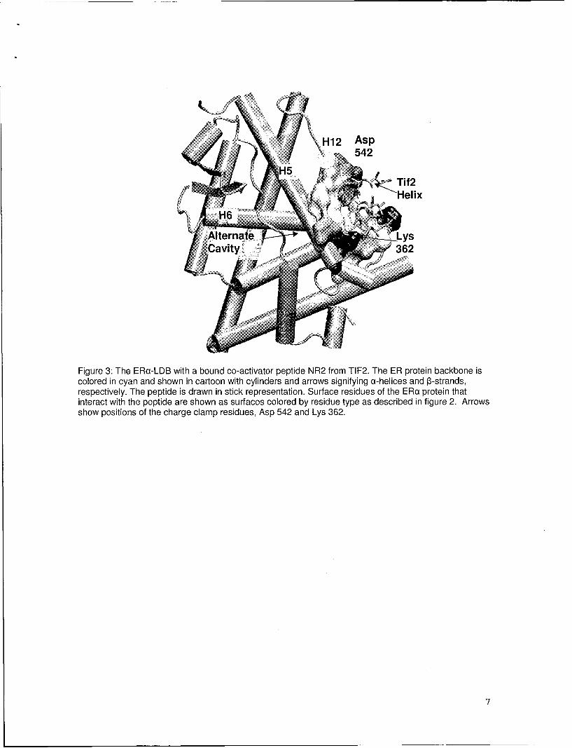
<H12 AspS542
H5 Tif2
Alternat e; LysSCavity~i 362
Figure 3: The ERa-LDB with a bound co-activator peptide NR2 from TIF2. The ER protein backbone iscolored in cyan and shown in cartoon with cylinders and arrows signifying a-helices and 13-strands,respectively. The peptide is drawn in stick representation. Surface residues of the ERa protein thatinteract with the peptide are shown as surfaces colored by residue type as described in figure 2. Arrowsshow positions of the charge clamp residues, Asp 542 and Lys 362.
7

Figure 4: The ERc-LDB before molecular dynamics and after 2ns of simulation time. The proteinbackbone is shown in ribbon representation and colored in cyan. Methionine 357 is shown in stickrepresentation. Surface residues that interact with the helical peptide of the co-activator protein are inblue. Residues that become accessible to solvent after simulation are shown in magenta. The differencein exposed surface area of the protein is about 200 A2. Before the simulation is started the sidechain ofMethionine 357 points in towards the alternate cavity were 3M5-PhlP is present. Early in the simulation,the sidechain switched positions to point out towards the solvent accessible surface. This disrupts atleast one small hydrophobic pocket that is required for optimal co-activator binding.
The resulting combination of CBIs and estrogenic compounds are currently beingre-ranked based on MD simulations. We have also received a 40,000 compound libraryto screen using our methods developed in this project and this work is continuing. (Task1.4)
Task 2: Biologically assay the effect of the CBI on the estrogen receptor activation.
The ability of potentially estrogenic compounds to activate the estrogen receptorwas assessed using a standard luciferase assay. This assay, a discussion of the cellsused for this experiment, and results of the effects of several heterocyclic amines on theER activation are described in Appendix A (Tasks 2.1 and 2.2).
In addition, using this assay, we were able to assess the effects of smallmolecule structure on ER activation. Using a series of heterocyclic amine compoundsthat differed only slightly in structure, we were able to show that PhIP was optimallyconfigured to activate the ER (figure 5).
8

130 -----
120, T
110
100 -jgo-
0LU'9 7oL
60
50-
40)
VO,1
10,0
Control Estradiol Genistein PhIP N-OH-PhIP 3-Methyl 3-Methyl-5- IQ MelQx IFP TMIPPhIP Phenyl
PhIP
Figure 5: Effect of heterocyclic amines on ER activation. Concentrations of all HAs tested was 4x10 7 M.E2 concentrations was 10-9 M. A discussion of the relevance of including genistein in this experiment isprovided in Appendix A.
Competition assays were used to determine the effect of simultaneous incubationof HAs (4x1 0-7M) and 10.9 M E2 on ERcx activation (Fig. 6). Treating MCF-7 cells withPhIP and E2 produces a response that is 7% less than E2 alone. N2-Hydroxy-PhIPinhibits the E2-mediated activation of ERc by 30%, and the other HAs, with theexception of TMIP, reduce E2-initiated Er activation to the levels of the control cells.Based on the docking data, we find that the compounds that are most strongly predictedto bind to the alternative binding cavity on the ERa protein, are the best inhibitors (Task2.3)
9

1 2 0 ._ _ _ ......... .... .......... ........
100
I80
6o - -
4 Tn-20 _ _
00
Figure 6. Luciferase Activation Based Competition assays, HAs and E2. Concentrations of all HAs testedwere 4xl 0 7M. E2 concentrations were 10-9 M. Total activation relative to E2 on the y-axis vs HAs on thex-axis.
We wanted to know how some HAs were inhibiting ERa activation while PhIP didnot. A simple competition assay was used in which E2 plus an HA were addedsimultaneously to the incubated cells. The resulting data are shown in Figure 6.Estrogen overcame the inhibitory effect of 2NOH-PhIP and TMP while in enhancing theactivating properties of PhIP. Other HAs were close to the control activity. These datalead us to believe that an alternative binding site is present at the level of the ERa-LDB.It is possible that other steps in the ER signaling pathway are the actual targets of theinhibitory HAs, NMR experiments are planned to confirm or deny binding to the ERaprotein.
10

Task 3: Spectroscopically measure the binding of the CBI to the ER LBD
Full length, recombinant human estrogen receptor a (hERa) is commerciallyavailable through Sigma Aldrich, Inc. It is supplied as 1.5 pM purified protein (750pmoles in 500 pI of 50 mM tris-HCI, pH 8.0, 500 mM KCI, 2 mM EDTA, 1 mM sodiumorthovanadate and 10% glycerol). Recombinant hERq is produced as an active, soluble66.4 kDa protein by a baculovirus expression system, and thus possesses post-translational modification. For NMR binding studies, it was first necessary to determinealternative buffer conditions in which the ER protein remained stable. Both tris andglycerol contribute large proton signals in the NMR data that interfere with the detectionof submicromolar quantities of sample. In addition, high salt concentrations can result inprotein precipitation when small volumes of compounds, such as PhIP and E2, whichare only soluble in organic solvents such as DMSO, are added. After numerous trials,we had determined that the protein was able to remain stable for a few days after beingexchanged after extensive dialysis (Spectrapor 7, MWCO 1000 dialysis tubing) into10mM Na2HPO 4, pH 8.0, 120mM NaCI, 2.7mM KCI (PBS) buffer (Task 3.1).
SVarious NMR spectroscopic techniques can be used to screen for interactionsbetween proteins and ligands (reviewed in [11, 12]. Transferred NOESY (trNOESY)experiments are routinely used to detect ligand binding to a target protein underconditions of fast exchange (ligands that bind with mM to mM dissociation constants)[13]. The intensity of each intra-ligand NOE crosspeak is governed by the population-weighted cross-relaxation rate. A strong negative NOE crosspeak is observed forbinders (black peaks), as opposed to weakly positive (red peaks) or zero NOEcrosspeaks for non-binders or in the absence of protein. Thus, the sign flip of the NOEcross peak between the free versus bound states acts as a simple binary filter todistinguish binders from non-binders.
11

a b
9:0 8.0 7.0 6.0 5.0 4.0 3.0 2.0 1.0 9.0 8.0 7.0 6.0 5.0 4.0 3•0 2.0 1.0ppm ppm
77
7 7
8. 4 8.ý2 8.0 7.8 7.6 7A ' 8'4 8.2 8'.0 7.8 7.86 7A4 7.2 8.4 8.2 8.0 7.8 7.6 7.4 7.2DI (ppm) 91 PI ppmI D r pp )
Figure 7: (a) ID H spectrum of 5.95 mM PhIP dissolved in deuterated DMSO. (b) ID-'H spectrum of 132mM estradiol dissolved in deuterated DMSO.(c) The expanded region of a 900 ms mixing time NOESYspectrum of 5.95 mM PhlP in DMSO exhibits weak positive NOE crosspeaks (boxed in red). (d) Theexpanded region of a 300 ms mixing time NOESY spectrum of 3 pM ER and 120 pM PhIP indicates thatPhlP binds as evidenced by the strong, negative crosspeaks (boxed in black).Concentration of PhlP is50X greater in (c) than in (d). Spectrum in (d) is plotted at 2 X lower intensity level relative to spectrum in(c) for presentation purposes. (e) The expanded 300 ms mixing time NOESY spectrum after addition of1.5 pM estradiol to the ERa+PhlP sample shows that PhIP does not bind (red peaks). Spectrum (e) isplotted at 2X lower intensity level relative to spectrum in (d). This result suggests that PhIP has beendisplaced from binding to ERa by estradiol, and that both ligands may be binding in the same site. Notethat the chemical shifts of PhIP change depending on solvent (DMSO in spectrum (c) versus aqueousbuffer in spectra (d) and (e)).
We observed a sign flip in NOE crosspeaks of PhIP from positive (red) tonegative (black) in the trNOESY spectra when PhIP was added to ERa, confirmingexperimentally that PhIP does indeed bind to ER (Figure 7c and d) (Task 3.2). SinceE2 is known to bind with high affinity to ERa (Kd 10-8-10-9 M), we could not use thetrNOESY method to detect its binding to ERa. However, since the E2's binding site onERa is known, it is possible to use this information to design a trNOESY competitionexperiment that can provide information about where PhIP is binding. If PhIP is bindingin a different site than estradiol, than we should be able to still observe negative (black)peaks because PhIP and estradiol are binding in two different sites, and estradiol is notdisplacing PhIP binding. In contrast, if PhIP is binding in the same site as estradiol,then adding estradiol to the PhIP/ERa mixture should competitively displace it since
12

estradiol binds much more strongly to ERa. In this case, we would observe anothersign flip of the PhIP peaks from black (binding) to red (not binding). Addition of estradiolto the PhIP/ERa mixture resulted in PhIP cross peaks flipping sign from black to red(Figure 7d and e) and thus suggesting that both PhIP and estradiol are binding in thesame site on Era (Task 3.3). This result is consistent with the results shown by us andothers that PhIP exposure increases MCF-7 cell proliferation and ERa activation. It alsoagrees with our computational model and with the idea that PhIP directly binding to ERais responsible for the effects observed. However, we can not completely rule out thepossibility that PhIP is binding in another site, and that estradiol prevents PhIP frombinding because of a conformation change in the protein.
Thus in order to further clarify the identity of the ligand binding site, we initiatedthe expression of 15N-labeled ERa ligand binding domain (LBD) (Task 3.4). Dr. MylesBrown (Harvard University) has generously provided us with the expression vector forthe hERa-LBD). The GST-LBD fusion protein exhibits binding affinity to estradiol (KD ~0.1 to 3.3 nM) that is comparable with reported values measured for wild-type MCF-7ERa expressed in vivo [14].
The hERa-LBD is expressed as a GST fusion protein in BL21 (DE3) cells. Cellgrowth is carried out for 16 hours at 37 -C with shaking at 300 rpm (INOVA shaker)using either an autoinduction method, as introduced by Studier [15] or with a modifiedversion of the autoinduction method described specifically for NMR studies [15, 16].Cells are harvested by centrifugation and lysed by sonication on ice in a buffercontaining 50mM Tris-CI, pH 7.5, 100mM NaCI, 20mM P3- mercaptoethanol, 0.5% NP-40, followed by centrifigation at 30,000g at 4-°C for 30min to remove cell debris.
Although the expression yields are very high for the GST-ERa-LBD protein, ascan be seen in Figure 8, the protein is insoluble. Currently, we are in the process ofmodifying the expression protocol (addition of glycerol, tween and other detergents, aswell as lower temperature) to increase the solubility of the protein. Once this isaccomplished, the resulting supernatant will then be diluted and bound to a glutathionesepharose 4B column (Amersham) [17]. After several washes with PBS buffer, thehERa-LBD protein will be cleaved from GST and eluted from the sepharose-GSTcolumn by treatment with thrombin (Amersham). Yields will be determined by theBradford protein assay (BioRad). The purified protein will then be concentrated intoPBS buffer, pH 7.4 by centrifugation using either a Centricon YM-3 or Ultra-4concentrators (Amicon) [16].
13

1 2 3 4 5 1 2 3
Figure 8: Polyacrylamide gel electrophoresis (4-12%) results showing high expression levels of GSTERci-LBD. (a) Most of the protein is in the insoluble fraction (lane 1: GST-ERa-LBD uninduced; lane 2:induced 1 hr. @ 370; lane 3: induced 2hrs. @ 370; lane 4. clear lysate (soluble fraction); lane 5. insolublefraction. (b) ERa expression in autoinducing minimal media, (lane 1: molecular weight markers; lane 2:Grew -~1 6hrs, 370, 300rpm, cell lysate; lane 3: 10 times dilution).
The availability of 15 N-Iabeled ERci-LBD protein will allow us to carry outStructure Activity Relationship by NMVR (SAR-by-NMR) studies [18] that will clearlydefine the binding site and orientation of not only PhIP, but other OBls and HAs.
Task 4: Submit manuscript for publication.
A large portion of the work detailed above in tasks 1-3 has been submitted andaccepted for publication in the American Chemical Society journal, Chemical Researchin Toxicology. At the time of this report, the projected publication date is October 5 'h
2005. We anticipate submission of a second manuscript describing the characterizationof the co-activator binding site interactions with ligands bound in the alternative cavitythrough 15 N NMVR and molecular dynamics simulations.
14

Key Research AccomplishmentsPosters and Accepted Abstracts:
* 19th National Meeting of the Protein Society San Diego CA, August 2004.• K.S. Kulp, B.J. Bennion, J.L. Montgomery, F.L. Lightstone, M.G. Knize, J.S.
Felton and L.M. Bennett "Dietary constituents affect receptor activation and cellproliferation in human breast cancer cells". American Association of CancerResearch Annual Meeting, March 27-31, 2004, Orlando Florida.
* B.J. Bennion, K.S. Kulp, J.L. Montgomery, F.L. Lightstone, M.G. Knize, J.S.Felton and L.M. Bennett "Dietary constituents affect receptor activation and cellproliferation in human breast cancer cells". American Chemical Society AnnualMeeting, March 27-31, 2004, Anaheim California.
* DOD BCRP Meeting "Era of Hope" Philadelphia PA, June 2005.• 3 6 th Meeting of The Environmental Mutagen Society San Francisco CA,
September 2005.
Manuscripts:"• "PhIP Carcinogenicity in Breast Cancer:Computational and Experimental
Evidence for Competitive Interactions with Human Estrogen Receptor " in pressChemical Research in Toxicology, Oct 2005.
Reportable OutcomesPresentations:
"* Biosciences Directorate Symposium LLNL, November 2004."• Biosciences Directorate Postdoctoral Symposium LLNL, July 2005.
Invited Talks-Lectures:• University of California-Davis Cancer Center, Sacremento CA, January 2005.* Chemistry Department University of the Pacific, Stockton CA, February 2005.* Bio-engineering Department University of the Pacific, Stockton CA, March 2005.• Edward Teller Education Center, Lawrence Livermore Natl. Lab., July 2005.
NIH R01 grant entitled "Dietary exposure to multiple heterocyclic amines may causefewer breast tumors than exposure to single carcinogens" to be submitted October 1,2005.
Conclusions
Computer aided docking discovered a food mutagen that could bind in the nativebinding cavity of ERa-LDB and possibly activate the estrogen-signaling pathway. Thecomputational work also predicted a possible secondary binding site for smallmolecules. In deed, we showed that disruption of the surface of the protein waspossible when this alternate binding site is occupied by small heterocyclic amines.Future work in this area would include a computationally based screening of a largerdatabase of commercially available compounds that are similar size and structure to theheterocyclic amines that antagonized ERa gene expression.
15

PhIP was also shown to increase proliferation of MCF-7 breast cancer cells aswell as activate ERa dependent gene expression. Other PhIP related heterocyclicamines decreased breast cancer cell growth and blocked ERa activation by estradiol.These findings corroborate the predictions made in task 1, i.e. PhIP was a weak agonistand that the other heterocyclic amines were strong antagonists to ERa dependent geneexpression.
NMR experiments showed that PhlP could bind to human ERa. E2 was alsoshown to displace bound PhIP from human Era. These two observations substantiatedcomputational predictions in task 1, i.e. P hIP was predicted to bind to the native bindingcavity at a reduced affinity compared to E2. In addition, we also developed expressiontechniques for 15N-labeled human ERa-LDB protein to accurately define the bindinglocation of small molecules. This research has resulted in a submitted manuscript thatwas accepted for publication in Chemical Research in Toxicology in 2005.In conclusion, we have shown that computational docking has predictive value, whichcan open new lines of research; moreover a possible new link between food mutagensand breast cancer was discovered.
16

References:1. Seeger, H. et al., "Effect of Tamoxifen and 2-methoxyestradiol alone and in
Combination on Human Breast Cancer Cell Proliferation." (2003) J. SteroidBiochem. And Mol. Biol. 84, 255-257.
2. Rodriguez, A.L., Tamrazi, A., Collins, M.L., Katzenellenbogen, J.A."Design,Synthesis, and in-Vitro Biological Evalulation of Small Molecule Inhibitors ofEstrogen Receptor alpha Coactivator Binding." (2003) J. Med. Chem. 47, 600-611.
3. Walkers, J.D., Fang, H., Perkins, R., Tong W.D. "QSARs for Endocrine DisruptionPriority Setting Database 2: The integrated 4-Phase Model." (2003) QSAR andCombinatorial Science 22, 89-105.
4. Witorsch, R.J. "Endocrine Disruptors: Can Biological Effects and EnvironmentalRisks Be Predicted." (2003) Regulatory Toxicology and Pharmacology 36, 118-130.
5. van Lipzig, Marola, M.H. et al., (2004) "Prediction of Ligand Binding Affinity andOrientation of Xenoestrogens to the Estrogen Receptor by Molecular DynamicsSimulations and the Linear Interaction Energy Method." J. Med. Chem. 47, 1018-1030.
6. Korach, K.S. and McLachlan, J.A. (1985) "The role of the estrogen receptor indiethylstilbestrol toxicity." Arch. Toxicol Suppl. 8, 33-42.
7. Knize, M.G., and J.S. Felton. 2005. Formation and human risk of carcinogenicheterocyclic amines formed from natural precursors in meat. Nutrition Reviews63(5):158-165.
8. Knize, M.G., R. Sinha, E.D. Brown, C.P. Salmon, O.A. Levander, J.S. Felton, and N.Rothman. (1998) "Heterocyclic Amine Content in Restaurant-CookedHamburgers, Steaks, Ribs, and Chicken." J. Ag. Food Chem. 46:4648-4651.
9. Brzozowski, A.M., A.C.W. Pike, Z. Dauter, R.E. Hubbard, T. Bonn, 0. Engstrom, L.Ohman, G.L. Greene, J.A. Gustafsson, andM. Carlquist. 1997. (1997) "Molecularbasis of agonism and antagonism in the oestrogen receptor." Nature 389, 753-758.
10. van Hoorn W.P. (2002) "Identification of a second binding site in the estrogenreceptor." J. Med. Chem. 45, 584-589.
11. Coles, M., Heller, M., and Kessler, H. (2003) "NMR-based screening technologies."DRUG DISCOVERY TODAY8, 803-810.
12. Meyer, B., and Peters, T. (2003) "NMR Spectroscopy techniques for screening andidentifying ligand binding to protein receptors." ANGEWANDTE CHEMIE-INTERNATIONAL EDITION 42, 864-890.
13. Clore, G.M., and A.M. Gronenborn. (1982). "Theory and application of thetransferred nuclear Overhauser effect to the study of the conformation of smallmolecules bound to proteins." J. Magn. Reson. 48, 402-417.
14. Halachmi, S., E. Marden, G. Martin, H. MacKay, C. Abbondanza, and M. Brown.(1994) "Estrogen receptor-associated proteins: possible mediators of hormone-induced transcription." Science 264, 1455-1458.
15. Studier, F.W. (2005) "Protein production by auto-induction in high-density shakingcultures." Protein Expr. Purif. 41, 207-234.
17

16. Tyler, R.C., H.K. Sreenath, S. Singh, D.J. Aceti, C.A. Bingman, J.L. Markley, andB.G. Fox. (2005) "Auto-induction medium for the production of [U-1i5N]- and [U-13C, U-1 5N]-labeled proteins for NMR screening and structure determination."Protein Expr. Purif. 40, 268-278.
17. Fabbro, D., D. Batt, P. Rose, B. Schacher, T.M. Roberts, and S. Ferrari. (1999)"Homogeneous purification of human recombinant GST-Akt/PKB from Sf9 cells."Protein Expr. Purif. 17, 83-88.
18. Hajduk, P.J., J. Dinges, G.F. Miknis, M. Merlock, T. Middleton, D.J. Kempf, D.A.Egan, K.A. Walter, T.S. Robins, S.B. Shuker, T.F. Holzman, and S.W. Fesik.(1997) "NMR-based discovery of lead inhibitors that block DNA binding of thehuman papillomavirus E2 protein." J. Med. Chem. 40, 3144-3150.
18

Appendix A
PhIP Carcinogenicity in Breast Cancer: Computational and Experimental Evidence forCompetitive Interactions with Human Estrogen Receptor
Brian J. Bennion 1 , Monique Cosman1 , Felice C. Lightstone', Mark G. Knizel, Jennifer L.
Montgomery', L. Michelle Bennett2, James S. Felton', Kristen S. Kulpl*
SBiosciences Directorate
Lawrence Livermore National Laboratory7000 East Ave. Livermore CA, 94551
2Center for Cancer Research, NCI, NIH, 31 Center Drive, 31/3A1 1, Bethesda, MD20892
Running Title: Heterocyclic amines in breast cancerKeywords: PhIP, Heterocyclic amines, Breast Cancer, Molecular Modeling, EstrogenReceptor ActivationFootnotes:This work was supported by grants from the DOD (BC032793 to BJB, MC, FCL) andNIH-NCI (CA55861 to MK, JM, JSF, and KSK)
*Corresponding Author:
Kristen S. KulpLawrence Livermore National LaboratoryBiology and Biotechnology Research ProgramMail Code L-452Phone: 925-422-6351Fax: 925-422-2282Email: [email protected]
Manuscript Information: 18 text, 5 figure pagesCharacter Count: 32417 Word Count: 5016Abstract: 201 wordsClassification: Cell and Tumor BiologyAbbreviations:ERox human estrogen receptor alphaLBD ligand binding domainE2 estradiolTrNOESY transferred nuclear Overhauser effect spectroscopyPhIP (2-amino-i -methyl-6-phenylimidazo[4,5-b]pyridine)MelQx (2-amino-3,8-dimethylimidazo[4,5-flquinoxaline)IFP (2-amino-1,6-dimethylfuro[3,2-e]imidazo[4,5-b]pyridine)N2-hydroxy-PhIP (2-hydroxyamino-l-methyl-6-phenylimidazo[4,5-b]pyridine)

Abstract
Many carcinogens have been shown to cause tissue specific tumors in animal models.
The mechanism for this specificity has not been fully elucidated and is usually attributed
to differences in organ metabolism. For heterocyclic amines, potent carcinogens that
are formed in well-done meat, the ability to either bind to the estrogen receptor and
activate or inhibit an estrogenic response will have a major impact on carcinogenicity.
Here we describe our work with the human estrogen receptor alpha (ERcX) and the
mutagenic/carcinogenic heterocyclic amines PhIP, MelQx, IFP, and the hydroxylated
metabolite of PhIP, N2-hydroxy-PhIP. We demonstrate that PhIP binds with the ligand
binding domain (LBD) both by computational docking and NMR analysis. This binding
competes with estradiol (E2) in the native E2 binding cavity of the receptor. In in vitro
assays, we find that PhIP, in contrast to the other heterocyclic amines, increases cell-
proliferation in MCF-7 human breast cancer cells and activates the ERcL receptor. We
also find that other heterocyclic amines and N2-hydroxy-PhIP inhibit ERoa activation. We
propose that the mechanism for the tissue specific carcinogenicity seen in the rat breast
tumors and the presumptive human breast cancer associated with the consumption of
well-done meat maybe mediated by this receptor activation.

Introduction
Changes in breast cancer incidence among immigrant populations suggest that
lifestyle factors, including diet, may be an important cause of the disease and a potent
clue for treatment. One dietary modification that is frequently found among immigrants
from eastern to western countries is an increase in the consumption of cooked muscle
meats. Well-done cooked muscle meats are known to contain potent mutagens and
mammary carcinogens (e.g. in rodents) belonging to the heterocyclic amine (HA) class
of chemical compounds (Felton et al., 2004). Three of the heterocyclic amines
commonly found in meats cooked under household conditions are: 2-amino-i -methyl-6-
phenylimidazo[4,5-b]pyridine (PhIP), 2-amino-3,8-dimethylimidazo[4,5-tjquinoxaline
(MelQx), and 2-amino-1,6-dimethylfuro[3,2-e]imidazo[4,5-b]pyridine, (IFP). The relative
amounts of these compounds formed during cooking depend on both meat type and
cooking conditions (Knize et al., 1998). PhIP is frequently the most mass abundant
heterocyclic amine produced during the cooking of beef, pork, and chicken, (Keating et
al., 2000; Knize et al., 1998; Norrish et al., 1999; Pais et al., 2000; Sinha et al., 1995;
Skog et al., 1997; Wakabayashi et al., 1992). Humans are routinely exposed to varying
amounts of these food-derived compounds, and there are studies supporting their role
in human carcinogenesis (Knize and Felton, 2005).
When given in the diets of rats, PhIP has been shown to cause the formation of
hormone-dependent mammary tumors (Ito et al., 1991). A powerful liver carcinogen,
MelQx also causes breast tumors in Sprague-Dawley rats but is a much less potent
mammary carcinogen than PhIP (Snyderwine, 2002; Wakabayashi et al., 1992). IFP
has been shown to be a potent mutagen, but has not yet been tested for carcinogenicity
in an animal model. Although the mechanism of carcinogenesis for these compounds

has not been fully elucidated, metabolic activation and subsequent formation of DNA
adducts is believed to be critical (Snyderwine, 2002; Snyderwine et al., 2003). The
metabolic activation of PhIP, a two-phase process, is representative of the activation
pathways of the other HAs. During Phase I metabolism, PhIP is oxidized via cytochrome
P4501 A2 (CYP1 A2) to a hydroxylated intermediate, 2-hydroxyamino-1 -methyl-6-
phenylimidazo [4,5-b] pyridine (N2-hydroxy-PhIP). N2-Hydroxy-PhIP is then converted to
a more biologically reactive form via Phase II metabolizing enzymes, primarily the
acetyltransf erases or sulfotransferases. This esterification generates electrophilic 0-
sulfonyl and O-acetyl esters, which have the capacity to bind DNA and cellular proteins.
In addition to diet, hormonal factors also play a role in mammary gland
tumorigenesis. Estrogen receptor cx (ERos), a member of the nuclear receptor family,
regulates estrogen-responsive gene expression upon ligand binding. Structurally, the
ERct protein contains four major domains: activation, DNA binding, hinge, and ligand
binding domains (LBD). Crystal structures of LBD-ERox with agonist and antagonists
bound have been published (Eiler et al., 2001; Henke et al., 2002; Pike et al., 1999;
Pike et al., 2001; Renaud et al., 2003; Ruff, 1999; Shiau et al., 1998; Shiau et al., 2002;
Warnmark et al., 2001). Ligand dependent ERos activation begins with 17-j3-estradiol
(E2) binding the ERoa monomer and releasing protein chaperones from the receptor
(Pratt and Toft, 1997). With E2 bound, ERoa monomers then dimerize (Kumar and
Chambon, 1988) and interact with co-activator and co-repressor proteins (Moras and
Gronemeyer, 1998). Transcription begins when all factors have been recruited and ERos
binds to the estrogen response element (ERE) of the specific gene.

Because exposure to PhIP alone is sufficient to cause tumors in rodent models,
PhIP may be important in mammary tumor development beyond general DNA adduction
and tumor initiation. Exposure to PhIP has been shown to increase cell proliferation in
mammary gland terminal end buds, potential sites of tumor development, suggesting
that PhIP promotes tumorigenesis by causing the further replication of PhIP-DNA
adducts in target cells (Snyderwine, 1999). This potential role in promotion is
strengthened by the findings of Pfau et al. and Gooderham et al. demonstrating that
PhIP exposure increases human breast cancer cell proliferation and activates ERcX
(Gooderham et al., 2002; Pfau et al., 2000) at concentrations that are less than those
required for mutagenic activity. Gooderham and coworkers have also shown that the
pure anti-estrogen ICI 182,780 abolishes PhIP transcriptional activity, further suggesting
that PhIP binds specifically to ERoa (Lauber et al., 2004). However, in studies on
ovarectomized Sprague-Dawley rats, PhIP did not produce any estrogenic response
(Kawamori et al., 2001), suggesting that PhIP is not a strong estrogen mimic in this
assay system. Other studies propose that PhIP promotes carcinogenesis by retarding
mammary gland development (Snyderwine et al., 1998), increasing serum prolactin
levels (Venugopal et al., 1999b) and inhibiting apoptosis (Venugopal et al., 1999a).
Here, we are the first to report that (1) PhIP binds directly to the ERa-ligand
binding domain and competes with estradiol in the native binding cavity, and (2) the
PhIP phase I metabolite, N2-hydroxy-PhIP, and two heterocyclic amines, MelQx and
IFP, inhibit ERoS activation. We also confirm the proliferative effect of PhIP on MCF-7
cells and the estrogen receptor activation shown by Lauber et al. (Lauber et al., 2004).
Taken together, these results suggest that PhIP activates the estrogenic response via
direct binding to ERos, thereby explaining its tumor specificity.
Methods
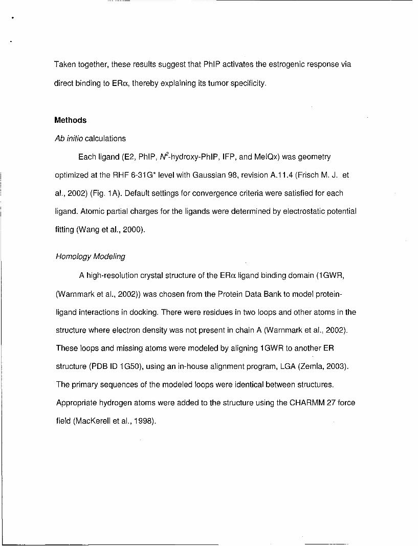
Ab initio calculations
Each ligand (E2, PhIP, N2-hydroxy-PhIP, IFP, and MelQx) was geometry
optimized at the RHF 6-31 G* level with Gaussian 98, revision A.1 1.4 (Frisch M. J. et
al., 2002) (Fig. 1 A). Default settings for convergence criteria were satisfied for each
ligand. Atomic partial charges for the ligands were determined by electrostatic potential
fitting (Wang et al., 2000).
Homology Modeling
A high-resolution crystal structure of the ERcx ligand binding domain (1 GWR,
(Warnmark et al., 2002)) was chosen from the Protein Data Bank to model protein-
ligand interactions in docking. There were residues in two loops and other atoms in the
structure where electron density was not present in chain A (Warnmark et al., 2002).
These loops and missing atoms were modeled by aligning 1GWR to another ER
structure (PDB ID 1G50), using an in-house alignment program, LGA (Zemla, 2003).
The primary sequences of the modeled loops were identical between structures.
Appropriate hydrogen atoms were added to the structure using the CHARMM 27 force
field (MacKerell et al., 1998).

Docking
The estrogen receptor protein (1GWR homology model) and ligands (E2, PhiP,
N2-hydroxy-PhIP, and MelQx) were prepared for the docking studies as described in the
Autodock manual (Morris et al., 1998). The original source code was modified to allow a
grid cube size of 128 points with a 0.375 A grid spacing. The resulting grid covered
-80% of the ligand binding domain. Using the lamarkian genetic algorithm option, the
total number of docking steps was set to 200 and cluster analysis was activated. Each
ligand pose was then grouped according to its position on the protein. A Boltzmann
probability, Ki, was then derived from the calculated free energy AGj, gas constant R,
and temperature, T(300 C) of each pose, i.
Ki = e(-AGi IRT) Equ. 1
Docking poses in each grouping, s, were then weighted according to Equ. 2.
E Ki,5i's
P - YKj iEqu. 2
Here, p, is the weighted probability of finding pose, i, in grouping, s. These probabilities
were then summed for each category.
Transferred Nuclear Overhauser Effect Spectroscopy (trNOESY)
All spectra were recorded at 300 C using a Varian Inova 600 MHz spectrometer. 1H-1 D
and phase sensitive 2D-nuclear Overhauser effect spectroscopy (NOESY) experiments
(900 ms mixing time) were carried out on 1.0 mg of PhIP (5.94 mM) or 27 mg of E2

(132.1 mM) dissolved in 750 gL deuterated DMSO) in the absence of ER(o protein (Fig.
1 B, C). Human, recombinant ERox protein was purchased from Sigma-Aldrich as 750
pmols of purified protein in 500 gL 50 mM Tris-HCL, pH 8.0, 500 mM KCL, 2 mM DTT,
1 mM EDTA, 1 mM sodium orthovanadate and 10% gylcerol. We first determined
alternative buffer conditions in which the ERoa protein remained stable. Both Tris and
glycerol contribute large proton signals in the NMR data that interfere with the detection
of micromolar quantities of PhIP and E2. In addition, high salt concentrations are not
compatible with adding small volumes of PhIP and E2, which are only soluble in organic
solvents such as DMSO, and results in precipitation of the protein. We determined that
ERcL remained stable for several days after being exchanged (using dialysis,
MWCO=10,000) into 50 mM Na2PO 4, pH 8.0,200 mM KCL, 1% P-mercaptoethanol.
Transferred NOESY (trNOESY) experiments (300 ms mixing time) were used to detect
whether binding occurred upon addition of 40 molar excess of PhIP (120 pM) to a 3 gM
solution of ER in NMR buffer. TrNOESY is useful in detecting binding when the ligand is
in fast exchange between the free and bound states (1 0-210-6 M range for the
dissociation constant). Adding molar excess of E2 to the solution of ER and PhIP results
in precipitation. This is most likely due to the fact that E2 strongly binds ERos. To
determine if E2 could displace PhIP from the binding pocket, 0.25 molar ratio of E2 (1.5
RM) was added to the 6 pIM ERcL protein-240 pLM PhIP solution. 300 increments were
collected in tj, each with 128 or 256 scans and 1024 complex data points collected in t2.
The total amount of DMSO in the protein/ligand samples did not exceed 2%. NMR data
was processed using VNMR software (Varian Inc., Palo Alto CA), and the 2D frequency
domain matrices were analyzed using FELIX (version 97, Accelrys, San Diego, CA).

Cell Culture
MCF-7 cells were obtained from American Type Culture Collection (ATCC,
Manassas, VA) and were grown in DMEM with 5% FBS, 1% non-essential amino acids,
10 pg/ml insulin, 2 mM L-glutamine, and 1% penicillin/streptomycin. All tissue culture
supplies, with the exception of the charcoal stripped fetal bovine serum, were obtained
from Invitrogen (Carlsbad, CA). Cells were maintained at 37 0C with 5% humidity. For
proliferation assays and estrogen responsive reporter assays, Opti-MEM (phenol red-
free medium) was used with 5% charcoal stripped fetal bovine serum (Clontech, Palo
Alto CA) to minimize estrogen-like activity contributed by the phenol red and serum
(Soto et al., 1995; Zacharewski, 1998).
Cell proliferation assay
For cell proliferation assays 3 x 10³ cells were seeded into each well of a 96-well
tissue culture plate in Opti-MEM medium. Twenty-four hours after plating, the medium
was replaced with medium supplemented with increasing concentrations of E2 (Sigma,
St. Louis MO), PhIP (Toronto Research Chemicals, Downsview, Ontario), MelQx
(Toronto Research Chemicals, Downsview, Ontario) and IFP, treated with the
appropriate concentration of vehicle or left untreated. IFP is a natural product isolated
from a heated mixture of creatine, glutamic acid and sucrose (Pais et al., 2000).
Heterocyclic amines were initially dissolved in DMSO and then serially diluted in
medium. DMSO concentration was never greater than 0.5%. Ethanol was used to
dissolve E2. The effect of the heterocyclic amines was determined by comparing cell
growth to the appropriate vehicle-treated cells and untreated cells.
The effect of test compounds on cell growth was evaluated 48 and 72 hours after
the initiation of treatment. Cell growth was quantified using an Aqueous Non-
Radioactive Cell Proliferation Assay (Promega, Madison WI) with absorbance of the
wells measured in a standard multi-well plate reader at 490nm. Briefly, this colormetric
assay measures the bioreduction of a tetrazolium compound (3-(4,5-dimethylthiazol-2-
yl)-5-(3-carboxymethoxyphenyl)-2-(4-sulfophenyl)-2H-tetrazolium, inner salt; MTS) to a
formazan product. The conversion of MTS is directly proportional to the number of living
cells in culture. Each experiment was performed at least three individual times with 4 -
6 replicates per experiment. Effect of chemical was determined by comparing cell
growth to the appropriate vehicle-treated cells and untreated cells. Error is presented as
the standard error of the mean. Statistical significance was assessed using Wilcoxon
Rank Sum Test.
o Estrogen Responsive Reporter Assay
Heterocyclic amines were tested for estrogenic and anti-estrogenic activity using
a standard estrogen responsive reporter plasmid containing three vitellogenin estrogen
responsive elements (EREs) upstream of the luciferase reporter gene (EREx3-Luc).
The EREx3-Luc was a kind gift of Diane Klotz, NIEHS. EREx3-Luc was co-transfected
with a commercially available control renilla reporter plasmid (pRL-TK, Promega,
Madison WI) to control for transfection efficiency and for the normalization of the results.
The reporter plasmids were transfected into MCF-7 cells (Klotz et al., 1996; Nodland et

al., 1997; Reel et al., 1996; Safe et al., 1998), which constitutively express ERc* and
have well-characterized responses to estrogenic stimuli. The ability of the compounds to
induce luciferase reporter gene activity was compared to 10.9 M E2 and appropriate
negative controls, as previously described (Charles et al., 2002).
MCF-7 cells were plated in 96-well plates, designed specifically for use in a
luminometer (Perkin Elmer, Wellesley, MA) at a density of 1X1 05 cells/well in Opti-MEM
medium. The cells were left untransfected or were co-transfected with 200 ng each of
luciferase reporter and renilla control plasmids using lipofectAMINETM 2000 (Invitrogen,
Carlsbad, CA) optimized for 96-well plate experiments according to directions. After a 5-
hour transfection, the untransfected cells were incubated with just culture medium to
quantify background levels. The transfected cells were incubated with the same culture
media alone or supplemented with heterocyclic amines, E2 or both. Control cells were
incubated with a corresponding amount of compound solvent at the highest
concentration.
The cells were incubated at 370C for 18 hours and then assayed for luciferase
and renilla activity using a dual luciferase/renilla reporter assay kit (Promega, Madison,
WI) according to directions. Each experimental condition was duplicated six to eight
times in the 96-well plate and each plate was read in the luminometer twice. Luciferase
activity was normalized to the renilla controls and compared to 10.9 M E2, standard in
estrogen responsive luciferase assays. Estrogen receptor-mediated activation was
determined by comparisons to negative and E2 controls. All experiments were repeated
more than 3 times and all data were normalized to E2 (10 9M) activation. For
competition assays, HAs (alone or in combinations) and/or E2 were added

simultaneously. Error is presented as the standard error of the mean. Statistical
significance was assessed using Wilcoxon Rank Sum Test.
Results
Docking Studies
Initial docking of E2 to the ER(x LBD as a control calculation reveal the ligand
pose as seen in the 1 GWR crystal structure, with the initial position of the ligand at least
15 A outside of the native binding cavity. PhIP and N2-hydroxy-PhIP were manually
docked in the same orientation as E2 to match the crystal structure (Brzozowski et al.,
1997), where the exocyclic amine and imidazole ring were overlaid on the E2 hydroxyl
group and 'A-ring', respectively. These initial structures started the docking trials of
PhIP, N2-hydroxy-PhIP, IFP, and MelQx, using the same grid of the protein as for E2.
Eleven groupings contain 99% of all docking poses for each HA. For E2 and PhIP, the
E2 binding cavity accounts for -98% and -89% of the total binding probability,
respectively. PhIP binds favorably into the E2 binding site although the calculated
probability of binding is lower when compared to E2 (Fig. 2A). Several poses of PhIP
are observed in the E2 binding site. One pose is rotated by 180' with the phenyl ring
pointing toward residues Glu353 and Arg394. However, the most favorable pose places
the exocyclic amine proximal to the hydrogen bonds between Glu353 and Arg394,
similar to the initial docking position and E2 pose in the co-crystal (Brzozowski et al.,
1997) (Fig. 2B). Side-chains from Leu387, Leu391, and Phe404 make hydrophobic
contacts with each face of the imidazo ring. A possible hydrogen bond between the
exocyclic primary amine and the carbonyl of Leu387 is also observed. The donor-H-

acceptor angle is greater than 1500, and the hydrogen bond distance is less than 2.6 A
(Fig. 2B). The phenyl ring of PhIP is further stabilized by hydrophobic contacts with
Leu346 on one face and by Met343 and Met421 around the edge of the ring. The face
of the phenyl ring opposite Leu346 is open to solvent. Furthermore, the phenyl ring is
turned relative to the rest of the molecule, which allows for a better fit to the binding
cavity wall and is consistent with the geometry-optimized structure (Fig. 2B).
Two significant groupings of N2-hydroxy-PhIP, IFP, and MelQx are observed, one
in the native E2 binding site and another 11 A away, close to helix 5. The lowest energy
pose of N2-hydroxy-PhIP is in the E2 binding site and is in the same orientation as PhIP
(i.e. all hydrophobic contacts were the same). However, the hydroxyl group does not
form any hydrogen bonds with protein side-chains or backbone. Instead of forming any
obvious hydrogen bonds, the hydroxyl group was further buried into a solvent channel
under the Glu353...Arg394 hydrogen bond bridge.
The planar and rigid IFP molecule assumes the same basic orientation of PhIP
and N2-hydroxy-PhIP; the exocyclic amine of IFP is pointed toward the Glu3530o°Arg394
hydrogen bond bridge. A hydrogen bond is also observed between the primary amine of
IFP and the carbonyl oxygen of Leu387. The binding orientation for MelQx in the E2
binding cavity is the opposite that of PhIP and N2-hydroxy-PhIP because the exocyclic
amine points toward His524, similar to the orientation of the d-ring of E2 in the crystal
structure (1 GWR). We observe a hydrogen bond with the side-chain hydroxyl group of
Thr347 and the exocyclic amine from MelQx. In both IFP and MelQx, the fused rings do
not appear to make optimal hydrophobic interactions in comparison with PhIP and N2-
hydroxy-PhIP.

NMR Ligand Binding Studies
We performed transferred NOESY (trNOESY) experiments (Clore et al., 1982;
Gronenborn and Clore, 1982; Roberts, 1999) to detect PhIP binding to ERca protein. An
observed sign flip in NOE crosspeaks of PhIP from positive (red) to negative (black) in
the TrNOESY spectra (Fig. 3A, B), is an indication of binding to ERc* protein. Adding E2
to the PhIP/ ERoS mixture causes the sign flip in the NOE crosspeaks to revert back to
positive (red), indicating that PhIP is no longer bound to ERa (Fig. 3C).
MCF-7 Human breast cancer cell assays
Incubating ATCC MCF-7 cells withl xl 0-9M tol xl 0-7 M E2 for 48 hours produces a
slight increase in cell proliferation (7-12%) that is significantly different from vehicle-
treated cells. By 72 hours the increase in cell number is 20% above control cells and by
96 hours the increase has risen to 40% (data not shown). This increase in cell number
is consistent with other reports of proliferation response for this MCF-7 cell line
(Rasmussen and Nielsen, 2002). Adding increasing doses of PhIP for 48 hours also
causes a 7-10% increase in proliferation that is significantly different from control cells,
but not significantly different from E2 at similar concentrations (Fig. 4). By 72 hours the
increase in proliferation reaches 20% for 4x1 0-8M PhIP, which is not significantly
different from the increase in cell number caused by a similar concentration of E2 (data
not shown) cells treated with N2-hydroxy-PhIP show a slight, non-significant, increase in
cell growth at low doses (4x1 09M). However at higher concentrations (4xl 0-6 M), cell

growth is inhibited. Treating cells with concentrations of MelQx and IFP higher than
4x1 0-8M for 48 hours causes a significant decrease in cell growth (Fig. 4).
A standard luciferase reporter assay was used to measure the effect of HAs on
ERc-dependent transcription activity. This assay relies on the ERo normally expressed
by the MCF-7 cells. Active ERI3 is not expressed in MCF-7 cells (Bardin et al., 2004;
Cestac et al., 2005; Paruthiyil et al., 2004; St-Laurent et al., 2005), and therefore ERP3
interactions cannot contribute to the results. As shown in figure 5A, increasing amounts
of PhIP causes a dose-dependent increase in ERa activity that is significantly different
than untreated cells. In contrast, increasing amounts of N2-hydroxy-PhIP and the other
HAs do not show this activation. Competition assays were used to determine the effect
of simultaneous incubation of HAs (4x1 0-7 M) and 10.9 M E2 on ERcX activation (Fig. 5B).
Treating MCF-7 cells with PhIP and E2 produces a response that is 13% less than E2
alone. N2-Hydroxy-PhIP inhibits the E2-mediated activation of ERoX by 40%, and MelQx
and IFP inhibit ERax activation to levels that are significantly lower than untreated cells.
Cell viability was measured under identical conditions and is not affected by incubation
with HAs separately or in combination with E2 at these concentrations (data not shown).
Effect of HA combinations on ERax activity
Competition assays were performed with PhIP, N2-hydroxy-PhIP, and MelQx to
determine the effect of simultaneous exposure to 4x10⁻⁷M PhIP and increasing amounts
of the other compounds (Fig. 5C). Treating MCF-7 cells with either a 1:1 or 1:3 molar
ratio of PhIP to other compounds significantly inhibits ERoa transcription activity to levels
that are lower than cells exposed to PhIP alone.

Discussion
Heterocyclic amines are known to be potent mutagens and rodent carcinogens,
but the complete mechanism of carcinogenicity for these compounds has not been
elucidated. This report is the first to show that PhIP binds directly to the ERcx-ligand
binding domain and competes with estradiol in the native binding cavity. We also
demonstrate the novel finding that N2-hydroxy-PhIP, the PhIP phase I metabolite, and
two heterocyclic amines, MelQx and IFP, inhibit ERax activation. Among the HAs tested,
activation of the receptor is limited to PhIP, and other HAs can act as potential anti-
estrogens. We also show that PhIP increases MCF-7 cell proliferation and activates
ERox dependent transcription, in agreement with results published by Lauber et al.
(Lauber et al., 2004).
Ph/P Bindinq to Erar
The docking of E2 to the ERox LBD validates our model of ERax LBD and the
docking algorithm for our system because the most stable E2 pose found was the same
orientation as the co-crystal E2 conformation. The most favorable binding pose of PhIP
was also found in the E2 binding site, even though the probability of PhIP binding is
lower than E2 binding to the E2 site. This lower binding probability is consistent with the
known high binding affinity of E2 to ER(x (Kd 10-9-1010 M) and our NMR results show
E2 competing out PhIP (see below).

We designed an NMR competition experiment implementing trNOESY
techniques that provides information about the PhIP binding site on the ERcX protein. If
PhIP binds the same site as E2, then adding E2 to the PhIP/ERct mixture should
displace PhIP since E2 binds more strongly to ERa than PhIP (as predicted
computationally). If PhIP is binding in a different site than E2, then the negative (black)
PhIP-bound peaks should remain. When E2 is added to the PhIP/ ERU mixture, a
reversion in the NOE crosspeaks from the negative (black) back to positive (red) is
observed, indicating that PhIP is no longer bound to ERos. These data highly suggest
that PhIP is binding to the ligand-binding domain. However, another possibility is that
PhIP is binding in another site, and E2 is preventing PhIP from binding because of a
conformation change in the protein. Although this possibility may exist, the
computational prediction of PhIP binding in the E2 binding site and our in vitro data on
stimulated cell proliferation (see below) leads us to the conclusion that PhIP is binding
in the E2 binding site competitively.
The effect of the HAs on the proliferative activity of whole cells was tested using
a modified E-SCREEN assay (Rasmussen and Nielsen, 2002). This assay is based on
the ability of estrogen-responsive MCF-7 cells to proliferate in the presence of
compounds that mimic estrogen. Although a standard assay for screening for
estrogenicity, the proliferative response of the MCF-7 cells varies depending on the
MCF-7 subline employed. In these studies we used ATCC wild-type MCF-7 cells, which
have been shown to vary in their response to estrogen incubation (Rasmussen and
Nielsen, 2002). Using these cells, we found that micromolar concentrations of PhIP
were able to stimulate proliferation up to 40% above vehicle-treated cells over a 72-hour

incubation. Lauber et. al. determined that the same increase in cell proliferation could
be caused by only nanomolar concentrations of PhIP (Lauber et al., 2004). These
authors used MCF-7 cells obtained from the European Collection of Cell Cultures
(ECACC) in their assays; presumably this MCF-7 cell variant is more sensitive to
estrogen or estrogen-like compounds. Nevertheless, both studies demonstrated
increased cell proliferation when incubated with PhIP.
After much discussion regarding the potential estrogenicity of PhIP and its
relevance to tumor progression (Gooderham et al., 2002; Lauber et al., 2000; Pfau et
al., 2000), we and others (Lauber et al., 2004) now confirm that PhIP activates ER and
stimulates cell growth. Both E2 and PhIP are able to activate ERos-dependent
transcription in MCF-7 cells transfected with a standard estrogen responsive reporter
plasmid containing three vitellogenin EREs upstream of the luciferase reporter gene. In
other words, PhIP is able to use the endogenous ERox present in MCF-7 cells to effect
transcription. Thirty minute pre-incubation with the complete anti-estrogen ICI 182,780
inhibited this increased activity (data not shown) (Lauber et al., 2004), further
suggesting that PhIP is effecting transcription through direct interaction with ERos. These
results and the fold-activation of ERcx transcription activity (compared to E2) in the
presence of micromolar concentrations of PhIP are in agreement with previous.work
(Lauber et al., 2004).
The in vitro data, without the supporting NMR and computational data, could also
be explained by PhIP acting on the ERox pathway at points upstream or downstream
from the receptor protein. For the present studies, the trNOESY results, together with
the in vitro data and docking results strongly support the idea that PhIP and E2 compete

for the same binding cavity within the ERu, LBD. Taken together, these results
demonstrate that PhlP initiates an estrogenic response in target cells by binding directly
with the ERos.
N2-Hydroxy-PhIP, MelQx and IFP Inhibition of ERa
Close examination of our docking data shows that PhIP makes a hydrogen bond
between the exocyclic-amine and the carbonyl-oxygen of Leu387 (Fig. 2B). N2-Hydroxy-
PhIP shares all the same hydrophobic contacts as PhIP. However, because of the non-
optimal placement of the hydroxy group, no hydrogen bonds are made with the protein,
decreasing its probability of binding in the E2 cavity (Fig. 2A). These data suggest that
the driving force in binding in the E2 cavity is hydrophobic and that electrostatic
interactions such as hydrogen bonds play only a secondary role.
In the cellular assays, N2-hydroxy-PhIP inhibited the ability of E2 to activate ERoa.
When compared to PhIP, the docking data predicts that the binding of N2-hydroxy-PhIP
should be weaker. The cell assays confirm these predictions. When N2-hydroxy-PhIP is
co-incubated with physiological concentrations of E2, much of the ER activation
remains, showing that N2-hydroxy-PhIP is not a particulary effective inhibitor.
Like PhIP and N2-hydroxy-PhIP, MelQx and IFP both contain an exocyclic amine
with a methyl group at the adjacent carbon. However, MelQx and IFP contain three
fused-heterocyclic rings, which limits hydrogen bonding and hydrophobic interactions
compared to PhlP. The low barrier rotation of the phenyl ring in PhIP optimizes non-
bonded contacts, which is not possible in MelQx and IFP. Our docking data suggest
that MelQx and IFP do not bind favorably in the E2 binding cavity because the fixed

conformation of the fused-rings does not allow optimization of hydrophobic contacts.
The lack of a favorable binding conformation is confirmed by the inability of these to
compounds to stimulate reporter gene transcription in our in vitro cell assay (Fig. 5).
When MelQx and IFP are co-incubated with physiological concentrations of E2,
ERc* activity is significantly inhibited. This inhibition is identical to results obtained when
the compounds are added alone. Compared to N2-hydroxy-PhIP, MelQx and IFP are
much more effective inhibitors so that E2 activity remains below control levels. These
results suggest that the mechanism of binding for MeIQX and IFP are different than
PhIP and N2-hydroxy-PhIP; MelQx and IFP may even bind in other regions of the ERca
protein.
Implications of Diet and Metabolism
The estrogenic potency of PhIP is comparable to other environmental estrogens
found in the diet. Genistein, a known phytoestrogen found in soy products, was
compared to PhIP in ERox activation assays (data not shown). At low concentrations
(4X1 0 7M) there was no statistical difference in the activation of the ERuX by either
compound, but higher concentrations of genistein elicited a much greater ERcX response
than higher concentrations of PhIP. When investigating compounds from food, it is
difficult to predict exposure levels for individual target cells. The concentrations of PhIP
examined here are orders of magnitude higher than what a single cell may be exposed
to after a meal of cooked meat. However, chronic exposure to PhIP over a lifetime may
add to the total estrogenic burden of the body.
In addition, it is not clear what the biological consequence of being exposed to
xeno-estrogens may be. Although exposure to environmental estrogens may increase

the activity of endogenous ERc, exposure to other compounds may inhibit the activation
(anti-xeno-estrogens), effectively canceling out the effect of the xeno-estrogens. In fact,
although exposure to PhIP may be stimulating ERca activity, simultaneous consumption
of the other HAs, MelQx and IFP, may diminish that effect. Our previous studies have
shown that when meat is cooked, the ratios of formation of PhIP:MelQx and PhIP:IFP
varies according to cooking conditions (Knize et al., 1998). In figure 5C, 1:1 ratios of
PhIP:MelQx and PhIP:IFP mixtures significantly inhibit the ability of PhIP to activate
ERoc. If the ratios are increased to 1:3, both MelQx and IFP completely prevent any
stimulatory action by PhIP. When cells are exposed to combinations of HAs and
metabolites, as would happen during dietary consumption of cooked meat, the PhIP-
mediated activation of ERc* can be abolished. Thus, the biological consequences of
exposure to HAs may depend upon many complex factors, including the ratio of HAs
formed in the meat, how well each compound is absorbed from the digestive tract, and
the concentration of the compounds at the target cells.
Metabolism of the compounds will also contribute to the complexity of the overall
human exposure. MCF- 7 cells contain active P450 metabolism; however most of the
activity is CYP1 Al. Our previous investigations have shown that these cells do not
significantly metabolize PhIP (K. S. Kulp, unpublished results), suggesting that
metabolites of the HAs are not confounding our measurements of the effects of the
compounds on cell proliferation and ERca activity. However, in humans consuming
cooked meats, hepatic and extra-hepatic metabolism of PhIP to N2-hydroxy-PhIP is
extensive. In fact, only a small percentage of an ingested and absorbed dose of PhIP is
excreted as the parent compound; the rest is metabolites (Malfatti et al., 1999). Our

results show that co-incubation of PhIP and N2-hydroxy-PhIP inhibits the stimulatory
effect of PhIP on the ERc and that the amount of inhibition depends upon the amount of
N2-hydroxy-PhlP present in the incubation. This suggests that the estrogenic potential
of PhIP may depend in large part on the extent of Phase I activation of the compound,
either in the liver or at the level of the target cell.
Conclusions
Here we show by experiment and computational methods that the food mutagen
PhIP can activate human ERcu by binding to the LBD (Figs. 2, 3), and that PhIP
stimulates MCF-7 breast cancer cell proliferation, suggesting a mechanism for tissue-
specific carcinogenesis of this carcinogen. We also show that IFP, MelQx, and the
primary hydroxylated metabolite of PhIP do not stimulate breast cancer cell proliferation
(Fig. 4). Moreover, MelQx, IFP and N2-hydroxy-PhIP inhibit estrogen receptor activation
(Fig. 5). Together, these results imply that these dietary constituents may play an active
role in both activating and inhibiting hormone sensitive cancers. More importantly,
understanding the mechanisms of action for the inhibitory HAs may lead to potent
therapeutics against breast cancer.
Acknowledgments
This work was supported by grants from the DOD (BC032793) and NIH-NCI (CA55861)
and was performed under the auspices of the U.S. DOE by University of California,
Lawrence Livermore National Laboratory under Contract W-7405-Eng-48.

References
Bardin, A., N. Boulle, G. Lazennec, F. Vignon, andP. Pujol. 2004. Loss of ER beta expression asa common step in estrogen-dependent tumor progression. Endocrine-Related Cancer11(3):537-551.
Brzozowski, A.M., A.C.W. Pike, Z. Dauter, R.E. Hubbard, T. Bonn, 0. Engstrom, L. Ohman,G.L. Greene, J.A. Gustafsson, and M. Carlquist. 1997. Molecular basis of agonism andantagonism in the oestrogen receptor. Nature 389(6652):753-758.
Cestac, P., G. Sarrabayrouse, C. Medale-Giamarchi, P. Rochaix, P. Balaguer, G. Favre, J.C.Faye, andS. Doisneau-Sixou. 2005. Prenylation inhibitors stimulate both estrogenreceptor alpha transcriptional activity through AF- 1 and AF-2 and estrogen receptor betatranscriptional activity. Breast Cancer Research 7(l):R60-R70.
Charles, G.D., C. Gennings, T.R. Zacharewski, B.B. Gollapudi, andE.W. Carney. 2002. Anapproach for assessing the estrogen receptor-mediated interactions in mixtures of threechemicals: A pilot study. Toxicological Sciences 68:349-360.
Clore, G.M., andA.M. Gronenborn. 1982. Theory and application of the transferred nuclearOverhauser effect to the study of the conformation of small molecules bound to proteins.J Magn Reson 48:402-417.
Clore, G.M., A.M. Gronenborn, C. Mitchinson, andN.M. Green. 1982. H-1-Nmr Studies onNucleotide Binding to the Sarcoplasmic-Reticulum Ca-2+ Atpase - Determination of theConformations of Bound Nucleotides by the Measurement of Proton-Proton TransferredNuclear Overhauser Enhancements. European Journal of Biochemistry 128(1):113-117.
Eiler, S., M. Gangloff, S. Duclaud, D. Moras, andM. Ruff. 2001. Overexpression, purification,and crystal structure of native ER alpha LBD. Protein Expression and Purification22(2): 165-173.
Fabbro, D., D. Batt, P. Rose, B. Schacher, T.M. Roberts, andS. Ferrari. 1999. Homogeneouspurification of human recombinant GST-Akt/PKB from Sf9 cells. Protein Expr Purif17(l):83-88.
Felton, J.S., M.G. Knize, L.M. Bennett, M.A. Malfatti, M.E. Colvin, andK.S. Kulp. 2004. Impactof environmental exposures on the mutagenicity/carcinogenicity of heterocyclic amines.Toxicology 198(1-3):135-145.
Frisch M. J., Trucks G. W., Schlegel H. B., Scuseria G. E., Robb M. A., Cheeseman J. R.,Zakrzewski V. G., Montgomery J. A. Jr., Stratmann R. E., Burant J. C., Dapprich S.,Millam J. M., Daniels A. D., Kudin K. N., Strain M. C., Farkas 0., Tomasi J., Barone V.,Cossi M., Cammi R., Mennucci B., Pomelli C., Adamo C., Clifford S., Ochterski J.,Petersson G. A., Ayala P. Y., Cui Q., Morokuma K., Rega N., Salvador P., Dannenberg J.J., Malick D. K., Rabuck A. D., Raghavachari K., Foresman J. B., Cioslowski J., Ortiz J.V., Baboul A. G., Stefanov B. B., Liu G., Liashenko A., Piskorz P., Komaromi I.,Gomperts R., Martin R. L., Fox D. J., Keith T., Al-Laham M. A., Peng C. Y.,Nanayakkara A., Challacombe M., Gill P. M. W., Johnson B., Chen W., Wong M. W.,Andres J. L., Gonzalez C., Head-Gordon M., Replogle E. S., anda.P.J. A. 2002. Gaussian98, Revision A. 11.4. Gaussian Inc, Pittsburg.

Gooderham, N.J., H. Zhu, S. Lauber, A. Boyce, andS. Creton. 2002. Molecular and genetictoxicology of 2-amino-i -methyl-6-phenylimidazo [4,5-b]pyridine (PhIP). Mutat Res-FundMol M 506:91-99.
Gronenborn, A.M., andG.M. Clore. 1982. Conformation of Nad+ Bound to Yeast and HorseLiver Alcohol-Dehydrogenase in Solution - the Use of the Proton-Proton TransferredNuclear Overhauser Enhancement. Journal of Molecular Biology 157(1): 155-160.
Hajduk, P.J., J. Dinges, G.F. Miknis, M. Merlock, T. Middleton, D.J. Kempf, D.A. Egan, K.A.Walter, T.S. Robins, S.B. Shuker, T.F. Holzman, andS.W. Fesik. 1997. NMR-baseddiscovery of lead inhibitors that block DNA binding of the human papillomavirus E2protein. J Med Chem 40(20):3144-3150.
Halachmi, S., E. Marden, G. Martin, H. MacKay, C. Abbondanza, andM. Brown. 1994. Estrogenreceptor-associated proteins: possible mediators of hormone-induced transcription.Science 264(5164):1455-1458.
Henke, B.R., T.G. Consler, N. Go, R.L. Hale, D.R. Hohman, S.A. Jones, A.T. Lu, L.B. Moore,J.T. Moore, L.A. Orband-Miller, R.G. Robinett, J. Shearin, P.K. Spearing, E.L. Stewart,P.S. Turnbull, S.L. Weaver, S.P. Williams, G.B. Wisely, andM.H. Lambert. 2002. A newseries of estrogen receptor modulators that display selectivity for estrogen receptor beta.Journal of Medicinal Chemistry 45(25):5492-5505.
Humphrey, W., A. Dalke, andK. Schulten. 1996. VMD: Visual molecular dynamics. Journal ofMolecular Graphics 14(1):33-&.
Ito, N., R. Hasegawa, M. Sano, S. Tamano, H. Esumi, S. Takayama, andT. Sugimura. 1991. Anew colon and mammary carcinogen in cooked food, 2-amino-1-methyl-6-phenylimidazo[4,5-b]pyridine (PhIP). Carcinogenesis 12:1503-1506.
Kawamori, T., N. Uchiya, K. Watanabe, T. Ohta, T. Sugimura, andK. Wakabayashi. 2001.Effects of heterocyclic amines with mammary gland potential on estrogenic response ofuterus in ovariectomized rats. Cancer Letters 162:31-37.
Keating, G.A., R. Sinha, D. Layton, C.P. Salmon, M.G. Knize, K.T. Bogen, C.F. Lynch, andM.Alavanja. 2000. Comparison of heterocyclic amine levels in home-cooked meats withexposure indicators. Cancer Causes and Control 11(8):731-739.
Klotz, D.M., B.S. Beckman, S.M. Hill, J.A. McLachlan, M.R. Walters, andS.F. Arnold. 1996.Identification of environmental chemicals with estrogenic activity using a combination ofin vitro assays. Environmental Health Perspectives 104(10): 1084-1089.
Knize, M.G., and J.S. Felton. 2005. Formation and human risk of carcinogenic heterocyclicamines formed from natural precursors in meat. Nutrition Reviews 63(5):158-165.
Knize, M.G., R. Sinha, E.D. Brown, C.P. Salmon, O.A. Levander, J.S. Felton, and N. Rothman.1998. Heterocyclic Amine Content in Restaurant-Cooked Hamburgers, Steaks, Ribs, andChicken. J. Ag. Food Chem. 46:4648-4651.
Kumar, V., andP. Chambon. 1988. The Estrogen-Receptor Binds Tightly to Its ResponsiveElement as a Ligand-Induced Homodimer. Cell 55(1): 145-156.
Lauber, S., S. Ali, andN.J. Godderham. 2000. In vitro oestrogenicity of the food-ferivedheterocyclic amine PhIP. Toxicology Letters 1 16(Suppl. 1):26-27.
Lauber, S.N., S. Ali, andN.J. Gooderham. 2004. The cooked food derived carcinogen 2-amino-I-methyl-6-phenylimidazo[4,5-b] pyridine is a potent oestrogen: a mechanistic basis for itstissue-specific carcinogenicity. Carcinogenesis 25(12):2509-2517.
MacKerell, A.D., D. Bashford, M. Bellott, R.L. Dunbrack, J.D. Evanseck, M.J. Field, S. Fischer,J. Gao, H. Guo, S. Ha, D. Joseph-McCarthy, L. Kuchnir, K. Kuczera, F.T.K. Lau, C.

Mattos, S. Michnick, T. Ngo, D.T. Nguyen, B. Prodhom, W.E. Reiher, B. Roux, M.Schlenkrich, J.C. Smith, R. Stote, J. Straub, M. Watanabe, J. Wiorkiewicz-Kuczera, D.Yin, andM. Karplus. 1998. All-atom empirical potential for molecular modeling anddynamics studies of proteins. Journal of Physical Chemistry B 102(18):3586-3616.
Malfatti, M.A., K.S. Kulp, M.G. Knize, C. Davis, J.P. Masengill, S. Williams, s. Nowell, S.MacLeod, K.H. Dingley, K.W. Turtletaub, N.P. Lang, andJ.S. Felton. 1999. Theidentification of [2-14C]2-amino-l-methyl-6-phenylimidazo[4,5-b]pyridine (PhIP)metabolites in humans. Carcinogenesis 20:705-713.
Merritt, E.A., andD.J. Bacon. 1997. Raster3D: Photorealistic molecular graphics.Macromolecular Crystallography, Pt B 277:505-524.
Moras, D., andH. Gronemeyer. 1998. The nuclear receptor ligand-binding domain: structure andfunction. Current Opinion in Cell Biology 10(3):384-391.
Morris, G.M., D.S. Goodsell, R.S. Halliday, R. Huey, W.E. Hart, R.K. Belew, andA.J. Olson.1998. Automated docking using a Lamarckian genetic algorithm and an empiricalbinding free energy function. Journal of Computational Chemistry 19(14): 1639-1662.
Nodland, K.I., M. Wormke, andS. Safe. 1997. Inhibition of estrogen-induced activity by 2,3,7,8-tetrachlorodibenzo-p-dioxin (TCDD) in the MCF-7 human breast cancer and other celllines transfected with vitellogenin A2 gene promoter constructs. Archives of Biochemistryand Biophysics 338:67-72.
Norrish, A.E., L.R. Ferguson, M.G. Knize, J.S. Felton, S.J. Sharpe, andR.T. Jackson. 1999.Heterocyclic amine content of cooked meat and risk of prostate cancer. J Natl Cancer I91:2038-2044.
Pais, P., M.J. Tanga, C.P. Salmon, andM.G. Knize. 2000. Formation of the mutagen IFP inmodel systems and detecti
on in restaurant meats, J. Agric. Fd. Chem. 47:1098-1108.Paruthiyil, S., H. Parmar, V. Kerekatte, G.R. Cunha, G.L. Firestone, andD.C. Leitman. 2004.
Estrogen receptor beta inhibits human breast cancer cell proliferation and tumorformation by causing a G(2) cell cycle arrest. Cancer Research 64(1):423-428.
Pfau, W., B. Gutendorf, andJ. Westendorf. 2000. Endocrine disruptors in fried meat: PhIP is anestrogen. Proceedings of the American Association for Cancer Research 41:310.
Pike; A.C.W., A.M. Brzozowski, R.E. Hubbard, T. Bonn, A.G. Thorsell, 0. Engstrom, J.Ljunggren, J.K. Gustafsson, andM. Carlquist. 1999. Structure of the ligand-bindingdomain of oestrogen receptor beta in the presence of a partial agonist and a fullantagonist. Embo Journal 18(17):4608-4618.
Pike, A.C.W., A.M. Brzozowski, J. Walton, R.E. Hubbard, A.G. Thorsell, Y.L. Li, J.A.Gustafsson, andM. Carlquist. 2001. Structural insights into the mode of action of a pureantiestrogen. Structure 9(2): 145-153.
Pratt, W.B., andD.O. Toft. 1997. Steroid receptor interactions with heat shock protein andimmunophilin chaperones. Endocrine Reviews 18(3):306-360.
Rasmussen, T.H., andJ.B. Nielsen. 2002. Critical parameters in the MCF-7 cell proliferationbioassay (E-Screen). Biomarkers 7(4):322-336.
Reel, J.R., J.C.V. Lamb, andB.H. Neal. 1996. Survey and assessment of mammalian estrogenbiological assays for hazard characterization. Fundamental and Applied Toxicology34(2):288-305.
Renaud, J., S.F. Bischoff, T. Buhl, P. Floersheim, B. Fournier, C. Halleux, J. Kallen, H. Keller,J.M. Schlaeppi, andW. Stark. 2003. Estrogen receptor modulators: Identification and

structure-activity relationships of potent ER alpha-selective tetrahydroisoquinolineligands. Journal of Medicinal Chemistry 46(14):2945-2957.
Roberts, G.C. 1999. NMR spectroscopy in structure-based drug design. Curr Opin Biotechnol10(l):42-47.
Roberts, G.C. 2000. Applications of NMR in drug discovery. Drug Discov Today 5(6):230-240.Ruff, M., Gangloff, M., Eiler, S., Duclaud, S., Wurtz, J. M., Moras, D. 1999. The Crystal
Structure of an Estrogen Nuclear Receptor Mutant Suggest a Molecular Mechanism forthe Agonism Antagonism Switch.
Safe, S., K. Connor, andK. Gaido. 1998. Methods for xenoestrogen testing. Toxicology Letters103:665-670.
Shiau, A.K., D. Barstad, P.M. Loria, L. Cheng, P.J. Kushner, D.A. Agard, andG.L. Greene.1998. The structural basis of estrogen receptor/coactivator recognition and theantagonism of this interaction by tamoxifen. Cell 95(7):927-937.
Shiau, A.K., D. Barstad, J.T. Radek, M.J. Meyers, K.W. Nettles, B.S. Katzenellenbogen, J.A.Katzenellenbogen, D.A. Agard, andG.L. Greene. 2002. Structural characterization of asubtype-selective ligand reveals a novel mode of estrogen receptor antagonism. NatureStructural Biology 9(5):359-364.
Sinha, R., N. Rothman, E. Brown, 0. Levander, C.P. Salmon, M.G. Knize, andJ.S. Felton. 1995.High concentrations of the carcinogen 2-amino-i -methyl-6-phenylimidazo- [4,5-b]pyridine (PhIP) occur in chicken but are dependent on the cooking method. CancerRes. 55:4516-4519.
Skog, K., K. Augustsson, G. Steineck, M. Stenberg, andM. Jagerstad. 1997. Polar and non-polarheterocyclic amines in cooked fish and meat products and their corresponding panresidues. Food and Chemical Toxicology 35(6):555-565.
Snyderwine, E.G. 1999. Mammary gland carcinogenesis by 2-amino-1-methyl-6-phenylimidazo[4,5-b]pyridine in rats: possible mechanisms. Cancer Letters 143:211-215.
Snyderwine, E.G. 2002. Mammary gland carcinogenesis by food-derived heterocyclic amines:metabolism and additional factors influencing carcinogenesis by 2-amino- 1-methyl-6-phenylimidazo[4,5-b]pyridine (PhIP). Environ Mol Mutagen 39(2-3): 165-170.
Snyderwine, E.G., C.D. Davis, H.A.J. Schut, andS.J. Roberts-Thomson. 1998. Proliferation,development and DNA adduct levels in the mammary gland of rats given 2-amino-I-methyl-6-phenylimidazo[4,5b]pyridine and a high fat diet. Carcinogenesis 19:1209-1215.
Snyderwine, E.G., H.S. Yoon, L.P. Knight-Jones, M. Tran, H.A.J. Schut, andM.S. Yu. 2003.Mutagenesis and DNA adduct formation in the mouse mammary gland exposed to 2-hydroxyamino-l-methyl-6-phenylimidazo-[4,5-b]pyridine in whole organ culture.Mutagenesis 18(1):7-12.
Soto, A.M., C. Sonnenschein, K.L. Chung, M.F. Fernandez, N. Olea, andF.O. Serrano. 1995.The E-SCREEN assay as a tool to identify estrogens: an update on estrogenicenvironmental pollutants. Environ Health Perspect 103 Suppl 7:113-122.
St-Laurent, V., M. Sanchez, C. Charbonneau, andA. Tremblay. 2005. Selective hormone-dependent repression of estrogen receptor beta by a p38-activated ErbB2/ErbB3 pathway.Journal of Steroid Biochemistry and Molecular Biology 94(1-3):23-37.
Studier, F.W. 2005. Protein production by auto-induction in high-density shaking cultures.Protein Expr Purif 41:207-234.
Tyler, R.C., H.K. Sreenath, S. Singh, D.J. Aceti, C.A. Bingman, J.L. Markley, andB.G. Fox.2005. Auto-induction medium for the production of [U- 15N]- and [U- 13C, U- 15N]-

labeled proteins for NMR screening and structure determination. Protein Expr Purif40(2):268-278.
Venugopal, M., R. Agarwal, A. Callaway, H.A.J. Schut, andE.G. Snyderwine. 1999a. Inhibitionof cell death in human mammary epithelial cells by the cooked meat-derived carcinogen2-amino- 1-methyl-6-phenylimidazo[4,5-b]pyridine. Biochemical and BiophysicalResearch Communications 266:203-207.
Venugopal, M., A. Callaway, andE.G. Snyderwine. 1999b. 2-Amino-l-methyl 6-phenylimidazo[4,5-b] pyridine (PhIP) retards mammary gland involution in lactatingSprague-Dawley rats. Carcinogenesis 20:1309-1314.
Wakabayashi, K., M. Nagao, H. Esumi, andT. Sugimura. 1992. Food-derived mutagens andcarcinogens. Cancer Research (suppl.) 52:2092s-2098s.
Wang, J.M., P. Cieplak, andP.A. Kollman. 2000. How well does a restrained electrostaticpotential (RESP) model perform in calculating conformational energies of organic andbiological molecules? Journal of Computational Chemistry 21 (12): 1049-1074.
Warnmark, A., T. Almlof, J. Leers, J.A. Gustafsson, andE. Treuter. 2001. Differentialrecruitment of the mammalian mediator subunit TRAP220 by estrogen receptors ERalpha and ER beta. Journal of Biological Chemistry 276(26):23397-23404.
Warnmark, A., E. Treuter, J.A. Gustafsson, R.E. Hubbard, A.M. Brzozowski, andA.C.W. Pike.2002. Interaction of transcriptional intermediary factor 2 nuclear receptor box peptideswith the coactivator binding site of estrogen receptor alpha. Journal of BiologicalChemistry 277(24):21862-21868.
Zacharewski, T. 1998. Identification and assessment of endocrine disruptors: limitations of invivo and in vitro assays. Environ Health Perspect 106 Suppl 2:577-582.
Zemla, A. 2003. LGA: a method for finding 3D similarities in protein structures. Nucleic AcidsResearch 31 (13):3370-3374.

Figure Legend
Figure 1. Structures of heterocyclic amines and E2 (A) focused on in this study and 1 D-1H spectra of 5.95 mM PhIP (B) and 132 mM E2 (C) dissolved in deuterated DMSO(100%) showing the relative position of the NMR peaks for each compound.
Figure 2. Boltzmann probability distributions for each ligand in the E2 binding cavity (A).Cut away view of the ligand-binding cavity of ERcx (B) with lowest energy docking posesof E2 (purple) and PhIP (colored-sticks). The surface is colored according to residuetype; nonpolar (white), polar uncharged (green). Key hydrogen bonding residues areshown in colored-stick representations (Glu 353, Arg 394, Leu 387, His 524). Figurewas made with VMD (Humphrey et al., 1996) and rendered with Raster3d (Merritt andBacon, 1997).
Figure 3. The expanded region of a 900 ms mixing time NOESY spectrum (A) of 5.95mM PhIP in DMSO (<2%) exhibits weak positive NOE crosspeaks (boxed in red). Theexpanded regions of 300 ms mixing time NOESY spectra of ERca with (B) addition of 40fold molar excess PhIP indicating that PhIP binds, as evidenced by the flip in sign tonegative crosspeaks (boxed in black), and (C) after addition of 0.25 molar ratio of E2 toa 1:40 ERu, + PhIP sample showing that PhIP does not bind (red peaks) when E2 ispresent.
Figure 4. Effect of PhIP, N2-Hydroxy-PhIP, MelQx, IFP and E2 on MCF-7 cellproliferation. Cells were treated with compounds for 48 hours. Error is presented asstandard error of the mean. * = significantly different from control, p<0.01. For PhIPtreatments only, * = significantly different from control, p<0.01 and not significantlydifferent from estradiol treatment at a corresponding molar concentration. X Estradiol* PhIP, E N2-hydroxy-PhIP, A MelQx and e IFP.
Figure 5. The effect of heterocyclic amines on estrogen receptor activation. Cells weretreated with compounds as described in Methods. Error is presented as standard errorof the mean. Compound concentrations are molar and E2 concentration is 10-9M in allexperiments. Increasing concentrations of Has (A). Nf-Hydroxy- PhIP, MelQx and IFPare significantly different from PhIP-treated cells at the same concentrations (p<0.01).All compound treatments are significantly different from control and estradiol (p<0.01).Competition assays with estradiol (B). Cells were treated with 4x1 0-7M compound aloneor compound with 10-9M E2 (hatched bars). Effect of combinations of heterocyclicamines on estrogen receptor activation (C). Simultaneous addition of PhIP and N2-hydroxy- PhIP, or PhIP and MelQx are significantly different from PhIP alone (*)(p<0.01) All compound treatments are significantly different from control and estradiol(p<0.01).

II
A HH1-NC H : N "' -N
IFP N N MelOx C.H3 OH
SEstradiol, E2
PhIP
9.08.07.06.05.04.03.02.01.0 9.08.07.0 6.05.04.03.02.01.0ppm ppm

A E2Z 1 RhIP _
.. Q 0 8 .... N 2-O H- ...................~0.
PhiP
~ 04 ~MeIx[FP
-N 0.2m
E2 Binding Cavity
B
Glu 353:r Mis24
M94,4J ....... et 421

A B , A",•,• C C i
Pos
E3 i----~--8. .8.2 8 7.767.472 8.4 6.2 c.0 87.16 7.: 2 8:.4 6.2 ".•7[1 [4 72
Dl (ppm) Dl (ppm) Dl (pprn)

120-
* E2 * PhIP.11i T * T *-
.-. 110 ; -
100 Melx0
80
• 8*_) *2 80
0
60
50 .
1.E-09 1.E-08 1.E-07 1.E-06 1.E-05
Concentration (M)

Ac:100--
"0~ 80-
a) 60 - _ _ _ _ _ _ _ _ _
20
L.
0Control E2 4x10 1 .2xl 0-6 410 1.x 01 410 1.221cr 6 410 1.x 21PhIP /V-hydroxy- Mel~x 1FP
PhIPB
60
a) 40
cz 20
Control E2 PhIP/V-hydroxy- MeIQx 1FF PhIPNf-hydroxy-MeIQx IFPPhIP PhIP
Compounds alone Compounds with 1 0-9ME2
~100-
0 80 -';- ) 60
c: a) 40
.o 20
0Control E2 4X1 0- 41x0- 1.2x10- 4x10⁷ 1.2x10O- 4x1 0- 1.2x10- 4x1 0- 1.2X10⁶
PhIP N2-hydroxy- Mel~x N'-hydroxy- MeIQxPhIP PhIP
Compounds alone Compounds with 4xl 0 7M PhIP

Appendix BStatement of Work
Computational Characterization and Prediction of Estrogen Receptor CoactivatorBinding Site Inhibitors
Task 1: Computationally predict the relative binding of CBIs in the coactivator site andthe CBI binding properties. (Months 1-12)(1.1) A minimum of three CBIs and a minimum of three estrogenic compounds will bedocked into the coactivator site and the estradiol site, respectively, using AutoDock.(Months 1-2)(1.2) The resulting combination of CBIs and estrogenic compounds will be rank orderedby effective binding. (Months 3-4)(1.3) From the resulting docked compounds, molecular dynamics (MD) simulations willbe run on the energetically favorably bound CBIs to investigate their effects on proteinbinding and stability. MD simulations will be carried out using NAMD and eachsimulation will be run for 2 ns. (Months 4-11)(1.4) The resulting combination of CBIs and estrogenic compounds will be re-rankedbased on the MD simulations. (Month 12)Task 2: Biologically assay the effect of the CBI on the estrogen receptor activation(Months 1-12)(2.1) Order and obtain the CBIs and estrogenic compounds. (Month 1)(2.2) Measure estrogen receptor activation by transfecting a reporter plasmid thatcontains 3 vitellogenin of estrogen response elements upstream of a luciferase reportergene in MCF-7 human breast cancer cells. Receptor activity will be measured in thepresence estradiol and each individual CBI. (Month 2-3)(2.3) Compare the effect of the CBIs on estradiol to their effect on the other estrogeniccompounds analyzed in the docking program. (Months 4-6)Task 3: Spectroscopically measure the binding of the CBI to the ER LBD.(3.1) Obtain the compounds used in the biological assay. (Month 1)Order and obtain a pure sample of the human ERg (Month 1)(3.2) If the CBIs demonstrate the ability to modulate estrogen receptor activation in thecells assays described in Task 2, they will be assayed for binding to Era by themselves,using a transferred nuclear Overhauser effect spectroscopy (trNOESY) assay. (Months4-6)(3.3) CBIs that bind ER and demonstrate activity in Task 2 will be assayed againstestradiol in a competition NMR assay to determine if they bind in the presence ofestradiol. If they bind in the presence of estradiol, then the CBIs are binding to anothersite. (Months 6-8)(3.4) To rank order the binding of CBIs, CBIs will be added individually to ERa, using anNMR assay (Months 8-10)Task 4: The results will be put into a draft of a paper to be submitted later (Month 12)