2.0 Review of Literature 2.1 Chemotherapy and...
32
Review of Literature 6 2.0 Review of Literature 2.1 Chemotherapy and Disease A disease is an impairment of the normal state of an organism or any of its components that hinders the performance of vital functions. It may be attributed to one or many factors e.g. malnutrition, industrial hazards, climate, specific infective agents (e.g. viruses, bacteria, fungi, protozoa, helminthes), inherent defects of the body (e.g. various genetic and immunologic anomalies) or combination of these. Infectious diseases are major hazard all over the world causing premature deaths (Pinner et al., 1996). Chemotherapy started with vaccination discovered by Edward Jenner but the term chemotherapy was originally coined by Paul Ehrlich who discovered the first effective chemotherapeutic agent- arsphenamine/salvarsan, which opened the door to future developments in chemotherapy and antibiotics. Discovery and development of sulfonamides was followed by a golden period (1935-1970) with flurry of discoveries of antibiotics leading to more than a dozen classes of antibiotics including sulphonamides, β lactams, aminoglycosides, chloramphenicol, tetracycline, macrolides, trimethoprim, rifamycins, quinolones and glycopeptides. It was the discovery of the most beneficial secondary metabolite i.e. penicillin from Penicillium notatum by Alexander Fleming in 1929, which revolutionized the field of modern medicine. Since then, a lot of compounds possessing antibiotic activity have been isolated from different fungi. In general naturally occurring substances are distinguished from the synthetic compounds by the name of antibiotics. Natural products are chemical compounds obtained as a result of of primary or secondary metabolism of living organisms (Bentley, 1997). The primary metabolites such as nucleic acids, fatty acids, polysaccharides and proteins) are present in all biological systems whereas secondary metabolites are diverse chemical compounds with varied biological functions. An important part of the natural products, the group of smaller molecular secondary metabolites of microorganisms usually exhibits some kind of biological activities. Vast majority of clinically relevant compounds, including antibacterial, antifungal, and antitumor have been derived from natural products (Shu, 1998). They represent the greatest single contribution of drug therapy for the health care of increasing population of the world and provide effective control against many
Transcript of 2.0 Review of Literature 2.1 Chemotherapy and...

Review of Literature
6
2.0 Review of Literature
2.1 Chemotherapy and Disease
A disease is an impairment of the normal state of an organism or any of its
components that hinders the performance of vital functions. It may be attributed to one or
many factors e.g. malnutrition, industrial hazards, climate, specific infective agents (e.g.
viruses, bacteria, fungi, protozoa, helminthes), inherent defects of the body (e.g. various
genetic and immunologic anomalies) or combination of these. Infectious diseases are
major hazard all over the world causing premature deaths (Pinner et al., 1996).
Chemotherapy started with vaccination discovered by Edward Jenner but the term
chemotherapy was originally coined by Paul Ehrlich who discovered the first effective
chemotherapeutic agent- arsphenamine/salvarsan, which opened the door to future
developments in chemotherapy and antibiotics. Discovery and development of
sulfonamides was followed by a golden period (1935-1970) with flurry of discoveries of
antibiotics leading to more than a dozen classes of antibiotics including sulphonamides, β
lactams, aminoglycosides, chloramphenicol, tetracycline, macrolides, trimethoprim,
rifamycins, quinolones and glycopeptides. It was the discovery of the most beneficial
secondary metabolite i.e. penicillin from Penicillium notatum by Alexander Fleming in
1929, which revolutionized the field of modern medicine. Since then, a lot of compounds
possessing antibiotic activity have been isolated from different fungi. In general naturally
occurring substances are distinguished from the synthetic compounds by the name of
antibiotics. Natural products are chemical compounds obtained as a result of of primary
or secondary metabolism of living organisms (Bentley, 1997). The primary metabolites
such as nucleic acids, fatty acids, polysaccharides and proteins) are present in all
biological systems whereas secondary metabolites are diverse chemical compounds with
varied biological functions. An important part of the natural products, the group of
smaller molecular secondary metabolites of microorganisms usually exhibits some kind
of biological activities. Vast majority of clinically relevant compounds, including
antibacterial, antifungal, and antitumor have been derived from natural products (Shu,
1998). They represent the greatest single contribution of drug therapy for the health care
of increasing population of the world and provide effective control against many

Review of Literature
7
microbial pathogens. Various biotic entities like actinomycetes, plants, mushrooms,
animals etc have been proved to be potential sources for various bioactive compounds
which help to fight different diseases. But as the increasing population is becoming
resistant to many commercially available antibiotics leading to the emergence of many
fatal diseases, so there is a real need to find and expand the spectrum of such sources
which can help in finding novel compounds to resolve the problem posed by resistance or
to expand the spectrum of the existing antimicrobial sources. Fungi are well known
source of bioactive compounds and the research for isolation of novel fungal metabolites
that blossomed more than 40 years ago is still very active now. At present, the search for
new producers of biologically active compounds that help them to survive and adapt to
their surroundings can be expected in these fungi with the greatest probability (Gloer,
1995; Grabley et al., 1999). Microbes have made a phenomenal contribution to the health
and well being of people throughout the world. They produce a number of secondary
metabolites which are an integral part of the pharmaceuticals currently available in the
market (Demain and Sanchez, 2009). Soil holds an enormous biodiversity that can be
screened for antibiotic producers. Because of huge expenditure on synthetic molecules
with effective antimicrobial properties, natural products are still a worth promise
(Newmann and Cragg, 2007). Fungi are important sources of secondary metabolites and
they continue to provide new chemical entities with novel biological activities (Baker and
Alvi, 2004). They have been a rich source of compounds for therapeutic applications
including antibacterial (Rancic et al., 2006; Lucas et al., 2007; Takahashi et al., 2009),
antifungal (Nicoletti et al., 2007; Omura et al., 2008), antiviral (Nishihara et al., 2000),
immunosuppressants and cholesterol-lowering agents (Grabley et al., 1992; Kwon et al.,
2002). Although many fungi have been listed, to possess antimicrobial activity, still there
is a need to explore more such organisms to meet the problem of emerging strains of
resistant microorganisms and to expand the spectrum of suitable organisms which can be
useful to meet the requirements of potent antimicrobials. The present study was thus
planned to isolate fungi from soil samples collected from different regions of Punjab,
India and to screen them for their antimicrobial potential.

Review of Literature
8
2.2 Antibiotics and their Mechanism of Action
Most antibiotics used for the treatment of bacterial infections may be categorized
either according to their principal mechanism of action or on the basis of their chemical
structure. The main mechanism of action of antibiotics includes (Fig 2.2.1; Table 2.2.1).
Ø “Inhibition of cell-wall synthesis.”
Ø “Disruption of cell membrane.”
Ø “Inhibition of protein synthesis.”
Ø Inhibition of DNA synthesis.
Ø “Interference with the synthesis of essential metabolites.”
Figure 2.2.1 Diagrammatic representation of different mode of action of antibiotics

Review of Literature
9
Table 2.2.1: Group of antibiotics with their mode of action
Inhibits Cell Wall Synthesis
Penicillins
(bactericidal in nature: acts by blocking the cross linking catalysed by the enzyme transpeptidase )
Group of
antibiotics
Drugs Antibacterial spectrum Possible side effects
Penicillins Aqueous
penicillin G
Streptococcus. pyogenes Hypersensitivity reaction
β –lactams Penicillin G Streptococcus. agalactiae Hemolytic anemia
Benzathine
penicillin G
C. perfringens
Procaine
penicillin G
Penicillin V
Aminopenicl
ins
Ampicillin Streptococcus. pyogenes Hypersensitivity reaction
Amoxicillin Streptococcus. agalactiae Hemolytic anemia
C. perfringens
Gram-negative:
E. coli
Penicillinase
-resistant-
penicillins
Methicillin Streptococcus. pyogenes Hypersensitivity reaction
Nafcillin Streptococcus. agalactiae Hemolytic anemia
Oxacillin C. perfringens Interstitial nephritis
Penicillinase
– susceptible
penicillins
Cloxacillin,
Dicloxacillin,
Amoxicillin,
Ampicillin,
Flucloxacillin
E. coli and Penicillinase –producing
Staph. aureus
Antipseudo
monal
penicillins
Carbenicillin Hypersensitivity reaction
Ticarcillin Pseudomonas aeruginosa Hemolytic anemia
Piperacillin Interstitial nephritis
Cephalosporins
First
generation
Cefazolin S. aureus Allergic reaction
Cephalexin S. epidermidis Coombs-positive anemia (3%)
Some Gram-negatives:
E. coli
Klebsiella
Second
generation
Cefoxitin Staph. aureus Allergic Reaction
Cefaclor Staph . epidermidis ETOH Disulfiram reaction
Cefuroxime Some Gram-negatives:
Cefamandole E. coli
Cefprozil Klebsiella
Third
generation
Cefixime,
Ceftriaxone
S. aureus Allergy, hypoprothrombinemia
Cefotaxime S. epidermidis ethanol Disulfiram reaction
Ceftazidime Gram-negative bacteria:
Cefoperazone Escherichia coli
Cefpodoxime Klebsiella spp.
Pseudomonas Hypersensitivity,
Fourth
generation
Cefepime Staph. aureus, Strep. pneumoniae, P.
aeruginosa, and Enterococci
Gastrointestinal upset and nausea
Fifth
generation
Ceftobiprole methicillin-resistant Staphy aureus,
penicillin-resistant Strep
pneumoniae, P. aeruginosa,
and Enterococci

Review of Literature
10
Other Cell Wall Inhibitors
Glycopeptide Vancomycin, Teicoplanin,
Decaplanin
MRSA Red man syndrome
(bactericidal:
disrupts
peptioglycan
cross-
linkage)
Staph. Aureus Nephrotoxicity
Staph. epidermidis “Ototoxicity”
β-lactam
antibiotic-
Inhibitors
Ampicillin-
Sulbactam,
Amoxicillin-
Clavulanic Acid
S . aureus Hypersensitivity Reaction
S. epidermidis Hemolytic anemia
Escherichia coli
Klebsiella spp.
Carbapene
ms
Imipenem
(+ cilastatin)
Enterococci,
Staphylococccal spp.
Listeria,
Streptococci
Enterobacteriaceae,
bacteroides
Acinetobacter species
diarrhea, nausea, vomiting, skin rash and pruritus.
Meropenem
Doripenem
Ertapenem
Aztreonam Aztreonam E. coli
P. mirabilis
Acinetobacter anitratus
Chest discomfort
cough
difficulty with breathing or troubled breathing
fever
Polymyxins Polymyxin B Topical Gram-negative infections May cause redness, pain and edema at the injection
site. If used as eye drops, may cause burning
sensation, itching or temporary blindness .
Polymyxin E
Nephrotoxicity
Bacitracin Bacitracin skin infection- associated bacteria. Nephrotoxicity (albuminuria, cylindruria, azotemia,
rising blood concentrations of the drug); GI effects
(nausea, vomiting); pain at injection site;
hypersensitivity reactions (rash)
Protein Synthesis Inhibition
Are bactericidal in nature and cause damage by binding irreversibly to 30S ribosomal subunit.
Aminoglycos
ides
Gentamicin Enterobacteriaceae Nephrotoxic in nature.
Neomycin Pseudomonas Ototoxic in nature
Amikacin
Tobramycin
Streptomycin
Tetra-
cyclines
Tetracycline Rickettsia Hepatotoxicity
(bacteriostati
c: blocks
tRNA)
Doxycycline Mycoplasma Tooth discoloration Impaired growth
Minocycline Spirochetes (Lyme's disease)
Demeclocycline

Review of Literature
11
Are bactericidal in nature and cause damage by binding irreversibly to 50S ribosomal subunit
Macrolides Erythromycin Some Species of Streptococcus Coumadin Interaction (cytochrome P450)
Azithromycin Haemophilus. Influenzae
Clarithromycin Mycoplamsa pneumonia
Chloram
phenicol
(broad
spectrum)
Chloramphenic
ol
Staphylococcus aureus, E. coli and
Streptococcus pneumoniae,, Neisseria
meningitidis, Strep
pneumoniae and Haemophilus
influenza
Aplastic Anemia
Lincosamides
Clindamycin Treatment of Staphylococcal
and Streptococcal infections.
Bacteroides fragilis
some anaerobic strains
Clostridium difficile–associated diarrhea
(pseudomembranous colitis)
clindamycin may cause esophagitis
Linezolid
(variable)
Linezolid VRE
Streptococcal spp.
MRSA
Side effects such as vomiting, Nausea, and
Vaginal candidiasis
Strepto
gramins
Quinupristin VRE, VRSA local irritation at peripheral administration sites,
centrally mediated myalgia and arthralgia, nausea,
and reversible rise in conjugated bilirubin -. Dalfopristin
Pristinamycin
Virginamycin
Inhibition of DNA Synthesis
Fluoroquinolones
(bactericidal by nature :mode is by inhibiting DNA gyrase )
First
generation
Nalidixic acid Crucial for treatment of nosocomial
infections
Adverse reactions are nausea, vomiting, and
diarrhea, insomnia and serious neurological
disorder myasthenia gravis. Overdose may also
cause tendon rupture.
(http://www.orthobullets.com/basic-science/9059/antibiotic-classification-and-mechanism)
Review of Literature
12
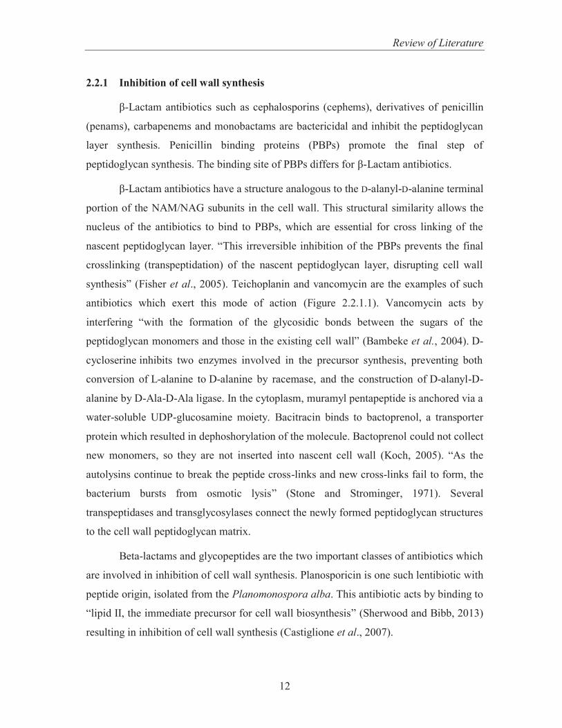
2.2.1 Inhibition of cell wall synthesis
β-Lactam antibiotics such as cephalosporins (cephems), derivatives of penicillin
(penams), carbapenems and monobactams are bactericidal and inhibit the peptidoglycan
layer synthesis. Penicillin binding proteins (PBPs) promote the final step of
peptidoglycan synthesis. The binding site of PBPs differs for β-Lactam antibiotics.
β-Lactam antibiotics have a structure analogous to the D-alanyl-D-alanine terminal
portion of the NAM/NAG subunits in the cell wall. This structural similarity allows the
nucleus of the antibiotics to bind to PBPs, which are essential for cross linking of the
nascent peptidoglycan layer. “This irreversible inhibition of the PBPs prevents the final
crosslinking (transpeptidation) of the nascent peptidoglycan layer, disrupting cell wall
synthesis” (Fisher et al., 2005). Teichoplanin and vancomycin are the examples of such
antibiotics which exert this mode of action (Figure 2.2.1.1). Vancomycin acts by
interfering “with the formation of the glycosidic bonds between the sugars of the
peptidoglycan monomers and those in the existing cell wall” (Bambeke et al., 2004). D-
cycloserine inhibits two enzymes involved in the precursor synthesis, preventing both
conversion of L-alanine to D-alanine by racemase, and the construction of D-alanyl-D-
alanine by D-Ala-D-Ala ligase. In the cytoplasm, muramyl pentapeptide is anchored via a
water-soluble UDP-glucosamine moiety. Bacitracin binds to bactoprenol, a transporter
protein which resulted in dephoshorylation of the molecule. Bactoprenol could not collect
new monomers, so they are not inserted into nascent cell wall (Koch, 2005). “As the
autolysins continue to break the peptide cross-links and new cross-links fail to form, the
bacterium bursts from osmotic lysis” (Stone and Strominger, 1971). Several
transpeptidases and transglycosylases connect the newly formed peptidoglycan structures
to the cell wall peptidoglycan matrix.
Beta-lactams and glycopeptides are the two important classes of antibiotics which
are involved in inhibition of cell wall synthesis. Planosporicin is one such lentibiotic with
peptide origin, isolated from the Planomonospora alba. This antibiotic acts by binding to
“lipid II, the immediate precursor for cell wall biosynthesis” (Sherwood and Bibb, 2013)
resulting in inhibition of cell wall synthesis (Castiglione et al., 2007).

Review of Literature
13
Figure 2.2.1.1 Diagrammatic representation of inhibition of cell wall synthesis
2.2.2 Disruption of Cell Membrane
The plasma membrane is a bilayered structure that separates the intracellular
organelles from the exterior micro environment. Plasma membrane plays a crucial role in
a number of cell functions by aiding in attachment to solid surfaces ,functioning of ion
conducting channels and promoting cell signal process. It also tethers together : the cell
wall and the cytoskeleton. Rapid depolarization may occur due to plasma membrane
disruption, which compromises membrane potential and subsequent inhibition of protein
and nucleic acid synthesis may result in cell death.
Antibiotics like Colistin and Polymyxin B are produced from Bacillus spp. These
antimicrobials have a general structure consisting of a cyclic peptide head attached to a
long hydrophobic tail, which interacts with the gram negative outer membrane and
cytoplamic membrane displacing bacterial counterions, causing outer membrane
destabilization. They exert their antimicrobial activity in a detergent like manner, where
the amino group (positive charged) of the peptide head electrostatiscally interacts with a
portion of LPS layer carrying the negative charge. This interaction results in the outer
membrane destabilization causing disruption of both outer and inner membranes
(Newton, 1956; Davis et al., 1971; Chen and Fenigold, 1973; Imai et al., 1975; Canepari
et al., 1990; Silverman et al., 2003; Falagas and Kasiakou, 2005; Steenbergen et al.,
Review of Literature
14
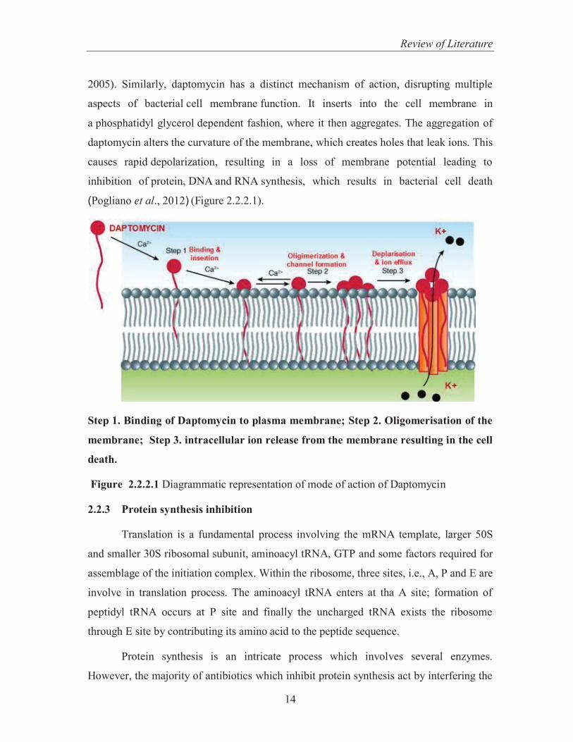
2005). Similarly, daptomycin has a distinct mechanism of action, disrupting multiple
aspects of bacterial cell membrane function. It inserts into the cell membrane in
a phosphatidyl glycerol dependent fashion, where it then aggregates. The aggregation of
daptomycin alters the curvature of the membrane, which creates holes that leak ions. This
causes rapid depolarization, resulting in a loss of membrane potential leading to
inhibition of protein, DNA and RNA synthesis, which results in bacterial cell death
(Pogliano et al., 2012) (Figure 2.2.2.1).
Step 1. Binding of Daptomycin to plasma membrane; Step 2. Oligomerisation of the
membrane; Step 3. intracellular ion release from the membrane resulting in the cell
death.
Figure 2.2.2.1 Diagrammatic representation of mode of action of Daptomycin
2.2.3 Protein synthesis inhibition
Translation is a fundamental process involving the mRNA template, larger 50S
and smaller 30S ribosomal subunit, aminoacyl tRNA, GTP and some factors required for
assemblage of the initiation complex. Within the ribosome, three sites, i.e., A, P and E are
involve in translation process. The aminoacyl tRNA enters at tha A site; formation of
peptidyl tRNA occurs at P site and finally the uncharged tRNA exists the ribosome
through E site by contributing its amino acid to the peptide sequence.
Protein synthesis is an intricate process which involves several enzymes.
However, the majority of antibiotics which inhibit protein synthesis act by interfering the

Review of Literature
15
translational steps involving either the 30S or 50S subunit of the bacterial ribosome. The
antibiotics act by inhibiting 30S initiation complex consisting of an mRNA template, 30S
subunit and f-met-tRNA, complexation of 30S initiation complex and the 50S ribosome
to form 70S subunit and the elongation process of the polypeptide chain (Figure 2.2.3.1).
Tetracyclines e.g. Doxycycline is a group of antibiotics which blocks the A site of the
ribosome, thus preventing the aminoacyl-tRNA binding.
“Aminoglycoside antibiotics have an affinity for the 30S ribosome subunit.
Streptomycin, one of the most commonly used aminoglycosides, interferes with the
creation of the 30S initiation complex. Kanamycin and tobramycin also bind to the 30S
ribosome and block the formation of the larger 70S initiation complex” (Kestell et al.,
2002).
Figure 2.2.3.1 Diagrammatic representation of protein synthesis inhibition
(Classes.midlandstech.edu)
Review of Literature
16
Erythromycin is one such representative of macrolides, forms a complex with50S
ribosome through its 23S rRNA component, by virtue of which the 50S subunit fails to
assemble. Also, roxithromycin, clarithromycin, Erythromycin exhibit their mode of
action by preventing the occurrence of the final transpeptidation step due to blockage
polypeptide exporting tunnel (Menninger and Otto, 1982; Usary and Champney, 2001;
Oyama et al., 2007).
Peptidyl transferase is an important enzyme required in the final step, i.e., protein
elongation. Antibiotics such as clindamycin and lincomycin inhibits peptidyl transferases
whereas macrolides are not the inhibitors for this enzyme (Chang et al., 1966).
Puromycin acts by causing termination in chain elongation process where it acts on 3’
terminus of aminocyl tRNA (Nathans, 1964; Azzam and Algranati, 1973).
An aminoglycoside, hygromycin B, bins to the 30S subunit of tRNA. It has been
reported that binding of hygromycin B deforms the A sit of ribosome which further
resulted in translocation of protein elongation (Gaha and Champney, 2007).
2.2.4 Inhibition of DNA synthesis
Interference with DNA synthesis requires several steps. These antimicrobial
agents hamper the synthesis of nucleotides or their interconversion, preventing DNA
from acting as a template and by interfering with the polymerization step of DNA
synthesis thereby inhibiting the replication and transcription step. Antimicrobial drugs
have been developed to target each of these steps (Figure 2.2.4.1). For example, the
antimicrobial rifampin binds to DNA-dependent RNA polymerase, thereby inhibiting the
initiation of RNA transcription. Quinolones are one such group of antibiotics which
readily inhibits topoisomerase II (DNA gyrase), a key enzyme required for DNA
replication (Nakamura and Yura, 1976; Schulz and Zillig, 1981; Campbell et al., 2001).
DNA gyrase causes uncoiling of the superhelical DNA strand by transient breakabe and
joining of its phosphodiester backbone. This opens up the DNA strand for subsequent
action by DNA/ RNA polymerases. Norfloxacin, levofloxacin and ciprofloxacin are some
of the antibiotics which exhibit this mode of action (Hooper, 2001).

Review of Literature
17
Figure 2.2.4.1 Diagrammatic representation of DNA synthesis inhibition
2.2.5 Interference with the synthesis of essential metabolites
Sulfonamides or sulfa drugs represent a category of compounds whose
mechanism is directed towards specific enzyme system. Sulfonamides are known to
competitively inhibit para-aminobenzoic acid (PABA) due to their structural similarity
with PABA. Many bacteria require para-aminobenzoic acid as a precursor to their
synthesis of the essential coenzyme tetrahydrofolic acid (THFA). PABA is a structural
part of the THFA acid molecule. In bacteria, dihydropteroate synthetase (DHPS) is an
important enzyme needed for synthesis of folic acid from PABA, as it is essential for
DNA synthesis. Sulfonamides act by competitively inhibiting DHPS, due to which
PABA is not converted into folic acid. Sulfonamides are usually bacteriostatic in nature
(Figure 2.2.5.1). The selective action of sulfonamides is explained by the fact that the
PABA molecule and the sulfonamide molecule are so similar that the sulfonamide may
enter the reaction in place of the PABA and block the synthesis of an essential cellular
constituent (Henry, 1943; Smith et al., 2000).

Review of Literature
18
Figure 2.2.5.1 Interference of antibiotics with essential metabolites synthesis in bacteria
2.3 Antimicrobial Resistance
Antimicrobial resistance is one of the biggest challenges all over the globe
specially in the treatment of nosocomial infections. Ernest Duchesne and Alexander
Fleming originally discovered the antibacterial potential of Penicillium spp. in 1928 by
isolation of penicillin (Cantas, 2013). Success stories of natural antibiotic penicillin, has
revolutionized the world. Since then, a number of compounds with antimicrobial
properties have been found. So these metabolites play a vital role in the development of
modern medicine. Since their discovery, antibiotics have saved millions of people from
life-threatening diseases. As bacterial resistance is increasing now a days, its treatment
with ongoing antibiotics is a big question worldwide (Pouillard, 2002; Levy and
Marshall, 2004; Alanis, 2005; Pallett and Hand, 2010). Supporting the foreseen
development of antimicrobial resistance, Alexander Fleming, the discoverer of penicillin,
once gave a statement in the New York Times that “the microbes are educated to resist

Review of Literature
19
penicillin and a host of penicillin fast organisms is bred out which can be passed to other
individuals and from them to others until they reach someone who gets a septicemia or
pneumonia which penicillin cannot save” (Levy, 2002). Active resistance is caused by
selective environmental pressure which is linked to a class of antibiotic whereas passive
resistance is a result of non-antibiotic linked processes. Antimicrobial resistance is
attributed to various factors which mainly include human practices. Resistance to
antibiotics is either natural or intrinsic and mutational or acquired. Thus, normally
susceptible populations of bacteria may become resistant to antimicrobial agents through
pre-existing factor in the microorganisms or it may be due to some factors acquired by
genetic changes or non genetic mechanisms. Genetic resistance may be chromosomal
(due to mutations in chromosomal DNA) and/or extra chromosomal resistance (due to
resistant plasmids occurring via transduction and conjugation). Misuse of antibiotics by
prescribing the antibiotics in a wrong manner by physicians leading to a development of
antimicrobial resistance (Arnold and Straus, 2005). The past few decades have seen an
alarming increase in the prevalence of antibiotic resistance microorganisms (Livermore,
2009). Over the last few decades multidrug resistant organisms are becoming difficult to
treat thus causing number of diseases worldwide. These includes VRE (Vancomycin
Resistant Enterococci), MRSA (methicillin/oxacillin-resistant Staphylococcus aureus),
PRSP - penicillin-resistant Streptococcus pneumonia, ESBLs - Extended-spectrum beta-
lactamases (which are resistant to cephalosporins and monobactams). MRSA and VRE
and PRSP are the leading cause of diseases in non- hospital associated settings.whereas
ESBL result in various nosocomial infections all over the globe (Toadr, 2009).
MRSA are resistant not only to methicillin’s action as well as Beta-lactams such as
cephalosporin and penicillin. But to several classes of antibiotics. Several troublesome
reports on MRSA, VRSA (Vancomycin-resistant S. aureus), Hospital-Associated MRSA
(HA-MRSA) or Community-Associated MRSA (CA-MRSA) are the major contributors
to the struggling medical history against the Staphylococcal infections. HA-MRSA
occurs most frequently in patients with weak immunity. Life threatening infections, such
as blood and surgical infections is commonly associated with MRSA. “More people in
the U.S. die from MRSA infection than AIDS. Methicilin resistant S. aureus was
responsible for an estimated 94,000 life threatening infections, as reported by US Centers

Review of Literature
20
for Disease Control and prevention (CDC). That same year, roughly 16,000 people in the
U.S. died from AIDS, according to CDC” (Klevens et al., 2007). “Each year in the
United States, at least 2 million people acquiring serious infections with bacteria that are
resistant to one or more of the commercially available antibiotics, as reported by US
department of Health and Human Services, (2013)”. It has been observed that Gram
negative bacteria are now a days the major contributors in developing resistance The
most serious gram negative infections are health care associated including pathogens like
Enterobacteriaceae, Pseudomonas aeruginosa etc.
2.3.1 Extended-Spectrum Beta-Lactamase (ESBL) - Producing Gram-Negative
Bacteria
“Extended-spectrum β-lactamases (ESBLs) are a group of plasmid-mediated,
diverse, complex and rapidly evolving enzymes that are posing a major therapeutic
challenge today in the treatment of hospitalized and community-based patients” (Rawat
and Nair, 2010) and are commonly found in Enterobacteriaceae. ESBLs hydrolyse
penicillins, oxyimino-cephalosporins (cefotaxime, ceftazidime), cephalosporins
(extended- and narrow-spectrum), monobactams such as Aztreonam. Most common
ESBL producers are E. coli and Klebsiella pneumoniae, and, but also include
Enterobacteriaeae. The ESBL encoding genes can be easily transferred to various
bacteria by means of plasmids, which may also carry genes for some other non- beta
Lactams such as chloramphenicol ,tetracyclines and sulfamethoxazole-trimethoprim, in
addition to ESBL thus proving their treatment to be a biggest challenge. ESBL producers
are mostly found in catheter tips, blood, sputum, peritoneal fluid etc. ESBLs owe their
emergence to overuse and misuse of antibiotics which are mostly having extended
spectrum as well as transmission from patient to patient or health care workers; where the
lower portion of GI tract mainly harbours these organisms (Paterson and Bonomo, 2005;
Sering et al., 2009). Thus we can say that the resistance problem is widespread and
continuously bothering the world. So to overcome the problem, researchers need to know
the mechanism of antibiotic resistance.

Review of Literature
21
2.4 Mechanism of Bacterial Resistance
There are different mechanisms by which bacteria offer resistance depending
upon the species (Figure 2.4.1): i) Antibiotic inactivation–antibiotic molecule is
inactivated directly. (Wright, 2005) (ii) Target modification leading to alteration in
antibiotic sensitivity (Lambert, 2005) (iii) “Efflux pumps and outer membrane (OM)
permeability changes” leading to reduction in intracellular drug concentration or (iv)
Target bypass, a mechanism , by virtue of which “ some bacteria become refractory to
specific antibiotics by bypassing the inactivation of a given enzyme” (Figure 2.4.2)
(Kumar and Scweizer, 2005).
Figure 2.4.1 Different mechanism of antibiotic resistance
This mechanism of resistance has been found in many sulfonamide- and
trimethoprim- resistant bacteria. Antibiotics such as trimethoprim and sulfonamides have
been found to inhibit dihydrofolate reductase (DHFR) and dihydropteroate synthase
(DHPS) enzymes respectively, which play a key role in tetrahydrofolate biosynthesis
(Mobashery and Azucena, 1999; Happi et al., 2005). Thus, the resistance mechanism
adopted by an organism depends upon the nature and target site of the antibiotic within
the cell and involvement of either the chromosomal mutation or resistance plasmid.

Review of Literature
22
Biology of Drug Resistance
Biochemical approach Genetic approach
Inactivation of Antibiotic
Group transfer
Hydrolysis
Redox process
Modification of Target
Alteration of Peptidoglycan structure
Interference with Protein structure
Interference with DNA synthesis.
Modification of Efflux pumps and Outer
Membrane
Permeability
Target bypass
Mutations
Hypermutators
Spontaneous mutations
Adaptive mutagenesis
Horizontal gene transfer
Plasmids
(conjugative) transponsons
Integrons
Figure 2.4.2 Diagrammatic Representation of Drug Resistance

Review of Literature
23
Table 2.4.1: Enzymatic approach for inactivation of antibiotics
Method Enzyme/ groups involved Target Antibiotics
Hydrolysis β –lactams Macrolides
Aminoglycosides
Chloramohenicol
Group transfer Acyl Type A streptogramin
Phorphoryl Aminoglycosides
Macrolides
Rifamycin
Peptide
Thiol Fosfomycin
Nucleotidyl Aminoglycoside
Lincosamide
ADP-ribosyl Rifamycin
Glycosyl Macrolide
Other Redox Tetracycline
Rifamycin
Type A streptogramin
Lyase Type B streptogramin
(Wright, 2005)
Antimicrobial resistance antagonizes the outcome of treatment, subsequently
increasing the spread of hospital associated cross-infection (French, 2005). These factors
demand for the development of new agents with better efficacy than the existing ones;
which is an important and foremost priority of various pharmaceutical industries
immensely focusing on combating various diseases and save the people from these
deadly infectious agents. Nowadays researchers are looking forward to novel
antimicrobials from various sources which can be further utilized for various
pharmaceutical and biotechnological purposes.

Review of Literature
24
2.5 Strategies to Combat Antibiotic Resistance
The overall goal for the researchers now days are hampering development and
subsequent spread of antimicrobial resistance by focusing activities around the following
aims.
1. Thorough knowledge of antimicrobial resistance.
2. “Conserve and steward the effectiveness of existing treatments”.
3. Promote the development of novel antimicrobials which could be of use for
diagnostics and novel therapies.
The present strategy is based on the development of novel antimicrobial
compounds from microbial sources or to find the novel microorganisms which can
further be useful for providing different metabolites of various pharmaceutical
importances. Nature is a great repository of such organisms which can be tapped for
various uses.
2.6 Antimicrobials from Plants
Medicinal plants are rich repository of various bioactive molecules that serve as
natural plant defense mechanisms against invasion by microorganisms, insects or for
combating infectious or parasitic agents or generated in response to stress conditions
(Cowan, 1999). Traditionally, a number of herbs are recommended for various ailments
and reported for their carminative, stomachic, anti-inflammatory, antioxidant,
antihypoglycemic, antihypertensive, antispasmodic, gastric acid suppressive etc (Eguale
and Giday, 2009). Various reports are available on antimicrobial potential of the plants
from all over the world (Arora and Kaur, 1999; Rojas et al., 2006; Palombo, 2009;
Sharker and Shahid, 2010).
Flavonoids, is a class of aromatic compounds commonly found in fruit,
vegetables etc. Flavonoids have been reported to possess bioactivities, viz., anti-
inflammatory (Okwu, 2004), antiallergic (Mills and Bone, 2000); anticancer; antiulcer
(Vidari et al., 2003); antibacterial (Xu and Lee, 2001; Ozcelik et al., 2008) and antiviral
properties (Li et al., 2002). Many plant extracts such as Terminalia chebula, Mahonia
bealei, Rabdosia rubescens, Rubus chingii, Scutellaria baicalensis, Magnolia officinalis
Review of Literature
25
and Rosa rugosa were reported by Miyasaki et al.,(2013) for antimicrobial activity
against Acinetobacter baumanniii. Compounds such as Ellagic acid, norwogonin and
chebulagic acid isolated from plant extracts of Rosa rugosa, Scutellaria baicalensis and,
Terminalia chebula have been reported to have antimicrobial activity against
Acinetobacter baumanniii.
In another report, the active compound isolated from Acalypha indica showed
better activity than standard antibiotic Clotrimazole (Solomon et al., 2005).
Plants derived phytoconstituents such as tannins, saponins also have potential
antimicrobial (Funatogawa et al., 2004; Yang et al., 2006), antiviral (Mengoni et al.,
2002); antiprotozoal (Wallace, 2004); antioxidant (Haridas et al., 2001); chemo
preventive (Mujoo et al., 2001) and hypoglycemic properties (Yoshikawa et al., 2001).
Besides so many biological properties of medicinal plants their commercial usage
is still under question. Medicinal plants consist of several chemicals which may either act
alone or in a synergistic manner. It is not easy to track these synergistic interactions since
the underlying mechanism has not yet been completely explored. Mostly plants based
medicines are locally restricted and downstreaming process is more studied in case of
microorganisms which help in commercial production of the antimicrobial agents. As, a
lot of medicinal plants have been studied for their biological activities but antibiotics
have been used for quick relief from any ailment. Herbs had been in use since time
immemorial but in case of severe infections, such as bouts of mastitis and UTI's; which
require quick relief; antibiotics such as erythromycin and ciprofloxacin play an important
role. When it comes to disease causing bacteria, we live in a world where bacteria
continually evolve resistance to older remedies and even older antibiotics. Microbial
secondary metabolites are one of the major sources of anti-bacterial, anti-fungal,
antitumor, anti-virus and immunosuppressive agents for clinical use. Present challenges
in microbial pharmaceutical development are the discovery of novel secondary
metabolites with significant biological activities. Novel antibiotics such as ceftriaxone,
clarithromycin and augmentin must enter the spectrum of the already existing
antimicrobial agents to fight against various diseases caused by drug resistant
microorganisms such as MRSA, VRSA, VRE etc (Ruiz et al., 2010). The interest of the

Review of Literature
26
pharmaceutical industries have been directed towards the synthesis of various bioactive
metabolites (Cardenas et al., 1998; Demain, 2002) such as cholesterol lowering drugs,
e.g., statins (Nicholls et al., 2007), anticancer drugs e.g. bleomycin, dactinomycin,
doxorubicin and staurosporin (Minotti et al., 2004). A lot of studies have been carried out
to search biologically active compounds from microbial sources like actinomycetes,
bacteria and fungi. Thus we can say that microbial world possesses a vast pool of
antimicrobial compounds which has been successfully exploited, but a major limitation is
that only less than 1% of the microbial world having been explored yet.
2.7 Antimicrobials from Actinomycetes
Actinomycetes are filamentous bacteria notably characterized by their antibiotic
producing ability. Some of the antibiotics produced by actinomycetes are
neomycin, erythromycin, streptomycin and tetracycline. The actinomycetes, particularly,
Streptomyces, has been widely used for the production of antimicrobial agents
(Argoudelis et al., 1987). Right from the discovery of streptomycin from Streptomyces
griseus, a lot many actinomycetes have been screened for antimicrobials till date.
Attimarad et al. (2012), isolated two antibacterial compounds from marine
actinomycetes, 7-demethoxy rapamycin; antimicrobial compound with broad spectrum
antimicrobial activity was isolated from Streptomyces hygroscopicus BDUS 49
(Parthasarathi et al., 2012). Actinomycetes provide a rich source of compounds for
therapeutic applications including antibacterial (Sibanda et al., 2010; Zhang et al., 2013),
antifungal (Sharma and Parihar, 2010; Bharti et al., 2010); antiviral (Takatsuki, 1969;
Sacramento et al., 2004; Ara et al., 2012), immunosuppressant and cholesterol-lowering
agents (Shigemori et al., 1998; Kumari et al., 2013). A lot of actinomycete strain from
different environment have been isolated and screened for antimicrobial activity. Seven
actinomycetes strains isolated from soil of Gwalior were shown to possess antimicrobial
activity against Escherichia coli, Methicillin resistant Staphylococcus aureus and
Vancomcin resistant Enterococci (Singh et al., 2012). Some actinomycetes have been
isolated from Himalayan soil and were found to possess better antimicrobial potential
against Staphylococcus aureus, Escherichia coli, Pseudomonas aeruginosa, Bortrytis
cinera and Trichophyton mentagrophytes (Duraipandiyan et al., 2010).

Review of Literature
27
Researchers not only worked on soil but marine environments are also the rich
source of actinomycetes having various biological activities. In another report, some
actinomycete strains, isolated from marine environment, were found to be active against
various pathogenic microorganisms such as Bacillus subtilis Staphylococcus species,
Vibrio fischeri, Pseudomonas species, Proteus vulgaris, Klebsiella pneumoniae revealed
to be the potent antimicrobial source (Valli et al., 2012). Similarly several endophytic
strains also rich in bioactive compounds with different biological activities have been
reported. An endosymbiotic actinomycete strain showed antibacterial potential against
various human clinical and reference strains like Micrococcus luteus, haemolytic
Streptococcus, Klebsiella pneumoniae, Staphylococcus epidermidis, Proteus mirabilis,
Escherichia coli, Enterococcus faecalis, Pseudomonas aeruginosa and Staphyloccus
aureus (Gandhimathi et al., 2008).Various compounds have been isolated from
actinomycetes with various biological activities. Novel pyridinum compound isolated
from marine actinomycetes, Amycolatopsis alba var. nov. isolated from Visakhapatnam
coast of Bay of Bengal, India, also showed better antibacterial activity (Dasari et al.,
2012). Kitouni et al (2005), isolated different actinomycete strains from north–east of
Algeria which showed antimicrobial potential. Actinomycetes isolated from water and
sediments of Lake Tana, Ethiopia displayed an immense antimicrobial potential against
Escherichia.coli, Pseudomonas aeruginosa, Salmonella typhi, Klebsiella pneumoniae,
and Staphylococcus aureus (Gebreyohannes et al., 2013)
Although this natural source has a lot of potential to be used for various biological
activities but for the search of newer antimicrobials we can look for more
microorganisms which can be easily identified first on the morphological basis so that its
further selection as a novel antimicrobial source could be easy which in case of
actinomycetes is difficult to identify at initial stages of growth.
2.8 Antimicrobial Activity of Bacteria
Bacteria are ubiquitous in nature and holds several biotechnological and
pharmaceutical applications. The polypeptide antibiotics such as polymyxin and
bacitracin) are reportedly produced by Bacillus polymyxa and Bacillus subtilis
respectively. Likewise, Bacillus cereus is a well known producer of zwittermicin.
Researchers are also making efforts to expand the range of bacteria that can be tapped for
Review of Literature
28
antibiotic research. Secondary metabolites from various bacterial species are well
reported for their biological activities such as antibacterial (Chelossi et al., 2004; Devi et
al., 2011; Beric et al., 2012); antioxidant activity (Amaretti et al., 2013; Shori, 2013). A
lot of compounds have been isolated from bacteria showing different biological activities
including antiviral (Yoshimizu et al., 1988). “Pseudoalteromonas flavipulchra JG1
produces a protein PfaP and a range of small-molecule compounds with inhibitory
activities against Vibrio anguillarum” (Yu et al., 2012). Not only from soil, marine
environment also possess such potential source of antimicrobial agents Likewise, bacteria
of Bacillus, Paenibacillus and Saccharothrix genera, isolated from Anadara broughtonii
inhabiting possessed broad spectrum antibacterial activity (Romanenko et al., 2008).
Wilson et al. (2009) studied one hundred and four marine isolates possessing
antimicrobial activity by cross dilution assay against S.aureus, P.aeruginosa, E.coli, and
two strains of Shewanella spp.
In another report, Pseudomonas fluroscence (H40, H41) and Pseudomonas
aeruginosa (H51); the bacterial species associated with sponge, was found to possess
better antibacterial activity against some resistant strains: multi drug resistant Klebsiella
pneumonia and vancomycin resistant Enterococcus faecium. Bacillus pumilus Pc 31 and
Pc 32, Pseudovibrio dentrificans Mm 37 strain were active against gram positive bacteria
(Santos et al., 2010). Members of heterotrophic bacteria Firmicutes, Gammaproteo
bacteria, Actinobacteria and Alphaproteobacteria isolated from Park Bay sediments
showed antimicrobial potential against Escherichia coli, , Proteus mirabilis,
Pseudomonas aeruginosa, Shigella boyodii, Salmonella typhi ,Staphylococcus aureus,
Staphylococcus epidermidis, Klebsiella pneumoniae, Vibrio vulnificus and Vibrio harveyi
(Nithya and Pandian, 2010). Thousands of spp. of lactic acid bacteria (LAB) and their
metabolites isolated from various sources have been studied for antimicrobial potential.
Antimicrobial activity of several strains of lactic acid bacteria (LAB) isolated from
Genestoso variety of cheese, possessed antibacterial activity against the five reference
varieties of Enterococcus faecalis, Staphylocccus aureus, Lactobacillus planatarum,
Clostridium tyrobutyrium, and Listeria monocytogenes (Gonzalez et al., 2007). The
evaluation of antimicrobial activities of Lactobacillus sakei, Pediococcus acidilactici,
Pediococcus pentosaceus was performed by (Cizeikiene et al., 2013) inhibiting the
growth of pathogenic bacteria belonging to Bacillus, Pseudomonas, Listeria and

Review of Literature
29
Escherichia genera in various degrees. These bacterial strains also showed fungicidal
activities against fungi and yeast such as Fusarium culorum, Penicillium chryosogenum,
Aspergillus fumigatus, Aspergillus versicolor, Penicillium expanusm, Aspergillus niger
and Candida perapsilosis. A lot many compounds have been purified from various
bacterial isolates isolated from different sources possessing antimicrobial activity.
Various compounds like “3 hydroxy decanoic acid, 5-oxododecanoic acid and 3-hydroxy-
5 dodecenoic acid” were isolated from Lactobacillus planatrum having a potential
antifungal activity (Ryu et al., 2014). Proteinaceous substance produced by Lactobacillus
paracasei sub sp. paracasei possessed bactericidal and fungistatic activities against
Bacillus subtilis, Helicobacter pylori, several Lactbacillus debrueckii species and yeast
strains Candida pseudointermedia, Candida blankii, Candida albicans and Saccharomyces
cerevisiae (Atanassova et al., 2003). A novel antimicrobial peptide produced by
Breviacillus laterosporus isolated and purified from the soil of mango plants showed a
broad range of antimicrobial activity against test organisms used (Zhao et al., 2012).
Thirty one aerobic and three anaerobic lactic acid bacteria isolated from
unfermented and fermenting cassava leaves and roots and showed antimicrobial potential
against indicator bacteria Escherichia coli, Salmonella enterica serotype typhimurium,
Bacillus cereus and Staphylococcus aureus (Anyogu et al., 2014). Four strains of lactic
acid bacteria viz. KT2W2G, KT2W2L, TS9S17 and TS9SI9 isolated from mangrove
forest in Southern Thailand produced bacteriocin like inhibitory substance (BLIS) and
showed an inhibition zone against Lactobacillus sakei subsp. sakei, Listeria
monocytogenes and Brochothrix thermosphacta by agar well diffusion assay (Hwanhlem
et al., 2014). Several bacterial isolates obtained from honeys (New Zealand), possessed
broad spectrum antibacterial potential against microorganisms such as Bacillus cereus,
Bacillus subtilis, Escherichia coli, Listeria monocytogenes, and Salmonella enteritidis
(Lee et al., 2008).
Endophytic strains from various sources are also known to be a rich source for
many compounds possessing antimicrobial activity. An endophytic bacterium Bacillus
loliquefaciens isolated from mangrove was found to be antagonistic to some fungal and
bacterial pathogens (Hu et al., 2010). Thus a number of bacteria have been shown as the
potential sources for various antimicrobial products, still there is a need to explore more
microorganisms which can easily reach to downstreaming process for the commercial

Review of Literature
30
production of antibiotics. Morphological screening of bacteria at flask level is difficult
while in case of fungi we can identify the genus apparently while looking at the colony
and further we can easily identify the contamination of another microorganism at flask
level. Screening of fungi for antimicrobials can be easier by looking at its morphology
2.9 Antimicrobials from Fungi
Fungi is a diverse group of eukaryotic organisms that include yeasts and molds.
This kingdom differs from plants, animals, protists, and bacteria due to a fact that fungal
cells have chitineous cell walls in contrast to cellulose- containing cell walls of plants and
some protists. “Fungi have a worldwide distribution, and grow in a wide range of
habitats, including extreme environments such as deserts or areas with high salt
concentrations (Vaupotic et al., 2008) or ionizing radiation (Dadachova et al., 2007) as
well as in deep sea sediments (Raghukumar, 1998). Some can survive the intense cosmic
radiations encountered during space travel (Sancho et al., 2007)”. Soil fungi play an
important role as major decomposers in the soil ecosystem. They also provide mankind
with very useful pharmaceutical products, such as antibiotics and other valuable
substances, including organic acids, enzymes, pigments and secondary metabolites used
in the food industry and fermentation. In addition, many soil fungi are biological control
agents for plant pathogens and insect pests (Puangsombat et al., 2010). Most common
genera of fungi found in soil are Alternaria, Aspergillus Cephalosporium, Botrytis,
Fusarium, Penicillium, Rhizopus, Verticillium, Trichoderma, Cladosporium,
Gliocladium, Monilia, Pythium, Mucor, Chaetomium etc. (Jackson, 1975). The present
study concerns the isolation of fungi from soil with an objective to expand the spectrum
of such fungi for the isolation of biologically active compounds which can be useful
antimicrobials. Aspergillus (sac fungi) comprises a diverse group of both beneficial and
pathogenic species which occurs predominately in soil and marine habitats. “Some
common species include Aspergillus fumigatus, responsible for the highest number of
human deaths from fungi, “Aspergillus flavus”, a destructive agricultural pest,
and Aspergillus nidulans, an important model organism” (Gibbons and Rokas, 2013.
Aspergillus is a well known genus which produces various bioactive metabolites for
ensuring its survival, fitness and reproduction and are of significant importance in fields
of genetics, bioengineering, ecology, and biochemistry. Despite such importance, a lot
more remains untapped from this diverse genus (Nutzmann et al., 2011).
Review of Literature
31
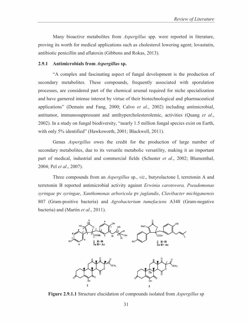
Many bioactive metabolites from Aspergillus spp. were reported in literature,
proving its worth for medical applications such as cholesterol lowering agent; lovastatin,
antibiotic penicillin and aflatoxin (Gibbons and Rokas, 2013).
2.9.1 Antimicrobials from Aspergillus sp.
“A complex and fascinating aspect of fungal development is the production of
secondary metabolites. These compounds, frequently associated with sporulation
processes, are considered part of the chemical arsenal required for niche specialization
and have garnered intense interest by virtue of their biotechnological and pharmaceutical
applications” (Demain and Fang, 2000; Calvo et al., 2002) including antimicrobial,
antitumor, immunosuppressant and antihypercholesterolemic, activities (Quang et al.,
2002). In a study on fungal biodiversity, “nearly 1.5 million fungal species exist on Earth,
with only 5% identified” (Hawksworth, 2001; Blackwell, 2011).
Genus Aspergillus owes the credit for the production of large number of
secondary metabolites, due to its versatile metabolic versatility, making it an important
part of medical, industrial and commercial fields (Schuster et al., 2002; Blumenthal,
2004; Pel et al., 2007).
Three compounds from an Aspergillus sp., viz., butyrolactone I, terretonin A and
terretonin B reported antimicrobial activity against Erwinia carotovora, Pseudomonas
syringae pv syringae, Xanthomonas arboricola pv juglandis, Clavibacter michiganensis
807 (Gram-positive bacteria) and Agrobacterium tumefaciens A348 (Gram-negative
bacteria) and (Martin et al., 2011).
Figure 2.9.1.1 Structure elucidation of compounds isolated from Aspergillus sp

Review of Literature
32
Diverse antimicrobial activity was found in the crude extract of Aspergillus
fumigatus where the ten compounds (Figure 2.9.1.2) namely “ linoleic acid (1); R (-)–
glycerol monolinoleate (2); bis-dethio–(bis methyl-thio)-gliotoxin; FR-49175 (3);
fumiquinazoline–F (4); fumiquanzoline–D (5); (Z-Z)-N,N–[1-[(4-Hydroxy phenyl)-
methylene]-2-[(4-Hydroxyphenyl)-methylene]-1,2-ethanediyl]-bis-formamide (6),
pyrazoline-3-one trimer (7), Tricho-9-ene-2α, 3α, 11α, 16-tetraol (8), 2-deoxy-thymidine
(9); and cerebroside (10)” against eleven microbial test strains (Shaaban et al., 2013).
Figure 2.9.1.2 Structure of some compounds isolated from Aspergillus fumigatus

Review of Literature
33
Two new compounds obtained through microbial transformation of Sch-64235
which is produced by the endophytic fungus Phomopsis sp. showed better IC50 value
against colonic epithelial cancer cells (Adelin et al., 2012). Dihydroxymethyl pyranone
isolated from Aspergillus candidus significantly showed potent antifungal activity, high
antioxidative activity when compared with α-tocopherol, and antitumor activity against
HEp-2 and HepG2 cells with IC50 of 7µg/ml (Elaasser et al., 2011).
Prenylated indole alkaloids (Figure 2.9.1.3), carneamides (1-3), quinazolinone
derivatives, carnequinazolines (5-7), aryl c-glycosides, carnequinazolines (5-7), aryl C-
lycosides, carnemycin (8,9) and a drimane sesquiteroenoid (10) from Aspergillus carneus
(Trichomoeae) have been shown to possess potent antimicrobial activities. These were
also examined for their cytotoxic activities (Zhuravleva et al., 2012)
Figure 2.9.1.3 Structure of some compounds isolated from Aspergillus carneus

Review of Literature
34
Similarly many more compounds have been isolated from Aspergillus till date
having various biological activities. In a study, Aspergillus janus was reported to
produce two compounds; janoxepin (1) and brevicompanine B (2) which possessed anti
malarial activity against Plasmodium falciparum with IC50 values of 28 and 35 mg/ml,
respectively (Sporge et al., 2005). Three new cyclopentapeptides; versicoloritides,
orcinol tetramer, tetracinol and two new lactones together with known metabolites:
diorcinol, glyantryine and cordyol C; obtained from extracellular culture broth of
Aspergillus versicolor were active against Staphylococcus aureus, Escherichia coli,
Enterobacter aerogens, Bacillus subtilis, Pseudomonas aeruginosa and Candida albicans
and proved to be better antimicrobial agents (Zhuang et al., 2011). Similarly lists of
endophytic strains have been known to produce various compounds with antimicrobial
activity. An endophyitc fungus, Aspergillus fumigatus CY018 isolated from Cynodon
dactylon, produced novel metabolites such as asperfumoid (1) and aspertumin (2) with
potent antifungal activity against Candida albicans (Liu et al., 2004).
Similarly, bioactive secondary metabolites isolated from Aspergillus ochraceus
identified as Campholene aldehyde, Lucenin-2 and 6-Ethyl oct-3-yl-2-ethylhexyl ester
showed potential antimicrobial activity against human pathogens like Klebsiella,
Pseudomonas, Staphylococcus aureus and Micrococcus (Meenupriya and Thangaraj, 2011).
2.9.2 Antimicrobials from Penicillium sp
Members of the Penicillium spp. are filamentous fungi. It was the discovery of
wonder drug penicillin that revolutionized the field of antibiotics and directed the interest
of the researchers towards natural resources having different biological activities. Since
then, a lot of bioactive compounds were purified from different fungi including
Penicillium janczewskii, Penicillium canescens (Kozlovskii et al., 1997; Furtado et al.,
2005), Penicillium sclerotiorium, Penicillium janthinellum, Penicillium citrinum
(Takahashi et al., 2008) and Myrothecium cinctum (Kobayashi et al., 2004) etc.
Penicillium spp. is widespread and is found in soil, decaying vegetation, and the air.
Multi drug resistant organisms such as methicillin resistant Staphylococcus
aureus, rifampicin-resistant S. aureus, Staphylococcus aureus and vancomycin resistant
Enterococcus faecium and Cryptococcus neofromans were found to be sensitive to

Review of Literature
35
Citrinin, a compound isolated from Penicillium sp FF01 associated with marine Fijian
sponge Melophlus sp. Citrinin also exhibited cytotoxicity against brine shrimp larvae,
indicating Penicillium sp as a promising source of natural bioactive metabolites
(Subramani et al., 2013) Six different compounds were obtained from Penicillium spp.
isolated from Brazilian cerrado soil having broad spectrum antimicrobial activity against
Listeria monoytogenes ATCC 19115, Streptococcus pyogenes ATCC 19615, Salmonella
typhimurium ATCC 13311, Candida albicans ATCC 18804 and Bacillus cereus ATCC
11778 making these compounds, a suitable starting compounds for biotechnological use
as a new drug lead (Petit et al., 2009). Not only Penicillium spp isolated from soil have
been evaluated for their different biological activities but the endophytic strains have also
been screened for their biological potential.
A strain of Penicillium sp. isolated from Mauritia flexuosa roots has been
reported to produce seven antimicrobial compounds namely glandicoline B (1),
ergosterol (2), brassicasterol (3), ergosterol peroxide (4), cerevisterol (5), mannitol (6)
and 1-O-α-D-glucopyranoside (7) (Figure 2.9.2.1) (Koolen et al., 2012)
Figure 2.9.2.1 Structure of some compounds isolated from Penicillium sp.

Review of Literature
36
Two endophytic Penicillium spp. isolated from Aristolochia macrophylla leaves
produced six metabolites: Orcinol, Cyclo(L-Pro–L-Val), Uracil, 4-Hydroxymellein, 8-
Methoxymellein and 5-Hydroxymellein exhibited potential bioactivity against fungi such
as Cladosporium cladosporioides and Cladosporium sphaerospermum and also possess
acetylcholinestrase inhibitory activity (Oliveira et al., 2009).
The most common Penicillium citrinum spp islolated from various sources have
been screened and different compounds have been isolated having antimicrobial activity.
Such potential was found in a sea derived fungus Penicillium citrinum which produced
five novel polyketides along with thirteen different known compounds, having better
antimicrobial potential with minimum inhibitory concentration of 16 µg/ml against
Staphylococcus aureus, methicillin-resistant Staphylococcus aureus and Candida
albicans (Fig 2.9.2.2). Not only antimicrobial but the various compounds have been
studied for their insecticidal properties. Nine new yaequinolones with insecticidal
properties have been reported from Penicillium sp. FKI-2140 (Uchida et al., 2006).
Figure 2.9.2.2 Structure of some compounds 1-18 isolated from Penicillium citrinum
PSU-F51

Review of Literature
37
Likewise, another another Penicillium sp. from Huperzia serrata produced a
novel compound, viz., (2S)-2,3-dihydro-7-hydroxy-6,8-dimethyl-2-[(E)-prop-1-enyl]-
chroman-4-one with anticancerous potential against some cancer cell lines such as
HepG2 and HeLa (Ying et al., 2011).
A lot more compounds have been isolated from Penicillium sp from different
environments but a lot remain untapped from various environments. As bacterial
resistance is spreading throughout the world, especially in all health care associated
pathogens revealing the steadily decreasing potencies of prevalent antibiotics (Gould,
2008). So there is a need to explore more microorganisms which may help in isolating
some novel biologically active compounds and to expand the spectrum of suitable
organisms which can be useful to meet the requirements of potent antimicrobials. The
present study was thus planned to isolate fungi from soil samples collected from different
regions of Punjab, India and to screen them for their antimicrobial activity. Different
physiochemical parameters such as pH, incubation period, temperature, carbon and
nitrogen sources play a significant role for antimicrobial production, so the present study
is directed towards optimization of such factors to enhance the production of
antimicrobial agents. The potential antimicrobial compounds (novel) have been isolated
and characterized. The purified compounds have been tested for various antimicrobial
studies such as MIC, VCC and post antibiotic effect. Biosafety of the purified compounds
has been evaluated by various methods such as Ames and MTT assay to check the
toxicity of these compounds. Further, the antimicrobial activity of these purified
compounds will be compared with some of the commercially available antibiotics.


![Literature Review - INFLIBNETshodhganga.inflibnet.ac.in/bitstream/10603/8178/11/11_chapter 02.pdf · CHAPTER 2 Literature Review ... Lee and sheriff [11] ... refrigeration cycles](https://static.fdocuments.in/doc/165x107/5a9e414b7f8b9a21488dd6fb/literature-review-02pdfchapter-2-literature-review-lee-and-sheriff-11-.jpg)