2 The Role of Coagulation in Arterial and Venous Thrombosis
21
From: Contemporary Cardiology: Antithrombotic Drug Therapy in Cardiovascular Disease Edited by: A.T. Askari and A.M. Lincoff (eds.), DOI 10.1007/978-1-60327-235-3_2 © Humana Press, a part of Springer Science+Business Media, LLC 2010 2 The Role of Coagulation in Arterial and Venous Thrombosis Kandice Kottke-Marchant CONTENTS Introduction Clinical Manifestations of Arterial and Venous Thrombosis Coagulation Physiology Regulation of Coagulation Coagulation Dysregulation in Arterial and Venous Thrombosis Summary References ABSTRACT The coagulation cascade is integral to the hemostatic process and serves to limit the amount of blood loss during trauma. However, derangements in this process can result in venous thrombosis and con- tribute to the development of arterial atherothrombotic disease. Indeed, in arterial thrombosis, the effects of thrombin may extend far beyond coagulation activation and play an important role in activation of a wide variety of cells and the inflammatory processes. Venous thrombosis and arterial thrombotic diseases have traditionally been thought of as separate processes; however, they share many similarities in patho- physiology and risk factors. The activation of the coagulation cascade underlies both arterial and venous thrombosis, and biological triggers, such as inflammation, prothrombotic microparticles, and endothelial activation provide a plausible link between the two. Key words: Coagulation cascade; Mechanism; Pathophysiology; Platelets; Thrombosis INTRODUCTION Hemostasis and thrombosis are related processes involving the coagulation system, platelets, endothe- lial cells, and the vascular wall. Indeed, Rudolf VIrchow, in the 1800s, postulated a triad of causes for thrombin formation: changes in the composition of blood, alterations in the vessel wall, and disruption of blood flow (1). Physiological hemostasis occurs as a protective response to vascular injury; exposure of blood components to subendothelial proteins stimulates activation of platelets and production of the key coagulation enzyme thrombin leading to formation of a fibrin meshwork (2,3). This process prevents 19
Transcript of 2 The Role of Coagulation in Arterial and Venous Thrombosis

BookID 148448_ChapID 2_Proof# 1 - 07/10/2009
From: Contemporary Cardiology: Antithrombotic Drug Therapy in Cardiovascular DiseaseEdited by: A.T. Askari and A.M. Lincoff (eds.), DOI 10.1007/978-1-60327-235-3_2
© Humana Press, a part of Springer Science+Business Media, LLC 2010
2 The Role of Coagulation in Arterial and Venous Thrombosis
Kandice Kottke-Marchant
Contents
IntroductionClinical Manifestations of Arterial and Venous ThrombosisCoagulation PhysiologyRegulation of CoagulationCoagulation Dysregulation in Arterial and Venous ThrombosisSummaryReferences
AbstrAct The coagulation cascade is integral to the hemostatic process and serves to limit the amount of blood loss during trauma. However, derangements in this process can result in venous thrombosis and con-tribute to the development of arterial atherothrombotic disease. Indeed, in arterial thrombosis, the effects of thrombin may extend far beyond coagulation activation and play an important role in activation of a wide variety of cells and the inflammatory processes. Venous thrombosis and arterial thrombotic diseases have traditionally been thought of as separate processes; however, they share many similarities in patho-physiology and risk factors. The activation of the coagulation cascade underlies both arterial and venous thrombosis, and biological triggers, such as inflammation, prothrombotic microparticles, and endothelial activation provide a plausible link between the two.
Key words: Coagulation cascade; Mechanism; Pathophysiology; Platelets; Thrombosis
INTRODUCTION
Hemostasis and thrombosis are related processes involving the coagulation system, platelets, endothe-lial cells, and the vascular wall. Indeed, Rudolf VIrchow, in the 1800s, postulated a triad of causes for thrombin formation: changes in the composition of blood, alterations in the vessel wall, and disruption of blood flow (1). Physiological hemostasis occurs as a protective response to vascular injury; exposure of blood components to subendothelial proteins stimulates activation of platelets and production of the key coagulation enzyme thrombin leading to formation of a fibrin meshwork (2,3). This process prevents
19
Antihrombotic Drug Therapy in Cardiovascular Disease
BookID 148448_ChapID 2_Proof# 1 - 07/10/2009 BookID 148448_ChapID 2_Proof# 1 - 07/10/2009
excessive bleeding or exsanguination after vascular injury or trauma. On the other hand, thrombosis can be considered “hemostasis in the wrong place and at the wrong time” (4), with dysregulation of one or more elements of the hemostatic system contributing to formation of a platelet-fibrin thrombus that often occludes blood vessels, leading to pathologic complications (5). If the thrombus occurs in the arterial system, distal ischemia can lead to acute coronary syndromes, myocardial infarctions, stroke, and peripheral extremity necrosis. Thrombus formation in the venous system, typically deep venous thrombosis, leads to local tissue congestion and decreased venous blood return, but dislodged thromboemboli may result in the potentially devastating complications of pulmonary infarction or, paradoxically, stroke.
CLINICAL MANIFESTATIONS OF ARTERIAL AND VENOUS THROMBOSIS
Thrombosis in the arterial system typically occurs superimposed on vascular abnormalities that are the result of other diseases, such as atherosclerosis or vasculitis (6,7). Arterial atherosclerotic plaques are well known to liberate prothrombotic and inflammatory substances, such as oxidized lipids (8), and express procoagulant molecules such as tissue factor (9). While coagulation activation is undoubt-edly involved in arterial thrombosis, platelet activation plays a crucial role, as described in Chap. 1, and antiplatelet drugs are the predominant pharmacologic therapy for this class of disorders (10).
Arterial thrombosis in this setting, leading to vascular occlusion, can result in vascular-bed-specific pathologies. Coronary artery occlusion results in acute coronary syndromes or myocardial infarction, while cerebrovascular occlusion results in thrombotic stroke, and occlusion of peripheral arteries leads to peripheral arterial disease and gangrene. Paradoxical embolism of a venous thrombus through a patent foramen ovale may also lead to cerebrovascular thrombosis (11).
In contrast to arterial thrombosis, venous thrombotic disorders are not usually associated with underlying vascular pathologies. Instead, venous thrombosis is associated with venous stasis or con-genital dysregulation of coagulation proteins or natural anticoagulants (12). Deep vein thrombi, most often involving the veins of the lower extremities, may dislodge and result in embolism to the pulmo-nary vasculature, with segmental pulmonary infarction and an attendant high morbidity rate (13). Prolonged atrial fibrillation is often complicated by thrombosis of the atrial appendages. While multi-factorial, stasis is a contributing factor in thrombosis observed with atrial fibrillation, the role of the coagulation system is highlighted by success with targeted anticoagulant therapies (14,15).
While a direct clinical link between arterial and venous thrombosis has not been demonstrated, atherosclerosis and venous thromboembolic disease share similar risk factors, such as age, obesity, diabetes mellitus, and the metabolic syndrome (16). The activation of the coagulation cascade under-lies both arterial and venous thrombosis, and biological triggers, such as inflammation, prothrombotic microparticles, and endothelial activation provide a plausible link between the two. Some disorders, such as the antiphospholipid antibody syndrome (APS) and heparin-induced thrombocytopenia (HIT), have clear clinical associations with both arterial and venous thrombosis (17). The thrombosis in HIT, for example, is thought to be multifactorial, with platelet activation, endothelial activation, and thrombin generation all contributing to the pathophysiology (18). In the following sections, the physi-ology of coagulation and its regulation will be described, followed by a discussion of coagulation-associated risk factors for both arterial and venous thrombosis.
COAGULATION PHYSIOLOGY
The coagulation system, together with endothelial cells and platelets, is responsible for maintaining blood in a fluid state, but, when activated, rapidly results in development of a fibrin clot by conversion of the plasma protein, fibrinogen, to the polymer fibrin by the key enzyme, thrombin (2,3) (See Fig. 1).
20

Antithrombotic Drug Therapy in Cardiology: The Role of Coagulation in Arterial and Venous Thrombosis
BookID 148448_ChapID 2_Proof# 1 - 07/10/2009
In addition to production of fibrin, thrombin has wide-reaching functions that range from platelet activa-tion to stimulation of endothelial cells, vascular smooth muscle cells (VSMC), monocytes, T lymphocytes, and fibroblasts, so the production and inhibition of thrombin is tightly regulated (19,20).
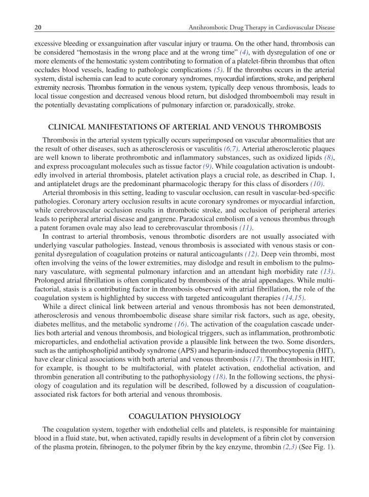
The coagulation system comprises proenzymes that typically reside in the intravascular space in an inactivated state together with cofactors, cations, and cell-associated phospholipid. Coagulation can be activated by two principal mechanisms, the intrinsic and the extrinsic pathways that converge to produce thrombin by the common pathway through a series of inter-related enzymatic reactions (21,22) (See Fig. 2). These classical pathways form the basis of the two most frequently performed coagulation tests, the prothrombin time (PT), which measures the extrinsic and common pathways; and the activated partial thromboplastin time (APTT), which measures the intrinsic and common pathways. However, the physiologic activation of coagulation in vivo is not so segregated, with the initiation phase occurring through tissue factor exposed during vascular injury leading to a subsequent propagation phase and further amplification of the process by thrombin, due to activation of factors, V, VIII, XI (23).
The Intrinsic PathwayOne mechanism of coagulation activation is the intrinsic pathway, so called because its compo-
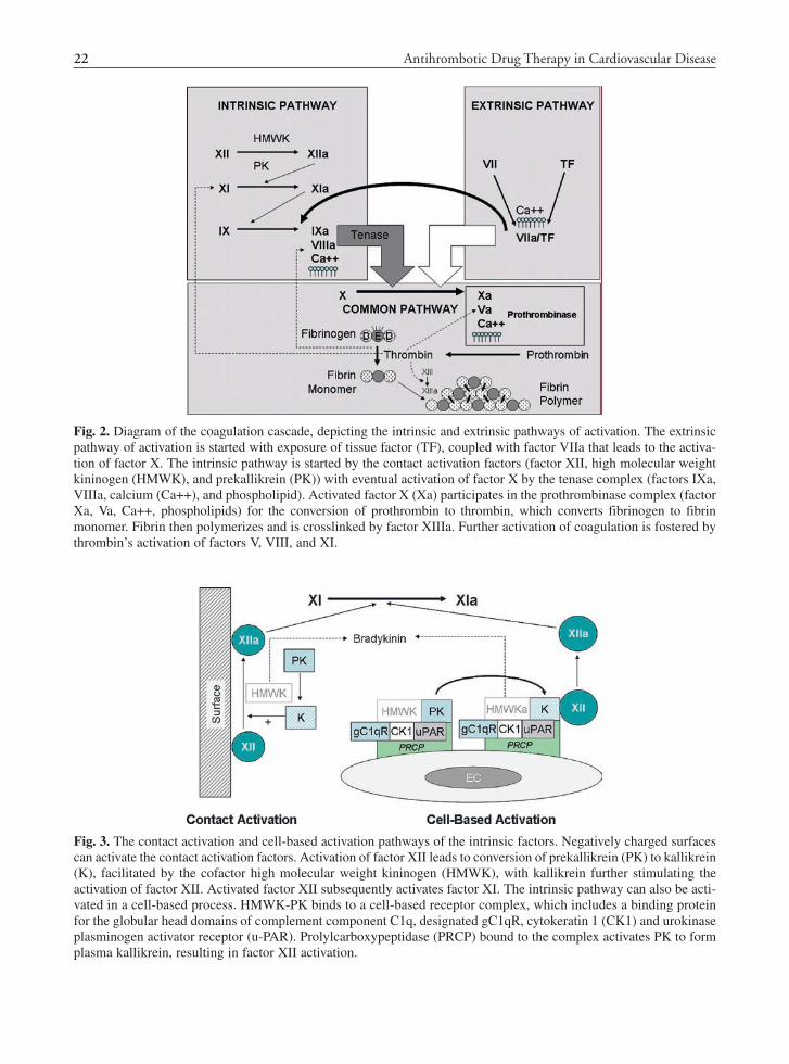
nents, factors XII, XI, IX, VIII, prekallikrein (PK), and high molecular weight kininogen (HMWK), are all plasma proteins and are “intrinsic” to the lumen of the blood vessel (24). The intrinsic pathway can be activated when factor XII undergoes autoactivation to factor XIIa on a negatively charged surface through a process called “contact activation” (25,26) (See Fig. 3). Negatively charged surfaces
Fig. 1. The elements of the hemostatic system, with the relationship between endothelial cells, the vascular wall, coagulation, coagulation inhibitors, and platelet activation. AT antithrombin, ProS protein S, APC activated protein C, TFPI tissue factor pathway inhibitor, ECM extracellular matrix.
21
Antihrombotic Drug Therapy in Cardiovascular Disease
BookID 148448_ChapID 2_Proof# 1 - 07/10/2009 BookID 148448_ChapID 2_Proof# 1 - 07/10/2009
Fig. 2. Diagram of the coagulation cascade, depicting the intrinsic and extrinsic pathways of activation. The extrinsic pathway of activation is started with exposure of tissue factor (TF), coupled with factor VIIa that leads to the activa-tion of factor X. The intrinsic pathway is started by the contact activation factors (factor XII, high molecular weight kininogen (HMWK), and prekallikrein (PK)) with eventual activation of factor X by the tenase complex (factors IXa, VIIIa, calcium (Ca++), and phospholipid). Activated factor X (Xa) participates in the prothrombinase complex (factor Xa, Va, Ca++, phospholipids) for the conversion of prothrombin to thrombin, which converts fibrinogen to fibrin monomer. Fibrin then polymerizes and is crosslinked by factor XIIIa. Further activation of coagulation is fostered by thrombin’s activation of factors V, VIII, and XI.
Fig. 3. The contact activation and cell-based activation pathways of the intrinsic factors. Negatively charged surfaces can activate the contact activation factors. Activation of factor XII leads to conversion of prekallikrein (PK) to kallikrein (K), facilitated by the cofactor high molecular weight kininogen (HMWK), with kallikrein further stimulating the activation of factor XII. Activated factor XII subsequently activates factor XI. The intrinsic pathway can also be acti-vated in a cell-based process. HMWK-PK binds to a cell-based receptor complex, which includes a binding protein for the globular head domains of complement component C1q, designated gC1qR, cytokeratin 1 (CK1) and urokinase plasminogen activator receptor (u-PAR). Prolylcarboxypeptidase (PRCP) bound to the complex activates PK to form plasma kallikrein, resulting in factor XII activation.
22

Antithrombotic Drug Therapy in Cardiology: The Role of Coagulation in Arterial and Venous Thrombosis
BookID 148448_ChapID 2_Proof# 1 - 07/10/2009
include the artificial reagents in the APTT assay, such as kaolin, celite, and silica, which explains the dependence of the APTT on the contact activation factors. However, the intrinsic pathway can be activated in vivo by substances such as articular cartilage, endotoxin, L-homocysteine, and the developing thrombus (27,28).
Activation of factor XII leads to conversion of PK to kallikrein, facilitated by the cofactor HMWK, with kallikrein further stimulating the activation of factor XII (27). Activated factor XII subsequently activates factor XI, with factor XIa activating factor IX. Factor IXa, together with factor VIIIa, phos-pholipids, and calcium form the tenase complex that activates factor X.
The intrinsic pathway can also be activated in a cell-based process, as the components can assem-ble on endothelial cells, platelets, and granulocytes (27,29). HMWK-PK binds to a cell-based receptor complex, which includes a binding protein for the globular head domains of complement component C1q, designated gC1qR, cytokeratin 1 (CK1) and urokinase plasminogen activator receptor (u-PAR) (27) (See Fig. 3). Prolylcarboxypeptidase (PRCP) bound to the complex activates PK to form plasma kallikrein, resulting in factor XII activation (30). Apart from activation of factor XI, this complex is involved in other physiologic activities through formation of bradykinin, which participates in fibri-nolysis activation and the production of antiplatelet molecules, nitric oxide and prostacyclin, from endothelial cells (27).
The role of the contact activation factors, XII, HMWK, and PK, has traditionally been discounted in physiological hemostasis, as patients with deficiencies of these proteins do not manifest a bleeding dia-thesis (31). Studies have shown that factor IX is principally activated not by the contact factors but by the tissue factor/factor VIIa complex, as described later, and physiologic hemostasis is thought to proceed principally through that extrinsic pathway (32). However, recent studies with factor XI and XII knockout mice have indicated that deficiency of these factors may impair formation of occlusive thrombi in arterial injury models and may be attractive targets for new antithrombotic agents (26,28,33).
The Extrinsic PathwayThe so-called Extrinsic Pathway of coagulation, comprising tissue factor and factor VII, is acti-
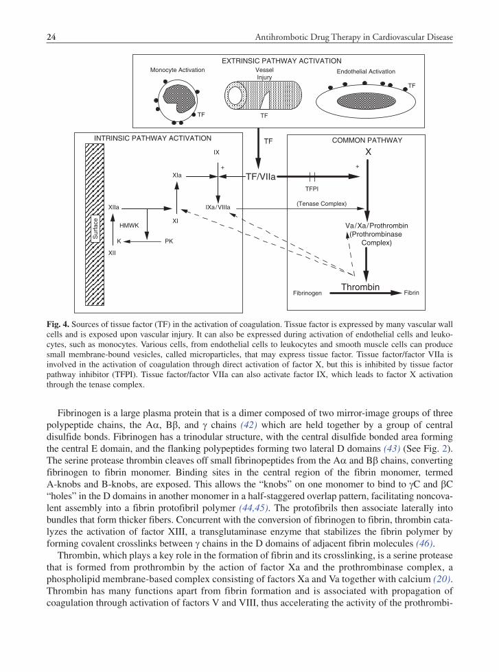
vated by tissue injury or cellular activation and is likely the primary mechanism for in vivo hemostasis (2,34). Tissue factor, complexed with activated factor VIIa in the presence of calcium and phospholipids, activates coagulation through conversion of factor X to Xa and also through activation of factor IX to IXa (35) (See Fig. 4).
Tissue factor is an intrinsic membrane glycoprotein expressed on many vascular wall cells, such as smooth muscle cells, pericytes, and fibroblasts (34,36). Constitutively expressed cell-based tissue factor can be exposed to the blood following vascular injury, but tissue factor expression can also be induced on vascular endothelial cells and leukocytes by thrombin and inflammatory stimuli (34,37,38). This induc-tion is well described for monocytes, but also may occur in neutrophils and eosinophils and is thought to play a role in the thrombotic consequences of disseminated intravascular coagulation and sepsis (37,39). Activation of many cells leads to production of minute membrane-bound microparticles, which may be a source of circulating tissue factor activity (40). Platelets may also play a role, as recent studies have shown that platelets can be stimulated to produce TF mRNA and synthesize TF protein (34,41).
Fibrin Formation and Common Pathway ActivationThe purpose of coagulation is the formation of an insoluble fibrin polymer as a hemostatic plug.
To this end, the transformation of plasma-based fibrinogen to crosslinked fibrin is accomplished by several mechanisms and is tightly regulated.
23
Antihrombotic Drug Therapy in Cardiovascular Disease
BookID 148448_ChapID 2_Proof# 1 - 07/10/2009 BookID 148448_ChapID 2_Proof# 1 - 07/10/2009
Fibrinogen is a large plasma protein that is a dimer composed of two mirror-image groups of three polypeptide chains, the Aa, Bb, and g chains (42) which are held together by a group of central disulfide bonds. Fibrinogen has a trinodular structure, with the central disulfide bonded area forming the central E domain, and the flanking polypeptides forming two lateral D domains (43) (See Fig. 2). The serine protease thrombin cleaves off small fibrinopeptides from the Aa and Bb chains, converting fibrinogen to fibrin monomer. Binding sites in the central region of the fibrin monomer, termed A-knobs and B-knobs, are exposed. This allows the “knobs” on one monomer to bind to gC and bC “holes” in the D domains in another monomer in a half-staggered overlap pattern, facilitating noncova-lent assembly into a fibrin protofibril polymer (44,45). The protofibrils then associate laterally into bundles that form thicker fibers. Concurrent with the conversion of fibrinogen to fibrin, thrombin cata-lyzes the activation of factor XIII, a transglutaminase enzyme that stabilizes the fibrin polymer by forming covalent crosslinks between g chains in the D domains of adjacent fibrin molecules (46).
Thrombin, which plays a key role in the formation of fibrin and its crosslinking, is a serine protease that is formed from prothrombin by the action of factor Xa and the prothrombinase complex, a phospholipid membrane-based complex consisting of factors Xa and Va together with calcium (20). Thrombin has many functions apart from fibrin formation and is associated with propagation of coagulation through activation of factors V and VIII, thus accelerating the activity of the prothrombi-
VesselInjury
TF/VIIa
X
Va/Xa/Prothrombin(Prothrombinase
Complex)
ThrombinFibrinogen Fibrin
IX
IXa/VIIIa
XI
XIa
TF
TFPI
++
Sur
face
XII
XIIa
HMWK
Monocyte Activation Endothelial ActivatIon
TF
TF
TF
EXTRINSIC PATHWAY ACTIVATION
INTRINSIC PATHWAY ACTIVATION COMMON PATHWAY
PKK
(Tenase Complex)
Fig. 4. Sources of tissue factor (TF) in the activation of coagulation. Tissue factor is expressed by many vascular wall cells and is exposed upon vascular injury. It can also be expressed during activation of endothelial cells and leuko-cytes, such as monocytes. Various cells, from endothelial cells to leukocytes and smooth muscle cells can produce small membrane-bound vesicles, called microparticles, that may express tissue factor. Tissue factor/factor VIIa is involved in the activation of coagulation through direct activation of factor X, but this is inhibited by tissue factor pathway inhibitor (TFPI). Tissue factor/factor VIIa can also activate factor IX, which leads to factor X activation through the tenase complex.
24

Antithrombotic Drug Therapy in Cardiology: The Role of Coagulation in Arterial and Venous Thrombosis
BookID 148448_ChapID 2_Proof# 1 - 07/10/2009
nase and tenase complexes, respectively (47) (Fig. 2). Thrombin is further involved with amplification of coagulation through its direct activation of factor XI. Indeed, individuals with factor XI deficiency typically only have bleeding when large amounts of thrombin are formed, such as during surgical procedures (48).
Thrombin, through a series of G-coupled protease-activated receptors (PARs), also activates plate-lets and other cells, such as endothelial cells and monocytes (19). Upregulation of PARs in smooth muscle cells is thought to play a key role in the pathogenesis of atherosclerosis and restenosis (49). Due to its wide-ranging effects, the activity of thrombin is closely regulated, as discussed later.
The activation of thrombin requires the action of factor Xa, another serine protease, in the prothrombinase complex resulting in cleavage of a peptide fragment, prothrombin fragment F1 + 2, from prothrombin (50). The formation of active factor Xa from the proenzyme factor X is considered the start of the common pathway and can be triggered by both tissue factor/factor VIIa from the extrinsic pathway and by the tenase complex (factors IXa, VIIIa, calcium, and phospholipids) from the intrinsic pathway (2). Having two separate mechanisms for factor Xa activation is thought to be associated with two temporal stages of blood coagulation (32,51), the initiation and propagation stages. In the initiation stage, factor X is initially activated by tissue factor/factor VIIa. This produces small amounts of thrombin, which activates factors V and VIII, leading to further thrombin produc-tion (47). After initiation, an inhibitor, tissue factor pathway inhibitor (TFPI), downregulates the ability of tissue factor/VIIa to activate factor X (52). Coagulation then enters the propagation phase, directing the activity of tissue factor/VIIa toward activation of factor IXa, resulting in a switch of the pri-mary activation of factor X to be via the intrinsic pathway tenase complex (51). Further thrombin production leads to the activation of factor XIa, which amplifies the propagation phase further.
Lessons from Studying In Vivo Coagulation ActivationMost studies that have been used to define the coagulation process have been performed in vitro,
often with highly purified single protein components. While these have helped delineate the processes involved, they may not necessarily indicate the relative importance of coagulation factors in hemos-tasis, hemorrhage, or thrombosis (145). For example, the processes of hemostasis and thrombosis may differ, as in vivo studies have shown defective thrombus formation in the absence of factors XII and XI, while patients with factor XII deficiency do not display a hemostatic defect (26,28). The clini-cal phenotype for deficiency of factors VIII and IX is hemophilia, a significant bleeding disorder, so this helps to indicate the importance of factor X activation by the tenase complex compared to tissue factor/VIIa. Contrary to the dogma that tissue factor is extrinsic to the blood vessel lumen, circulating tissue factor-bearing microparticles have been shown to be involved with in vivo thrombus develop-ment (53). It has been shown from in vivo studies that platelet thrombus formation and fibrin forma-tion may occur more simultaneously than sequentially, and other studies question the crucial role of platelet membrane phospholipids in the formation of fibrin (54,55). While these studies may challenge the classical paradigm of coagulation and its relationship with platelets, they also serve to underscore the complex and inter-related nature of the hemostatic process.
REGULATION OF COAGULATION
Coagulation activation involves numerous proteins and amplification steps, so its activation and propagation is closely regulated. The activities of thrombin and tissue factor, in particular, have several well-studied inhibitors and/or regulatory mechanisms. These regulatory mechanisms are of
25
Antihrombotic Drug Therapy in Cardiovascular Disease
BookID 148448_ChapID 2_Proof# 1 - 07/10/2009 BookID 148448_ChapID 2_Proof# 1 - 07/10/2009
clinical significance, as defective regulation due to congenital inhibitor deficiency may lead to clinical thrombosis, while pharmacologic use of the inhibitors has successfully controlled thrombosis. The endothelial cells that line the blood vessels also are able to regulate the function of many aspects of hemostasis from coagulation to platelet activation to fibrinolysis
Regulation of Tissue FactorClassically, since tissue factor was thought only to exist in locations extrinsic to the blood vessel
lumen, regulation of tissue factor activity has been thought to involve primarily vessel injury with exposure of constitutively expressed tissue factor to flowing blood. While this process is undoubtedly involved, other mechanisms for regulation of tissue factor exposure and activity likely play a role. Tissue factor can exist in an encrypted, inactive form, or a decrypted, active form; decryption may involve conformational or disulfide bonding changes (56,57). The major regulation of tissue factor/factor VIIa activity is through tissue factor pathway inhibitor b (TFPIb), a Kunitz-type proteinase inhibitor (52). TFPIb forms a quaternary complex with tissue factor, factors VIIa and Xa; the factor Xa within the complex is deacti-vated, but binding to adjacent tissue factor/factor VIIa prevents further factor X activation (38,58). TFPI may also further downregulate coagulation by causing TF-expressing cells to internalize cell surface TF/factor VIIa complexes (59). The importance of TFPI as a hemostatic regulator is highlighted by the embryonic lethality due to hemorrhage observed in TFPI knockout mice (60).
Regulation of ThrombinThe activity of thrombin is closely regulated by several direct and indirect mechanisms; these
involve antithrombin, a direct-acting inhibitor, or take advantage of thrombin’s proteolytic activities through the activation of protein C. Several naturally occurring thrombin inhibitory molecules have been developed by blood-sucking insects or envenoming snakes. One such molecule, hirudin, is a very potent direct thrombin inhibitor (DTI); OTIs have been developed as successful commercial anti-thrombotic agents, highlighting the importance of regulating endogenous thrombin activity (61).
Antithrombin (formerly called Antithrombin III) is a serine protease inhibitor that is a direct inhibitor of thrombin and other serine proteases (2,20). The reactive site loop (P1–P17) of antithrombin includes a scissile P
1-P
1¢ (Arg393-Ser394) bond that resembles the substrate for thrombin and other serine
proteases (62,63). Once thrombin cleaves the bond, the protease is covalently linked to the P1 residue and an inactive thrombin/antithrombin complex is formed (See Fig. 5). The ability of antithrombin to inhibit thrombin is accelerated approximately 1000-fold by heparin binding to arginine residues in the D-helix of antithrombin, with a resultant conformational change of the P1-P17 loop and exposure of the P1-P1¢ reactive center (64,65). The inactivated thrombin/antithrombin complex dissociates from heparin, allowing heparin to bind to another antithrombin molecule and catalyze the inactivation of yet more thrombin. In vivo, the luminal surface of endothelial cells is rich in heparan sulfate; bound antithrombin is thus maintained in an active conformation, ready to facilitate the rapid inactivation of thrombin.
Another thrombin regulatory mechanism is the thrombomodulin/protein C system, a multimolecular system that regulates blood coagulation, principally through the proteolytic degradation of activated factors V (fVa) and VIII (fVIIIa) (20,66,67) (See Fig. 5). Thrombin binds to an endothelial cell recep-tor, thrombomodulin, via the anion-binding exosite (68) and through conformational changes leads to a change in thrombin’s substrate specificity from fibrinogen to protein C with activation of protein C (69). Protein C interacts with the thrombomodulin/thrombin complex by binding to endothelial cell protein C receptor (EPCR), which appears to be an important step in physiological protein C activa-tion (70). In addition to activating protein C, the thrombomodulin/thrombin complex also is able to
26

Antithrombotic Drug Therapy in Cardiology: The Role of Coagulation in Arterial and Venous Thrombosis
BookID 148448_ChapID 2_Proof# 1 - 07/10/2009
activate thrombin activatable fibrinolysis inhibitor (TAFI), which results in inhibition of fibrin degra-dation and may also have a broad anti-inflammatory role (71,72). Activated protein C is released from the EPCR, and combines with protein S, another vitamin K-dependent factor, on endothelial or platelet phospholipid surfaces (66,67). This activated protein C complex is able to degrade factors Va and VIIIa, thus slowing the procoagulant drive (2).
Protein Z SystemAnother inhibitory mechanism of the coagulation system has been described, namely the protein
Z system. Protein Z is a vitamin-K dependent protein, with an inhibitor, known as protein Z-dependent protease inhibitor (ZPI) (73). ZPI has been shown to inhibit factor Xa in a process that requires protein Z, calcium, and phospholipids (74). ZPI has further been shown to inhibit factors XIa and IXa in a mechanism not requiring protein Z, calcium, or phospholipids (75,76). This inhibitory mechanism has been shown to play a role in hemostasis from in vitro studies and mouse models, but the role of ZPI and protein Z as an anticoagulant system in humans remains to be fully elucidated (73).
Role of EndotheliumEndothelial cells are involved in many aspects of coagulation regulation. As indicated earlier,
heparan sulfate and thrombomodulin are involved in inhibition of thrombin activity. Endothelial cells also inhibit platelet function through release of nitric oxide and prostacyclin and have profibrinolytic function through release of tissue plasminogen activator (tPA) (77). However, when activated by thrombin or inflammatory cytokines, endothelial cells can facilitate coagulation through expression of tissue factor, expression of procoagulant lipids, release of procoagulant microparticles, downregu-lation of thrombomodulin, and release of von Willebrand factor (78).
Fig. 5. Inhibition of thrombin. Thrombin (Factor IIa) is inhibited by antithrombin (AT) bound to heparans on endothe-lial surfaces. It is also regulated by binding to thrombomodulin (TM) on endothelial cells, where it converts protein C (Pro C) to activated protein C (APC). APC is a protease that degrades factors Va and VIIIa, thus decreasing further thrombin production.
27

Antihrombotic Drug Therapy in Cardiovascular Disease
BookID 148448_ChapID 2_Proof# 1 - 07/10/2009 BookID 148448_ChapID 2_Proof# 1 - 07/10/2009
Role of MicroparticlesMicroparticles are minute membrane-bound vesicles released from cell membranes by an exocyto-
sis process (79). They have been shown to arise from a wide variety of cells, including endothelial cells, platelets, monocytes, and erythrocytes (80). Microparticles are released from the surface of cells following apoptosis or activation; the release can be stimulated by a wide variety of agonists, such as cytokines, thrombin, and endotoxin, but also physical stimuli such as shear stress or hypoxia (79,81). During the formation of microparticles, membrane asymmetry is lost, with an exposure of anionic phospholipids, for example, phosphatidyl serine, to the external microparticle membrane (82). This provides a binding site for coagulation complexes, such as the tenase and prothrombinase complexes. Because microparticles arise from the cell membrane, their protein antigen composition is characteristic of their cell of origin and can be used to identify their source.
Circulating microparticles may contribute to the support of coagulation and development of throm-bosis or may have anticoagulant properties, depending on their number and cell of origin or the pro-ducing stimuli. The microparticle’s anionic phospholipid surface has been shown to support thrombin generation through assembly of procoagulant complexes, and may be associated with clinical proco-agulant states and other diseases (83,84,146). Tissue factor can be expressed by microparticles’ release by some cell types and may further contribute to their procoagulant activity (38,85). Depending on their cell surface antigens, microparticles may also contribute to the recruitment of cells to devel-oping thrombi. For example, monocyte microparticles expressing tissue factor and P selectin glyco-protein ligand-1 have been shown to bind to endothelial cells and platelets and to accumulate with the developing thrombus (86). However, not all microparticles are necessarily procoagulant. Microparticles arising from endothelial cells have been shown to have anticoagulant properties in some circum-stances, with increased expression of TFPI and expression of activated protein C (79,87).
Elevated microparticle levels have been described in many cardiovascular and thrombotic disorders and are often reported to correlate with clinical events (79–81). However, until the laboratory methods for quantifying and characterizing microparticles are more standardized, their use as a diagnostic or prognostic tool will be limited (79,88).
Interaction Between Coagulation and Platelet ActivationA striking feature of the hemostatic system is the degree of redundancy and interdependence of the
activation and regulatory processes involved. Not only are there multiple ways for coagulation to become activated, but there are also numerous and bidirectional associations between the coagulation system and platelets.
The coagulation system supports platelet activation and function in several ways. Thrombin is a potent activator of platelets through activation of PARs (19,89). Fibrinogen provides the bridge for platelet-platelet aggregation through its bivalent binding to the platelet glycoprotein IIb/IIIa (a
IIb/b
3)
receptor (90). Von Willebrand factor, a hemostatic protein released from endothelial cells, supports both platelet adhesion under shear and platelet aggregation (91).
On the other hand, activated platelets play a vital procoagulant role that serves as a link between platelet function and coagulation activation (92). Platelet membrane phospholipids undergo a rear-rangement during activation with a transfer of phosphatidyl serine from the inner table to the outer table of the platelet membrane, providing a binding site for phospholipid-dependent coagulation complexes that activate both factor X and prothrombin. Additionally, the GPIb/V/IX complex is thought to participate in activation of factors XI and XII. The platelet alpha granules release many procoagulant factors during platelet activation, including fibrinogen, von Willebrand factor, and factor V.
28
Antithrombotic Drug Therapy in Cardiology: The Role of Coagulation in Arterial and Venous Thrombosis
BookID 148448_ChapID 2_Proof# 1 - 07/10/2009
COAGULATION DYSREGULATION IN ARTERIAL AND VENOUS THROMBOSIS
The pathophysiology of arterial thrombotic diseases, mainly centered around cardiovascular atherosclerotic sequelae, is largely considered distinct from that of venous thrombotic disorders, such as deep vein thrombosis (DVT). Inflammation, the metabolic syndrome, lipid dysregulation, endothelial dysfunction, and platelet activation are characteristics of arterial thrombotic syndromes. Dysregulation of the coagulation system is thought to play a major role in the pathophysiology of venous thrombosis, highlighted by the well-described association between deficiency of the natural anticoagulants and venous thrombosis (93). However, evaluation of the risk factors for both disor-ders shows some similarities, particularly age and obesity, and there is some evidence that patients with venous thrombosis have a higher risk of cardiovascular disease (94,95). Indeed, some disorders such as antiphospholipid antibodies and HIT are complicated by both arterial and venous thrombo-sis, suggesting a unifying mechanism.
Coagulation Dysregulation and Venous Thromboembolic DiseaseVenous thrombosis typically manifests as a DVT, usually in the lower extremities, or as a pulmo-
nary embolism (PE) due to embolization of the thrombus to the pulmonary vasculature. A combina-tion of vascular stasis and coagulation dysregulation is thought to be principally involved in the pathophysiology of venous thrombosis, as there is usually little underlying vascular pathology. However, inflammatory and fibrinolytic mechanisms and production of tissue factor-bearing micro-particles may contribute (96). Venous thrombosis is relatively common, with an average annual inci-dence rate of 121.5 per 100,000 person-years in the USA and is the third most prevalent cardiovascular disease after coronary artery disease and stroke (97). The risk factors for venous thrombosis have been well studied and include both acquired and congenital risk factors (See Table 1). In general, venous thrombosis is considered a multifactorial or multigenetic disease, with the combination of more than one risk factor increasing the risk of developing venous thrombosis (98).
Congenital Thrombophilia
Recognition of familial tendencies for thrombosis initially led to a search for genetic abnormalities in the coagulation system. The genetic abnormalities first to be described in association with throm-bophilia were mutations leading to loss-of-function of natural anticoagulants, such as antithrombin, protein C, or protein S (99–101). These disorders are rare and heterozygous individuals have an increased risk for venous thromboembolism. In addition, elevated plasma levels of several coagulation proteins, including fibrinogen, factors VIII, IX, and XI, have been described to be associated with an increased risk of venous thrombosis (102). Furthermore, genetic polymorphisms associated with venous thrombosis and arterial vascular disease have been found in many of the procoagulant proteins, including prothrombin, fibrinogen, factors V, VII, XI, and XIII (103,147). Polymorphisms of EPCR, Z Protein inhibitor, fibrinolytic proteins, and enzymes in the transsulfuration pathway lead-ing to high homocysteine levels have also been described, but their association with venous throm-botic disease is not well established. The following discussion will concentrate on the two most well- described venous thrombotic risk factors, Factor V Leiden and the prothrombin G20210A mutation
Factor V Leiden
Patients resistant to the activity of APC were described by Dahlback in 1993 (104), and the molec-ular basis for this defect was shown to be a point mutation in the factor V gene located on chromo-some 1 (1691G-A), which was called Factor V Leiden (FVL) (105). The FVL mutation results in an arginine-glutamine substitution at amino acid 506 (Arg506Gln), the site of the first molecular cleav-
29

Antihrombotic Drug Therapy in Cardiovascular Disease
BookID 148448_ChapID 2_Proof# 1 - 07/10/2009 BookID 148448_ChapID 2_Proof# 1 - 07/10/2009
age of factor Va by APC. This results in diminished activated protein C cleavage of factor Va and continued formation of thrombin by the prothrombinase complex (i.e., APC resistance).
The FVL mutation is fairly common in the Caucasian population with a frequency of 2–15%, but is uncommon in African blacks and Asians (106,107). The mutation is detected in up to 40% of patients with venous thrombosis (103). FVL alone imparts approximately a five to eightfold increased risk for venous thrombosis in heterozygotes and 80-fold in homozygotes (108,109). Clinical studies have shown FVL to be a risk factor for deep venous thrombosis, pulmonary embolism, cerebral vein thrombosis, and superficial thrombophlebitis (110).
Prothrombin G20210A
Due to the central role of thrombin, increased prothrombin levels are a likely risk factor for venous thrombosis. In studying patients from families with unexplained thrombophilia, Poort et al. identified a G-A transition at nucleotide 20210 in the 3¢-untranslated portion of the prothrombin gene in 5 of 28 patients (111). The mutation is associated with increased levels of functionally normal prothrombin, with heterozygotes having a level about 50% higher than unaffected individuals (111). Prothrombin G20210A results in elevated prothrombin levels and a two to fivefold increased risk of venous throm-bosis (108,111)
Acquired Thrombophilia
Venous thrombosis is predominantly a disease of the aging, with the annual incidence increasing rapidly after age 45 to approximately 1,000 per 100,000 person-years by age 80 (112). Other acquired risk factors associated with venous thrombosis include malignancy, obesity, surgery, trauma, immo-bilization, central venous catheters, prior venous thrombosis, pregnancy, and hormone therapy (112,113) (See Table 1). Apart from age, malignancy has the strongest association with venous throm-bosis, with 20% of venous thromboembolic events occurring in the setting of malignancy, most com-monly with malignant brain tumors and cancer of the ovary, pancreas, colon, stomach, lung, and kidney (112,114). There has been a great deal of recent press regarding an increased risk of thrombo-sis associated with long-haul airplane travel, and epidemiologic studies have shown approximately a two to fourfold increased risk (115).
Two acquired risk factors for thrombosis, APS and HIT, merit discussion, as they are autoimmune-based disorders and are associated clinically with both venous and arterial thrombosis (17). HIT is a syndrome associated with thrombocytopenia and thrombosis developing approximately 5–14 days following heparin therapy (18). The inciting antigen in HIT is thought to be heparin/platelet factor 4 (116), with complexes of antibodies/heparin/platelet factor 4 binding to the FcgIIa receptor on plate-lets and endothelial cells, triggering platelet and endothelial activation with release of prothrombotic microparticles (117). In the APS, the antigen is variable, but is typically a protein associated with phospholipid membranes or cardiolipin; common antigens include b2-glycoprotein I and prothrombin (118), resulting in laboratory detection of anticardiolipin antibodies, anti-b2 glycoprotein I antibodies and/or the lupus anticoagulant. The APS is associated clinically with arterial thrombosis, venous thrombosis, and pregnancy complications in the setting of persistence of antiphospholipid antibodies for more than twelve weeks (119). The mechanism of thrombosis in patients with APS is still a matter of debate, but proposed mechanisms include platelet activation through binding b2 glycoprotein I, disruption of the Annexin V anticoagulant shield, and upregulation of endothelial cell tissue factor activity (17,120,121). Since both APS and HIT involve activation of platelets, coagulation, and endothelial cells, it is not surprising that both populations of patients demonstrate similar indicators of a hypercoagulable and inflammatory state, including increased thrombin generation, markers of platelet activation, endothelial cell dysfunction, and increased microparticles (17,79,122–124).
30

Antithrombotic Drug Therapy in Cardiology: The Role of Coagulation in Arterial and Venous Thrombosis
BookID 148448_ChapID 2_Proof# 1 - 07/10/2009
The Role of Coagulation in Arterial DiseaseLinks Between Coagulation and Atherosclerosis
Atherogenesis is a complex process involving endothelial cells, VSMC, macrophages, platelets, lipids, the inflammatory system, and cytokines that leads to the development of lipid-rich atheroscle-rotic plaques that result in vascular stenosis (9,125,126,148). Ongoing inflammation steers cellular proliferation and extracellular matrix production toward apoptosis, matrix degradation, and accumula-tion of necrotic material (9,126). Further disruption of a plaque made vulnerable by inflammation often leads to an acute thrombosis and vascular occlusion, due to platelet thrombus formation (127–129). Increased tissue factor expression by cells within the ruptured plaque can lead to coagulation activation, thrombin activation, and fibrin formation (23). If vascular occlusion does not occur with subclinical rupture, repetitive plaque rupture, thrombosis, and cellular proliferation lead to further
Table 1 Risk factors associated with venous thrombosis
Acquired Inherited
Major surgery Factor V LeidenTrauma, accidental and surgical Prothrombin gene mutation G20210ASolid and hematopoietic malignanciesCancer therapies
Antithrombin deficiency
Central venous catheters Protein C deficiencyLimb immobilizationHip, knee replacementProlonged cast or splintingStroke
Protein S deficiency
Bed ridden due to acute illness:Cardiopulmonary diseaseInfectious diseaseInflammatory diseaseMalignancy
Hyperhomocysteinemia*FVIII activity**Hereditary and environment contributions
Antiphospholipid antibody syndromeHeparin-induced thrombocytopeniaParoxysmal nocturnal hemaglobinuriaDisseminated intravascular coagulation/sepsisInflammatory bowel disease
Dysfibrinogenemia
Advancing age, especially >50ObesityPregnancy, oral contraceptives, hormone replacement therapiesLong-haul airline travel Controversial:
Factors IX and XIPlasminogen deficiencyHypofibrinolysisPAI-1 4G/5G mutationZPI deficiencyFactor XIII polymorphismsEndothelial protein C receptorThrombomodulin
References: (107,116,117)
31
Antihrombotic Drug Therapy in Cardiovascular Disease
BookID 148448_ChapID 2_Proof# 1 - 07/10/2009 BookID 148448_ChapID 2_Proof# 1 - 07/10/2009
luminal narrowing (130). The crucial role of both platelets and coagulation in the pathogenesis of acute coronary atherothrombosis is shown by the success of pharmacologic intervention with antiplatelet and anticoagulant drugs (131,132).
Tissue factor plays several roles in atherogenesis. Many cells within the atherosclerotic plaque express tissue factor (23). That tissue factor may play a role in pathogenesis of plaque-associated thrombosis is shown by animal studies demonstrating inhibition of thrombosis by treatment with recombinant-activated factor VII or TFPI (133,134). In addition, circulating tissue factor associated with cellular microparticles may also play a role in atherothrombosis. Procoagulant microparticles have been shown to be elevated in patients with acute coronary syndromes compared to patients with stable angina (79,135). However, other reports indicate that endothelial microparticles observed in acute myocardial infarction may have an anticoagulant phenotype, through expression of TFPI and activated protein C (87). An observation of increased levels of leukocyte-derived microparticles in subclinical atherosclerosis suggests a predictive value for microparticle measurement (84,136). While the role of circulating microparticles in atherothrombosis is not firmly established, drugs such as statins, which have been shown to decrease microparticle release, may play an additional role in treat-ment of coronary disease (137).
Thrombin generation certainly plays a role in atherothrombosis through its stimulation of fibrin production, but it has been shown to be an integral regulator as well due to its ability to activate platelets, and mediate inflammatory signals to macrophages, smooth muscle cells and endothelial cells (9,19,149). Thrombin, through action on endothelial PARs-1 and -2, stimulates expression of leukocyte adhesion molecules and secretion of proinflammatory chemokines such as monocyte chemotactic protein-1, platelet-derived growth factor (PDGF), IL-6 and IL-8 (9,138). Thrombin may also play a role in matrix remodeling due to its ability to stimulate endothelial production of matrix metalloproteinases (139). Further coagulation effects of thrombin on endothelial cells include release of von Willebrand factor, factor VIII, and tPA (19). Smooth muscle cells also express PAR-1 and -2, and thrombin may stimulate their proliferation, production of growth factors, production of extra-cellular matrix proteins, and expression of tissue factor (9,140). Thrombin activation of platelets through PAR-3 and -4 not only leads to platelet activation, but also to secretion of inflammatory mediators (PDGF, platelet factor 4, RANTES) and expression of CD40 ligand, that serves to attract leukocytes and activate them (141). As discussed previously, thrombin’s protease activity is usually short lived due to many inhibitory mechanisms. However, there is some evidence that thrombin may be sequestered in atherosclerotic lesions, resulting in persistent thrombin activity with consequent cellular effects and vascular remodeling (19).
Role of Thrombophilia Risk Factors in Arterial Thrombosis
Thrombophilia risk factors, as described earlier, are typically associated with an increased risk of venous thrombosis as most risk factors are due to increased function of coagulation proteins or decreased function of the natural anticoagulants. There is, however, a modest association between some of the thrombophilia risk factors with arterial thrombotic disease (142), especially in younger individuals or in association with oral contraceptive use. The association between factor V Leiden and myocardial infarction has been studied in several large meta-analyses, with an over-all modestly increased relative risk of MI in factor V Leiden carriers of 1.17 (95% CI 1.08–1.28) (143). Data from a meta-analysis of prothrombin G20210A and MI showed an increased odds ratio of 1.28 that was nonsignificant (95% CI, 0.94–1.73) (144). Despite single case reports, prospec-tive studies have not shown an association between deficiency of antithrombin, protein C, or protein S and MI (142).
32

Antithrombotic Drug Therapy in Cardiology: The Role of Coagulation in Arterial and Venous Thrombosis
BookID 148448_ChapID 2_Proof# 1 - 07/10/2009
SUMMARY
The coagulation proteins have a vital role in the hemostatic process, defending the body from exsanguination during trauma. However, it is clear that dysregulation of the coagulation system can variously lead to venous thrombosis and contribute to the development of arterial atherothrombotic disease. Indeed, in arterial thrombosis, the effects of thrombin may extend far beyond coagulation activation and play an important role in activation of a wide variety of cells and the inflammatory processes. Venous thrombosis and arterial thrombotic diseases have traditionally been thought of as separate processes; however, they share many similarities in pathophysiology and risk factors. The activation of the coagulation cascade underlies both arterial and venous thrombosis, and biological triggers, such as inflammation, prothrombotic microparticles, and endothelial activation provide a plausible link between the two.
REFERENCES 1. Virchow R (1856) Phlogose und thrombose in Gefasystem. Staatsdruckeri, Gesammelte Abhandlungen zur Wissen-
schaftlichen Medizin, Frankfurt 2. Dahlback B (2000) Blood coagulation. Lancet 355:1627–1632 3. Mann KG (1999) Biochemistry and physiology of blood coagulation. Thromb Haemost 82:165–174 4. Macfarlane RG (1977) Haemostasis: introduction. Br Med Bull 33:183–185 5. Colman RW (2006) Are hemostasis and thrombosis two sides of the same coin? J Exp Med 203:493–495 6. Badimon L, Chesebro JH, Badimon JJ. Thrombus formation on ruptured atherosclerotic plaques and rethrombosis on
evolving thrombi. Circulation 1992;86(upplIII):III-74 – III-85. 7. Badimon L, Vilahur G (2007) Platelets, arterial thrombosis and cerebral ischemia. Cerebrovasc Dis 24(Suppl 1):30–39 8. Fruhwirth GO, Loidl A, Hermetter A (2007) Oxidized phospholipids: from molecular properties to disease. Biochim
Biophys Acta 1772:718–736 9. Croce K, Libby P (2007) Intertwining of thrombosis and inflammation in atherosclerosis. Curr Opin Hematol 14(1):
55–6110. Lange RA, Hillis LD (2004) Antiplatelet therapy for ischemic heart disease. N Engl J Med 350:277–28011. Montessuit M, Pretre R, Bruschweiler I, Faidutti B (1997) Screening for patent foramen ovale and prevention of para-
doxical embolus. Ann Vasc Surg 11(2):168–17212. Rosendaal FR (1999) Venous thrombosis: a multicausal disease. Lancet 353:1167–117313. Tapson VF (2008) Acute pulmonary embolism. N Engl J Med 358:1037–105214. Hughes M, Lip GY: Guideline Development Group, National Clinical Guideline for Management of Atrial Fibrillation
in Primary and Secondary Care, National Institute for Health and Clinical Excellence. Stroke and thromboembolism in atrial fibrillation: a systematic review of stroke risk factors, risk stratification schema and cost effectiveness data. Thromb Haemost. 2008;99:295-304
15. Tan KT, Lip GY (2003) Atrial fibrillation: should we target platelets or the coagulation pathway? Card Electrophysiol Rev 7:370–371
16. Pradoni P (2007) Links between arterial and venous disease. J Intern Med 262:341–35017. Hoppensteadt DA, Walenga JM (2008) The relationship between the antiphospholipid syndrome and heparin-induced
thrombocytopenia. Hematol Oncol Clin North Am 22:1–1818. Warkentin TE (2007) Heparin-induced thrombocytopenia. Hematol Oncol Clin North Am 21:589–60719. Martorell L, Martinez-Gonzalez J, Rodriguez C, Gentile M, Calvayrac O, Badimon L (2008) Thrombin and protease-
activated receptors (PARs) in atherothrombosis. Thromb Haemost 99:305–31520. Lane DA, Philippou H, Huntington JA (2005) Directing thrombin. Blood 106:2605–261221. Macfarlane RG (1964) An enzyme cascade in the blood clotting mechanism, and its function as a biochemical amplifier.
Nature 202:498–49922. Davie EW, Ratnoff OD (1964) Waterfall sequence for intrinsic blood clotting. Science 145:1310–131223. Mackman N (2004) Role of tissue factor in hemostasis, thrombosis, and vascular development. Artioscler Thromb Vasc
Biol 24:1015–1022
33

Antihrombotic Drug Therapy in Cardiovascular Disease
BookID 148448_ChapID 2_Proof# 1 - 07/10/2009 BookID 148448_ChapID 2_Proof# 1 - 07/10/2009
24. Sainz PRA, Colman RA (2007) Fifty years of research on the plasma kallikrein-kinin system: from protein structure and function to cell biology and in-vivo pathophysiology. Thromb Haemost 98:77–83
25. Wiggins RC, Cochran CG (1979) The autoactivation of rabbit Hageman factor. J Exp Med 150:1122–113326. Gailani D, Tenne T (2007) Intrinsic pathway of coagulation and arterial thrombosis. Arterioscler Thromb Vasc Biol
27:2507–251327. Schmaier AH, McCrae KR (2007) The plasma kallikrein-kinin system: its evolution from contact activation. J Thromb
Haemost 5:2323–232928. Renne T, Pozgajova M, Gruner S, Schuh K, Pauer HU, Burfeind P, Gailani D, Nieswandt B (2005) Defective thrombus
formation in mice lacking coagulation factor XII. J Exp Med 280:28572–2858029. Motta G, Rojkjaer R, Hasan AAK, Cines DB, Schmaier AH (1998) High molecular weight kininogen regulates prekal-
likrein assembly and activation on endothelial cells: a novel mechanism for contact activation. Blood. 91:516–52830. Shariat-Madar Z, Mahdi F, Schmaier AH (2002) Identification and characterization of prolylcarboxypeptidase as an
endothelial cell prekallikrein activator. J Biol Chem 277:17962–1796931. Kaplan AP (1996) Intrinsic coagulation, thrombosis, and bleeding. Blood 87:209032. Butenas S, van’t Veer C, Mann KG (1997) Evaluation of the initiation phase of blood coagulation using ultrasensitive
assays for serine proteases. J Biol Chem 272:21527–2153333. Gailani D, Renne T (2007) The intrinsic pathway of coagulation: a target for treating thromboembolic disease. J Thromb
Haemost 5:1106–111234. Mackman N, Tilley RE, Key NS (2007) Role of the extrinsic pathway of blood coagulation in hemostasis and throm-
bosis. Arterioscler Thromb Vasc Biol 27:1687–169335. Mann KG, van’t Veer C, Cawthern K, Butenas S (1998) The role of the tissue factor pathway in initiation of coagulation.
Blood Coagul Fibrinolysis 9:S3–S736. Fleck RA, Rao LVM, Rapaport SI, Varki N (1990) Localization of human tissue factor antigen by immunostaining with
monospecific, polyclonal anti-human tissue factor antibody. Thromb Res 57:765–78137. Osterud B (1998) Tissue factor expression by monocytes: regulation and pathophysiological roles. Blood Coagul
Fibrinolysis 9(Suppl 1):9–1438. Monroe DM, Key NS (2007) The tissue factor-factor VIIa complex: procoagulant activity, regulation, and multitasking.
J Thromb Haemost 5:1097–110539. Franco RF, de Jonge E, Dekkers PEP, Timmerman JJ, Spek CA, van Deventer SJH, van Deursent P, van Kerkhoff L,
van Gemen B, ten Cate H, van der Poll T, Reitsma PH (2000) The in vivo kinetics of tissue factor messenger RNA expression during human endotoxemia: relationship with activation of coagulation. Blood 96:554–559
40. Aras O, Shet A, Bach RR, Hysjulien JL, Slungaard A, Hebbel RP, Escolar G, Jilma B, Hey NS (2004) Induction of microparticle- and cell-associated intravascular tissue factor in human endotoxemia. Blood 103:4545–4553
41. Panes O, Matus V, Saez CG, Quiroga T, Pereira J, Mezzano D (2007) Human platelets synthesize and express functional tissue factor. Blood 109:5242–5250
42. McKee PA, Rogers LA, Marler E, Hill RL (1966) The subunit polypeptides of human fibrinogen. Arch Biochem BIophys 116:271–279
43. Hantgan RR, Lord ST (2006) Fibrinogen structure and physiology. In: Colman RW, Marder VJ, Clowes AW, George JN, Goldhaber SZ (eds) Hemostasis and thrombosis. Basic principles and clinical practice, 5th edn. Lippincott Williams and Wilkins, Philadelphia, PA, pp 285–316
44. Weisel JW (1986) Fibrin assembly. Lateral aggregation and the role of the two pairs of fibrinopeptides. Biophys J 50:1079–1093
45. Yang Z, Mochalkin I, Doolittle RF (2000) A model of fibrin formation based on crystal structures of fibrinogen and fibrin fragments complexed with synthetic peptides. Proc Natl Acad Sci USA 97:14156–14161
46. Chen R, Doolittle RF (1969) Identification of the polypeptide chains involved in the cross-linking of fibrin. Proc Natl Acad Sci USA 63:420–427
47. Brummel KE, Paradis SG, Butenas S, Mann KG (2002) Thrombin functions during tissue factor-induced blood coagula-tion. Blood 100:148–152
48. Salomon O, Steinberg DM, Seligshon U (2006) Variable bleeding manifestations characterize different types of surgery in patients with severe factor XI deficiency enabling parsimonious use of replacement therapy. Haemophilia 12:490–493
49. Barnes JA, Singh S, Gomes AV (2004) Potease activated receptors in cardiovascular function and disease. Mol Cell Biochem 26:227–239
50. Tracy PB, Eide LL, Mann KG (1985) Human prothrombinase complex assembly and function on isolated peripheral blood cell population. J Biol Chem 260:2119–2124
34
Antithrombotic Drug Therapy in Cardiology: The Role of Coagulation in Arterial and Venous Thrombosis
BookID 148448_ChapID 2_Proof# 1 - 07/10/2009
51. Butenas S, Orfeo T, Brummel-ziedins KE, Mann KG (2007) Tissue factor in thrombosis and hemorrhage. Surgery 142:S2–S14
52. Baugh RJ, Broze GJ Jr, Krishnaswamy S (1998) Regulation of extrinsic pathway factor Xa formation by tissue factor pathway inhibitor. J Biol Chem 273:4378–4386
53. Gross PL, Furie BC, Merrill-Skoloff G, Chou J, Furie B (2005) Leukocyte-versus microparticle-mediated tissue factor transfer during arteriolar thrombus development. J Leukoc Biol 78:1318–1326
54. Furie B, Furie BC (2007) In vivo thrombus formation. J Thromb Haemost 5(Suppl 1):12–1755. Falati S, Gross P, Merrill-Skoloff G, Furie BC, Furie B (2002) Real-time in vivo imaging of platelets, tissue factor and
fibrin during arterial thrombus formation in the mouse. Nat Med 8:1175–118156. Dietzen DJ, Page KL, Tetzloff TA (2004) Lipid rafts are necessary for tonic inhibition of cellular tissue factor procoagu-
lant activity. Blood 103:3038–304457. Chen VM, Ahamed J, Versteeg HH, Berndt MC, Ruf W, Hogg PJ (2006) Evidence for activation of tissue factor by an
allosteric disulfide bond. Biochemistry 45:12020–1202858. Broze GJ Jr, Warren LA, Novotny WF, Higuchi DA, Girard JJ, Miletich JP (1988) The lipoprotein-associated coagula-
tion inhibitor that inhibits the factor VII-tissue factor complex also inhibits factor Xa: insight into its possible mecha-nism of action. Blood 71:335–343
59. Iakhiaev A, Pendurthi UR, Voigt J, Ezban M, Vijaya Mohan Rao L (1999) Catabolism of factor VIIa bound to tissue factor in fibroblasts in the presence and absence of tissue factor pathway inhibitor. J Biol Chem 274:36995–37003
60. Huang ZF, Higuchi D, Lasky N, Broze GJ Jr (1997) Tissue factor pathway inhibitor gene disruption produces intrauter-ine lethality in mice. Blood 90:944–951
61. Wong CK, White HD (2007) Direct antithrombins: mechanisms, trials, and role in contemporary interventional medi-cine. Am J Cardiovasc Drugs 7:249–257
62. Carrell RW, Evans DLI, Stein H (1991) Mobile reactive centre of serpins and the control of thrombosis. Nature 353:576–579
63. Carrell RW, Perry DJ (1996) The unhinged antithrombins. Brit J Haematol 93:253–25764. Jin L, Abrahams JP, Skinner R et al (1997) The anticoagulant activation of antithrombin by heparin. Proc Natl Acad Sci
USA 94:14683–1468865. Huntington JA, Olson ST, Fan B, Gettins PGW (1996) Mechanism of heparin activation of antithrombin. Evidence for
reactive center loop preinsertion with expulsion upon heparin binding. Biochemistry 35:8495–850366. Kottke-Marchant K, Comp P (2002) Laboratory issues in diagnosing abnormalities of protein C, thrombomodulin and
endothelial cell protein C receptor. Arch Pathol Lab Med 126:1337–134867. Dahlback B (1995) The protein C anticoagulant system: inherited defects as basis for venous thrombosis. Thromb Res
77:1–4368. Sadler JE, Lentz SR, Sheehan JP, Tsiang M, Wu Q (1993) Structure-function relationships of the thrombin-thrombo-
modulin interaction. Haemostasis 23:183–19369. Ye J, Esmon NL, Esmon CT, Johnson AE (1991) The active site of thrombin is altered upon binding to thrombomodulin:
two distinct structural changes are detected by fluorescence, but only one correlates with protein C activation. J Biol Chem 266:23016–23021
70. Taylor FB Jr, Peer GT, Lockhart MS, Ferrell G, Esmon CT (2001) Endothelial cell protein C receptor plays an important role in protein C activation in vivo. Blood 97:1685–1688
71. Bajzar L, Morser J, Nesheim M (1996) TAFI, or plasma procarboxypeptidase B, couples the coagulation and fibrinolytic cascade through the thrombin-thrombomodulin complex. J Biol Chem 271:16603–16608
72. Myles T, Nishimura T, Yun TH, Nagashima M, Morser J, Patterson AJ, Pearl RG, Leung LL (2003) Thrombin activat-able fibrinolysis inhibitor. A potential regulator of vascular inflammation. J Biol Chem 278:51059–51067
73. Corral J, Gonzalez-Conejero R, Hernandez-Esponosa D, Vicent V (2007) Protein Z/Z-dependent protease inhibitor (PZ/ZPI) anticoagulant system and thrombosis. Br J Haematol 137:99–108
74. Han X, Fiehler R, Broze GJ Jr (1998) Isolation of a protein Z-dependent plasma protease inhibitor. Proc Natl Acad Sci USA 95:9250–9255
75. Han X, Fiehler R, Broze GJ Jr (2000) Characterization of the protein Z-dependent protease inhibitor. Blood 96:3049–305576. Heeb MJ, Cabral KM, Ruan L (2005) Down-regulation of factor IXa in the factor Xase complex by protein Z-dependent
protease inhibitor. J Biol Chem 280:33819–3382577. Verhamme P, Hoylaerts MF (2006) The pivotal role of the endothelium in haemostasis and thrombosis. Acta Clin Belg
61:213–21978. Schouten M, Wiersinga WJ, Levi M, van der Poll T (2008) Inflammation, endothelium, and coagulation in sepsis. J
Leukoc Biol 83:536–545
35
Antihrombotic Drug Therapy in Cardiovascular Disease
BookID 148448_ChapID 2_Proof# 1 - 07/10/2009 BookID 148448_ChapID 2_Proof# 1 - 07/10/2009
79. Lynch SF, Ludlam CA (2007) Plasma microparticles and vascular disorders. Brit J Haematol 137:36–48 80. Piccin A, Murphy W, Smith O (2007) Circulating microparticles: pathophysiology and clinical implications. Blood
Rev 21:157–171 81. van Wijk MJ, Van Bavel E, Sturk A, Niuwland R (2003) Microparticles in cardiovascular diseases. Cardiovasc Res
59:277–287 82. Zwaal RFA, Schroit AJ (1997) Pathophysiologic implications of membrane phospholipid asymmetry in blood cells.
Blood 89:1121–1132 83. Pereira J, Alfaro G, Goycoolea M, Quiroga T, Ocqueteau M, Massardo L, Perez C, Saez C, Panes O, Matus V, Mezzano
D (2006) Circulating platelet-derived microparticles in systemic lupus erythematosus. Association with increased thrombin generation and procoagulant state. Thromb Haemost 95:94–99
84. Ardoin SP, Shanahan JC, Pisetsky DS (2007) The role of microparticles in inflammation and thrombosis. Scand J Immunol 66:159–165
85. Kushak RI, Nestoridi E, Lambert J, Selig MK, Ingelfinger JR, Grabowski EF (2005) Detached endothelial cells and microparticles as sources of tissue factor activity. Thromb Res 116:409–419
86. del Conde I, Shrimpton CN, Thiagarajan P, Lopez JA (2005) Tissue-factor-bearing microvesicles arise from lipid rafts and fuse with activated platelets to initiate coagulation. Blood 106:1604–1611
87. Steppich B, Mattisek C, Sobczyk D, Kastrati A, Schomig A, Ott I (2005) Tissue factor pathway inhibitor on circulating microparticles in acute myocardial infarction. Thromb Haemost 93:35–39
88. Hugel B, Zobairi F, Freyssinet JM (2004) Measuring circulating cell-derived microparticles. J Thromb Haemost 2:1846–1847
89. Lundblad RL, White GC II (2005) The interaction of thrombin with blood platelets. Platelets 16:373–385 90. Suehiro K, Smith JW, Plow EF (1996) The ligand recognition specificity of beta3 integrins. J Biol Chem
271:10365–10371 91. Reininger AJ, Heijnen HF, Schumann H, Specht HM, Schramm W, Ruggeri ZM (2006) Mechanism of platelet adhe-
sion to von Willebrand factor and microparticle formation under high shear stress. Blood 107:3537–3545 92. Kottke-Marchant K, Corcoran G (2006) The laboratory diagnosis of platelet disorders: an algorithmic approach. Arch
Pathol Lab Med 126:133–146 93. Rosendaal FR (1997) Risk factors for venous thrombosis: prevalence, risk and interaction. Semin Hematol 34:171–187 94. Prandoni P (2007) Venous thromboembolism and atherosclerosis: is there a link? J Thromb Haemost 5(Suppl
1):270–275 95. Prandoni P, Bilora F, Marchiori A, Bernardi E, Petrobelli F, Lensing AWA, Prins MH, Girolami A (2003) An associa-
tion between atherosclerosis and venous thrombosis. N Engl J Med 348:1435–1441 96. Wakefield TW, Myers DD, Henke PK (2008) Mechanisms of venous thrombosis and resolution. Arterioscler Thromb
Vasc Biol 28:387–391 97. Silverstein MD, Heit JA, Mohr DN, Petterson TM, O’Fallon WM, Melton JL III (1998) Trends in the incidence of deep
vein thrombosis and pulmonary embolism: a 25 year population-based study. Arch Intern Med 158:585–593 98. Seligsohn U, Zivelin A (1997) Thrombophilia as a multigenic disorder. Thromb Haemost 78:297–301 99. Egeberg O (1965) Inherited antithrombin III deficiency causing thrombophilia. Thromb Diath Haemorrh 13:516–530100. Griffin JH, Evatt B, Zimmerman TS, Kleiss AJ, Wideman C (1981) Deficiency of protein C in congenital thrombotic
disease. J Clin Invest 68:1370–1373101. Comp PC, Nixon RR, Cooper MR, Esmon CT (1984) Familial protein S deficiency is associated with recurrent throm-
bosis. J Clin Invest 74:2082–2088102. Nossent AY, Eikenboom JCJ, Bertina RM (2007) Plasma coagulation factor levels in venous thrombosis. Semin
Hematol 44:77–84103. Kottke-Marchant K (2002) Genetic polymorphisms associated with venous and arterial thrombosis – an overview. Arch
Pathol Lab Med 126:295–304104. Dahlback B, Carlsson M, Svensson PJ (1993) Familial thrombophilia due to a previously unrecognized mechanism
characterized by poor anticoagulant response to activated protein C. Proc Natl Acad Sci USA 90:1004–1008105. Bertina RM, Koeleman BPC, Koster T, Rosendaal FR, Dirven RJ, de Ronde H, van der Velden PA, Reitsma PH (1994)
Mutation in blood coagulation factor V associated with resistance to activated protein C. Nature 369:64–67106. Hoerl HD, Tabares A, Kottke-Marchant K (1996) The diagnosis and clinical manifestations of activated protein C
resistance: a case report and review of the literature. Vasc Med 1:275–280107. Rees DC, Cox M, Clegg JB (1995) World distribution of factor V Leiden. Lancet 345:1133–1134108. Bertina RM (1997) Factor V Leiden and other coagulation risk factor mutations affecting thrombotic risk. Clin Chem
43:1678–1683
36

Antithrombotic Drug Therapy in Cardiology: The Role of Coagulation in Arterial and Venous Thrombosis
BookID 148448_ChapID 2_Proof# 1 - 07/10/2009
109. Segers K, Dahlback B, Nicolaes GA (2007) Coagulation factor V and thrombophilia: Background and mechanisms. Thromb Haemost 98:530–542
110. Bertina RM (1999) Molecular risk factors for thrombosis. Thromb Haemost 82:601–609111. Poort SR, Rosendaal FR, Reitsma PH, Bertina RM (1996) A common genetic variation in the 3’-untranslated region of
the prothrombin gene is associated with elevated plasma prothrombin levels and an increase in venous thrombosis. Blood 88:3698–3703
112. Heit JA (2006) Epidemiology of venous thromboembolism. In: Colman RW, Marder VJ, Clowes AW, George JN, Goldhaber SZ (eds) Hemostasis and thrombosis. Basic principles and clinical practice, 5th edn. Lippincott Williams and Wilkins, Philadelphia, PA, pp 1227–1233
113. Olson JD, Eby C. Thrombophilia and Arterial and Venous Thrombosis in Adults. In: An Algorithmic Approach to Hemostasis Testing. K. Kottke-Marchent, Ed., CAP Press, Chicago, Chapter 17, October 29, 2008
114. Heit JA, O’Fallon WM, Petterson TM, Lohse CM, Silverstein MD, Mohr DN, Melton LJ III (2002) Relative impact of risk factors for deep vein thrombosis and pulmonary embolism: a population-based study. Arch Intern Med 162:1245–1248
115. Kuipers S, Schreijer AJ, Annegieter SC, Buller HR, Rosendaal FR, Middeldorp S (2007) Travel and venous thrombo-sis: a systematic review. J Intern Med 262:615–634
116. Greinacher A, Potzsch B, Amiral J, Dummel V, Eichner A, Mueller-Eckhardt C (1994) Heparin-associated thrombocy-topenia: isolation of th antibody and characterization of a multimolecular PF4-heparin complex as the major antigen. Thromb Haemost 71:247–251
117. Warkentin TE, Hayward CPM, Boshkov LK, Santos AV, Sheppard JA, Bode AP, Kelton JG (1994) Sera from patients with heparin-induced thrombocytopenia generate platelet-derived microparticles with procoagulant activity: an expla-nation for the thrombotic complications of heparin-induced thrombocytopenia. Blood 84:3691–3699
118. Lim W, Crowther MA (2007) Antiphospholipid antibodies: a critical review of the literature. Curr Opin Hematol 14:494–499
119. Miyakis S, Lockshin MD, Atsumi T et al (2006) International consensus statement on an update of the classification criteria for definite antiphospholipid syndrome (APS). J Thromb Haemost 4:295–306
120. Kaburaki J, Kuwana M, Yamamoto M, Kawai S, Ikeda Y (1997) Clinical significance of anti-annexin V antibodies in patients with systemic lupus erythematosus. Am J Hematol 54:209–213
121. Brandt JT (1991) The effects of lupus anticoagulant on the expression of tissue factor activity by cultured endothelial cells. Thromb Haemost 65:673
122. Mak K-H, Kottke-Marchant K, Brooks LM, Topol EJ (1998) In vitro efficacy of platelet glycoprotein IIb/IIIa antago-nist in blocking platelet function in plasma of patients with heparin-induced thrombocytopenia. Thromb Haemost 80:989–993
123. Walenga JM, Michal K, Hoppensteadt D et al (1999) Vascular damage correlates between heparin induced thrombocy-topenia and the antiphospholipid antibody syndrome. Clin Appl Thromb Hemost 5(Suppl 1):S76–S84
124. Dignat-George F, Camoin-Jau L, Sabatier F, Arnoux D, Anfosso F, Bardin N, Veit V, COmbes V, Gentile S, Moal V, Sanmarco M, Sampol J (2004) Endothelial microparticles: a potential contribution to the thrombotic complications of the antiphospholipid syndrome. Thromb Haemost 91:667–673
125. Hansson GK, Libby P (2006) The immune response in atherosclerosis: a double-edged sword. Nat Rev Immunol 6:508–519
126. Libby P (2002) Inflammation in atherosclerosis. Nature 420:868–874127. Weyrich A, Cipollone F, Mezzetti A, Zimmerman G (2007) Platelets in atherothrombosis: new and evolving roles. Curr
Pharm Des 13:1685–1691128. Davies MJ (1997) The composition of coronary artery plaques. N Engl J Med 336:1312–1314129. Nesto RW, Waxman S, Mittleman MA et al (1998) Angioscopy of culprit coronary lesions in unstable angina pectoris
and correlation of clinical presentation with plaque morphology. Am J Cardiol 81:225–228130. Burke AP, Kolodgie FD, Farb A, Weber DK, Malcom GT, Smialek J, Virmani R (2001) Healed plaque ruptures and
sudden coronary death: evidence that subclinical rupture has a role in plaque progression. Circulation 103:934–940131. Cohen M, Demers C, Gurfinkel EP, Turpie AG, Fromell GJ, Goodman S, Langer A, Califf RM, Fox KA, Premmereur
J, Bigonzi F (1997) A comparison of low molecular weight heparin with unfractionated heparin for unstable coronary artery disease. N Engl J Med 337:447–452
132. The PURSUIT Trial Investigators (1998) Inhibition of platelet glycoprotein IIb/IIIa with eptifibatide in patients with acute coronary syndromes. N Engl J Med 339:436–443
133. Chi L, Gibson G, Peng YW, Bouwley R, Brammer D, Rekhter M, Chen J, Leadley R (2004) Characterization of a tissue factor/factor VIIa-dependent model of thrombosis in hypercholesterolemia rabbits. J Thromb Haemost 2:85–92
37
Antihrombotic Drug Therapy in Cardiovascular Disease
BookID 148448_ChapID 2_Proof# 1 - 07/10/2009
134. Asada Y, Hara S, Tsuneyoshi A, Hatakeyama K, Kisanuki A, Marutsuka K, Sato Y, Kamikubo Y, Sumiyoshi A (1998) Fibrin-rich and platelet-rich thrombus formation on neointima: recombinant tissue factor pathway inhibitor prevents fibrin formation and neointimal development following repeated balloon injury of rabbit aorta. Thromb Haemost 80:506–511
135. Bernal-Mizrachi L, Jy W, Jimenez JJ, Pastor J, Maro LM, Horstman LL, de Marchena E, Ahn YS (2003) High levels of circulating endothelial microparticles in patients with acute coronary syndromes. Amer Heart J 145:962–970
136. Chironi G, Simon A, Hugel B, Del Pino M, Gariepy J, Freyssinet JM, Tedgui A (2006) Circulating leukocyte-derived micropar-ticles predict subclinical atherosclerosis burden in asymptomatic subjects. Arterioscler Thromb Vasc Biol 26:2775–2780
137. Tramontano AF, O’Leary J, Black AD, Muniyappa R, Cutaia MV, El-Sherif N (2004) Statin decreases endothelial microparticle release from human coronary artery endothelial cells: implication for the Rho-kinase pathway. Biochem Biophy Res Commun 320:34–38
138. Minami T, Sugiyama A, Wu SQ et al (2004) Thrombin and phenotypic modulation of the endothelium. Arterioscler Thromb Vasc Biol 24:41–53
139. Duhamel-Clerin E, Orvain C, Lanza F et al (1997) Thrombin receptor-mediated increase of two matrix metalloprotei-nases, MMP-1 and MMP-3, in human endothelial cells. Arterioscler Thromb Vasc Biol 17:1931–1938
140. Wu SQ, Aird WC (2005) Thrombin, TNF-alpha and LPS exert overlapping but nonidentical effects on gene expression in endothelial cells and vascular smooth muscle cells. Am J Physiol Heart Circ Physiol 289:H873–H885
141. Gawaz M, Langer H, May AE (2005) Platelets in inflammation and atherogenesis. J Clin Invest 115:3378–3384142. de Moerloose P, Boehlen F (2007) Inherited thrombophilia in arterial disease: a selective review. Semin Hematol
44:106–113143. Ye Z, Liu EHC, Higgins JPT, Keavney BD, Lowe GDO, Collins R et al (2006) Seven haemostatic gene polymorphisms
in coronary disease: meta-analysis of 66, 155 cases and 91, 307 controls. Lancet 367:651–658144. Kim RJ, Becker RC (2003) Assocation between factor V Leiden, prothrombin G20210A and methylenetetrahydro-
folate reductase C677T mutations and events of the arterial circulatory system: a meta-analysis of published studies. Am Heart J 146:948–957
145. Bodary PF, Eitzman DT (2009) Animal models of thrombosis. Curr Opin Hematol [epub ahead of print]146. Chironi GN, Boulanger CM, Simon A, Dignat-George F, Freyssinet JM, Tredgui A (2009) Endothelial microparticles
in diseases. Cell Tissue Res 335(1):143–51147. Reiner AP, Lange LA, Smith NL, Zakai NA, Cushman M, Folsom AR (2009) Common hemostasis and inflammation
gene variants and venous thrombosis in older adults from the Cardiovascular Health Study. J Thromb Haemost [epub ahead of print]
148. Libby P (2009) Molecular and celluar mechanisms of the thrombotic complications of atherosclerosis. J Lipid Res 50 Suppl:S352-7
149. Kastl SP, Speidl WS, Katsaros KM, Kaun C, Rega G, Assadian A, Hagmueller GW, Hoeth M, de Martin R, Ma Y, Mauer G, Huber K, Wojta J (2009) Thrombin induces the expression of oncostatin M via AP-1 activation in human macrophages: a link between coagulation and inflammation. Blood [epub ahead of print]
38

http://www.springer.com/978-1-60327-234-6