2. Radiology Perio - Toronto Academy of Dentistry · Periodontal&index 2. Periodontal&disease&index...
7
11/1/16 1 Is Radiologic Assessment of Alveolar Crest Height Useful to Monitor Periodontal Disease Activity? Based on DCNA chapter: Hattan A.M. Zaki, Kenneth R. Hoffmann, Ernest Hausmann, Frank A. Scannapieco Introduction • Periodontitis, which is caused by inflammatory processes initiated by bacteria that colonize as oral biofilms (dental plaque) on teeth, results in tissue destruction that manifests as gingival pockets, periodontal ligament destruction, and loss of tooth supporting alveolar bone Clinical Periodontal Examination Some Oftenused Periodontal Indices: 1. Periodontal index 2. Periodontal disease index 3. Gingival index 4. Sulcus bleeding index
Transcript of 2. Radiology Perio - Toronto Academy of Dentistry · Periodontal&index 2. Periodontal&disease&index...

11/1/16
1
Is Radiologic Assessment of Alveolar Crest Height Useful to Monitor Periodontal
Disease Activity?
Based on DCNA chapter:Hattan A.M. Zaki, Kenneth R. Hoffmann, Ernest Hausmann, Frank A. Scannapieco
Introduction• Periodontitis, which is
caused by inflammatory processes initiated by bacteria that colonize as oral biofilms (dental plaque) on teeth, results in tissue destruction that manifests as gingival pockets, periodontal ligament destruction, and loss of tooth supporting alveolar bone
Clinical Periodontal Examination
Some Often-‐used Periodontal Indices:1. Periodontal index2. Periodontal disease index3. Gingival index4. Sulcus bleeding index

11/1/16
2
Inherent limitations of traditional Periodontal Assessment
• Imprecise – all measures using probing result in ~2 mm SD
• Time consuming• Uncomfortable
Radiographic Assessments of Alveolar Bone Height
Criteria for ideal radiographic imaging:1. The radiograph should record the complete area
of interest to allow assessment of alveolar crest height and the presence of furcation involvement or vertical bone defects
2. Radiographic distortion should be minimized by use of proper x-‐ray projection geometry
3. Radiographs should have the optimal contrast and density
Intra-‐Oral Radiographs
Periapical Bitewing

11/1/16
3
Periapical Radiograph Projection Geometry
Parallel Projection techniqueBisecting Angle projection technique
Bitewing Radiographs – least distortion
Computer Assisted Assessment of Alveolar Bone
1. Assess the intra-‐and inter-‐user variations for indication of the various landmarks (CEJ , BD and BH)
2. Assess variations of various landmarks (CEJ, BD and BH) among different types of radiographs including periapical , bitewing and anterior periapical radiographs
3. Compare the pattern and rate of progression of alveolar bone loss between anterior teeth and posterior teeth.

11/1/16
4
Materials and Methods
• The non-‐standardized periapical and bitewing dental radiographs studied were obtained from one general dental practice and two periodontal practices
• Radiographs were digitized using Epson V700 film scanner and digitizer at 600 DPI sampling and 16-‐bit gray scale
• Baseline and longitudinal radiographs of the same tooth from the same patient were selected
Materials and Methods• In total, thirty-‐eight images (19 baseline images and 19 follow-‐up images) containing 128 teeth, were included in the analysis
• Four practicing dentists, one retired dentist, one dental resident and one trained non-‐dentist indicated landmarks for each image
• Each user then indicated the landmarks for each tooth twice, with the second indication separated by at least one week from the first indication to minimize the memory effect
Materials and Methods
11/1/16
5
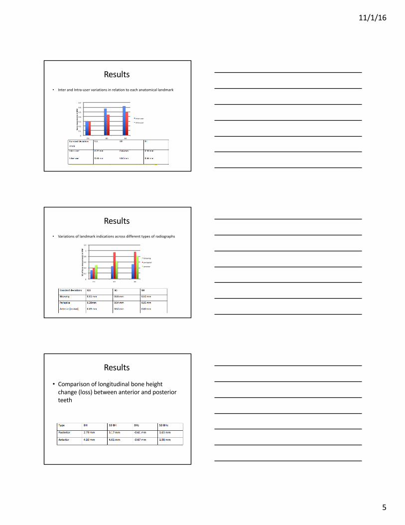
Results• Inter and Intra-‐user variations in relation to each anatomical landmark
0
0.1
0.2
0.3
0.4
0.5
0.6
0.7
CEJ BD BH
bone
mea
srmen
ts in
MM
Inter-‐user
intra-‐user
Results • Variations of landmark indications across different types of radiographs
0
0.2
0.4
0.6
0.8
1
1.2
CEJ BD BH
SD of b
one mea
surm
ents in
MM
bitewing
periapical
anterior
Results
• Comparison of longitudinal bone height change (loss) between anterior and posterior teeth

11/1/16
6
Summary • This study demonstrated that the CEJ has the lowest intra-‐
and inter-‐user variability (about 0.3 mm) for bitewing and periapical radiographs
• The BD results demonstrated a higher intra-‐ and inter-‐user variability (0.44 and 0.57mm, respectively) than those seen for the CEJ (0.3 mm each)
• The results also show that bitewing radiographs have the lowest variability in BD indications followed by anterior radiographs, while periapical radiographs show the highest BD variability
• Longitudinal BHc was slightly higher in anterior teeth compared to posterior teeth.
Conclusion • Periodontal probing and assessment of gingival
inflammation (bleeding on probing) is the mainstay of periodontal diagnosis and disease assessment
• CEJ is the most reliable landmark with posterior teeth assed with bitewings the most reliable and anterior teeth assessed by periapical radiographs the least reliable
• BD has a higher variability especially in periapicalradiographs
• The bitewing is the most reliable intra-‐oral radiographic technique for assessment of alveolar bone
Future Studies• Development of machine-‐learning tools for radiographic image
analysis. • Measure alveolar bone height at the distal and mesial edges of each
tooth and alveolar bone height change between two or more radiographs, i.e., two or more points in time automatically (the only user interaction will be to verify the analysis)
• Perform the measurements quickly (the measurements will be available for review within seconds after the radiograph is acquired)
• Make accurate (<0.5 mm) and reliable measurements (<0.5 mm precision for 95% of cases)
• Make the measurements non-‐invasively (only routine radiographs are needed, no additional burden to the dentist or patient to obtain measurements)
• Display the results in a summary report and annotated radiographs

11/1/16
7
Questions
• Would a computer-‐based automatic system to determine alveolar bone changes be useful in dental practice?
• How can this be used?