(2) Nuclear Reactions and Radioisotopes, (3) (5 ... · Available from- Superintendent of Documents,...
63
DOCUMENT RESUME ED 025 613 VT 006 292 By-Richardson, Harry D. Industrial Radiography Instructor's Guide. Atomic Energy Commission, Oak Ridge, Tenn. Div. of Nuclear Education and Training.; Office of Education (DHEW), Washington, D.C. Div. of Vocational and Technical Education. Repor t No- OE- 84034 Pub Date Mar 68 Note- 62p, Available from- Superintendent of Documents, U.S. Government Printing Office, Washington, D.C., 20402 (FS5.284:84034 $0.40). EDRS Price MF-$0.50 HC Not Available from EDRS. Descriptors-*Adult Vocational Education, Course Content, *Curriculum Guides, Radiation, *Radiographers, *Teaching Guides, *Trade and Industrial Education Identifiers- Industrial Radiography, *Nondestructive Testing The curriculum guide was developed for teacher use in an 80-hour course for industrial radiographers. The units include: (1) The Structure of Matter and Radiation, (2) Nuclear Reactions and Radioisotopes, (3) The Nature and Consequences of Radiation Exposure, (4) Radiation Attenuation, (5) Absorption of Radiation, (6) Radiation Detection and Measurement, (7) The Effect of Radiation on the Organs and Tissues of the Body, (8) Introduction to Radiography, (9) Elements of Radiography, (10) Radiographic Film, (11) Radiography Techniques, (12) Interpretation of Radiographs, and (13) Government Licensing, Health, and Transportation Regulations for Radiography. Each unit gives objectives, apparatus or aids required, content outline, and assignments in a study guide (VT 006 294) and text (VT 006 293). (EM)
Transcript of (2) Nuclear Reactions and Radioisotopes, (3) (5 ... · Available from- Superintendent of Documents,...

DOCUMENT RESUME
ED 025 613 VT 006 292
By-Richardson, Harry D.Industrial Radiography Instructor's Guide.Atomic Energy Commission, Oak Ridge, Tenn. Div. of Nuclear Education and Training.; Office of Education(DHEW), Washington, D.C. Div. of Vocational and Technical Education.
Repor t No- OE- 84034Pub Date Mar 68Note- 62p,Available from- Superintendent of Documents, U.S. Government Printing Office, Washington, D.C., 20402(FS5.284:84034 $0.40).
EDRS Price MF-$0.50 HC Not Available from EDRS.Descriptors-*Adult Vocational Education, Course Content, *Curriculum Guides, Radiation, *Radiographers,*Teaching Guides, *Trade and Industrial Education
Identifiers- Industrial Radiography, *Nondestructive TestingThe curriculum guide was developed for teacher use in an 80-hour course for
industrial radiographers. The units include: (1) The Structure of Matter and Radiation,(2) Nuclear Reactions and Radioisotopes, (3) The Nature and Consequences ofRadiation Exposure, (4) Radiation Attenuation, (5) Absorption of Radiation, (6)Radiation Detection and Measurement, (7) The Effect of Radiation on the Organs andTissues of the Body, (8) Introduction to Radiography, (9) Elements of Radiography,(10) Radiographic Film, (11) Radiography Techniques, (12) Interpretation ofRadiographs, and (13) Government Licensing, Health, and Transportation Regulationsfor Radiography. Each unit gives objectives, apparatus or aids required, contentoutline, and assignments in a study guide (VT 006 294) and text (VT 006 293). (EM)

INDUSTRIAL
RADIOGRAPHY
Instructor'sGuide

DISCRIMINATION PROHIBITED
Title VI of the Civil Rights Act of 1964 states "No person in the United.States shall on the ground of race, color, or national origin, be excluded fromparticipation in, be denied the benefits of, or be subjected to discriminationunder any program or activity receiving Federal financial assistance." There-fore, any program or activity making use of this publication and/or receivingfinancial assistance from the Department of Health, Education, and Welfaremust be operated in compliance with this law.

4lIII VA I 4/1.1111.1.
U.S. DEPARTMENT OF HEALTH, EDUCATION & WELFARE
OFFICE OF EDUCATION
THIS DOCUMENT HAS BEEN REPRODUCED EXACTLY AS RECEIVED FROM THEPERSON OR ORGANIZATION ORIGINATING IT. POINTS OF VIEW OR OPINIONSSTATED DO NOT NECESSARILY
REPRESENT OFFICIAL OFFICE OF EDUCATIONPOSITION OR POLICY.
U.S. ATOMIC ENERGY COMMISSIONGLENN T. SEABORG, Chairman
Robert E. Hollingaworth, General Manager
E002561.3
'INDUSTRIALRADIOGRAPHY
Instructor'sGuide
Developed y'ointly by the Division of V ocationaland Technical Education of the U.S. Office ofEducation and the Division of Nuclear Educa-tion and Training, U.S. Atomic Energy Com-mission.
Developed and first published pursuant to acontract with the U.S. Atomic Energy Comnzis-sion by Harry D. Richardson.
U.S. DEPARTMENT OFHEALTH, EDUCATION, AND WELFARE
JOHN W. GARDNER, SecretaryOffice of Education
HAROLD HOWE II, Commissioner

March 1968
Superintendent of Documents Catalog No. FS 5.284:84084
U.S. GOVERNMENT PRINTING OFFICE
WASHINGTON : 1968
For sale by the Superintendent of Documents, U.S. Government Printing OfficeWashington, D.C. 20402. Price 40 cents

Foreword
Industrial radiography is a vital factor in the growth of modern in-dustry. The training of industrial radiographers is of extreme importance
in meeting the needs of industry. They can assist in controlling production
costs, assure product quality and reliability, and support other relatedaspects of production and product competitiveness.
This 80-hour course is designed to provide the basic knowledge andskills necessary for the beginning radiographer. A trainee in this course is
expected to have a high school education. Using these fundamentalk he can
further develop his knowledge and skills through shop and field experiences
and additional study.
The lessons in the Student Guide and Laboratory Exercises are co-ordinated with the lessons in this Instructor's Guide. The subject matterselected from the Industrial Radiography Manual is presented in thisGuide in a sequence different from that in the Manual to give variety toclassroom sessions. This also permits laboratory exercises to be included in
earlier class sessions. Although it is desirable for a radiographer to haveknowledge of all the material in the Manual, the trainee cannot learn that
volume of material during a limited 80-hour program. Appendix D in the
Manual lists the paragraph numbers and their titles which are most im-
portant for the beginning radiographer to know. After mastering thismaterial he should learn the remainder of the material in the Manual and
its bibliography.
The instructor should have available copies of all items in the bibli-ography. Trainees should be encouraged to use these during their trainingprograms and for continued studying after returning to their places of
employment in order to become more knowledgeable and competent.
In addition to this Instructor's Guide, the material prepared and co-ordinated for this course includes *:
Industrial RadiographyManual
Industrial RadiographyStudent Guide and Laboratory Exercises
*Manual, Industrial Radiography, OE 84036, available from Superin-
tendent of Documents, U.S. Government Printing Office, Washington, D.C.
20402, price $1-25.Student Guide and Laboratory Exercises, Industrial Radiography,
OE 84035, available from Superintendent of Documents, U.S. Government
Printing Office, Washington, D.C. 20402, price 55 cents.

iv
The need for this course was identified by officials in the U.S. AtomicEnergy Commission. The Commission's first concern was to eliminateoverexposures to workers engaged in radiography. A second interest wasto increase the trained manpower in this expanding field. Content andformat for the course were identified by a committee of industry repre-sentatives working with representatives of the U.S. Atomic Energy Com-mission and 'the U.S. Office of Education, Division of Vocational andTechnical Education.
The writing of the manual was performed by Harry D. Richardson,Louisiana State University, under contractual arrangements with theDivision of Nuclear Education and Training, U.S. Atomic Energy Com-mission.
GRANT VENNAssociate Commissioner for
Adult, Vocational, and Library Programs
RUSSELL S. POORDirector, Division of
Nuclear Education and Training

Class Organization Plan
This Instructor's Guide is composed of lessons. Weighted times giverelative emphasis to more important topics. Instructors are encouraged todiscuss personal experiences and refer to radiography problems en-countered in industry.
These lessons are coordinated with the Student Guide lessons andthe Laboratory Exercises. The subject mattet for the lessons is takendirectly from the Industrial Radiography Manual. Other references listedin the bibliography should also be used.
Experience in teaching this material to radiography technicians in-dicates the students should be given ample classroom time to work
numerous problems. Personal observation by, and the assistance of, theinstructor are necessary to assure the student solves problems correctly.Preparing problems that realistically relate to shop and field situationsholds the student's interest.
The Student Guide lessons should be completed by the student andgiven to the instructor, These lessons should be graded and returned tothe student for filing in a notebook. Discussion of the lessons and thestudent's answers will provide follow-up review.
Announcement of written examinations has proved to be an impetusto the student's studying and diligence in class and laboratory work.

Contents
Pag e
FOREWORD
CLASS ORGANIZATION PLAN
LESSONS
IntroductionRadiography Demonstration
1
1
1. The Structure of Matter and Radiation 3
2. Nuclear Reactions and Radioisotopes 7
3. The Nature and Consequences of Radiation Exposure 10
4. Radiation Attenuation 16
5. Absorption of Radiation 18
6. Radiation Detection and Measurement 21
7. The Effect of Radiation on the Organs andTissues of the Body 25
8. Introduction to Radiography 29
9. Elements of Radiography 32
10. Radiographic Film 36
11. Radiography Techniques 40
12. Interpretation of Radiographs 43
13. Government Licensing, Health, and TransportationRegulations for Radiography 46
APPENDIXSuggested Typical Examination 53

Introduction Radiography Demonstration
TIME : One hour
OBJECTIVES:1. To introduce the general subject of radiography.
2. To identify for the students the apparatus to be used during the
training program.APPARATUS :
1. A radiation exposure device, either X-ray or gamma ray. (Noradiation exposure is desired in this demonstration so the isotopes should
be left in storage.)2. A "safety kit." (Refer to the laboratory experiments for this list
of apparatus.)3. Specimen to be radiographed. (A radiograph of this specimen
should be available.)4. Film holder and one sheet of film.5. Lead screens.6. Stop watch.7. Measuring tape.8. Lead letters and numbers.9. Penetrameters.
10. Masking tape.11. High intensity viewer.12. Sample radiographs.
PROCEDURE:The instructor will identify each piece of apparatus and very briefly
describe its function in radiography operations. A "dummy" arrangement
of apparatus will be used to demonstrate how an exposure is made. Items
in the safety kit will be used to initiate student thinkitg and actions inter-
relating good radiation safety practices with industrial operations which
use high intensity sources of ionizing radiation.At the end of this demonstration, the student t% ill be given course
manuals and instructed on how to use the manuals to apply his time most
advantageously during the entire training program.
1

The Structure Of Matter And Radiation
TIME: One hour
OBJECTIVES :1. To acquaint radiography students with the basic concepts con-
cerning the structure of matter.2. To develop a basic understanding of radioactivity and radiation.3. To provide information about certain kinds of radiation machines.
TEACHING AIDS :1. Chalkboard or chart pad.2. Figure references from Industrial Radiography Manual
a) Isotopes of Hydrogen (Fig. 1.7)b) Identifying Three Common Types of Radiation (Fig. 2.1)c) The Electromagnetic Spectrum (Fig. 2.3)
REFERENCE :Industrial Radiography Manual, Chapters 1 and 2.
LESSON:Introductory Statement:
Some basic understanding of the atomic structure of matter is neces-sary if the radiographer is to understand radiation and its application inindustry. The study of radiation must begin with more basic concepts suchas the fundamental particles, structure of the atom, isotopes, and elements,the basic building blocks of nature. There are 92 elements occurringnaturally and others have been made by man. Many of these elements areradioactive. Others may be made radioactive. These emit various kinds ofradiation through a process of radioactive decay until they reach a stablestate and are no longer radioactive.
Teaching Outline:I. The structure of matter
A. The atom and subatomic particles1. Electrons--small, very low weight particlesrevolve
around nucleus and carry unit negative charge.2. Protonsrelatively heavy particles compared to electrons,
have a positive charge equal, but opposite, to that ofelectrons.a) Number of protons in nucleus equals number of elec-
trons in orbit about nucleus of normal atoms.b) Number of protons in nucleus determines to what
element species the atom belongs.
Lesson 1
oz/

4
3. Neutronsnearly identical to proton in size and weightbut have no electrical charge; are bound in nucleus ofatoms along with protons.
B. The atom may be compared to the solar system (Fig. 1.2) * orelectron cloud model (Fig. 1.5) .
C. Elements are composed of atoms1. These atoms have same number of protons in nucleus
this is known as the atomic number of the element.2. They have similar physical characteristics.3. There are 92 naturally occurring elements.
a) Hydrogen is the first element, having one proton inits nucleus.
b) Uranium is the last naturally occurring element andhas 92 protons in its nucleus.
c) Transuranium elements 93 to 102 are man made.D. Compounds are composed of molecules.
1. Atoms of different elements may combine to form mole-cules, the smallest unit of a compound.
2. Two atoms of hydrogen and one atom of oxygen make amolecule of water (H20) .
3. Twelve carbon atoms, 22 hydrogen atoms, and 11 oxygenatoms compose a molecule of sugar (C12H22011).
E. Atomic weight and isotopes1. Weight of one oxygen atom has been arbitrarily set at 16
units.2. Other elements have atomic weights in relation to the
oxygen atom.3. The number of neutrons in the nuclei of atoms of same
element may vary.a) Atoms retain their same chemical characteristics but
their atomic weight varies.b) Isotopes of an element all contain the same number
of protons but differ in number of neutrons.c) The more neutrons the higher the atomic weight.d) Most samples of an element secured by man are mix-
tures of isotopes of the element.4. Compare isotopes of hydrogen and uranium.
a) Common form of hydrogen atom has one proton andone electron.
b) Deuterium has one proton and one neutron innucleus. Each isotope of hydrogen has one orbitalelectron (Fig. 1.7).
c) Uranium 238 has 92 protons, 146 neutrons, and 92orbital electrons.
d) Uranium 235 has 92 protons, 143 neutrons, and 92orbital electrons.
II. RadiationA. Excess energy possessed by unstable radioisotopes is emitted
in the form of radiation (Fig. 2.1).
* References are taken from the Industrial Radiography Manual.

B. Two types of radiation are particulate and electromagnetic
1. Particulate radiatiol is the movement of subatomic par-ticles through spacea) Alpha particles are nuclei of helium atoms having an
atomic weight of 4, travel relatively slowly, and
carry 2 positive charges. They travel only a fewcentimeters in air and will be stopped by a sheet of
paperb) Beta particles are high speed electrons having a
range of several feet in air, and may be stopped by a
few sheets of paper
2. Electromagnetic radiation consists of very short electro-
magnetic waves of energy having no charge or weight
a) This radiation has a wave quality and a particlequality
b) Gamma radiation has extremely short wavelengths,travels at the speed of light, is highly penetrating,and originates in nuclei of unstable atoms having ex-
cess energyc) X-radiation is produced when a stream of high-
energy electrons is slowed down upon striking a suit-able target. Electron transitions between orbital
shells give off photons of X-raysd) Frequency x wavelength = speed of light
e) Electromagnetic spectrum ranges from long wavelengths (low energy) at one extreme to very short
wave lengths (high energy) at the other extreme(Fig. 2.3).
III. Radiation machines
A. X-ray tubes have an electron generating filament and a target
anode sealed in a high vacuum
1. Positive charge on anode attracts negative electrons
2. When electrons strike target and are slowed down they re-
lease energy in the form of X-rays3. Also atoms in target acquire energy when orbital electrons
are dislodged. The excited atom attracts free electronsand emits X-rays in returning to a stable state
4. X-ray tubes have a wide application in medicine and in-
dustry
B. Van de Graaff generator is an electrostatic straight line ac-
celerator
1. Moving belt transfers charges to hollow metal sphere
2. The potential of several million volts is used to accelerate
charged particles down a tube to a target
C. Linear accelerator makes use of a high frequency oscillator
1. Alternate cylinders are connected together and connected
to the terminals of the oscillator
5

2. Charged particles are accelerated across gaps betweencylinders if cylinders are of correct lengths
3. Electrons may be made to approach the speed of light
D. Betatron is an electron accelerator which uses magnetic in-duction to accelerate electrons in a circular path1. Magnetic field is provided by large magnetic coils with a
laminated iron core operating on 60 or 180 cycle alter-nating current up to 400 cycles
2. Electron velocities acquire very high energy in a veryshort time
IV. Assign Student Guide Lesson 1.

Nuclear Reactions And Radioisotopes
TIME: One hour
OBJECTIVES:1. To acquaint radiography students with basic concepts of nuclear
reactions.2. To develop basic ideas about the activation of isotopes.3. To develop an understanding of the decay of radioactivity.
TEACHING AIDS:1. Chalkboard or chart pad2. Figure references from Industrial Radiography Manual
a) Chain Reaction of 1.1-235 (Fig. 3.2)b) Decay Schemes for Co-60 and Cs-137 (Fig. 3.8)e) Decay of Radioisotopes (Fig. 3.5)d) Co-60 Decay Curve: Cartesian Coordinates (Fig. 3.6)e) Co-60 Decay Curve : Semi-Log Coordinates (Fig. 3.'7)
REFERENCE:Industrial Radiography Manual, Chapter 3.
LESSON :
Introductory Statement:Efforts to secure nuclear energy became successful after Fermi used
neutrons to bombard atoms of elements. It was found that such a bombard-ment caused fission or the breaking apart of heavy atoms into lighter atoms.Some of the fission products would be radioactive. At the same time enor-mous energy was released.
Frequently an atom would capture a neutron in its nucleus and becomea radioactive isotope of the original element. These man-made radioactiveisotopes play an important role in radiography today.
Radiographers need some concept of the decay of radioactivity and ofradioactive half-life. Plotting radioactive decay is an important techniquesince this allows the person working with radioactive materials to know theamount of emission of a bit of such material at any given time.
Teaching Outline:I. Nuclear reactions
A. Nuclear fission1. Caused by neutron bombardment2. Binding energy holds atoms together3. Much energy released
Lesson 2
7

4. Uranium atom may capture neutrona) U-238 changed to neptuniumb) Neptunium is radioactive and decays to plutoniumc) Plutonium fissions and releases more neutrons (Fig.
3.1)d) May cause chain reaction
B. Chain reactions1. Fissionable isotopes include 13-235, plutonium, thorium
and protoactinium (Fig. 3.2)2. Fissionable atoms split approximately into equal frag-
ments (Fig. 3.3)3. Many neutrons released in atom fission4. Critical mass depends upon amount and shape of fission-
able material
C. Fission products1. Two groups of fission products are formed2. These cluster around isotopes of mass numbers 90 and 140
3. Barium, krypton, strontium, and cesium are frequentlyformed
4. There is excess of neutrons
II. Activation of isotopesA. More than 1500 isotopes known
1. Only a few naturally occurring radioactive isotopes2. Large number of man-made radioactive isotopes
B. Early production methods used machines to shoot fast par-ticles at atoms1. Cyclotron was an early device2. Alpha particles, protons and deuterons were used as
bullets
C. Radioisotopes produced in nuclear reactors
1. Some radioactive products, produced by fission itself2. Elements may be inserted in reactor and bombarded by
neutrons
D. Activation of atoms
1. Number of target nuclei being activated may be repre-sented by equation: A = Nf a t (par. 3-5)
2. Cobalt-59 may capture neutron and become radioactivecobalt-60. (Write equation on blackboard to show bal-ancing mass and charge.)
III. Decay of radioactivityA. Excess energy of nuclei of radioactive atoms emitted as radia-
tion1. Radiation is usually alpha and beta particles and gamma
rays2. Radioactive elements may decay in a series such as the
thorium, uranium or actinium series

3. These series may involve numerous steps (Table 3.1)4. Eventually a stable isotope is formed (par. 3-10) (Fig.
3.8)
B. Radioactive decay proceeds at a rate dependent upon totalnumber of atoms present at a given time
1. Rate of decay constantly changes since number of atomspresent is changing (Fig. 3.5)
2. Equation to represent decay takes the form N r= NoeAtwhere N = number of atoms remaining after time "t" (par. 3-7).
No r= number of atoms present at zero timee = base of natural logarithms = 2.718 . . .
--= decay constant of the radioisotopet = time
3. The radioactive half-life of an element is related to A. inthe equation on the preceding page: half-life = .693 -4- A.
C. The curie1. Rate of disintegrations of a radioisotope is referred to as
its activity2. The unit of measure of activity is the curie which is the
amount of any radioisotope that gives 3.7 x 10" disinte-grations per second
3. Millicurie and microcurie are commonly used as units ofmeasure of activity: 1,000 millicuries equal 1 curie ;1,000 microcuries equal 1 millicurie
D. Plotting radioactive decay1. Various kinds of instruments measure or count disinte-
grations2. If the count or measure of activity is known along with
the half-life for a given bit of radioactive material, a de-cay curve may be plotted (see Figures 3.6 and 3.7)
3. The measure of activity in curies may be plotted againsttime on cartesian coordinate paper or on semi-log paper(Figures 3.6 and 3.7)
4. Decay curve is a straight line on semi-log paper (Fig. 3.7)5. Pass out cartesian coordinate paper and let students plot
decay curve for a 32-curie source of Ir-192. Label the co-ordinates
6. Pass out semi-log coordinate paper and plot the same curveas for item 5 and label the coordinates. Place a legend onsheet to identify :a) Isotopeb) Sealed sourcec) Source manufacturer(Student must plot curves during class and instructor willcheck to determine that the work is neat and correct.)
IV. Assign Student Guide Lesson 2
9

The Nature and Consequences of Radiation Exposure
TIME : One hour
OBJECTIVES:
1. To inform radiography students that the risks of radiation exposureare similar to many other risks which they face daily and are not to beunduly feared on the one hand or taken too lightly on the other.
2. To acquaint radiography students with the standard measurementunits of radiation doses.
3. To acquaint radiography students with permissible radiation ex-posures and inform them how these standards relate to personnel moni-toring.
4. To inform radiography students about additional aspects of per-sonnel monitoring, including physical examinations, instrumentation andcontamination.
5. To introduce the separate problems of external and internal radia-tion exposure.
6. To inform radiography students of the different levels of radiationinjury and to acquaint them with the basic symptoms characteristic ofeach level.
TEACHING AIDS:1. Chalkboard or chart pad.2. Figure references from Industrial Radiography Manual
a) Pie chartEstimated Average Annual Gonad Exposures inthe U.S. (Fig. 6.1)
b) ChartRadiation "Banking" Concept for Radiation Workers(Fig. 6.3)
REFERENCE:
Industrial Radiography Manual, Chapter 6.
LESSON:
Introductory Statement:The major purposes of this lesson are to give students a perspective
for understanding radiation hazards in terms of other hazards of life andto introduce them to certain technical terms related to the measurementand symptoms of radiation injury. In the first instance it should be madeclear that all human activity involves certain risks, physical or mental.For example the bridge builder, the electrician, etc., face constant danger
10
Lesson 3

of physical injury, while the harried business executive or striving pro-fessional man is subject to mental breakdowns. The point of stress isthat these risks are commonly accepted and do not loom as important con-siderations to persons aspiring to become bridge builders, electricians,business executives or various types of professionals. The student shouldbe convinced that each occupation has its peculiar hazard and that radia-tion hazard, when known and understood, can be minimized or completelyavoided.
Radiography students in their future roles as technicians must have adegree of professionalism with regard to radiation. To this extent theymust become familiar with technical terms relating to the measurement ofradiation exposure and injury as well as those relating to radiation physicsand industrial radiography. In addition, a knowledge of the gross effectsof radiation injury provides the student with a frame of reference forunderstanding and appreciating the problem of personnel monitoring andcontamination.
Teaching Outline:
I. Radiation hazard in proper perspectiveA. Philosophy of risk evaluation
1. Radiation hazards should not be unreasonably feared orcompletely ignored
2. Most persons accept hazards in their work or other ac-tivities as a matter of coursea) Electricians, bridge builders, miners, farmers, etc.b) Records of National Safety Council show that all
modes of transportation present hazardsc) Many accidents occur in the home or at play, such as
falling downstairs or hunting accidents
B. All persons are constantly being exposed to some radiation1. "Background" radiation occurs in nature
a) Cosmic radiation from outer spacealtitude ac-counts for the fact that the level of such radiation istwice as high in Denver as New YorkThere are radioactive materials in the earth, some ofwhich are transferred to building materials
c) Radioactive materials are present in the body, whichhave been introduced in small amounts by food, waterand air
2. Man-made sourcesa) Medical devices, such as X-ray machines, are justi-
fiableb) Industrial machines are also justifiablec) Fluoroscopes for fitting shoes, etc., represent ex-
posure having questionable value (Fig. 6.1)
C. Sources of information about radiation's effect on man1. Animal experimentsmajor disadvantage is that dose-
effect relations cannot be assumed to be the same2. Occupational experience
a) Some workers received small doses of X and gamma
b)
11

rays before full effects were known. The symptomsand injuries of these persons were studied.
b) Persons working with medieal and industrial ma-chines have also been' studied.
3. Medical usespersons diagnosed or treated with radia-tion have been carefully studied over a period of time.
4. The atomic bombsa) The Hiroshima and Nagasaki bombs provided an op-
portunity to study effects of different levels of radia-tion over a period of time.
b) The Bikini bomb tests also provided such an op-portunity.
D. Radiation risk to radiographers must be evaluated in lightof preceding discussion.1. The proper conclusion is that radiatan can be dangerous
if received in sufficient quantity.2. With proper care, people can work with radioactive ma-
terials without any apparent effect on their ability to con-tinue living a normal life.
II. Measurement units of radiation dosesA. It is impossible to measure a quantity of radiation directly
since it can bring about a change in matter only to the extentof the energy actually absorbed.1. A given biological effect may also depend upon the
type and energy of the radiation.2. For these reasons, it is more convenient and practical to
measure exposure in purely physical terms and to usean additional factor to allow for relative biological ef-fectiveness.
B. Measurement terms1. Roentgen
a) A unit for measurement of penetrating external ra-diation (gamma) as it passes through air.
b) Defined asthe quantity of X or gamma radiationthat will produce one electrostatic unit of charge,either negative or positive, in one cubic centimeterof air at standard temperature and pressure (0°Cand 760 mm Hg)
c) The roentgen can be sub-divided into a more con-venient unit, the milliroentgen. 1,000 milliroentgensequal 1 roentgen.
2. Rem (roentgen equivalent man)a) Since the roentgen measures radiation in air only, the
rem is used to define biological effects on man. Therem is used to measure radiation dose produced up-on living tissue. (See par. 6-6.)
b) One roentgen of X or gamma radiation upon strik-ing the body produces a dose of one rem of bodilydamage.
c) The rem can be sub-divided into more convenient
12

units called millirem (mrem) which is one/thou-sandth of a rem.
3. Rad (Radiation absorbed dose)a) Defined as amount of radiation imparted to matter
by an ionizing particle per unit mass of irradiatedmaterial
b) The dose unit, the rad, represents an absorption of100 ergs of energy per gram of irradiated materialat the place of exposure
4. RBE (Relative biological effectiveness)a) Values of RBE have been worked out based on the
X-ray as an RBE of oneb) Values of RBE' have been worked out for all sources
of radiation for computing total exposure from agiven dose. (See par. 6-3.4)
c) Problem to be copied from text
III. Nature of Radiation Health Problem
A. External radiation1. Radiation being given off by certain radioactive materials
located outside of the body2. Can be visualized as a shower of tiny bullets
B. Internal radiation1. Results from radioactive materials getting into the body2. These materials tend to collect in certain body organs and
cause damagea) Biological half-lifeb) Effective half-life
IV. Levels and Symptoms of Radiation Injury
A. Classification of doses of radiation
1. Mild dosea) Exposure up to 25-50rb) Produces no detectable clinical effects.
2. Moderate dosea) 25r to 100-200rb) Produces some but not excessive damage (blood
changes, vomiting, diarrhea, loss of appetite)3. Median lethal dose
a) 200-300-400rb) Injury and disability certain, possibility up to 50%
mortality (drastic blood damages, anemia, sterility,epilation)
4. Lethal dosea) 300-600r or moreb) Death usually occurs in less than two weeks.
B. Common terms used to describe gross effects or radiationinjury1. Radiation sickness
a) Produced by a massive overdose of highly penetrating
13

external radiation to the whole body or a substantialportion of the body
b) Causes nausea, vomiting, diarrhea, hemorrhage, andlowering of the body's resistance to infection
2. Radiation injurya) Produced by an overdose of external radiation to
localized portion of the bodyb) Can cause injuries such as skin lesions and loss of
hair3. Radioactive poisoning
a) Produced by dangerous amounts of certain types ofradioactive materials introduced into the body bybreathing or swallowing or through wounds in theskin
b) Can cause such diseases as anemia and cancer
V. Personnel monitoring ( Chapter 13 presents Federal regulations,to be explained in detail at a later time, which govern all industrialinstallations using byproduct materials.)A. Permissible Exposurethat dose of ionizing radiation which
competent authorities have established as the maximum thatcan be absorbed without undue risk to human health1. Permissible occupational dose to whole body, accumulated
at any age, shall not exceed a total of 5 rem multipliedby the number of years beyond the age of 18
2. These recommendations are intended to protect the in-dividual and total population against both immediateand delayed radiation induced injury
B. Radiation "Banking" Concept (Fig. 6.3)1. Permissible rate of exposure set in accordance with what
is known as radiation "banking" concept2. Consider that each man has a radiation account to which
he adds 5 rem each year of his life after 18. He can drawon this account at the rate of 12 rem per year.
3. (Discuss all details of Fig. 6.3.)
C. Unusual exposure1. A person may be submitted to unusual exposure for
several different reasonsa) Non-continuous exposurea person is allowed to re-
ceive more exposure than recommended levels perday or week provided no further exposure issustained for a recommended period.
b) Fractional exposurecertain parts of the body suchas the hands and forearms are permitted to receivelarger doses of radiation.
c) Excessive exposure(1) If an individual receives exposure greater
than 100 rem in less than 10 years, he will haveto work under conditiohs of low or no radiationto prevent bodily injury.
(2) The National Committee on EadiP,don Protec-

tion and Measurement (NCRP) recommendsthat once-in-a-lifetime whole body emergencyor accidental exposure of 25 rem may be re-ceived. Although this exposure is not chargedagainst a person's total permissible occupa-tional dose, it should purposefully be receivedonly in extremely important or emergencysituations.
d) Medical diagnostic exposures are exempt from cal-culation of maximum permissible occupational ex-posure.
D. Physical examinations1. Pre-employment exam
a) Designed to eliminate abnormally radio-sensitive in-dividuals
b) Desirable for work with radioactive materials2. Follow-up physical
a) Designed to assure that maximum permissible levelsof exposure have not been exceeded
b) Afford a way of keeping track of the ability of thebody to respond to damage
E. Instrumentation1. Human inability to detect radiation makes mechanical
means of monitoring essential2. Personnel monitoring devices are therefore required by
law
F. Contamination1. Contamination hazards
a) Any contamination may find its way into the humanbody. Alpha and beta emitters are more hazardousinternally because of their short range. Alpha andbeta emitters are not usually considered to cause ex-ternal hazards.
b) Gamma emitters present hazards externally as wellas internally.
2. Safety precautionsa) New personnel should be made fully aware of safety
devices and emergency proceduresb) Exposed parts of body and clothing should be moni-
tored frequentlyc) Thorough searches should be made for contamination
at frequent intervalsd) Personnel should be forced to make a habit of clean-
liness and should thoroughly wash exposed parts oftheir bodies during the work day and before leavingarea
e) All persons responsible for radioactive materialsshould be thoroughly familiar with National Bureauof Standards Handbook 48.
VI. Assign Student Guide Lesson 3.
15

Radiation Attenuation
TIME : One hour
OBJECTIVES:1. To develop understanding and use of the inverse square law.2. To study factors related to attenuation of radiation.3. To present factors affecting radiation exposures.
TEACHING AIDS :1. Chalkboard or chart pad.2. Figure references from Industrial Radiography Manual
a) Variation of Radiation Intensity with Distance from Source(Fig. 4.4)
b) Dose Rate of Commonly Used Radioisotopes (Table 4.1)
REFERENCE:
Industrial Radiography Manual, Chapter 4, par. 4-4 and 4-5 ; Chapter6, par. 6-3.
LESSON :
Introductory Statement:In radiography the source of radiation may, for practical purposes, be
considered a point source. In this case, the intensity of radiation is in-versely proportional to the square of the distance from the source. Theinverse square law applies not only to radiation from radioactive materialsbut to all electromagnetic radiation such as radiowaves and light.
The effect of time on radiation exposure may be understood in terms ofthe definition of dose-rate in roentgens per hour (r/hr) or milli-roentgensper hour (mr/hr). If a radiographer works where the radiation intensity is50 milli-roentgens per hour, then in one hour, he would get 50 milli-remsof exposure and in two hours he would get 100 milli-rems of exposure.
Teaching Outline:I. Time is a factor in radiation exposure
A. Roentgen is a measure of the ionizing effect of radiationon air.1. The roentgen measure a definite amount of gamma or
X-radiation.2. Dose rate is expressed in roentgens per hour or milli-
roentgens per hour.
16
Lesson 4
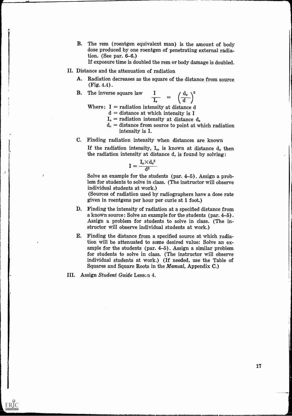
B. The rem (roentgen equivalent man) is the amount of bodydose produced by one roentgen of penetrating external radia-tion. (See par. 6-6.)If exposure time is doubled the rem or body damage is doubled.
II. Distance and the attenuation of radiationA. Radiation decreases as the square of the distance from source
(Fig. 4.4) .
B. The inverse square law do \2
dWhere: I = radiation intensity at distance d
d = distance at which intensity is II. = radiation intensity at distance dod = distance from source to point at which radiation
intensity is I.
C. Finding radiation intensity when distances are knownIf the radiation intensity, 10, is known at distance d0 thenthe radiation intensity at distance d, is found by solving:
I. X do2I =
d2
Solve an example for the students (par. 4-5). Assign a prob-lem for students to solve in class. (The instructor will observeindividual students at work.)(Sources of radiation used by radiographers have a dose rategiven in roentgens per hour per curie at 1 foot.)
D. Finding the intensity of radiation at a specified distance froma known source: Solve an example for the students (par. 4-5) .
Assign a problem for students to solve in class. (The in-structor will observe individual students at work.)
E. Finding the distance from a specified source at which radia-tion will be attenuated to some desired value: Solve an ex-ample for the students (par. 4-5) . Assign a similar problemfor students to solve in class. (The instructor will observeindividual students at work.) (If needed, use the Table ofSquares and Square Roots in the Manual, Appendix C.)
III. Assign Student Guide LessGn 4.
17

Absorption of Radiation
TIME: One hour
OBJECTIVES:1. To acquaint radiography students with the absorption character-
istics of the various types of radiation.2. To develop an understanding of thP bsorption of gamma rays in
shielding.3. To familiarize students with the half-value layer concept.4. To present the concept of reduction factors.
TEACHING AIDS:1. Chalkboard or chart pad.2. Figure references from Industrial Radiography Manual
a) Absorption of Radiation (Fig. 4.5).b) Half-Value Layers (Fig. 4.8)c) Relative Efficiency of Shielding Materials (Fig. 4.9)d) Broadbeam Radiation Reduction Factors (Figs. 4.10, 4.11,
4.12)
REFERENCE :Industrial Radiography Manual, Chapter 4
LESSON:Introductory Statement:
Of the types of radiation emitted by radioisotopes, gamma rays areof most interest to radiographers. Gamma rays can penetrate the mostdense materials and are not wholly stopped by any absorbing material.The intensity of gamma rays traversing an absorbing material plottedagainst the thickness of the absorbing material gives an exponential curve(Fig. 4.8).
There is little use in trying to compute the amount of absorbing mate-rial needed to stop all gamma or X-radiation, since a point is never reached
where all such radiation is stopped. It is convenient to employ the concept
of using adequate shielding to reduce the radiation intensity to values that
are acceptable for industrial workers.The use of half-value layers and reduction factors gives suitable results
for designing shields for industrial radiography.
Teaching Outline:I. The absorption of radiation
A. Alpha and beta rays may be stopped (Fig. 4.5).
18
Lesson 5

B. Gamma and X-rays are not completely stopped by shielding.1. The intensity of X or gamma rays after passing through
an absorber may be computed by the equation :
I=wheye I = initial intensity of gamma rays
e = base of natural logarithmsp, = linear absorption coefficient of absorber for
given gamma rayst = thickness of absorber in centimeters
2. Intensity of radiation plotted against thickness of ab-sorber gives exponential curvea) Study chart showing curve on cartesian coordinate
paper (Fig. 4.6).b) Study chart showing curve becomes a straight line
on semi-log coordinates (Fig. 4.7) .C. Linear absorption coefficients
1. Absorption coefficients vary with the energy of theradiation.
2. Also, absorption coefficients arenumber of the absorber.
3. Study Table 4.2 and note changes in coefficients withchanges in energy of gamma rays and changes in ma-terials.
4. Demonstrate use of table of absorption coefficients in aproblem.
5. Show plot of radiation intensity against thickness ofabsorber on cartesian and semi-log paper.
affected by the atomic
II. Half-value layersA. A half-value thickness is that thickness of absorber necessary
to reduce radiation intensity to half its initial value.1. A half-value thickness is related to the linear absorption
coefficient by the following equation:0.693
HVL
where HVL = half-value thickness (centimeters)4u, = linear absorption coefficient.
2. Compute half-value thickness for lead and water at somegiven value of energy for gamma rays, e.g., 0.1, 0.5, and1.0 Mev (Table 4.2 and Fig. 4.8)
B. The half-value layer idea may be used to work practical prob-lems in shielding (par. 4-7) (Fig. 4.9).Assign a problem for students to solve in class. (The in-structor will observe individual students at work.)
III. Reduction factorA. A reduction factor is the dose rate of radiation reaching a
point at some distance from a source divided by the dose ratereaching the same point with some shield interposed.
dose rate without shieldB. Reduction factor dose rate with shield
19

20
1. Reduction factor for a given radioisotope may be plottedagainst thickness of a given shielding material.
2. Such a graph may be used to compute shielding thicknessin practical problems (p. 35) (Figs. 4.10-4.12).
IV. Principles of Radiation Safety (Figure 4.13)
A. Personnel dosage is directly related to exposure time.
Allowable working =permissible exposure in mr/wk
time in hr/week exposure rate in mr/hrB. Lower personnel dosage will be received at longer distances
from a source. This is an application of the Inverse squareLaw (Table 4.3)
C. Shielding interposed will reduce personnel dosage.D. Many industrial radiation safety problems will require com-
binations of, or all of, the factors : (1) TIME, (2) DIS-TANCE, and (3) SHIELDING.Assign a similar problem for students to solve in class. (The
instructor will observe individual students at work.)
V. Assign Student Guide Lesson 5.

Radiation Detection and Measurement
TIME: One hour
OBJECTIVES :1. To develop a basic understanding of radiation detection and meas-
urement.2. To learn basic concepts of radiation measuring instruments.3. To familiarize students with the more commonly used radiation
measuring instruments required for industrial radiography.TEACHING AIDS :
1. Chalkboard and chart pad.2. Figure reference from Industrial Radiography Manual
The Pocket Dosimeter (Fig. 5.2)
REFERENCE :Industrial Radiography Manual, Chapter 5.
LESSON:Introductory Statement:
Radiation emitted from radioactive materials and X-ray machinescannot be detected by the human senses. Therefore, some kind of instru-ment must be used to determine the presence of radiation. Except forphotographic film techniques, almost all radiation detecting devices used byradiographers are based upon the ionization in a gas through which theradiation passes. These devices help provide protection to personnel fromradiation exposure and are necessary in making measurements related toradiography.
Teaching Outline:
I. Detection and measurement of radiationA. Detection is the determination of the presence of radiation.B. Measurement includes both detection and some measure of the
amount of radiation.C. Total dose exposure may be measured.
1. One type of instrument measures the accumulated ex-posure to radiation over a period of time. This is called adosimeter.
2. The milliroentgen is frequently used for measuring totaldose exposure.
3. Examples of dosimeters include: Lauritsen electroscope,the pocket dosimeter, the R-meter, and the film badge.
D. Dose-rate of exposure may be measured.
Lesson 6
21

1. Instruments used to measure dose-rate exposure are calledsurvey meters.
2. Milliroentgens per hour is a commonly used measure fordose rate.
3. Examples of survey meters include the Geiger countersand the ionization chambers as detectors.
II. Dosimeters
A. Lauritsen electroscope1. Is an ionization chamber instrument with a closed
chamber for collecting ions formed by radiation2. Chamber contains insulated electrode that has a fixed
and a movable part3. When electrode is charged the movable part is deflected
from the fixed part4. As ions in chamber are formed by radiation, those with a
charge opposite to electrode travel to it5. As ions neutralize charge on electrode the movable part is
deflected less and furnishes a measure of amount ofionization and therefore of radiation
6. A movable quartz fiber is viewed through a transparentscale and the amount of deflection can be measured
B. Pocket dosimeter (Fig. 5.2)1. Essentially a Lauritsen electroscope modified to about the
size of a fountain pen2. May be obtained to read in roentgens and milliroentgens3. Direct reading type can be held up to light to see the
hairline which moves across a scale, indicating dosagereceived
4. Nondirect reading type must be inserted in a reading de-vice to indicate dosage received. These are called pocketchambers
5. Due to insulator leakage, these devices are likely to givereadings higher than the actual radiation received.
C. R-Meter1. Is an ionization type instrument made in a variety of
styles2. Composed of a small chamber or roentgen meter and an
instrument for charging and measuring exposure of thesmall chamber
D. Film badges (Dismantle a film badge and film packet. De-scribe and allow students to examine all the parts.)1. Consists of small piece of X-ray film in a metal holder2. Is worn on outer clothing3. Measures gamma and X-radiation and high energy beta
radiation4. Film must be developed by controlled techniques and com-
pared to a set of control films to determine amount ofradiation received
5. The film is darkened in proportion to amount of radiationreceived
22

111. Survey metersA. Ionization chamber instruments
1. Ions in a gas caused by radiation move to electrodes ofopposite charges.
2. Electrodes are connected to a battery or other source ofvoltage.
3. Ions give up charges and cause a small current flowthrough battery and external circuit.
4. Current flow can be measured and thus give a measure ofthe radiation intensity required to produce the ionization.
5. Provide instantaneous measure of radiation intensity6. May read up to 500 roentgens ger hour
B. Geiger counter1. Usually measures low intensities of radiation (milli-
roentgens per hour) (Special designs permit higherranges.)
2. Uses a tube with high voltage and low gas pressure to in-crease sensitivity
3. One photon or particle entering the tube may start anavalanche of ions which will produce a voltage pulse.These pulses, through proper circuits, allow indication ofradiation received on galvanometer or audible signals.
4. May be used to read gamma, X, and beta radiation.5. Geiger counters may block out in a very high radiation
field (special circuit designs can prevent this) and giveno reading.
IV. Instrument Characteristics and CalibrationA. Characteristics of radiation measuring instruments should be
considered.1. Should detect desired radiation2. Should cover suitable range of dose or dose rate of
radiation3. Should hold calibration4. Should have a suitable time constant5. Batteries should be easily available6. Manufacturer should maintain good repair service
B. Radiographers need to calibrate instruments.1. Must secure a source of radiation of known intensity (Use
radiography source and manufacturer's calibrationcurve.)
2. Place instrument at distance to secure a certain reading.3. Make manual adjustments on instrument as directed in
the instrument manufacturer's instructions.4. Work an example for the class similar to illustrated in-
strument calibration (par. 5-6) .
V. Source Calibration (Fig. 5.6)for making radiography exposurecalculations and safety calculations it is mandatory that sourceemission rates be known. The process for determining this iscalled calibration and is accomplished according to this outline:
A. Place a dosimeter at a measured distance from the source.B. Expose the source for a measured time.
23

C. By calculations in par. 5-7, determine the dose-rate at 1 footfrom the source.
D. Divide the result in step 3 by the source emissivity (Table 4.1)Assign a similar problem for students to solve in class. (Theinstructor will observe individual students at work.)
E. Plot a source calibration curve similar to Fig. 5.6. (Be sureto label all parts of the curve sheet and identify the source.)Assign a similar problem for students to solve in class. (Theinstructor will observe individual students at work.)
VI. Assign Student Guide Lesson 6.

The Effect of Radiation on the Organs and Tissues of the Body
TIME: 30 minutes
OBJECTIVES :1. To acquaint the radiography student with the effect of radiation
on living matter.2. To inform radiography students of the relative sensitivity of
various body cells to radiation.3. To develop an understanding of the types of biological effects of
radiation and the factors related to these effects.4. To acquaint the radiography student with the specific effect of
radiation on the various organs and tissues of the body.5. To familiarize radiography students with the genetic effects of
radiation and the effect of radiation on the life span.
TEACHING AID:Chalkboard
REFERENCE :Industrial Radiography Manual, Chapter 7.
LESSON:Introductory Statement:
This lesson is designed principally for information purposes. It con-tains material with which the radiography student should be acquaintedbut which is not vital to the performance of his duties. The first pointto stress is that radiation affects living matter principally by altering theability of cells of the body to function normally. It should be made clearthat the various cells of the tissues and organs are not affected in the sameway. This fact explains why exposure of certain parts of the body can beconsiderably more critical in terms of injury than exposure of other parts.The student will have heard of the effect of radiation on the reproductiveorgans and may have undue fears founded on misinformation. He should beaware that radiation can and does produce genetic effects. There is evidencethat radiation dosage shortens the life span. However, the available evi-dence does not warrant undue fear of these consequences because of thehigh dosages required to produce measurable effects compared to dosageslikely to be received while working as an industrial radiographer.
Teaching Outline:I. Radiation effects on living matter
A. The body of man is composed of cells.1. Cells are the fundamental unit of structure of living
Lesson 7
25
organisms, just as the atom is the fundamental unit ofchemical structure.
2. A cell may be defined as "a restricted mass of protoplasmdifferentiated into cytoplasm, nucleus and a boundingmembrane."
B. Bodies of living organisms have varying tolerances to all kindsof injuries or abuses.1. The body is able to repair damages to itself through a
complicated process termed mitosis.2. Mitosis may be defined as a process whereby cell division
takes place and worn out or damaged cells are replaced.C. It has been determined that radiation affects the living organ
principally by altering the ability of cells to function nor-mally.1. This alteration comes about through the process of ioniza-
tion.2. The electrical charge of irradiated atoms in body cells is
changed and valence bond binding atoms into molecules isaffected in such a way as to cause chemical interactions.(See par. 7-6.)
D. Effects of radiation include :1. Cell to continue to grow until it reaches an abnormal
size2. Cell is altered so that its daughter cell is genetically dif-
ferent from it3. Daughter cells may die before they reproduce or divide
at a higher or lower rate than parent cells.E. In summary, the effect of radiation on a living organism is
produced at the cell level and is both physiological and chemi-cal in nature. The degree of damage depends on many condi-tions and factors.
II. RadiosensitivityA. Defined as the difference in response of the different tissues
and organs of the body to radiation1. Body cells which are more active in reproducing them-
selves and which are not fully mature are harmed most byradiation.
2. This fact can be explained in terms of the higher meta-bolic rate (rate of chemical change) of these cells, whichlowers their resistance.
B. Classification of body cells in order of radiosensitivity.1. Lymphocytes (white blood cells formed in the tissues of
the spleen and lymph nodes, etc.) are the most sensitive.2. Granulocytes (white blood cells formed in bone marrow)3. Basal cells (originators of the more complex cells of the
gonads, bone marrow, skin and alimentary canal)4. Alveolar cells (long cells which absorb oxygen from the
air)5. Bile duct cells (important in digestion)6. Cells of the tubules of the kidneys7. Endothelial cells (which cover the closed cavities of the
body)
26

8. Connective tissue cells (which support the organs .andspecialized tissues of the body)
9. Muscle cells10. Bone and nerve cells
C. Factors related to somatic radiation effects (those experiencedby the irradiated individual)1. The rate at which the dose is administered
a) Body repair can keep up with damage to a point.b) P:-.Ine dose over a shorter period of time would cause
greater damage2. The extent of the body irradiatedwhole body radiation
is more harmful than radiation to a fractional part of thebody.
3. The part of body irradiateda) Certain portions of the body are more radiosensitiveb) The upper abdomen generally highly radiosensitive
4. The age of the individualyoung persons are moresusceptible than mature adults, because their body cellsare in an accelerated stage of reproduction.
5. Biological variation among individualssome persons aremore resistant to radiation because of the make-up andfunctioning of their bodies.
III. The effect of radiation on various tissues and organs of the body
A. The blood and bone marrowradiation impairs the functionsof the blood so as to:1. Make the body more susceptible to disease2. Prevent blood clotting and thus prolong the healing of
wounds3. Bring on serious pathological conditions because the abil-
ity of blood to carry food and waste products is impairedB. The lymphatic system (Lymph is the liquid which bathes all
the cells of the body and transfers food and wastes from theblood.)1. Radiation which injures the spleen and other lymphatic
tissue stops the production of lymphocytes and impairsthe filtering of foreign substances out of the lymph.
2. This impairment is associated with increased bacterialactivity and reduces chances of recovery.
C. Skin and hair follicles1. Rather large doses of irradiation are usually necessary for
permanent damage, although skin reaction is one of firstclinical indications of radiation exposure.
2. Large doses of irradiation result in skin cancer and tem-porary baldness.
D. The digestive system1. Radiation impairs the secretion of digestive juices and
the reproduction of cells.2. Symptoms include vomiting, diarrhea and ulcers.
E. The liver and gall bladderradiation damage impairs abilityof these organs to function as part of the digestive system andin the metabolism process.
F. The endocrine system
27

1. Radiation serves to make this system function lessefficiently and to make the body more susceptible to heat,cold, injury and infection because the adrenaline supplycannot be replaced sufficiently.
2. Damage to thyroid gland decreases thyroxin productionand affects the basal metabolism rate.
3. Damage to the gonads (reproductive organs) can producesterility, although this is unusual, except for near lethaldoses. Under modern conditions of occupational exposure,there is no danger of sexual impotency.
G. The respiratory systemRadiation produces effects on the lungs by damaging theair sacs ; the lungs are especially susceptible to internalradiation, originating from air particles.
F. The urinary systemradiation damage can cause the cells ofthe uterus and bladder to break down and block the urinarytubes (uteres). One symptom is tenesmes (the urge to ridoneself of waste products w ithout being able to do so) .
I. The bones.1. Radiation presents a serious threat to the highly radio-
sensitive red bone marrow cells2. Tumor production is also possible under certain con-
ditions of internal radiation.J. The ey esradiation affects the eyes by promoting the develop-
ment og cataracts.
IV. Other effects of radiation.A. Effect of radiation on the life span.
1. Experiments on animals indicate radiation can affect thelife span.
2. Careful analysis of all evidence available suggests that ex-posure to permissible occupational dose rates, even froman early age, does not shorten life.
B. Genetic effects of radiation.1. Radiation can produce mutations.2. Because of inadequate evidence, it is difficult to appraise
genetic effects on (a) the individual or (b) the populationas a whole.
3. The fear expressed that radiation would increase thefrequency of abnormal births has not been validated bythe Japanese bomb victim experience.It is recommended that persons who receive radiationdoses to their reproductive organs wait for a period of sixmonths before conception of a child. This period permitsthe formation of sex cells which contain proportionallyfewer gene mutations.
V. Assign Student Guide Lesson 7.
28

Introduction To Radiography
TIME : 30 minutes
OBJECTIVES :
1. To introduce the student to the process of radiography.2. To acquaint the student with the procedures required in the mak-
ing of an industrial radiograph.3. To inform students of the industrial applications of radiography.4. To introduce certain terms and topics of importance to industrial
radiography.
TEACHING AIDS :
1. Chalkboards and/or chart pads.2. Figure reference from Industrial Radiography Manual
a) Radiograph Exposure Arrangement (Fig. 8.1)b) Sample radiographs (to be prepared or obtained by the in-
structor)
REFERENCE:Industrial Radiography Manual, Chapter 8.
LESSON:Introductory Statement:
This chapter is designed to serve as an introduction to the more de-tailed discussions of radiography which follow. Its major purpose is to givethe student an overall grasp of the process and purpose of industrialradiography and to present certain terms which are basic to the under-standing; of radiography. The last part of the chapter outlines the basictopics to be treated in depth in following chapters.
Teaching Outline:I. Introduction
A. Roentgen, a German, experimented with cathode rays in 1895.1. He discovered a new ray which caused a chemically coated
paper to glow.2. This ray could penetrate paper, wood, metal, etc.3. The ray also affected photographic plates.
B. The new ray was called the X-ray.C. In 1898 the Curies discovered that radium was radioactive.
1. The invisible ray given off by radium was called a gammaray.
Lesson 8
29

2. No use was made of radium as a source of radiation forradiography until the 1920's.
D. Later the Atomic Energy Commission produced and imple-mented distribution of artificially produced gamma raysources.
II. The process of radiography (Fig. 8.1)
A. Radiography is a process of testing materials which usespenetrating radiation such as X-rays or gamma rays.1. This allows the examination of the interior of objects or
assemblies which are invisible to the eye.2. This radiography process is called non-destructive testing
since objects are not damaged.B. In radiography, the rays passing through the material are
absorbed or changed.1. Change varies with the thickness and atomic number of
material.2. As a result, the intensity of the radiation beam emerging
from the object varies from point to point.3. Some kind of detector, such as a Geiger counter, films or
fluorescent screen can be used to detect radiation.
C. Industrial radiography is concerned only with recording"shadow" images of specimens on film. These "shadows" re-sult from differential radiation absorption in a specimen and
discontinuities.1. The three basic essentials in producing a radiograph are:
a) A source of radiationb) An object to be testedc) A cassette containing film
2. Radiation proceeds in a straight line and the amountreaching the film in the cassette depends on a number of
factors, i.e., radiation source emissivity, radiation energy,source to film distance (SFD), and specimen absorptivity.
3. Voids or breaks in steel, for example, can be detected be-
cause more radiation passes through these spots andmakes a dark spot on the film.
III. Applications of radiography include:
A. Testing and inspecting products of various kinds to determine
soundnessB. Inspection of welds on high pressure or high temperature ap-
paratusC. Examination of castingsD. Inspection and examination of nonmetallic materials such as
plastics, ceramics, concrete, paper, wood, textiles and foodproducts
E. Pipe wall thickness measurementsF. Other types of radiography, such as:
1. High speed or flash radiography2. Microradiography3. Stereography
30

IV. The radiographA. The processed film containing the visible images caused by
exposure to radiation is called a radiograph.B. The darkening of the radiograph is called density.C. Differences in density from one area to another are called
radiographic contrast.D. Sharpness of outline in the image on a radiograph is called
definition.E. Radiographic screens are used to shorten exposure time.
Such screens are called intensifying screens and are discussedin detail elsewhere.
F. The contrast of radiographs may be reduced by scatter.1. Materials not only absorb but scatter radiation in all di-
rections.2. The film thus receives radiation from the specimen, walls
and floors of rooms, etc., which may tend to make theimage hazy, unless reduced by screens, masks, diaphragmsor filters.
V. Industrial radiography involves four basic topics (to be discussedin detail later) .A. The source of radiationincluding maximum energy of
source and emission rate of sourceB. The specimen to be examinedincluding the mass density of
the material, atomic number, composition and thickness ofabsorption path
C. The film to be usedincluding speed and ability of film toresolve images
D. Geometric principlesincludes source size, source to filmdistance, specimen dimeAsion and shape, specimen to film dis-tance, and projected images
VI. Assign Student Guide Lesson 8.In addition to these outlined principles, every successful radi-ographer must know and practice these topics:
1. Radiography techniquesa) Exposure calculationsb) Exposure arrangements
2. Interpretation of radiographs

Elements Of Radiography
TIME : One hour
OBJECTIVES:1. To acquaint the student with the production of and characteristics
of X-radiation.2. To 'acquaint the studerit with the characteristics of gamma ray
sources.3. To develop a knowledge of the encapsulation of gamma ray
sources.4. To acquaint students with the geometric principles which apply to
shadow formation.5. To introduce problems caused by scattered radiation.
TEACHING AIDS :1. Chalkboard or chart pad.2. Figure references from Industrial Radiography Manual
a) Shadow Formation (Fig. 9.9) .
b) Geometrical Unsharpness, the Penumbral Shadow (Fig.9.10) .
c) Distortion of Shadows (Fig. 9.12).d) Shadow Formation Showing Enlargement of Image (Fig.
9.13).e) Sources of Scattered Radiation (Fig. 9.14).f) Backscatter from Floor, Walls, or Objects (Fig. 9.15).
REFERENCE:Industrial Radiography Manual, Chapter 9.
LESSON:Introductory Statement:
This chapter is designed to introduce the student to the basic principleswhich apply to the production and use of X and gamma radiation forradiography. Special emphasis should be given the geometric principleswhich must be considered in arranging radiography techniques. Studentsshould be encouraged to study the several diagrams which are included asthey are most helpful in understanding the formation of film images re-sulting from "radiation shadows" cast by the specimen and its discon-tinuities.
Teaching Outline:I. Characteristics of X-radiation
A. The energy of radiation is often expressed in electron volts.
32
Lesson 9

Because this is such a small unit, kiloelectron (key) andmillion electron volts (Mev) are the units most often used.
B. As the wavelength of X-radiation becomes smaller, the energyof the radiation becomes greater. The higher energy radiationmore easily penetrates dense materials.
C. Radiation from an X-ray tube consists of two parts.1. The continuous X-ray spectra, which covers a wide band
of wavelengthsa) This is the radiation of most use in industrial radi-
ography.b) A high tube voltage increases the intensity of con-
tinuous radiation.2. The characteristic X-ray spectra
a) X-radiation of a specific wave lengthb) Energy of this spectrum is small compared to con-
tinuous spectrum3. A broad energy spectrum is sometimes not desirable since
low energies are more readily scattered and absorbed.Filters are placed over tube windows to eliminate thelower energies.
II. Production of gamma raysradioisotopes for gamma radiographyare available from :
A. Naturally occurring materials (Radium-226)B. Fission products (Cesium-137)C. Activation by neutron bombardment (Cobalt-60, Iridium-
192, etc.)
III. Encapsulation of radioisotopes
A. In gamma radiation the energy emitted from decaying nucleiis used.1. It is mandatory that the radioactive chemicals be con-
tained so they will not come into contact with the humanbody.
2. This containment is accomplished by sealing the mate-rials in capsules.
3. This process is called encapsulation.Encapsulation is a highly specialized process requiring themost careful :1. Design2. Fabrication3. Quality control
C. Selection of materials and designs for capsules must consider:1. Resistance to corrosion from radioactive material and the
outside environment2. Damage to capsule caused by emitted radiation3. Resistance to erosion, abrasion, and structural stresses4. Sealing techniquesfusion welding has been proven ac-
ceptable.D. Double encapsulation
1. Some sources are now fabricated by sealing and testingin one capsule.
2. This capsule is then sealed and tested in a second capsule.
B.
33

E. Leak testing sealed sources (refer to Laboratory Exercise 15.)1. All capsules must be tested for leaks after sealing.2. Exterior surfaces must be decontaminated to the degree
that no more than 0.005 microcurie of removable contam-ination is present.
3. All radiography gamma ray sources must be leak testedat intervals not exceeding 6 months (par. 13-9.3).
IV. Characteristics of gamma radiography sourcesA. Radioisotopes used for industrial radiography have discrete
energies emitted (Fig. 3.8).1. Specimen thickness and atomic number determine the
energy (wave length or Mev) required for penetrationto make a radiograph.
2. In selecting a radioisotope for a particular specimen, thedesired characteristics are (Table 9.1) :a) Appropriate energyb) High specific activity (provides sources having
smaller dimensions)c) Long half-life
B. Metallic pellets are usually available for sources in the shapeof cylinders. One or more pellets may be sealed in a capsule.(Cesium-137 sources are prepared by pressure pelletizing theradioactive chemical compounds Cs Cl.)
V. Geometric principlesA. General considerations
1. Shadow formation (Fig. 9.9)a) Radiation beams behave much as light beams and
form shadowsb) Relation of (1) film to object and (2) source to film
distances determines size of shadow2. Sharpness of shadow depends on (Fig. 9.10) :
a) Size of sourceb) Ratio of the source to object distance and object to
film distance3. Distortion of shadows may occur because (Fig. 9.12) :
a) Plane of object and plane of the film may not beparallel
b) Object is not in contact with filmc) Radiation beam may not be directed perpendicular to
the surface of filmB. Enlargement (Fig. 9.13)
1. With extremely small sources of radiation, the specimenmay be placed some distance from the film.
2. The result will be enlarged images.C. Geometric principles of shadow formation are the basis for
the following recommendations :1. The radiation source should be as small as possible.2. The distance from the source to the film should be as great
as possible. (Economy of exposure time must be con-sidered in shop operations.)
3. The film should be as close to the specimen as possible.

4. The center of the radiation beam should be perpendicularto the center of the film.
5. The plane of maximum interest on the specimen should beParallel to the film.
VI. Specimen
A. Radiography specimens vary from thin low density plasticsto thick high density metallic materials.
B. Radiation interaction with and transmission through the sec-tion depend upon:1. Radiation energy2. Specimen atomic number3. Specimen thickness4. Density
C. Absorption coefficients indicate that :1. Radiation penetrates more easily as the energy increases.2. Radiation penetrates more easily as the material density,
atomic number, and composition decrease.D. Because of the above conditions, high energy radiation should
be used to radiograph thick dense specimens and low energyradiation should be used to radiograph thin low densityobjects.
VII. Scattered radiationA. Radiation interacts with any material (including air) and
may be scattered. Compared to the primary beam, scatter haslower energy and may have more or less intensity.
B. Most scatter reaching the film is forward scatter (Fig. 9.14) .
C. Hazy edges of images caused by scatter are called undercut.D. Back Scatter is caused by radiation interacting with objects
after passing the specimen (Fig. 9.15) .
VII. Assign Student Guide Lesson 9.
tt
35

Radiographic Film
TIME: One hour
OBJECTIVES :
1. To teach the students the characteristics of film used for in-dustrial radiography.
2. To acquaint students with the procedures for determining thefilms best suited for particular operations.
3. To inform students of the requirements and precautions for eachstep of film processing.
4. To introduce students to the type of facilities needed for filmprocessing.
TEACHING AIDS:1. Chalkboard or chart pad.2. Figure references from Industrial Radiography Manual
a) Film Characteristic Curve (Fig. 10.2)b) Relative Film Speed (Fig. 10.3)
REFERENCE :Industrial Radiography Manual, Chapter 10.
LESSON :
Introductory Statement:Radiographic quality is dependent upon film emulsion characteristics.
Speed and graininess must be considered in selecting techniques.The processing of film is a vital step in industrial and other types of
radiographic work. In order for the student to do a competent job, hemust not only know the mechanical steps involved but must understand thetheory as well. This chapter is designed to give the student the knowledgehe needs for processing film. It should be stressed throughout the discus-sion that the standards for film processing are high and that all measure-ments of solutions, time, and temperature should be made carefully.
Teaching Outline:I. Introduction
A. Film is a prime essential for radiography work.B. An industrial radiographic film is a thin, transparent, flexible,
plastic base coated with an emulsion composed of gelatin con-taining microscopic crystals of silver bromide.
C. This chapter is concerned with the sensitometric character-istics of radiographic film.
36
Lesson 10

II. Radiographic contrastA. Radiographic contrast is defined as a comparison of densities
on developed film areas.B. It depends on :
1. X-ray kilovoltage or isotope gamma ray energy2. Screen type3. Film contrast characteristics4. Film density5. Scattered radiation6. Subject contrast
III. Subject contrastA. Subject contrast is defined as the ratio of radiation intensities
passing through two selected portions of a specimen.B. Subject contrast varies according to the energy of the radia-
tion and specimen atomic number and thickness.
IV. Film contrast, H & D Curve (Fig. 10.2)A. Film is manufactured to give different film contrasts.B. Film contrast is best described by the "characteristic" curve
which relates film density to radiation exposurethe H & D
curve.C. Exposure (E) is defined as the product of the radiation in-
tensity (I) reaching the film and the radiation exposuretime (t). E = I x t.
D. Characteristic curves are used in selecting radiographic tech-nique as they show the relative speed and contrast of differenttypes of film (Fig. 10.3).
V. Speed
A. Film speed is inversely proportional to the exposure requiredto attain a certain densii;y.1. High speed (fast) film requires a low exposure2. Low speed (slow) film requires long exposure (Fig. 10.3) .
B. Industrial operations are always concerned with time.
VI. GraininessA. Film images are formed by multitudes of microscopic silver
crystals, called grains.B. Groups of grains produce an overall condition described as
graininess.1. All films have graininess to some extent.2. Graininess determines the fineness of detail that can be
perceived on film.
VII. In non-destructive testing the radiograph must interrelate filmcontrast, speed, and graininess to achieve an acceptable quality.
VIII. Radiographic screensA. Radiographic screens are used to intensify the photographic
action of radiation on the film.B. The two most used types are:
1. Lead foil screens, which are commonly used on both sidesof the films to improve quality
37

a) Such screens should be selected and handled with ex-treme care.
b) They should be cleaned carefully and should not con-tain irregularities or abrasions.
2. Fluorescent screens are used with film which has a high
sensitivity to blue light.a) Seldom used in industrial radiographyb) Should not be touched or cleaned without consulting
manufacturer's instructions
IX. Film processing (Use chemicals recommended by film manu-
facturers.)A. The chemical processing of film is an important and exacting
task.B. Several general precautions apply to film processing:
1. Specified limits should be observed for all chemical con-centrations, solution temperatures, and processing times.
2. Scrupulous cleanliness is mandatory.3. Equipment should be made from materials which do not
corrode in contact with processing chemicals.4. Suitable darkrooms and safe lights are required since
emulsions are sensitive to light.C. Developing involves:
1. Careful adherence to time and temperature recommenda-
tions, 5-8 minutes at 68°F2. Agitation of film3. Timely replenishment and/or replacement of developer
solutionD. Stop bath involves:
1. Allowing developer to drain from the film2. Immersion in stop bath for 30-60 seconds with moderate
agitationE. Fixation involves :
1. Allowing proper time-8 to 15 minutes at 68°F2. Vigorous agitation when first placed in solution3. Replenishment and/or replacement of solution at intervals
F. Washing involves:1. Use of adequate size tanks2. Running water3. Fresh uncontaminated water
G. Drying involves :1. Immersion of washed film in wetting agent when possible
2. Circulation of dry, warm air over film3. Use of filters for air
X. Film processing facilities
A. The location, design, and construction of film processing facil-
ities is important. Volume and character of work determines
specifications.B. The darkroom should have :
1. Adequate safe lights2. Corrosion resistant walls and floors3. Properly designed darkroom entrances
38

C. Equipment which meets all standards of size, safety, andcorrosion resistance
D. Film dryers with adequate filters and fast heating controlledto prevent melting the emulsion
E. Automatic film processing equipment
XII. Assign Student Cwide Lesson 10.
89

Radiography Techniques
40
TIME: Two hours
OBJECTIVES :
1. To inform students of practical guides for X and gamma radio-graphy exposure arrangements.
2. To teach students methods for calculating exposure time.3. To present principles for selecting practical radiography tech-
niques.4. To study causes and corrections of unsatisfactory radiographs.
TEACHING AID:Chalkboard
REFERENCE :Industrial Radiography Manual, Chapter 11.
LESSON:Introductory Statement:
The material preceding this lesson has been primarily concerned withprinziples. This lesson is to present techniques which have been tried andpnved to be practical for industrial radiography. Successful radiographersmust know and use the rules and guidelines in this lesson. Shop andlaboratory experience will be necessary 1:or the student to gain ability andconfidence to radiograph the many different industrial specimens.
Teaching Outline:I. Exposure calculations, defining and controlling variables.
A. Chemicals and film processing.1. Concentrations, replenishing.2. Timetemperature.3. Washing and drying.4. Cleanliness.
B. Films.1. Speed, graininess, contrast.2. Film factor, "F".
C. Screens.1. Metallic foil.2. Fluorescent.
D. Geometry (minimum criteria)1. SFD = 8 x specimen thickness (Fig. 11.1)2. SFD = diagonal length of radiograph (Fig. 11.2)
Lesson 11

E. Specimen1. Steel equivalent thickness (S.E.T.) ,.......:ceptable for
gamma ray energies within a limited range)2. Radiation penetration3. Absorption factor "A"
F. Source emissivity1. X-ray "ma"2. Gamma ray sources "S"
G. Exposure calculations1. Gamma ray (Fig. 11.3)
r
FAD 2r
S2. X-ray (Fig. 11.5)
H. Control of scatter1. Filters2. Screens3. Masks, diaphragms and blocking
II. Exposure arrangements
A. Location of source and film relative to specimen. B. General questions
1. Which arrangement will give the best radiographic imageof the specimen area that is likely to contain malu-facturing discontinuities?
2. Which arrangement will give the best radiographic imageof the specimen area that is likely to fail under imposedoperating stresses?
3. Which arrangement will allow the shortest exposure timewithout unnecessarily sacrificing radiographic quality?
4. Will the subject contrast require (a) single film, (b)double film, or (c) multiple exposure techniques?
5. Can beam or panoramic radiation be more appropriate?C. Emitted beam shapes
1. X and gamma ray2. Angular3. 3600 limited angle beam4. 3600 spherical beam
D. Source, film, specimen arrangements1. Flat specimens having uniform thickness2. Pipe
a) Small diameter, 11/2 inches or lessb) Large diameter
(1) Multiple exposures(2) Panoramic exposures
3. Pressure vessels4. Spheres and hemispheres5. Panoramic exposure for many specimens6. Techniques for specimens having large subject contrast
III. Unsatisfactory radiographs, causes and correctionsA. High density
1. Overexposure
41

2. Overdevelopment3. Scatter
B. Low density1. Underexposure2. Underdevelopment
a) Timeb) Depleted developerc) Temperature too low
C. High radiographic contrast1. Low energy2. High subject contrast3. High film contrast
D. Low radiographic contrast1. High energy2. Low subject contrast3. Low film contrast4. Underdevelopment
E. Poor definition1. Geometry2. Poor screen to film contrast3. Graininess4. Fluorescent screens
F. Fog1. Scatter2. Light leaks3. Chemical fog from overdevelopment4. Excessive film exposure to
a) Stray radiationb) Heatc) Humidityd) Vapors and gases
5. Film ageG. Streaks
1. Contaminated hangers2. Agitation3. Water spots during drying
H. Yellow stain1. Old, oxidized developer2. Omission of stop bath3. Improper fixation
I. White scum1_ Improperly mixed fixer2. Developer in fixer
J. Mechanical damage to emulsion1. Scratches2. Pressure marks3. Frilling and sloughing
K. Static electricity
IV. Assign Student Guide Lesson 11.
42

Interpretation Of Radiographs
TIME: Two hours
OBJECTIVES:1. To inform the student that the radiography process is not com-
plete until analysis and interpretation of the results have been made.2. To teach the concepts of interpreting test results in their relation-
ship to design and performance of materials and structures.3. To acquaint the student with the discontinuities likely to be found
in welds and castings.4. To present the general ideas relating to "quality levels" and "ac-
ceptance and comparison standards."5. To study specifications and reference radiographs.6. To review radiographic codes as they are used in industry.
TEACHING AIDS:1. Chalk board or chart pad.2. Figure references from Industrial Radiography Manual
a) Penetrameters (Fig. 12.1)b) A.S.M.E. Porosity Charts (Fig. 12.2)c) A.P.I. Standards for Field Welding of Pipelines (Fig. 12.3)
3. ASTM reference code card for steel welds.4. Sample Radiographs (to be prepared by the instructor for com-
parison of specimens to radiographs)
REFERENCES :1. Industrial Radiography Manual, Chapter 12.2. Radiographic reference codes and charts prepared by:
a) American Society of Mechanical Engineers.b) American Society for Testing and Materials.c) American Petroleum Institute.d) American Welding Society.e) U. S. Army Ordnance Department.f) U. S. Air Force.g) U. S. Navy.
LESSON :Introductory Statement:
The aim of this lesson is to teach the student that the capable radi-ographer must be able to interpret images of discontinuities observable on aradiograph. Engineering designs and specifications should be preparedto include considerations of material testing. The "Non-Destructive Test-
Lesson 12
43

ing" (radiography, in this program) of industrial materials and structureshas the objective to prove that the items have been fabricated in such amanner that contained discontinuities do not exceed certain conditions thatmight impair the usefulness or serviceability of the product.
Limited classroom time precludes thorough study and practice ofradiograph interpretation. The student must be introduced to the publishedand accepted code. Reference and comparison standards and porosity chartsprovide suitable guides for radiograph interpretation.
General discussions of weld and casting discontinuities should be re-lated to images on radiographs and charts.
Teaching Outline:I. Basic concepts of interpretation
A. General considerations1. Engineering design specifications2. Product serviceability
B. Acceptable quality levelsC. Quality levels and manufacturingD. Specifications and reference standardsreference radio-
graphs
II. Metal discontinuities (defects) general classifications
A. Cracks and breaksB. Holes and porosityC. Lack of fusionD. InclusionsE. Surface irregularities
III. Radiographic SensitivityA. Percentage sensitivityB. Penetrameters (Fig. 12.1)C. Reference radiographs and charts (Figs. 12,2 and 12.3)
IV. ASME codes (an example for discussion in this limited program)It is not intended to minimize the importance of other codes.
V. ASTM Radiographic reference materials
VI. Radiographs of welds
A. Gas holes and porosityB. Weld slag inclusionsC. Cracks or breaksD. Lack of penetrationE. Lack of fusionF. InclusionsG. External discontinuities
VII. Radiographs of castingsA. Gas holesB. PorosityC. Shrinkage cavitiesD. InclusionsE. MisrunsF. Cold shuts
44

G. CracksH. Core shiftI. Hot tearsJ. SegregationK. MicroshrinkageL. Surface irregularities
VIII. Assign Student Guide Lesson 12.

Government Licensing, Health, and Transportation
Regulations for Radiography
TIME : Two hours
OBJECTIVES:
1. To inform radiography students of the requirements and condi-
tions of licenses for use of byproduct materials.2. To inform radiography students of the general considerations
necessary for protection against radiation hazards.3. To acquaint radiography students with the precautionary proce-
dures and records, reports and notification procedures required of persons
holding licenses to use byproduct materials.4. To inform students of the qualifications and skills which a radio-
grapher and a radiographer's assistant must possess and to acquaintstudents with the various types of training programs for radiographers.
5. To inform students of the various government regulatory bodies
which govern transportation of radioactive materials and to provide the
student with a general knowledge of ICC, CAR, U. S. Postal Service, and
U. S. Coast Guard regulalons for ordinary shipments of radioactive ma-
terials.
TEACHING AIDS:
1. Chalkboard or chart pad.2. From Industrial Radiography Manual
a) Application for Byproduct Material LicenseUse of Sealed
Sources in Radiography (Form AEC-313R) (Fig. 13.1)
b) Occupational External Radiation Exposure History (FormAEC-4) (Fig. 13.2)
c) Current Occupational External Radiation Exposure (FormAEC-5) (Fig. 13.3)
d) Radiation Symbol and Signs (Figs. 13.4, 13.5, 13.6, and 13.7).
(This chart should be prepared with the proper colors.) (It is
desirable to give copies of the signs to students. These areavailable as (1) GPO 0-496032, and GPO 961771.)
e) AEC Regional Office Locations (Form AEC-3) (Fig. 13.8)
f) Interstate Commerce Commission Red Label and Blue Label
for Class D Poisons (Figs. 13.9 and 13.10)
REFERENCES :
1. Industrial Radiography Manval, Chapter 13.
46
Lesson 13

2. AEC Licensing Guide for INDUSTRIAL RADIOGRAPHY.3. Code of Federal Regulations: Title 10, Parts 20, 30, and 31 ;
Title 49, Parts 71-77; Title 14, Part 49 ; Title 46, Part 146.4. U. S. Postal Manual.
LESSON:Introductory Statement:
This lesson is designed to acquaint the student in a general waywith the regulations which apply to industrial radiography licenses, toradiation safety in such operations, and to the rules which govern thetransportation of radioactive materials. It should be stressed that the Codeof Federal Regulations and other pertinent Federal documents plus theregulations of the "Agreement States" and other "bodies" are the finalauthority on requirements and standards for industrial radiographic work.The Industrial Rcvliography Manual is not official, although it is based onthe appropriate regulations.
The importance of a thorough knowledge of government regulationsfor radiography students is obvious. Radiographers are called upon toaid in the preparation of applications for byproduct licenses and, undersuch licenses, have major responsibility for training programs and forestablishing and maintaining radiation protection standards in accordancewith established procedures and practices.
Teaching Outline:
I. The authority for AEC regulationsA. The AEC is responsible for the atomic energy program. The
Commission establishes regulations and licenses uses ofselected nuclear materials pursuant to the Atomic Energy Actof 1954.
B. In actual practice, the AEC sets standards for its operationsand for licenses on the basis of its operating and researchexperiences and on the recommendation of the Federal Radia-tion Council.
C. Regulations and safety rules are drawn to limit personnelradiation exposure below danger levels and to afford accept-able protection for the general public.
II. Licensing and policing authority of States and other "bodies"
A. The Atomic Energy Act was amended in 1959 so that theAEC could enter into an agreement with a State which per-mitted the State to assume certain regulatory authority.1. This authority is specifically limited to byproduct material
and source and special nuclear material.2. States accepting these responsibilities are called "Agree-
ment States."3. "Agreement States" regulations must be compatible with
AEC regulations and all such States must have reciprocalagreements with one another as well as competent per-sonnel to operate a regulatory program.
B. Other "bodies," such as port authorities, counties, cities, etc.,may also have regulatory power over radiation sources.
47
48
III. Requirements for a specific license to use byproduct materials for
radiographyA. Specific licenses are required of all persons who "manufacture,
produce, transfer, receive, acquire, own, possess, use, import,
or export" byproduct material.B. The first step in the application for a specific license is the
filing of Form AEC-313R (Figure 13.1).C. The general requirements which must be met for issuance of a
specific license are:1. The application must be for an authorized purpose.
2. The applicant's proposed equipment and facilities must be
adequate to protect health and minimize danger to life
and property.3. The applicant must be thoroughly qualified by training
and experience to use the byproduct material for the pur-
pose requested.4. The applicant must show that he has an adequate program
for training radiographers and radiographers' assistants.
D. All specific licenses are granted on the condition that detailed
records showing the receipt, transfer, export and disposal of
byproduct materials are kept and made available for periodic
inspection.
IV. Conditions and control and licenses
A. Violations of licensing requirements are punishable by fine
and imprisonment.B. Regulations do not allow transfer of a license to another
person except by written permission from the appropriate
authority.C. Each specific license includes an expiration date and all work
must be suspended at this time unless a renewal is granted on
the basis of an application.1. Such applications must be filed at least 30 days before
expiration of the old license.
2. The old license will remain in effect so long as the re-
newal application is pending.
D. Licenses may be revoked or suspended for two primary
reasons:1. The making of false statements in connection with the
application2. The discovery of conditions in violation of the license
E. A licensee may have his license amended to include new types
of operations by submitting an application for amendment.
V. General considerations for protection against radiation
A. Safety standards are designed to prevent individual workers
from receiving excessive radiation exposure and to safeguard
the general public.B. It is necessary that the radiographer be acquainted with the
precise meaning of the words used in the regulations, if he
is to practice safety in accordance with accepted standards.
Definitions should be reviewed at this point. (Students should
be encouraged to use the Glossary in Appendix A.)

C. In radiography operations it is impractical or impossible toprevent some exposure of individuals to radiation.1. Equipment designs and controlled procedures permit the
radiographer to work without receiving excessive radia-tion doses.
2. Exposure limits in restricted areas are:a) Whole body, head and trunk, active bloodforming
organs, lens of eyes, or gonads 13/4 remb) Hands and forearms, feet and ankles 183/4 remc) Skin of whole body 71/2 rem
3. Doses in excess of the limits prescribed are allowed pro-viding they do not exceed 3 rems per calendar -i.uarter anddo not exceed 5 (N-18) .
4. "Tolerance dose" for routine work must not exceed 100mr/w eek.
D. Radiation levels in unrestricted areas are permitted so longas they will not cause any individual to receive accumulateddosage to the whole body in excess of :1. 0.5 rem in one calendar year2. 2 millirem in one hour3. 100 millirem in seven consecutive days
E. Persons under 18 years of age are normally not allowea to beinvolved in radiography operations because of their greatersusceptibility to radiation damage.
F. Radiation signs and symbols must be used in certain placesand situations to be covered later. The prescribed symbol andsigns are shown in the Manual (Figs. 13.4-13.7) .
G. Other general conditions for protection against radiationinclude:1. The observance of specified limits on levels of radiation
for radiographic exposure devices and storage containers.2. The use of properly calibrated and operable survey meters
for surveys of radiation fields.3. The proper tagging of sealed sources not fastened to or
contained in a radiographic exposure device.VI. Precautionary Procedures and Records Required of Licensees.
A. Certain precautionary procedures must be outlined in anapplication for license. To be acceptable they must include :
1. Radiation surveys to evaluate the hazards incident to allradiographic operations. Survey records must be kept onfile for review.
2. Storage of licensed material in safe places.3. Personnel monitoring so that exposure limits can be care-
fully measured and controlled (Figs. 13.2 and 13.3)4. Use of caution signs, labels and signals as prescribed
(Figs. 13.4-13.7)5. Careful and complete instruction of personnel on all regu-
lations and procedures.6. Proper waste disposal.
B. Full reports and records of all radiographic operations mustbe maintained on the proper forms.
C. The proper authorities must be notified immediately of in-
49

cidents involving severe overexposure, release of excessiveradioactive materials, property damage, loss of facility use,and theft (Fig. 13.8)
VII. Qualifications and training of radiography personnel
A. The two types of radiography personnel are :1. Radiographerwho bears direct responsibility to the
licensee's management for compliance with regulations.He must be in direct attendance at the site of use ofradioactive materials.
2. Radiographer's Assistantwho manipulates equipmentand material related to radiography under the (Erectsupervision of a radiographer.
B. Radiographers and their assistants must meet certain
minimum training requirements and each applicant for alicense must have an adequate program for training such
personnel.1. Training programs must include
a) Initial trainingb) Periodic trainingc) On-the-job trainingd) Methods for determining qualifications of personnel
2. Arrangements may be made for the initial training to be
done by qualified consultants.
VIII. Organizational structure of radiography programs
A. Regulations require that each applicant for a specific license
submit a description of the overall organizational structure
of his radiography program.B. It is necessary to show specifically how authority is established
and responsibility delegated for operation of the program.
IX. Other requirements for industrial radiography operations
A. It is also required that applicants for licenses establish a
satisfactory internal inspection system to assure that ap-plicable regulations are followed.
B. All applicants for licenses must, in addition, pre )are detailed
operating and emergency procedures as part of their, applica-
tions. These procedures must be tailored to their specificoperations arid include:1. Specific instructions on the handling of exposure devices
and related equipment2. Specific instructions on the operation, use and limitations
of survey equipment3. Methods for controlling access to radiographic areas4. Methods and occasions for locking and securing exposure
devices and sealed sources5. Personnel monitoring devices required6. Instructions on transportation7. Instructions on records to be kept8. Instructions on what to do in case of malfunction
C. All sealed sources must be tested at intervals not to exceed
six months to detect leaks. (Refer to Laboratory Exercise 15.)

D. When accidents which threaten public health and safety occur,emergency radiological assistance may be obtained from theInteragency Committee on Radiological Assistance. Requestsshould be sent through the AEC-DOD Joint Nuclear Co-ordinating Committee.
X. Transportation of Radioactive Materials.A. All transportation of radioactive materials moving in inter-
state commerce by rail, water or public highway (except U. S.mail) is regulated by the Interstate Commerce Commission.The Civil Aeronautics Board controls air transportation, andwater transportation is under the jurisdiction of the U. S.Coast Guard. Interstate transportation is governed by localregulations.
B. ICC regulations are designed to minimize dangers to life andproperty incident to the transportation of radioactive mate-rials. To this end:1. ICC classifies radioactive materials as Class D poisons and
subdivides these materials into three groups for regula-tion purposes.a) Group Iincluding materials that emit gamma rays
only or both gamma and electrically charged cor-puscular raysis the group of principal concern toradiographers.
b) Group IIincludes materials that emit neutrons andeither or both types of radiation characteristic ofGroup I.
c) Group IIIincludes radioactive materials that emitelectrically charged corpuscular rays only.
2. The ICC exempts packages which pass the following testsfrom its regulations.a) Packages which are rugged enough to withstand
ordinary conditions of transportation without leak-age of radioactive material.
b) Packages in which the total activity of the contentsdoes not exceed 0.1 millicurie of radium or polonium ;that amount of strontium-89, strontium-90, orbarium-140 which disintegrates at a rate of morethan 5 million atoms per second ; and not more thanthat amount of any other radioactive substance whichdisintegrates at a rate of more than 50 million atomsper second.
3. Two criteria are accepted by the ICC for adequate packingand shielding.a) tinder normal conditions the degree of fogging of
undeveloped film must not exceed that produced by11.5 milliroentgens in 24 hours of penetrating gammarays of radium filtered by one-half inch of lead.
b) There must be no significant radioactive surface con-tamination on any part of the container.
4. ICC labeling regulations are as follows :a) All Group I and Group II packages must have a
51

Class D poison, Group I and Group II label (redlabel) attached (Fig. 13.9)
b) Packages of radioactive materials whose surfaceradiation is less than 10 mr/24 hr. (0.4 mr/hr.) butwhose radiation is too great for exemption, requirea Class D Poison, Group III label (blue label) (Fig.13.10)
5. Diamond shaped placards measuring 103/4 inches on eachside and bearing the words DANGEROUSRADIO-ACTIVE MATERIALS must be applied to railway carsand motor vehicles transporting any quantity of radio-active materials. Motor vehicles and trailers carryingradioactive materials must be marked and placarded oneach side and rear in letters not less than three inches high
on a contrasting background with the words: DANGER-OUSRADIOACTIVE MATERIALS.
6. Shippers of radioactive materials must describe thearticles on shipping orders, bills of lading and othershipping papers, and show thereon the color and kindof label applied to the package. Both red and blue label
packages must contain a certification on the label thatthe package contents are properly described, packed, andmarked.
7. Accidents and violations of ICC regulations must be re-ported to the Bureau of Explosives as quickly as possible.
C. Civil Air Regulations governing the transportation of radio-active materials are essentially the same as ICC regulations,with one major exception. Standards for storage aboard air-craft, because of distance and time factors, differ substantiallyfrom standa:eds applying to ground transportation.
D. Postal regulations governing the mailing of radioactive ma-terials state that any label required by federal law or federalrgencies must be placed on packages and the nature of the420!' ents must be plainly shown along with name and addressof mailer. In addition, the package must incorporate a "safe"design and must not emit from its exterior any significantalpha, beta or neutron radiations.
E. U.S. Coast Guard regulations governing water transportationof radioactive materials are the same as ICC regulations for
packaging and labeling requirements. However, the accepta-bility, handling and storage aboard ship are the responsibilityof the U.S. Coast Guard.
XI. Assign Student Guide Lesson 13.

Suggested Typical Examination
Industrial Radiography Course Examination
Name Date
Define these terms as related to AEC regukttions:1. Radiographer2. Survey3. Leak test4. Radiographer's assistant5. Personnel monitoring6. Banking concept7. Radiation area8. Occupational dose9. Sealed source
10. Survey11. Person12. Utilization log
Briefly answer the following:13. Describe how a calibration curve is useful to a radiographer.14. State some characteristics of a gamma ray.15. What is the difference between dose and dose-rate?16. Relative to personnel dosage, what is the effect of exposure time?17. Relative to personnel dosage, what is the effect of distance?18. Relative to personnel dosage, what is the effect of shielding?19. What is "reduction factor"?20. List the most important characteristics of a survey meter used by
radiographers.21. What is scattered radiation?22. Describe the three signs which are to be used by radiographers.
State the conditions under which each should be used.23. What are the allowable radiation intensities around radiography
exposure devices?24. List the records which are required to be maintained by radiography
installations.25. How may a radiographer dispose of decayed sources?26. For what two reasons may an AEC license be revoked?27. Why should you be careful when selecting a Geiger counter for survey-
ing around radiographic operations?28. When should a radiographer make surveys?29. Outline the minimum acceptable operating procedures.
Appendix
53

30. What markings are required on the outside of vehicles used for trans-porting radioisotopes?
Work these problems. Show all calculations on the test paper. If morespace is needed use the back of the sheets.31. The emission rate from a source is 5 roentgens per hour at 8 feet
from the source. Determine the emissivity at 1 foot from the source.32. A 5-curie source of Co-60 is to be used 10 feet from a group of men.
Determine the radiation intensity the men will receive when thesource is exposed.
33. In problem 32, determine the length of time the men can remain inthe radiation field without receiving more than 100 mrem.
34. A 30 curie source of 1r-192 is to be used 8 feet from a lathe operator.How thick must a concrete wall be built to prevent him from receivingmore than 2 mr/hr ? (Use half value layers.)
35. A Cs-137 source of radiation has an intensity of 10,000 mr/hr at adistance of 15 feet from the source. How much lead shielding willbe required. to reduce the dose-rate to 2.5 mr/hr? (Use reductionfactor.)
36. It is desired to calibrate an Ir-192 source. A dosimeter is chargedto 3 mr and exposed for 6 minutes at 10 feet from the source. Afterthe exposure the source of radiation had caused the dosimeter toread 157 mr. The calibration was made on January 1, 1965 by Mr.J. Ray de Ographer at the Cajun Exposure Emporium, Inc. Thesource was marked with serial number 147A5 by the E. N. CapsulatorCorporation.Determine the source activity and plot a decay curve for the source.(Be sure to properly place the proper information on the curve sheet.)
37. A survey meter has ranges 0-20, 0-200, and 0-2,000 mr/hr. It is de-
sired to calibrate this source on May 1, 1965. Use the source describedin problem 36.Describe the calibration procedure, limits of accuracy, and all calcu-lations for assuring this instrument is working properly. What wouldyou do if it were determined the instrument is not working properly?
tr U.S. GOVERNMENT PRINTING OFFICE: 19 68 0-254-302
54

r
- _
UNITED STATESGOVERNMENT PRINTING OFF E
DIVISION OF PUBLIC DOCUMENTS
WASHINGTON, D.C. 204
OFFICIAL BUSINESS
U.S. DEPARTMENT OF
HEALTH, EDUCATION, AND WELFARE
Office of Education
POSTAGE AND FEES PAIDU.S. GOVERNMENT PRINTING OF ICE
U.S. ATOMIC ENERGY COMMISSION
0E-84034