2+ Homeostasis in Gaucher’s Disease Fibroblasts: A ...ls15/segatori/Home_files/Chem Biol. 2011 Jun...
11
Chemistry & Biology Article Lacidipine Remodels Protein Folding and Ca 2+ Homeostasis in Gaucher’s Disease Fibroblasts: A Mechanism to Rescue Mutant Glucocerebrosidase Fan Wang, 1 Ann Chou, 2 and Laura Segatori 1,2, * 1 Department of Chemical and Biomolecular Engineering 2 Department of Biochemistry and Cell Biology Rice University, 6100 Main Street, Houston, TX 77005, USA *Correspondence: [email protected] DOI 10.1016/j.chembiol.2011.04.008 SUMMARY The hallmark of Gaucher’s disease cellular patho- genesis is the lysosomal accumulation of glucosyl- ceramide, which is caused by misfolding of mutated glucocerebrosidase (GC) and loss of lysosomal GC activity, and leads to depletion of [Ca 2+ ] ER . We demonstrate that modulation of Ca 2+ homeostasis and enhancement of the cellular folding capacity synergize to rescue the folding of mutated GC vari- ants. Lacidipine, an L-type Ca 2+ channel blocker that also inhibits [Ca 2+ ] ER efflux, enhances folding, trafficking, and activity of degradation-prone GC variants. Lacidipine remodels mutated GC proteo- stasis by simultaneously activating a series of distinct molecular mechanisms, namely modulation of Ca 2+ homeostasis, upregulation of the ER chap- erone BiP, and moderate induction of the unfolded protein response. However, unlike previously re- ported proteostasis regulators, lacidipine treatment is not cytotoxic but prevents apoptosis induction typically associated with sustained activation of the unfolded protein response. INTRODUCTION Gaucher’s disease (GD) is characterized by deficient lysosomal glucocerebrosidase (GC) activity and accumulation of GC substrate, glucosylceramide (Schueler et al., 2004). Mutations in GC-encoding gene (GBA; Hruska et al. [2008]) result in inactive GC variants, which are typically retrotranslocated from the endo- plasmic reticulum (ER) to the cytoplasm for ER-associated degradation (ERAD). A number of characterized missense muta- tions destabilize GC native structure without directly impairing its catalytic activity (Schmitz et al., 2005). As a result, these unstable GC variants retain biologic activity if forced to fold into their native 3D structure (Sawkar et al., 2002, 2006; Yu et al., 2007). Rescuing the function of mutated GC variants is an appealing alternative to the currently available therapeutic options (mainly enzyme replacement therapy [CerezymeÒ]), which are inade- quate for the treatment of neuronopathic forms of GD (Sidransky et al., 2007). Hence, considerable effort has been recently devoted to the design of strategies to rescue cellular folding, traf- ficking, and activity of mutated GC variants associated with neu- ronopathic GD (Mu et al., 2008a, 2008b; Ong et al., 2010; Wang et al., 2011), the most common being the severely destabilized L444P GC variant, which is associated with complete loss of activity and neuronopathic symptoms in homozygous patients (Grabowski, 1997). Ca 2+ ions, and particularly the gradient of [Ca 2+ ] between the ER (1 mM) and the cytoplasm (100 nM), play a signaling role in a number of fundamental cellular activities, including protein folding in the ER (Berridge et al., 1998; Bygrave and Benedetti, 1996). Ryanodine receptors (RyRs) and inositol 1,4,5-trisphos- phate (IP3) receptors on the ER membrane regulate [Ca 2+ ] ER efflux, whereas Ca 2+ -ATPases (SERCA pumps) transfer Ca 2+ from the cytoplasm into the ER (Baumann and Walz, 2001) (Figure 1A). In GD neurons, glucosylceramide accumulation causes excessive [Ca 2+ ] ER efflux via RyRs (Korkotian et al., 1999; Lloyd-Evans et al., 2003; Pelled et al., 2005). The correla- tion between the extent of GC variants’ residual activities in patient-derived fibroblasts and the clinical severity of the disease, including the occurrence of neuronopathic symptoms, has been established (Beutler et al., 1984; Meivar-Levy et al., 1994; Michelakakis et al., 1995). Hence, patient-derived fibro- blasts have been repeatedly used to investigate how Ca 2+ homeostasis modulation affects the folding of mutated GC vari- ants (Mu et al., 2008a; Ong et al., 2010; Wang et al., 2011). Ca 2+ homeostasis influences the biogenesis of secretory proteins and the activity of a number of ER chaperones (Michalak et al., 2002). We previously suggested that impairment of Ca 2+ homeostasis in GD fibroblasts, by compromising ER folding, hampers rescue of highly unstable, degradation-prone L444P GC folding (Wang et al., 2011). The application of RyR blockers was shown to counteract the effect of glucosylceramide accu- mulation on [Ca 2+ ] ER efflux, reestablish Ca 2+ homeostasis, and create an environment more conducive to native folding of L444P GC. However, it did not lead to a substantial rescue of L444P GC folding (Wang et al., 2011). L-type Ca 2+ channel (LTCC) (Figure 1A) blockers bind to high voltage-activated channels on the plasma membrane and lower cytosolic-free [Ca 2+ ](Hockerman et al., 1997; Triggle, 2006). Phenylalkylamines, benzothiazepines, and 1,4-dihydropyridines are the three main classes of LTCC blockers and include mole- cules that bind to three distinct LTCC receptor sites (Hockerman 766 Chemistry & Biology 18, 766–776, June 24, 2011 ª2011 Elsevier Ltd All rights reserved
Transcript of 2+ Homeostasis in Gaucher’s Disease Fibroblasts: A ...ls15/segatori/Home_files/Chem Biol. 2011 Jun...

Chemistry & Biology
Article
Lacidipine Remodels Protein Folding and Ca2+
Homeostasis in Gaucher’s Disease Fibroblasts:A Mechanism to Rescue Mutant GlucocerebrosidaseFan Wang,1 Ann Chou,2 and Laura Segatori1,2,*1Department of Chemical and Biomolecular Engineering2Department of Biochemistry and Cell Biology
Rice University, 6100 Main Street, Houston, TX 77005, USA*Correspondence: [email protected]
DOI 10.1016/j.chembiol.2011.04.008
SUMMARY
The hallmark of Gaucher’s disease cellular patho-genesis is the lysosomal accumulation of glucosyl-ceramide, which is caused by misfolding of mutatedglucocerebrosidase (GC) and loss of lysosomal GCactivity, and leads to depletion of [Ca2+]ER. Wedemonstrate that modulation of Ca2+ homeostasisand enhancement of the cellular folding capacitysynergize to rescue the folding of mutated GC vari-ants. Lacidipine, an L-type Ca2+ channel blockerthat also inhibits [Ca2+]ER efflux, enhances folding,trafficking, and activity of degradation-prone GCvariants. Lacidipine remodels mutated GC proteo-stasis by simultaneously activating a series ofdistinct molecular mechanisms, namely modulationof Ca2+ homeostasis, upregulation of the ER chap-erone BiP, and moderate induction of the unfoldedprotein response. However, unlike previously re-ported proteostasis regulators, lacidipine treatmentis not cytotoxic but prevents apoptosis inductiontypically associated with sustained activation of theunfolded protein response.
INTRODUCTION
Gaucher’s disease (GD) is characterized by deficient lysosomal
glucocerebrosidase (GC) activity and accumulation of GC
substrate, glucosylceramide (Schueler et al., 2004). Mutations
in GC-encoding gene (GBA; Hruska et al. [2008]) result in inactive
GC variants, which are typically retrotranslocated from the endo-
plasmic reticulum (ER) to the cytoplasm for ER-associated
degradation (ERAD). A number of characterized missense muta-
tions destabilize GC native structure without directly impairing its
catalytic activity (Schmitz et al., 2005). As a result, these unstable
GC variants retain biologic activity if forced to fold into their
native 3D structure (Sawkar et al., 2002, 2006; Yu et al., 2007).
Rescuing the function of mutated GC variants is an appealing
alternative to the currently available therapeutic options (mainly
enzyme replacement therapy [Cerezyme�]), which are inade-
quate for the treatment of neuronopathic forms of GD (Sidransky
766 Chemistry & Biology 18, 766–776, June 24, 2011 ª2011 Elsevier
et al., 2007). Hence, considerable effort has been recently
devoted to the design of strategies to rescue cellular folding, traf-
ficking, and activity of mutated GC variants associated with neu-
ronopathic GD (Mu et al., 2008a, 2008b; Ong et al., 2010; Wang
et al., 2011), the most common being the severely destabilized
L444P GC variant, which is associated with complete loss of
activity and neuronopathic symptoms in homozygous patients
(Grabowski, 1997).
Ca2+ ions, and particularly the gradient of [Ca2+] between the
ER (1 mM) and the cytoplasm (100 nM), play a signaling role in
a number of fundamental cellular activities, including protein
folding in the ER (Berridge et al., 1998; Bygrave and Benedetti,
1996). Ryanodine receptors (RyRs) and inositol 1,4,5-trisphos-
phate (IP3) receptors on the ER membrane regulate [Ca2+]ERefflux, whereas Ca2+-ATPases (SERCA pumps) transfer Ca2+
from the cytoplasm into the ER (Baumann and Walz, 2001)
(Figure 1A). In GD neurons, glucosylceramide accumulation
causes excessive [Ca2+]ER efflux via RyRs (Korkotian et al.,
1999; Lloyd-Evans et al., 2003; Pelled et al., 2005). The correla-
tion between the extent of GC variants’ residual activities in
patient-derived fibroblasts and the clinical severity of the
disease, including the occurrence of neuronopathic symptoms,
has been established (Beutler et al., 1984; Meivar-Levy et al.,
1994; Michelakakis et al., 1995). Hence, patient-derived fibro-
blasts have been repeatedly used to investigate how Ca2+
homeostasis modulation affects the folding of mutated GC vari-
ants (Mu et al., 2008a; Ong et al., 2010; Wang et al., 2011).
Ca2+ homeostasis influences the biogenesis of secretory
proteins and the activity of a number of ER chaperones (Michalak
et al., 2002). We previously suggested that impairment of Ca2+
homeostasis in GD fibroblasts, by compromising ER folding,
hampers rescue of highly unstable, degradation-prone L444P
GC folding (Wang et al., 2011). The application of RyR blockers
was shown to counteract the effect of glucosylceramide accu-
mulation on [Ca2+]ER efflux, reestablish Ca2+ homeostasis, and
create an environment more conducive to native folding of
L444P GC. However, it did not lead to a substantial rescue of
L444P GC folding (Wang et al., 2011).
L-type Ca2+ channel (LTCC) (Figure 1A) blockers bind to high
voltage-activated channels on the plasma membrane and lower
cytosolic-free [Ca2+] (Hockerman et al., 1997; Triggle, 2006).
Phenylalkylamines, benzothiazepines, and 1,4-dihydropyridines
are the three main classes of LTCC blockers and include mole-
cules that bind to three distinct LTCC receptor sites (Hockerman
Ltd All rights reserved

Figure 1. Treatment of GD Patient-Derived Fibroblasts with LTCC Blockers Enhances Mutated GC Activity
(A) [Ca2+]ER is regulated by RyRs, IP3 receptors, and SERCA pumps. L-type voltage-gated Ca2+ channels (LTCCs) mediate Ca2+ flow through the plasma
membrane. Lacidipine inhibits extracellular Ca2+ influx through LTCCs and ER Ca2+ efflux through RyRs.
(B–G) Relative L444P GC activities in cells treated with (B) lacidipine (20 mM), lercanidipine (20 mM), and nicardipine (20 mM) for 72 hr, and diltiazem (10 mM) and
verapamil (5 mM) for 120 hr. Relative GC activities were evaluated by normalizingGC activities measured in treated cells to the activity of untreated cells (left y axis)
(p < 0.01 if not specified; *p < 0.001). The corresponding fraction of WT GC activity is reported (right y axis). Experiments were repeated three times, and data
points are reported as mean ± SD. Relative L444P GC activities were also evaluated in L444P GC cells treated with a proteostasis regulator (MG-132, 0.6 mM;
celastrol, 0.6 mM) and an LTCC blocker: (C) lacidipine, (D) lercanidipine, and (E) nicardipine for 72 hr; and (F) diltiazem and (G) verapamil for 120 hr. GC activities
were obtained as described in (B) and are reported as mean ± SD.
(H–J) Relative N370S GC activities in cells treated with (H) lacidipine (20 mM) and diltiazem (10 mM) for 72 hr. Relative N370S GC activities in cells treated
a proteostasis regulator (MG-132, 0.6 mM; celastrol, 0.6 mM) and an LTCC blocker: (I) lacidipine, and (J) diltiazem.
See Figures S1 and S2.
Chemistry & Biology
Lacidipine Rescues Mutated GC Variants Folding
et al., 1997). Verapamil and diltiazem, prototypes of phenylalkyl-
amines and benzothiazepines, respectively, are FDA-approved
drugs for the treatment of hypertension and cardiac arrhythmias
Chemistry & Biology 18,
(Hockerman et al., 1997), and were previously reported to
partially rescue the folding of GC variants, but their mechanism
remains elusive (Mu et al., 2008a; Sun et al., 2009). We asked
766–776, June 24, 2011 ª2011 Elsevier Ltd All rights reserved 767
Chemistry & Biology
Lacidipine Rescues Mutated GC Variants Folding
whether this reported rescue of mutated GC folding is a general
property of LTCC blockers and whether their ability to modulate
intracellular [Ca2+] correlates with the resulting increase in
mutated GC activity. We investigated LTCC blockers with 1,4-di-
hydropyridine structure including the prototype nifedipine and
a series of second- and third-generation derivatives (Epstein,
1999; Pepine, 1989).We found that cell treatment with lacidipine,
a third-generation 1,4-dihydropyridine that antagonizes Ca2+
mobilization through LTCCs and RyRs (Gunther et al., 2008;
Wishart et al., 2008) (Figure 1A), results in enhanced folding, traf-
ficking, and activity of mutated GC variants. Particularly, we
show here that lacidipine functions as a proteostasis regulator
in patient-derived GD fibroblasts and rescues L444P GC folding
with considerably higher efficiency than any other Ca2+ channel
blocker reported in this study and previously (Mu et al., 2008a;
Ong et al., 2010; Wang et al., 2011). By investigating lacidi-
pine-induced modulation of intracellular [Ca2+] and of cellular
folding pathways, we demonstrated that lacidipine functions
by lowering cytoplasmic [Ca2+], remodeling the expression of
ER chaperone and the unfolded protein response (UPR), and
reducing cellular toxicity and apoptosis induction, thus acti-
vating mechanistically different cellular events previously
reported in association with distinct small molecule proteostasis
regulators (Wang et al., 2011).
RESULTS
Treatment with Small Molecule LTCC and RyR BlockersEnhances Folding, Trafficking, and Activity of MutatedGC in Patient-Derived FibroblastsWe investigated a series of LTCC blockers with 1,4-dihydropyr-
idine structure, particularly lacidipine, lercanidipine, nicardipine,
nifedipine, and nitrendipine (Triggle, 2003). Patient-derived fibro-
blasts harboring L444P GC were treated with a range of Ca2+
blocker concentrations for 5 days, and GC activities were evalu-
ated every 24 hr with the intact cell GC enzymatic activity assay
(Mu et al., 2008b) (see Figure S1 available online). Verapamil and
diltiazem, prototypes of the other two classes of LTCC blockers
(phenylalkylamines and benzothiazepines, respectively), were
included for comparison because they were previously reported
to partially rescue mutated GC folding (Mu et al., 2008a).
Culturing conditions resulting in maximal rescue of L444P GC
activity are reported in Figure 1B. L444P GC activity was
observed to increase up to 2.0-fold in cells treatedwith lacidipine
(20 mM, final medium concentration; p < 0.001) for 72 hr
compared to untreated cells, which corresponds to about 25%
of the WT cellular activity, and is expected to ameliorate GD
symptoms (Schueler et al., 2004). A milder increase in L444P
GC activity (1.2-fold; p < 0.01) was detected in the same cells
treated with lercanidipine and nicardipine (20 mM) for 72 hr,
compared to untreated cells (Figure 1B). Nifedipine and nitrendi-
pine treatment failed to rescue the activity of L444P GC (data not
shown). Lacidipine was observed to enhance the activity of
L444P GC to a considerably higher degree than diltiazem and
verapamil tested under the same conditions (Figure S1). Maximal
L444PGC activity increasewas observed upon diltiazem (10 mM,
1.4-fold) and verapamil (5 mM, 1.1-fold) treatment for 120 hr
(Figure 1B).
768 Chemistry & Biology 18, 766–776, June 24, 2011 ª2011 Elsevier
Similar to diltiazem and verapamil, lacidipine blocks LTCCs on
the plasma membrane as well as RyRs on the ER membrane
(Gunther et al., 2008). Nicardipine and lercanidipine are known
to block LTCCs and were reported to interfere with the release
of Ca2+ from the ER (Wishart et al., 2008), whereas nifedipine
and nitrendipine are thought to only interact with LTCCs
(Gunther et al., 2008). These reported binding interactions,
together with results from the GC activity assays reported above
(Figure 1B), suggest a correlation between the mechanism of
Ca2+ mobilization and the extent of L444P GC folding rescue.
Specifically, a higher increase in L444P GC activity results from
treatment with Ca2+ blockers that antagonize both LTCCs and
RyRs.
We asked whether treatment with proteostasis regulators,
such as MG-132 and celastrol, applied in combination with
LTCC blockers enhances the rescue of L444P GC folding, as
previously demonstrated for RyR blockers (Wang et al., 2011).
Proteostasis regulation was achieved via cell treatment with
either MG-132 (0.6 mM) or celastrol (0.6 mM), which are known
to rescue L444P GC folding through a mechanism distinct from
Ca2+ homeostasis modulation (Mu et al., 2008b). Patient-derived
fibroblasts were cultured in medium supplemented with an
LTCC blocker and a proteostasis regulator for up to 5 days,
and GC activity was measured every 24 hr (Figures 1C–1G; Fig-
ure S2). Coadministration of lacidipine (20 mM) and MG-132 for
72 hr resulted in a dramatic increase in L444P GC activity
compared to untreated cells (5.1-fold; p < 0.001) (Figure 1C),
which corresponds to 64% of wild-type GC activity and is signif-
icantly higher than what was observed treating the cells only with
lacidipine (2.0-fold, Figure 1B) or MG-132 (2.7-fold, Figure 1C).
Addition of lacidipine was observed to also enhance celastrol-
mediated increase in L444P GC activity (2.6-fold, Figure 1C).
Interestingly, lercanidipine (20 mM) and nicardipine (5 mM)
enhanced MG-132-mediated L444P GC activity rescue (4.3-
and 3.2-fold [p < 0.001], respectively) but failed to improve celas-
trol activity (Figures 1D and 1E). Diltiazem and verapamil were
observed to synergize with proteostasis regulators with lower
efficiency than 1,4-dihydropyridines. Particularly, treatment
with diltiazem (10 mM) and verapamil (5 mM) moderately
enhanced celastrol-mediated L444P GC folding rescue (2.1-
and 1.9-fold, respectively), and failed to alter MG-132-mediated
rescue (Figures 1F and 1G).
N370S GC is the most common GC variant exhibiting low
residual activity (Meivar-Levy et al., 1994). Cellular folding rescue
and enhancement of N370S GC activity were previously re-
ported (Mu et al., 2008a, 2008b; Offman et al., 2010; Sawkar
et al., 2002; Wang et al., 2011; Yu et al., 2007). As opposed to
L444P GC, N370S GC folding was previously shown to be
amenable to rescue with GC-specific chemical chaperones,
suggesting that the location and nature of these two mutations
have different destabilizing effects on the enzyme’s native
folding and cellular trafficking (Sawkar et al., 2002, 2005). In
addition, patients with GD carrying the N370S GC variant never
present neuronopathic GD symptoms typically associated with
L444P GC (Michelakakis et al., 1995). To verify whether lacidi-
pine-mediated rescue of mutated GC folding is restricted to
the L444P GC variant, GD patient-derived fibroblasts carrying
N370S GC were cultured in the presence of lacidipine and pro-
teostasis regulators, andGC activities weremeasured. Diltiazem
Ltd All rights reserved

Figure 2. Treatment of GD Patient-Derived Fibroblasts with Lacidi-
pine Promotes L444P GC Folding, Glycosylation, and Trafficking
(A)Western blot analyses of EndoH-treated and untreated total protein content
of L444PGC fibroblasts culturedwith lacidipine (20 mM),MG-132 (0.6 mM), and
celastrol (0.6 mM) for 48 hr and detected using GC-specific antibody. The solid
and dashed arrows indicate, respectively, EndoH-resistant and EndoH-
sensitive bands. PR, proteostasis regulator; Lac, lacidipine; Dil, diltiazem; MG,
MG-132; Cel, celastrol.
(B) Quantification of GC bands detected by western blot. Lower MW, EndoH-
sensitive bands corresponding to ER-retained GC were quantified and are
reported in the white portion of the bars, and quantification of higher MW,
EndoH-resistant bands corresponding to lysosomal GC are reported in the
black top portions. Band analyses and quantifications were conducted using
NIH ImageJ analysis software.
(C and D) Immunofluorescencemicroscopy of (C) GC and CNX (an ERmarker),
and (D) GC and LAMP1 (a lysosomal marker) in L444P GC fibroblasts. Cells
were treated with lacidipine (20 mM) and MG-132 (0.6 mM) for 48 hr. Colocal-
ization of CNX (red, column 1) and GC (gray, column 2) is shown in green
(column 3). Colocalization of LAMP1 (red, column 1) and GC (blue, column 2) is
also shown in green (column 3).
Chemistry & Biology
Lacidipine Rescues Mutated GC Variants Folding
Chemistry & Biology 18,
was previously shown to cause increase in N370S GC folding
(Mu et al., 2008a), hence it is reported here for comparison.
Similar to what was reported above for L444P GC fibroblasts, la-
cidipine (20 mM) treatment for 72 hr resulted in an increase in
N370S GC activity (1.8-fold; p < 0.001) (Figure 1H), which was
enhanced by the addition of MG-132 and celastrol (3.4- and
2.0-fold, respectively, Figure 1I). Diltiazem (10 mM, 1.4-fold; Fig-
ure 1H)-mediated increase in N370S GC activity was enhanced
by the addition of MG-132 and celastrol (3.1- and 1.7-fold,
respectively, Figure 1J). These results suggest that lacidipine
rescues the folding and activity of different mutated GC variants
and, thus, functions as a proteostasis regulator in GD patient-
derived fibroblasts.
In order to confirm that the increase in activity detected in cells
treated with lacidipine results from rescue of mutated GC folding
and trafficking to the lysosome, we tested L444P GC glycosyla-
tion state and cellular localization.
GC glycosylation state was investigated by endoglycosidase
H (EndoH) treatment. EndoH hydrolyzes high mannose, imma-
ture N-linked glycoproteins. EndoH treatment followed by GC
detection by western blot typically reveals a low MW band cor-
responding to partially glycosylated, ER-retained GC (EndoH-
sensitive) and a high MW band corresponding to fully glycosy-
lated, lysosomal GC (EndoH-resistant) (Maley et al., 1989). The
total protein content of cells cultured in media supplemented
with lacidipine (20 mM), MG-132 (0.6 mM), celastrol (0.6 mM), or
a combination thereof for 48 hr was subjected to EndoH treat-
ment, and GC was detected by western blot. A representative
western blot (Figure 2A) and quantification of EndoH-resistant
and EndoH-sensitive GC bands (Figure 2B) were reported. In
untreated cells nearly all L444P GC was detected as EndoH
sensitive, as expected (Mu et al., 2008b). However, a band cor-
responding to EndoH-resistant L444P GC was detected in cells
treated with lacidipine, and its intensity was comparable to that
detected in cells cultured with MG-132 or celastrol (the results
obtained from the experiments conducted with MG-132 and ce-
lastrol have been previously shown by Mu et al. [2008b], and are
reported here for comparison). Interestingly, lacidipine treatment
resulted in an �1.5-fold increase of total L444P GC and
a decrease of EndoH-sensitive fraction to 80% of total GC.
Cotreatment with lacidipine andMG-132 was observed to cause
a 2.5- and 1.7-fold increase in the EndoH-resistant pool of L444P
GC, compared to cells treated only with lacidipine and MG-132,
respectively (Figures 2A and 2B). This increase in mature, fully
glycosylated GC correlates with results obtained from GC enzy-
matic assays.
L444P GC cellular localization was evaluated using immuno-
fluorescence microscopy of L444P GC patient-derived fibro-
blasts treated with lacidipine (20 mM) and MG-132 (0.6 mM) for
48 hr and using antibodies specific for GC, for an ERmarker (Cal-
nexin, CNX), and for a lysosomal marker (LAMP-1). Colocaliza-
tion of GC and CNX (Figure 2C) and GC and LAMP-1 (Figure 2D)
is reported in green. L444P GC was barely detectable in
untreated cells due to extensive ERAD (Figures 2C and 2D), as
previously reported (Michelakakis et al., 1995). Analysis of
merged images revealed the presence of a large pool of enzyme
in the ER (Figure 2C) and in the lysosome (Figure 2D) in lacidi-
pine-treated cells, suggesting that lacidipine treatment
increases the pool of folded L444P GC that escapes ERAD
766–776, June 24, 2011 ª2011 Elsevier Ltd All rights reserved 769

Figure 3. LTCC Blockers Reduce Cytosolic
[Ca2+] Levels in Patient-Derived Fibroblasts
(A) L444P GD, (B) N370S GD, and (C) WT fibro-
blasts were cultured with lacidipine (20 mM) and
diltiazem (20 mM) for 5, 10, 20, 40, and 60 min,
respectively. Cytosolic [Ca2+] was evaluated by
measuring excitation 340/380 ratio of Fura-2
acetoxymethyl ester and normalized to that at time
zero. The data are reported as mean ± SD.
Chemistry & Biology
Lacidipine Rescues Mutated GC Variants Folding
and traffics to the lysosomes. Moreover, cotreatment with lacidi-
pine andMG-132 further increased the pool of ER and lysosomal
GC (Figures 2C and 2D), demonstrating that these twomolecules
synergize to rescue L444P GC folding and trafficking, and con-
firming the results obtained from enzymatic assays (Figures 1B
and 1C).
We previously showed that RyR inhibition creates an environ-
ment more amenable to L444P GC proteostasis than that of the
ER of untreated GD fibroblasts (Wang et al., 2011). The results
reported here suggest that combining inhibition of RyRs and
LTCCs enables direct rescue of L444P GC proteostasis.
However, lacidipine treatment seems to rescue L444P GC
folding and activity more efficiently than the other LTCC
blockers tested. This suggests that lacidipine is a more potent
modulator of intracellular [Ca2+] than other Ca2+ blockers used
here and previously (Mu et al., 2008a; Ong et al., 2010; Wang
et al., 2011) or that cell treatment with lacidipine rescues mutant
GC folding by activating other cellular mechanisms that influ-
ence the mutated GC folding free-energy diagram. The following
studies were conducted to investigate these hypotheses. Diltia-
zem was used as comparison in these studies because,
although it also inhibits LTCCs and RyRs and was reported to
enhance the folding of mutated GC variants (Mu et al., 2008a),
it is shown here to rescue L444P GC folding to a significantly
lower extent than lacidipine. In addition the mechanism involved
in diltiazem-mediated GC variant folding rescue still remains
elusive.
Lacidipine Depletes Cytosolic-Free [Ca2+] in GDPatient-Derived FibroblastsGlucosylceramide buildup causes [Ca2+]ER efflux and elevation
of cytosolic [Ca2+] in GD cells (Korkotian et al., 1999). Lacidipine
and diltiazem, by binding to LTCCs and RyRs, are expected to
lower cytosolic [Ca2+] and increase [Ca2+]ER, respectively. We
asked whether the larger increase in mutated GC variants
activity caused by cell treatment with lacidipine compared to dil-
tiazem correlates with their different effect on intracellular Ca2+
mobilization. Cytosolic-free [Ca2+] was evaluated by monitoring
changes in Fura-2 fluorescence (Ong et al., 2010) in L444P,
N370S, and wild-type GC fibroblasts treated with lacidipine or
diltiazem (Figure 3). Lacidipine treatment was observed to
deplete cytosolic [Ca2+] with higher efficiency than diltiazem
treatment in all cell types. In addition, depletion of cytosolic
[Ca2+] is markedly more enhanced in L444P GC than in N370S
GC cells, suggesting a correlation between LTCC blocker-medi-
770 Chemistry & Biology 18, 766–776, June 24, 2011 ª2011 Elsevier
ated Ca2+ homeostasis modulation and rescue of mutated GC
variants’ folding.
Lacidipine Treatment Upregulates BiP Expressionin L444P GC FibroblastsWe previously reported that the ER luminal chaperone BiP plays
a key role in L444PGC folding. Upregulation of BiP expression, in
combination with moderate UPR induction through MG-132
treatment, was shown to dramatically enhance the folding of
L444P GC (Wang et al., 2011). We asked whether cell treatment
with lacidipine influences the expression of ER chaperones and
conducted quantitative RT-PCR analyses to measure the
expression of the representative chaperones BiP, CNX, and Cal-
reticulin (CRT) in L444P GC fibroblasts treated with lacidipine
(20 mM), diltiazem (10 mM), MG-132 (0.6 mM), celastrol (0.6 mM),
or a combination thereof (Figures 4A–4C). BiP expression (Fig-
ure 4A) was dramatically upregulated by lacidipine treatment
(5.6-fold; p < 0.01), and lacidipine and MG-132 cotreatment
(13.1-fold; p < 0.01). Diltiazem treatment resulted in a milder
increase in BiP expression (1.8-fold), even when used in combi-
nation with MG-132 (3.1-fold). Although celastrol treatment was
observed to cause a modest increase in BiP expression (1.9-
fold), supplementing celastrol-containing medium with a LTCC
blocker did not influence BiP transcription.
ER chaperone expression in cells treated with lacidipine
(20 mM), diltiazem (10 mM), and MG-132 (0.6 mM) was confirmed
by western blot using chaperone-specific antibodies (Figure 4D).
BiP protein accumulation was enhanced by treatment with laci-
dipine alone or in combination with MG-132 compared to
untreated cells but only slightly enhanced by diltiazem and
MG-132 treatment. CNX and CRT protein levels did not seem
to be drastically altered. These results are consistent with RT-
PCR analyses and confirm the key role of BiP expression in
promoting native folding of L444P GC (Wang et al., 2011).
As opposed to what was previously reported for cells treated
with RyR blockers, which despite dramatically enhancing MG-
132-mediated L444PGC folding rescue do not directly modulate
the expression of ER chaperones (Wang et al., 2011), these
results indicate that lacidipine’s mechanism of action is based
on extensive remodeling of ER chaperone pathways. However,
we found that although cell treatment with lacidipine significantly
enhances BiP expression, it does not alter its cellular localiza-
tion. Particularly, immunofluorescence studies conducted to
test BiP localization in the ER and in the Golgi revealed that
BiP is still primarily localized in the ER (Figure S3).
Ltd All rights reserved

Figure 4. Treatment of Patient-Derived Fibroblasts with Lacidipine
Upregulates BiP Expression
(A–C) Relative mRNA expression levels of (A) BiP (p < 0.01), (B) CNX (p < 0.05),
and (C) CRT (p < 0.05) in L444P GC fibroblasts treated with lacidipine (20 mM),
diltiazem (10 mM), MG-132 (0.6 mM), and celastrol (0.6 mM) for 24 hr were
obtained by quantitative RT-PCR, corrected by the expression of the house-
keeping geneGAPDH, and normalized to those of untreated cells. The data are
reported as mean ± SD.
(D) Western blot analyses of BiP, CNX, CRT, and GAPDH (used as loading
control) accumulation in cells treated with lacidipine (20 mM) and MG-132
(0.6 mM) for 48 hr. Lac, lacidipine; Dil, diltiazem.
See Figure S3.
Chemistry & Biology
Lacidipine Rescues Mutated GC Variants Folding
Lacidipine Treatment Causes Modest Activation of AllThree Arms of the UPR but Does Not Induce Cytotoxicityin L444P GC Patient-Derived FibroblastsThe UPR is a tripartite signal-transduction cascade activated in
response to the accumulation of misfolded proteins in the ER.
UPR induction is mediated by the activation of three integral
ER membrane proteins, namely inositol requiring kinase 1
(IRE1), activating transcription factor 6 (ATF6), and double-
stranded RNA-activated ER kinase (PERK) (Schroder and Kauf-
man, 2005), which lead to the upregulation of UPR-related
genes, including chaperones and ERAD proteins. The expres-
sion of ATF6, PERK, and IRE1 was investigated to evaluate
UPR induction in GD patient-derived fibroblasts treated with
Chemistry & Biology 18,
lacidipine and diltiazem. Lacidipine was found to activate two
of the three arms of the UPR and with higher efficiency than dil-
tiazem. The increase in expression of UPR-associated genes in
cells treated with lacidipine was considerably enhanced by the
addition of MG-132. These results correlate with measurements
of L444P GC activity reported above, in which maximal increase
was obtained upon cotreatment with lacidipine and MG-132
(Figure 1).
Activation of IRE1 causes X-box binding protein-1 (Xbp-1)
mRNA cleavage (Ron and Walter, 2007). The product of Xbp-1
splicedmRNA acts as an activator of UPR target genes, whereas
the product of the unspliced Xbp-1 precursor acts as a repressor
(Ron and Walter, 2007). RT-PCR experiments followed by gel
electrophoresis were conducted to evaluate the accumulation
of the spliced and unspliced forms of Xbp-1 in L444P GC fibro-
blasts treated with LTCC blockers (lacidipine [20 mM] or diltiazem
[10 mM]) and a proteostasis regulator (MG-132 [0.6 mM] or celas-
trol [0.6 mM]) for 24 hr (Figure 5A). Treatment with MG-132
enhanced Xbp-1 splicing in L444P GC fibroblasts, as previously
reported (Mu et al., 2008b). Spliced Xbp-1 was barely detectable
in cells treated with lacidipine. However, a 4.8-fold increase in
spliced Xbp-1 accumulation was observed upon cotreatment
with lacidipine and MG-132 compared to treatment with MG-
132 only, recapitulating the synergistic effect of lacidipine and
MG-132 observed in enzymatic assays (Figures 5A and 5B). A
4.3-fold increase in spliced Xbp-1 was detected in L444P GC
fibroblasts treated with diltiazem and MG-132 compared to
that measured in fibroblasts treated with MG-132 only. Although
cell treatment with celastrol causes increase in Xbp-1 splicing
and in L444PGC folding rescue (Mu et al., 2008b), addition of ce-
lastrol to the media of lacidipine- or diltiazem-treated cells did
not increase Xbp-1 splicing. Taken together, these results
suggest a synergistic effect of LTCC blockers lacidipine and dil-
tiazem and the proteostasis regulator MG-132 on the activation
of the IRE1 arm of the UPR in L444P GC patient-derived fibro-
blasts. Among the culturing conditions investigated, the highest
degree of Xbp-1 splicing was observed in cells displaying the
maximum increase of L444P GC activity (lacidipine and MG-
132 treatment, Figure 1), suggesting a key role of IRE1 activation
in rescuing L444P GC folding.
The second arm of the UPR is mediated by ATF6 activation
(Ron and Walter, 2007). Quantitative RT-PCR was used to eval-
uate ATF6 expression in cells treated as described above. Laci-
dipine treatment resulted in ATF6 upregulation (2.1-fold), which
was further enhanced by the addition of MG-132 (3.5-fold), sug-
gesting that the ATF6 arm of the UPR is also activated by treat-
ment with lacidipine, particularly when used in combination with
MG-132 (Figure 5C). Diltiazem treatment barely affected ATF6
expression, and the addition of neither MG-132 nor celastrol to
diltiazem-supplemented media caused significant changes.
The third branch of the UPR is induced by PERK oligomeriza-
tion and phosphorylation of the eukaryotic translation initiation
factor-2 (eIF2a). eIF2a induces the expression of the transcrip-
tion factor ATF4 and a subset of ATF4 target genes, including
CHOP (Ron and Walter, 2007). Lacidipine treatment caused
CHOP upregulation (3.7-fold), which was considerably
enhanced by the addition of MG-132 (5.0-fold), indicating that
the PERK arm of the UPR is activated in response to lacidipine
treatment (Figure 5D). Treatment with diltiazem alone or in
766–776, June 24, 2011 ª2011 Elsevier Ltd All rights reserved 771

Figure 5. Treatment of Patient-Derived L444P GC Fibroblasts with
Lacidipine Results in Upregulation of the UPR without Induction of
ApoptosisCells were treated with lacidipine (20 mM), diltiazem (10 mM), MG-132 (0.6 mM),
and celastrol (0.6 mM) for 24 hr.
(A) Xbp-1 expression and splicing were determined by RT-PCR followed by gel
electrophoresis.
(B) Quantification of spliced Xbp-1 band intensities was conducted using the
NIH ImageJ analysis software. PR, proteostasis regulator; Lac, lacidipine; Dil,
diltiazem; MG, MG-132; Cel, celastrol.
(C–G) Relative mRNA expression levels of (C) ATF6, (D) CHOP, (E) BAK, (F)
BAX, and (G) Bcl-2 were obtained by quantitative RT-PCR and calculated as
described in Figure 4. The data are reported as mean ± SD (p < 0.01).
Chemistry & Biology
Lacidipine Rescues Mutated GC Variants Folding
combination with a proteostasis regulator did not cause signifi-
cant changes in CHOP expression (Figure 5D).
Prolonged induction of the UPR and inability of the ER folding
capacity to cope with the load of misfolded proteins lead to
apoptosis. A number of genes are involved in the regulation of
apoptosis induction, including the proapoptotic genes encoding
for Bcl-2 homologous antagonist (BAK) and Bcl-2-associated X
protein (BAX) (Scorrano et al., 2003), and the antiapoptotic
gene encoding the apoptosis regulator Bcl-2 (Rodriguez et al.,
772 Chemistry & Biology 18, 766–776, June 24, 2011 ª2011 Elsevier
2011). The expression of BAK, BAX, and Bcl-2 was investigated
in cells treated with LTCC blockers and proteostasis regulators
as described before. MG-132 and celastrol treatments caused
upregulation of the proapoptotic proteins BAK and BAX. Specif-
ically, MG-132 induced upregulation of BAK (2.1-fold, Figure 5E),
and celastrol induced upregulation of BAX (3.1-fold, Figure 5F).
Lacidipine did not significantly alter either BAX or BAK expres-
sion, whereas diltiazem caused upregulation of both BAK (1.9-
fold) and BAX (2.1-fold). Cotreatment with lacidipine and MG-
132 was compared to treatment with MG-132. When cells
were cotreated with lacidipine and MG-132, BAK expression
was barely altered, but BAX expression was observed to
decrease, compared to cells treated with MG-132 only. This is
interesting because lacidipine and MG-132 were shown to
have a synergic effect on the rescue of L444P GC activity (Fig-
ure 1C) and on UPR activation (Figures 5A–5D). Because
enhanced UPR activation typically leads to enhanced apoptosis
induction, both BAX and BAK expression would be expected to
increase in cells treated under these conditions. Particularly,
BAX expression was lowered 2.9-fold in cells treated with lacidi-
pine and MG-132, suggesting that the mechanism of lacidipine-
mediated L444P GC folding rescue involves inhibition of
apoptosis via BAX downregulation. Interestingly, when diltiazem
was used in combination with a proteostasis regulator, MG-132-
mediated BAK upregulation was enhanced (2.9-fold), but celas-
trol-mediated BAX upregulation was lowered, whichmay explain
why treatment with celastrol, but not with MG-132, results in
enhancement of L444P GC activity increase mediated by
diltiazem.
The expression of the antiapoptotic Bcl-2 encoding gene was
also evaluated (Figure 5G). Bcl-2 contributes to maintaining ER
Ca2+ homeostasis by reducing [Ca2+]ER efflux (Eckenrode
et al., 2010; Rong et al., 2009), and was found to be upregulated
in L444P GC fibroblasts cultured with either lacidipine or diltia-
zem (1.9-fold), underscoring the therapeutic potential of Ca2+
homeostasis modulation in L444P GC fibroblasts. MG-132 treat-
ment lowered Bcl-2 expression (0.8-fold), whereas celastrol
treatment did not seem to affect it. The addition of a proteostasis
regulator to lacidipine-treated cells resulted in lowered Bcl-2
expression, particularly it brought Bcl-2 expression back to the
level detected in untreated cells, whereas the addition of a pro-
teostasis regulator to diltiazem-treated cells resulted in substan-
tial downregulation of Bcl-2 (MG-132 and diltiazem: 2.3-fold;
celastrol and diltiazem: 2.0-fold). Similar to what was reported
above regarding the expression of proapoptotic genes, modula-
tion of Bcl-2 expression correlates with the ability of LTCC
blockers to rescue L444P GC folding when used alone or in
combination with an UPR-inducing proteostasis modulator.
We next tested whether lacidipine-mediated changes in the
expression of pro- and antiapoptotic genes translate in differ-
ences in cytotoxicity and cell death, a common marker of cells
treated for L444P GC folding rescue through UPR activators,
such as MG-132, tunicamycin, and thapsigargin (Wang et al.,
2011). L444P GC patient-derived fibroblasts treated with Ca2+
blockers and proteostasis regulators as described above were
tested using the CytoGLO� Annexin V-FITC Apoptosis Detec-
tion Kit to monitor membrane rearrangement (Annexin V binding)
and fragmentation (propidium iodide [PI] binding), which occur
during early and late apoptosis, respectively (Table 1; Figure S4).
Ltd All rights reserved

Table 1. Cell Toxicity Assay
Cell Treatment
Annexin V PI
Populationa Bindingb Populationa Bindingb
Lacidipine 1.80 ± 0.35 �2.92 ± 1.84 �3.30 ± 0.86 2.46 ± 1.26
MG-132 7.20 ± 0.12 47.36 ± 0.99 7.40 ± 0.38 27.87 ± 0.87
Lacidipine+MG-132 3.55 ± 0.14 40.77 ± 1.07 4.45 ± 0.26 14.53 ± 1.80
Celastrol 1.80 ± 0.86 10.40 ± 0.27 4.70 ± 0.23 18.98 ± 1.18
Lacidipine+celastrol 1.00 ± 0.53 8.34 ± 1.21 4.05 ± 0.95 13.20 ± 0.18
p < 0.01. Flow cytometry histograms are reported in Figure S4.aChange (%) in number of cells bound to Annexin V/PI compared with untreated cells.bChange (%) in Annexin V/PI-binding affinity compared with untreated cells.
Chemistry & Biology
Lacidipine Rescues Mutated GC Variants Folding
Treatment of L444P GC fibroblasts with lacidipine (20 mM) did
not cause cytotoxicity, and induction of apoptosis was not signif-
icantly altered compared to untreated cells. MG-132 (0.6 mM)
and celastrol (0.6 mM) resulted in 47.36% and 10.40% increase
in Annexin V binding, and 7.40% and 4.70% increase in dead
cell population, respectively. The addition of lacidipine to MG-
132 or celastrol-treated cells led to a decrease in Annexin V
binding to 40.77% and 8.34% and to a decrease in dead cell
population to 4.45%and 4.05%, respectively. These results indi-
cate that lacidipine treatment under conditions observed to
rescue mutated GC folding and induce UPR not only does not
cause cytotoxicity but also partially counteracts the cytotoxic
effect of UPR-inducing proteostasis regulators.
Interestingly, lacidipine mechanism of L444P GC folding
rescue differs significantly from that of RyR blockers reported
previously (Wang et al., 2011). Although cell treatment with
RyR blockers does not activate the UPR but rescues GD fibro-
blasts from UPR-induced toxicity, lacidipine treatment concur-
rently activates the UPR and ameliorates UPR-induced toxicity
in GD fibroblasts.
Lacidipine Treatment Upregulates GC ChromosomalExpression in L444P GC FibroblastsAs briefly alluded to before (Figures 2A and 2B), the total amount
of L444P GC seems to be enhanced by lacidipine treatment. Up-
regulation of GC gene (GBA) as well as of other genes encoding
for lysosomal enzymes involved in lipid metabolism was previ-
ously reported in cells treated for the rescue of L444P GC folding
through UPR induction. Particularly, we found that a number of
genes associated with the development of lysosomal storage
disorders, such as Niemann-Pick, Tay-Sachs, and Fabry
diseases, were upregulated upon MG-132-induced UPR
(Wang et al., 2011). This finding resonates with the general
increase in lipid metabolism that normally occurs during UPR
(Schroder and Kaufman, 2005) and was suggested as a poten-
tially therapeutic ‘‘side-effect’’ of mutated GC proteostasis regu-
lation via UPR activation (Wang et al., 2011).
Quantitative RT-PCR and western blot analyses were con-
ducted to understand whether the increase in cellular concentra-
tion of L444P GC observed in cells treated with lacidipine is due
to upregulation of GC expression in addition to L444P GC-
enhanced folding and lowered ERAD. L444P GC fibroblasts
were treated with lacidipine (20 mM), diltiazem (20 mM), MG-
132 (0.6 mM), celastrol (0.6 mM), or a combination thereof (Fig-
ure 6A). Lacidipine treatment was observed to enhance GC
Chemistry & Biology 18,
mRNA expression (3.1-fold) to an extent similar to MG-132
(3.1-fold) or celastrol (3.4-fold). Treatment with diltiazem resulted
in a lower increase in GC expression (2.5-fold), most likely re-
flecting diltiazem’s milder effect on UPR induction. Interestingly,
addition ofMG-132 resulted in increase in both lacidipine and dil-
tiazem-mediated GC upregulation (4.5- and 4.6-fold, respec-
tively), whereas celastrol slightly lowered it (2.9- and 2.2-fold,
respectively). These transcriptional changes were confirmed at
the translational level by western blot analyses (Figure 6B).
L444P GC fibroblasts were cultured with lacidipine (20 mM),
MG-132 (0.6 mM), and celastrol (0.6 mM) for 48 hr, and bands de-
tected with a GC-specific antibody were quantified using NIH
ImageJ analysis software (Figure 6C). The L444P GC content
of lacidipine-treated cells increased about 50% compared to
that of untreated cells, similarly to what was observed in MG-
132-treated cells. In addition the combination of lacidipine and
MG-132 caused a 2.3-fold increase in total L444P GC, which is
higher than what was observed in cells treated with either one
of these molecules, and is in perfect agreement with the results
obtained from quantitative RT-PCR. Celastrol treatment did not
significantly affect GC protein accumulation. In summary these
data indicate that GC chromosomal expression is enhanced
upon treatment with lacidipine, and suggest that GC upregula-
tion contributes to L444P GC folding rescue mediated by UPR
induction.
DISCUSSION
Ubiquitously expressed voltage-gated LTCCs support inward
current of Ca2+ ions. The function of Ca2+ ions as an intracellular
second messenger has been reported in many cellular
processes, ranging from gene expression to cardiac and smooth
muscle contraction. Because Ca2+ mediates both physiological
and pathological events, considerable effort has been devoted
to the study of Ca2+ channel antagonists, a chemically and phar-
macologically heterogeneous group of drugs widely used as
therapeutic agents as well as research tools. The prototypical
LTCC antagonists are diltiazem (a benzothiazepine), verapamil
(a phenylalkylamine), and nifedipine (a 1,4-dihydropyridine) (Trig-
gle, 2006). Diltiazem and verapamil are FDA-approved drugs for
the treatment of hypertension and cardiac arrhythmias (Hocker-
man et al., 1997). They were reported to rescue folding, traf-
ficking, and activity of GC variants in patient-derived fibroblasts
(Mu et al., 2008a),but failed to rescuemutatedGC activity inmice
(Sun et al., 2009). In an effort to discover small molecules that
766–776, June 24, 2011 ª2011 Elsevier Ltd All rights reserved 773
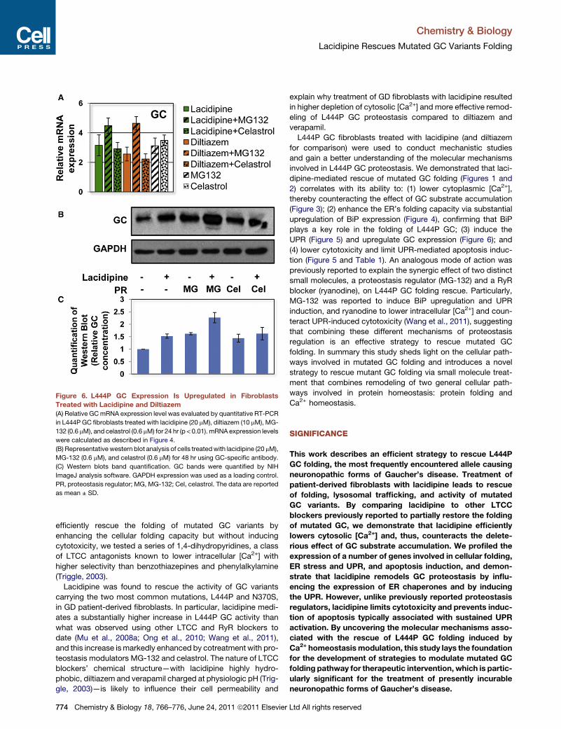
Figure 6. L444P GC Expression Is Upregulated in Fibroblasts
Treated with Lacidipine and Diltiazem(A) Relative GCmRNA expression level was evaluated by quantitative RT-PCR
in L444P GC fibroblasts treated with lacidipine (20 mM), diltiazem (10 mM), MG-
132 (0.6 mM), and celastrol (0.6 mM) for 24 hr (p < 0.01). mRNA expression levels
were calculated as described in Figure 4.
(B) Representative western blot analysis of cells treatedwith lacidipine (20 mM),
MG-132 (0.6 mM), and celastrol (0.6 mM) for 48 hr using GC-specific antibody.
(C) Western blots band quantification. GC bands were quantified by NIH
ImageJ analysis software. GAPDH expression was used as a loading control.
PR, proteostasis regulator; MG, MG-132; Cel, celastrol. The data are reported
as mean ± SD.
Chemistry & Biology
Lacidipine Rescues Mutated GC Variants Folding
efficiently rescue the folding of mutated GC variants by
enhancing the cellular folding capacity but without inducing
cytotoxicity, we tested a series of 1,4-dihydropyridines, a class
of LTCC antagonists known to lower intracellular [Ca2+] with
higher selectivity than benzothiazepines and phenylalkylamine
(Triggle, 2003).
Lacidipine was found to rescue the activity of GC variants
carrying the two most common mutations, L444P and N370S,
in GD patient-derived fibroblasts. In particular, lacidipine medi-
ates a substantially higher increase in L444P GC activity than
what was observed using other LTCC and RyR blockers to
date (Mu et al., 2008a; Ong et al., 2010; Wang et al., 2011),
and this increase ismarkedly enhanced by cotreatment with pro-
teostasis modulators MG-132 and celastrol. The nature of LTCC
blockers’ chemical structure—with lacidipine highly hydro-
phobic, diltiazem and verapamil charged at physiologic pH (Trig-
gle, 2003)—is likely to influence their cell permeability and
774 Chemistry & Biology 18, 766–776, June 24, 2011 ª2011 Elsevier
explain why treatment of GD fibroblasts with lacidipine resulted
in higher depletion of cytosolic [Ca2+] and more effective remod-
eling of L444P GC proteostasis compared to diltiazem and
verapamil.
L444P GC fibroblasts treated with lacidipine (and diltiazem
for comparison) were used to conduct mechanistic studies
and gain a better understanding of the molecular mechanisms
involved in L444P GC proteostasis. We demonstrated that laci-
dipine-mediated rescue of mutated GC folding (Figures 1 and
2) correlates with its ability to: (1) lower cytoplasmic [Ca2+],
thereby counteracting the effect of GC substrate accumulation
(Figure 3); (2) enhance the ER’s folding capacity via substantial
upregulation of BiP expression (Figure 4), confirming that BiP
plays a key role in the folding of L444P GC; (3) induce the
UPR (Figure 5) and upregulate GC expression (Figure 6); and
(4) lower cytotoxicity and limit UPR-mediated apoptosis induc-
tion (Figure 5 and Table 1). An analogous mode of action was
previously reported to explain the synergic effect of two distinct
small molecules, a proteostasis regulator (MG-132) and a RyR
blocker (ryanodine), on L444P GC folding rescue. Particularly,
MG-132 was reported to induce BiP upregulation and UPR
induction, and ryanodine to lower intracellular [Ca2+] and coun-
teract UPR-induced cytotoxicity (Wang et al., 2011), suggesting
that combining these different mechanisms of proteostasis
regulation is an effective strategy to rescue mutated GC
folding. In summary this study sheds light on the cellular path-
ways involved in mutated GC folding and introduces a novel
strategy to rescue mutant GC folding via small molecule treat-
ment that combines remodeling of two general cellular path-
ways involved in protein homeostasis: protein folding and
Ca2+ homeostasis.
SIGNIFICANCE
This work describes an efficient strategy to rescue L444P
GC folding, the most frequently encountered allele causing
neuronopathic forms of Gaucher’s disease. Treatment of
patient-derived fibroblasts with lacidipine leads to rescue
of folding, lysosomal trafficking, and activity of mutated
GC variants. By comparing lacidipine to other LTCC
blockers previously reported to partially restore the folding
of mutated GC, we demonstrate that lacidipine efficiently
lowers cytosolic [Ca2+] and, thus, counteracts the delete-
rious effect of GC substrate accumulation. We profiled the
expression of a number of genes involved in cellular folding,
ER stress and UPR, and apoptosis induction, and demon-
strate that lacidipine remodels GC proteostasis by influ-
encing the expression of ER chaperones and by inducing
the UPR. However, unlike previously reported proteostasis
regulators, lacidipine limits cytotoxicity and prevents induc-
tion of apoptosis typically associated with sustained UPR
activation. By uncovering the molecular mechanisms asso-
ciated with the rescue of L444P GC folding induced by
Ca2+ homeostasismodulation, this study lays the foundation
for the development of strategies to modulate mutated GC
folding pathway for therapeutic intervention, which is partic-
ularly significant for the treatment of presently incurable
neuronopathic forms of Gaucher’s disease.
Ltd All rights reserved

Chemistry & Biology
Lacidipine Rescues Mutated GC Variants Folding
EXPERIMENTAL PROCEDURES
GC Activity Assay
The intact cell GC activity assay was performed as described previously (Mu
et al., 2008b) and in the Supplemental Experimental Procedures.
Quantitative RT-PCR
RT-PCR was conducted as described previously (Wang et al., 2011) and in the
Supplemental Experimental Procedures, using the primers listed in Table S1.
Western Blot Analyses and Immunofluorescence Assays
Details are provided in the Supplemental Experimental Procedures.
Intracellular [Ca2+] Measurement
Fura-2, AM (AnaSpec) was used to measure cytosolic [Ca2+] according to
company’s instructions. Briefly, cells were incubated with 5 mM Fura-2, AM
and 0.05% (w/v) Pluronic F-127 (Invitrogen) at 37�C for 30 min. Following
two washing steps, fluorescence was measured (excitation 340 and 380 nm,
emission 510 nm). Fluorescence ratio of excitation 340/380 reflects relative
intracellular Ca2+ level. Additional details are provided in the Supplemental
Experimental Procedures.
Toxicity Assay
Toxicity assays were conducted as described previously (Wang et al., 2011)
and in the Supplemental Experimental Procedures.
Statistical Analysis
All data are presented as mean ± SD, and statistical significance was calcu-
lated using a two-tailed Student’s t test.
SUPPLEMENTAL INFORMATION
Supplemental Information includes Supplemental Experimental Procedures,
four figures, and one table and can be found with this article online at
doi:10.1016/j.chembiol.2011.04.008.
ACKNOWLEDGMENTS
We greatly appreciate Dichuan Li who helped analyze the colocalization
images.
Received: January 10, 2011
Revised: March 29, 2011
Accepted: April 19, 2011
Published: June 23, 2011
REFERENCES
Baumann, O., and Walz, B. (2001). Endoplasmic reticulum of animal cells and
its organization into structural and functional domains. Int. Rev. Cytol. 205,
149–214.
Berridge, M.J., Bootman, M.D., and Lipp, P. (1998). Calcium—a life and death
signal. Nature 395, 645–648.
Beutler, E., Kuhl, W., and Sorge, J. (1984). Cross-reacting material in Gaucher
disease fibroblasts. Proc. Natl. Acad. Sci. USA 81, 6506–6510.
Bygrave, F.L., and Benedetti, A. (1996). What is the concentration of calcium
ions in the endoplasmic reticulum? Cell Calcium 19, 547–551.
Eckenrode, E.F., Yang, J., Velmurugan, G.V., Foskett, J.K., and White, C.
(2010). Apoptosis protection by Mcl-1 and Bcl-2 modulation of inositol 1,4,5-
trisphosphate receptor-dependent Ca2+ signaling. J. Biol. Chem. 285, 13678–
13684.
Epstein, M. (1999). Role of a third generation calcium antagonist in the
management of hypertension. Drugs 57 (Suppl 1 ), 1–10.
Grabowski, G.A. (1997). Gaucher disease: gene frequencies and genotype/
phenotype correlations. Genet. Test. 1, 5–12.
Chemistry & Biology 18,
Gunther, S., Kuhn, M., Dunkel, M., Campillos, M., Senger, C., Petsalaki, E.,
Ahmed, J., Urdiales, E.G., Gewiess, A., Jensen, L.J., et al. (2008).
SuperTarget and Matador: resources for exploring drug-target relationships.
Nucleic Acids Res. 36, D919–D922.
Hockerman, G.H., Peterson, B.Z., Johnson, B.D., and Catterall, W.A. (1997).
Molecular determinants of drug binding and action on L-type calcium chan-
nels. Annu. Rev. Pharmacol. Toxicol. 37, 361–396.
Hruska, K.S., LaMarca, M.E., Scott, C.R., and Sidransky, E. (2008). Gaucher
disease: mutation and polymorphism spectrum in the glucocerebrosidase
gene (GBA). Hum. Mutat. 29, 567–583.
Korkotian, E., Schwarz, A., Pelled, D., Schwarzmann, G., Segal, M., and
Futerman, A.H. (1999). Elevation of intracellular glucosylceramide levels
results in an increase in endoplasmic reticulum density and in functional
calcium stores in cultured neurons. J. Biol. Chem. 274, 21673–21678.
Lloyd-Evans, E., Pelled, D., Riebeling, C., Bodennec, J., de-Morgan, A.,
Waller, H., Schiffmann, R., and Futerman, A.H. (2003). Glucosylceramide
and glucosylsphingosine modulate calcium mobilization from brain micro-
somes via different mechanisms. J. Biol. Chem. 278, 23594–23599.
Maley, F., Trimble, R.B., Tarentino, A.L., and Plummer, T.H., Jr. (1989).
Characterization of glycoproteins and their associated oligosaccharides
through the use of endoglycosidases. Anal. Biochem. 180, 195–204.
Meivar-Levy, I., Horowitz, M., and Futerman, A.H. (1994). Analysis of glucocer-
ebrosidase activity using N-(1-[14C]hexanoyl)-D-erythroglucosylsphingosine
demonstrates a correlation between levels of residual enzyme activity and
the type of Gaucher disease. Biochem. J. 303, 377–382.
Michalak, M., Robert Parker, J.M., and Opas, M. (2002). Ca2+ signaling and
calcium binding chaperones of the endoplasmic reticulum. Cell Calcium 32,
269–278.
Michelakakis, H., Dimitriou, E., Van Weely, S., Boot, R.G., Mavridou, I.,
Verhoek, M., and Aerts, J.M. (1995). Characterization of glucocerebrosidase
in Greek Gaucher disease patients: mutation analysis and biochemical
studies. J. Inherit. Metab. Dis. 18, 609–615.
Mu, T.W., Fowler, D.M., and Kelly, J.W. (2008a). Partial restoration of mutant
enzyme homeostasis in three distinct lysosomal storage disease cell lines by
altering calcium homeostasis. PLoS Biol. 6, e26.
Mu, T.W., Ong, D.S., Wang, Y.J., Balch, W.E., Yates, J.R., 3rd, Segatori, L.,
and Kelly, J.W. (2008b). Chemical and biological approaches synergize to
ameliorate protein-folding diseases. Cell 134, 769–781.
Offman, M.N., Krol, M., Silman, I., Sussman, J.L., and Futerman, A.H. (2010).
Molecular basis of reduced glucosylceramidase activity in the most common
Gaucher disease mutant, N370S. J. Biol. Chem. 285, 42105–42114.
Ong, D.S., Mu, T.W., Palmer, A.E., and Kelly, J.W. (2010). Endoplasmic retic-
ulum Ca2+ increases enhance mutant glucocerebrosidase proteostasis. Nat.
Chem. Biol. 6, 424–432.
Pelled, D., Trajkovic-Bodennec, S., Lloyd-Evans, E., Sidransky, E.,
Schiffmann, R., and Futerman, A.H. (2005). Enhanced calcium release in the
acute neuronopathic form of Gaucher disease. Neurobiol. Dis. 18, 83–88.
Pepine, C. (1989). Nicardipine, a new calcium channel blocker: role for
vascular selectivity. Clin. Cardiol. 12, 240–246.
Rodriguez, D., Rojas-Rivera, D., and Hetz, C. (2011). Integrating stress signals
at the endoplasmic reticulum: the BCL-2 protein family rheostat. Biochim.
Biophys. Acta 1813, 564–574.
Ron, D., and Walter, P. (2007). Signal integration in the endoplasmic reticulum
unfolded protein response. Nat. Rev. Mol. Cell Biol. 8, 519–529.
Rong, Y.P., Bultynck, G., Aromolaran, A.S., Zhong, F., Parys, J.B., De Smedt,
H., Mignery, G.A., Roderick, H.L., Bootman, M.D., and Distelhorst, C.W.
(2009). The BH4 domain of Bcl-2 inhibits ER calcium release and apoptosis
by binding the regulatory and coupling domain of the IP3 receptor. Proc.
Natl. Acad. Sci. USA 106, 14397–14402.
Sawkar, A.R., Cheng, W.C., Beutler, E., Wong, C.H., Balch, W.E., and Kelly,
J.W. (2002). Chemical chaperones increase the cellular activity of N370S
beta -glucosidase: a therapeutic strategy for Gaucher disease. Proc. Natl.
Acad. Sci. USA 99, 15428–15433.
766–776, June 24, 2011 ª2011 Elsevier Ltd All rights reserved 775

Chemistry & Biology
Lacidipine Rescues Mutated GC Variants Folding
Sawkar, A.R., Adamski-Werner, S.L., Cheng, W.C., Wong, C.H., Beutler, E.,
Zimmer, K.P., and Kelly, J.W. (2005). Gaucher disease-associated glucocere-
brosidases show mutation-dependent chemical chaperoning profiles. Chem.
Biol. 12, 1235–1244.
Sawkar, A.R., Schmitz, M., Zimmer, K.P., Reczek, D., Edmunds, T., Balch,
W.E., and Kelly, J.W. (2006). Chemical chaperones and permissive tempera-
tures alter localization of Gaucher disease associated glucocerebrosidase
variants. ACS Chem. Biol. 1, 235–251.
Schmitz, M., Alfalah, M., Aerts, J.M., Naim, H.Y., and Zimmer, K.P. (2005).
Impaired trafficking of mutants of lysosomal glucocerebrosidase in
Gaucher’s disease. Int. J. Biochem. Cell Biol. 37, 2310–2320.
Schroder, M., and Kaufman, R.J. (2005). The mammalian unfolded protein
response. Annu. Rev. Biochem. 74, 739–789.
Schueler, U.H., Kolter, T., Kaneski, C.R., Zirzow, G.C., Sandhoff, K., and
Brady, R.O. (2004). Correlation between enzyme activity and substrate storage
in a cell culture model system for Gaucher disease. J. Inherit. Metab. Dis. 27,
649–658.
Scorrano, L., Oakes, S.A., Opferman, J.T., Cheng, E.H., Sorcinelli, M.D.,
Pozzan, T., and Korsmeyer, S.J. (2003). BAX and BAK regulation of endo-
plasmic reticulum Ca2+: a control point for apoptosis. Science 300, 135–139.
776 Chemistry & Biology 18, 766–776, June 24, 2011 ª2011 Elsevier
Sidransky, E., LaMarca, M.E., and Ginns, E.I. (2007). Therapy for Gaucher
disease: don’t stop thinking about tomorrow. Mol. Genet. Metab. 90, 122–125.
Sun, Y., Liou, B., Quinn, B., Ran, H., Xu, Y.H., and Grabowski, G.A. (2009).
In vivo and ex vivo evaluation of L-type calcium channel blockers on acid
beta-glucosidase in Gaucher disease mouse models. PLoS One 4, e7320.
Triggle, D.J. (2003). 1,4-dihydropyridine calcium channel ligands: selectivity of
action. The roles of pharmacokinetics, state-dependent interactions, channel
isoforms, and other factors. Drug Dev. Res. 58, 5–17.
Triggle, D.J. (2006). L-type calcium channels. Curr. Pharm. Des. 12, 443–457.
Wang, F., Agnello, G., Sotolongo, N., and Segatori, L. (2011). Ca2+ homeo-
stasis modulation enhances the amenability of L444P glucosylcerebrosidase
to proteostasis regulation in patient-derived fibroblasts. ACS Chem. Biol. 6,
158–168.
Wishart, D.S., Knox, C., Guo, A.C., Cheng, D., Shrivastava, S., Tzur, D.,
Gautam, B., and Hassanali, M. (2008). DrugBank: a knowledgebase for drugs,
drug actions and drug targets. Nucleic Acids Res. 36, D901–D906.
Yu, Z., Sawkar, A.R., Whalen, L.J., Wong, C.H., and Kelly, J.W. (2007).
Isofagomine- and 2,5-anhydro-2,5-imino-D-glucitol-based glucocerebrosi-
dase pharmacological chaperones for Gaucher disease intervention. J. Med.
Chem. 50, 94–100.
Ltd All rights reserved