2 Anaphylaxis 3 Anterior Mediastinal Mass 4 Bradycardia 5 ...
30
1 Air Embolism 2 Anaphylaxis 3 Anterior Mediastinal Mass 4 Bradycardia 5 Bronchospasm 6-7 Cardiac Arrest 8 Difficult Airway 9-10 Fire: Airway / OR 11 Hyperkalemia 12 Hypertension 13 Hypotension 14 Hypoxia 15 Intracranial Pressure 16 Laryngospasm 17 Local Anesthetic Toxicity 18 Loss of Evoked Potentials 19 Malignant Hyperthermia 20 Massive Hemorrhage 21 Myocardial Ischemia 22 Pulmonary Hypertension 23 Tachycardia 24 Tamponade, Cardiac 25 Tension pneumothorax 26 Transfusion Reaction 27 Trauma 28 Maternal OB Hemorrhage Call for help! Code Team ___________ PICU ___________ Fire ___________ Overhead STAT ___________ ECMO ___________ Notify surgeon/team Pedi Crisis CRITICAL EVENTS CHECKLISTS For use in the peri-anesthesia setting Use expert clinical judgment when using this and all emergency manuals. Revision Jan 2020. Available at: http://www.pedsanesthesia.org/wpcontent/uploads/2018/03/SPACriticalEvent sChecklists.pdf
Transcript of 2 Anaphylaxis 3 Anterior Mediastinal Mass 4 Bradycardia 5 ...

1 Air Embolism
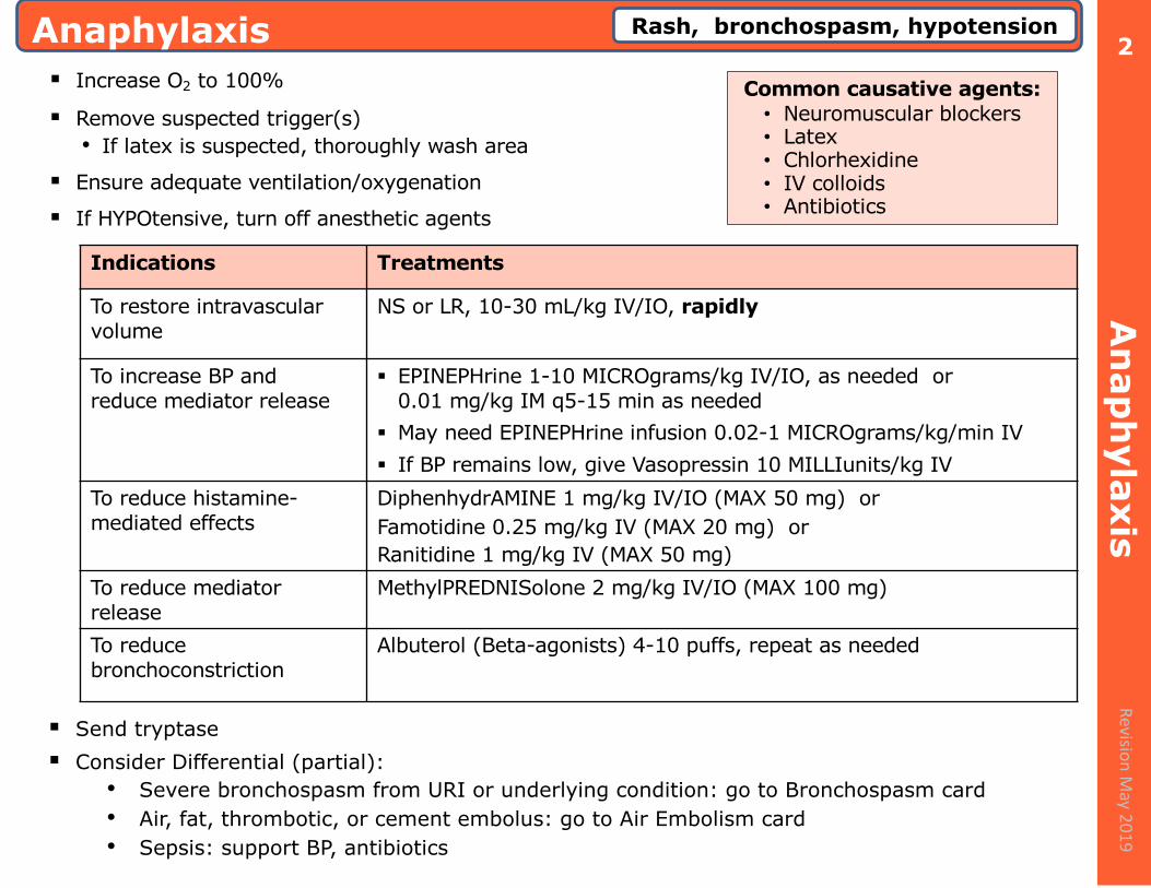
2 Anaphylaxis
3 Anterior Mediastinal Mass
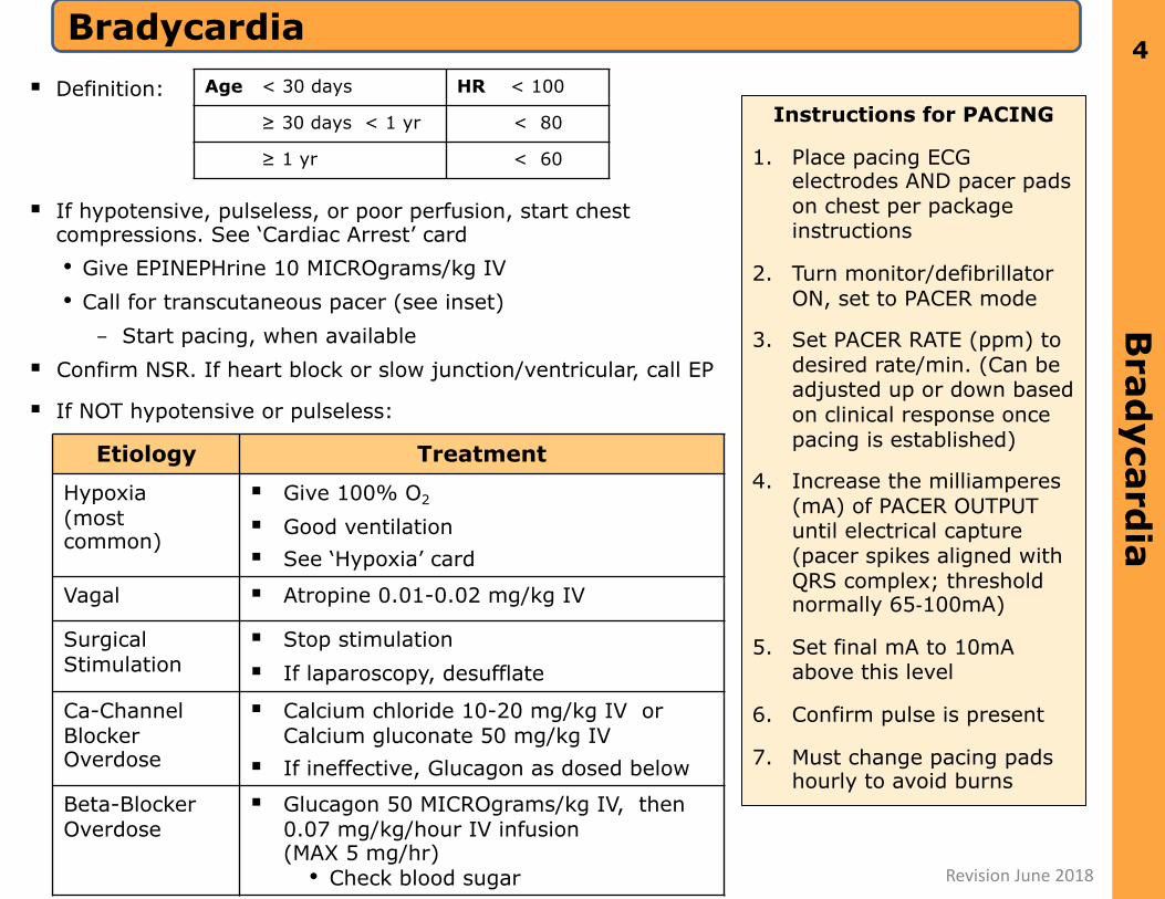
4 Bradycardia
5 Bronchospasm
6-7 Cardiac Arrest
8 Difficult Airway
9-10 Fire: Airway / OR
11 Hyperkalemia
12 Hypertension
13 Hypotension
14 Hypoxia
15 Intracranial Pressure
16 Laryngospasm
17 Local Anesthetic Toxicity
18 Loss of Evoked Potentials
19 Malignant Hyperthermia
20 Massive Hemorrhage
21 Myocardial Ischemia
22 Pulmonary Hypertension
23 Tachycardia
24 Tamponade, Cardiac
25 Tension pneumothorax
26 Transfusion Reaction
27 Trauma
28 Maternal OB Hemorrhage
Call for help!Code Team ___________PICU ___________Fire ___________Overhead STAT ___________ECMO ___________
Notify surgeon/team
Pedi Crisis
CRITICAL EVENTSCHECKLISTS
For use in the peri-anesthesia setting
Use expert clinical judgment when using this and all emergency manuals.
Revision Jan 2020. Available at: http://www.pedsanesthesia.org/wpcontent/uploads/2018/03/SPACriticalEventsChecklists.pdf

§ Notify team, stop nitrous oxide and volatile agents. Increase O2 to 100%.
§ Stop air entrainment: Find air entry point, stop source, and limit further entry.§ Ask surgeon:
- Flood wound with irrigation/soaked saline dressing- Stop all pressurized gas sources, e.g. laparoscope, endoscope- Place bone wax or cement on exposed bone edges
• Check for open venous lines or air in IV tubing
• Lower surgical site below level of heart (if possible)
• Perform Valsalva
§ Consider:
• Compress jugular veins intermittently if head or cranial case
§ If hypotensive:• Give EPINEPHrine 1-10 MICROgrams/kg IV, consider infusion EPINEPHrine 0.02-1
MICROgrams/kg/min IV or NOREPInephrine 0.05-2 MICROgrams/kg/min IV
• Chest compressions: 100-120/min to force air through lock, even if not in cardiac arrest
• If available, call for TEE/US
§ If cardiac arrest, see ‘Cardiac Arrest’ card
Air EmbolismA
ir Emb
olism↓ EtCO2 ↓ SaO2 ↓ BP, mill-wheel murmur
1
Revision Oct 2018
An
aph
ylaxis § Increase O2 to 100%
§ Remove suspected trigger(s)• If latex is suspected, thoroughly wash area
§ Ensure adequate ventilation/oxygenation
§ If HYPOtensive, turn off anesthetic agents
Anaphylaxis Rash, bronchospasm, hypotension2
Common causative agents:• Neuromuscular blockers• Latex• Chlorhexidine• IV colloids• Antibiotics
Indications Treatments
To restore intravascular volume
NS or LR, 10-30 mL/kg IV/IO, rapidly
To increase BP andreduce mediator release
§ EPINEPHrine 1-10 MICROgrams/kg IV/IO, as needed or 0.01 mg/kg IM q5-15 min as needed
§ May need EPINEPHrine infusion 0.02-1 MICROgrams/kg/min IV§ If BP remains low, give Vasopressin 10 MILLIunits/kg IV
To reduce histamine-mediated effects
DiphenhydrAMINE 1 mg/kg IV/IO (MAX 50 mg) or Famotidine 0.25 mg/kg IV (MAX 20 mg) or Ranitidine 1 mg/kg IV (MAX 50 mg)
To reduce mediator release
MethylPREDNISolone 2 mg/kg IV/IO (MAX 100 mg)
To reducebronchoconstriction
Albuterol (Beta-agonists) 4-10 puffs, repeat as needed
Revision May 2019
§ Send tryptase§ Consider Differential (partial):
• Severe bronchospasm from URI or underlying condition: go to Bronchospasm card• Air, fat, thrombotic, or cement embolus: go to Air Embolism card• Sepsis: support BP, antibiotics

§ Increase O2 to 100%
Anterior Mediastinal Mass A
nterior M
ediastin
al Mass
Intra-operative TreatmentsAirway collapse§ Increase FiO2
§ Add CPAP for spontaneous ventilation; add PEEP for controlled ventilation
§ Reposition to lateral or prone
§ Ventilate via rigid bronchoscope
Cardiovascular collapse§ Give fluid bolus
§ Reposition to lateral or prone
§ Ask surgeon for sternotomy and elevation of mass
§ Consider ECMO
Preoperative ConsiderationsHigh Risk Factors§ Etiology:
• Hodgkin’s and non-Hodgkin’s lymphoma
§ Clinical signs: • orthopnea, upper body edema,
stridor, wheezing
§ Imaging findings: • tracheal, bronchial, carinal, or great
vessel compression; SVC or RVOT obstruction; ventricular dysfunction; pericardial effusion
Anesthetic Plan
§ Perform surgery under local anesthesia, if possible
§ Pre-treat with irradiation or corticosteroids
§ Maintain spontaneous ventilation and avoid paralysis
§ Ensure availability of fiberoptic and rigid bronchoscope
§ Cardiopulmonary bypass or ECMO
§ Type and cross and sternal saw (for surgeons) available
3
Revision June 2018
Brad
ycardia
§ Definition:
§ If hypotensive, pulseless, or poor perfusion, start chest compressions. See ‘Cardiac Arrest’ card• Give EPINEPHrine 10 MICROgrams/kg IV• Call for transcutaneous pacer (see inset)
- Start pacing, when available§ Confirm NSR. If heart block or slow junction/ventricular, call EP
§ If NOT hypotensive or pulseless:
Bradycardia 4
Instructions for PACING
1. Place pacing ECG electrodes AND pacer pads on chest per package instructions
2. Turn monitor/defibrillator ON, set to PACER mode
3. Set PACER RATE (ppm) to desired rate/min. (Can be adjusted up or down based on clinical response once pacing is established)
4. Increase the milliamperes (mA) of PACER OUTPUT until electrical capture (pacer spikes aligned with QRS complex; threshold normally 65-100mA)
5. Set final mA to 10mA above this level
6. Confirm pulse is present
7. Must change pacing pads hourly to avoid burns
Age < 30 days HR < 100
≥ 30 days < 1 yr < 80
≥ 1 yr < 60
Etiology Treatment
Hypoxia (most common)
§ Give 100% O2
§ Good ventilation§ See ‘Hypoxia’ card
Vagal § Atropine 0.01-0.02 mg/kg IV
SurgicalStimulation
§ Stop stimulation§ If laparoscopy, desufflate
Ca-Channel BlockerOverdose
§ Calcium chloride 10-20 mg/kg IV orCalcium gluconate 50 mg/kg IV
§ If ineffective, Glucagon as dosed below
Beta-BlockerOverdose
§ Glucagon 50 MICROgrams/kg IV, then 0.07 mg/kg/hour IV infusion (MAX 5 mg/hr)• Check blood sugar Revision June 2018

BronchospasmB
ronch
ospasm
↓ EtCO2, upslope stage III EtCO2↑ airway pressures, ↓ SpO2
Non-Intubated Patient
§ If ETT in, go to ‘Intubated Patient’ column on this card (at the left)
§ Administer supplemental oxygen
§ Auscultate the chest, differentiate from stridor/extrathoracic airway obstruction
§ Consider inhaled albuterol (with spacer) 2.5-5 mg. If severe, 5-20 mg/hr inhaled
§ Consider chest radiograph
§ Consider IV steroids: methylprednisolone 1 mg/kg IV (MAX 60 mg) or dexamethasone 0.15-0.25 mg/kg (MAX 16 mg)
§ If severe, consider EPINEPHrine 1-2 MICROgrams/kg IV (MAX 1 mg) or 10 MICROgrams/kg subcutaneous/intramuscular (MAX 0.5 mg)
§ If severe, consider ICU and/or advanced airway management.
5Revision O
ct 2018
Intubated Patient
§ Increase FiO2 to 100%§ Auscultate the chest:• Equal breath sounds?• Endobronchial ETT?• Wheezing?
§ Check ETT:• Kinked? • Secretions/blood in ETT? Needs suctioning?
§ Consider albuterol 2-10 puffs, repeat as needed§ Consider deepening anesthetic§ If needed, give ketamine 1-2 mg/kg IV § If severe, consider
EPINEPHrine 1-2 MICROgrams/kg IV (MAX 1 mg)§ Consider IV steroids: methylprednisolone 2 mg/kg IV
(MAX 60 mg) or dexamethasone 0.15-0.25 mg/kg (MAX 16 mg)
§ Consider chest radiograph § For refractory bronchospasm, consider magnesium
sulfate 50-75 mg/kg (MAX 2 grams) bolused over 20 minutes, (CAUTION, may cause hypotension)
Differential Diagnosis§ Endobronchial intubation§ Mechanical obstruction of ETT• Kinking• Solidified secretions or blood• Overinflation of tracheal tube cuff
§ Inadequate depth of anesthesia§ URI/tobacco exposure§ Foreign body
§ Pulmonary edema§ Tension pneumothorax§ Aspiration pneumonitis§ Pulmonary embolism § Persistent coughing and straining§ Asthmatic attack§ Anaphylaxis

§ Notify team, call for help and code cart/defibrillator§ Increase oxygen to 100%. Turn off anesthetics§ If ETT, 100-120 chest compressions/min + 10 breaths/min. Avoid hyperventilation. § If no ETT, 15:2 compression:ventilation ratio (100-120 chest compressions/min + 8 breaths/min)§ For chest compressions, maximize EtCO2 > 10 mmHg (see next card for more details):
• Switch compressor every 2 min• Use sudden increase in EtCO2 for ROSC, Do NOT stop compressions for pulse check
§ Obtain defibrillator. Attach pads. If VF/VT, shock 2 joules/kg. Continue chest compressions x 2 minutes. § Start timer. Designate team leader. Assign roles. Designate a scribe/recorder. Notify family. Continue
with items in yellow box
Card
iacA
rrestCardiac Arrest Pulseless cardiac arrest
6
Hs and Ts: Reversible Causes
• Hypovolemia• Hypoxemia• Hydrogen ion (acidosis)• Hyperkalemia/Hypoglycemia• Hypothermia
• Tension Pneumothorax• Tamponade (Cardiac)• Thrombosi• Toxin (anesthetic, β-blocker)• Trauma (surgical or nonsurgical bleeding)
Revision Oct 2018
Repeat sequence below until return of spontaneous circulation: § If still in VF/VT, shock 4 joules/kg q2 min (up to 10 joules/kg on subsequent shocks)§ Resume chest compressions immediately regardless of rhythm§ EPINEPHrine 10 MICROgrams/kg IV q 3-5 min while in arrest (MAX 1 mg)
• If still no ROSC after second dose of EPINEPHrine, activate ECMO (if available) § Check pulse & rhythm q 2 min during compressor change§ Check for reversible causes (Hs and Ts) early and often (see table below)§ Lidocaine 1 mg/kg bolus (MAX 100 mg); may repeat (total: 2 doses) OR amiodarone 5 mg/kg
bolus; may repeat (total: 3 doses)
§ Repeat sequence in this box until return of spontaneous circulation

Cardiac Arrest: Supine/Prone Chest Compressions
§ If no midline incision: Compress with heel of hand on spine and second hand on top
§ If midline incision: Compress with heel of each hand under scapula
Compress with encircling technique:
§ If no midline incision: thumbs midline
§ If midline incision:thumbs lateral to incision
Prone:Infants
Su
pin
e/Pron
eC
hest com
pression
s7
Figure 1: From Dequin P-F et al. Cardiopulmonary resuscitation in the prone position: Kouwenhoven revisited. Intensive Care Medicine, 1996;22:1272Figure 2: From Tobias et al, Journal of Pediatric Surgery, 1994:29, 1537-1539Figure 3: Original artwork by Brooke Albright-Trainer, MD
Figure 2
Figure 1
Prone:Children/Adolescents
Figure 3
§ Chest compression instructions (see previous card for full CPR instructions):• Place patient on backboard, maintain good hand position; if prone, see instructions below• Maximize EtCO2 > 10 mmHg with force/depth of compressions• Allow full recoil between compressions• Switch compressor every 2 min• Use sudden increase in EtCO2 for ROSC, Do NOT stop compressions for pulse check
Revision Mar 2018

Difficu
lt Airw
ay, Un
expected
§ Increase O2 to 100% and maintain continuous oxygen flow during airway management
§ Call for help, surgical airway expert and cart, rigid bronchoscope and tracheostomy kit
§ If unable to mask ventilate, ask for 2-handed assistance and: • Insert oral and/or nasal airway; • If unsuccessful, insert supraglottic airway (e.g., LMA)• Decompress stomach with orogastric tube• Consider reversing rocuronium or vecuronium with
sugammadex (16 mg/kg). Call to obtain if not in OR.
§ If able to re-establish pt spontaneous ventilation:• Consider awakening patient• Consider reversal of neuromuscular blocker
§ After two attempts: change providers and consider alternative approaches to intubation (see table)
§ If macroglossia (e.g. Beckwith-Wiedemann, Pierre-Robin), or mediastinal mass, consider prone or lateral position
§ If still unable to ventilate:• Younger children: Emergency non-invasive airway
such as rigid bronchoscopy • In older children: Jet ventilation or emergency
invasive/surgical airway such as cricothyrotomyor tracheostomy
Difficult Airway, Unexpected
Alternative Approaches for
Intubation
• Different blade• Re-position head• Different provider• Video-laryngoscope• Bougie• Intubating LMA• Fiberoptic scope• Intubating stylet• Blind oral • Blind nasal
8
Revision May 2019
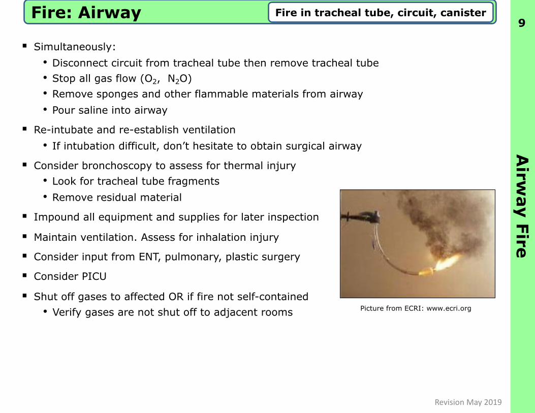
Airw
ay Fire Fire: Airway Fire in tracheal tube, circuit, canister
§ Simultaneously:• Disconnect circuit from tracheal tube then remove tracheal tube• Stop all gas flow (O2, N2O)• Remove sponges and other flammable materials from airway• Pour saline into airway
§ Re-intubate and re-establish ventilation• If intubation difficult, don’t hesitate to obtain surgical airway
§ Consider bronchoscopy to assess for thermal injury• Look for tracheal tube fragments• Remove residual material
§ Impound all equipment and supplies for later inspection
§ Maintain ventilation. Assess for inhalation injury
§ Consider input from ENT, pulmonary, plastic surgery
§ Consider PICU
§ Shut off gases to affected OR if fire not self-contained• Verify gases are not shut off to adjacent rooms
9
Picture from ECRI: www.ecri.org
Revision May 2019

OR
Fire OR Fire (non-airway) Fire in OR, equipment smoke,
fumes, flash/fire on patient
§ Simultaneously:• Stop flow of medical gases• Remove drapes and all burning and flammable material from patient• Make one attempt to extinguish fire by pouring saline on fire
§ If fire not extinguished on 1st attempt, use CO2 fire extinguisher
§ If fire persists:• Activate fire alarm• Remove patient from OR• Confine fire by closing all OR doors• Turn off O2 gas supply to OR
§ Maintain ventilation. Assess for inhalation injury
§ Consider input from ENT, pulmonary, plastic surgery
§ Consider PICU
§ Shut off gases to affected OR if fire not self-contained• Verify gases are not shut off to adjacent rooms
§ Impound all equipment and supplies for later inspection
10
Picture from ECRI: www.ecri.org
Revision May 2019

Hyp
erkalemia
Hyperkalemia Serum K+ > 6 mEq/L
Treatment:§ If hemodynamically unstable, start CPR/PALS§ Hyperventilate with 100% O2
§ IV calcium gluconate 60-100 mg/kg or calcium chloride 20 mg/kg• Directly visualize site to avoid infiltration• Flush tubing after calcium administration
§ Stop K+ containing fluids (LR/RBC); switch to NS§ Dextrose IV 0.5-1 g/kg and insulin IV 0.1 Unit/kg (MAX 10 units)§ Albuterol puffs or nebulized, once cardiac rhythm stable
§ Sodium bicarbonate IV 1-2 mEq/kg§ Furosemide IV 0.5-1 mg/kg§ Consider terbutaline 10 MICROgrams/kg load,
then 0.1-10 MICROgrams/kg/min
§ If cardiac arrest > 6 min, activate ECMO (if available)§ Dialysis if refractory to treatment§ If transfusion required, use washed or fresh RBC
Causes of Hyperkalemia: § Excessive intake: massive or “old” blood products, TPN, cardioplegia, KCl infusion§ Shift of K+ from tissues to plasma: crush injury, burns, succinylcholine, malignant
hyperthermia, acidosis§ Inadequate excretion: renal failure§ Pseudohyperkalemia: hemolyzed sample, thrombocytosis, leukocytosis
11
From: Slovis C, Jenkins R. BMJ 2002
Manifestations: • Tall peaked T
wave• Heart block• Sine wave• V fib or asystole
Revision Mar 2018

Hyp
ertension
Acute Hypertension Sustained high blood pressure refractory to treating reversible causes
§ In pediatrics, hypertension is almost always treated by addressing likely causes such as light anesthesia or measurement error:
• Ensure correct BP cuff size: cuff bladder width ~ 40% of limb circumference
• Ensure arterial line transducer is at level of heart
- Consider placing arterial line if not already present
* CAUTION: Anti-hypertensive drugs are almost never needed for routine pediatric cases. These medications are used almost exclusively for specialized cardiac, neurosurgical, or endocrine (pheochromocytoma) cases. Consult an expert before use. Rule-out increased ICP.
12
Action Drug (IV Dosing)
Direct smooth muscle relaxation
§ Sodium nitroprusside 0.5-10 MICROgrams/kg/min § HydrALAZINE 0.1-0.2 mg/kg (adult dose 5-10 mg)
β-Adrenergic blockade
§ Esmolol 100-500 MICROgrams/kg over 5 min, then 25-300 MICROgrams/kg/min
§ Labetalol (also a effect) 0.2-1 mg/kg q 10 min; 0.4-3 mg/kg/hour (infusion)
Calcium channel blockade
§ niCARdapine 0.5-5 MICROgrams/kg/min § Clevidipine 0.5-3.5 MICROgrams/kg/min
D1-dopamine agonist § Fenoldopam 0.2-0.8 MICROgrams/kg/min
Hypertensive Blood Pressure Range*
Age (yr) Systolic Diastolic
newborn >97 >70
1-3 >105 >61
4-12 >113 >86
Revision May 2019

HypotensionH
ypoten
sionC
ause
s
↓ Preload ↓ Contractility ↓ Afterload
§ Hypovolemia§ Vasodilation§ Impaired venous return
§ Tamponade§ Pulmonary embolism
§ IVC compression (prone, obese, surgical)
§ Pneumothorax/ pneumoperitoneum
§ Increased PIP or PEEP
§ Negative inotropic drugs (anesthetic agents)
§ Arrhythmias
§ Hypoxemia§ Heart failure (ischemia)§ Hypocalcemia/blood product
administration
§ Drug-induced vasodilation
§ Sepsis
§ Anaphylaxis§ Adrenal crisis
§ Hypocalcemia§ Thyroid crisis
Trea
tmen
t
§ Expand circulating blood volume (administer fluids rapidly, consider albumin)
§ Trendelenberg position
§ Place or replace IV; consider intraosseous line
§ Start inotrope infusion (DOPamine, EPINEPHrine), as needed
§ Consider calcium IV
§ Review ECG for rhythm disturbances or ischemia
§ Send ABG, Hgb, electrolytes
§ Start vasopressor infusion: phenylephrine, norepinephrine
§ Go to ‘Anaphylaxis’ card, if appropriate.
§ Administer steroids for adrenal crisis
13§ Ensure oxygenation/ventilation
§ Turn anesthetic agents down or off
§ Check cuff size and transducer position
§ Consider placing arterial line if not already present
§ Give appropriate treatment (see table below)
Sustained low blood pressure with patient at risk for end-organ hypoperfusion, typically > 20% below baseline
Age < 5th% Systolic BP (mmHg)*
* Numbers are only a guide and vary for individual
patients and situations
Preemie 47– 57
0 – 3 mo 62 – 69
3 mo – 1 yr 65 – 68
1 – 3 yr 68 – 74
4 – 12 yr 70 – 85
> 12 yr 85 – 92
Revision May 2019
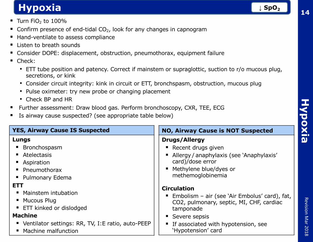
HypoxiaH
ypoxia
↓ SpO2 14§ Turn FiO2 to 100% § Confirm presence of end-tidal CO2, look for any changes in capnogram§ Hand-ventilate to assess compliance§ Listen to breath sounds§ Consider DOPE: displacement, obstruction, pneumothorax, equipment failure§ Check:• ETT tube position and patency. Correct if mainstem or supraglottic, suction to r/o mucous plug,
secretions, or kink• Consider circuit integrity: kink in circuit or ETT, bronchspasm, obstruction, mucous plug• Pulse oximeter: try new probe or changing placement• Check BP and HR
§ Further assessment: Draw blood gas. Perform bronchoscopy, CXR, TEE, ECG§ Is airway cause suspected? (see appropriate table below)
NO, Airway Cause is NOT SuspectedDrugs/Allergy§ Recent drugs given§ Allergy / anaphylaxis (see ‘Anaphylaxis’
card)/dose error§ Methylene blue/dyes or
methemoglobinemia
Circulation§ Embolism – air (see ‘Air Embolus’ card), fat,
CO2, pulmonary, septic, MI, CHF, cardiac tamponade
§ Severe sepsis§ If associated with hypotension, see
‘Hypotension’ card
YES, Airway Cause IS SuspectedLungs§ Bronchospasm§ Atelectasis§ Aspiration§ Pneumothorax§ Pulmonary Edema
ETT § Mainstem intubation§ Mucous Plug§ ETT kinked or dislodged
Machine § Ventilator settings: RR, TV, I:E ratio, auto-PEEP§ Machine malfunction
Revision Mar 2018

Increased
Intracran
ial Pressu
reIncreased Intracranial Pressure
§ If GCS < 9, respiratory distress, hemodynamic instability:• Secure airway• Provide sedation prior to transport
§ Keep PaCO2 30-35 mmHg and PaO2 > 60 mmHg§ Maintain cerebral perfusion pressure (discuss goal CPP with team)§ Discuss target ICP with neurosurgery, will often want ICP < 20§ Use vasopressors (phenylephrine or NOREPInephrine) as needed to maintain BP and CPP§ Consider head of bed at 30⁰
§ Hypertonic saline (3% saline via central venous catheter) 1-5 mL/kg over 20 min, then 0.1-2 mL/kg/hour; goal ICP <20 mmHg • Monitor serum sodium• Keep osmolarity <360 mOsm/L
§ If hypertonic saline not available, can give mannitol 0.25-1 g/kg, over 20 minutes to decrease ICP
§ Consider furosemide 1-2 mg/kg (starting MAX 20 mg) to decrease ICP§ Consider seizure prophylaxis: Keppra (levetiracetam) 10-30 mg/kg IV (MAX 2500 mg)
§ Refractory elevated ICP treatment, consider: • Barbiturate coma • Paralysis with non-depolarizing agent
§ AVOID: • Compression of neck vessels• Hyperthermia• Hyperglycemia & dextrose containing solutions (maintain glucose level < 200 mg/dL)
15Revision
Mar 2018
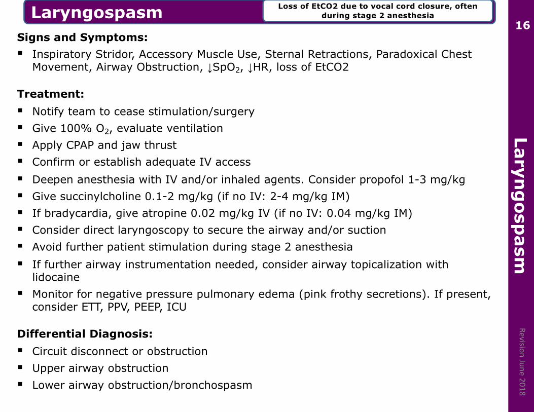
LaryngospasmLaryn
gosp
asmLoss of EtCO2 due to vocal cord closure, often
during stage 2 anesthesia16
Revision June 2018
Signs and Symptoms:§ Inspiratory Stridor, Accessory Muscle Use, Sternal Retractions, Paradoxical Chest
Movement, Airway Obstruction, ↓SpO2, ↓HR, loss of EtCO2
Treatment:§ Notify team to cease stimulation/surgery§ Give 100% O2, evaluate ventilation§ Apply CPAP and jaw thrust§ Confirm or establish adequate IV access§ Deepen anesthesia with IV and/or inhaled agents. Consider propofol 1-3 mg/kg§ Give succinylcholine 0.1-2 mg/kg (if no IV: 2-4 mg/kg IM)§ If bradycardia, give atropine 0.02 mg/kg IV (if no IV: 0.04 mg/kg IM)§ Consider direct laryngoscopy to secure the airway and/or suction§ Avoid further patient stimulation during stage 2 anesthesia§ If further airway instrumentation needed, consider airway topicalization with
lidocaine§ Monitor for negative pressure pulmonary edema (pink frothy secretions). If present,
consider ETT, PPV, PEEP, ICU
Differential Diagnosis:§ Circuit disconnect or obstruction§ Upper airway obstruction§ Lower airway obstruction/bronchospasm

Local An
esthetic To
xicityLocal Anesthetic Toxicity Hypotension, rhythm disturbance,
altered consciousness, seizures
§ Stop local anesthetic§ Request Intralipid kit
§ Secure airway and ventilation
§ Give 100% O2
§ Confirm or establish adequate IV access.
§ Confirm & monitor continuous ECG, BP, and SaO2
§ Seizure treatment: • Midazolam 0.05-0.1 mg/kg IV • Be prepared to treat resultant hypoventilation
§ Treat hypotension with small doses of EPINEPHrine 1 MICROgram/kg
§ Avoid propofol, vasopressin, calcium channelblockers and beta blockers
§ Start Intralipid therapy (see inset box)§ If cardiac instability occurs:• Start CPR/PALS- Continue chest compressions (lipid must circulate). May need prolonged
compressions
§ Consider: alert nearest cardiopulmonary bypass/ECMO center & ICU if no ROSC after 6 min§ Monitor and correct acidosis, hypercarbia and hyperkalemia§ Consider Differential (partial):
• Anaphylaxis: go to Anaphylaxis card• Air, fat, thrombotic, or cement embolus: go to Air Embolism card
17
Intralipid Dosing
§ Bolus Intralipid 20% 1.5 mL/kg over 1 min
§ Start infusion 0.25 mL/kg/min
§ Repeat bolus every 3-5 min up to 4.5 mL/kg total dose until circulation is restored
§ Double the rate to 0.5 mL/kg/min if BP remains low
§ Continue infusion for 10 min after hemodynamic stability is restored.
§ MAX total Intralipid 20% dose: 10 mL/kg over first 30 min
Revision June 2018
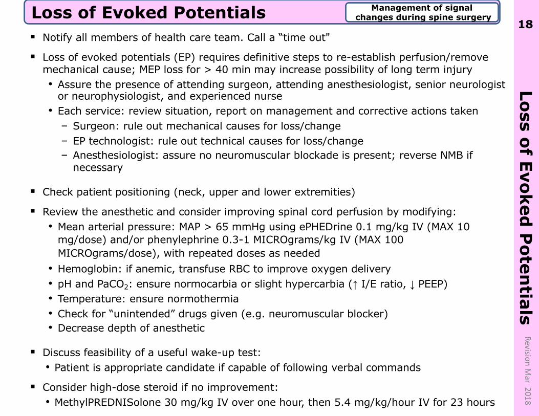
Loss of Evoked P
otentials
§ Notify all members of health care team. Call a “time out"
§ Loss of evoked potentials (EP) requires definitive steps to re-establish perfusion/remove mechanical cause; MEP loss for > 40 min may increase possibility of long term injury• Assure the presence of attending surgeon, attending anesthesiologist, senior neurologist
or neurophysiologist, and experienced nurse• Each service: review situation, report on management and corrective actions taken- Surgeon: rule out mechanical causes for loss/change- EP technologist: rule out technical causes for loss/change- Anesthesiologist: assure no neuromuscular blockade is present; reverse NMB if
necessary
§ Check patient positioning (neck, upper and lower extremities)
§ Review the anesthetic and consider improving spinal cord perfusion by modifying:• Mean arterial pressure: MAP > 65 mmHg using ePHEDrine 0.1 mg/kg IV (MAX 10
mg/dose) and/or phenylephrine 0.3-1 MICROgrams/kg IV (MAX 100 MICROgrams/dose), with repeated doses as needed
• Hemoglobin: if anemic, transfuse RBC to improve oxygen delivery• pH and PaCO2: ensure normocarbia or slight hypercarbia (↑ I/E ratio, ↓ PEEP)• Temperature: ensure normothermia• Check for “unintended” drugs given (e.g. neuromuscular blocker)• Decrease depth of anesthetic
§ Discuss feasibility of a useful wake-up test:• Patient is appropriate candidate if capable of following verbal commands
§ Consider high-dose steroid if no improvement:• MethylPREDNISolone 30 mg/kg IV over one hour, then 5.4 mg/kg/hour IV for 23 hours
Loss of Evoked Potentials Management of signalchanges during spine surgery
18 Revision M
ar 2018

Malig
nan
t Hyp
ertherm
iaMalignant Hyperthermia ↑ Temp ↑ HR ↑ CO2 acidosis
§ Get MH Cart, dantrolene, and help § Notify team and stop procedure, if possible§ Stop volatile anesthetic, succinylcholine. § Attach charcoal filter. Turn O2 flow to 10 L/min§ Hyperventilate patient to reduce EtCO2
§ Give dantrolene 2.5 mg/kg IV, rapidly, through large bore IV if possible, every 5 min until symptoms resolve. May need up to 10 mg/kg (if no response at this dose, consider alternative diagnoses)• Dantrium/Revonto: Assign dedicated person to mix these formulations of
dantrolene (20 mg/vial) with 60 mL non-bacteriostatic sterile water• Ryanodex: 250 mg is mixed with 5 mL non-bacteriostatic sterile water
§ Transition to non-triggering anesthetic§ Give sodium bicarbonate 1-2 mEq/kg IV for suspected metabolic acidosis§ Cool patient: • Apply ice externally to axilla, groin and around head• Infuse cold saline intravenously• NG and open body cavity lavage with cold water• Stop cooling when temperature < 38o C
§ Hyperkalemia treatment: • Calcium gluconate 30 mg/kg IV or calcium chloride 10 mg/kg IV;• Sodium bicarbonate 1-2 mEq/kg IV; • Regular insulin 0.1 units/kg IV (MAX 10 units) and dextrose 0.5-1 g/kg IV
§ VT or afib treatment: Do NOT use calcium channel blocker; give amiodarone 5 mg/kg§ Send labs: ABG or VBG, electrolytes, serum CK, serum/urine myoglobin, coagulation§ Place urinary catheter, maintain UO > 2 ml/kg/hr§ If cardiac arrest occurs, begin CPR & consider ECMO, see ‘Cardiac Arrest’ card§ If no response after 10 mg/kg dantrolene, consider other dx: sepsis, NMS, serotonin synd., myopathy,
pheochromocytoma§ Call ICU to arrange disposition. For post-acute management, see: http://www.mhaus.org
MH hotline 1-800-644-9737
19
Revision Mar 2018

Transfu
sion: M
assive Hem
orrhag
eMassive Hemorrhage Replacement > half total blood volume
(TBV) per hour or TBV < 24 hours
§ Notify Blood Bank immediately, send blood sample for type and cross
§ Activate institutional pediatric massive transfusion protocol. Consider RBC : FFP : Platelets = 2:1:1 or 1:1:1• Use un-crossmatched O negative PRBCs and AB+
plasma until crossmatched blood available
• Consider intraoperative blood salvage (e.g., Cell Saver)
§ Obtain additional vascular access if needed
§ Watch for hyperkalemia, if needed give calcium gluconate 60 mg/kg or calcium chloride 20 mg/kg while directly visualizing IV site (if peripheral)
§ Warm the room
§ Send labs/perform point of care testing q 30 min: CBC, platelets, PT/PTT/INR, fibrinogen, rapid TEG, ABG, Na, K, Ca, lactate
§ Blood product administration:• Use 140 micron filter for all products• Use a blood warmer for RBC and FFP
transfusion (NOT for platelets)• Consider use of rapid transfusion pumps • Monitor ABG, electrolytes, and temperature
§ When under control: call blood bank to terminate
20
§ HCT < 21% or Hgb < 7: • 4 ml/kg PRBC increases Hct by 3
§ Platelet count < 50,000 (< 100K for brain injury), rapid TEG-MA < 54mm:
• 10 ml/kg apheresed platelets increases platelet count by 30 –50k
§ INR > 1.5 (or > 1.3 brain injury), rapid TEG-ACT >120 sec:
• 10ml/kg thawed plasma increases coagulation factors by 20%
§ Fibrinogen < 100 mg/dL or rapid TEG-angle<66°, k value >120 sec: • 10 ml/kg pooled cryoprecipitate
increases fibrinogen by 30-50 mg/dL
§ Refractory hemorrhage
• Consider factor VIIa, up to 90 MICROgrams/kg
Treatment
Revision Mar 2018

Myocardial Ischem
iaMyocardial Ischemia ST changes on ECG
Treatment:§ Improve O2 Supply:• Increase O2 to 100% • Correct anemia• Correct hypotension
§ Decrease O2 Demand:• Reduce heart rate • Correct hypertension• Restore sinus rhythm
§ Drug therapy (rarely needed in peds, consult a pediatric cardiac expert):• NitroGLYCERIN 0.5-5 MICROgrams/kg/min• Consider heparin infusion 10 Units/kg bolus,
then 10 Units/kg/hour
Potential Causes:§ Severe hypoxemia§ Systemic arterial hypo- or hypertension§ Marked tachycardia§ Severe anemia§ Coronary air embolus§ Cardiogenic shock§ Local anesthetic toxicity
21
Recognition§ ST depression >0.5 mm in any lead § ST elevation >1 mm (2mm in
precordial leads)§ Flattened or inverted T waves§ Arrhythmia: VF, VT, ventricular
ectopy, heart block
Diagnostic studies§ 12-lead ECG:
• II, III, aVF for inferior (RCA)• V5 for lateral ischemia (LCx)• V2, V3 anterior ischemia (LAD)
§ Compare to previous ECGs§ Request Pediatric Cardiology
consult and echocardiogram
Revision Mar 2018
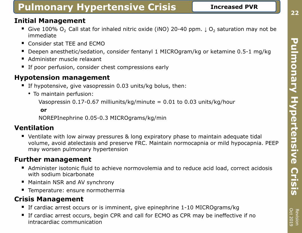
Pu
lmon
ary Hyp
ertensive C
risisPulmonary Hypertensive Crisis Increased PVR
22 Revision O
ct 2019
Initial Management§ Give 100% O2 Call stat for inhaled nitric oxide (iNO) 20-40 ppm. ↓ O2 saturation may not be
immediate§ Consider stat TEE and ECMO§ Deepen anesthetic/sedation, consider fentanyl 1 MICROgram/kg or ketamine 0.5-1 mg/kg§ Administer muscle relaxant§ If poor perfusion, consider chest compressions early
Hypotension management§ If hypotensive, give vasopressin 0.03 units/kg bolus, then: • To maintain perfusion:
Vasopressin 0.17-0.67 milliunits/kg/minute = 0.01 to 0.03 units/kg/hourorNOREPInephrine 0.05-0.3 MICROgrams/kg/min
Ventilation§ Ventilate with low airway pressures & long expiratory phase to maintain adequate tidal
volume, avoid atelectasis and preserve FRC. Maintain normocapnia or mild hypocapnia. PEEP may worsen pulmonary hypertension
Further management§ Administer isotonic fluid to achieve normovolemia and to reduce acid load, correct acidosis
with sodium bicarbonate§ Maintain NSR and AV synchrony§ Temperature: ensure normothermia
Crisis Management§ If cardiac arrest occurs or is imminent, give epinephrine 1-10 MICROgrams/kg§ If cardiac arrest occurs, begin CPR and call for ECMO as CPR may be ineffective if no
intracardiac communication
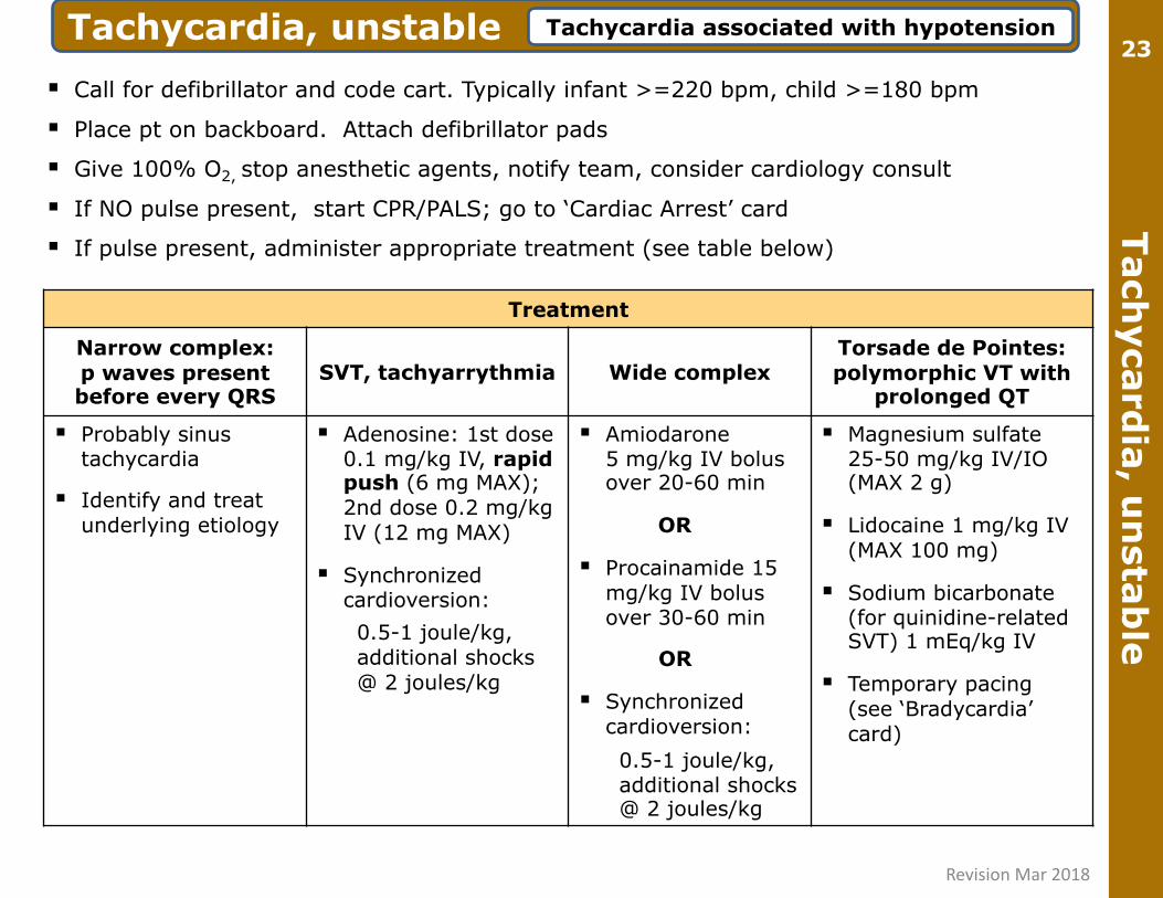
Tachycard
ia, un
stable
Tachycardia, unstable Tachycardia associated with hypotension
§ Call for defibrillator and code cart. Typically infant >=220 bpm, child >=180 bpm
§ Place pt on backboard. Attach defibrillator pads
§ Give 100% O2, stop anesthetic agents, notify team, consider cardiology consult
§ If NO pulse present, start CPR/PALS; go to ‘Cardiac Arrest’ card
§ If pulse present, administer appropriate treatment (see table below)
23
Treatment
Narrow complex:p waves present before every QRS
SVT, tachyarrythmia Wide complexTorsade de Pointes: polymorphic VT with
prolonged QT
§ Probably sinus tachycardia
§ Identify and treat underlying etiology
§ Adenosine: 1st dose 0.1 mg/kg IV, rapid push (6 mg MAX); 2nd dose 0.2 mg/kg IV (12 mg MAX)
§ Synchronized cardioversion:0.5-1 joule/kg, additional shocks@ 2 joules/kg
§ Amiodarone 5 mg/kg IV bolus over 20-60 min
OR
§ Procainamide 15 mg/kg IV bolus over 30-60 min
OR
§ Synchronized cardioversion:0.5-1 joule/kg, additional shocks@ 2 joules/kg
§ Magnesium sulfate 25-50 mg/kg IV/IO (MAX 2 g)
§ Lidocaine 1 mg/kg IV (MAX 100 mg)
§ Sodium bicarbonate (for quinidine-relatedSVT) 1 mEq/kg IV
§ Temporary pacing (see ‘Bradycardia’ card)
Revision Mar 2018

Tamponade, Cardiac Tamponade physiology occurs when increased pericardial pressure impairs diastolic filling
Signs & Symptoms§ Beck’s Triad: muffled heart tones, distended neck veins, decreased systolic blood pressure§ Pulsus Paradoxus: cyclic inspiratory decrease in systolic BP of more than 10mmHg§ Electrical Alternans: cyclic alteration in magnitude of p waves, QRS complex & t-waves§ Typical presentation of acute tamponade = sudden hypotension, tachycardia & tachypnea; patient
may be unable to lie flat
Tamp
ond
ade, C
ardiac
24
Diagnosis§ Echocardiography/ultrasound: diastolic compression or
collapse of RA/RV, leftward displacement of ventricular septum, exaggerated increase in RV size with reciprocal decrease in LV size during inspiration
Treatment - imaging is key in deciding treatment§ Pericardiocentesis awake/local for large effusions prior to
GA§ Surgical for postoperative tamponade (cause is often local
collections of clotted blood)
Anesthetic Considerations§ Progressive decrease in SV with an increased CVP à systemic hypotension à cardiogenic shock§ Goals: maintain sympathetic tone and CO via á HR and contractility/fluid bolus prn
• Induction: Ketamine (1-2 mg/kg IV), muscle relaxant• If CV collapse: EPINEPHrine 0.05-0.1 MICROgrams/kg IV bolus or infusion (0.01-0.1
MICROgrams/kg/min)• Access: Large bore PIV; arterial line ideal but should not delay treatment in hemodynamically
unstable patient• Avoid: cardiac depression, vasodilation, â HR; á airway pressure (will â venous return) so
may need small tidal volumes or hand ventilation
Differential Diagnosis§ CHF, PE§ If pulsus paradoxus: respiratory distress, airway obstruction, COPD, PE, RV infarction
First Published Nov 2018

Tension
Pn
eum
othorax
Tension Pneumothorax ↑ HR ↓ SpO2 ↓ BP tracheal deviation, mediastinal shift 25
§ Stop N2O; increase O2 to 100%
§ Secure airway with endotracheal tube
§ Reduce positive ventilation pressure
§ Consider CXR, lung ultrasound, transillumination to confirm diagnosis (see inset)
§ Administer vasopressors for circulatory collapse
§ Perform immediate needle decompression, then chest tube placement
§ Needle decompression:• 2nd rib space superior to 3rd rib, mid-clavicular line- 14-16g angiocath for teens/adults- 18-20g angiocath for infants/children
§ Chest tube insertion• 5-6th intercostal space, mid-axillary line
§ If no improvement in hemodynamics after a rush of air, consider:• Needle decompression of contralateral side• Presence of pneumopericardium• Scan both lungs with ultrasound or
transillumination to evaluate for alternate side or insufficiently decompressed pneumothorax
Needle decompression
Chest tube
Downloaded from: http://www.uwhealth.org/images/ewebeditpro/uploadimages/5384_Figure_1.jpg
Lung Ultrasound Instructions§ High frequency probe, place
longitudinally on chest, 2nd
intercostal space. Slide probe downwards to observe pleural sliding
• If see pleural sliding, 100% positive predictive value no pneumothorax
• If no pleural sliding, consider pneumothorax, ARDS, fibrosis, acute asthma, pleurodesis
Revision Mar 2018

Transfu
sion R
eactions
Transfusion Reactions Reactions may occur with any type of product. Important to determine type of reaction.
For All Reactions:§ Stop transfusion§ Disconnect donor product and IV tubing§ Infuse normal saline through clean tubing§ Examine blood product ID; determine correct pt§ Send product to Blood Bank
26
Hemolytic Non-Hemolytic Anaphylactic
Signs: Hemoglobinemia, hemoglobinuria, DIC, ↓ BP, ↑ HR, bronchospasm
§ Furosemide 1-2 mg/kg IV (MAX 40 mg)
§ Mannitol 0.25-1 g/kg
§ Support BP to maintain renal perfusion
§ Maintain urine output at least 1-2 mL/kg/hour
§ Prepare for cardiovascular instability
§ Send blood and urine sample to laboratory
Signs: ↓ BP, bronchospasm, pulmonary edema, fever, rash
§ Treat fever
§ Treat pulmonary edema
§ Observe for signs of hemolysis
Signs: Erythema, urticaria, angioedema, bronchospasm, tachycardia, shock
§ Support airway and circulation as necessary
§ EPINEPHrine 1-10 MICROgrams/kg IV
§ DiphenhydrAMINE 1 mg/kg IV (MAX 50 mg)
§ MethylPREDNISolone 2 mg/kg IV (MAX 60 mg)
§ Maintain intravascular volume
Revision Mar 2018
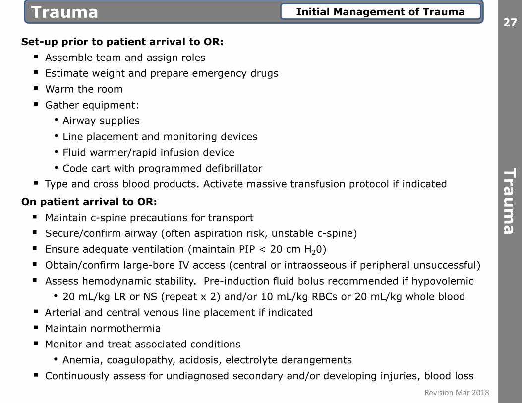
Set-up prior to patient arrival to OR:§ Assemble team and assign roles§ Estimate weight and prepare emergency drugs§ Warm the room§ Gather equipment:
• Airway supplies• Line placement and monitoring devices• Fluid warmer/rapid infusion device• Code cart with programmed defibrillator
§ Type and cross blood products. Activate massive transfusion protocol if indicated
On patient arrival to OR:§ Maintain c-spine precautions for transport§ Secure/confirm airway (often aspiration risk, unstable c-spine)§ Ensure adequate ventilation (maintain PIP < 20 cm H20)§ Obtain/confirm large-bore IV access (central or intraosseous if peripheral unsuccessful)§ Assess hemodynamic stability. Pre-induction fluid bolus recommended if hypovolemic
• 20 mL/kg LR or NS (repeat x 2) and/or 10 mL/kg RBCs or 20 mL/kg whole blood§ Arterial and central venous line placement if indicated§ Maintain normothermia§ Monitor and treat associated conditions
• Anemia, coagulopathy, acidosis, electrolyte derangements§ Continuously assess for undiagnosed secondary and/or developing injuries, blood loss
Traum
aTrauma Initial Management of Trauma
27
Revision Mar 2018

MATERNAL CRISIS 26

MA
TERN
AL P
ostpartu
m H
emorrh
age
MATERNAL Postpartum Hemorrhage Loss of >500mL after vaginal
birth, or >1,000mL after cesarean delivery
§ ATTENTION: This checklist is for ADULT-SIZED maternal patients ONLY
§ Prepare for crystalloid and blood product resuscitation
§ Obtain vascular access with 2 large-bore IVs
§ Call Blood Bank to activate Massive Transfusion with PRBC:FFP:platelet in a 4:2:1 ratio. Ask blood bank to prepare next round when each round is picked up.
• Give calcium chloride ADULT DOSE 200-500mg/Unit PRBCs, in separate line. Monitor for hyperkalemia
• Consider giving tranexamic acid early
• If refractory hemorrhage, consider fVIIa and cryoprecipitate or fibrinogen concentrate
§ Give uterotonics
§ Call for rapid transfuser or pressure bags
§ Warm room, patient and fluids (NOT platelets)
§ Send CBC, PT/PTT/INR, fibrinogen, calcium, K, ABG
Obstetric Interventions Consider
• Intrauterine balloon
• External uterine compression sutures
• Uterine artery ligation
• Hysterectomy
• Arterial line
• If awake, convert to general anesthesia
• Embolization in IR
• TEG/ROTEM monitoring
Treatment
ADULT MATERNAL Uterotonics:
§ Oxytocin ADULT DOSE 3-5 Units rapid infusion, then start 40 Units slow infusion
§ Methylergonovine (Methergine) ADULT DOSE 0.2mg IM NOT IV, may repeat in 2 hours (AVOID in HTN and pre-eclampsia)
§ Carboprost (Hemabate) ADULT DOSE 0.25mg IM NOT IV, may repeat q 15 minutes up to 8 doses (AVOID in asthma, pulmonary hypertension)
§ Misoprostol ADULT DOSE 800-1000 MICROgrams rectal
Hemostatics:
§ Tranexamic acid ADULT DOSE 1g
§ If low fibrinogen, give cryoprecipitate ADULT DOSE 10 units or Fibrinogen concentrate
§ If refractory hemorrhage, consider factor VIIa 90 MICROgrams/kg, up to 3 doses
28
Revision Dec 2018