(19) TZZ ¥¥Z Z T - PatentOrderpdfstore.patentorder.com/pdf/ep/0b1/ep2353010b1.pdf · AT BE BG CH...
25
Note: Within nine months of the publication of the mention of the grant of the European patent in the European Patent Bulletin, any person may give notice to the European Patent Office of opposition to that patent, in accordance with the Implementing Regulations. Notice of opposition shall not be deemed to have been filed until the opposition fee has been paid. (Art. 99(1) European Patent Convention). Printed by Jouve, 75001 PARIS (FR) (19) EP 2 353 010 B1 TEPZZ ¥5¥Z_ZB_T (11) EP 2 353 010 B1 (12) EUROPEAN PATENT SPECIFICATION (45) Date of publication and mention of the grant of the patent: 08.07.2015 Bulletin 2015/28 (21) Application number: 08875351.2 (22) Date of filing: 21.11.2008 (51) Int Cl.: G01N 33/68 (2006.01) (86) International application number: PCT/EP2008/066030 (87) International publication number: WO 2010/049010 (06.05.2010 Gazette 2010/18) (54) METHODS FOR DETECTION OF TOXEMIA VERFAHREN ZUM NACHWEIS EINER TOXÄMIE PROCÉDÉS POUR LA DÉTECTION DE LA TOXÉMIE (84) Designated Contracting States: AT BE BG CH CY CZ DE DK EE ES FI FR GB GR HR HU IE IS IT LI LT LU LV MC MT NL NO PL PT RO SE SI SK TR (30) Priority: 28.10.2008 US 109008 P (43) Date of publication of application: 10.08.2011 Bulletin 2011/32 (73) Proprietor: Murauski, Uladzimir A. 15745 Wildau (DE) (72) Inventor: Murauski, Uladzimir A. 15745 Wildau (DE) (74) Representative: Lawrence, John et al Barker Brettell LLP 100 Hagley Road Edgbaston Birmingham B16 8QQ (GB) (56) References cited: US-A1- 2003 170 912 • ALTAMENTOVA SVETLANA M ET AL.: "A fluorescence method for estimation of toxemia: Binding capacity of lipoproteins and albumin in plasma" CLINICA CHIMICA ACTA, vol. 271, no. 2, 23 March 1998 (1998-03-23), pages 133-149, XP9118415 ISSN: 0009-8981 DOI: 10.1016/S0009-8981(97)00244-1 • CALDARARU HORIA ET AL: "A Spin Probe Study of Mesoporous Silica Formation via a Neutral Templating Route" JOURNAL OF PHYSICAL CHEMISTRY PART B: CONDENSED MATTER, MATERIALS, SURFACES, INTERFACES & BIOPHYSICAL, vol. 107, no. 25, 2003, pages 6032-6038, XP009118577 Published on web: 4 June 2003 ISSN: 1520-6106 [retrieved on 2003-06-04] • SMIRNOVA, T. I.: "Binding of MRI contrast agents to albumin : a high-field EPR study" APPLIED MAGNETIC RESONANCE, CODEN: APMREI, vol. 31, no. 3-4, 2007, pages 431-446, XP9118566 ISSN: 0937-9347 • GURACHEVSKY ANDREY ET AL: "Application of spin label electron paramagnetic resonance in the diagnosis and prognosis of cancer and sepsis", CLINICAL CHEMISTRY AND LABORATORY MEDICINE, vol. 46, no. 9, September 2008 (2008-09), pages 1203-1210, ISSN: 1434-6621
Transcript of (19) TZZ ¥¥Z Z T - PatentOrderpdfstore.patentorder.com/pdf/ep/0b1/ep2353010b1.pdf · AT BE BG CH...

Note: Within nine months of the publication of the mention of the grant of the European patent in the European PatentBulletin, any person may give notice to the European Patent Office of opposition to that patent, in accordance with theImplementing Regulations. Notice of opposition shall not be deemed to have been filed until the opposition fee has beenpaid. (Art. 99(1) European Patent Convention).
Printed by Jouve, 75001 PARIS (FR)
(19)E
P2
353
010
B1
TEPZZ ¥5¥Z_ZB_T(11) EP 2 353 010 B1
(12) EUROPEAN PATENT SPECIFICATION
(45) Date of publication and mention of the grant of the patent: 08.07.2015 Bulletin 2015/28
(21) Application number: 08875351.2
(22) Date of filing: 21.11.2008
(51) Int Cl.:G01N 33/68 (2006.01)
(86) International application number: PCT/EP2008/066030
(87) International publication number: WO 2010/049010 (06.05.2010 Gazette 2010/18)
(54) METHODS FOR DETECTION OF TOXEMIA
VERFAHREN ZUM NACHWEIS EINER TOXÄMIE
PROCÉDÉS POUR LA DÉTECTION DE LA TOXÉMIE
(84) Designated Contracting States: AT BE BG CH CY CZ DE DK EE ES FI FR GB GR HR HU IE IS IT LI LT LU LV MC MT NL NO PL PT RO SE SI SK TR
(30) Priority: 28.10.2008 US 109008 P
(43) Date of publication of application: 10.08.2011 Bulletin 2011/32
(73) Proprietor: Murauski, Uladzimir A.15745 Wildau (DE)
(72) Inventor: Murauski, Uladzimir A.15745 Wildau (DE)
(74) Representative: Lawrence, John et alBarker Brettell LLP 100 Hagley Road EdgbastonBirmingham B16 8QQ (GB)
(56) References cited: US-A1- 2003 170 912
• ALTAMENTOVA SVETLANA M ET AL.: "A fluorescence method for estimation of toxemia: Binding capacity of lipoproteins and albumin in plasma" CLINICA CHIMICA ACTA, vol. 271, no. 2, 23 March 1998 (1998-03-23), pages 133-149, XP9118415 ISSN: 0009-8981 DOI: 10.1016/S0009-8981(97)00244-1
• CALDARARU HORIA ET AL: "A Spin Probe Study of Mesoporous Silica Formation via a Neutral Templating Route" JOURNAL OF PHYSICAL CHEMISTRY PART B: CONDENSED MATTER, MATERIALS, SURFACES, INTERFACES & BIOPHYSICAL, vol. 107, no. 25, 2003, pages 6032-6038, XP009118577 Published on web: 4 June 2003 ISSN: 1520-6106 [retrieved on 2003-06-04]
• SMIRNOVA, T. I.: "Binding of MRI contrast agents to albumin : a high-field EPR study" APPLIED MAGNETIC RESONANCE, CODEN: APMREI, vol. 31, no. 3-4, 2007, pages 431-446, XP9118566 ISSN: 0937-9347
• GURACHEVSKY ANDREY ET AL: "Application of spin label electron paramagnetic resonance in the diagnosis and prognosis of cancer and sepsis", CLINICAL CHEMISTRY AND LABORATORY MEDICINE, vol. 46, no. 9, September 2008 (2008-09), pages 1203-1210, ISSN: 1434-6621

EP 2 353 010 B1
2
5
10
15
20
25
30
35
40
45
50
55
Description
BACKGROUND OF THE INVENTION
1. Field of the Invention
[0001] The present invention relates generally to analysis of extracellular fluids that contain carrier proteins, and, moreparticularly, but not by way of limitation, to methods for prognosis and detection of toxemia in a patient by analyzing aserum sample from the patient.
2. Description of Related Art
[0002] For illustration, but without limiting the scope of the invention, the background is described with respect toanalyzing the blood of a human patient.[0003] In the early stages of toxemia, the ability of a patient’s body to evacuate toxins from the blood stream maybecome compromised. That is, the patient’s biological systems for evacuating toxins from the patient’s blood streammay stop functioning properly and begin to permit toxins to build in the patient’s body. In later stages of toxemia, thesetoxins generally reach relatively high levels (as compared to normal toxin levels in a healthy patient), and the toxins maybegin to cause noticeable symptoms such as illness, cell damage, organ failure, and the like. Currently known methodsof diagnosing toxemia may not permit recognition or diagnosis of toxemia until later stages when symptoms are alreadynoticeable. In these later stages, treatment may be less effective and may not be effective enough to prevent the deathof the patient.[0004] Analyzing hematologic parameters and/or measuring the concentration of various metabolites in blood samplesfrom a patient are known in the art and may be widely-used methods of diagnosing toxemia (which may also be knownin the art as toxaemia) in a patient, such as, for example, in a clinical setting. However, these known methods suffer anumber of shortcomings and/or drawbacks.[0005] One example of a known method may be referred to in the art as the "mean mass molecules" evaluation of asample of a patient’s blood serum. However, in the "mean mass molecules" evaluation, generally only a fraction ofunbound, free endogenous toxins in the serum may be detected. This fraction may also be limited in that it may containmostly toxins that are hydrophilic. "Absorbed" or "bound" hydrophobic toxins, which are respectively adsorbed on bio-logical membranes or bound on carrier proteins, may not be detected. The failure to detect absorbed hydrophobic toxinscan be detrimental to the treatment and recovery of the patient because these toxins may subsequently damage, ormay have already damaged, cells, organs or systems in the patient. Such damage, caused by these absorbed and/orbound hydrophobic toxins, may subsequently cause changes in hematological parameters and/or other symptoms oftoxemia.[0006] Because the "mean mass molecules" evaluation (as well as other known methods of detecting toxemia) maynot detect hydrophobic toxins, toxemia may not be detected until cells have been significantly damaged and/or hema-tologic changes or failures have already occurred. Stated otherwise, known methods of detecting toxemia may not detecttoxemia in its early stages, and instead, may only detect toxemia in later stages of its development when it may besubstantially harder to treat it effectively. In these later stages of toxemia, treatment may be inhibited by factors suchas, for example, reduced capacity of the carrier proteins to evacuate toxins, and/or reduced capacity of the patients liverto detoxify or remove the toxins from the carrier proteins.[0007] Soviet Union Patent, SU 1,459,656, published February 23, 1989, describes a method of diagnosing endog-enous toxemia by an evaluation of the ability of erythrocyte membranes to bind the fluorescent probe ANS. This methodmay permit detection of damage to erythrocyte membranes that has already occurred due to erythrocyte interaction withtoxins (either hydrophilic or hydrophobic). This method may permit relatively-earlier detection of toxemia in a patient,such as, for example, at a stage in which toxins have already damaged cell membranes but before the damage isextensive enough to cause failure of organs and/or systems of the patient. Practically speaking, this method may permitdetection of toxemia as much as approximately 6 to 12 hours before other hematological parameters show detectiblechanges or before other symptoms may appear. This method therefore still suffers from the shortcoming that it may notdetect toxemia before toxins have damaged cells of the patient, and therefore may not permit treatment early enoughduring the development of the disease to prevent or minimize the effects, complications, or symptoms of toxemia.[0008] United States Patent No. 7,166,474 describes a method of detecting changes in transport properties of albuminby using electron paramagnetic resonance (EPR) spectroscopy (which may also be known in the art as electron spinresonance "ESR" spectroscopy) to evaluate a sample that contains albumin (an albumin-containing sample). This methodcan include evaluating the albumin transport function with respect to long chain fatty acids by using a spin probe repre-sented by a spin-labeled fatty acid. Specifically, according to this method, the EPR-spectra of the spin probe can bemeasured in at least three aliquots of the sample, where each aliquot is mixed with significantly different concentrations

EP 2 353 010 B1
3
5
10
15
20
25
30
35
40
45
50
55
of the spin probe and a high concentration of ethanol. The concentration of ethanol is high enough to induce significantconformational changes of the albumin to enable evaluation of the conformational flexibility of the albumin. The parametersof albumin transport function are derived from measurements of the conformational changes induced in the albuminartificially by the high ethanol concentration. This method assumes that albumin molecules efficiently release boundsubstances to target objects at conditions that occur in a healthy patient, and therefore induces a conformational changefacilitating dissociation of albumin-bound ligands. While this may be useful in the method taught by this patent, theconformational changes induced by the high ethanol concentration prevents evaluation of the toxin-scavenging (toxin-binding) function of the albumin, because it assumes delivery of albumin-bound toxins to an excretion system, e.g. liver,rather than distribution of toxins throughout an organism in way similar to the delivery of nutrients to cells (permittingtoxins to damage cells), as can happen in toxemic patients. This method suffers from possible shortcomings that mayinclude, for example, the use of at least three aliquots of each sample, the fact that evaluation may be limited to certainalbumin parameters, and excessive dissociation of bound substances (e.g., toxins) from the albumin (and/or from otherserum proteins) resulting from the addition of relatively high-concentrations of ethanol. These possible shortcomingsmay contribute to variations in results and false-negatives in detecting the presence of protein-bound toxins in patients’blood. Altomentova et al (1998), Clinica Chimica Acta, Vol 271, no. 2, pp 133-149, relates to a fluorescence method forestimation of toxemia: Binding capacity of lipoproteins and albumin in plasma. US 2003/170912 relates to a method forthe esr-spectroscopic detection of changes in the transport properties of albumin in albumin-containing samples, esr-spectrometer for carrying out said method, and use of the method for diagnostic purposes and for controlling albumin-containing preparation. Gurachevsky et al (2008), Clin. Chem. Lab. Med. Vol 46(9); pp 1203-1210 relates to applicationof spin label electron paramagnetic resonance in the diagnosis and prognoses of cancer and sepsis.[0009] A continuing need therefore exists for improved, faster, less-expensive, and/or more-accurate methods ofdetecting toxemia, especially in its early or precursor stages.
SUMMARY OF THE INVENTION
[0010] The present invention is related to a method for detecting toxemia in a subject patient, comprising the stepsof: (a) mixing a labelled 16-DOXYL-stearic acid probe having a site to bind to albumin with an aliquot of a subject patient’sextracellular fluid containing albumin, the amount of probe such that the molar ratio of the probe to the albumin is in therange of 0.3 to 1.5; (b) mixing ethanol with the mixture of the aliquot and the probe, wherein the ethanol when added tothe mixture increases the solubility of the probe in the aliquot, and wherein the amount of ethanol added causes a portionof the probe to dissociate from the albumin without causing toxins to dissociate from the albumin; (c) analyzing themixture comprising the aliquot, the probe, and the ethanol to determine the binding efficiency of the albumin in thepatient’s extracellular fluid, wherein the binding efficiency is the concentration ratio of bound probe and unbound probeadjusted for the concentration of the added ethanol in the mixture and where the binding efficiency is calculated usingthe formula: 1/BE= (Sc/S-1) x (F/B) x P, where BE is binding efficiency, F is the concentration of unbound probe, B isthe concentration of bound probe, Sc is the critical solvent concentration at which all probe is dissociated from thealbumin and dissolved in the solution, S is the solvent concentration and P is the concentration of albumin; and (d)comparing the subject binding efficiency to at least one control binding efficiency of the albumin in the extracellular fluidof at least one non-toxemic control patient, wherein the amount of ethanol mixed with the aliquot and probe does notinduce conformational changes in the albumin, and wherein the volume of ethanol mixed with the aliquot makes up lessthan 30% of the volume of the aliquot, and optionally less than 10% of the volume of the aliquot. Various embodimentsof the present description can be suitable for detecting variations in carrier-protein functions, characteristics, and/orparameters by mixing various substances with a sample of extracellular fluid (that contains carrier proteins) from asubject patients, and analyzing the mixture. Examples of such extracellular fluids include blood serum, lymph fluid, andspinal fluid. Some embodiments of the present methods are suitable for prognosis (predicting the onset of), detection,or diagnosis of toxemia in its early or precursor stages. The present invention may be suitable, for example, for diagnosisand/or monitoring of toxemia in patients in clinical settings such as intensive care units. By way of further examples thepresent invention may be suitable for such diagnosis, prognosis, or monitoring of patients in post-surgery recovery, orpatients with or suffering from trauma, infection, sepsis, systemic inflammatory response syndrome (SIRS), stroke orbrain attack, infarction, or the like.[0011] In some embodiments, the methods of the present description can be suitable for detection, diagnosis, orprognosis of toxemia, by detecting or measuring the deviation of the binding efficiency of carrier proteins of a subjectpatient, relative to the binding efficiency of carrier proteins of a control patient. The binding efficiency of carrier proteinsof the control patient can be established, for example, by testing one or more control patients, by historical test resultsfrom patients with known conditions, by statistical aggregation of data from various other patients, or by any other suitablemeans that permits comparison with the subject patient in such a way as to provide an indication of whether the subjectpatient has, likely has, is likely to have, does not have, likely does not have, or is not likely to have toxemia. This deviationcan, for example, be detected or measured for a sample of serum taken from a subject patient by mixing an aliquot of

EP 2 353 010 B1
4
5
10
15
20
25
30
35
40
45
50
55
the serum sample with a labeled hydrophobic probe having a long-hydrocarbonchain. Specifically, parameters of bindingefficiency of a hydrophobic probe having a long-hydrocarbon chain to serum carrier proteins can correlate to the amountof toxic substances bound to the binding sites of the carrier proteins. This correlation can, for example, be caused bycompetitive binding on specific protein sites for substances with long hydrocarbon chains, and indirectly due to allostericmodification of the affinity of these sites upon ligand binding on other protein sites. Stated otherwise, when high levelsof toxins are present in the subject patient’s blood, the toxins may crowd and/or modify the binding sites of the carrierproteins, thereby preventing the hydrophobic probe from binding to those binding sites. In this way, hydrophobic toxinscan be detected as their levels rise, and even before they damage cells.[0012] The present description includes various embodiments of methods for detecting toxemia in a subject patient.Some embodiments of the present method can comprise obtaining an aliquot from a sample of a subject patient’sextracellular fluid containing carrier proteins. Some of the present methods can comprise mixing a labeled hydrophobicprobe with the aliquot. In some of these methods, the amount of probe can be such that the molar ratio of probe capableof binding to (with) carrier proteins to the carrier proteins is between about 0.3 and about 1.5. Some methods cancomprise mixing a solvent into the aliquot. In some of these methods, the solvent can be such that when added to thealiquot and probe the solubility of the probe is increased in the aliquot. In some of these methods, the amount of solventmixed with the aliquot can be sufficient to dissociate a portion of the probe from the carrier proteins without causingsignificant dissociation of toxins from the carrier proteins. In some methods, the amount of solvent mixed with the aliquotcan be described as sufficient to dissociate a portion of the probe from the carrier proteins without causing dissociationof toxins from the carrier proteins.[0013] Some methods comprise analyzing the mixture of the aliquot, probe, and solvent to determine the bindingefficiency of the carrier proteins. Some of these embodiments can comprise comparing the subject binding efficiency toat least one control binding efficiency for at least one non-toxemic control patient.[0014] Some methods can comprise analyzing the mixture of aliquot, probe, and solvent to determine a subject toxin-evacuation parameter of the carrier. Some of these methods can comprise comparing the subject toxin-evacuationparameter to at least one control toxin-evacuation parameter for a non-toxemic control patient. In some of these methods,analyzing the mixture can comprise measuring the concentrations of protein-bound and unbound probe in the mixture;deriving a subject binding efficiency of the carrier proteins from at least the concentrations of the protein-bound andunbound probe; and deriving a subject toxin-evacuation parameter of the carrier proteins as the square of the subjectbinding efficiency.[0015] The present description includes various kits for detecting toxemia in a subject patient from a sample of thesubject patient’s extracellular fluid containing carrier proteins. Some of the present kits can comprise a labeled hydro-phobic probe. In some of these kits, the amount of probe can be such that when mixed with an aliquot having a prede-termined volume of the extracellular fluid the molar ratio of probe capable of binding with (to) carrier proteins to thecarrier proteins is between about 0.3 and about 1.5. Some of the present kits can comprise a solvent. In some of thesekits, the solvent can be such that when mixed with the probe and the aliquot the solubility of the probe is increased inthe aliquot. In some of these kits, the amount of solvent can be sufficient to dissociate a portion of the probe from thecarrier proteins without causing significant dissociation of toxins from the carrier proteins. In some kits, the amount ofsolvent can be described as sufficient to dissociate a portion of the probe from the carrier proteins without causingdissociation of toxins from the carrier proteins.[0016] Some kits can comprise instructions for performing the various embodiments of the present methods usingone or more elements of the present kits. For example, some kits can comprise instructions for: obtaining an aliquothaving a predetermined volume of a sample of the subject patient’s extracellular fluid containing carrier proteins; mixinga labeled hydrophobic probe with the aliquot, the amount of probe such that the molar ratio of probe capable of bindingwith (to) carrier proteins to the carrier proteins is between about 0.3 and about 1.5; mixing a solvent with the aliquotsufficient to dissociate a portion of the probe from the carrier proteins without causing significant dissociation of toxinsfrom the carrier proteins; analyzing the mixture of the aliquot, probe, and solvent to determine the binding efficiency ofthe carrier proteins; comparing the subject binding efficiency to at least one control binding efficiency for a non-toxemiccontrol patient; measuring the concentrations of protein-bound and unbound probe in the mixture; deriving a subjectbinding efficiency of the carrier proteins from at least the concentrations of the protein-bound and unbound probe; and/orany other steps or limitations described herein.[0017] In some methods, the probe and the solvent can be mixed with one another prior to either being mixed withthe aliquot.[0018] In some methods and kits, the probe can comprise an organic molecule having between 8 and 28 Carbonatoms. In some methods and kits, the probe can comprise a fatty acid. In some of these methods and kits, the probecan comprise a long-chain fatty acid. In some of the present methods and kits, the probe can comprise 16-DOXYL-stearic acid. In some of the present methods and kits, the probe can comprise a phospholipid, a lysophospholipid, and/orthe like.[0019] In some of the present methods and kits, the amount of solvent is such that the solvent does not cause or

EP 2 353 010 B1
5
5
10
15
20
25
30
35
40
45
50
55
induce significant conformational changes to the carrier proteins. In some methods and kits, the amount of solvent maybe described as such that the solvent does not cause or induce conformational changes to the carrier proteins. In some,the amount of solvent is sufficient to increase the concentration of unbound probe in the mixture of aliquot, probe, andsolvent to at least 5 times greater than the concentration of unbound probe in a mixture of aliquot and probe without solvent.[0020] In some methods and kits, the solvent can comprise alcohol. In some methods and kits, the solvent can compriseethanol. In some methods and kits, the volume of the amount of solvent can be less than about 30% of the volume ofthe aliquot. In some methods and kits, the volume of the amount of solvent can be less than about 25% of the volumeof the aliquot. In some methods and kits, the volume of the amount of solvent can be less than about 20% of the volumeof the aliquot. In some methods and kits, the volume of the amount of solvent can be less than about 15% of the volumeof the aliquot. In Some methods and kits, the volume of the amount of solvent can be less than about 10% of the volumeof the aliquot. In some of these embodiments, the volume of the amount of solvent can be less than about 5% of thevolume of the aliquot.[0021] Some methods can comprise normalizing the subject binding efficiency to account for the reduction in carrier-protein concentration caused by the amount of solvent in the mixture. In some methods, the steps of deriving the subjectbinding efficiency and normalizing the subject binding efficiency can be performed simultaneously.[0022] Some methods can comprise normalizing the subject binding efficiency to account for the reduction in carrier-protein concentration caused by medical conditions of the patient.[0023] In some methods, the at least one control binding efficiency can comprise a range of control binding efficiencies.In some embodiments, the at least one non-toxemic control patient is also healthy.[0024] Some methods can comprise repeating one or more steps of the method for one or more additional aliquots.In some methods, the amount of solvent mixed with each of the one or more additional aliquots can be different fromthe amount of solvent mixed with the first or initial aliquot. In some methods, the subject binding efficiencies derived forthe first or initial aliquot and the one or more additional aliquots can be averaged to derive an average subject bindingefficiency. In some methods, the average subject binding efficiency can be compared in the step of comparing the subjectbinding efficiency to at least one control binding efficiency.[0025] In some methods and kits, the volume of the aliquot can be less than about 100 mL. In some of these methodsand kits, the volume of the aliquot can be about 50 mL.[0026] Some methods can comprise diagnosing, responsive to the subject binding efficiency being less than the atleast one control binding efficiency, the subject patient with toxemia.[0027] In some methods and kits, the probe can be labeled with a spin-label. In some methods and kits, the probecan be labeled with a radioactive label. In some methods and kits, the probe can be labeled with a fluorescent label. Insome methods, the extracellular fluid can be blood serum. In some methods, the extracellular fluid can be lymph fluid.In some methods, the extracellular fluid can be spinal fluid.[0028] Any methods can consist of or consist essentially of - rather than comprise/include/contain/have - any of thedescribed steps, elements, and/or features.[0029] Details associated with the embodiments described above and others are presented below.
BRIEF DESCRIPTION OF THE DRAWINGS
[0030] The following drawings illustrate by way of example and not limitation. For the sake of brevity and clarity, everyfeature of a given structure is not always labeled in every figure in which that structure appears. Identical referencenumbers do not necessarily indicate an identical structure. Rather, the same reference number may be used to indicatea similar feature or a feature with similar functionality, as may non-identical reference numbers.
FIGS. 1A-1B depict graphical representations of a number of labeled probes for use in various embodiments of thepresent methods.FIG. 2 depicts a schematic representation of one exemplary procedure for evaluating a mixture containing an aliquotof serum and an amount of labeled probe by EPR spectroscopy that is suitable for use in embodiments of the presentmethods.FIG. 3 depicts an exemplary EPR spectrum of a mixture of serum and a spin-labeled fatty acid probe (16-doxylstearic acid) obtained by an EPR spectroscopy method of analysis that is suitable for use in embodiments of thepresent methods.FIG. 4 depicts a graphical representation of the binding efficiency of serum carrier proteins, as assessed by thepresent methods, for a number of patients admitted to an intensive care unit (ICU).
DESCRIPTION OF ILLUSTRATIVE EMBODIMENTS
[0031] The term "coupled" is defined as connected, although not necessarily directly, and not necessarily mechanically;

EP 2 353 010 B1
6
5
10
15
20
25
30
35
40
45
50
55
two items that are "coupled" may be integral with each other. The terms "a" and "an" are defined as one or more unlessthis disclosure explicitly requires otherwise. The terms "substantially," "approximately," and "about" are defined as largelybut not necessarily wholly what is specified, as understood by a person of ordinary skill in the art.[0032] The terms "comprise" (and any form of comprise, such as "comprises" and "comprising"), "have" (and any formof have, such as "has" and "having"), "include" (and any form of include, such as "includes" and "including") and "contain"(and any form of contain, such as "contains" and "containing") are open-ended linking verbs. As a result, a method that"comprises," "has," "includes" or "contains" one or more steps possesses those one or more steps, but is not limited topossessing only those steps. Likewise, a step of a method that "comprises," "has," "includes" or "contains" one or morefeatures possesses those one or more features, but is not limited to possessing only those one or more features. Forexample, in a method that comprises the step of obtaining a sample of blood serum containing carrier proteins, the bloodserum includes the specified features but is not limited to having only those features. For example, in such a method,the blood serum could also contain water.[0033] Further, a device or structure that is configured in a certain way is configured in at least that way, but it canalso be configured in other ways than those specifically described.[0034] Various methods may include testing, analyzing, and/or evaluating of a sample of extracellular fluid from asubject patient. For clarity and brevity, embodiments are described below for testing blood serum of a subject patient.However, the embodiments, features, steps, and particulars described below can also be applied to other extracellularfluids such as, for example, lymph fluid, spinal fluid, and the like.[0035] Some methods include obtaining a serum sample from a subject patient. A serum sample can be obtained, forexample, by drawing blood from the patient and using centrifugation to substantially isolate the serum from the blood.Other suitable methods may also be used to obtain the serum sample from the subject patient. An aliquot can also beobtained from this subject sample. Such an aliquot can be separated from the sample or can include the entire sample.[0036] Some methods include mixing a labeled hydrophobic probe with the aliquot. The labeled hydrophobic probemay also be referred to herein as the "labeled probe," the "hydrophobic probe," or the "probe." The probe is (or comprises)an organic molecule, and/or is selected to be capable of binding with (to) carrier proteins (e.g., albumin) of the extracellularfluid (e.g., serum or plasma)., The probe can comprise a suitable number of Carbon atoms. For example, the probe cancomprise between 8 and 28 Carbon atoms, including 8, 9, 10, 11, 12, 13, 14, 15, 16, 17, 18, 19, 20, 21, 22, 23, 24, 25,26, 27, or 28 Carbon atoms, or any smaller range between 8 and 28 Carbon atoms, e.g., between 14 and 22, between16 and 20, or between 18 and 22 Carbon atoms. In another example, the probe can comprise less than 8 Carbon atomsor more than 28 Carbon atoms. In some embodiments, the probe can comprise a hydrocarbon chain such as, for example,a hydrocarbon chain having any suitable number of Carbon atoms. The probe can comprise a hydrocarbon moleculewhose structure is branched and/or whose structure comprises a ring structure. The probe can comprise a hydrocarbonmolecule comprising elements other than Carbon or Hydrogen, such as, for example, Chlorine, Phosphorous, and/orNitrogen.[0037] The probe can comprise a hydrocarbon chain such as, for example, a fatty acid, a long-chain fatty acid, amedium-chain fatty acid, or the like. Examples of probes suitable for some embodiments of the present method include:16-DOXYL-stearic acid, free radical; 5-DOXYL-stearic acid, free radical; 16:0-16 PC DOXYL, free radical (1- Palmitoyl-2-S tearoyl-(16-DOXYL)- sn-Glycero-3-Phosphocholine); 18:0 PC C13 (1,2- Di stearo yl [1-13C] - sn-Glycero-3 -Phosphocholine), Carbon-13 labeled; and Oleic acid-1,2,3,7,8,9,10-13C7, Carbon-13 labeled. Such labeled probes areavailable from commercial suppliers including, for example, (1) Sigma-Aldrich, Inc., St. Louis, Missouri, U.S.A., www.sig-maaldrich.com; (2) TCI America, Portland, Oregon, U.S.A. and Wellesley Hills, Massachusetts, U.S.A, www.tciameri-ca.com; (3) Fluorochem, Derbyshire, U.K., www.fluorochem.co.uk; and (4) Avanti Polar Lipids Inc., Alabaster, Alabama,U.S.A., www.avantilipids.com.[0038] The probe may be suspended in a volume of liquid so as to, for example, enable accurate measurement of theamount of the probe. This liquid is referred to herein as the "suspension liquid" or "liquid" so as not to be confused withthe solvent described herein (even though the suspension liquid may be or comprise a solvent as that term is used ina more general sense). In some of these examples, the suspension liquid may be (or comprise) the solvent, which isdescribed below. In some of these examples, the probe may be suspended in the solvent in an amount desired formixing the aliquot, probe, and solvent without further addition of solvent; or may be suspended in an amount of solventsmaller than is desired, such that additional solvent must be separately added to the mixture. For example, if 10 20.65mL of solvent are mixed with a 50 mL aliquot: (1) the probe may be suspended in 10 mL of solvent and the suspensionadded to the aliquot; or (2) the probe may be suspended in 5 mL of solvent, the suspension added to the aliquot, and 5mL of solvent (without probe) also mixed with the aliquot. Examples of suitable solution liquids, for use where the probesolution and solvent are mixed while both are liquid, include the materials described below for the solvent.[0039] In others of these (in which the probe may be suspended in a volume of liquid), the probe solution may beadded to a container first and the liquid portion of the liquid/probe solution permitted to evaporate, leaving substantially-only the probe in the container. In such embodiments, the aliquot of serum may then be added such that the probe thendissolves into the serum. Examples of suitable liquids for this suspension/evaporation method include both the liquids

EP 2 353 010 B1
7
5
10
15
20
25
30
35
40
45
50
55
described below for the solvent, as well as other liquids (including solvents known in the art as non-polar solvents) thatare not miscible with water, such as, for example, methyl ethyl ketone, methyl acetate, diethyl ether, dichloro methane,benzene, pentane, cyclo pentane, etc.[0040] The amount of probe mixed with the aliquot is sufficient to permit a representative level of binding of the probeto the carrier proteins, i.e., a level of binding that is sufficient to be representative of the binding ability of the carrierproteins in the subject patient’s body at the time the sample was taken from the subject patient. The amount of probeis also small enough to prevent (or to not permit) saturation of the fatty acid binding sites on the carrier proteins. Theamount of probe is such that the molar ratio of probe (that is capable of binding with carrier proteins) to the carrierproteins is between about 0.3 and about 1.5 so as to, for example, encourage binding of the probe carrier proteins in amanner that is representative of the circulatory system of the patient. In some examples, the molar ratio of the probe tothe carrier proteins is greater than, less then, or between any of about 0.3, 0.4, 0.5, 0.6, 0.7, 0.8, 0.9, 1, 1.1, 1.2, 1.3,1.4, or 1.5. In other examples, the molar ratio of the probe concentration to the carrier-protein concentration can be anysuitable ratio that permits the present methods to function as described.[0041] The concentration of carrier proteins in the serum can be approximated from various texts known in the art.,The concentration of carrier proteins can be approximated as the expected concentration of carrier proteins in a healthypatient. In such examples, results can later be adjusted (as described below) to account for any reduction in carrier-protein concentration expected as a result of the medical conditions of the patient (e.g., toxemia). In some examples,the concentration of carrier proteins can be approximated as the expected concentration of carrier proteins in a patientwith the same medical conditions of the subject patient (e.g., toxemia, cancer, and/or the like), and the amount of probeadjusted accordingly so as to, for example, reduce or eliminate the need to adjust results later to account for a reductionin carrier-protein concentration caused by medical conditions of a subject patient.[0042] Some methods include mixing a solvent with the aliquot to, for example, increase the solubility of the hydrophobicprobe in the water of the aliquot. As used specifically herein, the "solvent" mixed with the aliquot is a substance (orcombination of substances) capable of increasing the solubility of the probe in the mixture (of at least aliquot, probe,and solvent), and can comprise one or more substances generally known in the art as solvents (e.g., polar solvents)and/or one or more substances not generally considered to be solvents (e.g., solids, water-based or other solutions,and the like). In some examples, the solvent may include a mixture of two or more substances. In some examples, thesolvent is selected to be capable of causing some dissociation of the probe from the carrier proteins (e.g., albumin) inthe aliquot. In some examples, the solvent can be (or comprise) a liquid that is miscible with, or soluble in, water (and/orthat may be known in the art as a polar solvent), such as, for example, methanol, ethanol, acetonitrile, dimethyl sulfoxideDMSO), tetrahydrofuran (THF), acetic acid, formamide, ethylene glycol, glycerin, water, and the like. In some examples,the solvent may be added at any suitable stage or in any suitable manner, including, for example, to the aliquot alone,to the probe alone, to a mixture of only the aliquot and probe, or to the complete mixture of aliquot, probe, and solvent.[0043] Some examples of the present methods include mixing water with any component of the mixture or the entiremixture at any suitable stage, including, for example, to the aliquot alone, to the probe alone, to a mixture of only thealiquot and probe, or to the complete mixture of aliquot, probe, and solvent. Some methods include mixing additiveswith the water, with the solvent described above, with any other component of the mixture, and/or or with the completemixture, to adjust or modify the isotonic properties of the sample mixture. For example, some methods include mixingan amount of NaCl with water to achieve a concentration of about 0.9% NaCl to simulate the isotonic properties of bloodand the normal cells of the body, as may be done in other areas of medicine and biological sciences.[0044] In some examples where the solvent is selected to be capable of causing dissociation of the probe from thecarrier proteins (e.g., albumin), the amount of solvent added to the aliquot is such that a portion of the probe is dissociatedfrom (caused to not bind or stop binding with) the carrier proteins without causing significant dissociation of toxins fromthe carrier proteins. As used here, "significant dissociation of toxins" is dissociation from carrier proteins that substantiallyaffects the representative nature of the binding properties exhibited by the carrier proteins in the aliquot (relative to thebinding properties the same carrier proteins exhibited in the subject patient’s body at the time the sample was obtainedfrom the subject patient). This lack or relatively minimal amount of dissociation of toxins may also be described as,simply, "without causing dissociation of toxins." As used here, "without causing dissociation of toxins" does not necessarilymean that no toxins are dissociated from carrier proteins, and in fact, some toxins may be dissociated from carrierproteins. Instead, as used here, "without causing dissociation of toxins" means that the binding properties exhibited bythe carrier proteins in the aliquot are still representative of the binding properties the same carrier proteins exhibited inthe subject patient’s body at the time the sample was obtained from the subject patient).[0045] In such examples, and in contrast to U.S. Patent No. 7,166,474 (described above in background section), theamount of solvent added to the aliquot is also such that the solvent does not induce significant conformational changesto the carrier proteins. As used here, "significant conformational changes" are conformational changes to the carrierproteins that substantially affect the representative nature of the binding properties exhibited by the carrier proteins inthe aliquot (relative to the binding properties the same carrier proteins exhibited in the subject patient’s body at the timethe sample was obtained from the subject patient). For example, a conformational change would substantially affect the
EP 2 353 010 B1
8
5
10
15
20
25
30
35
40
45
50
55
representative nature of the binding properties if it caused the carrier proteins to release toxins so as to bind a measurablygreater amount of probe than the carrier proteins would have bound without such conformational changes. This lack orrelatively minimal amount of conformational changes in carrier proteins may also be described as, simply, "withoutcausing (or inducing) conformational changes to the carrier proteins" or "does not cause (or induce) conformationalchanges to the carrier proteins." As used here, this does not necessarily mean that no conformational changes arecaused to or induced in the carrier proteins, and in fact, some conformational changes may occur. Instead, as usedhere, "without causing conformational" and/or "does not cause conformational" mean that the binding properties exhibitedby the carrier proteins in the aliquot are still representative of the binding properties the same carrier proteins exhibitedin the subject patient’s body at the time the sample was obtained from the subject patient).[0046] In some examples, the amount of solvent is such that the concentration of unbound (free) probe in the mixture(of aliquot, probe, and solvent) is at least a multiple greater than (e.g., 5, 6, 7, 8, 9, 10, 20, 50, 100, 500, 1000, 5000,10000, or 100000 times greater than) the concentration of unbound (free) probe before the addition of the solvent (e.g.,in a mixture of only the aliquot and probe). In other embodiments, the amount of solvent is such that the concentrationof unbound (free) probe in the mixture (of aliquot, probe, and solvent) is in a range between about a lower multiple andabout a higher multiple greater than (e.g., between about 5 and about 10, between about 10 and about 100, or betweenabout 100 and about 1000 times greater than) the concentration of unbound (free) probe before the addition of thesolvent (e.g., in a mixture of only the aliquot and probe).[0047] In some examples, the volume of the amount of solvent mixed with the aliquot is a percentage of the volumeof the aliquot. For example, for a 50 mL aliquot, the volume of solvent added may be between about 1% and about 30%,i.e., between about 0.5 mL and about 15 mL, or in any individual percentage or range of percentages within this range,e.g., about 10%, about 20%, between about 5% and about 20%, between about 10% and about 20%, or the like. Insome examples, the volume of solvent mixed with the aliquot is greater than about and/or less than about any of: 1%,2%, 3%, 4%, 5%, 6%, 7%, 8%, 9%, 10%, 11%, 12%, 13%, 14%, 15%, 16%, 17%, 18%, 19%, 20%, 21%, 22%, 23%,24%, 25%, 26%, 27%, 28%, 29% and 30%. In some examples, a specific volume of solvent mixed with the aliquot iswithin this range, such as, for example, for an aliquot of 50 mL, about 1 mL, about 5 mL, about 10 mL, about 15 mL, about20 mL, or any other suitable volume of solvent can be added.[0048] The addition of solvents in such amounts can reduce variations and/or errors by largely overcoming the influenceof stochastic factors such as, for example, influences caused by various trace substances in the aliquot. For example,an aliquot of serum may have trace amounts of alcohol that vary significantly relative to trace amounts of alcohol in adifferent aliquot of serum. Such significant relative differences may introduce variations that can affect the repeatabilityand reliability of the results achieved. By adding the solvent (in an amount to achieve the effects described above) priorto analyzing the mixture, and then adjusting the results to account for the concentration of the added solvent (e.g., tonormalize the results by negating the effects on bound-and unbound-probe concentrations caused by the added solvent),the relative significance of such variations can be reduced. This reduction in relative significance can increase therepeatability and reliability of the results achieved by the present methods.[0049] In some examples, the probe and the solvent can be mixed together prior to mixing either of the probe or solventwith the aliquot. In some examples, a surfactant may also be added to the mixture to promote, encourage, or assist thebinding of probe to the carrier proteins in the aliquot of serum. In such examples, the surfactant may be added at anysuitable stage or in any suitable manner, including, for example, to the aliquot alone, to the probe alone, to a mixture ofonly the aliquot and probe, or to the complete mixture of aliquot, probe, and solvent. Examples of suitable surfactantsinclude nonionic detergents such as Tween 20 and Triton X-100, which may be available from suppliers such as Sigma-Aldrich, Inc., St. Louis, Missouri, U.S.A., www.sigmaaldrich.com. Some methods may include incubating the mixture (ofprobe, solvent, and aliquot of serum) for a period of time including, for example, greater than or less than about any of10, 9, 8, 7, 6, 5, 4, 3, 2, or 1 minutes. Some methods may include agitation of the mixture (e.g., by shaking, or by shakingat between, greater than, less than, or between about 5 and 8 Hz for a period of time that can be separate from or atleast partially (including wholly) concurrent with the incubation, including, for example, less than about any of 10, 9, 8,7, 6, 5, 4, 3, 2, or 1 minutes. Additionally, some methods may include incubating and/or agitating the mixture at abouta predetermined temperature such as, for example, 37° C (approximate human body temperature), or the like. In othermethods, the mixture can be agitated and/or incubated for any suitable period of time, at any suitable frequency, and/orat any suitable temperature.[0050] As described in more detail below, some methods include analyzing the mixture of the aliquot, probe, andsolvent to determine the binding efficiency of carrier proteins including, for example, as a function of concentrations ofresidual-free and protein-bound fractions of the probe in the aliquot (adjusted for the added solvent by multiplying bythe factor inversely proportional to solvent concentration). As an example, some methods include measuring the con-centration of each of the protein-bound and unbound (free) fractions of the probe, and the binding efficiency derivedfrom these concentrations (and adjusted for the change in concentration caused by the addition of the solvent). Thepresence of toxemia is indicated if, in the subject sample (aliquot), the binding efficiency of carrier proteins is reducedrelative to a control range exhibited by carrier proteins in non-toxemic control subjects, and if the degree of reduction in

EP 2 353 010 B1
9
5
10
15
20
25
30
35
40
45
50
55
binding efficiency is higher than the reduction of carrier-protein concentration. In some methods, the reduction in bindingefficiency can be normalized to account for the reduction in carrier-protein concentration, such that the normalizedbinding efficiency can be compared directly to the control range. The control subjects may be any one subject or groupof subjects suitable for comparison, including, for example, one or more non-toxemic ICU patients, non-toxemic subjects,healthy subjects, or the like.[0051] Investigations of solutions of carrier proteins have revealed that the binding constant Kb (which may also beknown in the art as the inverse of the dissociation constant or as the ratio of the association and dissociation coefficients)of carrier proteins (e.g., albumin) in hydrophilic solution, e.g. serum, with respect to hydrophobic substances (e.g., bindingof long chain fatty acids to serum albumin), can be reduced by the addition of a solvent (e.g., alcohol), as illustrated bythe linear function of the inverse of solvent concentration:
where Kb is the binding constant, S is the solvent concentration, Sc is the critical concentration of the solvent in a solutionat which the probe is completely soluble (will completely dissociate from the carrier proteins and dissolve in the solution),and K is the constant equal to binding constant Kb in a solution containing the solvent at concentration of Sc/2. Onepossible explanation for this is that hydrophobic forces in hydrophilic solutions are functions of the entropy and theenthalpy of the solution, and the introduction of such solvents can modify each of the entropy and the enthalpy of thesolution, in some instances, for example, as a linear function of solvent concentration.[0052] Without the addition of solvent to the aliquot, other factors such as, for example, concentrations of variousmetabolites and serum proteins, temperature, pH variations, and the like, can influence the entropy of the water mediumand cause significant variations in the hydrophobic forces in the aliquot. This can result in variations in the concentrationof the unbound (free) fraction of the hydrophobic probe. In contrast, the addition of an amount of solvent that increasesthe concentration of unbound probe in the mixture (of aliquot, probe, and solvent) to at least 5 times greater than theconcentration of unbound probe before the addition of the solvent (e.g., a mixture with only the aliquot and probe) canovercome the influence of stochastic factors on the aliquot, and thereby significantly reduce variations in measuredresults down, for example, to 20% or less.[0053] Concentrations of protein-bound and unbound (free) fractions of the probe can be measured by any suitablemethods or techniques. For example, the concentrations of protein-bound and unbound (free) fractions of the labeledprobe in the mixture of the aliquot, probe, and solvent can be measured by: measurement of radioactivity (e.g., wherethe probe has a radioactive label, such as Carbon-13), fluorescence spectroscopy (e.g., where the probe has a fluorescentlabel), EPR-spectroscopy (e.g., where the probe has a spin label), luminescent spectroscopy (e.g., where the probe hasa luminescent label), or the like. However, without the addition of polar solvent at a relatively low concentration in thealiquot, the concentration of unbound-free probe in the aliquot may be below a minimal concentration measurable withsuch methods of measurements. Some methods include mixing a solvent with the aliquot, where the amount (volume,concentration, or the like) is such that the solvent does not induce significant conformational changes to carrier proteinsin the aliquot and/or significant dissociation of protein-bound metabolites and toxins. In some methods, the amount ofsolvent is such that the concentration of unbound probe in the mixture is increased to a level sufficient for accuratemeasurement by the method of measurement used. For example, in some methods, the amount of solvent mixed withthe aliquot is the smallest amount necessary to increase the concentration of unbound probe in a mixture of probe,aliquot, and solvent to at least about five times greater than the concentration of unbound probe in a mixture of only thesame probe and aliquot.[0054] Some methods can include one or more intermediate or preparation steps for preparing the mixture after mixingthe aliquot, probe, and solvent but before measuring the concentrations of protein-bound and unbound (free) fractionsof the probe. For example, for probes having radioactive or fluorescent labels, the mixture can be incubated in a dialysistube divided into two volumes by a membrane that is permeable to the probe but not permeable to carrier proteins inthe mixture so as to separate the mixture into two portions, a first portion containing carrier proteins and unbound probe,and a second portion containing unbound probe but substantially no carrier proteins. After this incubation, the probeconcentration can be measured for each of the carrier-protein-containing and carrier-protein-free portions of the mixture.Measurement of the concentrations of protein-bound probe and/or unbound probe depends of the type of labeled probeused. The first separated portion can be analyzed using either measurement of radioactivity or fluorescence, as appro-priate to the type of labeled probe. If the measurement method is unsuitable for direct measurement of free probe (e.g.,spectroscopy of some fluorescent labels), the second separated portion can be mixed with a portion of carrier proteinsand the concentration of the previously-unbound probe analyzed similar to the portion of protein-bound probe as it isdescribed above.[0055] By way of another example, for a spin-labeled probe, the mixture can be analyzed using an EPR-spectrometer

EP 2 353 010 B1
10
5
10
15
20
25
30
35
40
45
50
55
to obtain an EPR spectrum of the probe such that the EPR spectrum is simultaneously indicative of different spectralcomponents corresponding to protein-bound and free fractions of the probe. More specifically, spectral analysis of theEPR-spectrum can permit separation of single spectral components, identification of their relation to protein-bound andunbound fraction of spin probe, and approximation or estimation of their relative concentrations.[0056] Binding efficiency (BE) of the carrier proteins is the concentration ratio of bound probe and unbound probe. Insome embodiments of the present methods, binding efficiency is derived, calculated, or otherwise determined once themixture of aliquot, probe, and solvent has reached binding equilibrium (with respect to the binding of probe to carrierproteins). Binding efficiency (BE) and/or BE parameters of serum carrier proteins can be calculated using the protein-bound probe concentration (B) and unbound probe concentration (F), and solvent concentration (S) in the mixture. Forexample, BE can be calculated with either:
OR
where Sc (specified above), P is the concentration of carrier proteins in the sample, and N is the number of binding sitesfor the probe on the carrier protein molecule.[0057] Formula (3), above, differs from Formula (2) by a factor (N-B/P) that adjusts for the effect of binding saturation.For some serum samples, in which carrier proteins are mostly presented by serum albumin, generally N=7 and the factor(N-B/P) is generally close to 6.[0058] In some methods, a toxin evacuation parameter (TEP) can be derived, calculated, or otherwise determined forthe carrier proteins, as an alternative or in addition to the binding efficiency BE. The toxin evacuation parameter can beindicative to the ability of the carrier proteins to bind toxins relative to the ability of the carrier proteins to release boundtoxins, and can be calculated as the square of binding efficiency (BE):
[0059] The present methods were performed for non-toxemic control patients to determine control values for non-toxemic (and/or healthy) patients, including control values such as mean and a range of normal variation. The mean BEof the control patients was assigned a value of 100%. The control range of BE variation in healthy control patients wasbetween about 40% and about 160%. In some examples, the control range used for comparison to the BE of the subjectpatient may be any suitable value or subset range between about 40% and about 160% including, for example, betweenabout 50% and about 150%, between about 40% and about 100%, or the like. In some examples, a control value maybe used (where a subject patient’s BE below the control value indicated the presence or likely onset of toxemia). In suchembodiments, the control value used may be any suitable value between about 20% and about 160%, including, forexample, 20%, 25%, 30%, 35%, 40%, 43%, 45%, 50%, 55%, 60%, 65%, or any other suitable value in the disclosedrange. In some examples, different control values can indicate, and/or can be used to diagnose, varying degrees of risk.For example, a BE above 40% can indicate a low risk of having or developing toxemia, a BE between 20% and 40%can indicate a moderate risk of having or developing toxemia, and a BE below 20% can indicate a high risk of havingor developing toxemia. In some examples, BE can be calculated relative to (as a percentage of) the mean value of thecontrol range.[0060] As discussed above, the presence of toxemia can be indicated if, in the subject aliquot, the binding efficiency(BE) of carrier proteins is reduced relative to the control range or value, and if the degree of reduction in BE is higherthan the reduction of carrier-protein concentration from a carrier-protein concentration that would be expected in controlpatients. For example, the concentration of carrier proteins may be reduced by the addition of solvent; and the accuracyof the derived BE can be improved if the derived BE is adjusted for this reduction. By way of another example, theconcentration of carrier proteins may be reduced (relative to a healthy patient) in a subject patient with toxemia; and theaccuracy of the derived BE can be improved if the derived BE is adjusted for this reduction. Reduction in carrier-proteinconcentration will not always be known exactly, and adjustment for reduction in carrier-protein concentration (that mayresult from several sources) can be approximated from various data, e.g., expected concentrations of carrier proteinsfor healthy and/or toxemic patients.[0061] Some methods can comprise diagnosing a patient with toxemia response to, for example, the BE of the subject

EP 2 353 010 B1
11
5
10
15
20
25
30
35
40
45
50
55
patent being lower than a control value or range of BE. Some methods can comprise transmitting a comparison of thesubject BE and the control BE value or range to a clinician, physician, or the like, including, for example, a doctor thatis responsible for diagnosing the subject patient. Some methods can comprise transmitting a comparison of the subjectBE and control BE value or range to an insurance carrier of the subject patient. Some methods may comprise using theBE or other parameters derived for a subject patient to monitor the efficiency and/or efficacy of hemosorption, hemodi-alysis, antiseptic treatments, antibiotic treatments, anti-inflammatory treatments, and/or other therapies or treatments.Some methods can comprise testing blood, plasma, plasma products and/or albumin solutions prior to injection orinfusion of such plasma products or albumin solutions into a patient. Some methods can comprise evaluating or monitoringa patient’s response to hemosorption, hemodialysis, antiseptic treatments, antibiotic treatments, anti-inflammatory treat-ments, and/or other therapies or treatments. Some methods can comprise testing or evaluating the health of a subjectpatient to assess the patient’s readiness or suitability for a task, assignment, and/or deployment, such as, for example,submarine duty, military deployment, specific line of employment, and/or specific tasks or group of tasks, deploymentor assignment in medical wards or toxic cleanup activities where the risk of exposure toxins may be high.[0062] Some methods may include using the results for triage purposes such as determining whether and when totreat various patients relative to each other. For example, where there are three patients, one with a BE of 2%, one witha BE of 10%, and one with a BE of 15%; the patient with a BE of 2% may be too ill to have a reasonable likelihood ofrecovery; and the patients with BEs of 10% and 20% may both have a reasonable likelihood of recovery, but the patientwith a BE of 10% may have a more immediate need for treatment to capitalize on the reasonable likelihood of recovery.In such a situation, if resources are limited, the patient with a BE of 10% can be treated first, the patient with a BE of20% can be treated second, and the patient with a BE of 2% can be treated last (if sufficient resources are available)or may be comforted with pain killers or the like.[0063] Some methods can comprise evaluating the viability of donor organs (e.g., liver, kidney, or the like) by testinga sample of the donor’s blood serum or other extracellular fluid. Some methods can comprise evaluating the competence(health and/or functionality) of transplanted organs (e.g., liver, kidney, or the like) in patient’s body. Some methods maycomprise evaluating a competence of patient’s protective (immune) system in response to exotoxins (e.g., snake venom,poisons, food poisoning, or the like). Some methods may comprise evaluating blood derivatives (e.g., donated wholeblood, plasma, serum, or the like) for carrier-protein competence (health or functionality), carrier-protein deficiencies,and/or the like.
Example of EPR Spectroscopy
[0064] Some methods can use EPR spectroscopy to analyze the mixture of aliquot, probe, and solvent (some of whichprobe is generally bound to carrier proteins in the aliquot). One example of a suitable labeled probe is depicted in FIG.4, which depicts a suitably labeled stearic acid molecule, 16-doxyl stearic acid. The mixture can be placed into the EPRspectrometer and exposed to both a high magnetic field and microwave power, and various properties (such as theproperties described herein) of the mixture measured (directly or indirectly).
1. Binding of Spin-Labeled Probe to Carrier Proteins in the Sample
[0065] Spin-labeled fatty acid probes (e.g., as shown in FIG. 1A-1B) can be used to study carrier proteins by EPRspectroscopy. Spin-labeled fatty acid probes may also be referred to herein as "fatty acid probes" (and more generally"labeled probes" or "probes"). One exemplary procedure includes mixing an amount of labeled fatty acid probe (e.g. 16-doxyl stearic acid, a fatty acid labeled with a nitroxide radical) with a small (i.e., 50 ml) amount of serum or plasma. Themolar ratio of the probe to carrier proteins (e.g., albumin) can be in the range between about 0.3 and about 1.5 so asto, for example, permit a level of binding that is sufficient to be representative of the binding ability of the carrier proteinsin the subject patient’s body at the time the sample was taken from the subject patient while preventing saturation of thefatty acid binding sites on the carrier proteins. The labeled probe can also be mixed with a polar solvent such as, forexample, ethanol. The binding affinity of carrier proteins for the labeled probe can be reduced by the ethanol to increasethe number of unbound probe molecules in the mixture, as described above. After mixing the probe and solvent withthe aliquot of serum, the resultant mixture can be incubated with constant agitation (e.g., at 5-8 Hz) for 10 min at 37° C.[0066] This exemplary procedure is schematically depicted in FIG. 2. The embodiment of the present methods depictedin FIG. 2 includes: (1) placing the probe into a container, (2) mixing an aliquot of serum with the probe in the container,(3) mixing solvent with the aliquot and probe in the container, (4) placing the mixture (of aliquot, probe, and solvent) intoa pipette (before or after incubation), (5) placing the pipette into, and analyzing the mixture with, the EPR spectrometerto obtain EPR spectra of the mixture, and (6) processing the measurements to obtain the concentrations of protein-bound and unbound probe, and determining the binding efficiency of the carrier proteins in the serum of the aliquot. Thesteps and/or order of steps depicted and/or described in this embodiment of the present methods are not intended tobe limiting. Other embodiments of the present methods may omit any of these steps, and/or may include other steps,

EP 2 353 010 B1
12
5
10
15
20
25
30
35
40
45
50
55
may include any combination of these or other steps in any suitable order.[0067] Interaction between the stearic acid probe and serum carrier proteins may be mostly specific to albumin. Theaffinity of albumin for 16-doxyl stearic acid (the labeled probe) may generally be similar to its affinity for unlabeled stearicacid (which may be relatively high, e.g., a binding constant of about 109 mol-1). In blood serum of a healthy patient (andin the absence of binding site saturation), the abundance of albumin relative to other serum proteins and the presenceof several high-affinity binding sites for long chain fatty acids may result in 99% or more of the stearic acid probe beingbound exclusively to albumin.
2. Instrumentation
[0068] Following incubation of the probe with the sample, an amount of the mixture can be placed into a glass capillarytube. The tube can then be inserted into an EPR spectrometer (e.g., Model No. EPR 01-08 available from MedInnovationGmbH, Wildau, Germany). In the EPR spectrometer, the mixture is exposed to both a high magnetic field and microwavepower. This exposure induces resonance of the spin label and absorption of microwave power. An EPR spectrum canthereby be generated by scanning measurements of the magnetic field strength and absorption of microwave power.Other EPR spectrometers, such as conventional X-Band EPR spectrometers, or other EPR spectrometers operatingwith a microwave frequency of approximately 9-10 GHz, can be also used for obtaining these measurements. Thesample can be maintained at 37°C during the measurement process to mimic physiologic conditions.
3. Data processing
[0069] The EPR spectrum obtained with the spin probe can be analyzed using a simulation process. Simulation canbe performed using least-square fitting of a model spectrum to the measured spectrum. In this way, the EPR spectrumof the spin probe can be calculated using the appropriate model and parameters of the site where the spin probe is situated.[0070] The EPR spectrum obtained will generally consists of a large set of data points containing some amount ofmeasurement noise or error. If the parameters of the binding site model are accurately established, an ideal experimentalspectral curve can generally be derived. This task may be more complex when there are several sites that can bind thespin probe. In this situation, these different binding sites can be considered to improve accuracy when deriving the modelspectrum. A number of different tools have been developed that enable the derivation of a composite model spectrumfor compounds that possess several binding sites for the spin probe.[0071] Analysis of the EPR spectrum generated from the stearic acid probe bound to albumin (as described above)reveals four distinct spectral components. The major portion of the spectrum is represented by two components, asrepresented by lines B and C in FIG. 3. Each of these major spectra components is representative of the portion of fattyacid probe bound to carrier proteins (e.g., albumin) (as may correspond to the pictorial representation of carrier proteinscapable of binding with (to) the probe). The two remaining components are representative of free or unbound fatty acidspresent in the solution. The unbound fatty acids may be present singularly in solution, as represented by line D in FIG.3 (as may correspond to the pictorial representation of a carrier protein that is saturated by toxins), or may be organizedinto clusters of fatty acid micelles, as represented by line E in FIG. 3. The process of simulation generally determinesthe values of ideal spectrum parameters representing the equation that provides the best curve fit of the simulated andmeasured spectra. These parameters can include the intensity of each spectral component as well as specific EPRparameters determining the position, width, and shape of spectrum lines.[0072] Each EPR spectrum reflects the structural and functional characteristics of the protein that impact the bindingof the probe to albumin. One technique that is employed for the generation of EPR spectra includes a sample mixture(of serum aliquot, probe, and solvent). The characteristics of albumin that can be assessed from the EPR spectrum thatis generated can include the concentration of the fatty acid probe that is bound to albumin, and the concentration ofunbound probe. Another characteristic that can be generated is an estimation of changes in protein conformation (sig-nificant conformational changes are prevented and/or minimized in the present methods) at the albumin binding site forfatty acids (certain parameters of the EPR spectrum indicate the mobility of the fatty acid probe at its binding site onalbumin; such mobility can be influenced by several parameters, and those parameters can be used to prove an absenceof significant conformational changes of albumin molecule due to excessive concentration of a polar solvent in a samplemixture).
Exemplary Data Obtained Through Testing of the Present Methods
[0073] The present methods have been tested on ICU-patients suffering from post-surgery diseases and on healthysubjects. The common methods employed in this testing are discussed first, and the results of this testing on severalspecific cases are discussed below. The samples tested were obtained at the intensive care unit (ICU) of the BlokhinRussian Oncological Scientific Center of the Russian Academy of Medical Science, Moscow, Russia. These samples

EP 2 353 010 B1
13
5
10
15
20
25
30
35
40
45
50
55
were then frozen at -30° C, and later investigated by the inventor of the present methods.[0074] Blood serum was obtained by whole blood centrifugation. An aliquot of 50 ml of serum from each patient wasused for each test. A spin probe of 2-(14-carboxytetradecyl)-2-ethyl-4,4-dimethyl-3-oxazolidinyloxy (purchased fromFluorochem Ltd., Derbyshire, UK) was mixed into the aliquot at a concentration of 0.58 mmol/l. A solvent, 10 ml of ethanol,was mixed into the aliquot. The mixture was then incubated for 10 min at 37° C with continuous agitation in a standardshaker operated at about 5-8 Hz.[0075] After incubation, the probe was placed into a glass capillary (e.g., Model No. RM-40, available from KABELABORTECHNIK GmbH, Numbrecht-Elsenroth, Germany).[0076] The EPR spectrum of the mixture was then measured, as described below. The above-described capillary wasplaced into the resonator of an EPR spectrometer for spectroscopic analysis. The spectroscopy parameters were asfollows: microwave power 15 mW at frequency 9.32 GHz; magnetic field 3325 G with scan range 120 G; modulationamplitude 2 G; data accumulation by three scans each with 4096 measured points and a sweep time 60 s. The capillarytemperature was 37° C, and was controlled within +/- 0.2° C.[0077] The EPR spectrum was analyzed by computer using an EPR-spectrum simulation with nonlinear least-squaresfits. The spectrum model included five components. The first two are the S and W components which represent portionsof the probe that were bound differently on carrier proteins. As shown here, the S and W components primarily differedby spectral parameters of A tensors (hyperfine splitting tensor): Ai(S) = 30.02 Oe, A⊥(S) = 9.02 Oe, All (W) = 21.5 Oe,and A⊥(W) = 13.35 Oe. Parameters of the g tensor used in the spectral calculations of these two components were:gi(S) = 1.9983, g⊥(S) = 2.0019, gi (W) = 1.9990, and g⊥(W) = 2.0013. The third F component represented the unboundspin probe residing free in the sample. The parameters of the F component were: Ai(F) = A⊥(F) = 15.6 Oe, and gi(F) =g⊥(F) = 2.0008. The width of spectrum line for the F component, L(F) = 0.42 Oe, was significantly different from thewidth of the spectrum line for the S and W components, L(S) = L(W) = 3.45 Oe. The fourth and fifth componentsrepresented minor fractions of the probe that were not related to protein-bound or unbound-free probe, but to the probemolecules aggregated into micelles (M) and ones associated with free-lipids (P). The parameters of the M componentwere: Ai(M) = A⊥(M) = 0, gi(M) = g⊥(M) = 2.0014, and L(M) = 11.96 Oe. The parameters of the P component were:Ai(P) = A⊥(P) = 14.2 Oe, gi(P) = g⊥(P) = 2.0012, and L(P) = 1.1 Oe. The considering of the M and F components in thespectrum model can improve accuracy of the analysis of concentrations of protein-bound and unbound-free probes, butis not necessarily required in embodiments of the present methods.[0078] During the EPR-spectrum simulation with nonlinear least-squares fits, relative concentrations of all spectralcomponents as well as precise values of spectral parameters were determined. At the specified ethanol concentration,the majority of the (spin) probe, i.e., 90 to 99%, was found to be bound on carrier proteins (mostly on serum albumin,which generally makes up about 90% of the total carrier proteins in serum). The unbound fraction of the spin probe wasfound to have a relative concentration of 0.5 to 10%, and was found to vary somewhat among samples of differentpatients, and among different samples taken under different clinical conditions (clinical statuses) of the same patient.[0079] FIG. 4 depicts the results of this analysis of binding efficiency of carrier proteins in serum of eight differentpatients (A - H, described below) suffering from post-surgery disease, observed during the time the patients wereadministered in the ICU.
(A) Patient with septic shock
[0080] Clinical diagnosis: Patient A was diagnosed with lymphoma, septic shock, acute respiratory failure, and acutekidney failure.[0081] Microbiological data: Candida alb and Candida spp were discovered in pleural cavity.[0082] From the time of Patient A entered the ICU, binding efficiency (BE) was drastically reduced to the range ofabout 2-10%, and remained very low (as illustrated by line A in FIG. 4). Dysfunction of the toxin evacuation of serumcarrier proteins in Patient A was observed in the early stages of the sepsis-related toxemia. Patient A expired.
(B) Patient with severe sepsis
[0083] Clinical diagnosis: Patient B was diagnosed with severe sepsis, general peritonitis and intestinal haemorrhage.[0084] Microbiological data: Pseudomonas aeruginosa was discovered in bronchoscopy and drainage.[0085] BE for patient B was initially reduced and gradually decreased (as illustrated by line B in FIG. 4). Patient B expired.
(C) Patient with sepsis
[0086] Clinical diagnosis: Patient C was diagnosed with sepsis and hepatic failure.[0087] Microbiological data: Cholangiostoma -E.faecium was discovered in drainage.[0088] Binding efficiency (BE) was drastically reduced compared the control range. Specifically, BE for Patient C was

EP 2 353 010 B1
14
5
10
15
20
25
30
35
40
45
50
55
reduced to about 10% as a result of sepsis-related toxemia (as illustrated by line C in FIG. 4). Patient C was treatedwith antibiotic therapy, and was significantly rehabilitated by the sixth (6th) day following admission to the ICU. BE forPatient C varied (and largely correlated to) the course of treatment, e.g., BE was lower initially and increased with thereduction of toxemia caused by the antibiotic therapy. Patient C was successfully discharged from the ICU on the tenth(10th) day after admission.
(D) Patient with severe sepsis
[0089] Clinical diagnosis: Patient D was diagnosed with severe sepsis, peritonitis, bilateral pneumonia, chronic renalfailure, and liver failure.[0090] Microbiological data: Sputum - Ps. aeruginosa, Acinetobacter, Candida alb were discovered.[0091] Drastic reduction of binding efficiency (BE) was observed on the first day of admission to the ICU (as illustratedby line D in FIG. 4). Patient D was treated with antibiotic therapy, and was partially rehabilitated by the eighth (8th) dayfollowing admission to the ICU, however, at the ninth (9th) day, acute exacerbation from liver failure and thrombocytopeniaoccurred that debilitated the patient. Patient D expired.
(E) Patient who did not exhibit outward signs of toxemia
[0092] Clinical diagnosis: Patient E was diagnosed with partially compensated respiratory failure.[0093] Binding efficiency (BE) remained relatively high (compared to toxemic patients) during the time of observation(as illustrated by line E in FIG. 4). Patient E was successfully discharged from the ICU on the fourth (4th) day afteradmission.
(F) Patient who did not exhibit outward signs of toxemia
[0094] Clinical diagnosis: Patient F did not show complications during the period of time of his administration in the ICU.[0095] Binding efficiency (BE) remained relatively high (compared to toxemic patients) during the time of observation(as illustrated by line F in FIG. 4). Patient F was successfully discharged from the ICU.
(G) Patient who did not exhibit outward signs of toxemia
[0096] Clinical diagnosis: Patient G did not show complications during the period of time of his administration in the ICU.[0097] Reducing binding efficiency (BE) observed at second day after admission, then parameter BE showed gradualincrease during following period of time and remained relatively high (compared to toxemic patients) from fourth (4th)day on (as illustrated by line G in FIG. 4). Patient G was successfully discharged from the ICU on the sixth (6th) dayafter admission.
(H) Patient who did not exhibit outward signs of toxemia
[0098] Clinical diagnosis: Patient H diagnosed with nephropathy and partially compensated respiratory failure com-bined with Chronic Obstructive Pulmonary Disease.[0099] Binding efficiency (BE) remained relatively high (compared to toxemic patients) during the time of observation(as illustrated by line H in FIG. 4). Patient H was successfully discharged from the ICU on the third (3rd) day after admission.
(J) Healthy person
[0100] Volunteer J observed to be without evidence of any diseases.[0101] Binding efficiency (BE) for volunteer J is omitted from FIG. 4 for clarity. However, BE for volunteer J remainedat approximately 120% at the time of observation.
(K) Healthy person
[0102] Volunteer K observed to be without evidence of any diseases[0103] Binding efficiency (BE) for volunteer K is omitted from FIG. 4 for clarity. However, BE for volunteer K remainedat approximately 100% at the time of observation.[0104] Analysis of the clinical’ ’relevance of the parameter BE (as measured for these patients with the present methods)indicated that significant reduction of binding efficiency (BE) indicated a high probability of a subject patient developingtoxemia two days earlier than other known laboratory parameters and clinical indices. The changes in BE observed in

EP 2 353 010 B1
15
5
10
15
20
25
30
35
40
45
50
55
the course of patient administration in the ICU correlated with the clinical condition and the course of disease for everyobserved patient.[0105] Some embodiments of the description include a kit including materials for performing the various steps of thepresent methods. In one example of a kit for detecting toxemia in a subject patient from a sample of the subject patient’sblood serum containing carrier proteins, the kit includes an amount of labeled probe and an amount of solvent. Theamounts of probe and solvent are such that when mixed with aliquot of serum having about a predetermined volume(e.g., 50, 60, 70, 80, 90, 100, or more uL), the mixture will achieve the results described above, and/or have the proportionsof ingredients described above.[0106] The kit can include components for performing the various steps and/or portions of the present methods, asdescribed herein. The kit can further include instructions for performing the various steps or portions of the presentmethods, as described above.[0107] The methods can comprise or be limited to any combination of the steps and/or features characteristics de-scribed, unless the context explicitly or necessarily precludes the combination. For example, the method can includemixing probe with an aliquot and measuring the concentrations of bound and unbound probe; and another embodimentcan include mixing probe and solvent with the aliquot, measuring the concentrations of bound and unbound probe, andnormalizing the concentrations to substantially negate the changes in concentrations caused by the added. By way ofanother example, the kits can include a pipette and an amount of probe; another example can include an amount ofprobe and an amount of solvent; and another example can include an amount of probe, an amount of liquid solution,and an amount of solvent.
Claims
1. A method for detecting toxemia in a subject patient, comprising the steps of:
(a) mixing a labelled 16-DOXYL-stearic acid probe having a site to bind to albumin with an aliquot of a subjectpatient’s extracellular fluid containing albumin, the amount of probe such that the molar ratio of the probe to thealbumin is in the range of 0.3 to 1.5;(b) mixing ethanol with the mixture of the aliquot and the probe, wherein the ethanol when added to the mixtureincreases the solubility of the probe in the aliquot, and wherein the amount of ethanol added causes a portionof the probe to dissociate from the albumin without causing toxins to dissociate from the albumin;(c) analyzing the mixture comprising the aliquot, the probe, and the ethanol to determine the binding efficiencyof the albumin in the patient’s extracellular fluid, wherein the binding efficiency is the concentration ratio ofbound probe and unbound probe adjusted for the concentration of the added ethanol in the mixture and wherethe binding efficiency is calculated using the formula: 1/BE= (Sc/S-1) x (F/B) x P, where BE is binding efficiency,F is the concentration of unbound probe, B is the concentration of bound probe, Sc is the critical solventconcentration at which all probe is dissociated from the albumin and dissolved in the solution, S is the solventconcentration and P is the concentration of albumin ; and(d) comparing the subject binding efficiency to at least one control binding efficiency of the albumin in theextracellular fluid of at least one non-toxemic control patient,wherein the amount of ethanol mixed with the aliquot and probe does not induce conformational changes in thealbumin, andwherein the volume of ethanol mixed with the aliquot makes up less than 30% of the volume of the aliquot, andoptionally less than 10% of the volume of the aliquot.
2. The method of claim 1, wherein step (c) comprises:
(c1) measuring the concentrations of protein-bound and unbound probe in the mixture;(c2) deriving the subject binding efficiency of the albumin from at least the concentrations of the protein-boundand unbound probe; and optionally,(c3) deriving a subject toxin-evacuation parameter of the albumin as the square of the subject binding efficiency.
3. The method of any of claims 1 to 2, wherein the amount of ethanol mixed with the aliquot and probe increases theconcentration of unbound probe in the mixture of the aliquot, the probe, and the ethanol to at least 5 times greaterthan the concentration of unbound probe in a mixture of the aliquot and the probe without the ethanol.
4. The method of any one of claims 1 to 3, further comprising the step of:

EP 2 353 010 B1
16
5
10
15
20
25
30
35
40
45
50
55
(e) normalizing the subject binding efficiency to account for the reduction in albumin concentration caused bythe ethanol in the mixture;optionally, wherein steps (c2) and (e) are performed simultaneously.
5. The method of any one of claims 1 to 4, further comprising the step of:
(f) normalizing the subject binding efficiency to account for the reduction in albumin concentration caused bymedical conditions of the patient.
6. The method of any one of claims 1 to 5, wherein the at least one control binding efficiency comprises a range ofcontrol binding efficiencies of the albumin in the extracellular fluid of each of a plurality of control patients.
7. The method of claim 2, further comprising:
(e1) repeating steps (a), (b), (c1), and (c2) for each of two or more aliquots, wherein a different amount of ethanolis used for each of the two or more repititions;optionally, wherein the subject binding efficiencies derived for the two or more repetitions are averaged to derivean average subject binding efficiency, and wherein the average subject binding efficiency is utilized in step (d).
8. The method of any one of claims 1 to 7, wherein the volume of the aliquot is less than 100 mL, and optionally, lessthan 50 mL.
9. The method of any one of claims 1 to 8, wherein the probe is labelled with a spin-label, a radioactive label, or afluorescent label.
10. The method of any one of claims 1 to 9, wherein the extracellular fluid includes: blood serum, blood plasma, lymphfluid, or spinal fluid.
11. The method of any one preceding claim , where the binding efficiency is calculated using the formula: 1/BE=(Sc/S-1)) x (FB) x P x (N-B/P), where BE is binding efficiency, F is the concentration of unbound probe, B is the concentrationof bound probe, Sc is the critical solvent concentration at which all probe would be dissociated from the albuminand dissolved in the solution, P is the concentration of albumin, S is the solvent concentration and N is the numberof binding sites for the probe on the albumin molecule.
Patentansprüche
1. Ein Verfahren zum Nachweis einer Toxämie bei einem Patienten, bestehend aus den folgenden Schritten:
(a) der Mischung einer markierten 16-DOXYL-Stearinsäure-Sonde, die eine Stelle zur Anbindung an Albuminaufweist, mit einem Aliquot eines Albumins, das Extrazellulärflüssigkeit eines Patienten enthält, die Sonden-menge ist dabei so, dass das Molverhältnis der Sonde zum Albumin im Bereich von 0,3 bis 1,5 liegt;(b) der Mischung von Ethanol mit der Mischung des Aliquot und der Sonde, wobei das Ethanol, wenn es derMischung beigefügt wird, die Löslichkeit der Sonde im Aliquot erhöht und wobei die Menge des hinzugefügtenEthanols dazu führt, dass sich ein Teil der Sonde vom Albumin dissoziiert, ohne dass sich Toxine vom Albumindissoziierten;(c) der Analyse der Mischung, bestehend aus dem Aliquot, der Sonde und dem Ethanol, um die Bindungseffizienzdes Albumin in der Extrazellulärflüssigkeit des Patienten zu bestimmen, wobei die Bindungseffizienz das Kon-zentrationsverhältnis von gebundener und ungebundener Sonde, angepasst an die Konzentration des hinzu-gefügten Ethanols in der Mischung, bedeutet und wobei die Bindungseffizienz unter Verwendung der folgendenFormel berechnet wird: 1/BE= (Sc/S-1) x (F/B) x P, wobei BE die Bindungseffizienz, F die Konzentration derungebundenen Sonde, B die Konzentration der gebundenen Sonde, Sc die kritische Lösungskonzentration ist,bei der alle Sonden vom Albumin dissoziiert und in der Lösung gelöst werden, S ist die Lösungskonzentrationund P die Konzentration des Albumin; und(d) dem Vergleich der Patienten-Bindungseffizienz mit mindestens einer Kontroll-Bindungseffizienz des Albuminin der Extrazellulärflüssigkeit mindestens eines nicht-toxämischen Kontrollpatienten,wobei die mit dem Aliquot und der Sonde gemischte Menge des Ethanols nicht die konformativen Änderungenim Albumin blockiert, und
EP 2 353 010 B1
17
5
10
15
20
25
30
35
40
45
50
55
wobei das Volumen des mit dem Aliquot gemischten Ethanols weniger als 30% des Aliquot-Volumens undoptional weniger als 10% des Aliquotvolumens ausmacht.
2. Das Verfahren in Anspruch 1, wobei Schritt (c) Folgendes umfasst:
(c1) die Messung der Konzentrationen der protein-gebundenen und ungebundenen Sonde in der Mischung;(c2) die Ableitung der Patienten-Bindungseffizienz des Albumin von mindestens den Konzentrationen der pro-tein-gebundenen und ungebundenen Sonde; und optional,(c3) die Ableitung eines Toxin-Evakuierungs-Parameters des Albumin im Patienten als Quadrat der Patienten-Bindungseffizienz.
3. Das Verfahren in Anspruch 1 bis 2, wobei die mit dem Aliquot und der Sonde gemischte Ethanol-Menge die Kon-zentration der ungebundenen Sonde in der Mischung aus Aliquot, Sonde und Ethanol um mindestens das 5-fachegegenüber der Konzentration der ungebundenen Sonde in einer Mischung des Aliquot und der Sonde, ohne Ethanol,erhöht.
4. Das Verfahren eines der Ansprüche 1 bis 3, darüberhinaus bestehend aus dem folgenden Schritt:
(e) der Normalisierung der Patienten-Bindungseffizienz, die sich durch die Reduzierung in der Albumin-Kon-zentration, aufgrund des Ethanols im Gemisch, erklärt;optional, wobei die Schritte (c2) und (e) gleichzeitig ausgeführt werden.
5. Das Verfahren eines der Ansprüche 1 bis 4, darüberhinaus bestehend aus dem folgenden Schritt:
(f) der Normalisierung der Patienten-Bindungseffizienz, die sich durch die Reduzierung in der Albumin-Kon-zentration, aufgrund des medizinischen Zustandes des Patienten, erklärt;
6. Das Verfahren eines der Ansprüche 1 bis 5, wobei die mindestens eine Kontroll-Bindungseffizienz einen Bereichfür die Kontroll-Bindungseffizienz des Albumin in der Extrazellulärflüssigkeit jedes einer Vielzahl von Kontroll-Pati-enten umfasst.
7. Das Verfahren in Anspruch 2, darüberhinaus bestehend aus:
(e1) der Wiederholung der Schritte (a), (b), (c1) und (c2) für zwei oder mehr Aliquote, wobei eine unterschiedlicheMenge Ethanol für jede der zwei oder mehr Wiederholungen verwendet wird;optional, wobei die für die zwei oder mehr Wiederholungen abgeleiteten Patienten-Bindungseffizienzen gemitteltwerden, um eine durchschnittliche Patienten-Bindungseffizienz abzuleiten, und wobei der Durchschnitt derPatienten-Bindungseffizienz in Schritt (d) verwendet wird.
8. Das Verfahren eines der Ansprüche 1 bis 7, wobei das Volumen des Aliquot unter 100 mL und optional unter 50 mLliegt.
9. Das Verfahren eines der Ansprüche 1 bis 8, wobei die Sonde mit einem Spin-Label, einem radioaktiven Label odereinem fluoreszierenden Label markiert ist.
10. Das Verfahren eines der Ansprüche 1 bis 9, wobei die Extrazellulärflüssigkeit Folgendes einschließt: Blutserum,Blutplasma, Lymphflüssigkeit oder Rückenmarksflüssigkeit.
11. Das Verfahren eines der vorhergehenden Ansprüche, wobei die Bindungseffizienz unter Verwendung der folgendenFormel berechnet wird: 1/BE= (Sc/S-1) x (F/B) x P x (N-B/P), wobei BE die Bindungseffizienz, F die Konzentrationder ungebundenen Sonde, B die Konzentration der gebundenen Sonde, Sc die kritische Lösungskonzentration ist,bei der alle Sonden vom Albumin dissoziiert und in der Lösung gelöst werden, P ist die Konzentration des Albumin,S die Lösungskonzentration und N die Anzahl der Bindungsstellen für die Sonde am Albumin-Molekül.
Revendications
1. Un procédé de détection de la toxémie chez un patient sujet, comprenant les opérations suivantes :

EP 2 353 010 B1
18
5
10
15
20
25
30
35
40
45
50
55
(a) le mélange d’une sonde 16-DOXYL-acide stéarique marquée possédant un site à lier à de l’albumine avecune aliquote d’un fluide extracellulaire d’un patient sujet contenant de l’albumine, la quantité de sonde étanttelle que le rapport molaire de la sonde sur l’albumine se situe dans la plage de 0,3 à 1,5,(b) le mélange d’éthanol avec le mélange de l’aliquote et de la sonde, où l’éthanol, lorsqu’il est ajouté aumélange, augmente la solubilité de la sonde dans l’aliquote et où la quantité d’éthanol ajoutée amène une partiede la sonde à se dissocier de l’albumine sans amener des toxines à se dissocier de l’albumine,(c) l’analyse du mélange contenant l’aliquote, la sonde et l’éthanol de façon à déterminer l’efficacité de liaisonde l’albumine dans le fluide extracellulaire du patient, où l’efficacité de liaison est le rapport de concentrationde sonde liée et de sonde non liée ajusté en fonction de la concentration de l’éthanol ajouté dans le mélangeet où l’efficacité de liaison est calculée au moyen de la formule : 1/BE = (Sc/S-1) x (F/B) x P, où BE est l’efficacitéde liaison, F est la concentration de sonde non liée, B est la concentration de sonde liée, Sc est la concentrationde solvant critique à laquelle la totalité de la sonde est dissociée de l’albumine et dissoute dans la solution, Sest la concentration de solvant et P est la concentration d’albumine, et(d) la comparaison de l’efficacité de liaison du sujet à au moins une efficacité de liaison témoin de l’albuminedans le fluide extracellulaire d’au moins un patient témoin non toxémique,où la quantité d’éthanol mélangée à l’aliquote et la sonde n’induit pas de modifications conformationnelles dansl’albumine, etoù le volume d’éthanol mélangé à l’aliquote représente moins de 30% du volume de l’aliquote, et éventuellementmoins de 10% du volume de l’aliquote.
2. Le procédé selon la Revendication 1, où l’opération (c) comprend :
(c1) la mesure des concentrations de sonde liée à une protéine et de sonde non liée dans le mélange,(c2) la dérivation de l’efficacité de liaison du sujet de l’albumine à partir d’au moins les concentrations de lasonde liée à une protéine et de la sonde non liée, et éventuellement,(c3) la dérivation d’un paramètre d’évacuation de toxine chez le sujet de l’albumine sous la forme du carré del’efficacité de liaison du sujet.
3. Le procédé selon l’une quelconque des Revendications 1 à 2, où la quantité d’éthanol mélangée à l’aliquote et à lasonde augmente la concentration de sonde non liée dans le mélange de l’aliquote, de la sonde et de l’éthanol jusqu’àau moins 5 fois la concentration de sonde non liée dans un mélange de l’aliquote et de la sonde sans éthanol.
4. Le procédé selon l’une quelconque des Revendications 1 à 3, comprenant en outre l’opération suivante :
(e) la normalisation de l’efficacité de liaison du sujet de façon à prendre en compte la réduction de concentrationen albumine provoquée par l’éthanol dans le mélange, éventuellement, où les opérations (c2) et (e) sont exé-cutées simultanément.
5. Le procédé selon l’une quelconque des Revendications 1 to 4, comprenant en outre l’opération suivante :
(f) la normalisation de l’efficacité de liaison du sujet de façon à prendre en compte la réduction de concentrationen albumine provoquée par des états pathologiques du patient.
6. Le procédé selon l’une quelconque des Revendications 1 à 5, où la au moins une efficacité de liaison témoincomprend une plage d’efficacités de liaison témoins de l’albumine dans le fluide extracellulaire de chaque patientd’une pluralité de patients témoins.
7. Le procédé selon la Revendication 2, comprenant en outre :
(e1) la répétition des opérations (a), (b), (c1) et (c2) pour chaque aliquote parmi les deux ou plus aliquotes, oùune quantité différente d’éthanol est utilisée pour chaque répétition des deux ou plus répétitions,éventuellement, où les efficacités de liaison du sujet dérivées pour les deux ou plus répétitions sont moyennéesde façon à dériver une efficacité de liaison du sujet moyenne, et où l’efficacité de liaison du sujet moyenne estutilisée à l’opération (d).
8. Le procédé selon l’une quelconque des Revendications 1 à 7, où le volume de l’aliquote est inférieur à 100 mL, etéventuellement inférieur à 50 mL.

EP 2 353 010 B1
19
5
10
15
20
25
30
35
40
45
50
55
9. Le procédé selon l’une quelconque des Revendications 1 à 8, où la sonde est marquée avec un marqueur de spin,un marqueur radioactif ou un marqueur fluorescent.
10. Le procédé selon l’une quelconque des Revendications 1 à 9, où le fluide extracellulaire contient : du sérum sanguin,du plasma sanguin, du fluide lymphatique ou du fluide spinal.
11. Le procédé selon l’une quelconque des Revendications précédentes, où l’efficacité de liaison est calculée au moyende la formule : 1/BE = (Sc/S-1)) x (F/B) x P x (N-B/P), où BE est l’efficacité de liaison, F est la concentration desonde non liée, B est la concentration de sonde liée, Sc est la concentration de solvant critique à laquelle la totalitéde la sonde serait dissociée de l’albumine et dissoute dans la solution, P est la concentration d’albumine, S est laconcentration de solvant et N est le nombre de sites de liaison pour la sonde sur la molécule d’albumine.

EP 2 353 010 B1
20
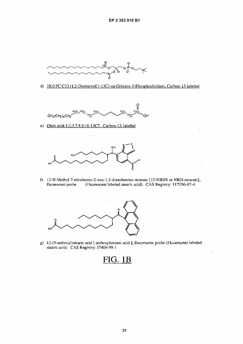
EP 2 353 010 B1
21

EP 2 353 010 B1
22

EP 2 353 010 B1
23

EP 2 353 010 B1
24
EP 2 353 010 B1
25
REFERENCES CITED IN THE DESCRIPTION
This list of references cited by the applicant is for the reader’s convenience only. It does not form part of the Europeanpatent document. Even though great care has been taken in compiling the references, errors or omissions cannot beexcluded and the EPO disclaims all liability in this regard.
Patent documents cited in the description
• SU 1459656 [0007]• US 7166474 B [0008] [0045]
• US 2003170912 A [0008]
Non-patent literature cited in the description
• ALTOMENTOVA et al. Clinica Chimica Acta, 1998,vol. 271 (2), 133-149 [0008]
• GURACHEVSKY et al. Clin. Chem. Lab. Med., 2008,vol. 46 (9), 1203-1210 [0008]


![(19) TZZ ¥ T - patentimages.storage.googleapis.com · 2,176,476, US Patent 4,992,474 and Tetrahedron Letters, vol. 35(50), pages 9375-9378, 1994. [0010] The present invention discloses](https://static.fdocuments.in/doc/165x107/5c25e6c309d3f2ae178c11cf/19-tzz-t-2176476-us-patent-4992474-and-tetrahedron-letters-vol.jpg)