19 Original 18
7
April 2008 - Vol.2 127 European Journal of Dentistry Gingival recession is described as “the exposure of the root surface by an apical shift in the position of the gingiva”. 1 Gingival recession increases in both prevalence and severity with age, 2,3 and the mandibular incisor region is a commonly affected area. This problem often causes increased susceptibility for root caries, poor esthetics, and dentine hypersensitivity. 4,5 Gingival recession is seen both in populations with high and poor standards of oral hygiene. 6 Tissue trauma caused by vigorous tooth Kemal Ustun a Zafer Sari b Hasan Orucoglu c Ismet Duran d Sema S. Hakki d ABSTRACT Gingival recession is displacement of the soft tissue margin apically leading to root surface exposure. Tooth malpositions, high muscle attachment, frenal pull have been associated with gingival tissue recession. Occlusal trauma is defined as injury resulting in tissue changes within the attachment apparatus as a result of occlusal forces. Trauma from occlusion may cause a shift in tooth position and the direction of the movement depends on the occlusal force. We present the clinical and radiological findings and the limitation of periodontal treatment of a severe gingival recession in a case with traumatic occlusion. A 16 years old male, systemically healthy and non- smoking patient presented to our clinic with severe gingival recession of mandibular canines and incisors. Clinical evaluation revealed extensive gingival recession on the vestibules of mandibular anterior segment. Patient has an Angle class III malocclusion and deep bite. To maintain the teeth until orthodontic therapy and maxillofacial surgery, mucogingival surgeries were performed to obtain attached gingiva to provide oral hygiene and reduce inflammation. After mucogingival surgeries, limited attached gingiva was gained in this case. Regular periodontal maintenance therapy was performed at 2 month intervals to preserve mandibular anterior teeth. Multidisciplinary approach should be performed in this kind of case for satisfactory results. Unless occlusal relationship was corrected, treatment of severe gingival recession will be problematic. For satisfactory periodontal treatment, early diagnosis of trauma from occlusion and its treatment is very important. (Eur J Dent 2008;2:127-133) Key words: Severe gingival recession; Malocclusion; Mucogingival surgery. a Konya Oral Health Center, Konya, Turkey. b Assoc. Prof., Selcuk University, Faculty of Dentistry, Department of Orthodontics, Konya, Turkey. c Assist. Prof., Selcuk University, Faculty of Dentistry, Department of Endodontics, Konya, Turkey. d Assoc. Prof., Selcuk University, Faculty of Dentistry, Department of Periodontology, Konya, Turkey. Corresponding author: Sema S. HAKKI Selcuk University, Faculty of Dentistry Department of Periodontology Campus, 42079, Konya, TURKEY Fax: +90 332 241 0062 E-mail: [email protected]; [email protected] Severe Gingival Recession Caused by Traumatic Occlusion and Mucogingival Stress: A Case Report INTRODUCTION
-
Upload
karin-uchima -
Category
Documents
-
view
36 -
download
2
Transcript of 19 Original 18

April 2008 - Vol.2127
European Journal of Dentistry
Gingival recession is described as “the exposure of the root surface by an apical shift in the position of the gingiva”.1 Gingival recession increases in both prevalence and severity with age,2,3 and the mandibular incisor region is a commonly affected area. This problem often causes increased susceptibility for root caries, poor esthetics, and dentine hypersensitivity.4,5
Gingival recession is seen both in populations with high and poor standards of oral hygiene.6
Tissue trauma caused by vigorous tooth
Kemal Ustuna
Zafer Sarib Hasan Orucogluc Ismet Durand
Sema S. Hakkid
AbstrActGingival recession is displacement of the soft tissue margin apically leading to root surface
exposure. Tooth malpositions, high muscle attachment, frenal pull have been associated with gingival tissue recession. Occlusal trauma is defined as injury resulting in tissue changes within the attachment apparatus as a result of occlusal forces. Trauma from occlusion may cause a shift in tooth position and the direction of the movement depends on the occlusal force. We present the clinical and radiological findings and the limitation of periodontal treatment of a severe gingival recession in a case with traumatic occlusion. A 16 years old male, systemically healthy and non-smoking patient presented to our clinic with severe gingival recession of mandibular canines and incisors. Clinical evaluation revealed extensive gingival recession on the vestibules of mandibular anterior segment. Patient has an Angle class III malocclusion and deep bite. To maintain the teeth until orthodontic therapy and maxillofacial surgery, mucogingival surgeries were performed to obtain attached gingiva to provide oral hygiene and reduce inflammation. After mucogingival surgeries, limited attached gingiva was gained in this case. Regular periodontal maintenance therapy was performed at 2 month intervals to preserve mandibular anterior teeth. Multidisciplinary approach should be performed in this kind of case for satisfactory results. Unless occlusal relationship was corrected, treatment of severe gingival recession will be problematic. For satisfactory periodontal treatment, early diagnosis of trauma from occlusion and its treatment is very important. (Eur J Dent 2008;2:127-133)
Key words: Severe gingival recession; Malocclusion; Mucogingival surgery.
a Konya Oral Health Center, Konya, Turkey.b Assoc. Prof., Selcuk University, Faculty of Dentistry, Department of Orthodontics, Konya, Turkey.c Assist. Prof., Selcuk University, Faculty of Dentistry, Department of Endodontics, Konya, Turkey.d Assoc. Prof., Selcuk University, Faculty of Dentistry, Department of Periodontology, Konya, Turkey.
Corresponding author: Sema S. HAKKISelcuk University, Faculty of Dentistry Department of Periodontology Campus, 42079, Konya, TURKEYFax: +90 332 241 0062E-mail: [email protected]; [email protected]
Severe Gingival Recession Caused by Traumatic Occlusion and Mucogingival Stress: A Case Report
IntroductIon
European Journal of Dentistry128
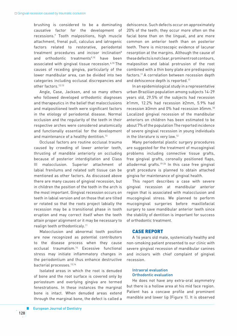
brushing is considered to be a dominating causative factor for the development of recessions.7 Tooth malpositions, high muscle attachment, frenal pull, calculus and iatrogenic factors related to restorative, periodontal treatment procedures and incisor inclination8 and orthodontic treatments9,10 have been associated with gingival tissue recession.6,10 The causes of receding gingiva, particularly of the lower mandibular area, can be divided into two categories including occlusal discrepancies and other factors.12,13
Angle, Case, Jackson, and so many others who followed developed orthodontic diagnoses and therapeutics in the belief that malocclusions and malpositioned teeth were significant factors in the etiology of periodontal disease. Normal occlusion and the regularity of the teeth in their respective arches were considered anatomically and functionally essential for the development and maintenance of a healthy dentition.13
Occlusal factors are routine occlusal trauma caused by crowding of lower anterior teeth, thrusting of mandible anteriorly on occluding because of posterior interdigitation and Class III malocclusion. Superior attachment of labial frenilums and related soft tissue can be mentioned as other factors. As discussed above there are many causes of gingival recession, but in children the position of the tooth in the arch is the most important. Gingival recession occurs on teeth in labial version and on those that are tilted or rotated so that the roots project labially the recession may be a transitional phase in tooth eruption and may correct itself when the teeth attain proper alignment or it may be necessary to realign teeth orthodonticaly.11
Malocclusion and abnormal tooth position are now recognized as potential contributors to the disease process when they cause occlusal traumatism.14 Excessive functional stress may initiate inflammatory changes in the periodontium and thus enhance destructive bacterial processes.13,16
Isolated areas in which the root is denuded of bone and the root surface is covered only by periosteum and overlying gingiva are termed fenestrations. In these instances the marginal bone is intact. When denuded areas extend through the marginal bone, the defect is called a
dehiscence. Such defects occur on approximately 20% of the teeth; they occur more often on the facial bone than on the lingual, and are more common on anterior teeth than on posterior teeth. There is microscopic evidence of lacunar resorption at the margins. Although the cause of these defects is not clear, prominent root contours, malposition and labial protrusion of the root combined with a thin bony plate are predisposing factors.11 A correlation between recession depth and dehiscence depth is reported.17
In an epidemiological study in a representative urban Brazilian population among subjects 14-29 years old, 29.5% of the subjects had recession ≥1mm, 12.2% had recession ≥2mm, 5.9% had recession ≥3mm and 0% had recession ≥5mm.18
Localized gingival recession of the mandibular anteriors on children has been estimated to be about 7% of the population The reported incidence of severe gingival recession in young individuals in the literature is very low.12
Many periodontal plastic surgery procedures are suggested for the treatment of mucogingival problems including connective tissue grafts, free gingival grafts, coronally positioned flaps, allodermal grafts.19-24 In this case free gingival graft procedure is planned to obtain attached gingiva for maintenance of gingival health.
This report describes a case with severe gingival recession at mandibular anterior region that is associated with malocclusion and mucogingival stress. We planned to perform mucogingival surgeries before maxillofacial surgery to save mandibular anterior teeth since the stability of dentition is important for success of orthodontic treatment.
cAsE rEPort A 16 years old male, systemically healthy and
non-smoking patient presented to our clinic with severe gingival recession of mandibular canines and incisors with chief complaint of gingival recession.
Intraoral evaluationOrthodontic evaluationHe does not have any extra-oral asymmetry
but there is a hollow area at his mid face region. Patient has a concave profile and prominent mandible and lower lip (Figure 1). It is observed
Gingival recession caused by traumatic occlusion

April 2008 - Vol.2129
European Journal of Dentistry
Ustun, Sari, Orucoglu, Duran, Hakki
that lower right second premolar is semi erupted, mandibular canines have distolingual axial rotation and there is diestema between lower left first and second premolar. Upper right first premolar has been extracted and there is diestema between maxillary incisors (Figure 2). 5 mm negative over jet and 7 mm overbite has been detected. Molar relationship is Angle Class III at both sides. Upper right and left canine have not erupted yet. Upper right and left second molars and mandibular left first premolar are still erupting. Anterior maxilla is narrower than the mandible transversally.
Periodontal evaluationThe intra-oral examination showed severe
hard and soft tissue loss on the vestibules of mandibular anterior segment (Figure 3). Moderate gingival recessions were also noted in other teeth including #12,13,14,15,20 (Table 1). All the vestibular gingiva of canines and incisors recessed to the apexes of these teeth. There were not any calculus on the vestibules of these teeth but dental plaque was present. Slight mobility of mandibular anterior incisors was observed. Pathologic periodontal pocket was not detected (Table 2).
Radiographic examinationIn the cephalometric analysis maxilla is
sagitally retrognathic and mandible is severely prognathic, the skeletal pattern is Class III (Figure 4a). Mandible’s volume is larger than its normal size and it is rotated posteriorly. The upper incisors are front positioned and protruded, the lower incisors are front positioned and retruded. The interincisal angle has been increased. It has been observed in the soft tissue analyses that the nasolabial angle has been decreased and according to “E plane” lower lip is highly protruded and upper lip is retruded. (Table 3)
In the panoramic radiograph analysis upper right canine is impacted and can be forced to erupt but upper left canine is impacted horizontally that it can not be forced to erupt (Figure 4b). The teeth including #1,16,17,32 are not present.
Endodontic treatmentHowever, affected teeth were vital and
they respond to electric pulp testing (Vitality Scanner, Analytic Technology, Glendora, CA, USA), endodontic treatments of teeth were performed due to severe recessions of gingiva and hypersensitivity.
Periodontal treatmentInitial phase therapy including scaling and
root planning was performed and oral hygiene instructions were given to the patient. Occlusal adjustment was performed to prevent undesirable
Tooth numberGingival recession Width of keratinized tissue
Pre-op Post-op Pre-op Post-op
#12 2 mm 0 mm -
#13 3 mm 0 mm -
#14 2 mm 1 mm -
#15 2 mm 1 mm -
#20 6 mm 2 mm -
#22 13 mm 13 mm 0 mm 1 mm
#23 14 mm 13 mm 0 mm 2 mm
#24 15 mm 14 mm 0 mm 2 mm
#25 15 mm 14 mm 0 mm 2 mm
#26 13 mm 12 mm 0 mm 2 mm
#27 10 mm 10 mm 0 mm 1 mm
#28 5 mm 0 mm -
Table 1. Gingival recession and width of keratinized tissue measurements of 16 year-old case. Data were given only
in affected teeth. Post-op measurements were conducted after periodontal surgery for mandibular anterior teeth.

European Journal of Dentistry130
contacts for mandibular incisors after endodontic therapy. Due to socioeconomic condition of patient, orthodontic treatment and maxillofacial surgery was postponed. Free gingival auto-graft was planned to save teeth, and enhance plaque removal and reduce inflammation around the teeth until orthodontic therapy and maxillofacial surgery. As a first step, free gingival auto-graft was conducted on the left lateral incisor and canine region. Two months after the first surgery, a second gingival auto-graft was performed on the central incisors and right lateral incisor and canine.
rEsuLtsHowever enough vestibular sulcus was obtained
on day 10 after surgeries, a highly limited amount of
attached gingiva was remained at the end of 1 month (Figure 5). Since the result is reversible, additional mucogingival surgery was not planned. Case will be assessed after orthodontic and maxillofacial surgery for periodontal condition. Regular periodontal maintenance therapy was performed at 2 month intervals to preserve mandibular anterior teeth. Multidisciplinary approach should be performed in this case for satisfactory results.
dIscussIonMucogingival deficiencies, especially of the
mandibular incisors, and the development of localized pathologic recession have been related to various etiologic factors. These, either individually or in combination, are minimally attached gingiva, thin labial alveolar bone, severe labial inclination, orthodontic treatment,9,10 fenestration of the alveolar bone, or occlusal trauma. Orthodontic intervention that improves the anatomic and functional environment can limit the recession and, in some cases, induce “creeping or spontaneous reattachment”.28
The presumption that the correction of malocclusion is predictably beneficial to the future health of the periodontium is questioned in the literature. Clinical experience and anecdotal reports from astute observers have identified localized etiologic effects of maloccluded and malpositioned teeth on periodontal health.12
In this case, occlusal trauma and mucogingival stress could be predisposing factors for gingival recession. Patient has 5 mm negative over jet and Figure 1. Extraoral view of case.
Figure 2. Intraoral upper and lower occlusal view of case. Figure 3. Intraoral view of case.
Figure 4. a) Cephalometric radiograph of the case (please note the position of lower incisors) b) Panoramic radiograph of the case.
Figure 5. Intraoral view of case at 3 months after surgery.
Gingival recession caused by traumatic occlusion
Number of teeth present 25
Plaque index ( 0-3 ) 2.1±0.35
Gingival index ( 0-3 ) 1.95±0.59
Pocket depth (mm) average±sd 1.87±0.71
Table 2. Mean values of clinical measurements of case
at first appointment.

April 2008 - Vol.2131
European Journal of Dentistry
Ustun, Sari, Orucoglu, Duran, Hakki
7 mm overbite. The upper anteriors are likely to cause labial movement of the lower anteriors into the thin labial plate of bone and severe fenestrations are formed. In combination with the mucogingival stress the gingiva receded in an accelerated manner.
To evaluate the possible etiologic relationship of malocclusion and malposition of teeth to periodontal disease, a study of the clinic population at the Columbia University, School of Dental and Oral Surgery, Division of Periodontics, was undertaken. The sample for the cross-sectional study included 516 subjects. A detailed examination of the health of the periodontium and the amount of gingival inflammation and periodontal destruction was recorded for every tooth in every subject.13 Similarly, observations of the occlusion and characteristics of the occlusion and tooth malposition were recorded. The data revealed that occlusion, as defined by Angle, was
not statistically related to periodontal disease. No health differences were found between subjects with Class I occlusions and those with Class II. Subjects with Class II Division 1 and Division 2 occlusions exhibited no differences in health for either anterior or posterior teeth. When unilateral Class II occlusions were studied, the maloccluded side was as healthy as the normally occluded side. The number of subjects with Class III occlusions was too small for data analysis; although, in that small group, there was no preponderance of disease.25-27
Reports about rare cases may provide beneficial information to clinicians about this question. Keeping in mind these edge cases may help them to make correct prognosis of dentitions and correct treatment plans.
It is known that in some cases of extreme anterior overbite, direct trauma to the gingiva from the incisal edges of the mandibular
Skeletal Measurements
1 SNA (dg) 81.0
2 SNB (dg) 87.0
3 ANB (dg) -6.0
4 A to N-I FH (mm) -2.4
5 Pg to N-I FH (mm) 6.7
6 FMA (dg) 31.0
7 SNGoGn (dg) 39.0
8 Mandibular length (Co-Gn) (mm) 125.4
9 Midfacial length (Co-A) (mm) 86.5
10 Maxillomand. difference ( 8-9 ) 40.0
Dental measurements
11 Overjet (mm) 5.0
12 Overbite (mm) 7.0
13 Mx1-NA(mm) 9.0
14 Mx1-NA (dg) 30.0
15 Md1- NB (mm) 8.0
16 IMPA (dg) 71.0
17 Interincisal angle (dg) 140.0
Soft Tissue Measurements
18 Nasolabial angle (dg) 89.0
19 Lower lip E-plane (mm) 1.0
20 Upper lip E-plane (mm) 5.5
Table 3. Cephalometric measurements of case.

European Journal of Dentistry132
incisors may result in palatal recession of the maxillary incisors. Similarly, in severe Class II Division 2 malocclusions with linguo-version of the maxillary incisors, functional trauma can cause marginal recession of the labial gingiva of the mandibular incisors. As far as we know this is the first report about Class III malocclusions with deep bite causing marginal recessions. This recession, although not the result of periodontitis, represented a significant loss of attachment.
In this case both the mucogingival problem and occlusal trauma act together and caused severe gingival recession. Although tooth loss from gingival recession is not a common condition, this case displayed that when malocclusion, fenestrations and mucogingival problems combine it does not seem to be so far.
rEFErEncEs1. Glickman I, Carranza FA. Clinical periodontology. 5th ed.
Philadelphia: W.B. Saunders; 1979. p. 100-101.
2. Beck JD. Periodontal implications: older people. Ann
Peridontol 1996;1:322-357.
3. Albandar JM. Global risk factors and risk indicators for
periodontal diseases. Periodontology 2000 2002;29:177-
206.
4. Tillis TSI, Keating JG. Understanding and managing dentine
hypersensitivity. J Dent Hyg 2002;76:296-309.
5. Drisko CH. Dentine hypersensitivity-dental hygiene and
periodontal considerations. Int Dent J 2002;52supp1:385-
393.
6. Wennstrom JL, Pini Prato G. Mucogingival therapy-
periodontal plastic surgery in: Lindhe J, Karring T, Lang
NP. Periodontology and Implant Dentistry. 4th edition, Oxford
Blackwell Munksgaard; 2003. p. 579.
7. Radentz WH, Barnes GP, Cutright DE. A survey of factors
possibly associated with cervical abrasion of tooth surfaces.
J Periodontol 1976;47:148-154.
8. Geiger AM, Wasserman BH. Relationship of occlusion
and periodontal disease: Part IX-Incisor inclination and
periodontal status. Angle Orthod 1976;46:99-110.
9. Reed J. Recession of mandibular anterior gingival following
orthodontics in adults. Todays FDA 2005;17:45.
10. Zimmer B, Seifi-Shirvandeh N. Changes in gingival
recession related to orthodontic treatment of traumatic
deep bites in adults. J Orofac Orthop 2007;68:232-44.
11. Carranza FA. The role of iatrogenic and other local factors
in: Carranza FA, Newman MG. Clinical Periodontology. 8th
edition, WB Saunders Company; 1996 p.167.
12. Rose GJ. Receding mandibular labial gingiva on children.
Angle Orthod 1967;37:147-150.
13. Geiger AM. Malocclusion as an etiologic factor in
periodontal disease: A retrospective assay. Am J Orthod
Dentofacial Orthop 2001;120:112-115.
14. Goldman H, Schluger S, Fox L, Cohen DW. Periodontal
Therapy, 7th ed. Philadelphia: W.B. Saunders; 1990. p.52.
15. Schwartz M, Lamster I, Fine J. Clinical guide to periodontics.
Philadelphia: W.B. Saunders; 1995;312-315.
16. Solberg WC. The role of morphofunctional occlusal factors
in periodontal disease. In: Carranza FA, editor. Glickman’s
Clinical Periodontology, 7th ed. Philadelphia:W. B.
Saunders; 1990. p.422-431.
17. Lost C. Depth of alveolar bone dehiscence in relation to
gingival recessions. J Clin Periodontol 1984;11:583-589.
18. Susin C, Haas AN, Opperman RV, Haugejorden O, Albandar
JM. Gingival recession: Epidemiology and risk indicators in
a representative urban Brazilian population. J Periodontol
2004;75:1377-1386.
19. Wennstrom J. Mucogingival theraphy. Ann Periodontol
1996;1:671-701.
20. Bouchard P, Mallet J, Borghetti A. Decision making
in aesthetics: Root coverage revisited. Periodontol
2000;2001:97-120.
21. Pagliaro U, Nieri M, Franceschi D, Clauser C, Pini-Prato
G. Evidence-based mucogingival therapy. Part 1: A critical
review of the literature on root coverage procedures. J
Periodontol 2003;74:709-740.
22. Clauser C, Nieri M, Franceschi D, Pagliaro U, Pini-Prato
G. Evidence-based mucogingival theraphy. Part 2: Ordinary
and individual patient data meta-analyses of surgical
treatment of recession using complete root coverage as
the outcome variable. J Periodontol 2003;74:741-756.
23. Harris RJ. Root coverage with connective tissue grafts:
An evaluation of short and long term results. J Periodontol
2002;73:1405-1411.
24. Müller HP, Eger T, Schorb A. Gingival dimensions after
root coverage with free connective tissue grafts. J Clin
Periodontol 1998;25:424-430.
25. Geiger AM, Wasserman BH, Thompson RH Jr, Beube EE,
Goodman SF, Pomerantz J, et al. Relationship of occlusion
and periodontal disease. I. A system for evaluating
periodontal status. J Periodontol 1971;42:364-370.
26. Wasserman BH, Thompson RH, Geiger AM, Goodman SF,
Pomerantz J, Turgeon LR, et al. Relationship of occlusion
and periodontal disease. II. Periodontal status of the study
population. J Periodontol 1971;42:371-378.
27. Geiger AM, Wasserman BH, Thompson RH Jr, Turgeon
LR. Relationship of occlusion and periodontal disease. V.
Relation of classification of occlusion to periodontal status
and gingival inflammation. J Periodontol 1972;43:554-560.
Gingival recession caused by traumatic occlusion

April 2008 - Vol.2133
European Journal of Dentistry
Ustun, Sari, Orucoglu, Duran, Hakki
28. Geiger AM. Mucogingival problems and the movement
of mandibular incisors: a clinical review. Am J Orthod
1980;78:511-527.


![Washington State Register, Issue 19-18 WSR 19-18-015 WSR ...lawfilesext.leg.wa.gov/law/wsr/2019/18/19-18PROP.pdfWSR 19-18-035 Washington State Register, Issue 19-18 Proposed [ 2 ]](https://static.fdocuments.in/doc/165x107/5f4acb3bcdd69703016a40e9/washington-state-register-issue-19-18-wsr-19-18-015-wsr-wsr-19-18-035-washington.jpg)