Crime and Trial. Before the trial Crime occurs Crime occurs Investigation Investigation.

January 1990 Volume 17, No.1
U.S. Department of Justice Federal Bureau of Investigation
Crime Laboratory Digest
Review Article In This Issue
Use of HPLC with Diode Array Spectrophotometric Detection in Forensic Drug Analysis
$.:&'...., 1 ....... 1
9.98 mjn 482 nm
T. w •• C'rr.' 1"""o:J
Swjvel 45 deg Tjlt 35 deg
If you have issues viewing or accessing this file contact us at NCJRS.gov.
EDITOR Barry L. Brown
Quantico, Virginia
William Anderson Reno, Nevada
F. Samuel Baechtel Quantico, Virginia
Robert Cravey Santa Ana, California
Harold Deadman Quantico, Virginia
Peter DeForest New York, New York
ASSISTANT EDITOR Denise K. Bennett Quantico, Virginia
EDITORIAL BOARD
Dean Fetterolf Quantico, Virginia
Barry Gaudette Ottawa, Canada
Robert Koons Quantico, Virginia
Dale Moreau Quantico, Virginia
PUBLICATIONS COMMITTEE
ASSOCIATE EDITOR David T. Stafford
Memphis, Tennessee
Robert Sibert Washington, D.C.
Robert Spalding Washington, D.C.
Irving Stone Dallas, Texas
Phillip Wineman Rockville, Maryland
Thomas Munson St. Cloud, Minnesota
AMERICAN SOCIETY OF CRIME LABORATORY DIRECTORS
David T. Stafford*, Chairman Memphis, Tennessee
Arnold Melnikoff Missoula, Montana
Gerald Smith Knoxville, Tennessee
Kenneth Jonmaire Lockport, New York
*These individuals also serve on the editorial board.
PUBLICATION POLICY
Rondal Bridgemon Tucson, Arizona
John Murdock* Martinez, California
The Crime Laboratory Digest is published quarterly by the FBI Laboratof'j in cooperation with the American Society of Crime Laboratory Directors (ASCLD). It is intended to serve as a rapid means of communication between crime laboratories, permitting information of interest and value to be disseminated among crime laboratory scientists.
Inclusion of an article in the Crime Laboratory Digest in no way represents an endorsement or recommendation of any part of that article by the Federal Government, the Department of Justice or the FBI. Contributing authors assume total responsibility for the contents and accuracy of their submission. Que3tions or requests concerning an article should be directed to the contributing agency.
All submissions are subject to editorial review in accordance with the editorial policy established by the FBI Laboratory and ASCLD. The editorial staff of the Crime Laboratory Digest reserves the right to edit all articles for style, grammer and punctuation. Comments and letters to the editor are encouraged and will be published when appropriate and as space permits. These should be forwarded to:
Crime Laboratory Digest - Editor FSRTC, FBI Academy
Quantico, Virginia 22135
Contents
Message from the Assistant Director in Charge of the FBI Laboratory
Editor's Column
Review Article: The Use of HPLC with Diode Array . Spectrophotometric Detection in Forensic Drug Analysis
Barry K. Logan, H. Steve Nichols, G. Scott Fernandez and David T ... Stafford discuss the benefits of combining diode array ultraviolet visible spectrophotometric detection with HPLC for increased efficiency in drug analysis/identification.
Feature Article: The Effect of Luminol on the Serological Analysis of Dried Bloodstains
Robert R. J. Grispino examines the effects of luminol applications on the complete serological analysis of dried bloodstains and addresses the advantages and disadvantages of using the luminol test at crime scenes.
DNA Technology Seminar Videotape Order Form
Meeting Announcements
Instructions for Submitting Articles
Crime Laboratory Digest Subscriber Form
U.s. Department of Justice Natlonallnstitute of Justice
131354
131353-131354
This document has been reproduced exactly as received from the person or organization originating it. Points of view or opinions stated in this document are those of the authors and do not necessarily represent the official position or poliCies of the National Institute of Justice.
Permission to reproduce this Q ~ material has been granted by
Public IXJI!1ain/El3I .IL-8. Deparb'Jent of !TU8±ic...,e~ __ to the National Criminal Justice Reference Service (NCJRS).
Further reproduction outside of the NCJRS system requires permission of the ~ owner.
CRIME LABORATORY DIGEST Vol 17, No.1, January 1990 ISSN NO. 0743-1872
1
2
5
13
24
25
28
29
Mont. 24
I 11 Minn. VT 33 (488)
(3,131) (362) 20 NH 76 (3,294) ~ OI'~ell:l.
MA 102 (5,047) S. Oak. (334) Wyo. H13 CT 54 (2,7 45)
12 (~80) Ri 105 (12,218) (212) Neb. NJ 170 (9,331 )
15 DE 139 (15,664) MD 507 (24,642) (466)
Col. OC 521 (28,835) 31 Kan.
(2,442) , 35 I. __ .,..,,.
Ariz. I 31 N. Mex.
16 (3,408) (1,008)
Texas 212 140~·i\ Foreign Governments
(15,803) (1.978) . ~ ~;R Australia 2 (297) Bahamas 6 (87)
British West Indies 4 (903) ,Q
.. ~.-
\j '-} Canada 4 (420) '(.\ Cayman Islands 1 (95)
Hawai~ Jamaica 1 (12) 101 {> (13,269)
FBIIDOJ
I Feature Miele I
The Effect of Luminol on the Serological Analysis of Dried Human Blood~stains
Robert R. J. Grispino Serology Unit
FBI Laboratory Washington, D. C. 20535
To a great degree today, crime laboratories are being provided with crime scene evidence which has been treated with luminol reagent mixtures by law enforcement personnel at the scene. Laboratory examiners are then requested to perform a complete serological analysis of the questioned samples. However, uninformed crime scene personnel sometimes opt to use the luminol test as a preferred field blood test at all crime scenes. Since the reagents are commercially available in packaged crime scene kits, the luminol test is susceptible to abuse and misuse by untrained officers. This article discusses the effects of luminol sprays on the complete serological analysis of neat dried bloodstains. In addition, the advantages and disadvantages of the luminol test are addressed, and suggestions are offered concerning the proper application and protocol of the luminol test at crime scenes. In summary, the results of this study demonstrate that the use of luminol denatures most blood enzymes after a short exposure, thus causing further serological comparisons to be questionable.
REVIEW
The compound 3 aminophthalhydrazide (5- amino 2, 3- dihydrophthalazine 1, 4- dione) was first synthesized by Schmitz in 1902. At the time of synthesis, he noticed that the compound exhibited a strong blue fluorescence in aciti solutions. In 1927, Lommel first observed a blue chemiluminescence after oxidation of the compound in alkaline solution. His work was never published, but Albrecht, one of his coworkers, confirmed and published Lommel's original findings in 1928. Albrecht also pointed out that blood and fresh potato juice caused the chemical to exhibit strong luminescence in the presence of hydrogen peroxide (HPJ. In 1934, Huntress et al. termed this compound luminol as a producer of light.
13
In 1936, Gleu and Pfannstiel confirmed Albrecht's observations regarding luminol and blood. They noted that luminol would not chemiluminesce in the presence of boiled vegetable peroxidases and observed that luminol mixed with pure hematin produced the most brilliant blue luminescence.
The first proposed forensic use of luminol as a preliminary blood test was reported by Specht in 1937. He sprayed blood on bushes, stone walls, rusty iron fences, furniture, stone steps and a garden. After allowing the blood to remain exposed to the elements for 14 days, Specht sprayed a luminol reagent mixture onto the blood and photographed the results. 'fhe mixture was 0.1% luminal in 5% aqueous sodium carbonate (Na2C03) with a 15% solution of HP2 added immediately before spraying. All bloodstained areas glowed with blue light for 10 to 15 minutes. Blood was also detected in water, soapy water and sewage. The luminol test worked well with both fresh and old bloodstains; in fact, the older the bloodstain, the more pronounced the positive reaction.
Proescher and Moody confirmed Specht's findings in 1939 using Specht's spray mixtures. They detected bloodstains on paper, fabrics and iron pipes exposed to the elements for 3 years, with 3-year-old putrefied blood exhibiting brilliant luminescence. In addition, they observed that dried and decomposed blood elicited a stronger and longer lasting luminol reaction than fresh blood. When the luminescence disappeared, it could be reproduced by application of fresh luminol spray. Dried bloodstains were made luminescent many times. Fresh dried bloodstains were made more luminescent by spraying the blood with 1 to 2% hydrochloric acid solution before luminol application. The luminol reaction was elicited with both animal and human blood. Hematin was detected in a dilution of 1:108
• Most importantly, Proescher and Moody (1939) made the following emphatic supportive statement on luminol's application to forensic serology:
CRIME LABORATORY DIGEST Vol 17, No.1, January 1990
"Luminol does not interfere with the spectroscopic, chemical or precipitation tests for the definite identification of blood. Hematin and hemochromogen crystals of dried blood were obtained after rep~atedly treating blood with luminol. The precipitin test can be applied if the blood is not decomposed."
Lytle and Hedgecock (1978) experimented with the effects of alkaline luminol/sodium perborate (NaB OJ mixtures on blood. They concluded:
" .... luminol is relatively nondestructive to the surroundings (it is noncorrosive and nonstaining) and to the blood (it does not prevent subsequent identi-fication tests or ABO blood grouping analysis although it does interfere with electrophoretic analysis of.. .. erythrocyte acid phosphatase and phosphoglucomutase)."
They recommended luminol as "a good field test" which was "sensitive and reasonably specific for blood."
The luminol test is known to be extremely sensitive to the presence of blood. In 1986, Thornton et al. stated that the unaided eye could detect blue chemiluminescence of luminol in blood diluted 1:104
• With the use of an infrared starlight scope, blue chemiluminescence of luminol could be detected in blood solutions of 1:106 to 5:106
•
In the luminolliterature (Gundennann 1965; Schneider 1970; Gaennsslen 1983; Kraul and Meyer 1941; Wei and White 1971; Roswell and White 1978), researchers have reported false positive results (that is, chemiluminescence of luminal in the absence of blood) from alkaline luminol in the presence of iron, copper, hypochlorites, manganese peroxide and ferricyanide. False negative results were obtained with luminol sprayed on cadmium-coated materials bearing blood. Cadmium has been employed as a corrosion resistant coating for materials.
The chemistry of the luminol reaction should be reviewed so that the results of these experiments can be better understood. Chemiluminescence is defined as the production of light by chemical means. It occurs when an electron moves from an excited state to a ground state. The electron is raised to an excited state by chemical energy. It requires a chemical reaction which will supply at least 40 to 70 kcal./mol energy :t$ in tht'i case in free radical reactions. It does not require excitation by radiant energy as does fluorescence (Wildes and White 1973).
14
The c:hemical basis of the luminol test is an oxidatjon reaction. An alkaline solution of luminol will oxidize in the presence of HP2 and a hematin-catalyzed peroxidase system. Hydrogen peroxide is the oxidizing agent. Blood is a hematin-catalyzed peroxidase system. The resulting oxidation reaction is visualized by a blue chemiluminescence.
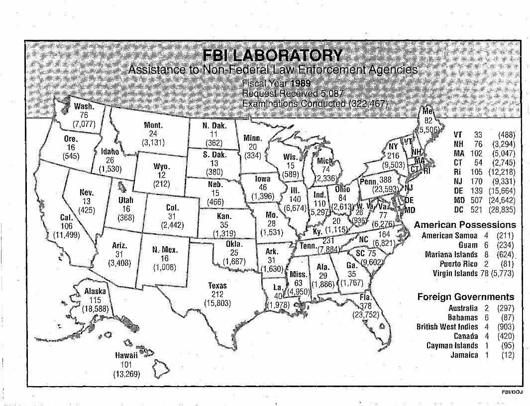
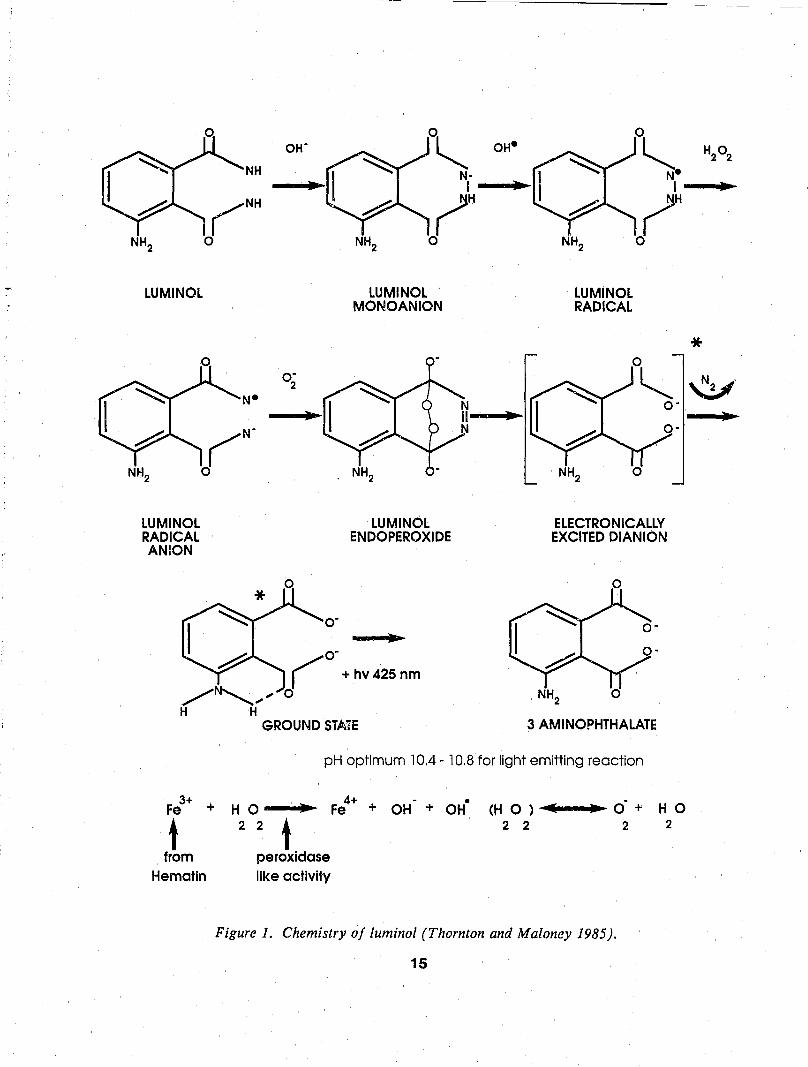
In 1985, Thornton and Maloney reviewed the chemistry of the luminol reaction'. Figure 1 is an amalgamation presented by Thornton of the work of a number of researchers on a proposed oxidation pathway for luminol. Not all of the steps of the oxidation pathway have been elucidated to date. The important points are:
1) Oxygen is required. 2) The anion of luminol is the reactant. 3) Free radicals are involved which provide the
40 to 70 kcal./mol of chemical energy needed for chemiluminescence (Brune:irett et al. 1972).
4) Nitrogen is the product of the reaction. 5) The entire conversion pathway is unknown.
MATERIALS AND METHODS
The luminol spray regimen utilized two separate mixtures consisting of the following:
Luminol Mixture 1 (Specht 1937)
Part 1 - 0.1 g luminol and 5 g Na2C03 carbonate in 50 ml distilled water.
Part 2 - 0.7 g NaB02 in 50 ml 95% ethanol.
Luminol Mixture 2 (modification)
Part 1 - 0.1 g luminol and 5 g Na2C03 in 90 ml distilled water.
Parts 1 and 2 were always prepared separately in advance and were only mixed together immediately prior to use. Application to the questioned stains was made directly via an aerosol sprayer or indirectly via dropper bottle to a distilled water dampened cotton swabbing of the stained area in question. Luminol mixtures were applied as a spray in a completely dark, well ventilated room.
Positive and negative controls were employed in all tests; these consisted of known human bloodstains, a copper penny, sterile cotton swabs and washed cotton sheeting.
NH2
LUMINOl
LUMINOl RADICAL ANION
NH
N-
# /,~~ H H
OW
•
O· 2
..
o~ N· I
~H NH2 0
LUMINOl MONOANION
lUMINOl ENDOPEROXIDE
O· r +hv42Snm o
GROUND STATE
OH-
•
•
0
~-N I
NH2
~H 0
lUMINOl RADICAL
ELECTRONICAllY EXCITED DIAN ION
o
NH2
3 AMINOPHTHALATE
pH optimum 10.4 - 10.8 for light emitting reaction
3+ Fe
t from
Hematin
+ H 0 •
2 2 t peroxidase like activity
4+ • • Fe + OH + OH (H 0 ) ..... 11{ ..... -...~ O· + H 0
2 2 2 2
Figure 1. Chemistry of luminal (Thornton and Maloney 1985).
15
H20 2
•
* ~.
•
Sixty-two dried bloodstains were tested to determine how sensitive these procedures would be in our laboratory. Stains of neat group A blood were tested ranging from 100 1'1 of blood to serial dilutions of the same blood from 1:10 to 1:108
• The stains were on washed cotton sheeting, #1 Whatman filter paper and gtass microscope slides. All stains were air-dried in a ventilating hood.
Forty-five dried human bloodstains consisting of 100 1'1 of neat group A, AB and 0 blood (15 each type) were deposited on the same fabric, paper and glass materials. These stains were exposed to luminol mixtures 1 and 2. An addi.tional 80 swabbings were developed. The specimens were then analyzed serologically through the following stages:
1) Phenolphthalein Preliminary Blood Test -
Following the Kastle-Meyer protocol (Kastle 1909; Kastle and Shedd 1901), reduced phenolphthalein is applied first to a damp swabbing of the suspected stain; then a 3% solution of HP2 is applied to the swab, all via dropper bottle. A positive result consists of a swab color change from colorless to pink/red.
2) Hemochromogen Confirmatory Blood Test -
Hemochromogen crystal formation is microscopically observed and evaluated ~fter application of Takayama reagent and heat to a portion of the luminol treated and untreated bloodstains (Takayama 1912).
3) Origin Determination -
Antihuman serum was produced in rabbits after the ID<;'tnod of Proom (1943). Buffered saline extrac~ of luminol-sprayed and unsprayed human bloodstains were reacted against the prepared rabbit antihuman sera according to the protocol of Ouchterlony (1948; 1949a; 1949b; 1949c; 1968). Identity precipitin bands creating a fused chevron effect were indicative of a positive result (see following).
4) ABO Grouping -
Forward testing of ABO antigens was accomplished using the absorption-elution technique (Kind 1960a; 1960b, Outterridge 1962; 1965a; 1965b). Reverse testing of ABO antibodies was accomplished via the Lattes crust technique
16
(Lattes 1927; 1928). Conclusive identifications were made only when the forward and reverse tests agreed. Both techniques relied on the evaluation of microscopic agglutination of indicator erythrocytes and corresponding ABO antibodies. The strength of agglutination was evaluated according to the following criterion:
+4 - one solid grape-like cluster aggregate of erythrocytes
+3 - several large agglutinates +2 - medium size agglutinates on a clear
background +1 - small aggregates (triplet or doublet cell
clusters) o - no agglutination
5) P,,)lymorphic Blood Enzyme Analysh. ~ (Wraxall et al. 1978)
Phospboglucomutase (PGM) - Isoelectric FocusiIilg (IE F) sub typing (Budowle 1984a; 1985; Budowle et al. 1986)
Erythrocyte Acid Phosphatase (EAP) - IEF (Budowle 1984b)
Esterase D/Glyoxalase I (EsD/GLO) - aga.r electrophoresis (Budowle 1984 b; 1985; Budowle and Gambel 1988)
Peptidase A - agar electrophoresis (Parkin 1978) Adenosine Deaminase/ Adenylate Kinase (ADA/ AK) - agar electrophoresis (Murch et al. 1986)
6) Serum Proteins -
Haptoglobin (HP) - Polyacrylamide Gel Electrophoresis (Budowle and Chow 1985)
Group Specific Component (Gc) and Transferrin (Tf) - Immunofixation (Alper and Johnson 1969)
Duplicate sets, 15 for each group, of group A bloodstains (neat to 10-8 dilutions), neat group AB and neat group 0 stains on washed cotton sheeting, paper and glass materials were stored at room temperature for 2 months. These items were considered aged stains for the purposes of this study. They were tested using the same protocol as the fresh stains.
Blood used in this study was collected voluntarily via finger prick from employees of the FBI Laboratory.
,-,
RESULTS
Sensitivity Evaluations:
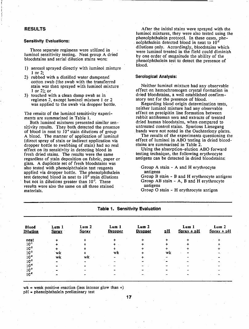
Three separate regimens were utilized in luminol sensitivity testing. Neat group A dried bloodstains and serial dilution stains were:
1) aerosol sprayed directly with luminol mixture I or 2;
2) rubbed with a distilled water dampened cotton swab (the swab with the transferred stain was then sprayed with luminol mixture 1 or 2); or
3) touched with a clean damp swab as in regimen 2, except luminol mixture I or 2 was applied to the swab via dropper bottle.
The resuits of the luminol sensitivity experiments are summarized in Table 1.
Both luminol mixtures presented similar sensitivity results. They both detected the presence of blood in neat to 10-1 stain dilutions of' group A blood. The manner of application of luminol (direct spray of stain or indirect application via dropper bottle to swabbing of stain) had no real effect on its sensitivity in detecting blood in fresh dried stains. The results were the same regardless of stain deposition on fabric, paper or glass. A duplicate set of fresh bloodstains was also tested with phenolphthalein test reagents applied via dropper bottle. The phenolphthalein test detected blood in neat to to-3 stain dilutions but not in dilutions greater than to-3
• These results were also the same on all three stained materials.
After the initial stains were sprayed with the luminol mixtures, they were also tested using the phenolphthalein protocol. In these cases, phenolphthalein detected blood in neat to to-2
dilutions only. Accordingly, bloodstains which were luminol treated in the fieJd could diminish by one order of magnitude the ability of the phenolphthalein test to detect the presence of blood.
Serological Analysis:
Neither luminol mixture had anv observable effect on hemochromogen crystal formation in dried bloodstains, a well established confirmatory test for the presence of blood.
Regarding blood origin determination tests, neither luminol mixture had any observable effect on precipitin line formation between rabbit antihuman sera and extracts of treated dried human bloodstains, when compared to untreated control stains. Spurious Liesegang bands were not noted in the Ouchterlony plates.
The results of the experiments questioning the effect of luminol in ABO testing in dried bloodstains are summarized in Table 2.
Using the absorption-elution ABO forward testing technique, the following erythrocyte antigens can be detected in dried bloodstains:
Group A stain - A and H erythrocyte antigens
Group B stain - Band H erythrocyte antigens Group AB stain - A, Band H erythrocyte
antigens Group 0 stain - H erythrocyte antigen
Table 1. Sensitivity Evaluation
Blood Lum 1 Lum 2 Lum 1 Dilution Sl!!n Spray Dropper
neat + + + to-I + + + 10-1 + + + 10-3 wk + wk 10~ wk wk to-5 10-6 to-7 10-1
wk "" weak positive reaction (less intense glow than +) pH - phenolphthalein preliminary test
Lurn 2 Lum 1 Lum 2 Dropper ill Spray + pH Spray + pH
+ + + + + + + + + + + + + wk +
17
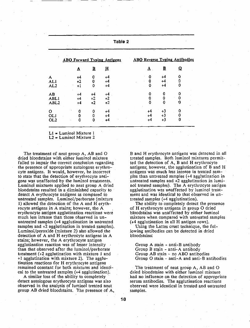
Table 2
ABQ Forward Tl:~inK Anti2~ns
A It H
A +4 0 +4 ALl +2 0 +4 AL2 +1 0 +4
AB +4 +4 +4 ABLI +4 +2 +2 ABL2 +4 +2 +2
0 0 0 +4 OLI 0 0 +4 OL2 0 0 +4
L 1 == Luminal Mixture 1 L2::o: Luminol Mixture 2
The treatment of neat group A, AB and 0 dried bloodstains with either luminol mix.ture failed to impair the correct conclusion regarding the presence of appropriate autologous erythrocyte antigens. It would, however, be incorrect to state that the detection of erythrocyte antigens was unaffected by the luminol treatments. Luminol mixtures applied to neat group A dried bloodstains resulted in a diminished capacity to detect A erythrocyte antigens as compared to untreated samples. Luminol/perborate (mixture 1) allowed the detection of the A and H erythrocyte antigens in A stains; however, the A erythrocyte antigen agglutination reactions were much less intense than those observed in ununtreated samples (+4 agglutination in untreated samples and +2 agglutination in treated samples). Luminol/peroxide (mixture 2) also allowed the detection of A and H erythrocyte antigens in A stains; however, the A erythrocyte antigen agglutination reaction was of lesser intensity than that observed after the luminol/perborate tl'eatment (+2 agglutination with mixture 1 and +1 agglutination with mixture 2). The agglutination reactions for H erythrocyte antigens remained constant for both mixtures and identical to the untreated samples (+4 agglutination).
A similar loss of the ability to completely detect autologous erythrocyte antigens was also observed in the analysis of luminol treated neat group AB dried bloodstains. The presence of A,
18
ABQ Reverse Tl:ning Antibodies
A l! Q
0 +4 0 0 +4 {)
0 +4 0
0 0 0 0 0 0 0 0 '0
+4 +3 0 +4 +3 0 +4 +3 0
Band H erythrocyte antigens was detected in all treated samples. Both luminal mixtures permitted the detection of A, Band H erythrocyte antigens; however, the agglutination of Band H antigens was much less intense in treated samples than untreated samples (+4 agglutination in untreated samples and +2 agglutination in luminal treated samples). The A erythrocyte antigen agglutination was unaffected by luminol treatment and was identical to that observed in untreated samples (+4 agglutination).
The ability to completely detect the presence of H erythrocyte antigens in group 0 dried bloodstains was unaffected by either luminol mixture when compared with untreated samples (+4 agglutination in all H antigen rows).
Using the Lattes crust technique, the following antibodies can be detected in dried bloodstains:
Group A stain - anti-B antibody Group B st!!.in - anti-A antibody Group AB stain - no ABO antibodies Group 0 stain - anti-A and anti-B antibodies
The treatment of neat group A, AB and 0 dried bloodstains with either luminol mixture had no influence on the detection of appropriate serum antibodies. The agglutination reactions observed were identical in treated and untreated samples.

Polymorphic Blood Enzyme Analysis
PGM Subtyping:
The untreated group A bloodstain was identified as PGM subtype 2-2+, while the untreated AB and 0 stains were PGM subtype 1+2+. After treatment with either of the luminol mixtures, the 2-2+ bands were still readable, but intensity was reduced compared to the untreated control sample. New spurious bands or unexplained enzyme activity were not noted on the electrophoretic plates.
The PGM subtype bands 1+2+ in the other stains were barely readable after treatment with the luminol/perborate mixture. New spurious bands or unexplained enzyme activity were not noted. The luminol/peroxide treatments rendered unreadable PGM activity in the AB and 0 samples.
EAP:
The untreated group A stain was identified as EAP type B, the AB stain was EAP type A and the 0 stain was EAP type BA. All luminol treated samples showed an intense fluorescence under ultraviolet light at the anode with no cathodic migration. Thus, these typing results were considered inconclusive for reporting purposes.
Other Blood Enzymes:
Enzyme activity in the following enzyme systems was not detected after treatment with either luminol mixture:
EsD/GLO Peptidase A ADA/AK
Serum Protein Analysis
HP:
The untreated group A stain was identified as HP type 2-1, the AB stain was HP type 2 and the type 0 stain was HP type 1. After treatment with either bminol mixture, heavy brown streaking was noted in the polyacrylamide gels. New spurious bands were not observed in luminol treated samples. Th,e brown streaking failed to interfere with the ~orrect reading of Haptoglobin types.
19
Gc and Tf:
All samples were identified as Gc type I. The untreated group A and 0 stains were identified as Tf type C while the untreated AB stain was identified as Tf type CD. In the Gc immunofixation system, all luminol treated samples were readable with light discrete bands noted. In the Tf immunofixation system, all luminol treated samples were readable, but streaking was prevalent in the gel. The band staining was much lighter than in untreated samples. New spurious bands or unexplained activity were not noted. after luminol treatment in either the Gc or Tf systems.
Aged Stains
Duplicate sets of the stains were stored for 2 months at room temperature to simulate the aging of forensic samples. These stains were subjected to the same serological protocols as the fresh samples described previously. The results of these experiments are provided. in Tables 3 and 4.
Aging did not reduce the sensitivity of either luminol preparation. Hemochromogen crystal formation and antihuman sera precipitation were unaffected by either luminol mixture.
Using the absorption-elution ABO forward testing technique, many similarities were noted in the effects of luminol on aged stains as compared to fresh stains. A diminished capacity to detect A erythrocyte antigens was observed after luminol treatments of aged A stains (+4 agglutination in untreated aged stains and +2 agglutination in luminol treated aged stains). As
Table 3. Sensitivity Evaluation of Aged Stains
Lum 1 Lum 2 ~ Spray ill
neat + + + 10'\ + + + 10'2 + + + 10'3 + + wk 10-4 wk wk wk lO's to 10-3
wk = weak positive reaction (less intense glow than +) pH - phenolphthalein preliminary test
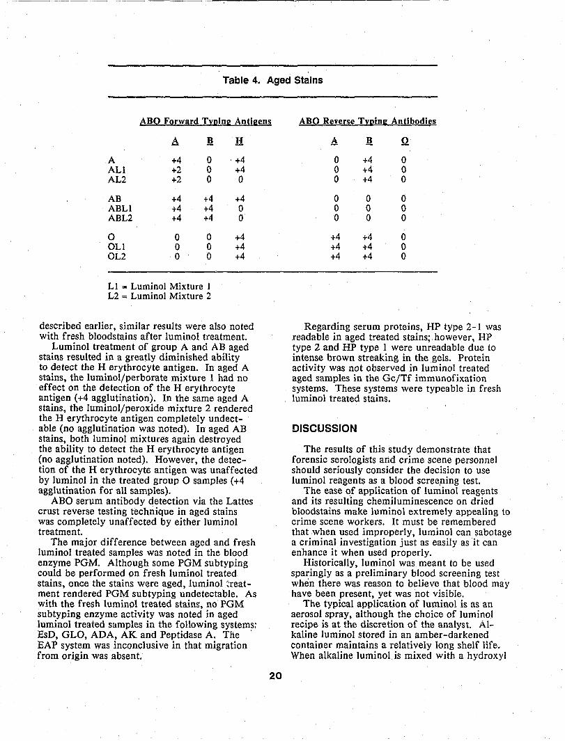
Table 4. A.ged Stains
ABQ FQrward TYl!in.:; Anti.:;en~
A J!
A +4 0 ALl +2 0 AL2 +2 0
AB +4 +4 ABLl +4 +4 ABL2 +4 +4
0 0 0 OLI 0 0 OL2 0 0
Ll ... Luminol Mixture 1 L2 = Luminol Mixture 2
H
+4 +4 0
+4 0 0
+4 +4 +4
described earlier, similar results were also noted with fresh bloodstains after luminol treatment.
Luminol treatment of group A and AB aged stains resulted in a greatly diminished ability to detect the H erythrocyte antigen. In aged A stains, the luminoljperborate mixture 1 had no effect on the detection of the H erythrocyte antigen (+4 agglutination). In the same aged A stains. the luminol/peroxide mixture 2 rendered the H erythrocyte antigen completely undectable (no agglutination was noted). In aged AB stains, both luminol mixtures again destroyed the ability to detect the H erythrocyte antigen (no agglutination noted). However, the detection of the H erythrocyte antigen was unaffected by luminol in the treated group 0 samples (+4 agglutination for all samples).
ABO serum antibody detection via the Lattes crust reverse testing technique in aged stains was completely unaffected by either luminol treatment.
The major difference between aged and fresh luminol treated samples was noted in the blood enzyme PGM. Although some PGM subtyping could be performed on fresh luminol treated stains, once the stains were aged, luminol ~reatment rendered PGM subtyping undetectable. As with the fresh luminol treated stains, no PGM subtyping enzyme activity was noted in aged luminol treated samples in the following systems: EsD, GLO, ADA, AK and Peptidase A. The EAP system was inconclusive in that migration from origin was absent.
20
ABQ R~'ferse TYl!inK Antibodies
A J! 0
0 +4 0 0 +4 0 0 +4 0
0 0 0 0 0 0 0 0 0
+4 +4 0 +4 +4 0 +4 +4 0
Regarding serum proteins, HP type 2-1 was readable in aged treated stains; however. HP type 2 and HP type 1 were unreadable due to intense brown streaking in the gels. Protein activity was not observed in luminol treated aged samples in the Gc/Tf immunofixation systems. These systems were typeable in fresh luminol treated stains.
DISCUSSION
The results of this study demonstrate that forensic serologists and crime scene personnel should seriously consider the decision to use luminol reagents as a blood screening test.
The ease of application of luminol reagents and its resulting chemiluminescence on dried bloodstains make luminol extremely appealing to crime scene workers. It must be remembered that when used improperly, luminol can sabotage a criminal investigation just as easily as it can enhance it when used properly.
Historically, luminol was meant to be used sparingly as a preliminary blood screening test when there was reason to believe that blood may have been present, yet was not visible.
The typical application of luminol is as an aerosol spray, although the choice of luminol recipe is at the discretion of the analyst. Alkaline luminol stored in an amber-darkened container maintains a relatively long shelf life. When alkaline luminol is mixed with a hydroxyl
ion source, the shelf life of the mixture is approximately 1 hour. Therefore, it is suggested that separate luminol reagents be included in crime scene kits for fresh mixing in appropriate circumstances.
In this series of experiments, NaB02 and HP2 were the chosen hydroxyl ion sources. The NaB02 did not readily enter into solution and constantly clogged the aerosol sprayer after mixture with alkaline luminol. The HP2 solution readily mixed with alkaline luminol and was much easier to apply to the stains.
As previously discussed, luminol can be applied via direct spray or indirect swab/dropper bottle method with no loss in sensitivity to blood. If the spray regimen is implemented, the following suggestions are offered:
1) The luminol mixture should be sprayed in a darkened, well ventilated room. Luminol has been shown to be moderately toxic to the Hver and kidneys (Schneider 1970); therefore human exposure to the spray should be limited.
2) Known blood and a copper penny should be included as positive controls when using the luminol spray as indkators of the success and degree of relative intensity of the chemiluminescence reaction.
3) False positive results may be obtained with luminol. A metal staple or carpet tack in a rug or a rusted metal vehicle interior will glow after treatment with luminol, simulating a positive blood reaction.
4) A camera should be available to immediately photograph any observed chemiluminescence. Respraying with luminol will restore any faded glow (Thornton and Murdock 1966; Zweidinger et al. 1973).
If the swab regimen is implemented, it should duplicate that of the phenolphthalein test (that is, a dampened swab is touched to a suspected stain and the swab then treated with luminol). Any swabbed area which results in a chemiluminescent glow should be well marked and preserved for future analysis using standard techniques.
The results of this study have shown that luminol routinely denatures most blood enzymes after a short exposure in neat bloodstains. Only limited serum proteins could be determined in luminol treated samples. The ability to determine as complete a biochemical profile as possible on a blood sample is the ultimate goal of
21
the forensic serologist. The luminol reagent severely compromises this ability and greatly diminishes the profile comparison possibilities.
The luminol spray regimen is appropriate for a determination of invisible blood traces on large areas such as carpets, walls, flooring or the carpeted interior of a vehicle when no blood is obvious. In these cases, if blood is present, it is there in such low concentrations as to usually preclude further ABO or enzyme analysis. Thus, nothing is lost or compromised by luminol spray application. What is gained is the ability to screen a large item or area quickly, easily and efficiently for the possible presence of blood. Luminol spray application may develop a stain pattern which could be of interest to investigators or could suggest a mechanism by which the crime took place.
Of paramount importance is the understanding that luminol remains a preliminary blood screening test wh!ch alone is insufficient to conclusively establish the presence of blood. The appropriate use of iuminol at a crime scene should be discussed and evaluated on a caseby-case basis. Luminol is a serologically destructive reagent when used improperly. If preliminary screening tests must be employed at a crime scene, the following guideline should be observed: With visible blood, preserve the stain, package it appropriately and send it to a crime laboratory for analysis. If no visible blood is present, consider the use of luminol.
REFERENCES
Albrecht, H. O. (1928). Uber die chemiluminescenz des aminophthalsaurehydrazids, Z. Physiol. Chern. 136:321.
Alper, C. and Johnson, A. ( 1969). Immunofixation electrophoresis - A technique for the study of protein polymorphisms, Vox. Sang. 17:445-452.
Brundrett, R. B., Roswell, D. F. and White, E. H. (1972). Yields of chemically produced excited states, J. Am. Chern. Soc. 94:7536-7541.
Budowle, B. (1984a). Phosphoglucomutase-l sub typing of human bloodstains on ultrathin layer polyacrylamide gels, Electrophoresis 5:165-167.
Budowle, B. (1984b). Rapid electrofocusing of erythrocyte acid phosphatase, Electrophoresis 5:254-255.
Budowle. B. (1984c). Typing of esterase D by isoelectdc focusing, Electrophoresis 5:314-316.
Budowle B. (1985). An agarose gel method for typing phosphoglucomutase-I, esterase D or glyoxalase I, J. Forensic Sci. 30:1216-1220.
Budowle. B. and Chow. G. H. (1985). Discontinuous polyacrylamide gel electrophoresis for typing haptoglobin in bloodstains, J. Forensic Sci. 30:893-897.
Budowle. B. and Gambel. A. M. (1988). A hybrid ampholyte focusing technique for esterase D subtyping of evidentiary material, J. Forensic Sci. 33:738-743.
Budowle. B .• Murch. R. S .• Davidson. L. C .• Gambel. A. M. and Kearney. J. J. (1986). Subtyping phosphoglucomutase-l in semen stains and bloodstains: A report on the method, J. Forensic Sci. 31:1341-1348.
Gaennsslen. R. (1983). Sourcebook in Forensic Serology, Immunology and Biochemistry. United States Department of Justice, Washington, D. C.
Gleu. K. and Pfannstiel. K. {1936}. Uber 3-aminophthalsaurehydrazid, J. Prakt. Chern. 146:137.
Gundermann. K. D. (1965). Chemiluminescence in organic compounds, Angew. Chemie (international edition) 4:566-573.
Huntress. E .• Stanley. L. and Parker. A. (1934). The preparation of 3 aminophthalhydrazide for use in the demonstration of chemiluminescence, J. Am. Chern. Soc. 56:241-242.
Kastle. J. H. (1909). Chemical Tests for Blood. United States Hygienic Laboratory Bulletin 51. United States Public Health and Marine Hospital Service, U. S. Government Printing Service, Washington, D.C.
Kastle. J. and Shedd. O. (1901). Phenolphthalein as a reagent for the oxidizing ferments, Am. Chern. J. 26:526-539.
Kind. S. (1960a). Absorption-elution grouping of dried blood smears, Nature 185:397-398.
Kind. S. (1960b). Absorption-elution grouping of dried bloodstains on fabrics, Nature 187:789-790.
22
Kraut. R. and Meyer. H. (1941). 1st der Nachweis von blutspuren durch 3 aminophthalsaurehydrazid ein kennzeichnemdes verfahren? Angew. Chemie 54:213-215.
Lattes, L. (1927). Praktische erfahrungen uber blutgruppenbestimmung in flecken, Dtsch. Z. Gesamte Gerichtt. Med. 9:402-419.
Lattes. L. (1928). Blutgruppendiagnose von blutflecken, Ukr. Zentralbl. Blutgruppen Forsch. 2:36-46.
Lytle, L. and Hedgecock, D. G. (1978). Chemiluminescence in the visualization of forensic bloodstains, J. Forensi~ Sci. 23:550-562.
Murch. R. S .. Gambel, A. M. and Kearney. J. J. (1986). A double origin electrophoretic method for the simultaneous separation of adenosine deaminase, adenylate kinase and carbonic anhydrase II, J. Forensic Sci. 3.1.:1349-1356.
Ouchterlony. O. (1948). Antigen-antibody reactions in gels, Arkh. Kemi. Mineral. Geol. 26B:14.
Ouchter[ony. O. (J949a). Antigen-antibody reactions in gels, Acta. Pathol. Microbiol. Scand. 26:507-515.
Ouchterlony O. (1949b). Antigen-antibody reactions in gels II. Factors determining the site of the precipitate, Arkh. Kemi. 1:43-48.
Ouchterlony. O. (1949c). Antigen-antibody reactions in gels III. The time factor, Arkh. Kemi 1:55-59.
Ouchterlony. O. (1968). Handbook of Immunodiffusion and Immunological Electrophoresis. Ann Arbor Science Publishers, Inc., Ann Arbor, Michigan.
Outterridge. R. A. (1962). Absorption-elution method of grouping bloodstains, Nature 195:818-~19.
Outterridge. R. A. (1965a). The biological individuality of dried human bloodstains, J. Forensic Sci. Soc. 5:22-51.
Outterridge. R. A. (1965b). Recent Advances in the Grouping of Dried Blood and Secretion Stains. In: Methods of Forensic Science, Vol. 4, pp. 299-332. Edited by A. S. Curry. Interscience, New York.

Parkin. B. H. (1978). The typing of peptidase A in bloodstains, J. Forensic Sci. Soc. 18:65-67.
Proescher. F. and Moody. A. M. (1939). Detection of blood by means of chemiluminescence, J. Lab. Clin. Med. 24:1183-1189.
Prom.'!.. H. (1943). The preparation of precipitating sera for the identification of animal species, J. Pathol. Bacteriol. 55:419-426.
Roswell. D. F. and White. E. H. (1978). The chemiluminescence of luminol and related hydrazides, Methods Lnzymol. 57:409-499.
Schmitz. A. (1902). Uber das hydrazid der trimensinsaure und der hemimellitsaure, Inaug. disseration Heidelberg cited in Curtius T. and Semper A. (1913) Ber. Btsch. Chern. Ges. 46:1162.
Schneider. H. W. (1970). A new long lasting luminol chemiluminescent cold light, J. Chern. Educ. 47:519-522.
Specht. W. (1937). Die chemiluminescenz des Hamins ein hilfsmittel zur auffindung und erkennung forensisch wichtiger blutspuren, Angew. Chemie 50:155-157.
Takayama. M. (1912). A method for identifying blood by hemochromgen crystallization, Kokubyo Gakkai Zasshi 306:463-481.
23
Thornton. J. I. and Murdock. J. E. (1966). Photography of the luminol reaction in crime scenes, Criminologist 10: 15-19.
Thornton. J. I. and Maloney. R. S. (1985). The chemistry of the luminol reaction - Where to from here?, CAC Newsletter, September, pp.9-17.
Thornton. J. I .. Guarino. K .. Rios. F. G. and Cashman. P. J. (1986). Enhancement of the luminol test by means of light amplification, J. Forensic Sci. 31:254-257
Wei. C. C. and White. E. H. (1971). An efficient chemiluminescent hydrazide: Benzo (ghi) perylene-l, 2-dicarboxylic acid hydrazide, Tetrahedron Letters 39:3559-3562.
Wildes. P. D. and White. E. H. (1973). Differences between excited states produced chemically and photochemically. Ion pairs of excited states derived from luminol, J. Am. Chern. Soc. 95:2610-2617.
Wraxall. B .• Bordeaux. J. and Harmor. G. (1978). Final Report - Bloodstain Analysis System. Aerospace Corporation Sub Contract #67854.
Zweidlinger. R. A .• Lytle. L. T. and Pitt. C. G. (1973). Photography of bloodstains visualized by luminol, J. Forensic Sci. 18:296-302.

ORDER FORM
YES- I want the following DNA Technology in Forensic Science YHS tapes:
Order No. Unit Price How Many? Total Amount Genome Structure ...
DNA17404 $50 X = $ DNA Analysis ...
DNA17411 $50 X = $ Restriction Endonucleases ...
DNA17405 $50 X = $ tv1itochondrial DNA. ..
DNA17406 $50 X = $ Probe L'lbclling ...
DNA17407 $50 X = $ Restriction Fragment...
DNA17408 $50 X = $ Use of PCRIDOT Blot...
DNA17409 $50 x = $ DNA Research ...
DNA17410 $50 x := $ Group Discussion 1
DNA174J2 $50 X := $ Group Discussion 2
DNA17413 $50 x = $
Total Amount of Order $
r have enclosed:
o Check 0 Money Order 0 An Official Purchase Order # _______ _ Made payable to: National Archivcs Trust Fund (NAC)
o Charge to my: 0 VISA 0 MasterCard
Credit Card # ------------------Exp. Datc ____ Signature ___________ _
Ship to:
Daytime Phone __________ _
24
Send your order to:
Natiomd AudioVisual Center 8700 Edgeworth Drive Capitol Heights, MD 20743
National AudioVisual Center ... 0 public service of your government