1435 Bleck Sat 2002
114
Intra- and Extra-Cranial ICU Neuromonitoring Thomas P. Bleck MD FCCM Louise Nerancy Eminent Scholar in Neurology Professor of Neurology, Neurological Surgery, and Internal Medicine Director, Neuroscience Intensive Care Unit The University of Virginia
-
Upload
medicineandhealth14 -
Category
Health & Medicine
-
view
388 -
download
0
Transcript of 1435 Bleck Sat 2002
Intra- and Extra-CranialICU Neuromonitoring
Thomas P. Bleck MD FCCMLouise Nerancy Eminent Scholar in Neurology
Professor of Neurology, Neurological Surgery, and Internal MedicineDirector, Neuroscience Intensive Care Unit
The University of Virginia

Relationships to disclose

Disclosures
• Research support from NIAID, NINDS, Alsius, Pharmos
• Consultant for USAMRICD, Alsius, Pfizer, Ortho-Biotech, ESP/Pharma
• Lecturer for ESP/Pharma, Ortho-McNeil
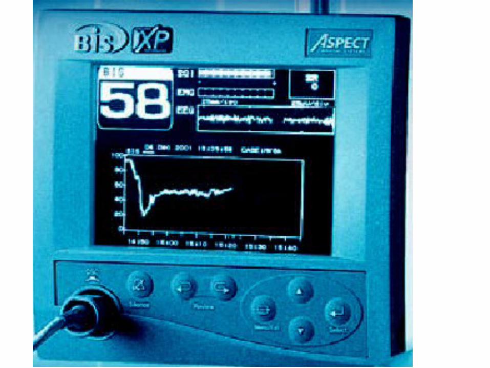
• I once got a turkey sandwich from Aspect, the manufacturer of the BIS monitor
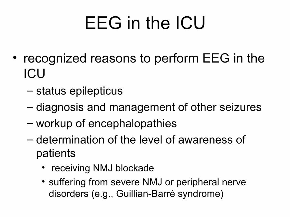
EEG in the ICU
• recognized reasons to perform EEG in the ICU– status epilepticus
– diagnosis and management of other seizures– workup of encephalopathies– determination of the level of awareness of
patients• receiving NMJ blockade• suffering from severe NMJ or peripheral nerve
disorders (e.g., Guillian-Barré syndrome)
Technical aspects of EEG in the ICU
• electrical safety
• electrodes– stability
– infection risk– CT/MR compatibility
• artifact reduction• data management
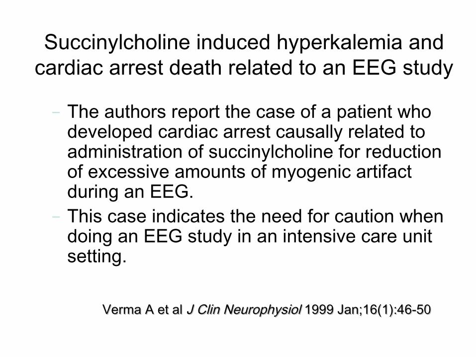
Succinylcholine induced hyperkalemia and cardiac arrest death related to an EEG study
– The authors report the case of a patient who developed cardiac arrest causally related to administration of succinylcholine for reduction of excessive amounts of myogenic artifact during an EEG.
– This case indicates the need for caution when doing an EEG study in an intensive care unit setting.
Verma A et al Verma A et al J Clin NeurophysiolJ Clin Neurophysiol 1999 Jan;16(1):46-50 1999 Jan;16(1):46-50
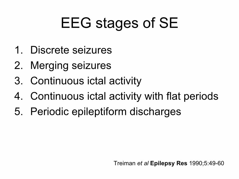
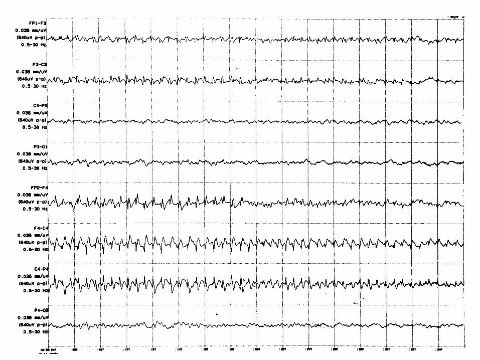
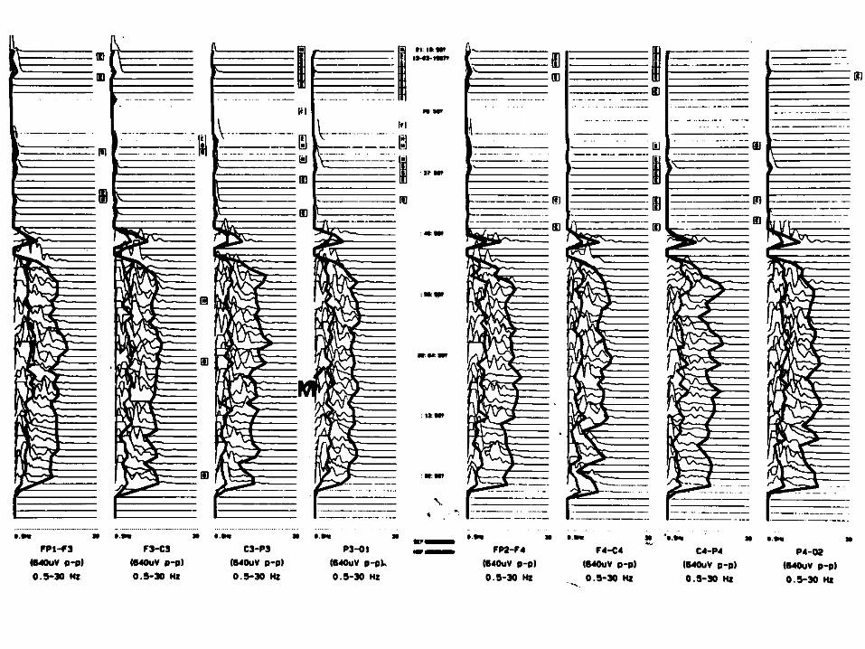
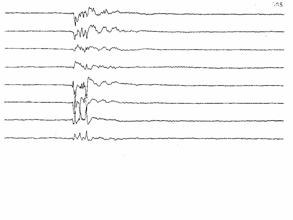
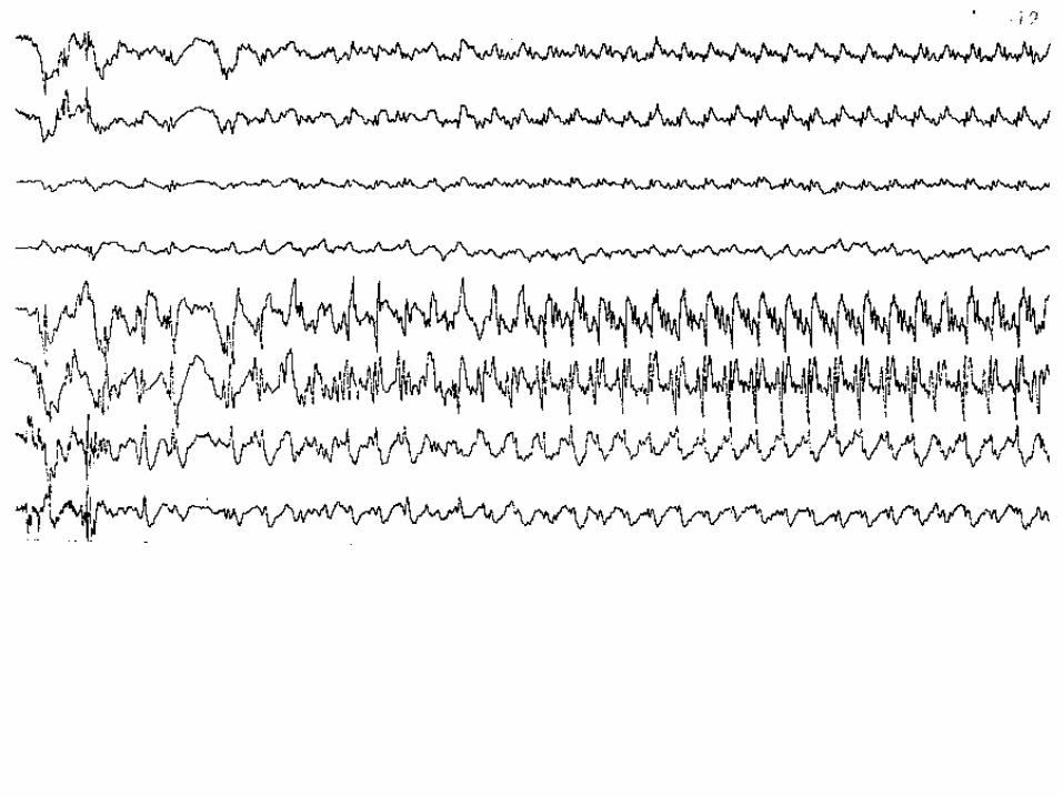
EEG stages of SE
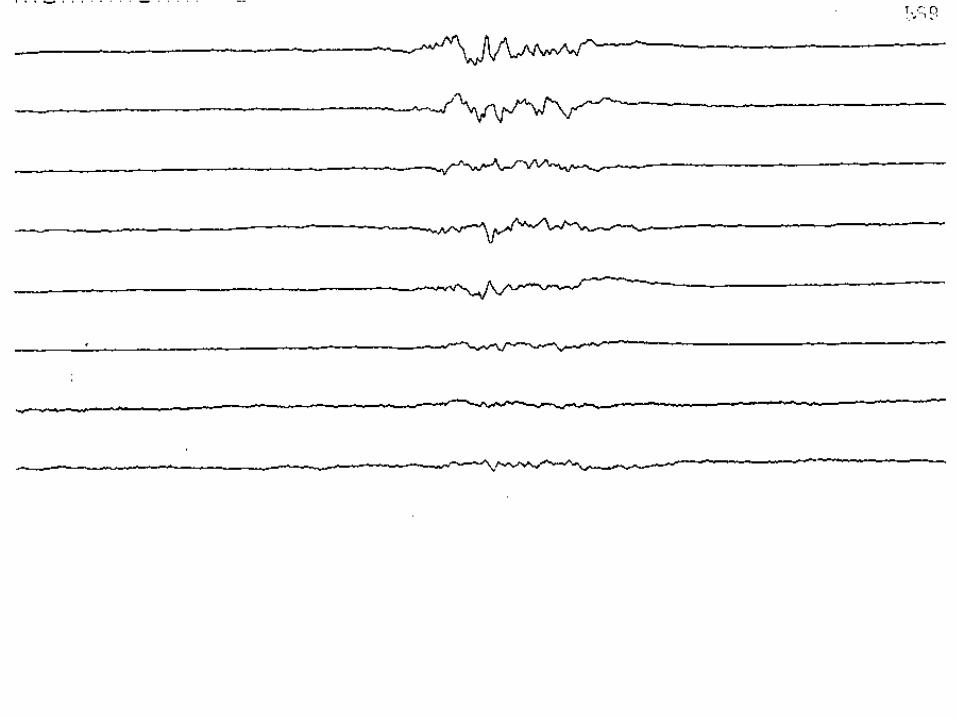
1. Discrete seizures
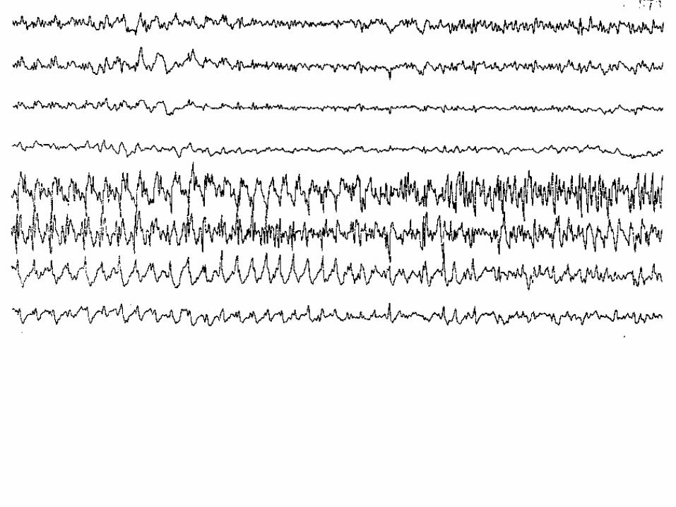
2. Merging seizures
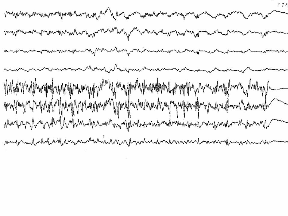
3. Continuous ictal activity
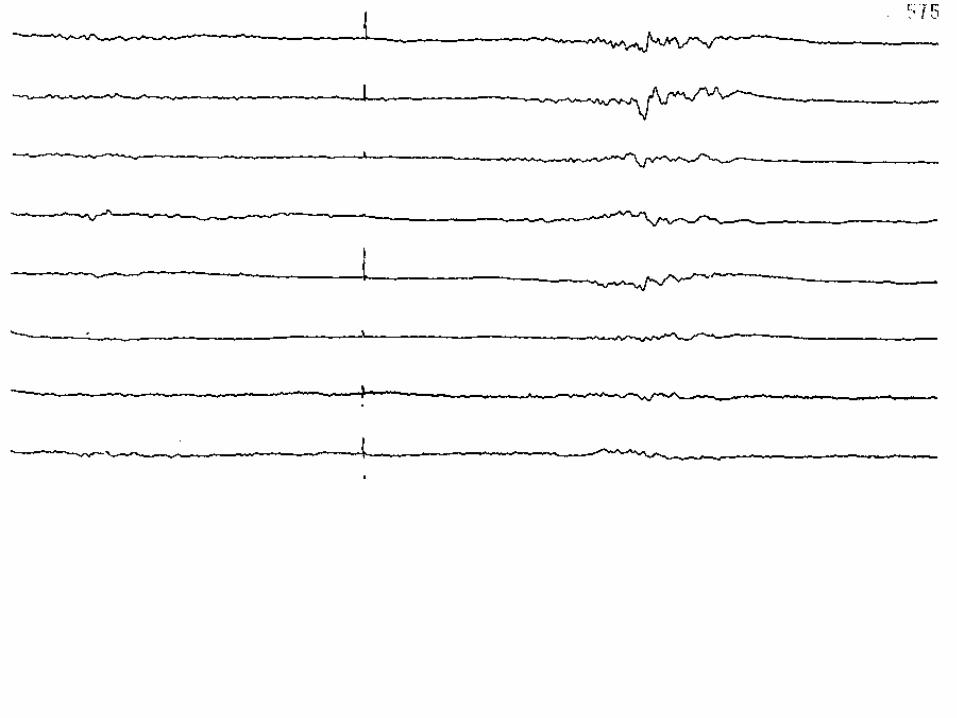
4. Continuous ictal activity with flat periods
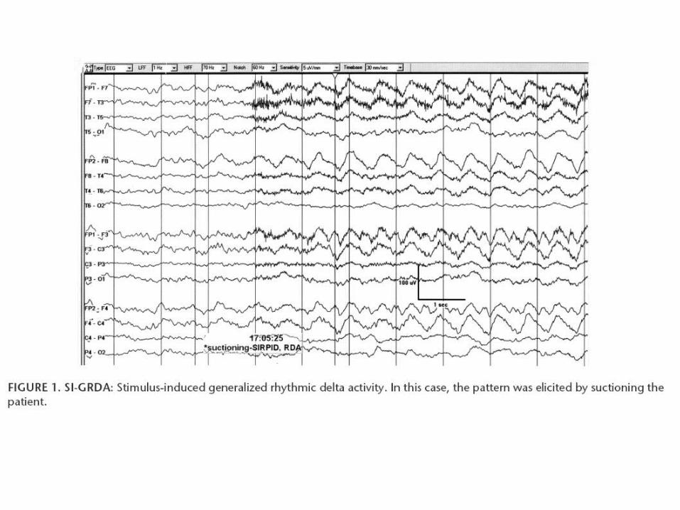
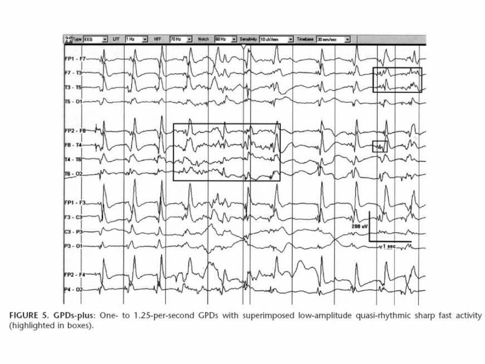
5. Periodic epileptiform discharges
Treiman et al Epilepsy Res 1990;5:49-60
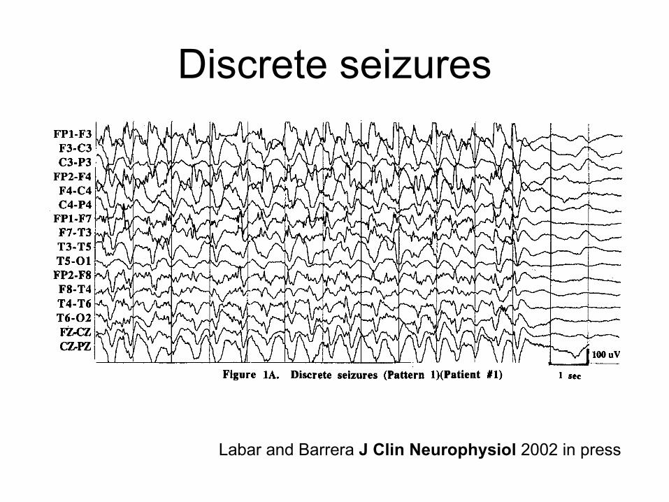
Discrete seizures
Labar and Barrera J Clin Neurophysiol 2002 in press
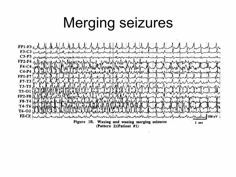
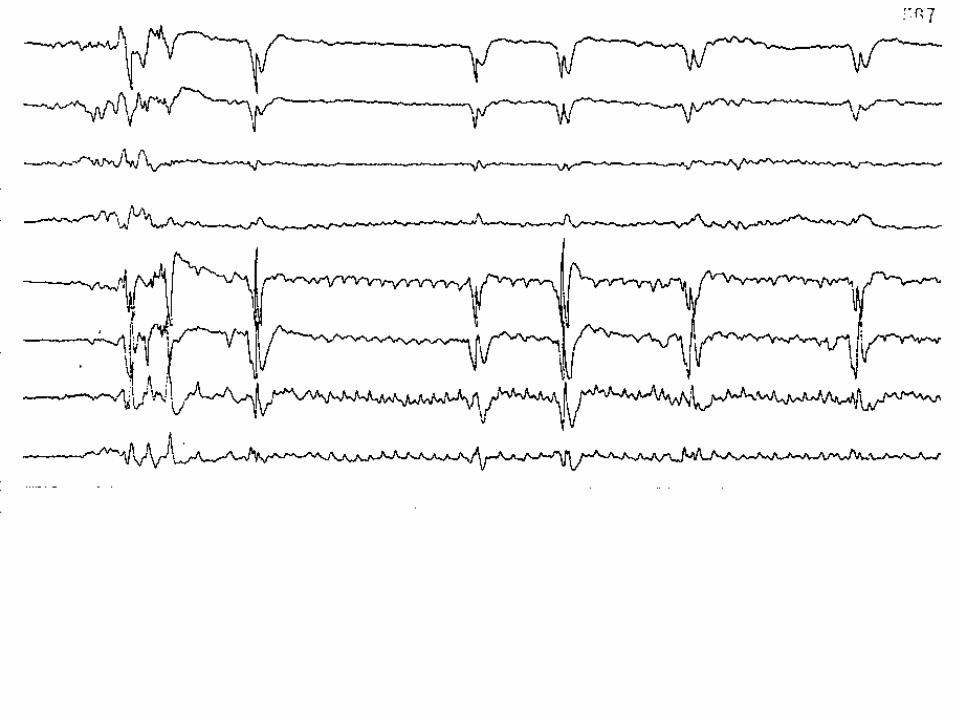
Merging seizures
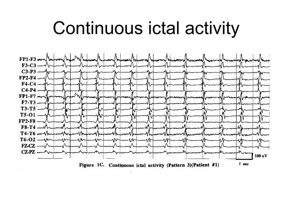
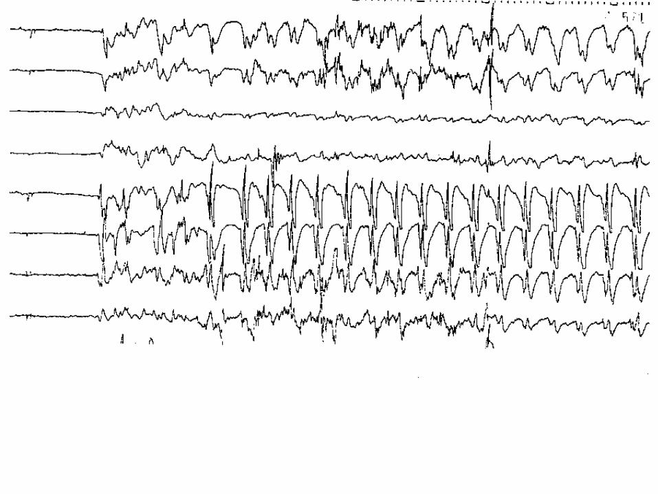
Continuous ictal activity
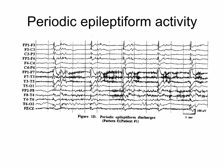
Periodic epileptiform activity

Residual electrographic SE after control of visible SE in VACSP 265
• 130 overt GCSE patients in whom EEG monitoring was begun within 30 minutes of start of treatment
• 26/130 (20%) remained in electrographic SE after motor movements had stopped (twitchless electrical activity)
Faught Epilepsia 1998
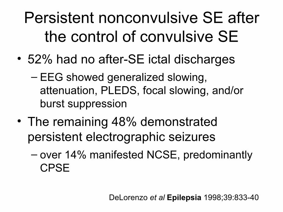
Persistent nonconvulsive SE after the control of convulsive SE
• 52% had no after-SE ictal discharges– EEG showed generalized slowing,
attenuation, PLEDS, focal slowing, and/or burst suppression
• The remaining 48% demonstrated persistent electrographic seizures – over 14% manifested NCSE, predominantly
CPSE
DeLorenzo et al Epilepsia 1998;39:833-40
Unsuspected NCSE
• 8% of a consecutive series of patients referred for EEG because of coma had NCSE
Towne et al Neurology 2000;54:340-5
Burst-suppression myths
• EEG burst-suppression has been demonstrated to be necessary for RSE control
• achieving burst-suppression means that the patient will not have seizures
• the burst-suppression pattern is easily recognized and taught, even for non-neurologists


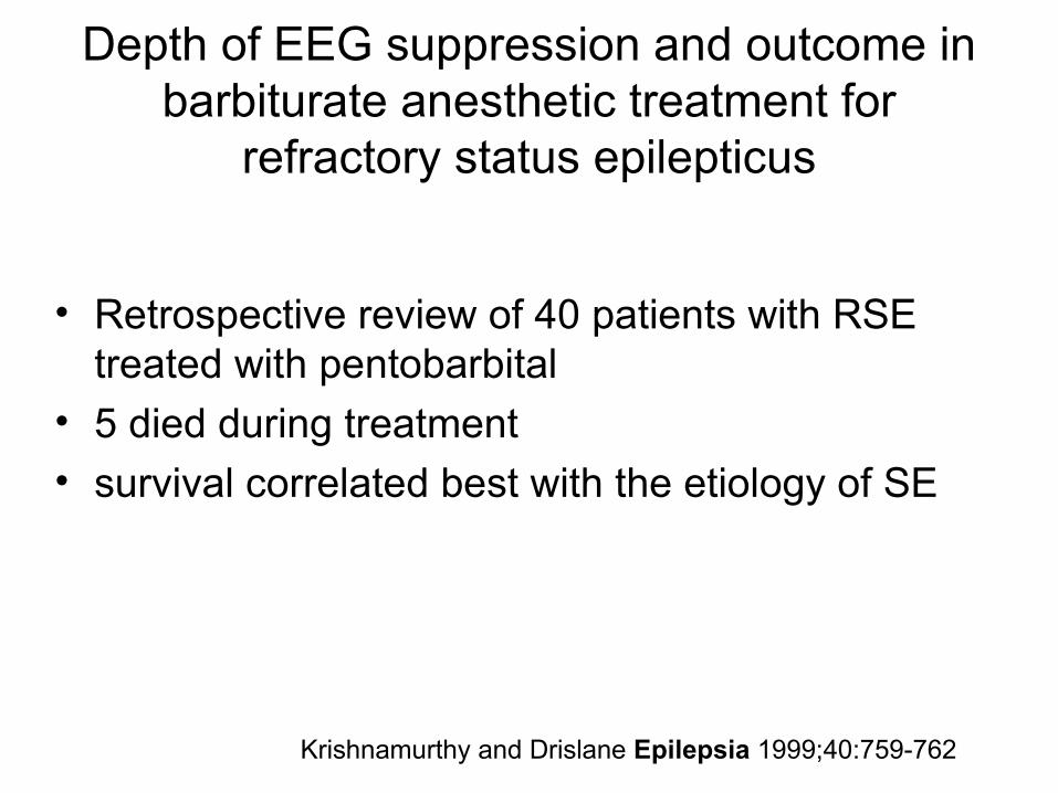
Depth of EEG suppression and outcome in barbiturate anesthetic treatment for
refractory status epilepticus
• Retrospective review of 40 patients with RSE treated with pentobarbital
• 5 died during treatment• survival correlated best with the etiology of SE
Krishnamurthy and Drislane Epilepsia 1999;40:759-762
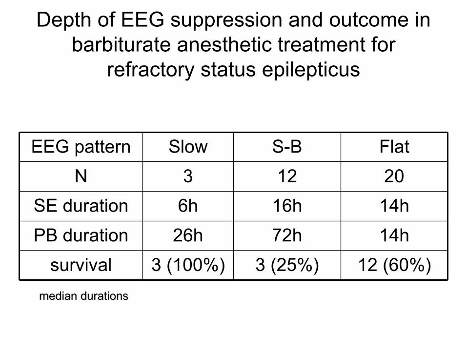
Depth of EEG suppression and outcome in barbiturate anesthetic treatment for
refractory status epilepticus
12 (60%)3 (25%)3 (100%)survival
14h72h26hPB duration
14h16h6hSE duration
20123N
FlatS-BSlowEEG pattern
median durationsmedian durations
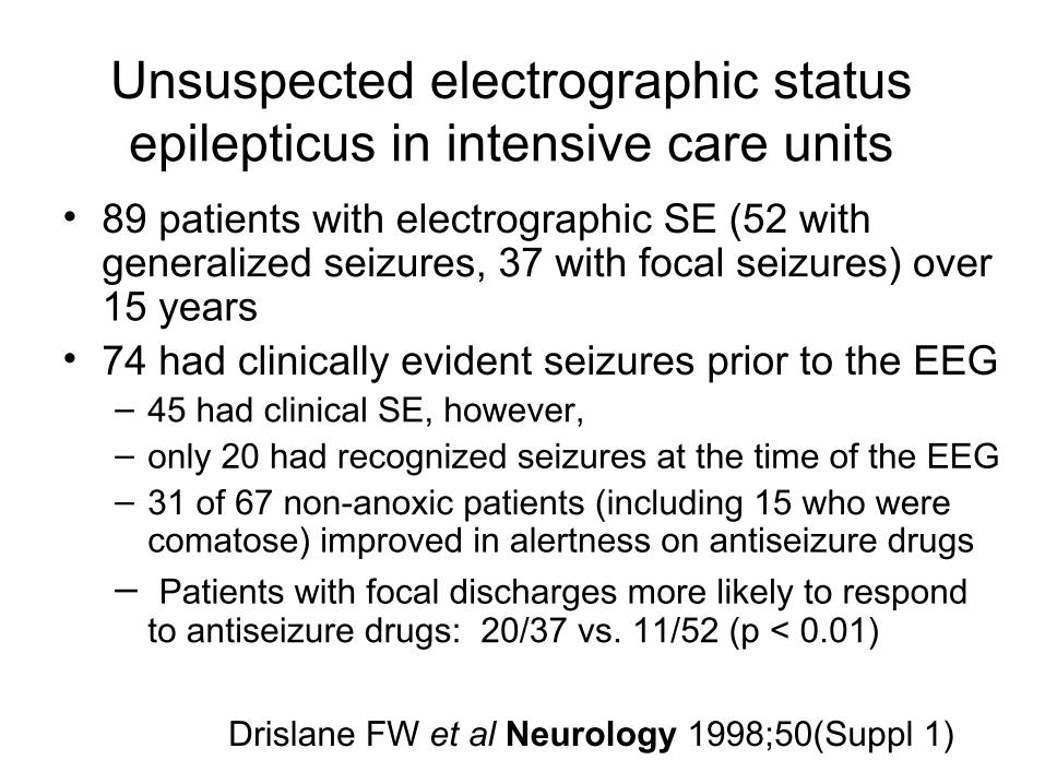
Unsuspected electrographic status epilepticus in intensive care units
• 89 patients with electrographic SE (52 with generalized seizures, 37 with focal seizures) over 15 years
• 74 had clinically evident seizures prior to the EEG – 45 had clinical SE, however,– only 20 had recognized seizures at the time of the EEG– 31 of 67 non-anoxic patients (including 15 who were
comatose) improved in alertness on antiseizure drugs
– Patients with focal discharges more likely to respond to antiseizure drugs: 20/37 vs. 11/52 (p < 0.01)
Drislane FW et al Neurology 1998;50(Suppl 1)
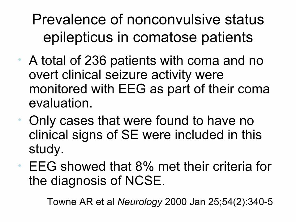
Prevalence of nonconvulsive status epilepticus in comatose patients
• A total of 236 patients with coma and no overt clinical seizure activity were monitored with EEG as part of their coma evaluation.
• Only cases that were found to have no clinical signs of SE were included in this study.
• EEG showed that 8% met their criteria for the diagnosis of NCSE.
Towne AR et al Neurology 2000 Jan 25;54(2):340-5
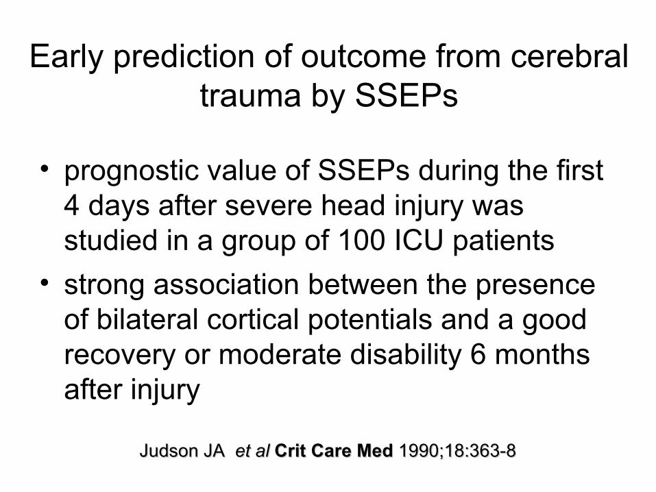
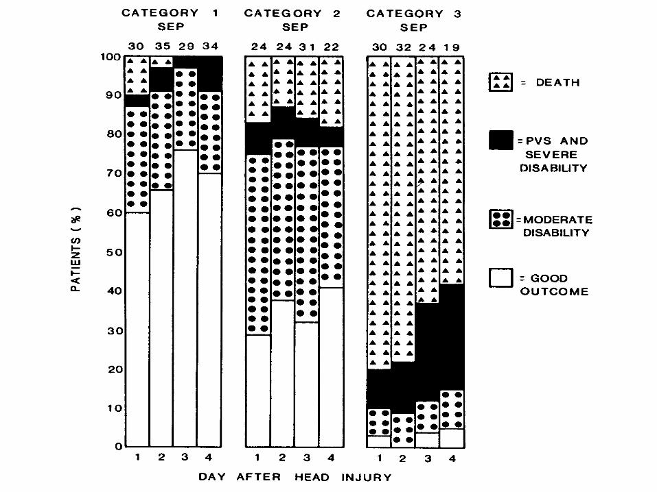
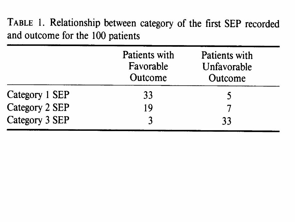
Early prediction of outcome from cerebral trauma by SSEPs
• prognostic value of SSEPs during the first 4 days after severe head injury was studied in a group of 100 ICU patients
• strong association between the presence of bilateral cortical potentials and a good recovery or moderate disability 6 months after injury
Judson JA Judson JA et al et al Crit Care MedCrit Care Med 1990;18:363-8 1990;18:363-8
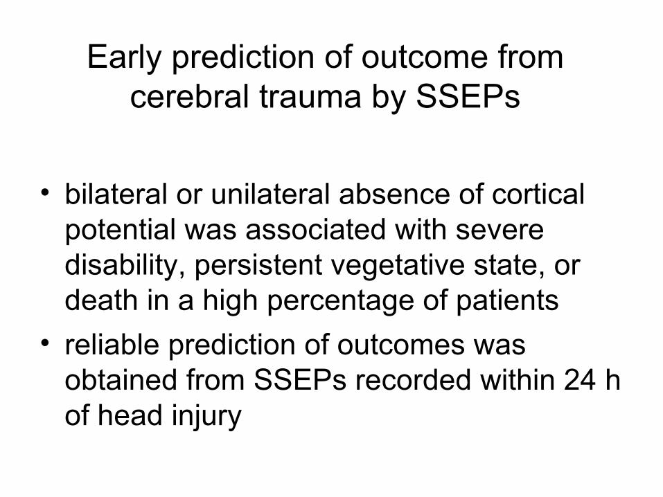
Early prediction of outcome from cerebral trauma by SSEPs
• bilateral or unilateral absence of cortical potential was associated with severe disability, persistent vegetative state, or death in a high percentage of patients
• reliable prediction of outcomes was obtained from SSEPs recorded within 24 h of head injury
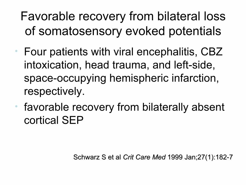
Favorable recovery from bilateral loss of somatosensory evoked potentials
• Four patients with viral encephalitis, CBZ intoxication, head trauma, and left-side, space-occupying hemispheric infarction, respectively.
• favorable recovery from bilaterally absent cortical SEP
Schwarz S et al Schwarz S et al Crit Care MedCrit Care Med 1999 Jan;27(1):182-7 1999 Jan;27(1):182-7
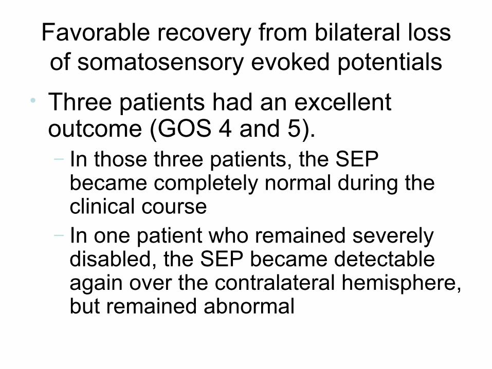
Favorable recovery from bilateral loss of somatosensory evoked potentials
• Three patients had an excellent outcome (GOS 4 and 5). – In those three patients, the SEP
became completely normal during the clinical course
– In one patient who remained severely disabled, the SEP became detectable again over the contralateral hemisphere, but remained abnormal
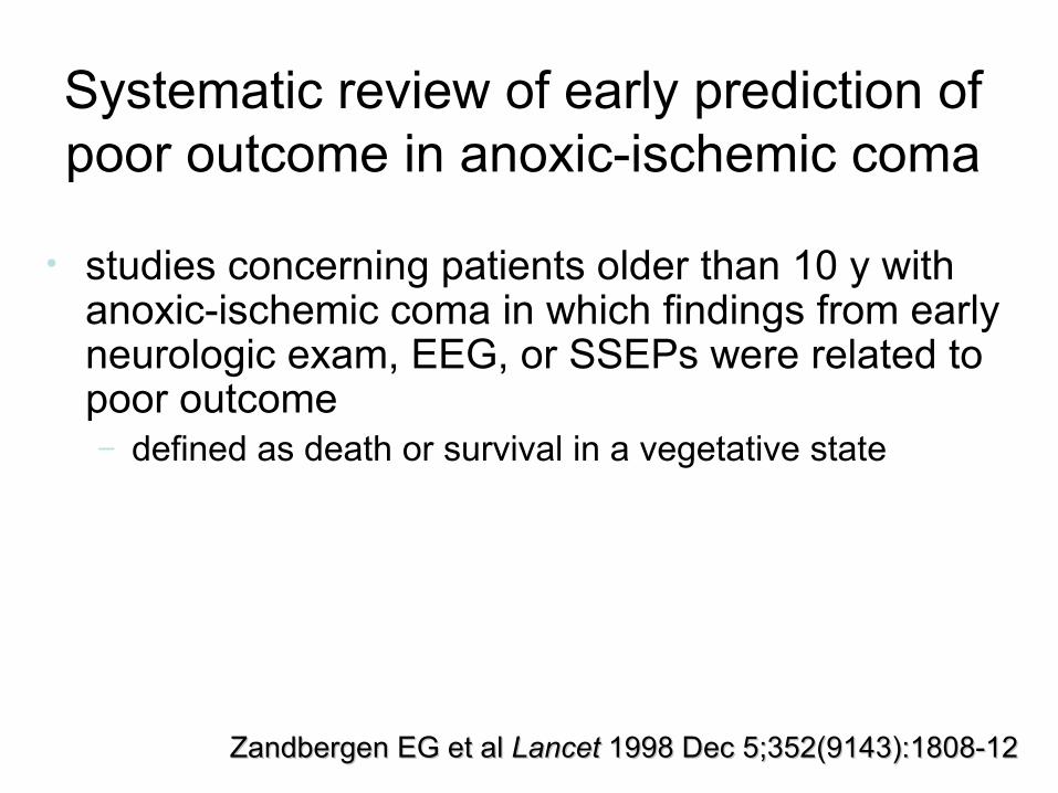
Systematic review of early prediction of poor outcome in anoxic-ischemic coma
• studies concerning patients older than 10 y with anoxic-ischemic coma in which findings from early neurologic exam, EEG, or SSEPs were related to poor outcome– defined as death or survival in a vegetative state
Zandbergen EG et al Zandbergen EG et al LancetLancet 1998 Dec 5;352(9143):1808-12 1998 Dec 5;352(9143):1808-12
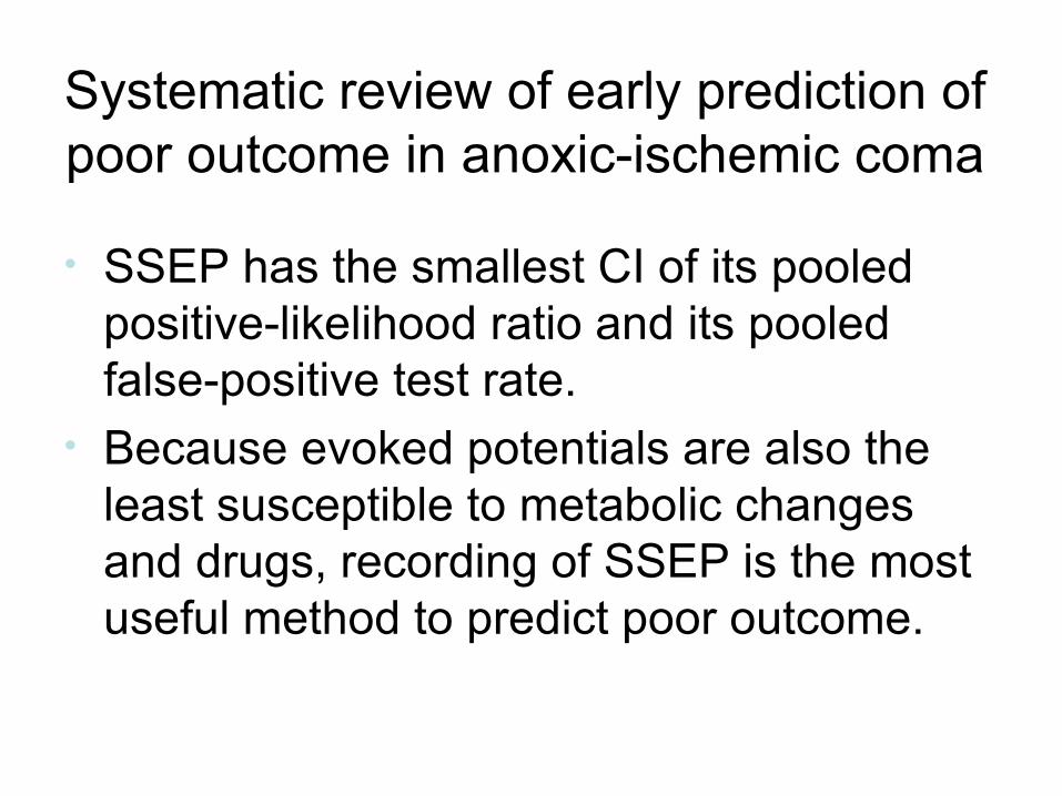
Systematic review of early prediction of poor outcome in anoxic-ischemic coma
• SSEP has the smallest CI of its pooled positive-likelihood ratio and its pooled false-positive test rate.
• Because evoked potentials are also the least susceptible to metabolic changes and drugs, recording of SSEP is the most useful method to predict poor outcome.
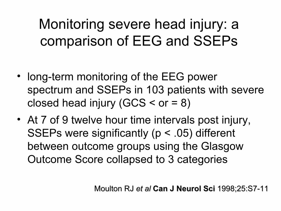
Monitoring severe head injury: a comparison of EEG and SSEPs
• long-term monitoring of the EEG power spectrum and SSEPs in 103 patients with severe closed head injury (GCS < or = 8)
• At 7 of 9 twelve hour time intervals post injury, SSEPs were significantly (p < .05) different between outcome groups using the Glasgow Outcome Score collapsed to 3 categories
Moulton RJ Moulton RJ et alet al Can J Neurol SciCan J Neurol Sci 1998;25:S7-11 1998;25:S7-11
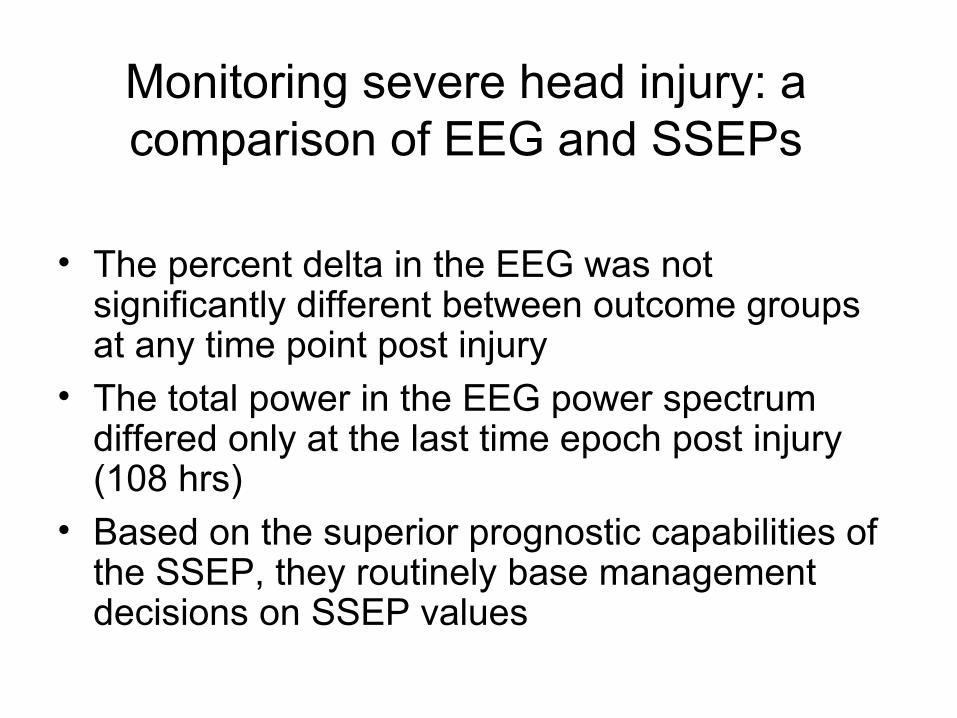
Monitoring severe head injury: a comparison of EEG and SSEPs
• The percent delta in the EEG was not significantly different between outcome groups at any time point post injury
• The total power in the EEG power spectrum differed only at the last time epoch post injury (108 hrs)
• Based on the superior prognostic capabilities of the SSEP, they routinely base management decisions on SSEP values
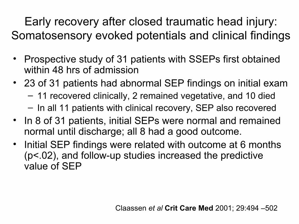
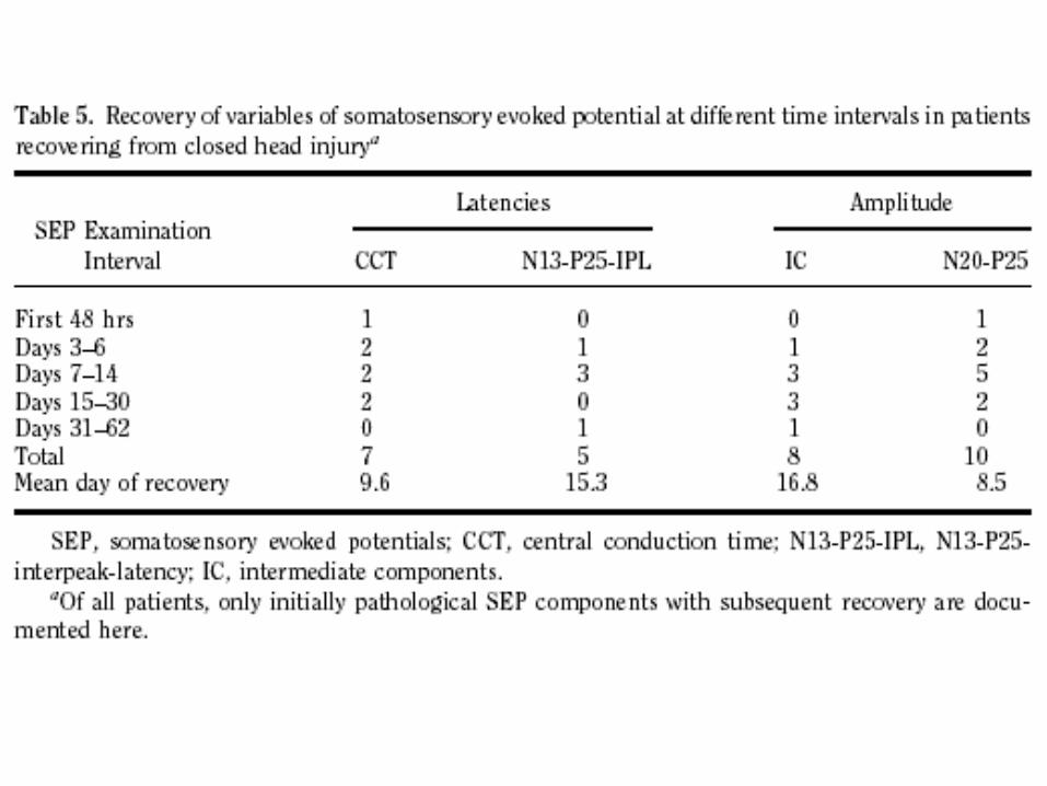
Early recovery after closed traumatic head injury: Somatosensory evoked potentials and clinical findings
• Prospective study of 31 patients with SSEPs first obtained within 48 hrs of admission
• 23 of 31 patients had abnormal SEP findings on initial exam– 11 recovered clinically, 2 remained vegetative, and 10 died– In all 11 patients with clinical recovery, SEP also recovered
• In 8 of 31 patients, initial SEPs were normal and remained normal until discharge; all 8 had a good outcome.
• Initial SEP findings were related with outcome at 6 months (p<.02), and follow-up studies increased the predictive value of SEP
Claassen et al Crit Care Med 2001; 29:494 –502
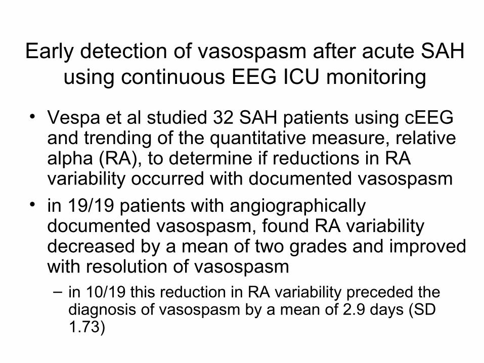
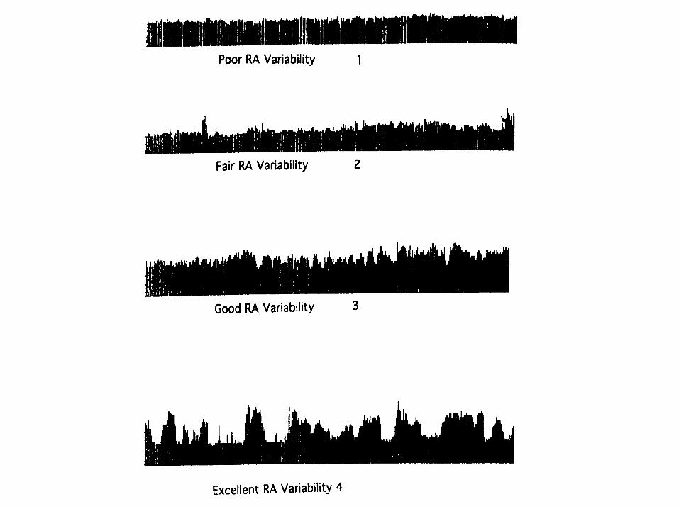
Early detection of vasospasm after acute SAH using continuous EEG ICU monitoring
• Vespa et al studied 32 SAH patients using cEEG and trending of the quantitative measure, relative alpha (RA), to determine if reductions in RA variability occurred with documented vasospasm
• in 19/19 patients with angiographically documented vasospasm, found RA variability decreased by a mean of two grades and improved with resolution of vasospasm– in 10/19 this reduction in RA variability preceded the
diagnosis of vasospasm by a mean of 2.9 days (SD 1.73)
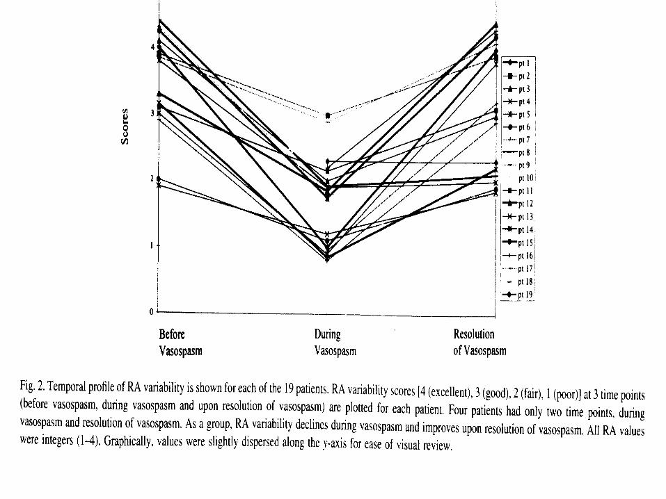
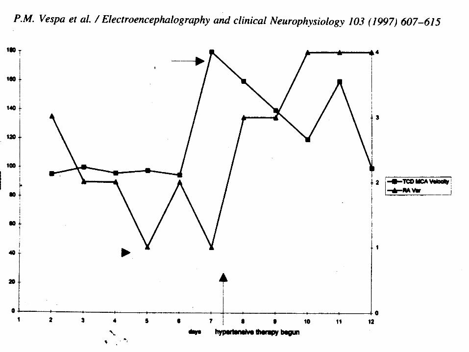
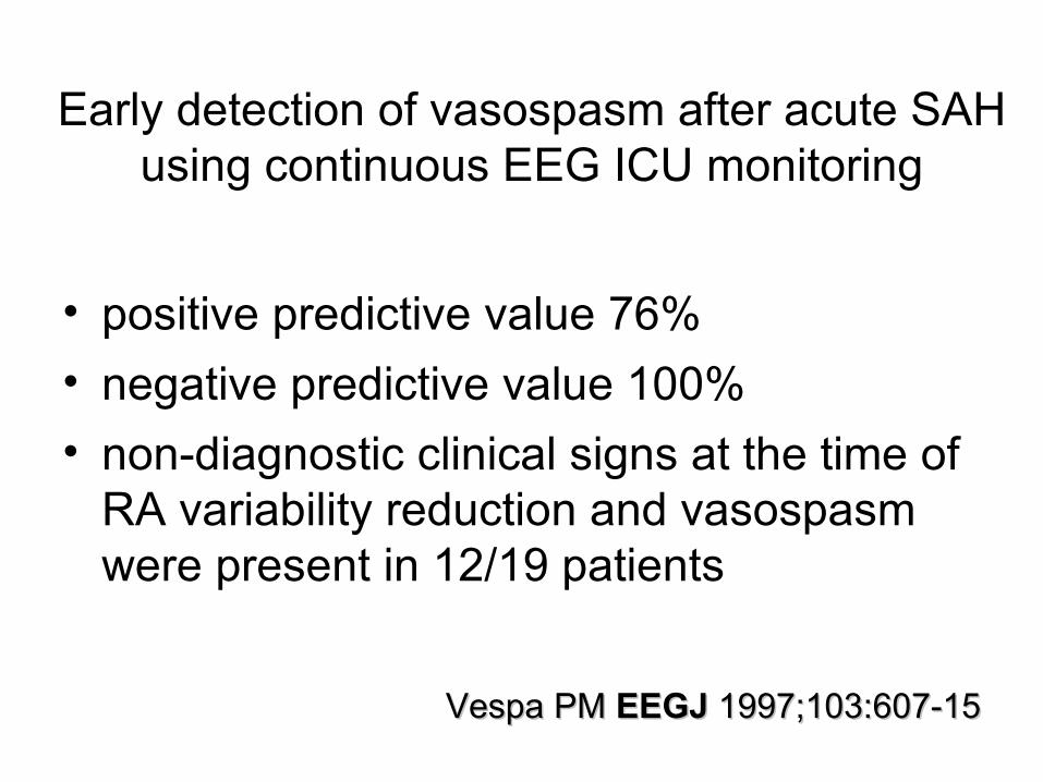
Early detection of vasospasm after acute SAH using continuous EEG ICU monitoring
• positive predictive value 76%
• negative predictive value 100%
• non-diagnostic clinical signs at the time of RA variability reduction and vasospasm were present in 12/19 patients
Vespa PM Vespa PM EEGJEEGJ 1997;103:607-15 1997;103:607-15
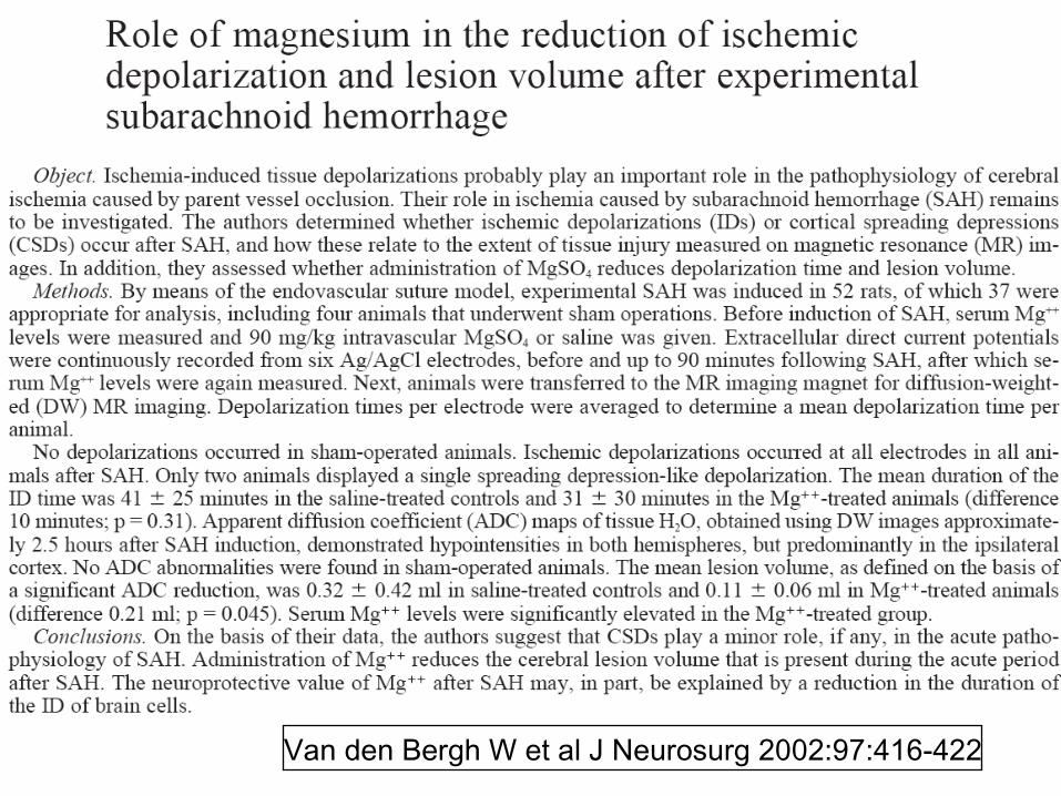
Van den Bergh W et al J Neurosurg 2002:97:416-422
J N
eiro
surg
199
9;91
:750
-760
J N
eiro
surg
199
9;91
:750
-760
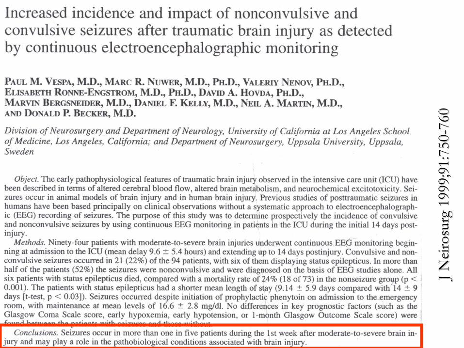
Increased incidence and impact of nonconvulsive and convulsive seizures after traumatic brain injury as detected by
continuous EEG monitoring
• Purpose: to determine prospectively the incidence of convulsive and nonconvulsive seizures using continuous EEG monitoring in patients in the ICU during the initial 14 days post-injury.
• 94 patients with moderate-to-severe brain injuries underwent continuous EEG monitoring beginning at admission to the ICU (mean delay 9.6+/-5.4 hours) and extending up to 14 days postinjury.
Vespa PM et al Vespa PM et al J NeurosurgJ Neurosurg 1999 Nov;91(5):750-60 1999 Nov;91(5):750-60
Increased incidence and impact of nonconvulsive and convulsive seizures after traumatic brain injury as detected by
continuous EEG monitoring
• Convulsive and nonconvulsive seizures occurred in 21 (22%) of the 94 patients, with six of them displaying SE
• In more than half of the patients (52%) the seizures were nonconvulsive and were diagnosed on the basis of EEG studies alone.
Increased incidence and impact of nonconvulsive and convulsive seizures after traumatic brain injury as detected by continuous
EEG monitoring
• All six patients with SE died, compared with a mortality rate of 24% (18 of 73) in the nonseizure group (p<0.001).
• The patients with SE had a shorter mean length of stay – 9.14±5.9 d compared with 14±9 d, p<0.031.
• Seizures occurred despite initiation of prophylactic PHT on admission to the ED, with maintenance at mean levels of 16.6±2.8 mg/dl.
Increased incidence and impact of nonconvulsive and convulsive seizures after traumatic brain injury as detected by continuous
EEG monitoring
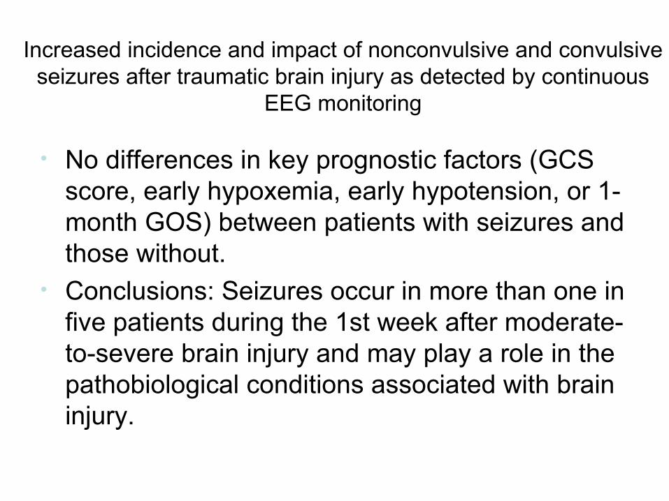
• No differences in key prognostic factors (GCS score, early hypoxemia, early hypotension, or 1-month GOS) between patients with seizures and those without.
• Conclusions: Seizures occur in more than one in five patients during the 1st week after moderate-to-severe brain injury and may play a role in the pathobiological conditions associated with brain injury.
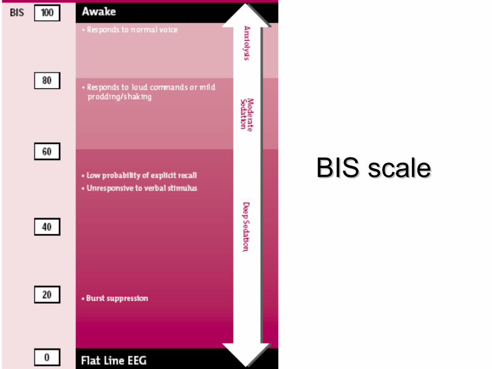
BIS scaleBIS scale
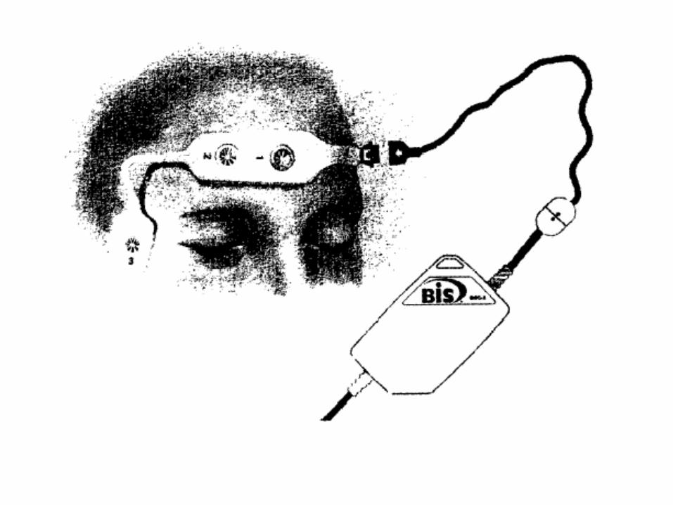
Detection of brain death onset using the bispectral index in severely comatose patients
• (this is a bad idea)
Vivien et al Intensive Care Med 2002;28:419–425
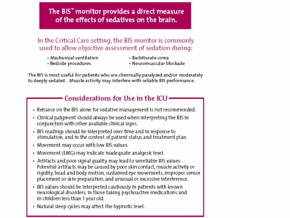
Is the bispectral index appropriate for monitoring the sedation level of mechanically ventilated SICU patients?
• Design and setting: Prospective convenience sample in a 12-bed anesthesiological-surgical ICU of a university hospital.
• Patients: 19 consecutive patients without any central neurological diseases requiring mechanical ventilation for more than 24 h.
Frenzel Frenzel et alet al Intensive Care Med Intensive Care Med 2002; 28:178–1832002; 28:178–183
Is the bispectral index appropriate for monitoring the sedation level of mechanically ventilated SICU patients?
• Measurements: BIS version 3.12 and clinical depth of sedation assessed by the– modified Observers’s Assessment of Alertness/
Sedation Scale, – modified Glasgow Coma Scale, – modified Ramsay Scale, – Cook Scale, and – Sedation-Agitation Scale
• Measured twice daily while patients were intubated and once daily after extubation until discharged from ICU.
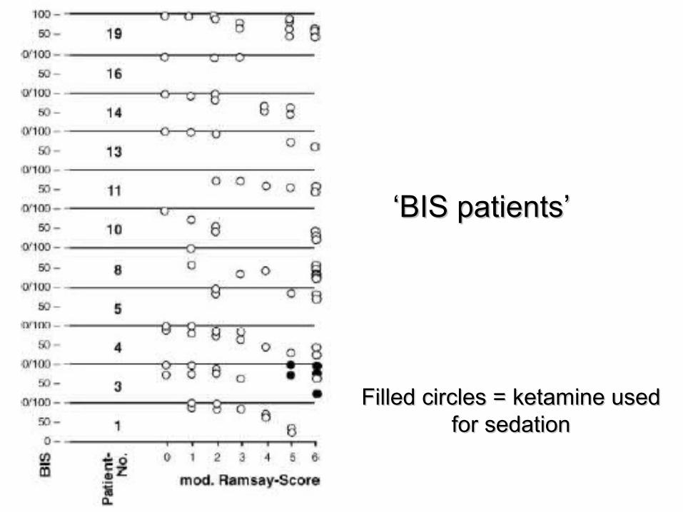
‘‘BIS patients’BIS patients’
Filled circles = ketamine usedFilled circles = ketamine usedfor sedationfor sedation
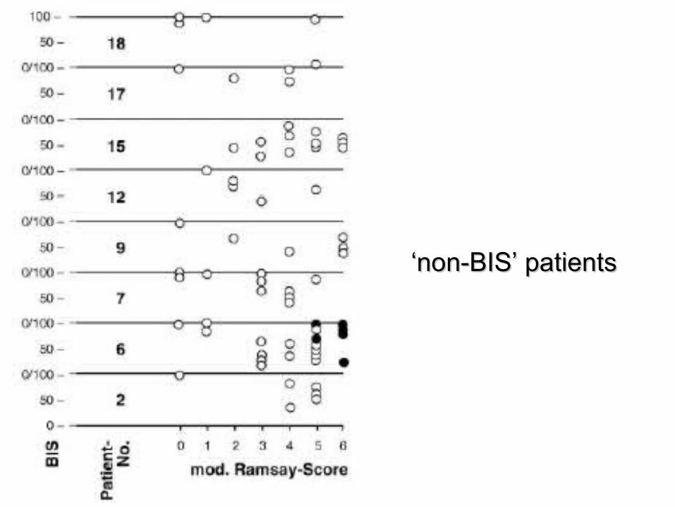
‘‘non-BIS’ patientsnon-BIS’ patients
Is the bispectral index appropriate for monitoring the sedation level of mechanically ventilated SICU patients?
• There was a moderate correlation between BIS and each sedation score in 11 patients (58%, “BIS patients”) and no correlation in 8 patients (42%, “non-BIS patients”).
• Found no parameters distinguishing between these two groups.
• On average eight measurements were necessary to establish a statistical correlation.
Is the bispectral index appropriate for monitoring the sedation level of mechanically ventilated SICU patients?
• Conclusions: BIS is correlated only in some ICU patients with the clinical assessment of their sedation level as based on various scores.
• At deeper sedation levels the interindividual differences increase.
• There were no criteria found to distinguish patients with and without correlation.
• This suggests that the BIS is not suitable for monitoring the sedation in a heterogeneous group of surgical ICU patients.
Sedation modulates recognition of novel stimuli and adaptation to regular stimuli in critically ill adults
• Studied 22 endotracheally intubated, mechanically ventilated patients chemically sedated with narcotics and a benzodiazepine (n=12), or narcotics and propofol (n=10) using sedation protocols.
• Evaluated the level of sedation using an automated auditory or visual P300 device.
Clifford and Buchman Crit Care Med 2002 30:609-616
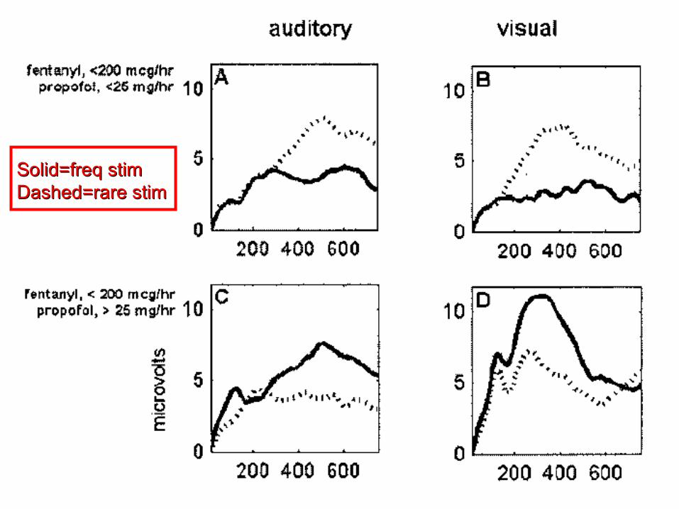
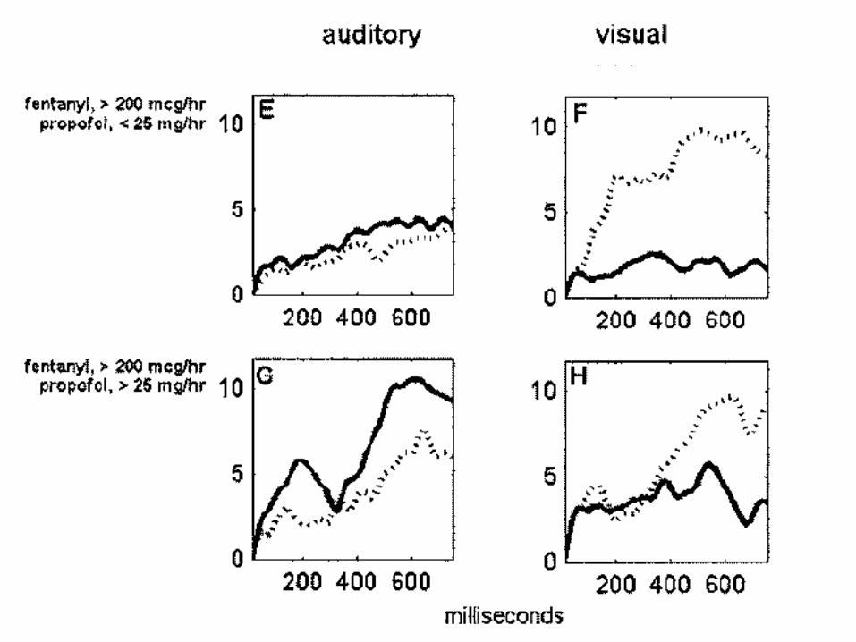
Solid=freq stimSolid=freq stimDashed=rare stimDashed=rare stim
Continuous measurement of cerebral blood flow velocity using transcranial Doppler reveals significant moment-to-moment variability of data in healthy volunteers and in patients with subarachnoid hemorrhage
Venkatesh B et al Crit Care Med 2002; 30:563–569
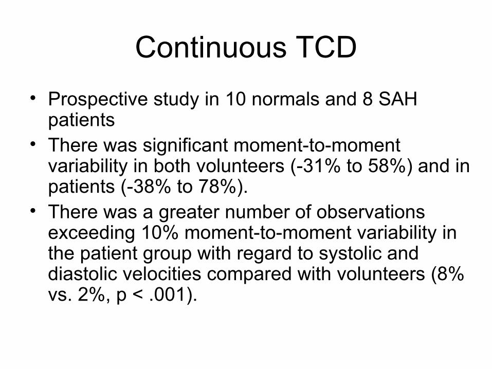
Continuous TCD
• Prospective study in 10 normals and 8 SAH patients
• There was significant moment-to-moment variability in both volunteers (-31% to 58%) and in patients (-38% to 78%).
• There was a greater number of observations exceeding 10% moment-to-moment variability in the patient group with regard to systolic and diastolic velocities compared with volunteers (8% vs. 2%, p < .001).
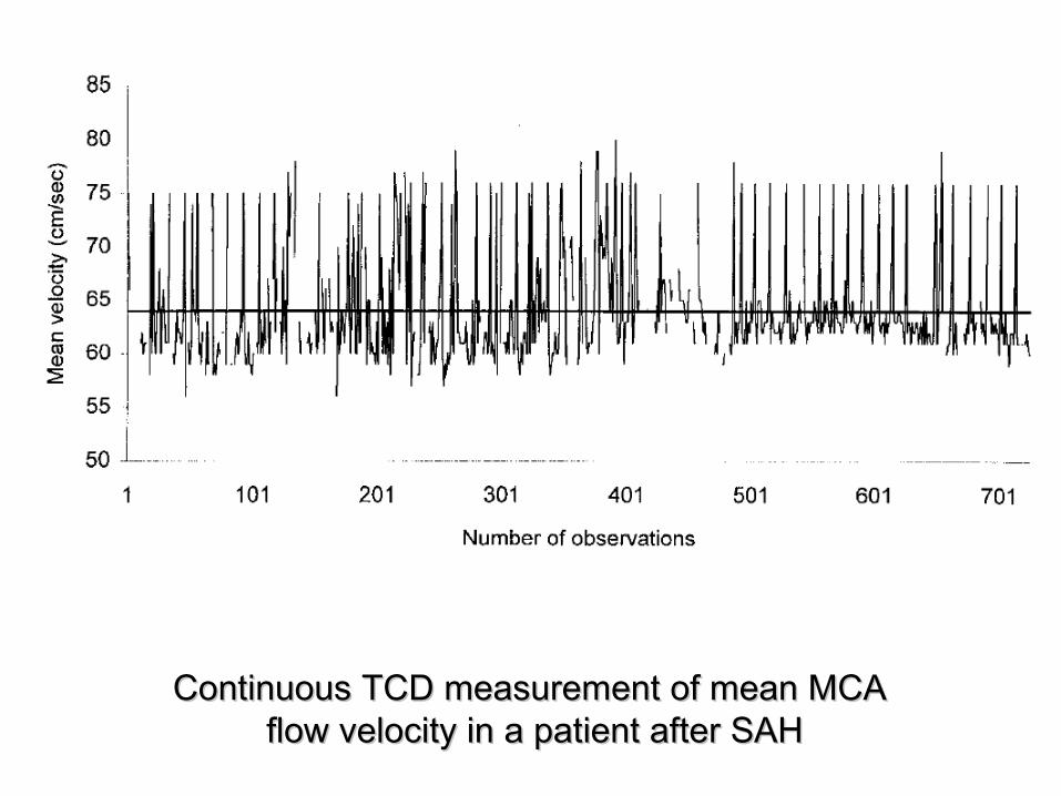
Continuous TCD measurement of mean MCA Continuous TCD measurement of mean MCA flow velocity in a patient after SAHflow velocity in a patient after SAH
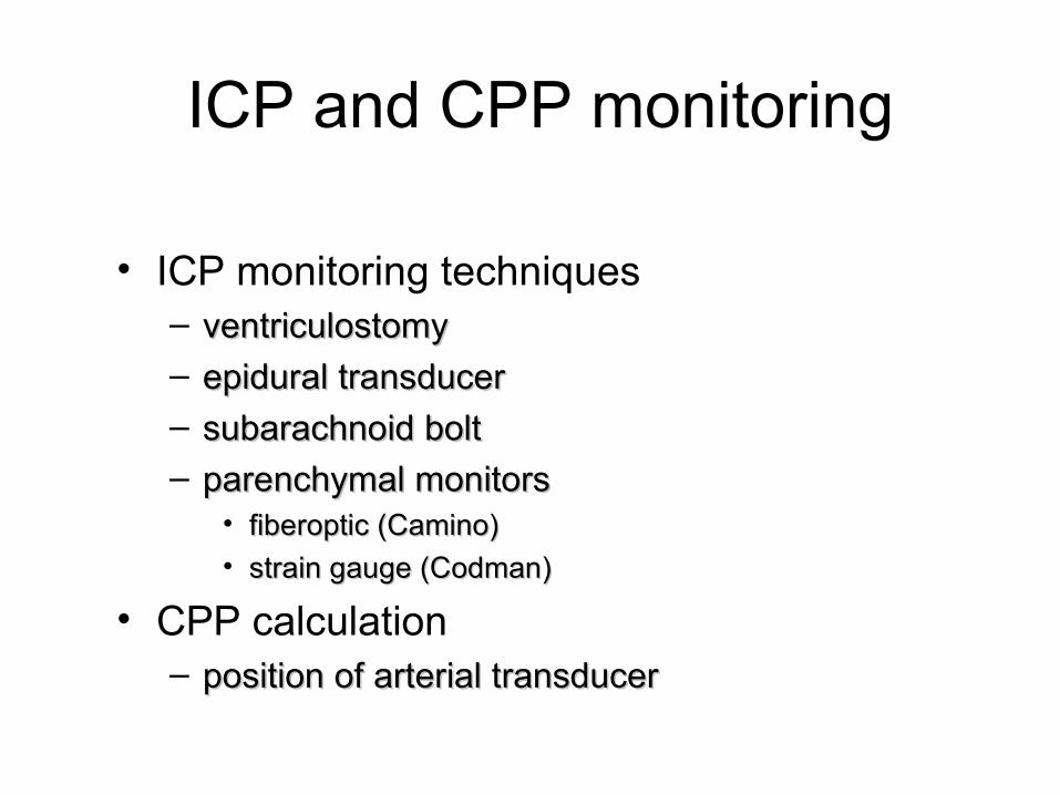
ICP and CPP monitoring
• ICP monitoring techniques– ventriculostomyventriculostomy– epidural transducerepidural transducer– subarachnoid boltsubarachnoid bolt– parenchymal monitorsparenchymal monitors
• fiberoptic (Camino)fiberoptic (Camino)• strain gauge (Codman)strain gauge (Codman)
• CPP calculation– position of arterial transducerposition of arterial transducer
Cerebral perfusion pressure
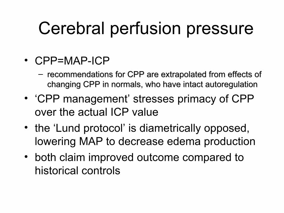
• CPP=MAP-ICP– recommendations for CPP are extrapolated from effects of recommendations for CPP are extrapolated from effects of
changing CPP in normals, who have intact autoregulationchanging CPP in normals, who have intact autoregulation
• ‘CPP management’ stresses primacy of CPP over the actual ICP value
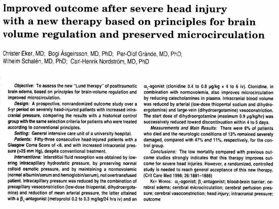
• the ‘Lund protocol’ is diametrically opposed, lowering MAP to decrease edema production
• both claim improved outcome compared to historical controls
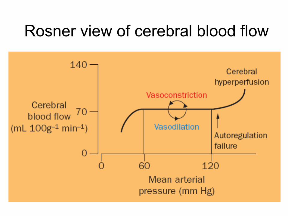
Rosner view of cerebral blood flow
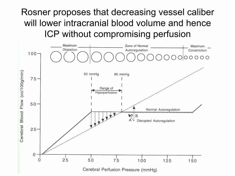
Rosner proposes that decreasing vessel caliber will lower intracranial blood volume and hence
ICP without compromising perfusion
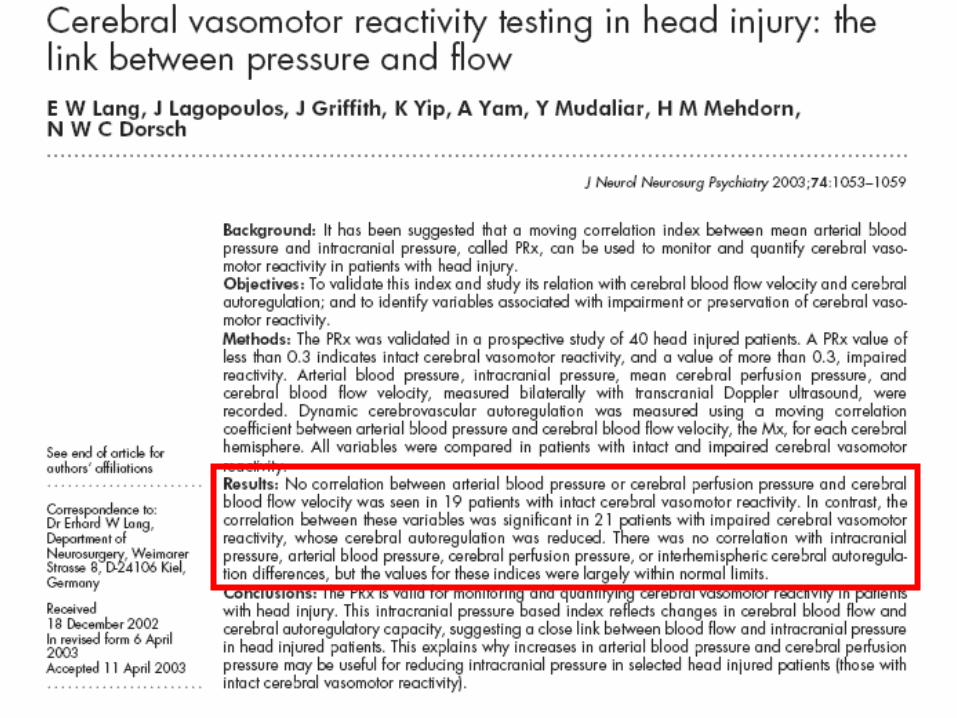
Intact autoregulation
Lang et al JNNP 2003;74:1053-1059
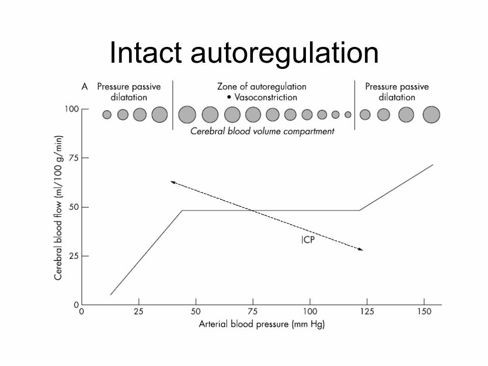
Example of intact autoregulation
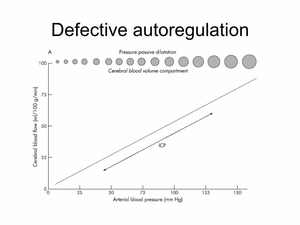
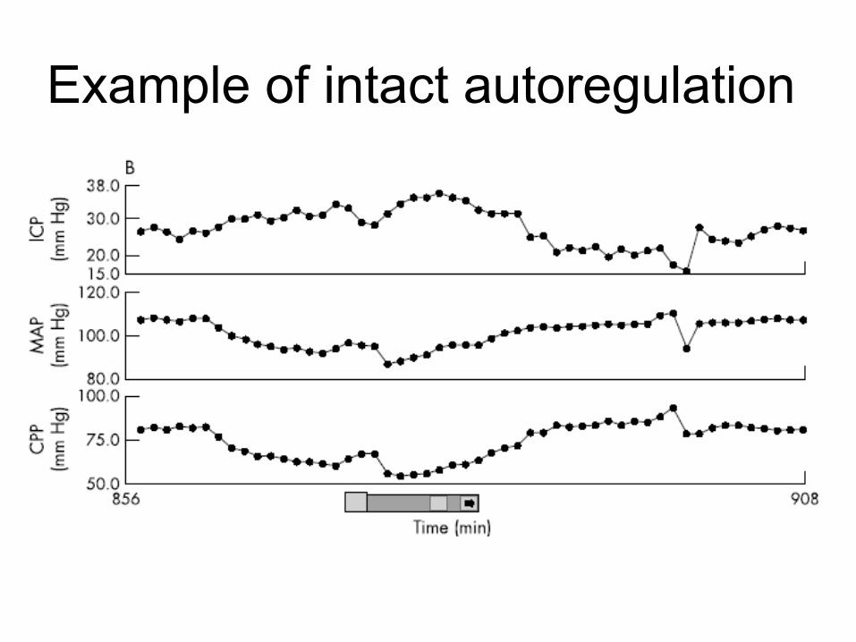
Defective autoregulation
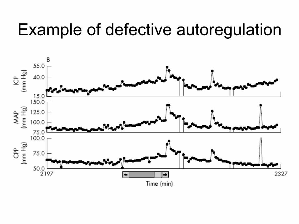
Example of defective autoregulation
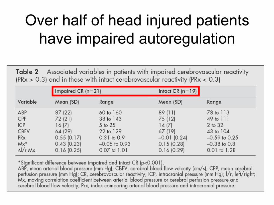
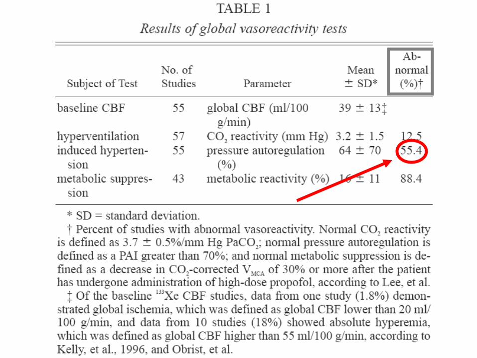
Over half of head injured patients have impaired autoregulation
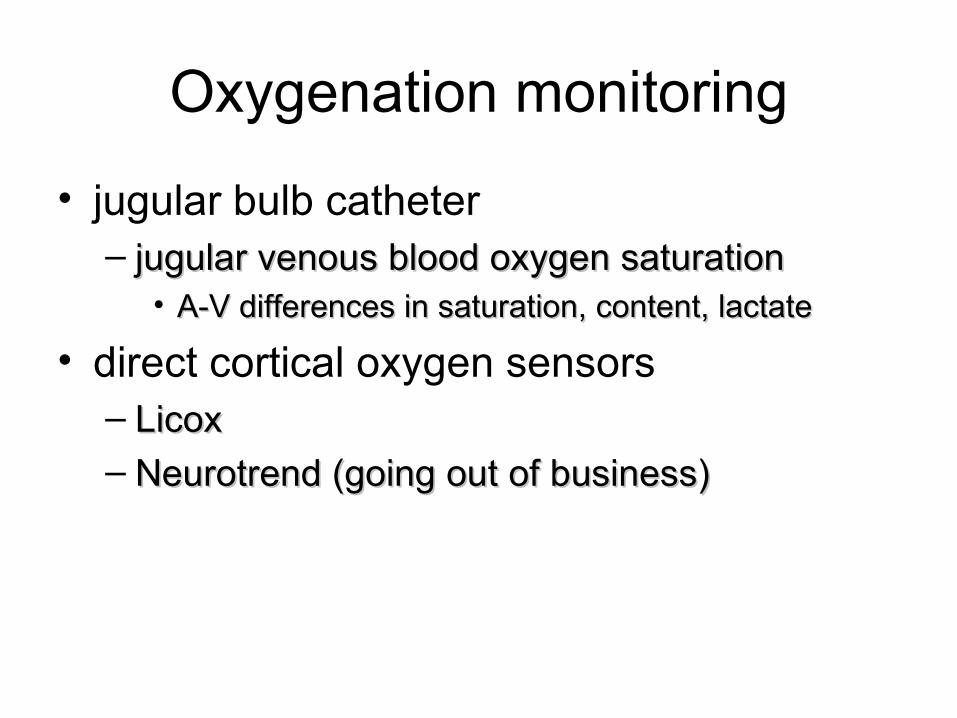
Oxygenation monitoring
• jugular bulb catheter– jugular venous blood oxygen saturationjugular venous blood oxygen saturation
• A-V differences in saturation, content, lactateA-V differences in saturation, content, lactate
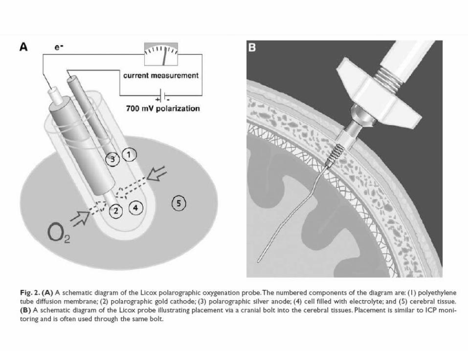
• direct cortical oxygen sensors – LicoxLicox– Neurotrend (going out of business)Neurotrend (going out of business)
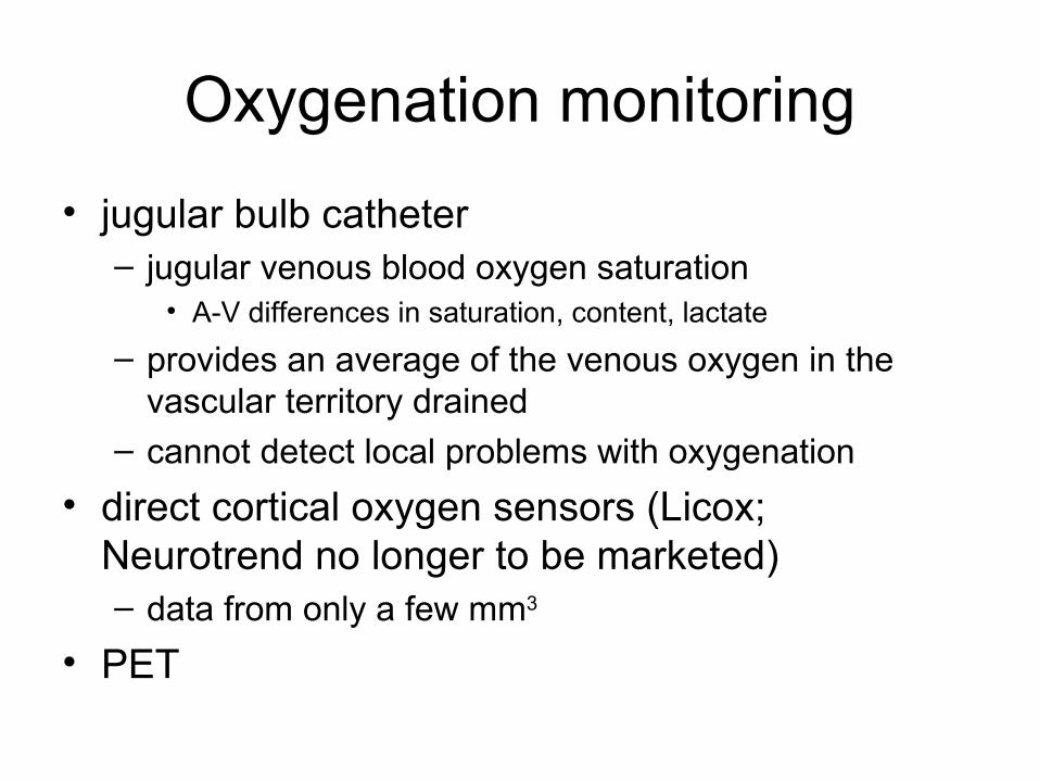
Oxygenation monitoring
• jugular bulb catheter– jugular venous blood oxygen saturation
• A-V differences in saturation, content, lactate
– provides an average of the venous oxygen in the vascular territory drained
– cannot detect local problems with oxygenation
• direct cortical oxygen sensors (Licox; Neurotrend no longer to be marketed)– data from only a few mm3
• PET
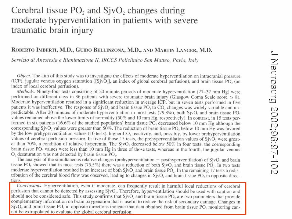
J Neurosurg
2002;96:97-10
2J N
eurosurg 2002
;96:97-102

Moderate hyperventilation lowered ICP and improved CPP…
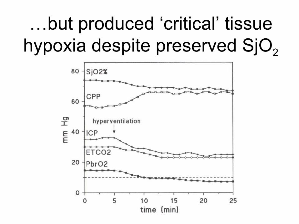
…but produced ‘critical’ tissue hypoxia despite preserved SjO2
J Neu
rosurg 2002;96
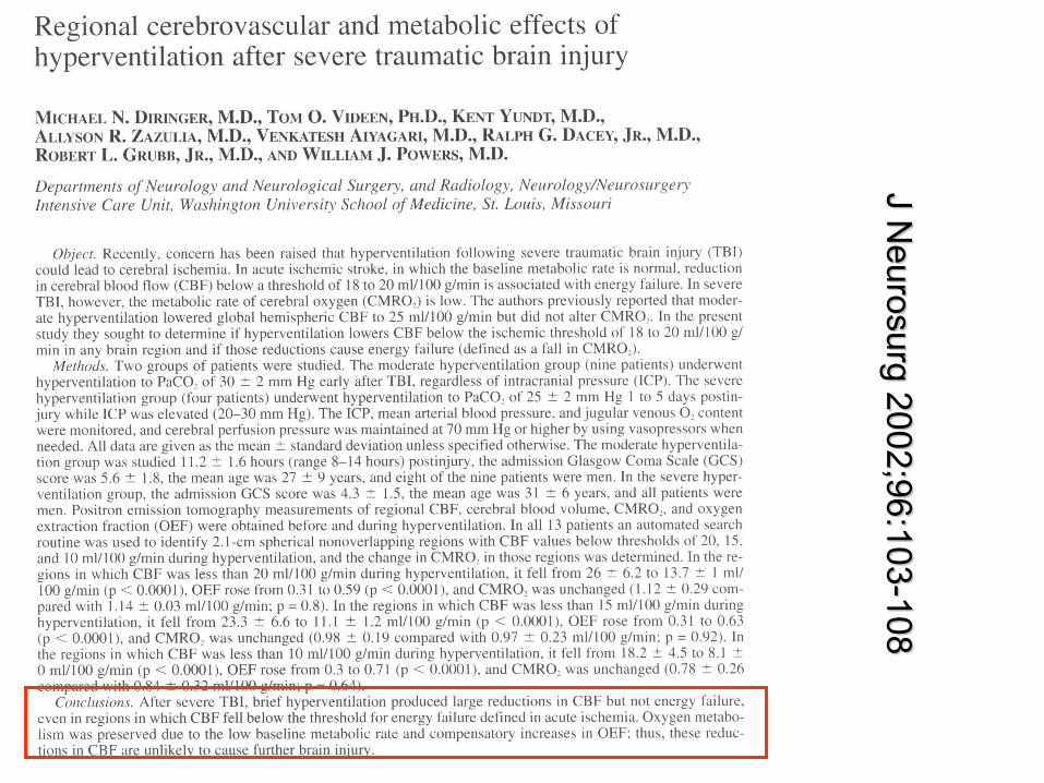
:103-108J N
eurosurg 200
2;96:103-108
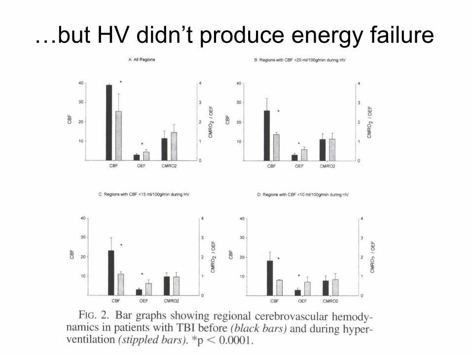
…but HV didn’t produce energy failure
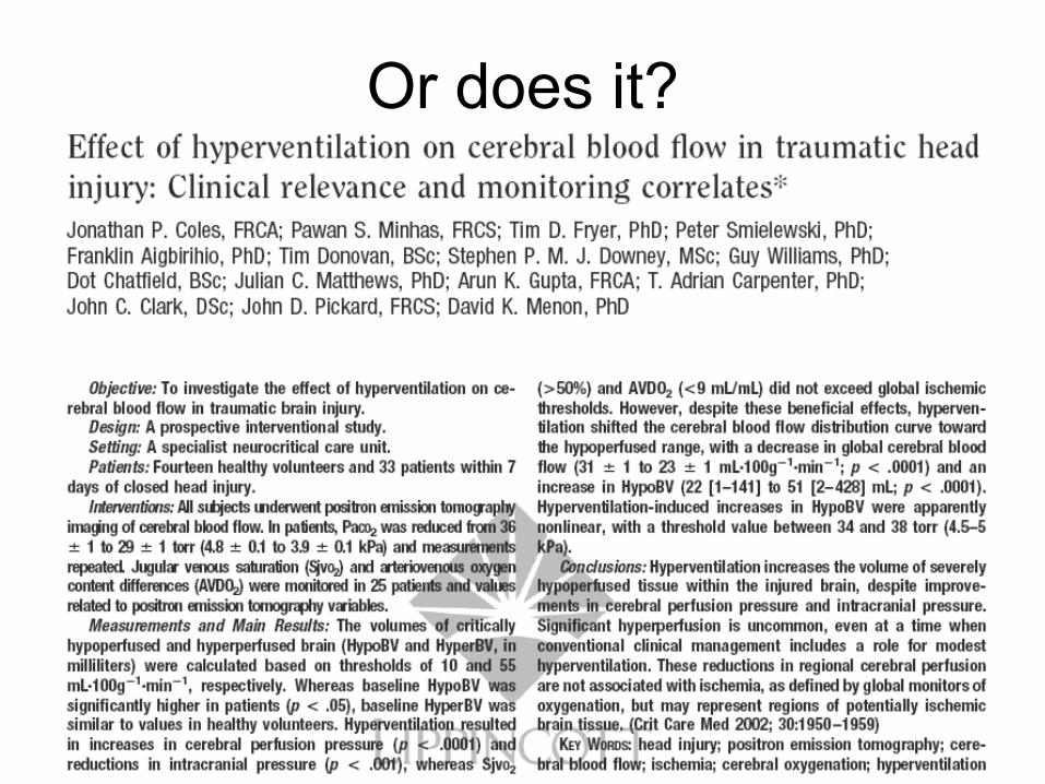
Or does it?
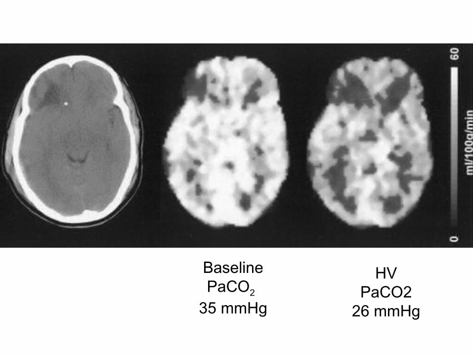
BaselinePaCO2
35 mmHg
HVPaCO2
26 mmHg