

1,2 , Jennifer Christie , Mark S Duxbury - jbc.org · miRNAs drive chemoresistance will guide the...
23
1 Homo sapiens systemic RNA interference defective-1 transmembrane family member 1 (SIDT1) mediates contact-dependent small RNA transfer and microRNA-21-driven chemoresistance* Mohamed O Elhassan 1,2 , Jennifer Christie 2 , Mark S Duxbury 1,2 1 Clinical Surgery, University of Edinburgh, Royal Infirmary of Edinburgh, Edinburgh, EH16 4SA, UK and 2 Pancreatic Cancer Biology Group, Edinburgh Cancer Research UK Centre, Institute of Genetics and Molecular Medicine, Western General Hospital, Crewe Road South, Edinburgh EH4 2XR, UK To whom correspondence should be addressed: Mark Duxbury, Pancreatic Cancer Biology Group, Edinburgh Cancer Research Centre, Western General Hospital, Crewe Road South, Edinburgh, EH4 2XR, Tel.: +44(0)131 7773500, E-mail: [email protected]. *Running title: SIDT1 mediates contact-dependent small RNA transfer Keywords: SIDT1, systemic, RNAi, microRNA, miR-21, pancreatic, cancer, adenocarcinoma, gemcitabine Background: The SID family is a highly conserved group of transmembrane channel-like proteins. Results: SIDT1 facilitates rapid contact- dependent intercellular small RNA transfer and mediates chemoresistance driven by microRNA- 21 in human adenocarcinoma cells. Conclusion: By mediating small RNA transfer, SIDT1 contributes to cancer chemoresistance mechanisms. Significance: A better understanding of non-cell autonomous RNA-based intercellular communication may yield novel anticancer therapeutics. SUMMARY Locally initiated RNA interference (RNAi) has the potential for spatial propagation, inducing posttranscriptional gene silencing in distant cells. In Caenorhabditis elegans, systemic RNAi (sysRNAi) requires a phylogenetically conserved transmembrane channel, SID-1. Here, we show that a human SID-1 orthologue, SIDT1, facilitates rapid, contact-dependent, bidirectional small RNA transfer between human cells, resulting in target-specific non-cell-autonomous RNAi. Intercellular small RNA transfer can both homotypic and heterotypic. We show SIDT1- mediated intercellular transfer of microRNA- 21 to be a driver of resistance to the nucleoside analogue gemcitabine in human adenocarcinoma cells. Documentation of a SIDT1-dependent small RNA transfer mechanism, and the associated phenotypic effects on chemoresistance in human cancer cells, raises the possibility that conserved sysRNAi pathways contribute to the acquisition of drug resistance. Mediators of non-cell autonomous RNAi may be tractable targets for novel therapies aimed at improving the efficacy of current cytotoxic agents. INTRODUCTION RNA interference (RNAi) is initiated locally by double stranded RNA (dsRNA), but has the capacity to propagate systemically (sysRNAi), inducing non-cell autonomous posttranscriptional gene silencing in distant cells. Although best described as an anti-viral mechanism in plants (1-3), sysRNAi also occurs in animals (4). In Caenorhabditis elegans, sysRNAi is dependent on a member of the systemic RNA interference defective (SID) family of channels, SID-1 (5). SID-1 was initially identified following a screen of C. elegans mutants lacking the wild-type sysRNAi phenotype (5,6). However, a range of organisms, including mice and humans, exhibit striking SID gene conservation (7-9). SID channels have relative specificity for small RNA molecules (10,11). While organism-wide sysRNAi phenomena are not apparent in mammals, both SID-1 and its human orthologue SIDT1 have been shown to facilitate siRNA uptake in human systems (12-14). The increased uptake of extracellular siRNA into human cells that SIDT1 mediates can result in highly specific posttranscriptional gene silencing (12,13). We previously demonstrated that SIDT1 functions as a transmembrane channel for small interfering RNA (siRNA) and localizes to the plasma http://www.jbc.org/cgi/doi/10.1074/jbc.M111.318865 The latest version is at JBC Papers in Press. Published on December 15, 2011 as Manuscript M111.318865 Copyright 2011 by The American Society for Biochemistry and Molecular Biology, Inc. by guest on July 28, 2018 http://www.jbc.org/ Downloaded from
Transcript of 1,2 , Jennifer Christie , Mark S Duxbury - jbc.org · miRNAs drive chemoresistance will guide the...

1
Homo sapiens systemic RNA interference defective-1 transmembrane family member 1 (SIDT1) mediates contact-dependent small RNA transfer and microRNA-21-driven chemoresistance*
Mohamed O Elhassan1,2, Jennifer Christie2, Mark S Duxbury1,2
1Clinical Surgery, University of Edinburgh, Royal Infirmary of Edinburgh, Edinburgh, EH16 4SA, UK and 2Pancreatic Cancer Biology Group, Edinburgh Cancer Research UK Centre, Institute of
Genetics and Molecular Medicine, Western General Hospital, Crewe Road South, Edinburgh EH4 2XR, UK
To whom correspondence should be addressed: Mark Duxbury, Pancreatic Cancer Biology Group,
Edinburgh Cancer Research Centre, Western General Hospital, Crewe Road South, Edinburgh, EH4 2XR, Tel.: +44(0)131 7773500, E-mail: [email protected].
*Running title: SIDT1 mediates contact-dependent small RNA transfer
Keywords: SIDT1, systemic, RNAi, microRNA, miR-21, pancreatic, cancer, adenocarcinoma,
gemcitabine Background: The SID family is a highly conserved group of transmembrane channel-like proteins. Results: SIDT1 facilitates rapid contact-dependent intercellular small RNA transfer and mediates chemoresistance driven by microRNA-21 in human adenocarcinoma cells. Conclusion: By mediating small RNA transfer, SIDT1 contributes to cancer chemoresistance mechanisms. Significance: A better understanding of non-cell autonomous RNA-based intercellular communication may yield novel anticancer therapeutics. SUMMARY Locally initiated RNA interference (RNAi) has the potential for spatial propagation, inducing posttranscriptional gene silencing in distant cells. In Caenorhabditis elegans, systemic RNAi (sysRNAi) requires a phylogenetically conserved transmembrane channel, SID-1. Here, we show that a human SID-1 orthologue, SIDT1, facilitates rapid, contact-dependent, bidirectional small RNA transfer between human cells, resulting in target-specific non-cell-autonomous RNAi. Intercellular small RNA transfer can both homotypic and heterotypic. We show SIDT1-mediated intercellular transfer of microRNA-21 to be a driver of resistance to the nucleoside analogue gemcitabine in human adenocarcinoma cells. Documentation of a SIDT1-dependent small RNA transfer mechanism, and the associated phenotypic effects on chemoresistance in human cancer cells, raises the possibility that conserved
sysRNAi pathways contribute to the acquisition of drug resistance. Mediators of non-cell autonomous RNAi may be tractable targets for novel therapies aimed at improving the efficacy of current cytotoxic agents. INTRODUCTION RNA interference (RNAi) is initiated locally by double stranded RNA (dsRNA), but has the capacity to propagate systemically (sysRNAi), inducing non-cell autonomous posttranscriptional gene silencing in distant cells. Although best described as an anti-viral mechanism in plants (1-3), sysRNAi also occurs in animals (4). In Caenorhabditis elegans, sysRNAi is dependent on a member of the systemic RNA interference defective (SID) family of channels, SID-1 (5). SID-1 was initially identified following a screen of C. elegans mutants lacking the wild-type sysRNAi phenotype (5,6). However, a range of organisms, including mice and humans, exhibit striking SID gene conservation (7-9). SID channels have relative specificity for small RNA molecules (10,11). While organism-wide sysRNAi phenomena are not apparent in mammals, both SID-1 and its human orthologue SIDT1 have been shown to facilitate siRNA uptake in human systems (12-14). The increased uptake of extracellular siRNA into human cells that SIDT1 mediates can result in highly specific posttranscriptional gene silencing (12,13). We previously demonstrated that SIDT1 functions as a transmembrane channel for small interfering RNA (siRNA) and localizes to the plasma
http://www.jbc.org/cgi/doi/10.1074/jbc.M111.318865The latest version is at JBC Papers in Press. Published on December 15, 2011 as Manuscript M111.318865
Copyright 2011 by The American Society for Biochemistry and Molecular Biology, Inc.
by guest on July 28, 2018http://w
ww
.jbc.org/D
ownloaded from
2
membrane in human cells (12). This observation led us to hypothesize that SIDT1 might also play a role in the complex contact-dependent intercellular communication that is not only essential for normal histogenesis but, when dysregulated, also drives malignant progression and therapeutic resistance. Small RNAs have a capacity to convey highly specific sequence-encoded signaling information (15). The microRNA (miRNA) system plays critical roles in the genesis, progression and cytotoxic drug resistance of a range of human malignancies (16). Both the functional complexity of the ‘miRNome’, and the diversity of miRNA targets, suggest regulation of gene function by miRNAs can be extremely subtle and adaptable (17). Within the tumor microenvironment, contact-dependent intercellular communication that is critical to the development of chemoresistance (18-20) is directly influenced by perturbation of the miRNome (21). This form of intercellular communication may represent an opportunity for novel targeted therapies. MicroRNA-21 (miR-21), a relatively well-characterized ‘oncogenic’ miRNA, is widely overexpressed in human cancer, and promotes therapeutic resistance in a number of human cancers (22-25). Pancreatic ductal adenocarcinoma is almost universally resistant to the nucleoside analogue gemcitabine, the agent that remains the mainstay of non-surgical therapy for this cancer. In vitro, in vivo and clinical resistance to gemcitabine is closely associated with levels of miR-21, which targets key apoptotic regulators, e.g. p53, phosphatase and tensin homolog (PTEN), Akt and repressors of Ras signaling (26,27). An improved understanding of the mechanisms through which miRNAs drive chemoresistance will guide the development of urgently needed novel approaches that may increase the efficacy of existing cytotoxic agents, such as gemcitabine.
Here we show that, by mediating rapid contact-dependent intercellular transfer of small RNAs, SIDT1 facilitates non-cell autonomous sequence-specific gene silencing in human cells, and can influence the development of chemoresistance in human adenocarcinoma cell populations.
EXPERIMENTAL PROCEDURES Cell culture and reagents Human HEK293 and BxPC3 cells were purchased from the American Type Culture Collection (ATCC, Teddington, UK) and maintained as previously described (28). Gemcitabine (Eli Lilly, UK), and 18-alpha-glycerretinic acid (Sigma, UK) were dissolved in phosphate buffered saline (PBS). RNase blend (Cambio, UK) pre-treatment was performed using 5 units at 37ºC for 30 min. Trypsinisation using 0.25% Trypsin with ethylenediaminetetraacetic acid (EDTA, Invitrogen, UK) was stopped by the addition of complete medium containing 10% fetal bovine serum (FBS). Human pancreatic stellate cells were obtained from surgical resection specimens under the ethically approved Edinburgh Pancreatic Biorepository scheme, using the outgrowth technique, detailed elsewhere (29). Stable far-red membrane linker Cell surface labelling was performed in accordance with the manufacturer’s instructions (Sigma). In brief, 106 cells were washed in PBS and the pellet was re-suspended in 1 ml Diluent C in a 15 ml tube. Fluorescent CellVue Claret lipophilic probe was diluted at a final concentration of 3µM in 1 ml of Diluent C in a 15 ml tube and the solution was rapidly added to cells and incubated for 5 min at room temperature with occasional agitation. 2 ml FBS was added and after 1 min of incubation at room temperature, cells were washed 3 times in complete culture medium. shRNA, oligonucleotides, plasmids and transfection and electroporation pCMV6-AC, pCMV6-AC-tGFP, pCMV6-AC-tGFP-SIDT1 (NM_017699) and pCMV6-Connexin-43/GJA1 (NM_000165) plasmids originated from Origene. TurboGFP was excised by NotI/PmeI digestion, fill-in and ligation to derive pCMV-AC-SIDT1. Virus-incompetent pTRIPZ-based shRNA vectors (Open Biosystems) were used for microRNA expression. A miR-21 dual luciferase reporter construct was engineered using oligonucleotides designed to include SgfI and PmeI sites the (see supplemental table for oligonucleotide sequences). A miR-21-resistant single base mismatch insert served as a control. Oligonucelotides were directionally cloned into the corresponding sites of the psiCHECK2 vector (Promega, UK), in accordance with the
by guest on July 28, 2018http://w
ww
.jbc.org/D
ownloaded from
3
manufacturer’s protocol. Renilla luciferase substrate luminescence was normalized to that of firefly luciferase substrate to allow quantification of Renilla luciferase-miR-21 target sequence mRNA degradation by miR-21. The Dual-Glo luciferase assay system (Promega, UK) was read using a VICTOR3-1420 multi-label reader (Perkin Elmer, UK). Transfection was performed using Lipofectamine 2000 (Invitrogen, UK) according to the manufacturer’s protocol. Stable cell lines were derived using G418 (0.3mg/ml) or puromycin (5µg/ml, both from Sigma, UK) selection, as appropriate. All constructs were verified by sequencing. SIDT1-specific siRNA and mismatch control and Cy3 siRNA (supplemental material) were obtained from Dharmacon, Sigma and Eurogentec. Lucifer yellow introduction was performed by electroporation in accordance with the manufacturer’s cell-type specific protocols using the Nucleofector™ system (Lonza, Switzerland). Gap junction intercellular communication as quantified by flow cytometric quantification of Lucifer yellow transfer as previously described (30). Recipient cells were labeled with far red membrane linker as above. Direct coculture and flow cytometric analysis of intercellular siRNA transfer Cocultured labeled cell subpopulations were encouraged to conjugate by centrifugation at 500rpm for 1 minute and cocultured at 37ºC or 4ºC. Following coculture, cells were washed and resuspended in 5mM EDTA/PBS and kept on ice. Multiparametric flow cytometry data were obtained from 10000 single cell events with stringent doublet exclusion gating FACSAria™ II using FACSDiva software (BD Biosciences, UK). Flow cytometry data were analysed using FlowJo V8 (TreeStar, Switzerland). Viable single cells were identified based on FSC and SSC characteristics (width, height, area). Indirect Transwell coculture and conditioned medium Upper or lower compartments of each Transwell chamber (0.4µm pore diameter, 12 well format) were populated with cells at the specified ratios, in accordance with the manufacturer’s instructions (Corning, UK). Cells were incubated for the specified times at 37ºC, collected using 5mM EDTA/PBS and analysed for Cy3-siRNA acquisition by flow cytometry, as described above. Immunoblotting and antibodies Cell extracts and gels were prepared and performed as previously described (28). Goat
anti-SIDT1 polyclonal antibody was obtained from Cambridge BioScience, UK, rabbit anti-SIDT1 polyclonal and anti-alpha-smooth muscle actin monoclonal antibodies were obtained from Sigma, UK, and mouse anti-beta-actin monoclonal monoclonal antibody was obtained from AbCam, UK. Signal intensities were quantified and normalized to that of beta-actin. Blots were performed in triplicate. Mean densitometric values (± SD) are shown. Proliferation, cytotoxicity, colony formation and apoptosis assays Cellular proliferation and cytoxic effects of gemictabine were quantified using the 3-(4,5,-dimethylthiazol-2-yl)-2,5-diphenyltetrazolium bromide (MTT) assay (Trevigen, Gaithersburg, USA), as previously described (31). MTT correlates closely with [3H]-thymidine incorporation in pancreatic cancer cell lines (32). Gemcitabine-induced cytotoxicity was determined after 24 hours of exposure. Plates were read at a wavelength of 570 nm, corrected to 560 nm, and normalized to controls. Readings were obtained from four biological replicates, with 10 determinations for each condition tested. The concentration of gemcitabine required to inhibit cellular proliferation by 50% (IC50) was calculated using Microsoft Excel software with semi-log curve fitting regression analysis. Caspase 3 activity was quantified using the ApoTarget colorimetric assay in accordance with the manufacturer’s protocol. Relative absorbance at 405nm was quantified using a VICTOR3-1420 multi-label reader (Perkin Elmer, UK). Apoptotic cells were quantified by fluorescent terminal deoxynucleotidyl transferase-mediated nick end labeling, as previously described (33). Colony formation in the presence of 1 µM gemcitabine was quantified as described elsewhere (34) following Giemsa staining. tGFP was quantified using a microplate reader (excitation: 485nm, emission: 530nm), as described elsewhere (27). tGFP was normalized to MTT OD560
with background subtraction. RESULTS Analysis of stable HEK293-derived transfectant cell lines HEK293 cells were transfected with pCMV-based plasmids encoding SIDT1 alone, or in combination with turbo green fluorescent protein (tGFP), and the aminoglycoside 3’-
by guest on July 28, 2018http://w
ww
.jbc.org/D
ownloaded from

4
phosphotransferase neomycin resistance selection marker. Levels of SIDT1 expression were quantified by Western blotting in the following stable transfectants, which were derived using G418 selection: HEKSIDT1, which overexpresses SIDT1; HEKSIDT1/tGFP, which overexpresses SIDT1 and tGFP; and HEKSIDT1-
tGFP, which overexpresses a fusion protein comprising SIDT1 and C-terminus tGFP. Stable tGFP (HEKtGFP) and empty vector transfectants (HEKVector) served as controls. The electrophoretic migration of SIDT1 and SIDT1-tGFP fusion protein was consistent with predicted respective molecular masses of 94kDa and 120kDa (Fig. 1a). The transfectant cell lines demonstrated no significant differences in respective rates of cellular proliferation, or fraction of apoptotic cells (TUNEL), under standard culture conditions (supplemental material 1). SIDT1 facilitates rapid contact-dependent siRNA transfer between human cells A direct coculture assay was used to investigate the role of SIDT1 in contact-dependent siRNA transfer. ‘Donor’ (HEKSIDT1 or HEKVector) and ‘Acceptor’ (HEKSIDT1/tGFP or HEKtGFP) cell subpopulations were subjected to direct coculture, allowing cell-cell contact as schematized (Fig 1b). Cy3-labelled 21-mer siRNA was introduced into ‘Donor’ cells by electroporation alone to eliminate potentially confounding effects of persisting transfection reagent. To mitigate against ‘Donor’ epifluorescence signal decay, as might occur due to Cy3-siRNA degradation, and to control for trogocytosis or cell fusion events, ‘Donor’ cells were co-labeled with a far-red fluorescent plasma membrane linker. This linker is highly persistent (t1/2 = 12 days), biochemically inert, does not affect cell viability or membrane function and has an emission spectrum that is readily distinguishable from that of Cy3 (35-37). Far-red label transfer to unlabelled cells was not observed during any direct or indirect coculture experiments. Potential artifact arising from contact-independent media-borne siRNA transfer was minimized by post-electroporation washing and RNase treatment, which degrades extracellular RNA to nucleoside monophosphates (supplemental material 2a) (38,39). SIDT1-overexpressing (HEKSIDT1 and HEKSIDT1/tGFP) and control (HEKVector and
HEKtGFP) ‘Donor’ and ‘Acceptor’ cells were cocultured at 1:1 ratios. Following coculture for 90 min, cell conjugates were disrupted to form single cell suspensions by EDTA treatment and agitation. Additional RNase treatment ensured removal of cell surface-associated Cy3-siRNA and free RNA that may have been released from lysed cells. Intercellular Cy3-siRNA transfer to ‘Acceptor’ cells was quantified by flow cytometry using stringent doublet exclusion. We quantified Cy3-siRNA-positive, tGFP-positive, far-red-negative ‘Acceptor’ cells, defining a new subset of ‘Acceptor’ cells that had acquired Cy3-siRNA from ‘Donor’ cells (Fig 1c I-VI). Transfer of Cy3-siRNA between HEKSIDT1
‘Donor’ and HEKSIDT1/tGFP ‘Acceptor’ cells was insensitive to RNase treatment and occurred rapidly (Fig 1c VII). In contrast, transfer of Cy3-siRNA between HEKVector ‘Donor’ and HEKtGFP ‘Acceptor’ cells was negligible (Fig 1c I). Direct coculture using HEKSIDT1 ‘Donor’ and HEKtGFP ‘Acceptor’ cells (Fig 1c II), as well as HEKVector ‘Donor’ and HEKSIDT1/tGFP ‘Acceptor’ cells (Fig 1c III), resulted in no significant difference in siRNA transfer, i.e. SIDT1 increased Cy3-siRNA acquisition regardless of whether it was overexpressed by ‘Donor’ or ‘Acceptor’ cells, indicating that facilitation of intercellular siRNA transfer by SIDT1 overexpression is bidirectional. Transfer of Cy3-siRNA from HEKSIDT1 to HEKSIDT1/tGFP was abolished by pre-incubation with polyclonal anti-SIDT1 (10 µg/ml, Fig 1c VI). Although free Cy3-siRNA was eliminated from the culture medium, we were cognizant that nascent or RNase-resistant exosome-borne Cy3-siRNA arising from ‘Donor’ cells could also potentially contribute to the ‘Acceptor’ Cy3-siRNA signal, through contact-independent acquisition. To control for contact-independent Cy3-siRNA transfer, we performed indirect coculture of identical ‘Donor’ and ‘Acceptor’ cell groups, separated by permeable (0.4µm diameter pore) Transwell insert membranes. In addition, ‘Acceptor’ cells were exposed to cell-free (0.4µm filtered) ‘Donor’ conditioned medium. After either 90 min indirect co-culture, or 90 min exposure to ‘Donor’ conditioned medium, Cy3 epifluorescence was quantified by flow cytometry, as above. No transfer of Cy3-siRNA to either HEKSIDT1/tGFP or HEKtGFP ‘Acceptor’ cells was detected following either indirect coculture with ‘Donor’ cells, or following exposure to ‘Donor’ conditioned
by guest on July 28, 2018http://w
ww
.jbc.org/D
ownloaded from
5
medium (supplemental material 2b). Contact independent uptake of extracellular Cy3-siRNA therefore did not account for the Cy3 signal acquired by the ‘Acceptor’ cell subpopulation. SIDT1-mediated Cy3-siRNA transfer is gap junction-independent The contribution of GJIC to Cy3-siRNA acquisition was predicted to be small in HEK293 cells, given their low levels of connexin junction formation and GJIC (40-42). However, gap junction-mediated intercellular transfer of small RNAs has been reported in some cell types (43,44). We therefore took steps to distinguish the contribution of SIDT1 to the acquisition of Cy3-siRNA by ‘Acceptor’ cells from that of GJIC. We reasoned that enhanced intercellular Cy3-siRNA transfer could result from either native gap junction-mediated siRNA transfer, or from facilitation of GJIC by SIDT1 overexpression. GJIC was quantified in the direct coculture system by flow cytometric measurement of Lucifer yellow transfer following electroporation-mediated Lucifer yellow loading, a quantitative approach that correlates with the scratch-loading Lucifer yellow transfer assay (30,45). Intercellular Lucifer yellow transfer was minimal in all combinations of HEKSIDT1, HEKSIDT1-tGFP, HEKSIDT1/tGFP, HEKtGFP and HEKVector following 90 min coculture. GJIC was inhibited without cytotoxicity in all transfectant HEK- and BxPC3-derived cell lines following pretreatment with the specific small molecule gap junction inhibitor 18-alpha-glycyrrhetinic acid (AGA, 25µM, supplemental material 3) (46,47). Coculture and flow cytometric analysis of Cy3-siRNA transfer was repeated following pretreatment with AGA prior to 90min coculture at 37°C, as above. Cy3-siRNA transfer was not significantly affected by AGA. Together, these observations indicate that SIDT1 overexpression does not facilitate GJIC, nor is the resulting increase in Cy3-siRNA transfer dependent on functional GJIC. Induction of non-autonomous miRNA activity through physical contact with a subpopulation of miR-21-overexpressing BxPC3 cells MicroRNA-21 (miR-21) is a critical driver of resistance to the nucleoside analogue gemcitabine in human pancreatic adenocarcinoma, an exemplar of a chemoresistant human malignancy (26,27). The
human pancreatic ductal adenocarcinoma cell line BxPC3 was selected as a model system as it exhibits relatively low levels of miR-21 activity under standard culture conditions (26). We stably transfected a subpopulation of BxPC3 cells with a non-viral pTRIPZ-derived miR-21 expression construct. These cells (BxPC3miR21) generate miR-21 in a doxycycline-inducible manner. Irrelevant miRNA-generating transfectants (BxPC3miRN/S, derived from #RHS4346) served as controls. A miR-21 reporter cell line (BxPC3CkmiR21) was derived from BxPC3 by stable transfection of a psiCHECK-2-based miR-21 reporter construct. This dual luciferase reporter system allows Renilla luciferase activity, which decreases in the presence of miR-21, to be normalized to firefly luciferase, controlling for variations in reporter construct abundance. A single nucleotide mismatch reporter cell line (BxPC3CkmiR21mm) was employed to confirm the specificity of the reporter system for miR-21 (supplemental material 4). BxPC3miR21 and BxPC3CkmiR21 cells were subjected to direct coculture at high total cell density (80-90% cell-cell contact) at BxPC3miR21:BxPC3CkmiR21 ratios ranging from 1:10 to 1:1000, in the presence or absence of 1µg/ml doxycycline. Doxycycline-induced BxPC3miR21 activation (confirmed by RFP epifluorescence) led to a decrease in normalized Renilla luciferase activity of directly cocultured BxPC3CkmiR21 reporter cells, reflecting increased miR-21 activity within the BxPC3CkmiR21 reporter cell subpopulation. Direct coculture with BxPC3miRN/S had no effect on normalized Renilla activity of BxPC3CkmiR21 cells, confirming specificity for the miR-21 sequence. Significant decreases in normalized Renilla lucifierase activity were observed even when BxPC3miR21 cells were present as a minority of 0.1% (Fig 2a). This non-autonomous increase in miR-21 activity within the BxPC3CkmiR21 subpopulation was not observed when BxPC3CkmiR21 were subjected to indirect coculture with BxPC3miR21 cells (0.4µm pore diameter Transwell, Fig 2a). Non-autonomous miR-21 activity is SIDT1-dependent Our previous observations led us to hypothesize that the SIDT1 channel could facilitate miR-21 transfer between contacting cells. SIDT1 channel protein expression varies considerably
by guest on July 28, 2018http://w
ww
.jbc.org/D
ownloaded from
6
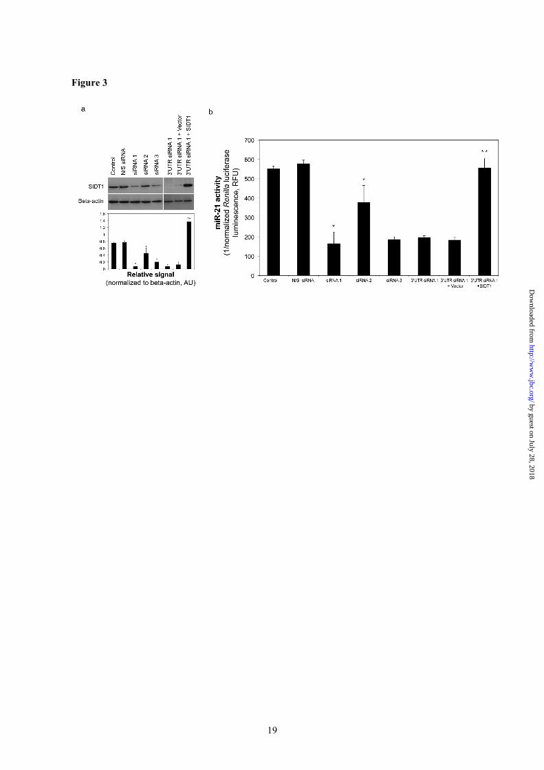
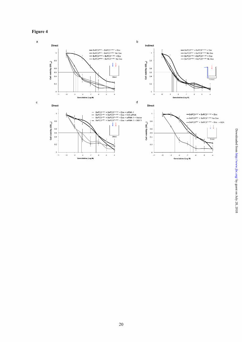
between pancreatic adenocarcinoma and immortalized normal ductal epithelial cell (HPDE4) lines. Among adenocarcinoma cells, BxPC3 expresses relatively high levels of SIDT1 (Fig 2b). Increased miR-21 activity in BxPC3CkmiR21 induced by the doxycycline-activated minority BxPC3miR21 subpopulation was abrogated by pre-treatment with siRNAs directed against different regions of the SIDT1 sequence, including the SIDT1 3’ un-translated region (3’UTR), but not control siRNA (Fig 3). The specificity of this siRNA-induced effect was further confirmed by a ‘rescue step’ in which a SIDT1 expression construct lacking the SIDT1 3’UTR target sequence, or empty vector control, was co-transfected with SIDT1-3’UTR siRNA 1. SIDT1 overexpression ‘rescued’ the abrogation of miR-21 induction in the BxPC3CkmiR21 reporter subpopulation that was observed with SIDT1-3’UTR siRNA 1 treatment (Fig 3). A minority subpopulation of miR-21-overexpressing adenocarcinoma cells increases global cellular chemoresistance to gemcitabine Given the ability of a minority subpopulation of BxPC3miR21 cells to increase miR-21 activity in physically contacting BxPC3CkmiR21 cells, we examined the effect of a 1% subpopulation of BxPC3miR21 on the gemcitabine IC50 for the total mixed cell population. Cells were exposed to clinically relevant concentrations of gemcitabine (48-52) and the IC50 derived for the whole cell population. The gemcitabine IC50 was increased from 6 × 10⁻⁸ M to 3 × 10⁻⁶ M by direct coculture of doxycycline-activated BxPC3miR21 with BxPC3CkmiR21 cells (ratio 1:100). Importantly, firefly luciferase activity normalized to total cell number remained constant, indicating preservation of the BxPC3miR21:BxPC3CkmiR21 ratio over the duration of the experiment. The gemcitabine IC50 approximated control levels when doxycycline was withheld, and was unaffected by direct coculture of BxPC3miRN/S with BxPC3CkmiR21, confirming specificity of the effect to the miR-21 sequence (Fig 4a). Transfer of mir-21 and IC50 were unaffected by 25µM AGA (Fig 4d and supplemental material 3). SIDT1 deficiency attenuates chemoresistance induced by a minority miR-21-overexpressing subpopulation of adenocarcinoma cells The increase in global cellular chemoresistance to gemcitabine that miR-21 induced was cell contact-dependent, the IC50 being unaffected
when BxPC3miR21 and BxPC3CkmiR21 cells were subjected to indirect coculture (BxPC3miR21:BxPC3CkmiR21 = 1:100, Fig 4b). The miR-21-induced increase in IC50 was significantly attenuated when cells were treated with SIDT1-3’UTR siRNA 1, but not control siRNA. A SIDT1 re-expression ‘rescue step’, as described above, confirmed that the decrease in gemcitabine IC50 induced by SIDT1-3’UTR siRNA 1 could be abolished by restoring levels of SIDT1 (Fig 4c). Corresponding impairment of colony forming capacity and increased caspase 3 activities were observed when BXPC3miR21 and BxPC3CkmiR21 were cocultured in the presence of gemcitabine following treatment with SIDT1-3’UTR siRNA 1. Reintroduction of SIDT1 abolished these effects on colony formation and caspase activities (Fig 5). SIDT1 contributes to cell adhesion mediated drug resistance (CAM-DR) Given the capacity for small RNA transfer observed between adenocarcinoma cells, we examined whether a similar process might contribute to the complex tumor-stromal cell interactions that can enhance chemoresistance in a range of human cancers (53). Human pancreatic stellate cells (hPSC) were isolated from surgical resection specimens and their morphology and immunophenotype (54) confirmed (Fig. 6a). SIDT1 was relatively highly expressed in all three hPSC lines tested (Fig. 6b). We repeated the flowcytometric Cy3-siRNA transfer assay, as before, and observed rapid Cy3-siRNA transfer between adenocarcinoma and hPSC cells that was sensitive to anti-SIDT1 antibody treatment (Fig. 6c). In order to quantify CAM-DR, we subjected BxPC3tGFP cells to direct coculture with hPSC in the presence of 1µM gemcitabine and measured tGFP fluorescence, which correlates with adenocarcinoma cell number (27), normalizing GFP fluorescence to MTT-based total cell quantification. BxPC3tGFP proliferation in the presence of clinically relevant gemcitabine levels was markedly increased by hPSC coculture. Furthermore, this effect was abrogated by treatment with 10µg/ml anti-SIDT1 antibody, but not control matched immunoglobulin. Although this aspect of SIDT1 biology requires further investigation, this result demonstrates that heterotypic intercellular small RNA transfer can occur and that SIDT1 contributes to this process.
by guest on July 28, 2018http://w
ww
.jbc.org/D
ownloaded from

7
In summary, SIDT1 facilitates rapid bidirectional, contact-dependent, RNase-insensitive transfer of Cy3-siRNA that is independent of GJIC. Contact-independent siRNA transfer was insignificant in comparison to SIDT1-mediated contact-dependent Cy3-siRNA acquisition. SIDT1-dependent Cy3-siRNA intercellular transfer is not restricted to adenocarcinoma cells and can occur between stromal cells, influencing CAM-DR. DISCUSSION Although organism-wide sysRNAi is not apparent in mammals, significant phylogenetic molecular conservation suggests that sysRNAi pathways may be relevant to human physiology and pathophysiology (7). The C. elegans orthologue of SIDT1, SID1, has recently been shown to be a dsRNA-gated channel capable of selective bidirectional intercellular dsRNA transfer (11). Our findings firstly demonstrate that SIDT1 facilitates contact-dependent small RNA transfer and non-cell autonomous posttranscriptional regulation; secondly, they support the assertion that small RNA-based signaling represents a further level of adaptive capacity and complexity within the tumor microenvironment; and thirdly, that disruption or exploitation of sysRNAi pathways may have therapeutic utility, particularly as a means of impairing the acquisition of resistance to cytotoxic agents. Contact-dependent intercellular communication not only maintains normal tissue organization, but can also drive neoplasia. However, to date, studies of SID family proteins have generally focused on these proteins as conduits for the contact-independent uptake of free small RNAs from the extracellular milieu (12,13). In addition to characterizing the role of SIDT1 in the context of contact-dependent small RNA intercellular transfer, this study provides new evidence that contact-dependent non-cell autonomous RNAi can shape therapeutic resistance in pancreatic cancer and that SIDT1 can act as a mediator of this form of RNA-based intercellular communication. The ‘miRNome’ is a highly complex and adaptable system with each miRNA exerting pleiotropic effects. Recent studies illustrate miRNA biogenesis to be exquisitely sensitive to cell context, miRNA levels increasing in a contact-dependent manner (55). miR-21 was the focus of this study as it promotes
chemoresistance to gemcitabine in human adenocarcinoma cells (22-25). The ability of a minority subpopulation of miR-21-overexpressing cells to influence global chemoresistance through a contact-mediated, SIDT1-dependent mechanism raises the intriguing possibility that subgroups of cells within a heterogeneous tumor population can influence resistance within the wider tumor microenvironment through contact-dependent non-cell autonomous RNAi. Subpopulations of drug tolerant tumor cells employ dynamic survival strategies that result in therapeutic resistance (34). Parallels can be drawn with microbial resistance, in which a small number of tolerant organisms can influence the ‘fitness’ of the populations as a whole. Similarly, cells exposed to cytotoxic drug may, through small RNA-based communication, influence survival pathways within contacting cells over significant distances. The rapid nature of SIDT1-mediated small RNA transfer is particularly striking and significantly precedes RNA transfer via contact-independent mechanisms such as exosomal shuttling and free RNA transfer (38). Tumor cells that are capable of rapid adaptation are more likely to gain selective advantage in the presence of a toxic perturbation. Rapid small RNA transfer and resulting posttranscriptional gene regulation would allow more timely adaptive changes than those resulting from ‘classic’ genetic mutation. This study supports the premise that small RNAs have the capacity to act as signaling intermediaries. Absence of disseminating fluorescent protein expression to ‘Acceptor’ cells in longer-term coculture studies confirms a degree of RNA type- and size-specificity, i.e. SIDT1 does not facilitate functional mRNA transfer. These observations are in keeping with the relative specificity for dsRNA exhibited by SID1 (11). Our results demonstrate SIDT1-mediated siRNA transfer to be independent of GJIC. GJIC can increase the susceptibility of cancer cells to cytotoxic agents through connexin-mediated ‘bystander effects’ in pancreatic cancer (56). BxPC3 cells in which GJIC is artificially increased exhibit ‘bystander’ cytotoxicity when exposed to gemcitabine (56-58). Interestingly, connexin expression and GJIC are frequently decreased in cancer, re-expression of connexins commonly suppressing tumorgenicity (59). In
by guest on July 28, 2018http://w
ww
.jbc.org/D
ownloaded from

8
contrast to the relatively non-selective nature of connexin-mediated communication, SIDT1 overexpression does not increase Lucifer yellow transfer. SIDT1 may therefore represent a means by which tumor cells can adapt to maintain small RNA-based intercellular communication, without experiencing greater ‘bystander’ cytotoxicity that increased GJIC would incur. Recent data from other groups indicate that non-cell-autonomous small RNA effects may be of general relevance to human cancer. Katakowski et al reported microvesicle-independent microRNA transfer between U87 human glioma cells (60). While this study did not directly examine chemoresistance, the authors demonstrated non-autonomous microRNA effects in contacting cells. Zhao et al have demonstrated that SNB19 glioma cells can undergo intercellular transfer of PTEN-silencing siRNA in coculture (61). Interestingly, this effect was only observed in direct (contact-dependent), but not indirect (contact-independent) coculture. The PTEN tumor suppressor mRNA is a target of miR-21 and a number of other micro-RNAs. PTEN deficiency is clinically associated with chemoresistance in a range of human cancers. These data also suggest that non-autonomous gene silencing can result in molecular events that promote clinical chemoresistance.
We have demonstrated that SIDT1 dependent small RNA transfer can also operate between adenocarcinoma and stromal cells, in this case pancreatic stellate cells. The important chemoprotective effects of direct tumor-stromal cell contact are increasingly recognized (27). Cell adhesion-mediated drug resistance (CAM-DR) that develops in the tumor microenvironment may represent a future therapeutic opportunity. Follicular dendritic cells can protect B-cell lymphoma cells from drug-induced apoptosis through contact-mediated microRNA-dependent mechanisms (21,62). miR-181a was found to be increased by direct
contact between dendritic and lymphoma cells, but not when cells were cultured under indirect coculture conditions, suggesting that free RNA, RNA bound to proteins, e.g. Argonaute or lipoproteins, or exosomal RNA transfer is less likely to mediate the effects on chemoresistance in this setting. This is in keeping with our observations. Although the possibility that the non-autonomous increase in miR-181a levels could result from intercellular RNA transfer was not explored, interestingly, SIDT1 is also relatively overexpressed in dendritic cells. The clinical implications of SIDT1 expression levels are likely to be complex and will be influenced by the prevailing miRNome within the tumor. Preliminary studies of primary breast cancer mRNA and microRNA expression arrays are consistent with the hypothesis that SIDT1 may have ‘permissive’ role in some of the phenotypic effects of miR-21 overexpression. High levels of miR-21 expression result in poorer survival for patients with SIDT1 overexpressing tumors relative to those with low SIDT1 expression (supplemental material 5). Further clinical studies are ongoing to address this question. In conclusion, SIDT1 mediates contact-dependent siRNA and miRNA transfer, and non-cell-autonomous RNAi, which can enhance pancreatic adenocarcinoma chemoresistance to gemcitabine that is driven by miR-21. SIDT1-dependent small RNA transfer may also contribute to CAM-DR. While sysRNAi in humans appears not to be the organism-wide phenomenon observed in C. elegans, comparable processes may support adaptation to perturbations and selective pressures within the tumor microenvironment, such as those induced by cytotoxic therapy. Therapeutic exploitation of sysRNAi pathways may have utility in human cancer and warrants further experimental evaluation.
by guest on July 28, 2018http://w
ww
.jbc.org/D
ownloaded from

9
References
1. Chitwood, D. H., and Timmermans, M. C. (2010) Nature 467, 415-419 2. Herr, A. J., and Baulcombe, D. C. (2004) Cold Spring Harb Symp Quant Biol 69, 363-370 3. Baulcombe, D. (2004) Nature 431, 356-363 4. Jose, A. M., and Hunter, C. P. (2007) Annu Rev Genet 41, 305-330 5. Winston, W. M., Molodowitch, C., and Hunter, C. P. (2002) Science 295, 2456-2459 6. Feinberg, E. H., and Hunter, C. P. (2003) Science 301, 1545-1547 7. Li, H., Coghlan, A., Ruan, J., Coin, L. J., Heriche, J. K., Osmotherly, L., Li, R., Liu, T., Zhang, Z., Bolund, L., Wong, G. K., Zheng, W., Dehal, P., Wang, J., and Durbin, R. (2006) Nucleic Acids Res 34, D572-580 8. Tomoyasu, Y., Miller, S. C., Tomita, S., Schoppmeier, M., Grossmann, D., and Bucher, G. (2008) Genome Biol 9, R10 9. Su, A. I., Wiltshire, T., Batalov, S., Lapp, H., Ching, K. A., Block, D., Zhang, J., Soden, R., Hayakawa, M., Kreiman, G., Cooke, M. P., Walker, J. R., and Hogenesch, J. B. (2004) Proc Natl Acad Sci U S A 101, 6062-6067 10. Hunter, C. P., Winston, W. M., Molodowitch, C., Feinberg, E. H., Shih, J., Sutherlin, M., Wright, A. J., and Fitzgerald, M. C. (2006) Cold Spring Harb Symp Quant Biol 71, 95-100 11. Shih, J. D., and Hunter, C. P. (2011) RNA 12. Duxbury, M. S., Ashley, S. W., and Whang, E. E. (2005) Biochem Biophys Res Commun 331, 459-463 13. Wolfrum, C., Shi, S., Jayaprakash, K. N., Jayaraman, M., Wang, G., Pandey, R. K., Rajeev, K. G., Nakayama, T., Charrise, K., Ndungo, E. M., Zimmermann, T., Koteliansky, V., Manoharan, M., and Stoffel, M. (2007) Nat Biotechnol 25, 1149-1157 14. Tsang, S. Y., Moore, J. C., Huizen, R. V., Chan, C. W., and Li, R. A. (2007) Biochem Biophys Res Commun 357, 480-486 15. Dinger, M. E., Mercer, T. R., and Mattick, J. S. (2008) J Mol Endocrinol 40, 151-159 16. Macfarlane, L. A., and Murphy, P. R. (2010) Curr Genomics 11, 537-561 17. Inui, M., Martello, G., and Piccolo, S. (2010) Nat Rev Mol Cell Biol 11, 252-263 18. Patek, P. Q., Lin, Y., and Case, P. G. (1989) Proc Soc Exp Biol Med 190, 234-239 19. Kobayashi, H., Man, S., Graham, C. H., Kapitain, S. J., Teicher, B. A., and Kerbel, R. S. (1993) Proc Natl Acad Sci U S A 90, 3294-3298 20. Green, S. K., Frankel, A., and Kerbel, R. S. (1999) Anticancer Drug Des 14, 153-168 21. Lwin, T., Lin, J., Choi, Y. S., Zhang, X., Moscinski, L. C., Wright, K. L., Sotomayor, E. M., Dalton, W. S., and Tao, J. (2010) Blood 116, 5228-5236 22. Krichevsky, A. M., and Gabriely, G. (2009) J Cell Mol Med 13, 39-53 23. Bai, H., Xu, R., Cao, Z., Wei, D., and Wang, C. (2011) FEBS Lett 585, 402-408 24. Misawa, A., Katayama, R., Koike, S., Tomida, A., Watanabe, T., and Fujita, N. (2010) Oncol Res 19, 23-33 25. Pan, X., Wang, Z. X., and Wang, R. (2011) Cancer Biol Ther 10, 1224-1232 26. Giovannetti, E., Funel, N., Peters, G. J., Del Chiaro, M., Erozenci, L. A., Vasile, E., Leon, L. G., Pollina, L. E., Groen, A., Falcone, A., Danesi, R., Campani, D., Verheul, H. M., and Boggi, U. (2010) Cancer Res 70, 4528-4538 27. Fujita, H., Ohuchida, K., Mizumoto, K., Egami, T., Miyoshi, K., Moriyama, T., Cui, L., Yu, J., Zhao, M., Manabe, T., and Tanaka, M. (2009) Cancer Sci 100, 2309-2317 28. Duxbury, M. S., Ito, H., Ashley, S. W., and Whang, E. E. (2004) J Biol Chem 279, 23176-23182 29. Bachem, M. G., Schunemann, M., Ramadani, M., Siech, M., Beger, H., Buck, A., Zhou, S., Schmid-Kotsas, A., and Adler, G. (2005) Gastroenterology 128, 907-921 30. Kiang, D. T., Kollander, R., Lin, H. H., LaVilla, S., and Atkinson, M. M. (1994) In Vitro Cell Dev Biol Anim 30A, 796-802 31. Duxbury, M. S., and Whang, E. E. (2007) Biochem Biophys Res Commun 354, 190-196 32. Raitano, A. B., Scuderi, P., and Korc, M. (1990) Pancreas 5, 267-277
by guest on July 28, 2018http://w
ww
.jbc.org/D
ownloaded from
10
33. Duxbury, M. S., Ito, H., Benoit, E., Waseem, T., Ashley, S. W., and Whang, E. E. (2004) Cancer Res 64, 3987-3993 34. Sharma, S. V., Lee, D. Y., Li, B., Quinlan, M. P., Takahashi, F., Maheswaran, S., McDermott, U., Azizian, N., Zou, L., Fischbach, M. A., Wong, K. K., Brandstetter, K., Wittner, B., Ramaswamy, S., Classon, M., and Settleman, J. (2010) Cell 141, 69-80 35. Bantly, A. D., Gray, B. D., Breslin, E., Weinstein, E. G., Muirhead, K. A., Ohlsson-Wilhelm, B. M., and Moore, J. S. (2007) Immunol Invest 36, 581-605 36. Horan, P. K., and Slezak, S. E. (1989) Nature 340, 167-168 37. Wallace, P. K., Palmer, L. D., Perry-Lalley, D., Bolton, E. S., Alexander, R. B., Horan, P. K., Yang, J. C., and Muirhead, K. A. (1993) Cancer Res 53, 2358-2367 38. Rechavi, O., Erlich, Y., Amram, H., Flomenblit, L., Karginov, F. V., Goldstein, I., Hannon, G. J., and Kloog, Y. (2009) Genes Dev 23, 1971-1979 39. Rocha, J. J., Korolchuk, V. I., Robinson, I. M., and O'Kane, C. J. (2011) PLoS One 6, e19087 40. Toyofuku, T., Yabuki, M., Otsu, K., Kuzuya, T., Hori, M., and Tada, M. (1998) J Biol Chem 273, 1519-1528 41. John, S. A., Kondo, R., Wang, S. Y., Goldhaber, J. I., and Weiss, J. N. (1999) J Biol Chem 274, 236-240 42. McSpadden, L. C., Kirkton, R. D., and Bursac, N. (2009) Am J Physiol Cell Physiol 297, C339-351 43. Wolvetang, E. J., Pera, M. F., and Zuckerman, K. S. (2007) Biochem Biophys Res Commun 363, 610-615 44. Valiunas, V., Polosina, Y. Y., Miller, H., Potapova, I. A., Valiuniene, L., Doronin, S., Mathias, R. T., Robinson, R. B., Rosen, M. R., Cohen, I. S., and Brink, P. R. (2005) J Physiol 568, 459-468 45. el-Fouly, M. H., Trosko, J. E., and Chang, C. C. (1987) Exp Cell Res 168, 422-430 46. Goldberg, G. S., Moreno, A. P., Bechberger, J. F., Hearn, S. S., Shivers, R. R., MacPhee, D. J., Zhang, Y. C., and Naus, C. C. (1996) Exp Cell Res 222, 48-53 47. Lahlou, H., Fanjul, M., Pradayrol, L., Susini, C., and Pyronnet, S. (2005) Mol Cell Biol 25, 4034-4045 48. Abbruzzese, J. L., Grunewald, R., Weeks, E. A., Gravel, D., Adams, T., Nowak, B., Mineishi, S., Tarassoff, P., Satterlee, W., Raber, M. N., and et al. (1991) J Clin Oncol 9, 491-498 49. Bhargava, P., Marshall, J. L., Fried, K., Williams, M., Lefebvre, P., Dahut, W., Hanfelt, J., Gehan, E., Figuera, M., Hawkins, M. J., and Rizvi, N. A. (2001) Cancer Chemother Pharmacol 48, 95-103 50. van Riel, J. M., Peters, G. J., Mammatas, L. H., Honeywell, R. J., Laan, A. C., Ruyter, R., van den Berg, F. G., Giaccone, G., and van Groeningen, C. J. (2009) Eur J Cancer 45, 2519-2527 51. Grunewald, R., Abbruzzese, J. L., Tarassoff, P., and Plunkett, W. (1991) Cancer Chemother Pharmacol 27, 258-262 52. Veltkamp, S. A., Beijnen, J. H., and Schellens, J. H. (2008) Oncologist 13, 261-276 53. Dalton, W. S. (1999) Drug Resist Updat 2, 285-288 54. Bachem, M. G., Schneider, E., Gross, H., Weidenbach, H., Schmid, R. M., Menke, A., Siech, M., Beger, H., Grunert, A., and Adler, G. (1998) Gastroenterology 115, 421-432 55. Hwang, H. W., Wentzel, E. A., and Mendell, J. T. (2009) Proc Natl Acad Sci U S A 106, 7016-7021 56. Garcia-Rodriguez, L., Perez-Torras, S., Carrio, M., Cascante, A., Garcia-Ribas, I., Mazo, A., and Fillat, C. (2011) Mol Cancer Ther 10, 505-517 57. Cottin, S., Ghani, K., de Campos-Lima, P. O., and Caruso, M. (2010) Mol Cancer 9, 141 58. Ammerpohl, O., Trauzold, A., Schniewind, B., Griep, U., Pilarsky, C., Grutzmann, R., Saeger, H. D., Janssen, O., Sipos, B., Kloppel, G., and Kalthoff, H. (2007) Br J Cancer 96, 73-81 59. Zhang, Z. Q., Zhang, W., Wang, N. Q., Bani-Yaghoub, M., Lin, Z. X., and Naus, C. C. (1998) Carcinogenesis 19, 1889-1894 60. Katakowski, M., Buller, B., Wang, X., Rogers, T., and Chopp, M. (2010) Cancer Res 70, 8259-8263
by guest on July 28, 2018http://w
ww
.jbc.org/D
ownloaded from

11
61. Zhao, T. Y., Zou, S. P., Alimova, Y. V., Wang, G., Hauser, K. F., Ghandour, M. S., and Knapp, P. E. (2006) J Neurochem 98, 1541-1550 62. Lin, J., Lwin, T., Zhao, J. J., Tam, W., Choi, Y. S., Moscinski, L. C., Dalton, W. S., Sotomayor, E. M., Wright, K. L., and Tao, J. (2011) Leukemia 25, 145-152 63. Enerly, E., Steinfeld, I., Kleivi, K., Leivonen, S. K., Aure, M. R., Russnes, H. G., Ronneberg, J. A., Johnsen, H., Navon, R., Rodland, E., Makela, R., Naume, B., Perala, M., Kallioniemi, O., Kristensen, V. N., Yakhini, Z., and Borresen-Dale, A. L. (2011) PLoS One 6, e16915
by guest on July 28, 2018http://w
ww
.jbc.org/D
ownloaded from
12
ACKNOWLEDGEMENTS We thank Drs Nick Gilbert, Bernard Ramsahoye, Prof John Iredale and members of the Frame and Brunton laboratories for helpful advice and reagents. We thank Dr Jacqueline Dickson for the pTRIPZ-IRES-GFP construct, Elizabeth Freyer (Flow cytometry facility, Medical Research Council Human Genetics Unit, Edinburgh) for technical assistance with flow cytometry and Dr Andy Sims for bioinformatics advice. MSD is supported by an NHS Education for Scotland Clinician Scientist Fellowship. This work was supported by Chief Scientist Office project grant CZB/4/693 and by The Melville Trust for the Care and Cure of Cancer. ABBREVIATION LIST GJIC, gap junction intercellular communication; RFP, red fluorescent protein; RNAi, RNA interference; dsRNA, double stranded RNA; SID-1, systemic RNA interference defective protein 1; SIDT1, SID-1 transmembrane family member 1; siRNA, short interfering RNA; tGFP, turbo green fluorescent protein
by guest on July 28, 2018http://w
ww
.jbc.org/D
ownloaded from
13
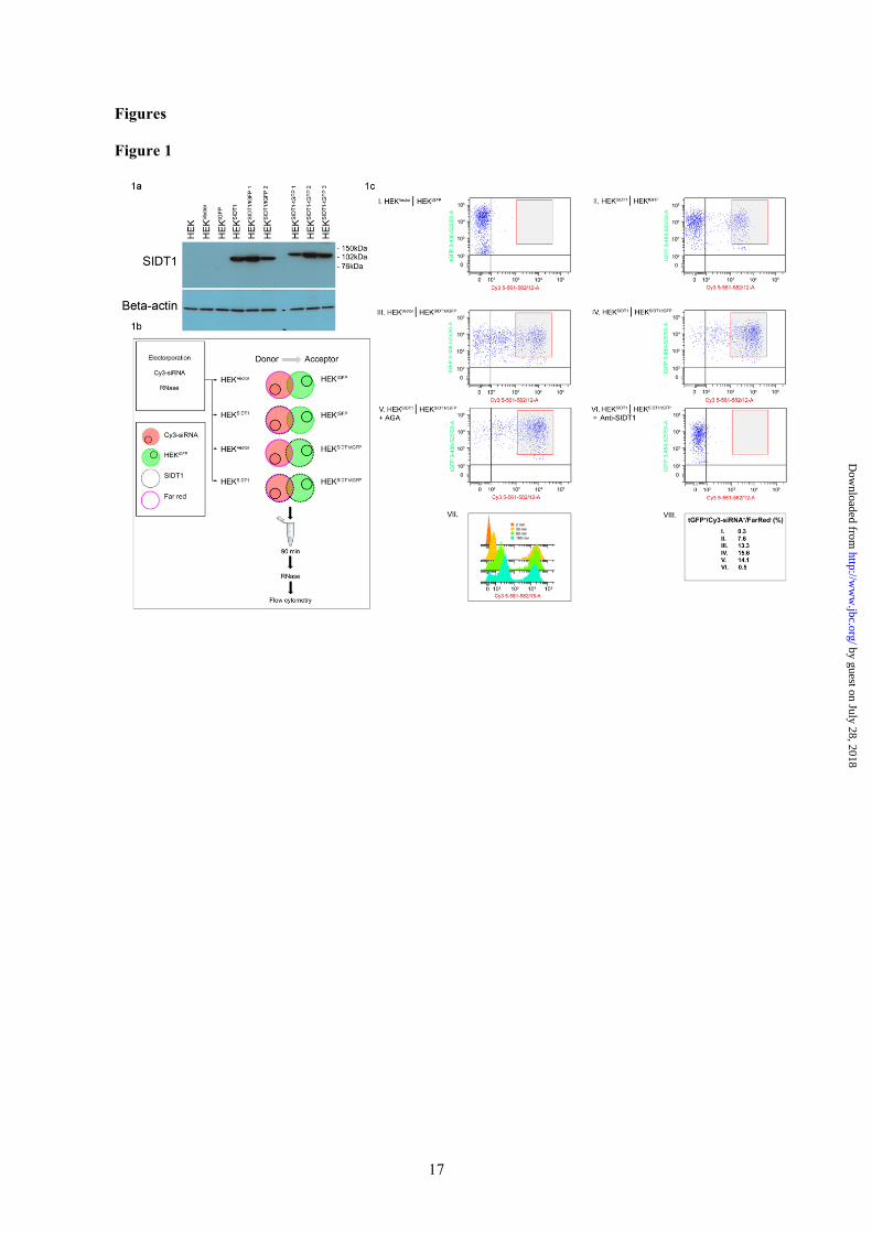
Figure Legends Figure 1. SIDT1 facilitates rapid contact-dependent intercellular Cy3-siRNA transfer between ‘Donor’ and ‘Acceptor’ subpopulations. (a). Characterisation of SIDT1 protein expression in HEK293-dervied stable transfectant cell lines. Representative Western blot analysis of SIDT1 expression in control HEK293 cells (HEKVector), tGFP control (HEKtGFP), SIDT1 (HEKSIDT1), SIDT1 and tGFP (SIDT1/tGFP, clone 1 and 2) and SIDT1-tGFP fusion (HEKSIDT1-tGFP) transfectants (clone 1-3). Beta-actin loading control. Electrophoretic mobility of SIDT-1 with C-terminal fusion tGFP protein (SIDT1-tGFP) corresponded to predicted Mr of 120kDa, versus 94kDa for SIDT1. (b). Schematic representation of siRNA transfer coculture experimental design. ‘Donor’ subpopulations comprised stable HEKVector or HEKSIDT1 transfectants. ‘Acceptor’ subpopulations comprised HEKtGFP or HEKSIDT1/tGFP transfectants. The ‘Donor’ subpopulation was tagged with far-red membrane linker and Cy3-siRNA introduced by electroporation. ‘Donor’ and ‘Acceptor’ subpopulations were directly cocultured for 90 min, 37°C, single cell suspensions generated and analysed by flow cytometry. Quantification of tGFP+/Cy3-siRNA+/FarRed- cells allowed ‘Donor’ to ‘Acceptor’ Cy3-siRNA transfer to be measured. (c). (I) In the absence of SIDT1 overexpression, Cy3-siRNA transfer was negligible. (II-IV) SIDT1 overexpression resulted in a marked increase in Cy3-siRNA transfer. SIDT1 overexpression in either ‘Donor’ or ‘Acceptor’ subpopulation was sufficient for this increase in Cy3-siRNA transfer to occur, indicating that SIDT1 facilitates bidirectional siRNA transfer. (V) Preincubation with alpha-glycyrrhetinic acid (AGA) had no effect on Cy3-siRNA transfer efficiency, indicating the effects of SIDT1 overexpression on Cy3-siRNA transfer to be independent of GJIC. Indirect coculture and conditioned medium exposure (90 min in each case) did not result in significant ‘Acceptor’ Cy3-siRNA acquisition (supplemental material 3b). (VI) Cy3-siRNA transfer was abolished by pre-incubation with anti-SIDT1 antibody. (VII) Time course of Cy3-siRNA transfer from HEKSIDT1 ‘Donor’ to HEKSIDT1/tGFP ‘Acceptor’ cells. Histogram representation of tGFP+/Cy3-siRNA+/FarRed- ‘Acceptor’ cells at specified time points. (VIII) Data presented are typical of quadruplicate biological repeat experiments. In each sample, 10000 single-cell events were recorded. Figure 2. A minority subpopulation increases non-autonomous miR-21 activity in adenocarcinoma cells. (a). BxPC3miR21 (doxycycline-inducible miR-21) or irrelevant miRNA (BxPC3miRN/S) controls were directly cocultured with miR-21 reporter cells (BxPC3CkmiR21) at the indicated ratios (± 1µg/ml doxycycline). Renilla luciferase luminescence (levels decreased by miR-21) was normalized to firefly luciferase luminescence to allow quantitative comparison (relative luminescence units, RLU). Doxycycline-induced BxPC3miR21 activation increased miR-21 activity in BxPC3CkmiR21 reporter cells. Coculture of BxPC3CkmiR21 with BxPC3miRN/S had no effect on normalized Renilla activity in BxPC3CkmiR21. Indirect coculture was insufficient to induce non-autonomous miR-21 activity. First white column indicates BxPC3CkmiR21 in standard monoculture. * P < 0.05 versus BxPC3miR21 + BxPC3CkmiR21, no doxycycline by multifactorial ANOVA, n = 4. (b). Representative Western blot analysis of SIDT1 expression in pancreatic adenocarcinoma (BxPC3, MIAPaCa2, Capan2) and immortalized normal ductal epithelial cells (HPDE4), demonstrating differential expression of SIDT1. Densitometric quantification (means ± SD) of SIDT1 signal, normalized to that of beta-actin. Mean values (± SD), n = 3.
by guest on July 28, 2018http://w
ww
.jbc.org/D
ownloaded from

14
Figure 3. SIDT1 is required for intercellular miR-21 transfer. (a). Knockdown of SIDT1 expression by siRNA was quantified by Western blotting. Three siRNAs targeting different regions of the SIDT1 sequence were compared. Mean (± SD) densitometric values are presented, n = 3. Levels of SIDT1 were most effectively decreased by siRNA 1, which targets the 3’UTR, but could be rescued by co-transfection of the SIDT1 expression vector (lacks the siRNA 1 3’UTR target), but not by control vector. (b). BxPC3CkmiR21 reporter activation by the minority (1%) BxPC3miR21 subpopulation was abrogated by transfection of siRNA 1, but not control siRNA, prior to coculture. SIDT1 ‘rescue’ restored BxPC3CkmiR21 miR-21 induction despite siRNA 1. Triplicate means (± SD). SIDT1 signal normalized to that of beta-actin. * P < 0.05 versus N/S siRNA; ** P < 0.05 versus 3’UTR siRNA 1 + vector, by multifactorial ANOVA, n = 5. Figure 4. SIDT1 deficiency abrogates chemoresistance induced by a minority miR-21-expressing subpopulation of pancreatic adenocarcinoma cells. Chemoresistance was quantified by MTT assay and IC50 values derived. (a) The IC50 of gemcitabine was significantly increased by direct coculture of doxycycline-activated BxPC3miR21 with BxPC3CkmiR21 cells (ratio 1:100). (b) The alteration in IC50 was minimal when the minority subpopulation was indirectly cocultured (Transwell). The gemcitabine IC50 was unaffected by coculture of BxPC3miRN/S with BxPC3CkmiR21. Means (± SD) are presented, n = 3. (c). The increase in IC50 induced by miR-21 was attenuated when cells were treated with SIDT1-3’UTR siRNA 1, but not by control mismatch siRNA. SIDT1 ‘rescue’ abrogated the effect of SIDT1-3’UTR siRNA 1. (d) Gap junction inhibition using 25µM alpha-glycyrrhetinic acid (AGA) did not significantly affect the increase in IC50 induced by miR-21. Figure 5. SIDT1 knockdown abrogates miR-21-induced chemoresistance. (a). Caspase 3 activities were quantified 24h following exposure to 1µM gemcitabine by colorimetric caspase 3 assay. Relative absorbances at 405nm with background subtraction are presented. Mean values (± SD) from four biological replicates. (b). Representative images of colony formation capacity for each condition are shown. Figure 6. Pancreatic stellate cell-induced cell adhesion mediated drug resistance (CAM-DR) is attenuated by anti-SIDT1 antibody.
(a). Pancreatic stellate cells (hPSC), expanded from pancreatic resection specimens, were immunocytochemically characterized. Representative image of PSC immunophenotype: smooth muscle actin, green; vitamin A granule autofluorescence, red; DAPI, blue. Magnification x40. Phase contrast image of PSC, Magnification x20.
(b). Patient-derived hPSC express SIDT1 protein. Western analysis of SIDT1 expression in patient-derived PSC cells (1-3). HEK293 cells serve as a negative control.
by guest on July 28, 2018http://w
ww
.jbc.org/D
ownloaded from

15
(c). Small RNA transfer between adenocarcinoma cells and hPSC is disrupted by anti-SIDT1 antibody. Flow cytometric analysis of Cy3-siRNA transfer between BxPC3 cells and hPSC. BxPC3 cells loaded with Cy3-siRNA (electroporation) and labeled with far red fluorescent linker were directly cocultured with hPSC (green fluorescent), 90 min. The subpopulation of Cy3-positive, far red negative cells (indicating Cy3-siRNA acquisition) was significantly reduced by anti-SIDT1 antibody.
(d). Stellate cell-induced CAM-DR is inhibited by anti-SIDT1 treatment. 5 x 10³ BxPCtGFP cells were directly cocultured with confluent hPSC for 72h in the presence of 1µM gemcitabine with anti-SIDT1 antibody or immunoglobulin control. tGFP fluorescence, attributable to the BxPC3 adenocarcinoma subpopulation, measured (Ex: 485nm/Em: 530nm) and normalized to MTT colorimetric readout of cell viability (OD560, with hPSC background subtraction). Direct coculture with hPSC increased adenocarcinoma cellular proliferation in the presence of gemcitabine, relative to standard monoculture. Anti-SIDT1 significantly decreased hPSC-induced CAM-DR. Mean values (± SD), n = 4.
by guest on July 28, 2018http://w
ww
.jbc.org/D
ownloaded from

16
Table 1. Oligonucleotide sequences
miR-21 5’-UAGCUUAUCAGACUGAUGUUGA-3’
miR-21 reporter oligonucleotides
5’-CGCAGTAGAGCTCTAGTTCAACATCAGTCTGATAAGCTAGTTT-3’
5’-AAACTAGCTTATCAGACTGATGTTGAACTAGAGCTCTACTGCGAT-3’.
A miR-21-resistant single base mismatch control oligonucleotides
5’-GCAGTAGAGCTCTAGTTCAACATCAGAAGATAAGCTAGTTT-3’
5’-AAACTAGCTTATCTTCTGATGTTGAACTAGAGCTCTACTGCGAT-3’.
SIDT1-specific siRNA 1 target sequence
5’-CUCUCAGGAUGAACGUAUU-3’
SIDT1-specific siRNA 2 target sequence
5’-CUACUGGGAUAGAUGUUUU-3’
SIDT1-specific siRNA 3 target sequence
5’-GAGCAAUUAUGGGACAAUAUU-3’
Cy3/control siRNA 5’-UAGCGACUAAACACAUCAAUU-3’
by guest on July 28, 2018http://w
ww
.jbc.org/D
ownloaded from
17
Figures Figure 1
by guest on July 28, 2018http://w
ww
.jbc.org/D
ownloaded from
Mohamed O. Elhassan, Jennifer Christie and Mark S. DuxburymicroRNA-21-driven chemoresistance
member 1 (SIDT1) mediates contact-dependent small RNA transfer and Homo sapiens systemic RNA interference defective-1 transmembrane family
published online December 15, 2011J. Biol. Chem.
10.1074/jbc.M111.318865Access the most updated version of this article at doi:
Alerts:
When a correction for this article is posted•
When this article is cited•
to choose from all of JBC's e-mail alertsClick here
Supplemental material:
http://www.jbc.org/content/suppl/2011/12/16/M111.318865.DC1
by guest on July 28, 2018http://w
ww
.jbc.org/D
ownloaded from