12-CRS-0106 REVISED 8 FEB 2013 Non-invasive Microwave Breast Cancer Detection - A Comparative Study...
17
Non-invasive Microwave Breast Cancer Detection - A Comparative Study Arezoo Modiri, Kamran Kiasaleh University of Texas at Dallas
-
Upload
john-kennedy -
Category
Documents
-
view
214 -
download
0
Transcript of 12-CRS-0106 REVISED 8 FEB 2013 Non-invasive Microwave Breast Cancer Detection - A Comparative Study...

Non-invasive Microwave Breast Cancer Detection - A Comparative Study
Arezoo Modiri, Kamran KiasalehUniversity of Texas at Dallas

http://www.cancer.org/acs/groups/content/@epidemiologysurveilance/documents/document/acspc-031941.pdf
Why New modality for Breast Cancer Detection in Needed?

Why Microwave-Based Diagnosis?
Frequent checkups need in-vivo, inexpensive, non-invasive, and convenient methods with
acceptable accuracy.
X-Ray:
- Ionizing,
- High false negative detection rate
(20%)
- In many cases, Painful
MRI:
- Expensive
- Not tolerable for some
Women (e.g. implant cases)
Ultrasound:
- Operator-dependent
- High false positive detection rate
(The false-positive rate is three times of that of X-ray => unnecessary biopsy.)
http://www.cancer.gov/cancertopics/factsheet/detection/mammograms
Why Microwave-Based Diagnosis?
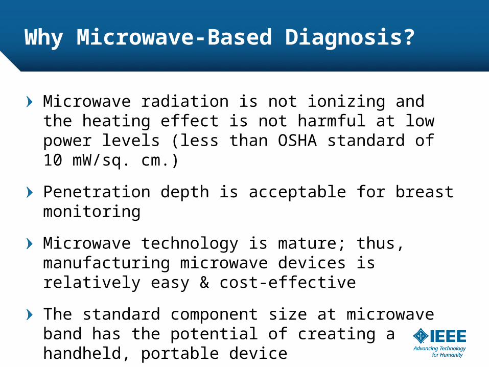
Microwave radiation is not ionizing and the heating effect is not harmful at low power levels (less than OSHA standard of 10 mW/sq. cm.)
Penetration depth is acceptable for breast monitoring
Microwave technology is mature; thus, manufacturing microwave devices is relatively easy & cost-effective
The standard component size at microwave band has the potential of creating a handheld, portable device

Are Any Other Research Groups Working On This Subject?
Dr. Paul Meaney – DartMouthDr. Susan Hagness – University of WisconsinDr. Elise Fear – University of CalgaryDr. Magda El-shenawee – University of ArkansasDr. Sima Noghanian – University of North DakotaDr. Natalia Nikolova – McMaster UniversityDr. John Stang – Duke University

Other Studies Target Clinical Applications

This Study’s Ultimate Goal
Portable, Self-Examine Tool which compensates for the defects of mammography by making check ups easier and more affordable for women and sending them to X-ray or MRI monitoring only when a signature is detected.

3D Radiator Design
The 3D radiator design was done in Ansoft HFSS
The digital phantom created by Ansoft was used
In order to have a full coverage of the tissue, hemisphere shape was chosen with 16 curled bent dipole antennas
Design frequency was chosen to be 1.2GHz since this was the longest antenna we could fit inside our structure
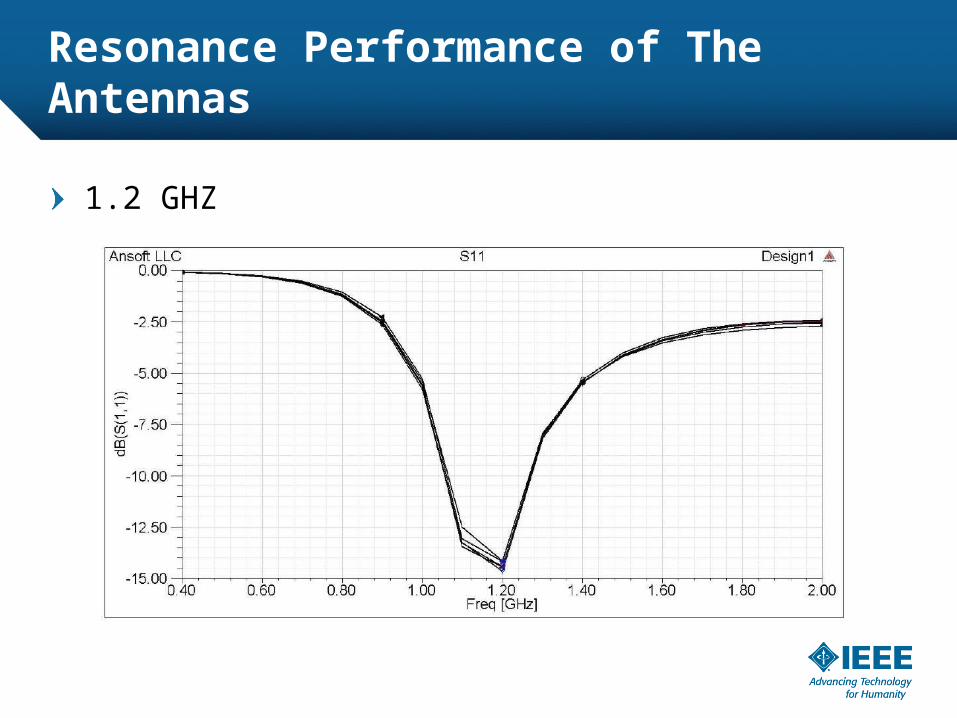
Resonance Performance of The Antennas
1.2 GHZ

The Two Versions of The Radiating Structure
One without conductive cover
One with a conductive cover added for Electromagnetic Shielding– Cause no interference
– Accept no interference
– Only the outer surface of the structure is covered by a conductive layer

Different Tumor Cases Are Considered
Different tumor shapes
Different tumor sizes
Different tumor locations

Electric Field Changes Are Studied
Both magnitude and phase contrasts are considered.
Cancerous model is exactly same as the normal one except for having one of the tumors inside it

How Signatures Are Analyzed
The signatures above a certain threshold are
added up.

Simulation Results for Two Tumor Cases

Specific Absorption Rate on Cut PlaneOSHA Compliance
SAR is 3.5W/Kg at the hottest spot.
The sphere is filled with fat.
1000 centimeter cube of fat is almost equal to 0.9Kg.
At the hottest spot, the power distribution is equal to (3mW/cubic cm) which is well
below the OSHA standard of (10 mW/square cm)
Conclusion
By studying a variety of tumor cases, it was shown that, overall, adding a conductor cover as electromagnetic shielding, not only creates an interference-free environment for measurement, but also significantly increases the cancer detection chance.