1174 IEEE TRANSACTIONS ON BIOMEDICAL ENGINEERING, VOL…jerby/68a.pdf · 1174 IEEE TRANSACTIONS ON...
9
1174 IEEE TRANSACTIONS ON BIOMEDICAL ENGINEERING, VOL. 53, NO. 6, JUNE 2006 Microwave Drilling of Bones Yael Eshet, Ronit Rachel Mann, Abby Anaton, Tomer Yacoby, Amit Gefen, Member, IEEE, and Eli Jerby*, Member, IEEE Abstract—This paper presents a feasibility study of drilling in fresh wet bone tissue in vitro using the microwave drill method [Jerby et al., 2002], toward testing its applicability in orthopaedic surgery. The microwave drill uses a near-field focused energy (typically, power under W at 2.45-GHz frequency) in order to penetrate bone in a drilling speed of mm/s. The effect of microwave drilling on mechanical properties of whole ovine tibial and chicken femoral bones drilled in vitro was studied using three-point-bending strength and fatigue tests. Properties were compared to those of geometrically similar bones that were equivalently drilled using the currently accepted mechanical rotary drilling method. Strength of mid-shaft, elastic moduli, and cycles to failure in fatigue were statistically indistinguishable between specimen groups assigned for microwave and mechanical drilling. Carbonized margins around the microwave-drilled hole were % the hole diameter. Optical and scanning electron microscopy studies showed that the microwave drill produces sub- stantially smoother holes in cortical bone than those produced by a mechanical drill. The hot spot produced by the microwave drill has the potential for overcoming two major problems presently associated with mechanical drilling in cortical and trabecular bone during orthopaedic surgeries: formation of debris and rupture of bone vasculature during drilling. Index Terms—Carbonization, mechanical properties, orthopaedic surgery, thermal damage. I. INTRODUCTION D RILLING in bones for orthopaedic and dental purposes has been a common practice for decades, using a variety of drilling bits [1], [2]. Rotary drills used today are efficient; however, they suffer several drawbacks including debris and chips spread resulting in foreign-body-reactions, substantial hematoma at the drilling site, heat generation, difficulties in attaining geometrical accuracy, and wobbling [1], [2]. An alter- native, more recent approach employs lasers for bone drilling [3], however, this method may be too costly for large-scale use in the clinical setting. This paper presents a novel approach for drilling of bones, using near-field microwaves [4], and provides Manuscript received November 25, 2004; revised September 25, 2005. This work was supported by the Israel Science Foundation under Grant 1270/04. As- terisk indicates corresponding author. Y. Eshet is with the Departments of Biomedical Engineering and Physical Electronics and Faculty of Engineering, Tel-Aviv University, Tel Aviv 69978, Israel (e-mail: [email protected]). R. R. Mann is with the Departments of Biomedical Engineering and Faculty of Engineering, Tel-Aviv University, Tel Aviv 69978, Israel. A. Anaton and T. Yacoby are with the Departments of Physical Electronics and Faculty of Engineering, Tel-Aviv University, Tel Aviv 69978, Israel. A. Gefen is with the Departments of Biomedical Engineering and Faculty of Engineering, Tel-Aviv University, Tel Aviv 69978, Israel. *E. Jerby is with the Departments of Physical Electronics and Faculty of Engineering, Tel-Aviv University, Ramat Aviv, Tel Aviv 69978, Israel (e-mail: [email protected]). Digital Object Identifier 10.1109/TBME.2006.873562 Fig. 1. Debris around the hole after mechanical drilling in vitro in cortical bone of chicken femora. the microwave energy characteristics for in vitro drilling in fresh (wet) bones as well as the results of mechanical and histological tests conducted in the microwave-drilled bones. A. Surgical Drilling in Hard Tissues: Review Up-to-Date Drilling in bone is a common surgical procedure, which may be required during preparation for insertion of a fixative or- thopaedic implant such as nail, screw or wire [5], or before in- sertion of a bone graft to enhance bone healing [6]. Presently, mechanical rotary drillers are the only type used in the clinical setting. Rotary drilling is performed at a wide range of speeds, from low to moderate ( rpm) up to ultra-high ( rpm) [7]. During such rotary drilling, bone debris accumulates around the drilling site (Fig. 1). Although measures are taken to carefully clean bone debris at the preclosure stage of surgery, remaining particles may induce a foreign-body-reaction around the implantation site. Such effects may delay bone healing or interfere with the process of osseointegration of the implant [8]. Rotary drilling also ruptures the vasculature at the drilling site. Substantial hematomas around the sites of rotary drilling were demonstrated using histology of rat femoral diaphysis [5]. Hematoxylin and eosin (H&E) histological staining in the same study also revealed coagulum material in the blood vessel lu- mens around the drilling site. The coagulum partially or fully occluded the vasculature at the site of drilling [5], imposing a second inhibiting effect on bone healing and osseointegration. Worse still, the ruptured vasculature and lymph systems provide a portal for infections and for entrance of toxins or wear parti- cles that may originate from the implant [9]. Since the stability of an orthopaedic fixation or a bone graft highly depend on the quality and quantity of the host bone, the above disadvantages of mechanical drilling may lead to longer postoperative recovery periods. Excessive heat generation in bone tissue during mechanical drilling has been reported, and was attributed mainly to friction during penetration of the rotary drill [7]. The heat generated by the friction between the drilling bit and bone tissue was shown to increase moderately between low and high drilling speeds, 0018-9294/$20.00 © 2006 IEEE
Transcript of 1174 IEEE TRANSACTIONS ON BIOMEDICAL ENGINEERING, VOL…jerby/68a.pdf · 1174 IEEE TRANSACTIONS ON...

1174 IEEE TRANSACTIONS ON BIOMEDICAL ENGINEERING, VOL. 53, NO. 6, JUNE 2006
Microwave Drilling of BonesYael Eshet, Ronit Rachel Mann, Abby Anaton, Tomer Yacoby, Amit Gefen, Member, IEEE, and
Eli Jerby*, Member, IEEE
Abstract—This paper presents a feasibility study of drilling infresh wet bone tissue in vitro using the microwave drill method[Jerby et al., 2002], toward testing its applicability in orthopaedicsurgery. The microwave drill uses a near-field focused energy(typically, power under 200 W at 2.45-GHz frequency) in orderto penetrate bone in a drilling speed of 1 mm/s. The effectof microwave drilling on mechanical properties of whole ovinetibial and chicken femoral bones drilled in vitro was studiedusing three-point-bending strength and fatigue tests. Propertieswere compared to those of geometrically similar bones that wereequivalently drilled using the currently accepted mechanicalrotary drilling method. Strength of mid-shaft, elastic moduli,and cycles to failure in fatigue were statistically indistinguishablebetween specimen groups assigned for microwave and mechanicaldrilling. Carbonized margins around the microwave-drilled holewere 15% the hole diameter. Optical and scanning electronmicroscopy studies showed that the microwave drill produces sub-stantially smoother holes in cortical bone than those produced bya mechanical drill. The hot spot produced by the microwave drillhas the potential for overcoming two major problems presentlyassociated with mechanical drilling in cortical and trabecular boneduring orthopaedic surgeries: formation of debris and rupture ofbone vasculature during drilling.
Index Terms—Carbonization, mechanical properties, orthopaedicsurgery, thermal damage.
I. INTRODUCTION
DRILLING in bones for orthopaedic and dental purposeshas been a common practice for decades, using a variety
of drilling bits [1], [2]. Rotary drills used today are efficient;however, they suffer several drawbacks including debris andchips spread resulting in foreign-body-reactions, substantialhematoma at the drilling site, heat generation, difficulties inattaining geometrical accuracy, and wobbling [1], [2]. An alter-native, more recent approach employs lasers for bone drilling[3], however, this method may be too costly for large-scale usein the clinical setting. This paper presents a novel approach fordrilling of bones, using near-field microwaves [4], and provides
Manuscript received November 25, 2004; revised September 25, 2005. Thiswork was supported by the Israel Science Foundation under Grant 1270/04. As-terisk indicates corresponding author.
Y. Eshet is with the Departments of Biomedical Engineering and PhysicalElectronics and Faculty of Engineering, Tel-Aviv University, Tel Aviv 69978,Israel (e-mail: [email protected]).
R. R. Mann is with the Departments of Biomedical Engineering and Facultyof Engineering, Tel-Aviv University, Tel Aviv 69978, Israel.
A. Anaton and T. Yacoby are with the Departments of Physical Electronicsand Faculty of Engineering, Tel-Aviv University, Tel Aviv 69978, Israel.
A. Gefen is with the Departments of Biomedical Engineering and Faculty ofEngineering, Tel-Aviv University, Tel Aviv 69978, Israel.
*E. Jerby is with the Departments of Physical Electronics and Faculty ofEngineering, Tel-Aviv University, Ramat Aviv, Tel Aviv 69978, Israel (e-mail:[email protected]).
Digital Object Identifier 10.1109/TBME.2006.873562
Fig. 1. Debris around the hole after mechanical drilling in vitro in cortical boneof chicken femora.
the microwave energy characteristics for in vitro drilling infresh (wet) bones as well as the results of mechanical andhistological tests conducted in the microwave-drilled bones.
A. Surgical Drilling in Hard Tissues: Review Up-to-Date
Drilling in bone is a common surgical procedure, which maybe required during preparation for insertion of a fixative or-thopaedic implant such as nail, screw or wire [5], or before in-sertion of a bone graft to enhance bone healing [6]. Presently,mechanical rotary drillers are the only type used in the clinicalsetting. Rotary drilling is performed at a wide range of speeds,from low to moderate ( rpm) up to ultra-high (rpm) [7]. During such rotary drilling, bone debris accumulatesaround the drilling site (Fig. 1). Although measures are takento carefully clean bone debris at the preclosure stage of surgery,remaining particles may induce a foreign-body-reaction aroundthe implantation site. Such effects may delay bone healing orinterfere with the process of osseointegration of the implant[8]. Rotary drilling also ruptures the vasculature at the drillingsite. Substantial hematomas around the sites of rotary drillingwere demonstrated using histology of rat femoral diaphysis [5].Hematoxylin and eosin (H&E) histological staining in the samestudy also revealed coagulum material in the blood vessel lu-mens around the drilling site. The coagulum partially or fullyoccluded the vasculature at the site of drilling [5], imposing asecond inhibiting effect on bone healing and osseointegration.Worse still, the ruptured vasculature and lymph systems providea portal for infections and for entrance of toxins or wear parti-cles that may originate from the implant [9]. Since the stabilityof an orthopaedic fixation or a bone graft highly depend on thequality and quantity of the host bone, the above disadvantages ofmechanical drilling may lead to longer postoperative recoveryperiods.
Excessive heat generation in bone tissue during mechanicaldrilling has been reported, and was attributed mainly to frictionduring penetration of the rotary drill [7]. The heat generated bythe friction between the drilling bit and bone tissue was shownto increase moderately between low and high drilling speeds,
0018-9294/$20.00 © 2006 IEEE

ESHET et al.: MICROWAVE DRILLING OF BONES 1175
and excessively when chips clog the flutes of the drill [7]. Tem-peratures of 89 C up to 185 C were recorded in distances of0.5 mm from the mechanically drilled holes [10], [11].
Rotary mechanical drills are difficult to be guided accurately.When touching the smooth bone cortex, rotary drills tend toslide and may dislocate or misalign [2]. Moreover, the diam-eter of the hole created is larger than the drill bit diameter, dueto wobbling effects [1]. After prolonged or repeated use, thedulling of the drill-bit edges increases heat generation and de-creases geometrical accuracy. Several attempts for improvingthe drilling process have been described, all concerned with drillbit geometry [1], drilling mechanism [12] or optimization of pa-rameters (applied force, drilling speed, etc.).
Laser ablation of hard tissues including bones has been com-pared to rotary drills for a variety of wavelengths and pulsestructures [3]. Under proper cooling conditions, the results ob-tained with laser ablation generally show high accuracy andclean cuts. The main disadvantage in laser drilling is the absentor delayed healing of the ablation site due to photo-acoustic andthermal damage effects [3].
Taken together, the literature above indicates that a newdrilling method is needed in orthopaedic surgery. Specifically,such new method should not produce bone debris, should notrupture blood vessels in bone during penetration and shouldnot involve wobbling effects during drilling. Microwave-baseddrilling can potentially fill these requirements.
B. Microwave-Tissue Interactions
Microwave interactions with biological tissues were studiedduring the last several years [13], [14] but no attempt was madeto employ microwave energy for the purpose of drilling inhard tissues. Reported microwave-tissue interactions includeheat generation by resistive losses of moving charged-ionsand oscillations of charged molecules, and heat transfer byinduced movements of charged ions. At temperatures over 50C, tissues undergo vaporization and carbonization. Higher
temperatures may cause desiccation, protein denaturation, co-agulation and finally welding and cavitation [13], [14]. It is notyet determined whether RF radiation has additional nonthermaleffects on biological tissues, though a recent study indicated thepossibility of its carcinogenicity [13]. Tumor ablation by RFradiation has drawn a growing interest recently [15]. The aimof this procedure is to cause coagulation necrosis of canceroustissue and, thus, tumor lysis and ablation. A percutaneouselectrode is inserted under imaging guidance (CT, MRI or US)to ablate subdermal lesions. The method was also used onbones for ablation of tumors such as osteoid osteoma [16] andmetastases [17].
C. The Microwave Drill
A new method for general purpose drilling which employsnear-field microwaves was recently introduced by Jerby et al.[4]. The microwave drill device is depicted in Fig. 2(a). Thecoaxial electrode radiates the microwave energy in the nearfield, thus producing a confined hot spot under the tip of theelectrode. The hot spot increases the local dielectric lossesof the material in a thermal runaway process [18], allowingthe electrode to penetrate deeper into the molten region. This
Fig. 2. Scheme of the microwave drill system. (a) Microwave energy is con-centrated in a small hot spot in front of the drilling bit, thus enabling its inser-tion into the bone. (b) Block diagram of the microwave drill cascade consistingof a 2.45-GHz magnetron tube protected by an isolator, a reflectometer unit,an impedance-matching tuner (to minimize the reflected power), and the mi-crowave drill device [shown schematically in (a)].
mechanism has no rotating or vibrating parts, and it does notproduce debris particles. The heat generated under the drillingtip is anticipated to immediately fuse the bone vasculaturecrossing the drilling path, thus eliminating hematomas andrelated coagulum. The microwave drill apparatus (Fig. 2) isconsiderably less expensive than any laser-based drill.
The objectives of this study were to 1) characterize themicrowave parameters (power, drilling speed) for drilling infresh wet bones in vitro; 2) determine if microwave drillinghas degrading effects on mechanical strength and stiffness offresh whole bones in vitro compared with standard mechanicaldrilling; 3) approximate the extent of tissue carbonization at themargins of holes produced by the microwave drill in vitro.
II. METHODS
A. Experimental Setup of the Microwave Drill
The experimental setup of the microwave drill is shown inFig. 2(b). The microwave cascade consists a power-controlledmagnetron (2.45 GHz) protected by an isolator, a reflectometerfor measuring the transmitted and reflected wave power, an E-Htuner for impedance matching, and a transition from the WR340rectangular waveguide to the coaxial microwave drill deviceshown in Fig. 2(a). The coaxial structure was cooled by pres-surized air ( bars). A constant external force of N wasapplied axially on the center electrode by means of weights (2Kg), mounted so that their center of mass was aligned with theaxis of the electrode.
For drilling in bovine trabecular bone (from the proximaltibial diaphysis), an effective power of 150–200 W wasrequired to produce 2.4-mm-diameter holes within 2–5 s. Forovine tibias with diaphysial cortical thickness of – mm,an effective microwave power of W was sufficient fordrilling 2.4-mm-diameter penetrating holes (using an electrode
1176 IEEE TRANSACTIONS ON BIOMEDICAL ENGINEERING, VOL. 53, NO. 6, JUNE 2006
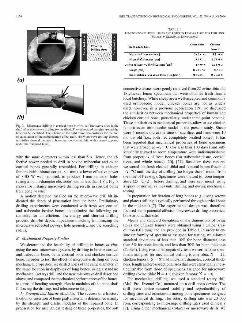
Fig. 3. Microwave drilling in cortical bone in vitro. (a) Transverse slice in theshaft after microwave drilling (ovine tibia). The carbonized margins around thehole can be identified. The scheme on the right frame demonstrates the methodof calculation of the carbonization effect ratio. (b) Microwave drilling showedno visible thermal damage to bone marrow (ovine tibia, with marrow exposedunder the fractured bone).
with the same diameter) within less than 5 s. Hence, the ef-fective power needed to drill in bovine trabecular and ovinecortical bones generally resembled. For drilling in chickenfemora (with thinner cortex, mm), a lower effective powerof W was required, to produce 1-mm-diameter holes(using a 1-mm-diameter electrode) within less than 1.8 s. Fig. 3shows for instance microwave drilling results in cortical ovinetibia bone in vitro.
A motion detector installed on the microwave drill bit in-dicated the depth of penetration into the bone. Preliminarydrilling experiments were conducted with fresh wet corticaland trabecular bovine bones, to optimize the following pa-rameters for an efficient, low-energy and shortest drillingprocess: drill-bit depth, impedance matching (minimizing themicrowave reflected power), hole geometry, and the scorchingradius.
B. Mechanical Property Studies
We determined the feasibility of drilling in bones in vitrousing the new microwave system, by drilling in bovine corticaland trabecular bone, ovine cortical bone and chicken corticalbone. In order to test the effect of microwave drilling on bonemechanical properties, we drilled holes of the same diameter, inthe same location in diaphyses of long bones, using a standardmechanical (rotary) drill and the new microwave drill describedabove, and compared the mechanical performances of the bones,in terms of bending strength, elastic modulus of the bone shaftfollowing the drilling, and tolerance to fatigue.
1) Strength and Elastic Modulus: The success of a fracturefixation or insertion of bone graft material is determined mainlyby the strength and elastic modulus of the repaired bone. Inpreparation for mechanical testing of these properties, the soft
TABLE IDIMENSIONS OF OVINE TIBIAS AND CHICKEN FEMORA USED FOR DRILLING
(MEANS � STANDARD DEVIATIONS)
connective tissues were gently removed from 22 ovine tibia and18 chicken femur specimens that were obtained fresh from alocal butchery. While sheep are a well-accepted and commonlyused orthopaedic model, chicken bones are not as widelyused, however, in a previous publication [19] we discussedthe similarities between mechanical properties of human andchicken cortical bone, particularly, under three-point bending.These similarities in mechanical properties allow to use chickenfemora as an orthopaedic model in the present study. Sheepwere 5 months old at the time of sacrifice, and hens were 10months old (i.e., both had completely ossified bones). It hasbeen reported that mechanical properties of bone specimensthat were frozen at C (for less than 100 days) and sub-sequently thawed to room temperature were indistinguishablefrom properties of fresh bones (for trabecular tissue, corticaltissue and whole bones) [20], [21]. Based on these reports,we stored the fresh cleaned tibial and femoral bones frozen at
C until the day of drilling (no longer than 1 month fromthe time of freezing). Specimens were thawed to room temper-ature (25 C) 2 h before drilling, and were kept moist (usinga spray of normal saline) until drilling and during mechanicaltesting.
In preparation for fixation of long bones (e.g., using screwsand plates) drilling is typically performed through cortical bonein the mid-shaft [7]. The experimental design was, therefore,focused on the potential effects of microwave drilling on corticalbone around that site.
Means and standard deviations of the dimensions of ovinetibias and chicken femora were obtained using a caliper (res-olution 0.01 mm) and are provided in Table I. In order to en-sure uniformity of specimens assigned for testing, we allowedstandard deviations of less than 10% for bone diameter, lessthan 5% for bone length, and less than 30% for bone thickness(Table I). Using two-tailed unpaired -tests we verified that spec-imens assigned for mechanical drilling (ovine tibia: ;chicken femora: ) had mid-shaft diameter, cortical thick-ness, length and cross-sectional area that were statistically indis-tinguishable from those of specimens assigned for microwavedrilling (ovine tibia: ; chicken femora: ).
For mechanical drilling, we used a standard rotary drill(MultiPro, Dremel Co.) mounted on a drill press device. Thedrill press device ensured stability and reproducibility ofdrilling sites and orientations among bone specimens assignedfor mechanical drilling. The rotary drilling rate was 20 000rpm, corresponding to mid-range drilling rates used clinically[7]. Using either mechanical (rotary) or microwave drills, we

ESHET et al.: MICROWAVE DRILLING OF BONES 1177
Fig. 4. Mechanical testing of drilled ovine tibias using three-point bending. (a) Experimental apparatus. (b) Scheme of the experiment showing the lower andupper supporting jigs (triangles). The upper jig applies a flexural force F which is balanced by reaction forces F=2 at each supporting lower jig. Lower jigs areL distance apart. The drilled hole is positioned opposed to the point of application of the flexural force. (c) Schematic cross-section through the bone mid-shaftdefining the major and minor diameters of the shaft.
created a single penetrating hole in the center of the shaft, withdiameter of mm and mm for the ovine tibiasand chicken femora, respectively. In mechanical drills, pene-tration speed was mm/s. In microwave drills, penetrationspeed was mm/s for drilling in ovine tibias, andmm/s for drilling in chicken femora. The center of the shaftwas identified and marked for each specimen as half the bonelength (Table I).
After drilling, each bone was subjected to a three-pointbending test at a deflection rate of 1 mm/min using an Instron5544 testing machine [Fig. 4(a)]. The span between the lowersupports [ , Fig. 4(b)] was scaled for bone length, and rangedbetween 13 and 14 cm for ovine tibias, and was set as 4.6 cmfor chicken femora. The upper support was pressing againstthe bone cortex at the side opposed to the location of the hole[Fig. 4(b)], so that bending-related tensile stresses were appliedaround the hole. A load cell with maximum capacity of 2 KNwas used to measure the applied load , which was recorded
as function of the flexural displacement caused by theupper support [Fig. 4(b)]. The failure load was used tocalculate the strength of bone under bending [22]
(1)
where is the minor diameter and is the moment of inertia ofthe cross-sectional area around the axis of the major diameter(Fig. 4(c), Table I). The slope of the force-displacement curve,
, was further used to calculate the elastic modulus ofcortical bone tissue [22]
(2)
We compared each mechanical property across groupsassigned for mechanical and microwave drilling, separately for

1178 IEEE TRANSACTIONS ON BIOMEDICAL ENGINEERING, VOL. 53, NO. 6, JUNE 2006
ovine tibias and chicken femora, using 2-tailed unpaired -tests.A value less than 0.05 was considered statistically significantin all statistical tests.
2) Fatigue: Fatigue studies were conducted in chickenfemora to simulate the endurance of drilled bones to a morephysiological loading scenario (i.e., repetitive, and less thanultimate strength loads). We assigned bones to three experi-mental groups: 1) drilled by microwave ; 2) drilledmechanically ; 3) controls, which were not drilled
. One-way ANOVA for each geometrical dimension(Table I) confirmed that all bone dimensions were insignificantacross groups and hence, geometrical uniformity of specimenswas verified. Specimen preparation and the drilling processwere as described in Section B–1.
Chicken femora from all groups were subjected to fatiguein three-point bending. The load amplitude was set as 220 N,which is % of the load to failure in three-point bendingfor the bone geometries considered herein (based on prelimi-nary three-point bending strength studies, ). The loadingspeed for fatigue was set as 50 mm/min, and the span betweenthe lower supports was again set as 4.6 cm. To determine ifmicrowave drilling had a different effect on cycles to fatiguefailure compared with mechanical drilling and controls, we rana one-way analysis of variance (ANOVA).
C. Area of Carbonization
Subsequent to mechanical testing we measured the area oftissue carbonization induced by the hot spot for ovine tibiasdrilled using the microwave drill. For that purpose, slices ofbone from both sides of the microwave-generated hole werecut transversally [Fig. 3(a)] using an electrically powered di-amond-coated disk saw (MultiPro, Dremel Co.). Measurementsof area of carbonization were conducted using image analysissoftware (SigmaScan Pro, SPSS Inc.) on digital photos of thebone slices (taken at a high-resolution of 2.3 mega pixels). Agraph paper was included in the images for calibration of di-mensions. We measured the areas of carbonization in the mar-gins of the hole and the projected area of the hole (“effectivedrilling area”) on each slice as shown in Fig. 3(a), right frame.We then divided the effective drilling area plus its carbonizedmargins by the net effective drilling area to obtain a carboniza-tion effect ratio. Ideally, when minimal carbonization occurs,this ratio should approach unity.
D. Optical and Scanning Electron Microscopy (SEM)
Additional chicken femora were drilled and assigned foroptical and SEM (microwave-drilled: ; mechanicallydrilled: ). Segments of 9 mm 9 mm were cut fromthese bones using a disk saw, so that they contained the hole intheir center. Samples were first studied under digital optical mi-croscopy (Axiolab A, Zeiss Co., reflective, magnification ).Measurements of spots of carbonization in microwave-drilledspecimens were taken using a special ruler for optical mi-croscopy (resolution 100 m) and a designated micrographimage processing software (SigmaScan Pro).
Second, samples were prepared for SEM studies (JSM840A,Jeol Co., MA, USA). In preparation for SEM, we mounted
Fig. 5. The microwave effective power (solid line) and drilling depth (dashedline) versus time during microwave drilling through (a) mid-shaft of ovine tibialbone (cortical bone) and (b) bovine trabecular bone.
the bone samples on aluminum discs, using conductive carbonpaint. The samples were coated with gold using a sputter coater(SC500, Polaron Co., UK). The acceleration voltage of theSEM was set as 15 KV and the filament current wasAmperes.
III. RESULTS
A. Characterization of Microwave Parameters
The variation of effective microwave power with depth ofpenetration and time during the drilling process is demonstratedin Fig. 5 for ovine cortical [Fig. 5(a)] and bovine trabecular[Fig. 5(b)] bone specimens. The effective microwave power fordrilling in cortical and trabecular bone components in vitro waskept under W and holes were produced after no more than5 s. The effective microwave power varied during the drillingprocess (Fig. 5) due to the variations in the microwave load-impedance and the consequent reflection from the bone as thedrilling process evolved.
B. Mechanical Property Studies
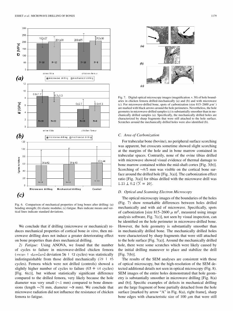
1) Strength and Elastic Modulus: Unpaired 2-tailed -testsshowed that mechanical properties of ovine tibias subjectedto microwave drilling were statistically indistinguishable fromthose of tibias drilled mechanically [bending strengthMPa, elastic modulus GPa, Fig. 6(a) and (b)]. Simi-larly, mechanical properties of chicken femora drilled withmicrowave or drilled mechanically were indistinguishable[bending strength MPa, elastic modulus GPa,Fig. 6(a), (b)].
ESHET et al.: MICROWAVE DRILLING OF BONES 1179
Fig. 6. Comparison of mechanical properties of long bones after drilling: (a)bending strength; (b) elastic modulus; (c) fatigue. Bars indicate means and ver-tical lines indicate standard deviations.
We conclude that if drilling (microwave or mechanical) re-duces mechanical properties of cortical bone in vitro, then mi-crowave drilling does not induce a greater deteriorating effecton bone properties than does mechanical drilling.
2) Fatigue: Using ANOVA, we found that the numberof cycles to failure in microwave-drilled chicken femora( deviation cycles) was statisticallyindistinguishable from those drilled mechanically (cycles). Femora which were not drilled (controls) showed aslightly higher number of cycles to failure ( cycles)[Fig. 6(c)], but without statistically significant differencecompared to the drilled femora, very likely because the holediameter was very small ( mm) compared to bone dimen-sions (length mm, diameter mm). We conclude thatmicrowave radiation did not influence the resistance of chickenfemora to fatigue.
Fig. 7. Digital optical microscopy images (magnification� 30) of hole bound-aries in chicken femora drilled-mechanically (a) and (b) and with microwave(c). For microwave-drilled bone, spots of carbonization (size 815–2600 �m )are marked with black arrows around the hole perimeters. Nevertheless, the holegeometry in microwave-drilled samples (c) is substantially smoother than in me-chanically drilled samples (a). Specifically, the mechanically drilled holes arecharacterized by sharp fragments that were still attached to the hole surface.Scratches around the mechanically drilled holes were also identified (b).
C. Area of Carbonization
For trabecular bone (bovine), no peripheral surface scorchingwas apparent, but crosscuts sometime showed slight scorchingat the margins of the hole and in bone marrow contained intrabecular spaces. Contrarily, none of the ovine tibias drilledwith microwave showed visual evidence of thermal damage tobone marrow contained within the mid-shaft cortex [Fig. 3(b)].Scorching of mm was visible on the cortical bone sur-face around the drilled hole [Fig. 3(a)]. The carbonization effectratio [Fig. 3(a)] for tibias drilled with the microwave drill was
.
D. Optical and Scanning Electron Microscopy
The optical microscopy images of the boundaries of the holes(Fig. 7) show remarkable differences between holes drilledmechanically and with aid of microwave. Specifically, spotsof carbonization [size 815–2600 m , measured using imageanalysis software, Fig. 7(c)], not seen by visual inspection, canbe identified on the hole perimeter in microwave-drilled bone.However, the hole geometry is substantially smoother thanin mechanically drilled bone. The mechanically drilled holeswere characterized by sharp fragments that were still attachedto the hole surface [Fig. 7(a)]. Around the mechanically drilledhole, there were some scratches which were likely caused bythe initial drilling maneuver to place and stabilize the drill[Fig. 7(b)].
The results of the SEM analyses are consistent with thoseof optical microscopy, but the high-resolution of the SEM de-tected additional details not seen in optical microscopy (Fig. 8).SEM images of the entire holes demonstrated that hole geom-etry is substantially smoother in microwave drilling [Fig. 8(a)and (b)]. Specific examples of defects in mechanical drillingare the large fragment of bone partially detached from the holesurface [marked by arrow “A” in Fig. 8(a), right frame], sharpbone edges with characteristic size of 100 m that were still

1180 IEEE TRANSACTIONS ON BIOMEDICAL ENGINEERING, VOL. 53, NO. 6, JUNE 2006
Fig. 8. SEM images of chicken femora drilled with microwave (left column) and mechanically (right column): (a) view of the holes from above; (b) and (c) typicalimperfections attached to the hole perimeters. Hole geometry is substantially smoother in microwave drilling (a). Typical defects in mechanical drilling are thelarge fragment of bone partially detached from the hole surface (arrow “A”), sharp bone edges still attached to the contour of the hole (arrow “C”), and scratches[(c), right frame]. Typical imperfections in microwave drilling are strut-like elements around the hole perimeter (arrow “B”) and small lumps (region “D”), whichwere apparently formed by melted bone minerals. Magnifications and scales are provided under each SEM frame.
attached to the contour of the hole after drilling [arrow “C,”Fig. 8(b), right frame], and scratches with width of about 10 maround the hole, which were probably caused by drill vibrationsduring the first contact with bone [Fig. 8(c), right frame]. SEMimages of microwave drilling also showed some typical imper-fections likely to be related with the heat generated during thedrill. Specifically, we observed strut-like elements around the
hole perimeter, with length of less than 1 m and thickness ofless than 0.2 m [arrow “B,” Fig. 8(b), left frame], which ap-pear to be hardened fibers of melted bone minerals. A seconddefect characteristic of microwave drilling was small lumps [di-mensions of 100–200 m, region “D” in Fig. 8(c), left frame],apparently of melted bone minerals, which were pushed out ofthe hole by the microwave electrode.

ESHET et al.: MICROWAVE DRILLING OF BONES 1181
IV. DISCUSSION
The feasibility of drilling in cortical and trabecular bonetissues using microwave radiation was demonstrated in this invitro study. The hot spot produced by the microwave drill hasthe potential for overcoming a major problem currently relatedwith mechanical drilling in bone: formation of debris leading toforeign-body-reaction (Figs. 1, 6, and 7). When passing throughblood vessels in bone, the microwave drill can potentially weldthe vessels contacting the hot spot (as opposed to ruptureof vessels during mechanical drilling). Hence, the risk forinfection may be lower in microwave drilling. The microwavedrilling process is relatively quick and is geometrically precise(no mechanical wobbling is involved). Drilling penetrationcan be monitored and controlled on a timescale in the orderof fractions of a second (Fig. 5). Importantly, the microwavedrill system is substantially more economical than laser-baseddrills. The microwave drill technology is versatile and withtailored design, can be considered for a wide range of clinicalapplications. These may include insertions of orthopaedicpins, nails and screws into bones, wire fixation, neurosurgery,dentistry, cranial surgeries, open-chest cardiac surgeries, andany other procedure requiring accurate, carefully controlledbone drilling.
In vitro drilling in bone provided several encouraging results:1) microwave drilling did not degrade the mechanical proper-ties of bone in vitro more than rotary drilling did; 2) carboniza-tion at the margins of the hole in cortical bone was relativelysmall ( % of the effective hole diameter); 3) holes producedby microwave drilling were substantially smoother than thoseproduced mechanically, and no partially attached bone frag-ments were observed under microscopy (optical or SEM) in mi-crowave-drilled holes.
We assume that carbonized bone in vitro represents the min-imal volume of damaged bone tissue around the microwavedrilling site. We cannot rule out further tissue damage in non-carbonized regions in vivo in bones drilled with microwave, e.g.,due to thermal and microwave radiation effects, and this shouldbe quantified in in vivo (animal) studies. However, it may bepossible to reduce the carbonization margins further before pro-ceeding to in vivo studies, using adaptive control on the mi-crowave power, by a liquid or air cooling mechanism. Basicstudies on thermal properties and thermal tolerance of bonetissue under microwave radiation can also be beneficial in orderto minimize the carbonized margins of the hole.
Some limitations in this study should be recognized. First, invitro bone specimens, rather than living bones, were used for therotary and microwave drilling tests. Although bone specimenswere obtained fresh and kept moist with a saline spray duringtesting to closely represent the living bone, the important in vivoeffects such as viability of osteocytes, tissue necrosis, and hy-peremia could not be analyzed at this stage and are awaitingfurther studies. The interaction of microwave heating with fluidflow in bone vessels is similarly not considered in vitro. Second,a drill press was used during the rotary drilling experimentsrather than a hand-held orthopaedic surgical drill. To replicateclinical conditions as closely as possible, a drill speed of 20 000rpm, representative of actual orthopaedic drills was used for all
rotary drilling maneuvers [7]. The authors of this paper believethat the advantages of site reproducibility and stability providedby the drill press apparatus were required for the present exper-imental design, which was aimed at comparing “ideal” rotaryand microwave drills.
Safety of the patient undergoing surgery and of the staff whooperates the drill is a critical issue to consider at this stage ofresearch, as microwave radiation may be hazardous. A tailoreddesign of the microwave drill and its screening for each specificsurgical application is expected to significantly reduce the radi-ation exposure of both the patient and staff to a permitted min-imum. It should be noted that existing RF ablation procedures[e.g., 16] use RF power of up to 150 W for about 15 min (s), much longer than needed for microwave drilling in bones.Operated in a similar power level, microwave drilling throughcortical bone only lasts 2–3 s (Fig. 5). Nevertheless, research ef-forts are required to determine whether microwave drilling in-duces a risk for carcinogenic effects, and how such risk can beminimized.
REFERENCES
[1] S. Saha, S. Pal, and J. A. Albright, “Surgical drilling: Design andperformance of an improved drill,” J. Biomech. Eng., vol. 245, pp.252–109, 1982.
[2] K. L. Wiggins and S. Malkin, “Drilling of bone,” J. Biomech., vol. 553,pp. 559–9, 1976.
[3] J. T. Payne, G. M. Peavy, L. Reinisch, and D. C. Van Sickle, “Corticalbone healing following laser osteotomy using 6.1 �m wavelength,”Lasers Surg., Med., vol. 38, pp. 43–29, 2001.
[4] E. Jerby, V. Dikhtyar, O. Aktushev, and U. Groslick, “The microwavedrill,” Science, vol. 587, pp. 589–298, 2002.
[5] S. Chiba, K. Okada, K. Lee, G. V. Segre, and R. M. Neer, “Molecularanalysis of defect healing in rat diaphyseal bone,” J. Veterinary Med.Sci., vol. 603, pp. 608–63, 2001.
[6] M. J. Park, M. C. Lee, and S. C. Seong, “A comparative study of thehealing of tendon autograft and tendon-bone autograft using patellartendon in rabbits,” Int. Orthop., vol. 35, pp. 39–25, 2001.
[7] S. R. Davidson and D. F. James, “Drilling in bone: Modeling heat gen-eration and temperature distribution,” J. Biomech. Eng., vol. 305, pp.314–125, 2003.
[8] J. F. Kay, L. Gilman, and T. C. May, “The tri-spade drill for endosseousdental implant installation,” J. Oral Implantol., vol. 424, pp. 8–17,1991.
[9] S. B. Goodman, M. Lind, Y. Song, and R. L. Smith, “In-vitro, in-vivo,and tissue retrieval studies on particulate debris,” Clin Orthop., vol. 25,pp. 34–352, 1998.
[10] M. B. Abouzgia and D. F. James, “Measurements of shaft speedwhile drilling through bone,” J. Oral Maxillofac. Surg., vol. 1308, pp.1315–53, 1995.
[11] A. R. Eriksson and T. Alberktsson, “Temperature threshold levelsfor heat-induced bone tissue injury: A vital-microscopic study in therabbit,” J. Prosthet. Dent., vol. 101, pp. 107–50, 1983.
[12] L. S. Nichter, S. Richlin, P. M. Navarrette, and K. Kosari, “The biome-chanical efficacy of an oscillating K-wire driver,” Ann. Plast. Surg., vol.289, pp. 292–29, 1992.
[13] G. S. Gazelle, S. N. Goldberg, L. Solbiati, and T. Livraghi, “Tumor ab-lation with radio-frequency energy,” Radiology, vol. 633, pp. 646–217,2000.
[14] A. Copty, F. Sakran, M. Golosovsky, M. D. Davidov, and A. Frenkel,“Low-power near-field microwave applicator for localized heating ofsoft matter,” Appl. Phys. Lett., vol. 5109, pp. 5111–84, 2004.
[15] M. Mashevich, D. Folkman, A. Kesar, A. Barbul, R. Korenstein, E.Jerby, and L. Avivi, “Exposure of human peripheral blood lymphocytesto electromagnetic fields associated with cellular phones leads to chro-mosomal instability,” Bioelectromagnetics, vol. 82, pp. 90–24, 2003.
[16] I. Ghanem, L. M. Collet, K. Kharrat, E. Samaha, H. Deramon, P. Mertl,and F. Dagher, “Percutaneous radiofrequency coagulation of osteoidosteoma in children and adolescents,” J. Pediatr. Orthop. B., vol. 244,pp. 252–12, 2003.

1182 IEEE TRANSACTIONS ON BIOMEDICAL ENGINEERING, VOL. 53, NO. 6, JUNE 2006
[17] M. R. Callstrom, “Painful metastases involving bone: Feasibility of per-cutaneous CT- and US- guided radio-frequency ablation,” Radiology,vol. 87, pp. 97–224, 2002.
[18] E. Jerby, O. Aktushev, and V. Dikhtyar, “Theoretical analysis of the mi-crowave-drill near-field localized heating effect,” (in 034 909) J. Appl.Phys., vol. 97, 2005.
[19] N. Passi and A. Gefen, “Trabecular bone contributes to strength of theproximal femur under mediolateral impact in the avian,” J. BiomechEng., vol. 198, pp. 203–127, 2005.
[20] J. C. Goh, E. J. Ang, and K. Bose, “Effect of preservation medium onthe mechanical properties of cat bones,” Acta. Orthop. Scand., vol. 465,pp. 467–60, 1989.
[21] F. Linde and H. C. Sorensen, “The effect of different storage methodson the mechanical properties of trabecular bone,” J. Biomech., vol.1249, pp. 1252–26, 1993.
[22] J. M. Gere and S. P. Timoshenko, Mechanics of materials. Boston,MA: PWS-KENT, 1972.
Yael Eshet was born in Tel-Aviv, Israel, in 1971. Shereceived the M.D., degree in 1996, the B.Sc. degreein electrical engineering in 2005, the B.Sc. degree inphysics in 2005, and the M.Sc. degree in biomed-ical engineering in 2004, all from Tel-Aviv Univer-sity, Tel-Aviv, Israel.
She is currently a second-year Medical Residentwith the Department of Imaging, Sheba MedicalCentre, , Tel-Hashomer, Israel. Her residency com-bines active work as a physician and research work.She is also participating in her free time in a research
group in the Department of Biomedical Engineering and Physical Electronics,Faculty of Engineering, Tel-Aviv University, Israel.
Ronit Rachel Mann was born in Ramat-Gan, Israel,in 1976. She received the B.Sc. degree in mechan-ical engineering from the Technion, Haifa, Israel, in2003. She is currently working toward the M.Sc. de-gree, specializing in biomedical engineering. Her re-search is microwave drilling in bone and in particularand the effects of microwave drilling on bone mor-phology and mechanical properties.
Abby Anaton was born in Istanbul, Turkey, in 1977.He received the B.Sc. degree in electrical engineeringfrom Tel Aviv University, Tel-Aviv, Israel, in 2003.
He is currently working toward the M.Sc. degree.His thesis concerns microwave heating of concrete.
His areas of interest are microwave system engi-neering and computational electromagnetics.
Tomer Yacoby was born in Israel in 1978. He re-ceived the B.Sc. degree in electrical engineering fromTel Aviv University, Tel-Aviv, Israel, in 2003. He iscurrently working toward the M.Sc. degree, special-izing in electromagnetics.
His research areas are microwave heating andprocessing of materials, and microwave systemengineering.
Amit Gefen (S’96–M’01) was born in Ramat-Gan,Israel, in 1971. He received the B.Sc. degree inmechanical engineering and the M.Sc. and Ph.D.degrees in biomedical engineering from Tel AvivUniversity, Tel Aviv, Israel, in 1994, 1997, and 2001,respectively.
During 2002–2003, he was a Postdoctoral Fellowat the Injury Biomechanics Laboratory of the Bio-engineering Department at the University of Pennsyl-vania, Philadelphia. He is currently a Senior Lecturerwith the Department of Biomedical Engineering, in
the Faculty of Engineering of Tel Aviv University. His research interests and ex-perience are in studying normal and pathological effects of mechanical factorson the structure and function of tissues, with emphasis on the musculoskeletalsystem.
Eli Jerby (M’04) received the M.Sc. and Ph.D. de-grees in electrical engineering, from Tel-Aviv Uni-versity, Tel-Aviv, Israel, in 1980 and 1989, respec-tively.
He worked at the Massachusetts Institute of Tech-nology with the late Prof. G. Bekefi on free-electronlasers and cyclotron-resonance masers. Since his re-turn to Tel Aviv University in 1991, he has developedand studied several microwave sources and applica-tions, including the microwave-drill invention and itsexperimental, theoretical, and practical aspects.