Continuous Processing of Nanocellulose and Polylactic Acid ...
Role of Polylactic Acid in Bone Regeneration –A Systematic Review
Tamilanbu Rajendran1 Suresh Venugopalan2
1.Postgraduate student, saveetha dental college& hospital
2. Reader, saveetha dental college & hospitalNo – 162 Poonamalle High Road, Chennai – 600077, Chennai, Tamil nadu, India
Abstract: Bone regeneration is a complex, physiological process of bone formation, which can be seen during normal fracture healing, and is involved in continuous remodeling throughout adult life. However, there are complex clinical conditions in which bone regeneration is required in large quantity, such as for skeletal reconstruction of large bone defects created by trauma, infection, tumor resection and skeletal abnormalities. The use of bone grafts is the standard to treat skeletal fractures, or to replace and regenerate lost bone. The most commonly used is auto graft, its use can lead to complications such as pain, infection, scarring, blood loss, and donor-site morbidity. An ideal bone graft or scaffold should be made of biomaterials that imitate the structure and properties of natural bone. However, creating living tissue constructs that are structurally, functionally and mechanically comparable to the natural bone has been a challenge so far. An electronic search was conducted for articles in PubMed database to screen for articles from 1966 to sept 2014 discussing the role of polylactic acid in bone regeneration using selected keywords. Critical appraisal was done for selected articles, Search yields a total of 383 articles of which 318 full text article were obtained, 30 articles were selected after reading the abstract out of which 10 met the inclusion criteria and 308 were excluded and hand search was done with the reference of the selected articles. The article selected used polylactic acid for bone regeneration. The present Systematic review indicates, when polylactic acid is modified with osteoinductive material such as beta tricalcium phosphate and hydroxyl appetite crystals shows better acceptance than plain polylactic acid.
Keywords: Polylactic acid, Polylactic acid scaffold, 3d printed polylactic acid Osseo conduction, bone regeneration, bone repair, bone remodeling, Osseo induction
INTRODUCTION: Bone, as a living tissue has the ability to heal by itself, however if the defect exceeds the critical size it will not heal spontaneously. [1] So helping measures are required for completion of healing process of such defect where various bone grafts and bone substitutes have been tried by several clinicians to healing of those large defects. A bone graft is defined as an implanted material that promotes bone healing alone or in combination with other material(s) [2]. The selection of an ideal bone graft relies on several factors such as tissue viability, defect size, graft size, shape and volume, biomechanical characteristics, graft handling, cost, ethical issues, biological characteristics, and associated complications [3] The materials used in bone grafting can be divided into several major categories, including auto grafts, allografts, and xenografts. Synthetic and biologically based, tissue-engineered biomaterials and combinations of these substitutes are other options. Autogenous bone are gold standard for bone regenerative material for several reasons including biocompatibility, no adverse reaction and they enclose osteoblasts that participate in new bone formation. There are limitations to obtaining autogenous grafts, however such as donor site morbidity and limited donor site. [4] One of the most recent developments in the defect is application of barrier membranes to occlude skeletal defects against invasion of soft tissue and thus allow osseous regeneration to fill the space underneath the
membrane. This technique is termed as guided tissue regeneration or guided bone regeneration. [5] This systematic review was attempted to analyze existing literature on polylactic acid and their role on osseous regeneration of fractured bone and induced defects in both human and animal models.
MATERIAL AND METHODS: SOURCES USED: An electronic search was conducted for articles in PubMed database to screen for articles from 1966 to sept 2014 discussing the role of polylactic acid in bone regeneration selected keywords and additionally hand searching was done. SEARCH TERMS USED IN PUBMED: (Maxilla) OR Mandible) AND (Polylactic acid) OR Polylactic acid scaffolds) OR PLA) OR PLA scaffolds) OR Poly-L-lactide) OR Poly-L-lactide scaffolds) OR PLLA) OR PLLA Scaffolds) OR Polylactide) OR Polylactide scaffolds) OR 3d printed polylactic acid) OR 3d printed PLA) OR 3d printed poly-L-lactide) OR 3d printed PLLA) OR 3d printed Polylactide)) OR Polylactic acid screws) OR Polylactic acid powder) OR Polylactic acid fibers) OR Polylactic acid plates) OR 3d printed polylactic acid) OR 3d printed polylactic acid scaffold)))) AND (Osseo conduction [Mesh Terms]) OR Bone regeneration [Mesh Terms]) OR Bone formation) OR Bone growth) OR Bone repair) OR Osseo induction) OR Bone remodeling) OR Hematoxin eosin staining) OR Micro radiograph) OR Histophotometry) OR SEM)
Tamilanbu Rajendran et al /J. Pharm. Sci. & Res. Vol. 7(11), 2015, 960-966
960

SELECTION OF STUDIES: Titles and abstracts of the search were initially screened for relevant articles and hand search of selected journals and in the reference selected articles were also done. And articles were further screened using inclusion and exclusion criteria. Inclusion criteria: Studies that fulfilled the following criteria were included in the study.
1. Studies on polylactic acid 2. Studies on different types of polylactic acid used 3. Studies on Osteogenesis 4. Studies on both humans and animals 5. Studies with follow up of minimum up to 4 weeks duration
Exclusion Criteria: 1. Studies on surface treated polylactic acid 2. Studies on scaffolds other than polylactic acid 3. Studies on regeneration other than bone.
4. Articles in other languages 5. Review article
Results of the electronic search: The systematic search revealed total of 383articles of which 318 full text article were obtained. 30 articles were selected after reading the abstract out of which 10 met the inclusion criteria and 308 were excluded. The article selected used polylactic acid for bone regeneration. Finally, selected articles were subjected to data extraction. The search flow chart is as shown in Fig.1.
RESULT:
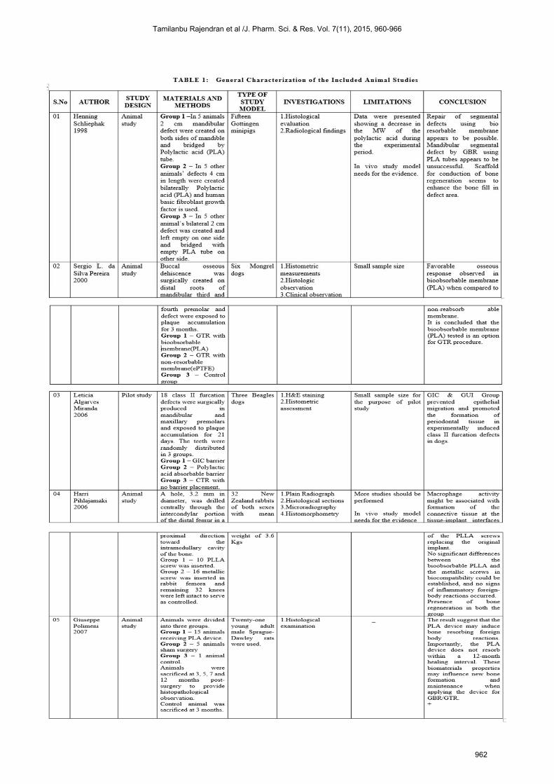
Table: 1 General Characterization of the Included Animal Studies Table: 2 Characterization of Included Human Studies Table: 3 Polylactic Acid Used as a Scaffold or Graft Material Table: 4 Polylactic Acid Used as Plates and Screws.
Tamilanbu Rajendran et al /J. Pharm. Sci. & Res. Vol. 7(11), 2015, 960-966
961
Tamilanbu Rajendran et al /J. Pharm. Sci. & Res. Vol. 7(11), 2015, 960-966
962

Tamilanbu Rajendran et al /J. Pharm. Sci. & Res. Vol. 7(11), 2015, 960-966
963

Tamilanbu Rajendran et al /J. Pharm. Sci. & Res. Vol. 7(11), 2015, 960-966
964

DISCUSSION:
Literature search performed in PubMed reveled insufficient cohesive database or studies with similar parameters. Although polylactic acid material was used in various studies they were either used as polylactic acid in tube forms, scaffolds, or fibers, screws and as plates they have been compared with surface modified polylactic acid. Because of the insufficient cohesive database of the study, meta-analysis were not performed. The literature search indicated towards the increase in use of polylactic acid material as a synthetic graft and in screws and plate forms. And also a viable option as an alternative to other graft materials. The interactive factors which play a role in osseous regeneration are discussed below. 1. BONE REGENERATION USING POLYLACTIC ACID Animal study conducted Using polylactic acid tubes on one side of the mandible and empty defect on other side, another group treated with polylactic acid combined with recombinant human basic fibroblast growth factor (rhbFGF) there were no bone regeneration seen with polylactic acid tube. Group combined with growth factor showed adequate bone regeneration. Author suggested that use of beta tricalcium phosphatase scaffold along with polylactic acid will have an advantage in bone regeneration Sergio. L.da & silva Pereira studied with GTR using polylactic acid and non-resorbable membrane (polytetrafluoroethylene) it shows that polylactic acid has increase in bone area width compared with non-resorbable membrane. Bone formation is confirmed using histological analysis and histometric analysis. [2] Yasuji HARADA studied using helical and non-helical (PLLA) fibers using piezoelectric charge under tensile stress. The relationship between tensile forces applied to PLLA fibers which in turn generates piezoelectricity was accessed and same piezoelectric stimulus was correlated to osteogenic stimulation. Significantly there was higher ossification was observed around helical PLLA Fibers [6] Letica algavarves Miranda studied using an experimental barrier of resin-modified GIC in one group and polylactic acid barrier in another group. It shows that both GIC barrier and polylactic acid barrier showed epithelial migration and promotes formation of new periodontal tissues, GIC group is more effective than polylactic group in terms of regeneration. If the scaffolds using for bone regeneration is porous in nature, there will be faster degradation of the scaffolds. Bone regeneration were evaluated using (micro CT). [9] 2. DEGRADATION OF POLYLACTIC ACID: When polylactic acid is used as the synthetic scaffolding material, it is important to measure the degradation of the polymer. Only 2 studies measured the degradation of PLA in animals, shows that polylactic acid has slow degrading property that starts only after 4 weeks of duration and the complete degradation will be taken place only after 12 months. [3, 5]
3. HISTOLOGICAL FINDINGS: Many studies reported that PLA membrane was surrounded by external surface of bone and connective tissues. [2] Leticia alga rives and harri studied that defect was completely filled with connective tissues and varying amount of bone and epithelial proliferation at 8 weeks shows bone regeneration. [3] Giuseppe study shows PLA was surrounded by fibrous capsule and multinucleated giant cell, some macrophages and lymphocytes confirms there were evidence of new bone formation. [5]. Presence of numerous spindle shaped mesenchymal cells were observed in PLA fiber in 2 weeks. PLA fiber surface was attached to trabecular bone where osteoblast lines of ossification were observed on the bone around the fiber. [6] Polylactic acid was covered with smooth connective tissue at 4 weeks post operatively, presence of spindle shaped cells associated with Type I and type III collagen is seen in 4, 8 and 16 weeks. [7] Use of pla in humans Many studies has been used polylactic acid in humans in plate and screw forms for fracture fixation and in socket preservation. PLA has given promising results in union of fractured bone in humans. [10] Limitations: In the reviewed articles, limitations of the studies were no specific form or peculiar type of polylactic acid material was repeatedly tried, still more detailed analysis of polylactic acid is required. The review of the selected articles suggests that polylactic acid scaffold can be used for osseous regeneration of fractured bone. More intensive investigations are necessary to identify the factors that influence the efficiency of osseous regeneration.
CONCLUSION: Repair of segmental defect using the bio absorbable membrane (PLA) appears to be a promising alternative for synthetic graft which can replace the entire lost bone or can be used as the scaffolding material for GTR / GBR and in fractured bone. The present review indicates, when polylactic acid is modified with osteoinductive material such as beta tricalcium phosphate and hydroxyl appetite crystals shows better acceptance in animal model than plain polylactic acid
REFERENCES: 1. Henning Schliephake and Georg Berding- Evaluation of bone
healing in patients with bone grafts and end osseous implants using single photon emission tomography (SPECT) - Clinical Oral Implants Research Volume 9 Issue 1, pages 34–42, February 1998.3xe
2. Silva Pereira, Sergio L. da Comparison of Bioabsorbable and Non-Resorbable Membranes in the Treatment of Dehiscence-Type Defects. A Histomorphometric Study in Dogs - Journal of periodontology (Chicago, Ill.: American Academy of Periodontology), Vol. 71, No. 8 (2000), p. 1306-131.
3. Harri Pihlajamaki - Long-term tissue response to bio absorbable poly-l-lactide and metallic screws: An experimental study - Harri Pihlajamaki October 2006Volume 39, Issue 4, Pages 932– 937.
4. Scandinavica - A resin-modified glass ionomer cement barrier for treating degree II furcation defects: A pilot study in dogs Acta Odontologica Scandinavica Volume 64, Issue 1, 2006.
Tamilanbu Rajendran et al /J. Pharm. Sci. & Res. Vol. 7(11), 2015, 960-966
965

5. G. Poimeni.et al - Histopathological Observations of a Polylactic Acid-Based Device Intended for Guided Bone/Tissue Regeneration.clin Implant Dent Relat Res
6. Yasuji Harada - Hydroxyapatite/Poly-L-lactide acid screws have better biocompatibility and femoral burr hole closure than does Poly-L-lactide acid alone - J Biomater Appl February 2014 28: 954-962.
7. Hanako Nishimoto & Takeshi Kokubu & Atsuyuki Inui & Yutaka Mifune & Kotaro Nishida & Hiroyuki - Ligament regeneration using an absorbable stent-shaped poly-L-lactic acid scaffold in a rabbit International Orthopedics (SICOT) (2012) 36:2379–238
8. Guillermo E. Chacon, DDS James P. Ellis, DDS, MS John R. Kalmar, DDS, PhD Edwin A. McGlumphy - Using resorbable screws for fixation of cortical onlay bone grafts: An in vivo study in rabbits - - journal of oral maxillofacial surgery November 2004Volume 62, Issue 11, Pages 1396–1402.
9. Letica algavarves Miranda - A resin-modified glass ionomer cement barrier for treating degree II furcation defects: A pilot study in dogs - Acta Odontologica Scandinavica Volume 64, Issue 1, 2006 37- 41
10. Takahio Suzuki - Resorbable poly-l-lactide plates and screws for the treatment of mandibular condylar process fractures: a clinical and radiologic follow-up study. J Oral Maxillofacial Surg 2004; 62(8):919-24
11. Asano K, Matsuno T, Tabata Y, Satoh T. Preparation of thermoplastic poly (L-lactic acid) membranes for guided bone regeneration. Int J Oral Maxillofacial Implants. 2013 Jul-Aug; 28(4).
12. Reyes R, Pec MK, Sánchez E, Del Rosario C, Delgado A, Évora C. Comparative, osteochondral defect repair: stem cells versus chondrocytes versus bone morphogenetic protein-2, solely or in combination. Eur Cell Mater. 2013 Jul8; 25:351-65.
13. Pan H, Hao S, Zheng Q, Li J, Zheng J, Hu Z, Yang S, Guo X, Yang Q. Bone induction by biomimetic PLGA copolymer loaded with a novel synthetic RADA16-P24 peptide in vivo. Mater Sci Eng. C Mater Biol Appl. 2013 Aug 1; 33(6):3336-45.
14. Harada Y, Kadono K, Terao T, Suzuki M, Ikada Y, Tomita N. Helical conformation endows poly-l-lactic acid fibers with a piezoelectric charge under tensile stress. J Vet Med Sci. 2013; 75(9).
15. Gupta A, Woods MD, Illingworth KD, Niemeyer R, Schafer I, Cady C, Filip P, El-Amin SF 3rd. Single walled carbon nanotube composites for bone tissue engineering. J Orthop Res. 2013 Sep; 31(9):1374-81.
16. Prokop A, Jubel A, Hahn U, Dieters Hagen M, Bleidistel M, Peters C, Hoff A, Rehm KE. A comparative radiological assessment of polylactide pins over 3 years in vivo. Biomaterials. 2005 Jul; 26(19):4129-38.
17. Keskin DS, Tezcaner A, Korkusuz P, Korkusuz F, Hasirci Collagen-chondroitin sulfate-based PLLA-SAIB-coated rhBMP-2 delivery system for bone repair. Biomaterials. 2005 Jun; 26(18):4023-34.
18. Stavropoulos A, Sculean A, Karring T. GTR treatment of intrabony defects with PLA/PGA copolymer or collagen bioresorbable membranes in combination with deproteinized bovine bone (Bio-Oss). Clin Oral Investing. 2004 Dec; 8(4):226-32. Epub 2004
19. Honda MJ, Yada T, Ueda M, Kimata Cartilage formation by serial passaged cultured chondrocytes in a new scaffold: hybrid poly (L-
lactide-epsilon-caprolactone) sponge. J Oral Maxillofacial Surg. 2004 Dec; 62(12):1510-6.
20. Borden M, Attawia M, Khan Y, El-Amin SF, Laurencin CT. Tissue-engineered bone formation in vivo using a novel sintered polymeric microsphere matrix. J Bone Joint Surg Br. 2004 Nov; 86(8):1200-8.
21. Kandziora F, Pflugmacher R, Scholz M, Eindorf T, Schnake KJ, Haas NP. Bioabsorbable interbody cages in a sheep cervical spine fusion model. Spine (Phila Pa 1976). 2004 Sep 1; 29(17):1845-55
22. Silva GA, Costa FJ, Coutinho OP, Radin S, Ducheyne P, Reis RL Synthesis and evaluation of novel bioactive composite starch/bioactive glass micro particles. J Biomed Mater Res A. 2004 Sep 1; 70(3):442-9.
23. Yu X, Botchwey EA, Levine EM, Pollack SR, Laurencin CT. Proc Natl Acad Sci Bioreactor-based bone tissue engineering: the influence of dynamic flow on osteoblast phenotypic expression and matrix mineralization.2004 Aug 3;101(31):11203-8
24. Van Eijk F, Saris DB, Riesel J, Willems WJ, Van Blitterswijk CA, Verbout AJ, Dhert WJ. Tissue engineering of ligaments: a comparison of bone marrow stromal cells, anterior cruciate ligament, and skin fibroblasts as cell source. Tissue Eng. 2004 May-Jun; 10(5-6):893-903.
25. Kaito T, Myoui A, Takaoka K, Saito N, Nishikawa M, Tamai N, Ohgushi H, Yoshikawa Potentiation of the activity of bone morphogenetic protein-2 in bone regeneration by a PLA-PEG/hydroxyapatite composite. Biomaterials. 2005 Jan; 26(1):73-9.
26. Pflugmacher R, Eindorf T, Scholz M, Gumnior S, Krall C, Schleicher P, Haas NP, Kandziora F. Chirurg. Biodegradable cage. Osteointegration in spondylosis of the sheep cervical spine.2004 Oct; 75(10):1003-12.
27. Liao SS, Cui FZ, Zhang W, Feng QL. Hierarchically biomimetic bone scaffold materials: Nano-HA/collagen/PLA composite. J Biomed Mater Res B Appl Biomater. 2004 May 15; 69(2):158-65.
28. Schmidmaier G, Wildemann B, Ostapowicz D, Kandziora F, Stange R, Haas NP, Raschke M. Long-term effects of local growth factor (IGF-I and TGF-beta 1) treatment on fracture healing. A safety study for using growth factors. J Orthop Res. 2004 May; 22(3):514-9.
29. Kokubo S, Mochizuki M, Fukushima S, Ito T, Nozaki K, Iwai T, Takahashi K, Yokota S, Miyata K, Sasaki N. Long-term stability of bone tissues induced by an osteoinductive biomaterial, recombinant human bone morphogenetic protein-2 and a biodegradable carrier. Biomaterials. 2004 May; 25(10):1795-803.
30. Ogawa R, Mizuno H, Watanabe A, Migita M, Shimada T, Hyakusoku Osteogenic and chondrogenic differentiation by adipose-derived stem cells harvested from GFP transgenic mice. Biochem Biophys Res Commun. 2004 Jan 23; 313(4):871-7.
31. Patist CM, Mulder MB, Gautier SE, Maquet V, Jerome R, Oudega Freeze-dried poly (D, L-lactic acid) macro porous guidance scaffolds impregnated with brain-derived neurotrophic factor in the transected adult rat thoracic spinal cord. Biomaterials. 2004 Apr; 25(9):1569-82
32. Arosarena OA, Collins WL. Defect repair in the rat mandible with bone morphogenic protein 5 and prostaglandin. Arch Otolaryngology Head Neck Surg. 2003 Oct;129(10):1125-30
Tamilanbu Rajendran et al /J. Pharm. Sci. & Res. Vol. 7(11), 2015, 960-966
966