1.1 Plant and Animal Cells - Wikispaces · understand the cell. Plant and Animal Cells ... a...
18
8 UNIT A Tissues, Organs, and Systems of Living Things The Discovery of the Cell When the microscope was invented in the mid-1600s, it became possible for scientists to look at the previously invisible world of the cell. Imagine the strange and beautiful structures that appeared before the eyes of these scientists. Today, we use sophisticated electron microscopes that allow us to not only see the cell in detail but also to get a glimpse of some amazing sights (Figure 1.1). Robert Hooke was the first to describe cells in 1663 (Figure 1.2). He thought that the cells were the passages for fluids in a plant. Today, we understand that a cell is the basic building block of life. Every living organism is made of cells. A cell takes in nutrients from its environment and releases waste products into its environment. A cell can also divide to make copies of itself. A cell contains everything that it needs to live and grow. Using Technology to Study the Cell In the early days of cell biology, scientists used simple light microscopes to view sliced sections of living cells. These microscopes helped scientists see and study the external structure of a cell but revealed few details about the tiny specialized working parts within the cell. Here is a summary of what you will learn in this section: • Cells have special structures that enable them to perform important life functions. • Scientists use technology, such as the microscope, to understand the cell. Plant and Animal Cells Figure 1.1 A piece of moss, as seen through a microscope, shows many cells filled with chloroplasts, an organelle involved in photosynthesis. The cells are shown at a magnification of 500. 1.1 Figure 1.2 Robert Hooke’s drawing of cork cells, as seen under a microscope. He used the term “cells” based on what he saw.
Transcript of 1.1 Plant and Animal Cells - Wikispaces · understand the cell. Plant and Animal Cells ... a...

8 UNIT A Tissues, Organs, and Systems of Living Things
The Discovery of the CellWhen the microscope was invented in the mid-1600s, it becamepossible for scientists to look at the previously invisible world of thecell. Imagine the strange and beautiful structures that appeared beforethe eyes of these scientists. Today, we use sophisticated electronmicroscopes that allow us to not only see the cell in detail but also to geta glimpse of some amazing sights (Figure 1.1).
Robert Hooke was the first to describe cells in 1663 (Figure 1.2). He thought that the cells were the passages for fluids in a plant. Today,we understand that a cell is the basic building block of life. Every livingorganism is made of cells. A cell takes in nutrients from itsenvironment and releases waste products into its environment. A cellcan also divide to make copies of itself. A cell contains everything thatit needs to live and grow.
Using Technology to Study the CellIn the early days of cell biology, scientists used simple light microscopesto view sliced sections of living cells. These microscopes helpedscientists see and study the external structure of a cell but revealed fewdetails about the tiny specialized working parts within the cell.
Here is a summary of what youwill learn in this section:
• Cells have special structuresthat enable them to performimportant life functions.
• Scientists use technology, suchas the microscope, tounderstand the cell.
Plant and Animal Cells
Figure 1.1 A piece of moss, as seen through a microscope, shows many cells filled withchloroplasts, an organelle involved in photosynthesis. The cells are shown at a magnification of 500�.
1.1
Figure 1.2 Robert Hooke’s drawingof cork cells, as seen under amicroscope. He used the term“cells” based on what he saw.
ist10_ch01.qxd 7/22/09 3:23 PM Page 8

9Cells are the basic unit of life and often combine with other cells to form tissues.
Advances in technology, such as the development of the electronmicroscope (Figure 1.3), have allowed biologists to learn detailedinformation about different cell parts and their functions. Technologyhas also made the process of learning about the cell easier. For example,the electron microscope can produce images that are 1000 times moredetailed than the light microscope (Figure 1.4).
The discovery of the cell is an example of how scientific knowledgedepends on technology. As our technology continues to improve, ourknowledge and understanding of the cell will continue to expand.
A2 Quick Lab
What We Remember about the Cell
Cells come in a variety of shapes and sizes. However,there are some structures that are common to cells.There are also some differences. This activity will giveyou an opportunity to review the information that youknow about the cell.
PurposeTo create a graphic organizer that shows what youremember about the cell
Procedure
1. Work in a small group of 2–4 students.
2. Brainstorm for two minutes with your group aboutwhat you remember about the cell. You may wishto use words, pictures, or phrases. Think aboutthe different parts of the cell, the functions ofthese parts, or different examples of cells.
3. Create a graphic organizer using the words,pictures, or phrases that you came up with in step 2.
Questions
4. Sometimes, we remember things better if we canvisualize an example or illustration. What type ofcell did you visualize when you werebrainstorming about the cell?
5. There are many parts in a cell. Sometimes, it iseasier to remember the functions of the differentcell parts by using analogies to everyday things.For example, we may say that the cell has a partthat acts like a brain. Use an analogy to describeone specific part of the cell that you placed inyour graphic organizer.
6. Did your group find that it was easier toremember the parts of the cell, functions of thecell, or examples of cells? Explain.
Figure 1.4 Red blood cells viewed through ascanning electron microscope (magnification3700�)
Figure 1.3 The world’s most powerfulelectron microscope, the Titan 80-300Cubed, was installed at McMasterUniversity in Hamilton, Ontario, inOctober 2008.
ist10_ch01.qxd 7/22/09 3:23 PM Page 9

Cell Parts and Their FunctionsAll living things are made of cells. Our bodies are made up of between10 trillion (1013) and 100 trillion (1014) cells. A cell is the basic unit of life.Each cell contains smaller parts called organelles. These organelles havespecial functions that maintain all the life processes of the cell, including:
• intake of nutrients • exchange of gases
• movement • waste removal
• growth • reproduction
• response to stimuli
There are two types of cells: plant cells and animal cells (Figures 1.5 and 1.6).
10 UNIT A Tissues, Organs, and Systems of Living Things
rough endoplasmic reticulum
smooth endoplasmic reticulum
chromatin
nucleus
cell membrane
Golgi apparatus
mitochondrion
chloroplast
cytoskeleton
cell wall
wall of adjacent cell
central vacuole
cytoplasm
ribosomes (small brown dots)
nucleolus
Figure 1.5 A plant cell
WORDS MATTER
The word “cell” is derived from theLatin word cellula, meaning smallcompartment. The word “cyto,” as incytoplasm, is from the Greek rootmeaning cell.
ist10_ch01.qxd 7/22/09 3:23 PM Page 10

Structures and Organelles in CellsA cell contains structures and organelles that carry out variousfunctions. Although all cells must perform the tasks that maintain life,not all cells are identical. Therefore, some structures and organelles arethe same in both plant and animal cells while other structures andorganelles differ between plant and animal cells.
11Cells are the basic unit of life and often combine with other cells to form tissues.
chromatinnucleolus
nucleus
cell membrane
Golgi apparatus
mitochondrionlysosome
cytoplasm
ribosomes(small brown dots)
cytoskeleton
vacuole
rough endoplasmicreticulum
smooth endoplasmicreticulum
Figure 1.6 An animal cell
ist10_ch01.qxd 7/22/09 3:23 PM Page 11

Cell MembraneEvery cell has a cell membrane that forms a protectivebarrier around the cell (Figure 1.7). The cell membrane ismade of a double layer of lipids. A lipid is a fat-likemolecule that does not dissolve in water. The cellmembrane is designed to allow different substances tomove through it.
One process for moving substances across the cellmembrane is called diffusion. Diffusion depends on theconcentration of the substance on both sides of membrane.The amount of dissolved particles, called solutes, in asolution is the concentration. When a substance ispresent in different concentrations on either side of the cellmembrane, the particles will diffuse, or move, from an areaof high concentration to an area of lower concentration(Figure 1.8).
CytoplasmAll cells contain cytoplasm, a jelly-like substance that fills the cell andsurrounds the organelles (Figure 1.7). Cytoplasm contains the nutrientsrequired by the cell to carry on its life processes. The organelles aresuspended in the cytoplasm. The physical nature of the cytoplasmallows the nutrients and organelles to move within the cell.
NucleusThe nucleus is the control centre organelle of the cell (Figure 1.7). Itcontrols all the activities in a cell, including growth and reproduction.The nucleus is surrounded by the nuclear envelope, which containspores to allow the transport of materials. Most nuclei also contain asmall dense area called the nucleolus.
12 UNIT A Tissues, Organs, and Systems of Living Things
cellmembrane
area of lowconcentration
area of highconcentration movement of
substances
substances
(a) (b) (c )
Figure 1.8 (a) There is a higher concentration of substances on one side of the cell membrane. (b) The substances move to the side that has a lower concentration until a balanced state, called equilibrium, isattained. (c) When equilibrium is reached, the substances diffuse across the cell membrane in both directions.
Figure 1.7 A cell showing the cell membrane,cytoplasm, and large nucleus (magnification 6000�)
cellmembrane
nucleus
cytoplasm
ist10_ch01.qxd 7/22/09 3:23 PM Page 12
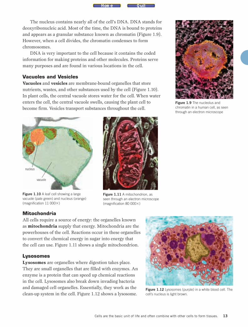
The nucleus contains nearly all of the cell’s DNA. DNA stands fordeoxyribonucleic acid. Most of the time, the DNA is bound to proteinsand appears as a granular substance known as chromatin (Figure 1.9).However, when a cell divides, the chromatin condenses to formchromosomes.
DNA is very important to the cell because it contains the codedinformation for making proteins and other molecules. Proteins servemany purposes and are found in various locations in the cell.
Vacuoles and VesiclesVacuoles and vesicles are membrane-bound organelles that storenutrients, wastes, and other substances used by the cell (Figure 1.10). In plant cells, the central vacuole stores water for the cell. When waterenters the cell, the central vacuole swells, causing the plant cell tobecome firm. Vesicles transport substances throughout the cell.
MitochondriaAll cells require a source of energy: the organelles knownas mitochondria supply that energy. Mitochondria are thepowerhouses of the cell. Reactions occur in these organellesto convert the chemical energy in sugar into energy thatthe cell can use. Figure 1.11 shows a single mitochondrion.
LysosomesLysosomes are organelles where digestion takes place.They are small organelles that are filled with enzymes. Anenzyme is a protein that can speed up chemical reactions in the cell. Lysosomes also break down invading bacteriaand damaged cell organelles. Essentially, they work as theclean-up system in the cell. Figure 1.12 shows a lysosome.
13Cells are the basic unit of life and often combine with other cells to form tissues.
Figure 1.12 Lysosomes (purple) in a white blood cell. Thecell’s nucleus is light brown.
Figure 1.11 A mitochondrion, asseen through an electron microscope(magnification 80 000�)
Figure 1.10 A leaf cell showing a largevacuole (pale green) and nucleus (orange)(magnification 11 000�)
Figure 1.9 The nucleolus andchromatin in a human cell, as seenthrough an electron microscope
nucleus
vacuole
ist10_ch01.qxd 7/22/09 3:23 PM Page 13
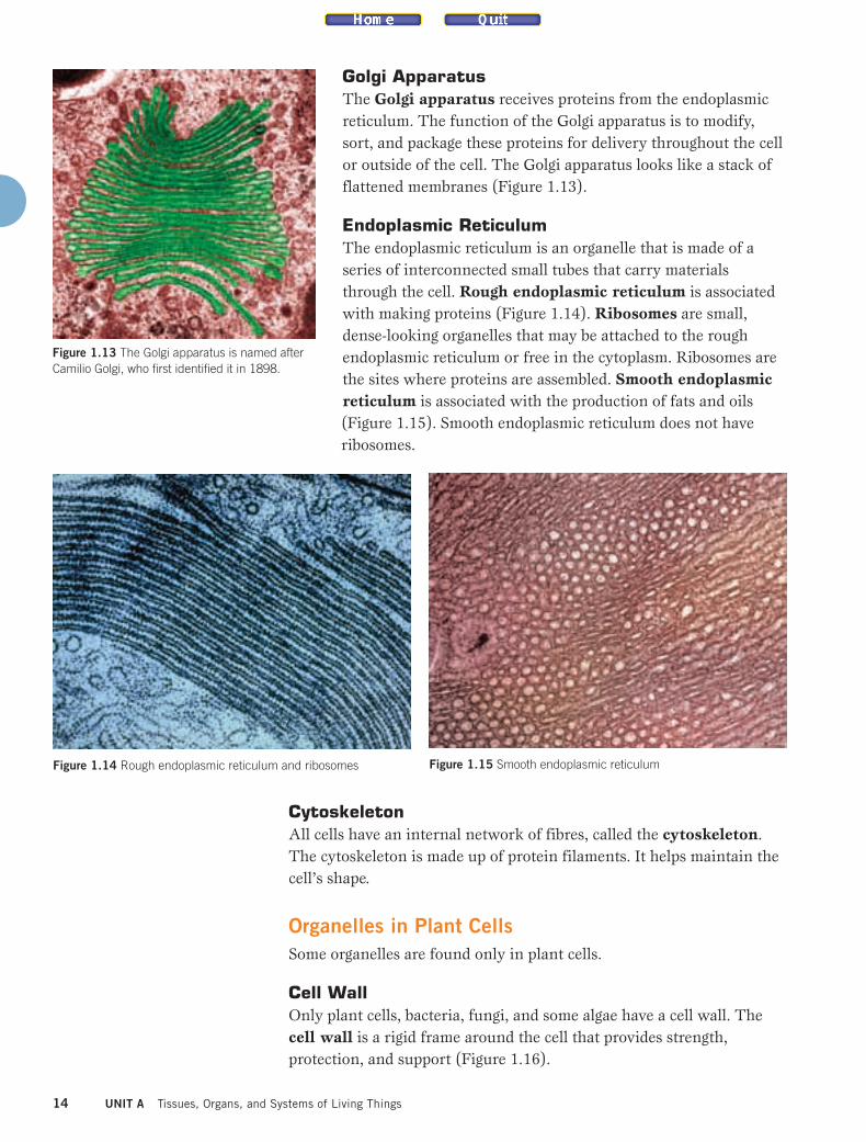
Golgi ApparatusThe Golgi apparatus receives proteins from the endoplasmicreticulum. The function of the Golgi apparatus is to modify,sort, and package these proteins for delivery throughout the cellor outside of the cell. The Golgi apparatus looks like a stack offlattened membranes (Figure 1.13).
Endoplasmic ReticulumThe endoplasmic reticulum is an organelle that is made of aseries of interconnected small tubes that carry materialsthrough the cell. Rough endoplasmic reticulum is associatedwith making proteins (Figure 1.14). Ribosomes are small,dense-looking organelles that may be attached to the roughendoplasmic reticulum or free in the cytoplasm. Ribosomes arethe sites where proteins are assembled. Smooth endoplasmicreticulum is associated with the production of fats and oils(Figure 1.15). Smooth endoplasmic reticulum does not haveribosomes.
CytoskeletonAll cells have an internal network of fibres, called the cytoskeleton.The cytoskeleton is made up of protein filaments. It helps maintain thecell’s shape.
Organelles in Plant CellsSome organelles are found only in plant cells.
Cell WallOnly plant cells, bacteria, fungi, and some algae have a cell wall. Thecell wall is a rigid frame around the cell that provides strength,protection, and support (Figure 1.16).
14 UNIT A Tissues, Organs, and Systems of Living Things
Figure 1.14 Rough endoplasmic reticulum and ribosomes Figure 1.15 Smooth endoplasmic reticulum
Figure 1.13 The Golgi apparatus is named afterCamilio Golgi, who first identified it in 1898.
ist10_ch01.qxd 7/22/09 3:23 PM Page 14

15Cells are the basic unit of life and often combine with other cells to form tissues.
One Word Connectsto Another Word
In the passage on chloroplasts,
note the way in which each term
is connected to another term.
Create a concept map to show the
connections. Begin with a top
bubble with the term
“chloroplast,” and then connect
the other terms as you read the
paragraph. Try this strategy with
another paragraph that contains
new terms.
During Reading
Figure 1.16 A leaf cell showing the cell wall and many chloroplasts (magnification 1000�)
cell wall
chloroplasts
chloroplast
stroma
granum
thylakoids
Figure 1.17 Photosynthesis takes place in the chloroplast in a plant cell.
ChloroplastsChloroplasts are found only in plant cells and some algae. Theseorganelles contain a green substance called chlorophyll. Chlorophyll usesenergy from the Sun to convert carbon dioxide and water into sugar andoxygen in a process called photosynthesis. Figure 1.17 shows theinternal structure of a chloroplast. The chloroplast is made up of littlesacs known as thylakoids. Thylakoids are stacked together in a waythat resembles a stack of coins. They are surrounded by a thick fluidcalled stroma. A stack of thylakoids is called a granum; chloroplasts mayhave many grana. You can think of the thylakoids as being “solarcollectors.” They collect light energy from the Sun, which is used duringthe process of photosynthesis to produce carbohydrates. Thecarbohydrates are used for the growth of the plant.
ist10_ch01.qxd 7/22/09 3:23 PM Page 15

Differences between Plant and Animal CellsCell walls and chloroplasts are only found in plant cells. However, thereare other differences between plant and animal cells:
• Plant cells contain a specialized chemical compound calledchlorophyll, a pigment that makes photosynthesis possible.
• Plant cells have a large central vacuole. Vacuoles in animal cellstend to be small.
• Some plant cells store energy in the form of starch or oils, such ascornstarch and canola oil. Animal cells store energy in the formof glycogen, a carbohydrate, or as lipids in the form of fats.
• Some animal cells have specialized compounds: for example,hemoglobin in red blood cells and cholesterol in other cells.
• Animal cells have centrioles, which are paired structures thatare involved in cell division. Plant cells do not have centrioles.
The Microscope as a Tool for Cell ResearchThe cell is very small — too small to be seen with the unaided eye. Oncethe microscope was developed, scientists were able to see and study thecell. Today, biologists use different types of microscopes to explore cellstructure and function. This knowledge is useful in assessing our healthbecause cells can be viewed under a microscope to look forabnormalities.
Compound Light Microscope A compound light microscope uses light focussed through differentlenses to form a magnified image of a specimen or object. Figure 1.18shows a compound light microscope.
16 UNIT A Tissues, Organs, and Systems of Living Things
Learning Checkpoint
1. What is an organelle?
2. What is the function of vacuoles and vesicles?
3. Describe the relationship between the functions of the endoplasmic reticulum
and the Golgi apparatus.
4. Explain the role of the thylakoids in the process of photosynthesis.
5. State two similarities and two differences between plant and animal cells.
Suggested Activities •• A6 Inquiry Activity on page 24• A3 Quick Lab on page 21• A4 Quick Lab on page 21
ist10_ch01.qxd 7/22/09 3:23 PM Page 16

17Cells are the basic unit of life and often combine with other cells to form tissues.
Part Function
1. Tube Separates the ocular lensfrom the objective lens
2. Revolving nosepiece Holds the objective lenses
3. Objective lenses Magnify specimen; threelenses are usually 4�,10�, and 40�
4. Stage Supports the slide forobservation
5. Diaphragm Allows light to pass throughthe specimen
6. Condenser lens Focusses light onto thespecimen
7. Lamp Supplies the light thatpasses through thespecimen
8. Base Provides a stable platformfor the microscope
9. Fine adjustmentknob
Sharpens an image
10. Coarse adjustmentknob
Moves the stage up ordown to focus on thespecimen
11. Stage clips Hold the slide in positionon the stage
12. Arm Holds the tube in placeand is used to carry themicroscope
13. Eyepiece or ocularlens
Magnifies the specimen,usually by 10�; single lens
Figure 1.18 This compound light microscopeis commonly found in science classrooms.
Table 1.1 Parts of a Microscope
1
2
4
5
6
3
13
12
11
10
7
8
9
ist10_ch01.qxd 7/22/09 3:23 PM Page 17

MagnificationThe first microscope had a magnification of 20�, which meant that itproduced an image that was enlarged by about 20 times. A compoundlight microscope has a series of lenses, which permits a higher level ofmagnification. For example, the compound light microscope has amaximum magnification of 1000� to 2000�; this means that the imageis 1000 to 2000 times bigger than the actual object. To find the totalmagnification, you multiply the power of the objective lens by thepower of the ocular lens (eyepiece).
A photo taken through a microscope is called a micrograph. Amicrograph shows the magnified image of a specimen. To produce amicrograph, either a camera is attached to a microscope in place of theeyepiece or a special microscope that has a camera and an eyepiece is used.
18 UNIT A Tissues, Organs, and Systems of Living Things
Example Problem 1.1
Determine the total magnification of a microscope if themagnification of the objective lens is 10� and the magnification ofthe ocular lens is 10�.
GivenMagnification of objective lens = 10�
Magnification of ocular lens = 10�
RequiredTotal magnification = ?
Analysis and SolutionMultiply the magnification of the objective lens by themagnification of the ocular lens to get the total magnification.(10�)(10�) = 100�
ParaphraseTherefore, the total magnification is 100�.
Practice Problems
1. Determine the totalmagnification of amicroscope with anobjective lens of 100� andan ocular lens of 10�.
2. Determine the totalmagnification of amicroscope with anobjective lens of 4� and an ocular lens of 10�.
3. Determine the totalmagnification of amicroscope with anobjective lens of 40� and an ocular lens of 10�.
ResolutionRegardless of the magnification, being able to see clear detail in animage depends on the resolution, or resolving power, of the microscope.Resolution is the ability to distinguish between two objects that are veryclose together. For example, look at Figure 1.19. You may be able to seethe individual dots in A and B, but it is hard to see the dots in D. Thisis because most people can only see dots that are 0.1 mm or larger.Using a compound light microscope, we can see individual objects thatare closer together than 0.1 mm.
Suggested Activity •A5 Inquiry Activity on page 22
ist10_ch01.qxd 7/22/09 3:23 PM Page 18

ContrastIt can be difficult to see the cell parts because both the cell and itsbackground may be pale or transparent. Scientists use stains to improvethe contrast between a cell’s structures and the background and toproduce better images. Two common stains are methylene blue andiodine. In fluorescence microscopy, fluorescent substances are added tothe cells. When the cells are placed in ultraviolet light, the fluorescentsubstances glow (Figure 1.20).
19Cells are the basic unit of life and often combine with other cells to form tissues.
Figure 1.20 A micrograph showing nerve cells that have been stained with afluorescent stain.
Figure 1.19 Can you see the individual dots that make up the circles in A, B, C, and D?
A B C D
ist10_ch01.qxd 7/22/09 3:23 PM Page 19

Electron MicroscopesAn electron microscope uses a beam of electrons instead of light. Thetransmission electron microscope (TEM) is capable of magnifications ofup to 1 500 000� (Figure 1.21). Since a beam of electrons can passthrough thin slices, only thin sections of cells can be examined. Thismeans that an electron microscope cannot be used to look at living cells— only dead cells can be observed.
A scanning electron microscope (SEM) provides information aboutthe surface features of a specimen (Figure 1.22). The SEM operates upto a magnification of 300 000� and produces three-dimensional imagesof cells.
A photograph taken through either a TEM or an SEM is called anelectron micrograph. An electron micrograph provides detailedinformation about the surface and texture of a cell, the shape and size ofthe particles in the cell, and the arrangement of the materials in a cell.
As a result of new technology, research on cells has led to majorbreakthroughs in medicine and industry. For example, the scanningtunnelling microscope (STM) and the atomic force microscope (AFM)produce images of molecules within cells, which help scientistsunderstand the structure and function of molecules within the cell.
20
Figure 1.21 In a transmission electron microscope, the electronstravel down the microscope column and pass through thespecimen. An image forms on a fluorescent screen at the bottom ofthe column.
Figure 1.22 A researcher using a scanning electron microscope
UNIT A Tissues, Organs, and Systems of Living Things
Take a closer look at either themitochondrion or the lysosome.Briefly describe the function of theorganelle. Find out how theelectron microscope has improvedthe understanding of the structureand function of this organelle. Usea graphic organizer to record yourthoughts and your sources. Beginyour research at ScienceSource.
Take It Further
ist10_ch01.qxd 7/22/09 3:23 PM Page 20

21Cells are the basic unit of life and often combine with other cells to form tissues.
A3 Quick Lab
PurposeTo create a model of a plant or an animal cell
Procedure
1. Select the type of cell — plant or animal — thatyou will model.
2. You will work in partners. Decide which of the cellparts you will include in your model. For each cellpart, decide on the shape, size, and texture.
3. Create your model using the modelling clay, andshare it with the class.
Questions
4. How do you think that the shape and structure ofa specific cell part relates to its function? Explainyour answer.
5. In this activity, you created a scientific model ofthe cell. What are some limitations of your model?
Cells on Display
A4 Quick Lab
It is useful to record your observations when using amicroscope. A sketch is a basic drawing that provideslittle detail but is accurate in scale and in proportion(Figure 1.23).
PurposeTo practise drawing sketches of cells Procedure
1. Your teacher will display a prepared slide on anLCD projector. Study the cell carefully.
2. Draw a sketch of the cell showing the externalstructures. Make sure that your sketch reflectsaccurate scale and proportion.
3. Repeat step 2 for the other slides. Be sure toinclude a title for each sketch.
Questions
4. What aspects of sketching did you find easy?What aspects did you find difficult?
5. What could you do to improve your sketches?
Practice Makes Perfect!
Figure 1.23 A labelled sketch of an amoeba
Materials & Equipment• coloured modelling clay
Materials & Equipment• LCD projector • pen and/or pencil
• prepared slides • transparent ruler
• paper
ist10_ch01.qxd 7/22/09 3:23 PM Page 21

22 UNIT A Tissues, Organs, and Systems of Living Things
A5 Inquiry Activity
A compound light microscope magnifies the image ofa specimen. The magnification depends on thecombination of lenses used. While it is interesting andinformative to view objects under a microscope, it isdifficult to know the actual size of the object beingobserved. To learn how to estimate the size of anobject, you will compare it with something you alreadyknow — the diameter of the field of view, which is theentire area that you see when you look through theocular lens. You will then estimate the size of plantand animal cells. You will record your observations inthe form of a labelled biological diagram.
QuestionHow can a compound light microscope be used toestimate the size of a plant or animal cell?
Procedure
Part 1 — Determining the Size of the Fieldof View
1. Review the proper handling and use of themicroscope in Skills Reference 10.
2. Copy Table 1.2 in your notebook. Record themagnification for each power.
3. Set up yourmicroscope and placea transparent metricruler on the stage, sothat it covers abouthalf of the stage, asshown in Figure 1.24.
4. Observe the rulerunder low power.Move the ruler so thatyou are measuring thediameter (width) of thelow-power field of viewfrom left to right. Setone of the millimetredivisions at the edge ofthe field of view, asshown in Figure 1.25.
Creating Biological Diagrams of Plant and Animal Cells
SKILLS YOU WILL USE■ Using equipment, materials, and
technology accurately and safely■ Communicating ideas,
procedures, and results in avariety of forms
CAUTION: Practise proper techniques in handling themicroscope and slides.
• compound lightmicroscope
• pen and/or pencil
• paper
• transparent metric ruler
• prepared slides of plant and animal cells
Materials & Equipment
Figure 1.24 Set-up formeasuring the diameter ofthe field of view
Figure 1.25 Move the ruler so that you can measure thediameter of the field of view. Line up a millimetre mark at theedge of the circle.
Skills References 2, 6, 10
Field Magnification
FieldDiameter(mm)
FieldDiameter(µm)
low power
high power
Table 1.2 Microscope Magnification and FieldDiameter
ist10_ch01.qxd 7/22/09 3:23 PM Page 22
23Cells are the basic unit of life and often combine with other cells to form tissues.
A5 Inquiry Activity (continued)
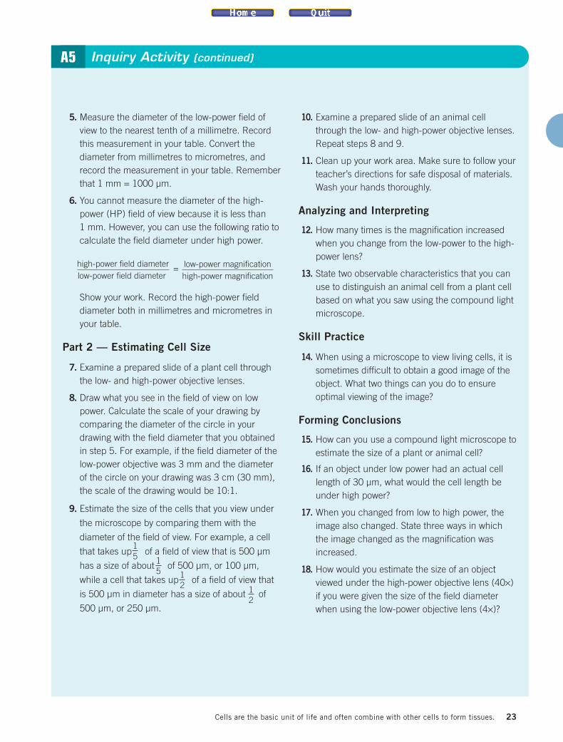
5. Measure the diameter of the low-power field ofview to the nearest tenth of a millimetre. Recordthis measurement in your table. Convert thediameter from millimetres to micrometres, andrecord the measurement in your table. Rememberthat 1 mm = 1000 µm.
6. You cannot measure the diameter of the high-power (HP) field of view because it is less than 1 mm. However, you can use the following ratio tocalculate the field diameter under high power.
Show your work. Record the high-power fielddiameter both in millimetres and micrometres inyour table.
Part 2 — Estimating Cell Size
7. Examine a prepared slide of a plant cell throughthe low- and high-power objective lenses.
8. Draw what you see in the field of view on lowpower. Calculate the scale of your drawing bycomparing the diameter of the circle in yourdrawing with the field diameter that you obtainedin step 5. For example, if the field diameter of thelow-power objective was 3 mm and the diameterof the circle on your drawing was 3 cm (30 mm),the scale of the drawing would be 10:1.
9. Estimate the size of the cells that you view under
the microscope by comparing them with the
diameter of the field of view. For example, a cell
that takes up of a field of view that is 500 µm
has a size of about of 500 µm, or 100 µm,
while a cell that takes up of a field of view that
is 500 µm in diameter has a size of about of
500 µm, or 250 µm.
10. Examine a prepared slide of an animal cellthrough the low- and high-power objective lenses.Repeat steps 8 and 9.
11. Clean up your work area. Make sure to follow yourteacher’s directions for safe disposal of materials.Wash your hands thoroughly.
Analyzing and Interpreting
12. How many times is the magnification increasedwhen you change from the low-power to the high-power lens?
13. State two observable characteristics that you canuse to distinguish an animal cell from a plant cellbased on what you saw using the compound lightmicroscope.
Skill Practice
14. When using a microscope to view living cells, it issometimes difficult to obtain a good image of theobject. What two things can you do to ensureoptimal viewing of the image?
Forming Conclusions
15. How can you use a compound light microscope toestimate the size of a plant or animal cell?
16. If an object under low power had an actual celllength of 30 µm, what would the cell length beunder high power?
17. When you changed from low to high power, theimage also changed. State three ways in whichthe image changed as the magnification wasincreased.
18. How would you estimate the size of an objectviewed under the high-power objective lens (40×)if you were given the size of the field diameterwhen using the low-power objective lens (4×)?
high-power field diameterlow-power field diameter
= low-power magnificationhigh-power magnification
1 5 1
5 1 2 1
2
ist10_ch01.qxd 7/22/09 3:23 PM Page 23

24 UNIT A Tissues, Organs, and Systems of Living Things
A6 Inquiry Activity
There are some similarities and some differencesbetween plant cells and animal cells that can be seenusing a compound light microscope. You will look atcells from the human body and from an onion to seethe similarities and the differences.
QuestionWhat similarities and differences between plant andanimal cells can be seen using a microscope?
Procedure
Part 1 — Examining Animal Cells
1. Review the proper handling and use of themicroscope in Skills Reference 10. Set up yourmicroscope.
2. Take a small piece of clear adhesive tape, andstick it on the inside of your wrist. Remove thetape, and place it sticky side up on the slide.
3. Verify that cells are present by looking at yourslide at low power and medium power.
4. Make a wet mount of your cells. Add a drop ofmethylene blue stain to the slide at one edge ofthe cover slip.
5. Place a piece of torn paper towel against the edgeof the cover slip on the side opposite that of thestain. The stain should move under the cover slip
toward the paper towel. When all of the cells arestained, remove the paper towel.
6. Place the slide on the microscope, and observethe cells.
7. Create a labelled diagram of your skin cells.Include the magnification and scale.
Part 2 — Examining Plant Cells
8. Obtain a small section of onion. Use the tweezersto pull off a thin transparent layer of cells.
9. Prepare a wet mount of the onion cells. Add adrop of iodine stain, and follow the stainingprocedure in step 5.
10. Place the slide on the microscope, and observethe cells.
11. Create a labelled diagram of the onion cells.Include the magnification and scale.
12. Clean up your work area. Make sure to follow yourteacher’s directions for safe disposal of materials.Wash your hands thoroughly.
Analyzing and Interpreting
13. Both the plant and animal cells used in thisactivity are specialized cells that form the outerlayer of the organism. Describe how theappearance and shape of the cells enable them toaccomplish their task of covering and protection.
14. Explain how the cells appeared to be differentwhen viewed at different magnifications.
15. Explain why it is necessary to use onionmembrane that is only one cell in thickness.
Skill Practice
16. Explain how the use of contrast (light levels anduse of stain) improved your understanding of thecells that you were viewing.
Forming Conclusions
17. Describe the similarities and differences that youobserved between the plant and animal cells.
Examining Plant and Animal Cells
SKILLS YOU WILL USE■ Conducting inquiries safely■ Observing, and recording
observations
CAUTION: Practise proper techniques in handling themicroscope and slides. Use care when staining. Coveryour staining work area with a paper towel.
• clear adhesive tape
• compound lightmicroscope
• methylene blue stain
• microscope slides andcover slips
• onion epidermis
• iodine stain
• paper
• paper towel
• pen and/or pencil
• tweezers
Materials & Equipment
Skills References 2, 6, 10
ist10_ch01.qxd 7/22/09 3:23 PM Page 24

Key Concept Review1. What five life processes do cells perform?
2. List the five organelles that are common toplant and animal cells. What are theirfunctions?
3. What are three differences between plant andanimal cells?
4. Why can the granum and thylakoid structuresbe described as “solar collectors”?
5. Prepare a table that summarizes theorganelles and structures found in plant andanimal cells.
6. Explain how fluorescence microscopy works.
7. Name two types of electron microscopes thatare used by cell biologists.
8. What is the name of the image created by anelectron microscope?
9. Explain why the cell can be considered to bethe “building block” of life.
10. Explain the importance of contrast inmicroscopy.
11. What two things can you do to create contrastwhen you use a compound light microscopeto study a specimen?
Connect Your Understanding12. Explain why a cell biologist would choose to
use an electron microscope rather than a lightmicroscope. When would a light microscopebe preferred?
13. What details of a microscope would you needto know to determine the total magnificationof the system?
14. Explain why you would expect the cells of adesert plant, such as a cactus, to havethickened cell walls.
15. Think about the function of themitochondria. You have been asked to viewcells taken from the leg muscle of an athleteand cells taken from the skin of an elderlyindividual. What differences in the number ofmitochondria would you see in the twosamples? Explain your thinking.
16. Explain how a microscope may be used toassess human health.
17. Write a short paragraph that compares andcontrasts plant and animal cells byconsidering structures, presence ofspecialized compounds, and forms of energystorage.
18. The scientist shown below is looking at cellsthrough a fluorescent microscope. How hasthe development of technology aided ourunderstanding of cells?
Reflection19. Describe three things about plant and animal
cells that you did not know before you startedworking on this section.
For more questions, go to ScienceSource.
1.1 CHECK and REFLECT
25Cells are the basic unit of life and often combine with other cells to form tissues.
Question 18
ist10_ch01.qxd 7/22/09 3:23 PM Page 25