1 Zygomatic complex fractures Management of Maxillofacial Trauma.
50
1 Zygomatic complex Zygomatic complex fractures fractures Management of Maxillofacial Trauma
-
Upload
ross-mckinney -
Category
Documents
-
view
233 -
download
10
Transcript of 1 Zygomatic complex fractures Management of Maxillofacial Trauma.

11
Zygomatic complex fracturesZygomatic complex fractures
Management of Maxillofacial Trauma
22
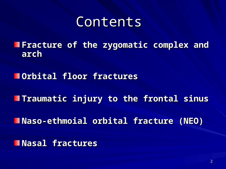
Contents Contents
Fracture of the zygomatic complex and archFracture of the zygomatic complex and arch
Orbital floor fracturesOrbital floor fractures
Traumatic injury to the frontal sinusTraumatic injury to the frontal sinus
Naso-ethmoial orbital fracture (NEO)Naso-ethmoial orbital fracture (NEO)
Nasal fracturesNasal fractures
33
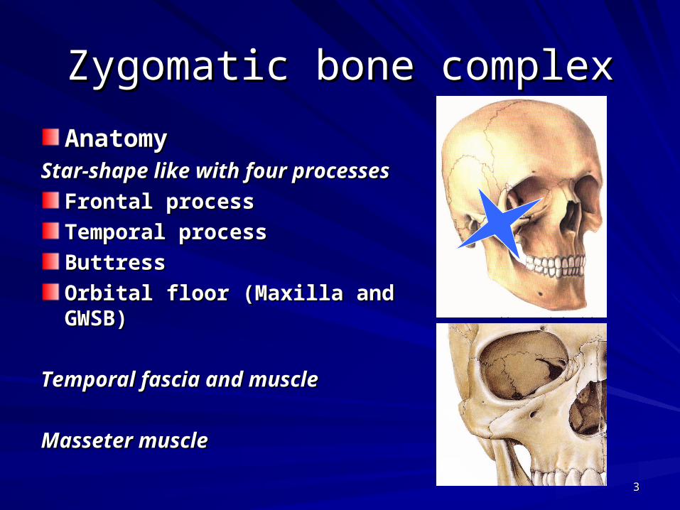
Zygomatic bone complexZygomatic bone complex
AnatomyAnatomyStar-shape like with four processesStar-shape like with four processes
Frontal processFrontal process
Temporal processTemporal process
ButtressButtress
Orbital floor (Maxilla and GWSB)Orbital floor (Maxilla and GWSB)
Temporal fascia and muscleTemporal fascia and muscle
Masseter muscleMasseter muscle
44
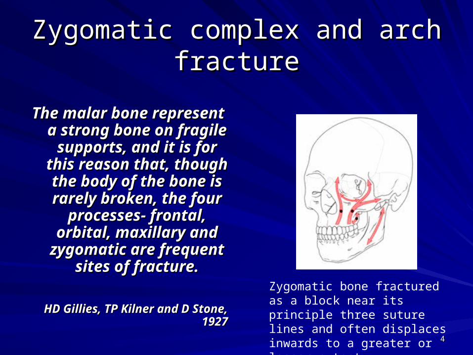
Zygomatic complex and arch Zygomatic complex and arch fracturefracture
The malar bone represent The malar bone represent a strong bone on fragile a strong bone on fragile
supports, and it is for supports, and it is for this reason that, though this reason that, though the body of the bone is the body of the bone is rarely broken, the four rarely broken, the four
processes- frontal, processes- frontal, orbital, maxillary and orbital, maxillary and
zygomatic are frequent zygomatic are frequent sites of fracture.sites of fracture.
HD Gillies, TP Kilner and D Stone, HD Gillies, TP Kilner and D Stone, 19271927
Zygomatic bone fractured as a block near its principle three suture lines and often displaces inwards to a greater or lesser extent.
55
Occurrence Occurrence
Observed in (>50%) of middle third Observed in (>50%) of middle third fracture fracture (in developed countries due to assaults)(in developed countries due to assaults)
The zygomatic arch fracture can be The zygomatic arch fracture can be isolated in most of the casesisolated in most of the cases
•As isolated fracture•In combination with other middle third fracture
•With internal orbital fracture (blow out)
66
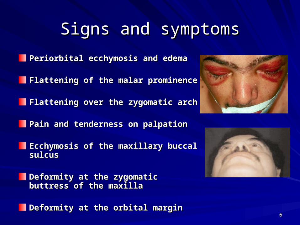
Signs and symptomsSigns and symptoms
Periorbital ecchymosis and edemaPeriorbital ecchymosis and edema
Flattening of the malar prominenceFlattening of the malar prominence
Flattening over the zygomatic archFlattening over the zygomatic arch
Pain and tenderness on palpationPain and tenderness on palpation
Ecchymosis of the maxillary buccal sulcusEcchymosis of the maxillary buccal sulcus
Deformity at the zygomatic buttress of the Deformity at the zygomatic buttress of the maxillamaxilla
Deformity at the orbital marginDeformity at the orbital margin
77
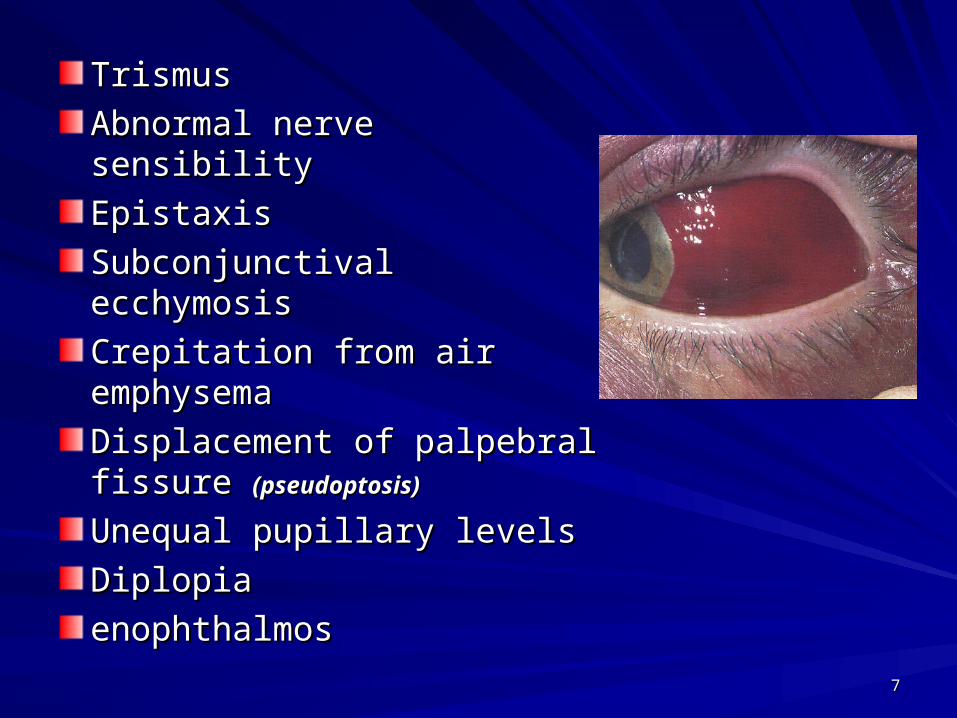
TrismusTrismus
Abnormal nerve sensibilityAbnormal nerve sensibility
EpistaxisEpistaxis
Subconjunctival ecchymosisSubconjunctival ecchymosis
Crepitation from air Crepitation from air emphysemaemphysema
Displacement of palpebral Displacement of palpebral fissure fissure (pseudoptosis)(pseudoptosis)
Unequal pupillary levelsUnequal pupillary levels
DiplopiaDiplopia
enophthalmosenophthalmos
88
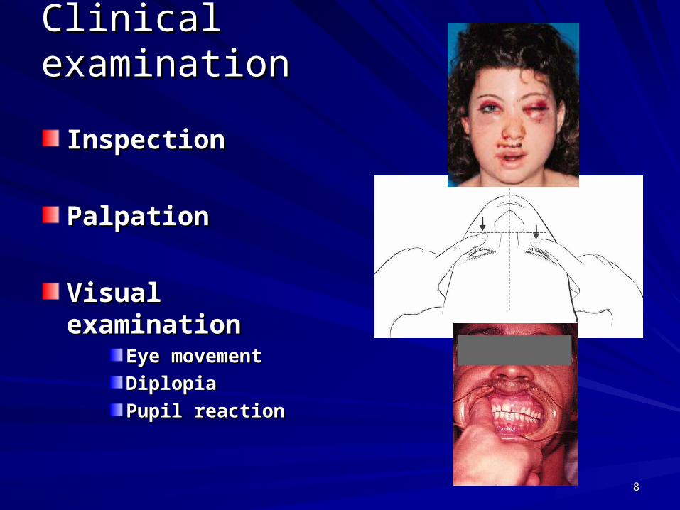
Clinical examinationClinical examination
InspectionInspection
PalpationPalpation
Visual examinationVisual examinationEye movementEye movement
DiplopiaDiplopia
Pupil reactionPupil reaction
99
Radiographical evaluationRadiographical evaluation
Nothing is more valuable to the surgeon in Nothing is more valuable to the surgeon in determining the extent of injury and the determining the extent of injury and the
position of the fragments-both before and position of the fragments-both before and after operation- than a good skiagram after operation- than a good skiagram
(radiograph)(radiograph)
HD Gillies, TP Kilner and D Stone, 1927HD Gillies, TP Kilner and D Stone, 1927
1010
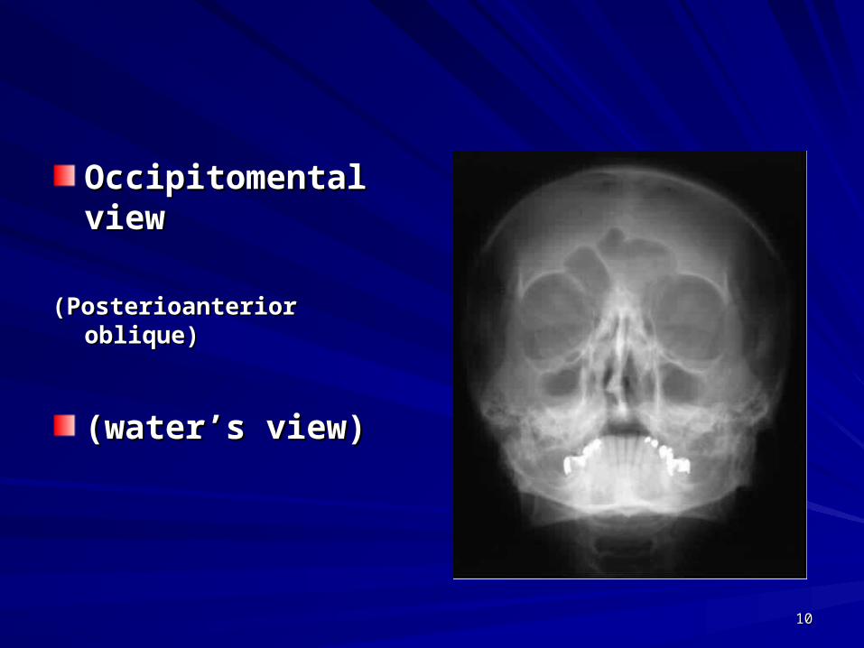
Occipitomental viewOccipitomental view
(Posterioanterior oblique)(Posterioanterior oblique)
(water’s view)(water’s view)
1111
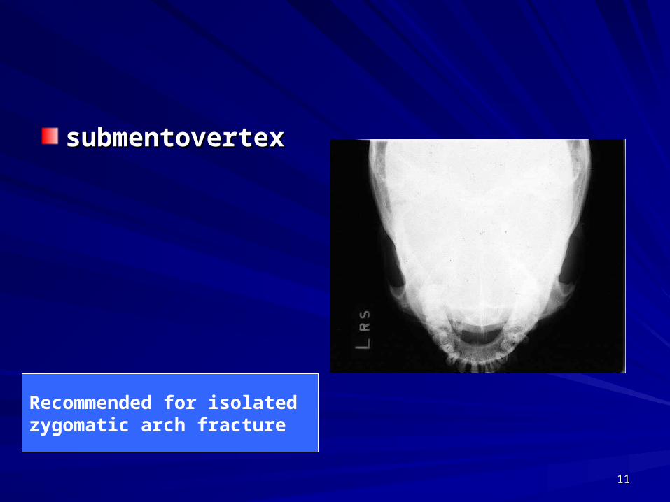
submentovertexsubmentovertex
Recommended for isolated zygomatic arch fracture
1212
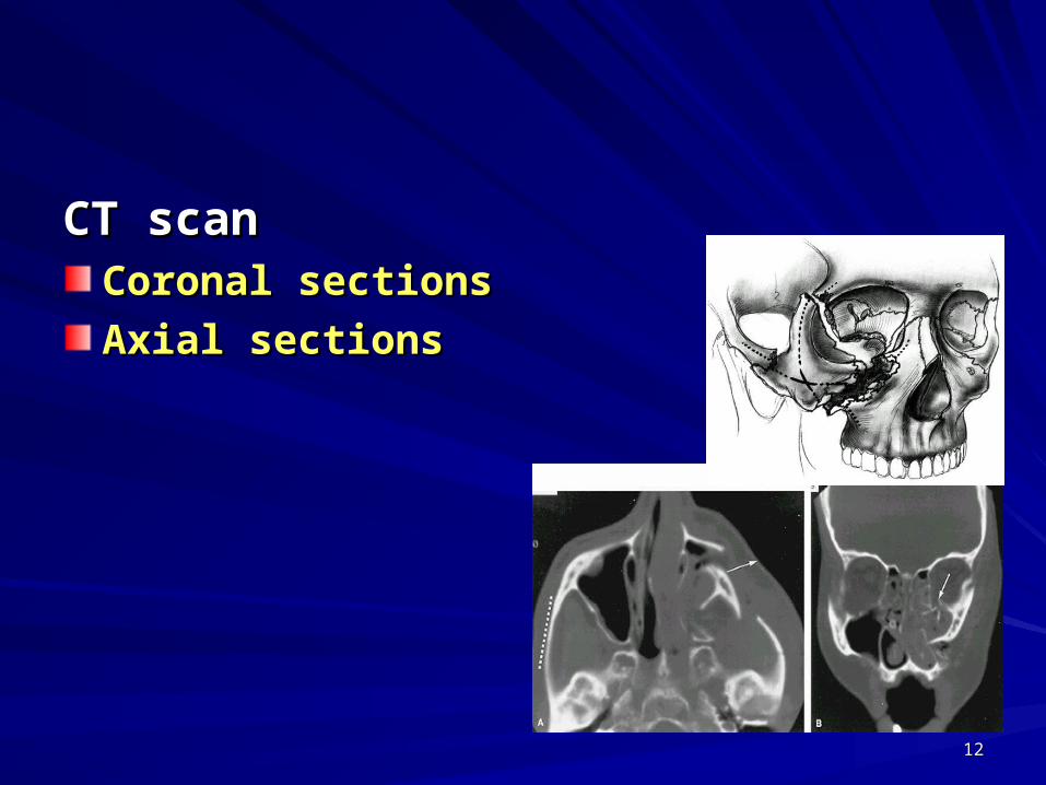
CT scanCT scanCoronal sectionsCoronal sections
Axial sectionsAxial sections
1313
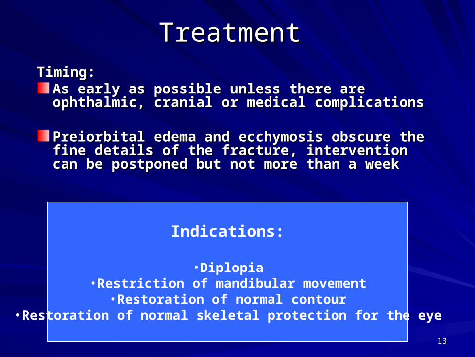
Treatment Treatment
Timing:Timing:As early as possible unless there are ophthalmic, As early as possible unless there are ophthalmic, cranial or medical complicationscranial or medical complications
Preiorbital edema and ecchymosis obscure the Preiorbital edema and ecchymosis obscure the fine details of the fracture, intervention can be fine details of the fracture, intervention can be postponed but not more than a weekpostponed but not more than a week
Indications:
•Diplopia•Restriction of mandibular movement
•Restoration of normal contour•Restoration of normal skeletal protection for the eye
1414
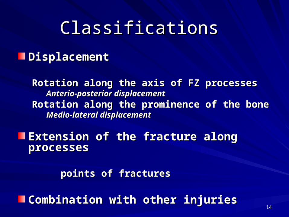
Classifications Classifications
DisplacementDisplacement
Rotation along the axis of FZ processesRotation along the axis of FZ processesAnterio-posterior displacementAnterio-posterior displacement
Rotation along the prominence of the boneRotation along the prominence of the boneMedio-lateral displacementMedio-lateral displacement
Extension of the fracture along processesExtension of the fracture along processes
points of fracturespoints of fractures
Combination with other injuriesCombination with other injuries
1515
Treatment Treatment
The methods of treating a fractured malar bone The methods of treating a fractured malar bone recommended by the various writers who have recommended by the various writers who have
reported cases include simple digital manipulation reported cases include simple digital manipulation under genre real anesthesia, external manipulation under genre real anesthesia, external manipulation
by means of a cow-horn dental forceps grasping the by means of a cow-horn dental forceps grasping the edges of the bone, traction and elevation by means edges of the bone, traction and elevation by means
of wire or heavy bone elevators passed through of wire or heavy bone elevators passed through small local external incisions, and elevation via small local external incisions, and elevation via
incision in the mucosa of the ginigival sulcus at the incision in the mucosa of the ginigival sulcus at the canine fossa. Our technique, which has now been canine fossa. Our technique, which has now been
used successfully in a number of cases, differs from used successfully in a number of cases, differs from those mentioned. those mentioned.
HD Gillies, TP Kilner and D Stone, 1927HD Gillies, TP Kilner and D Stone, 1927
1616
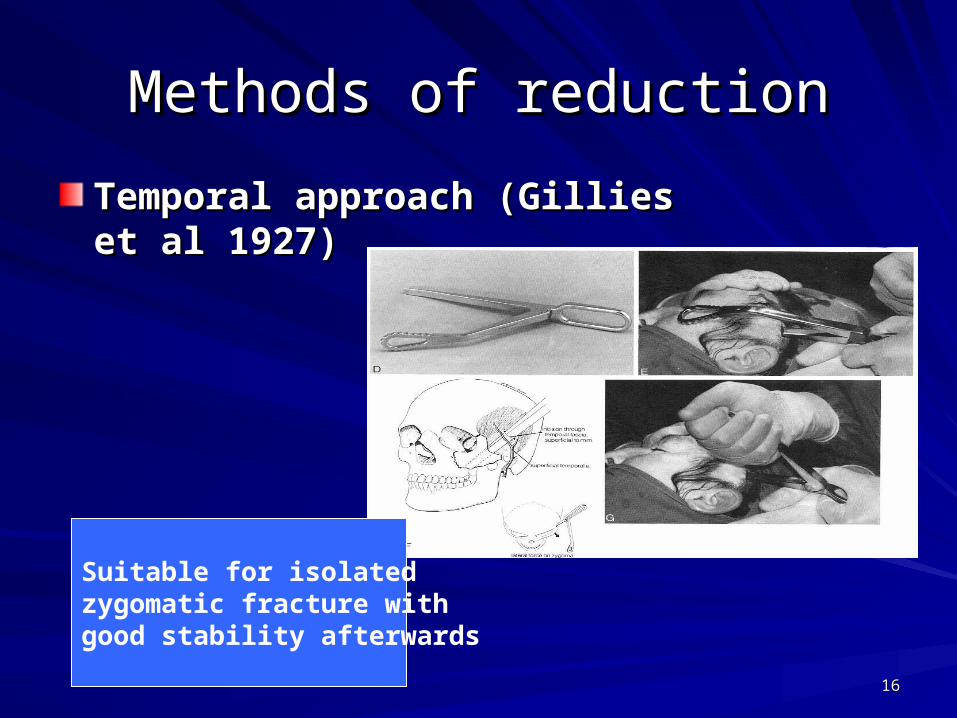
Methods of reductionMethods of reduction
Temporal approach (Gillies et al Temporal approach (Gillies et al 1927)1927)
Suitable for isolated zygomatic fracture with good stability afterwards

1717
Methods of reductionMethods of reduction
Percutaneous approach (malar hook, Percutaneous approach (malar hook, Carroll-Girard bone screw)Carroll-Girard bone screw)
Suitable for displaced zygomatic fracture with highStability after reduction

1818
Methods of reductionMethods of reduction
Buccal sulcus Buccal sulcus approach (Keen approach (Keen 1909)1909)
Elevation from Elevation from eyebrow approacheyebrow approach
(the same principle of Gillies (the same principle of Gillies
approach)approach)

1919
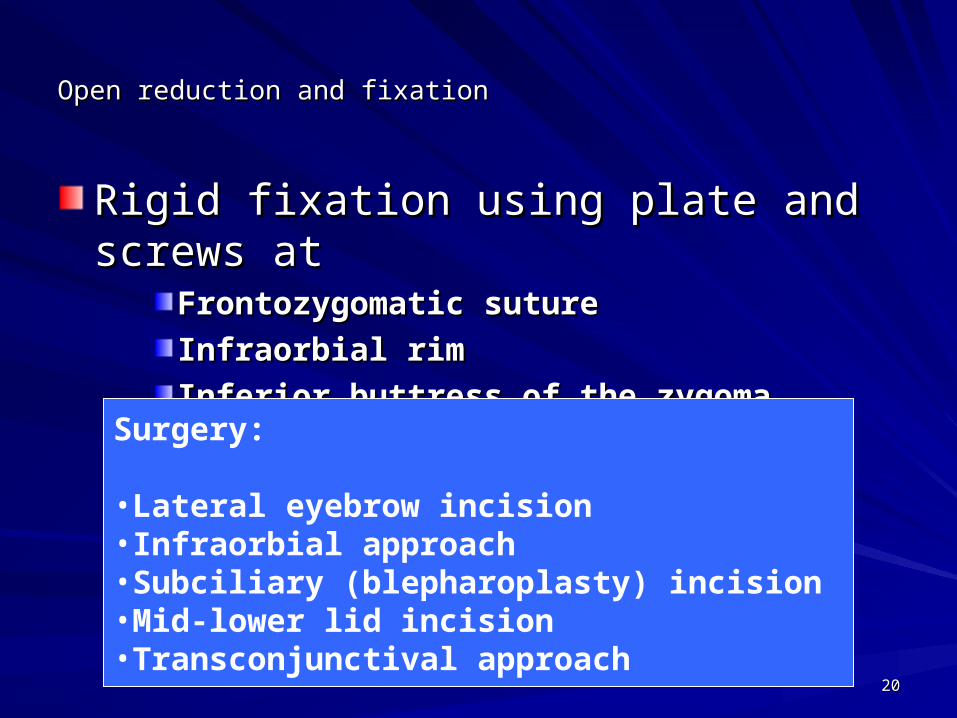
Open reduction and fixationOpen reduction and fixation
Transosseous wiring atTransosseous wiring at–Frontozygomatic sutureFrontozygomatic suture– Infraorbial rimInfraorbial rim
Surgery:
•Lateral eyebrow incision
•Infraorbital approach
2020
Open reduction and fixationOpen reduction and fixation
Rigid fixation using plate and screws atRigid fixation using plate and screws atFrontozygomatic sutureFrontozygomatic suture
Infraorbial rimInfraorbial rim
Inferior buttress of the zygomaInferior buttress of the zygoma
Surgery:
•Lateral eyebrow incision•Infraorbial approach•Subciliary (blepharoplasty) incision•Mid-lower lid incision•Transconjunctival approach
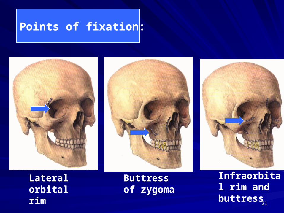
2121
Infraorbital rim and buttress
Lateral orbital rim
Buttress of zygoma
Points of fixation:
2222
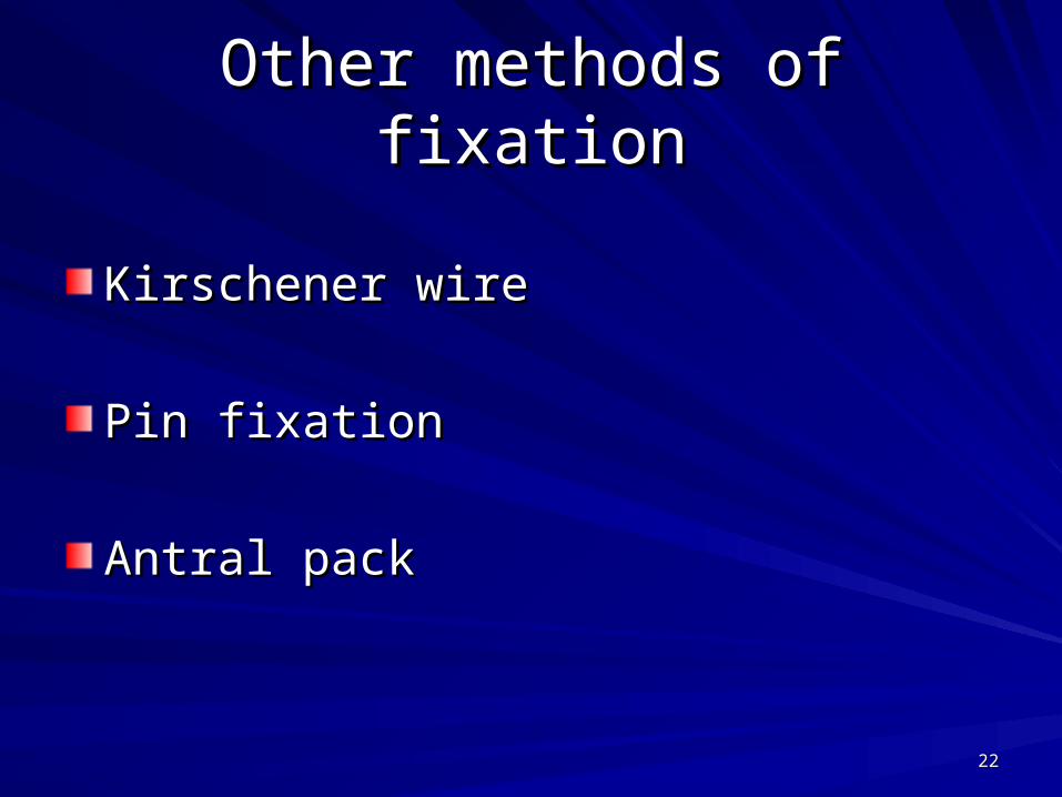
Other methods of fixationOther methods of fixation
Kirschener wireKirschener wire
Pin fixationPin fixation
Antral packAntral pack
2323
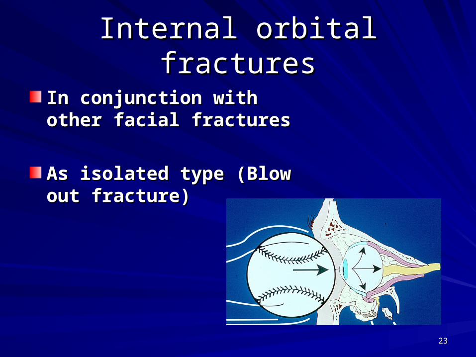
Internal orbital fracturesInternal orbital fractures
In conjunction with other In conjunction with other facial fracturesfacial fractures
As isolated type (Blow out As isolated type (Blow out fracture)fracture)
2424
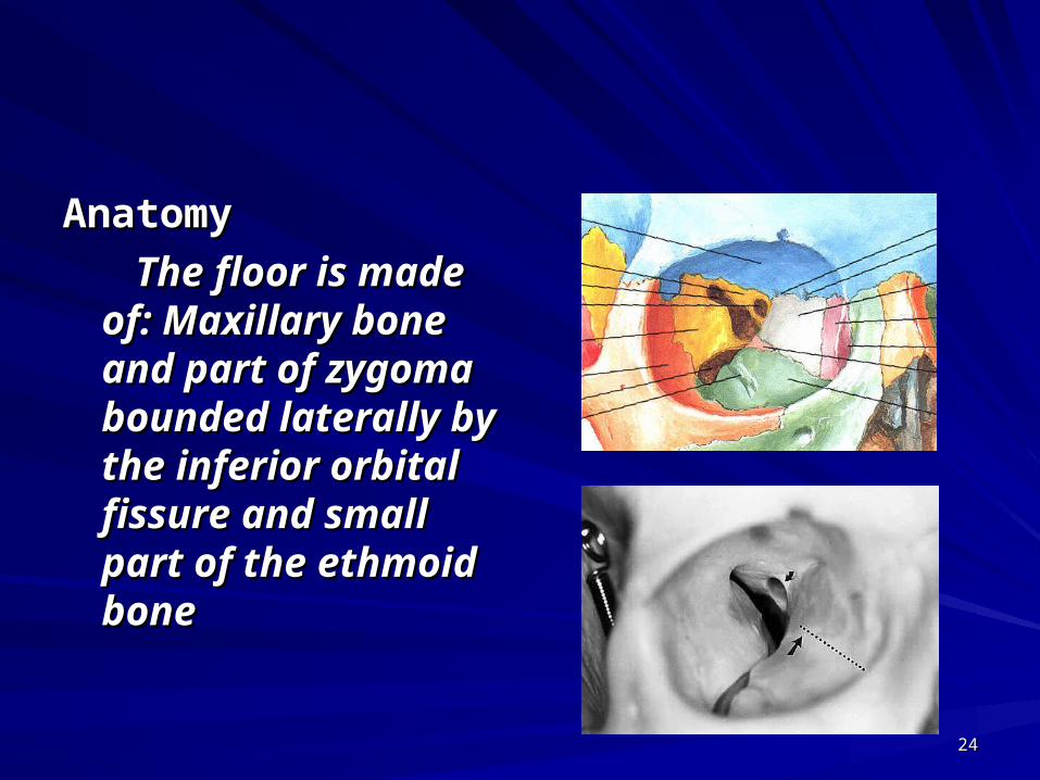
AnatomyAnatomy
The floor is made of: The floor is made of: Maxillary bone and Maxillary bone and part of zygoma part of zygoma bounded laterally by bounded laterally by the inferior orbital the inferior orbital fissure and small fissure and small part of the ethmoid part of the ethmoid bonebone
2525
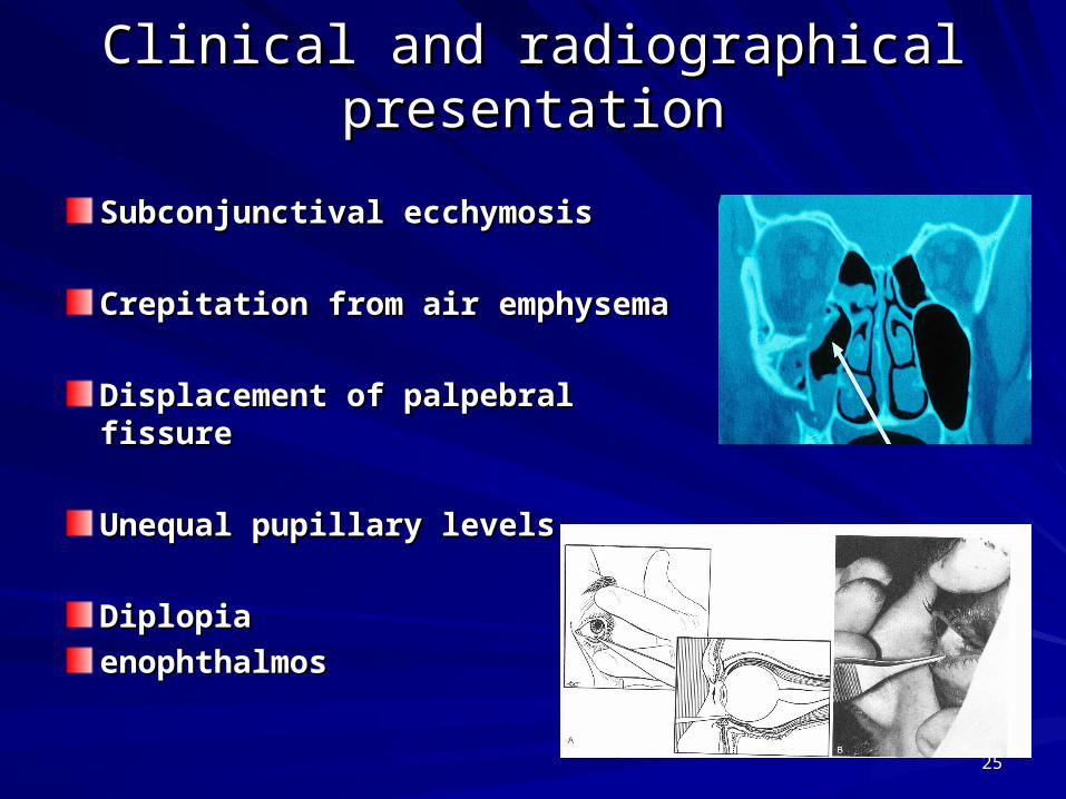
Clinical and radiographical presentationClinical and radiographical presentation
Subconjunctival ecchymosisSubconjunctival ecchymosis
Crepitation from air emphysemaCrepitation from air emphysema
Displacement of palpebral fissureDisplacement of palpebral fissure
Unequal pupillary levelsUnequal pupillary levels
DiplopiaDiplopia
enophthalmosenophthalmos
2626
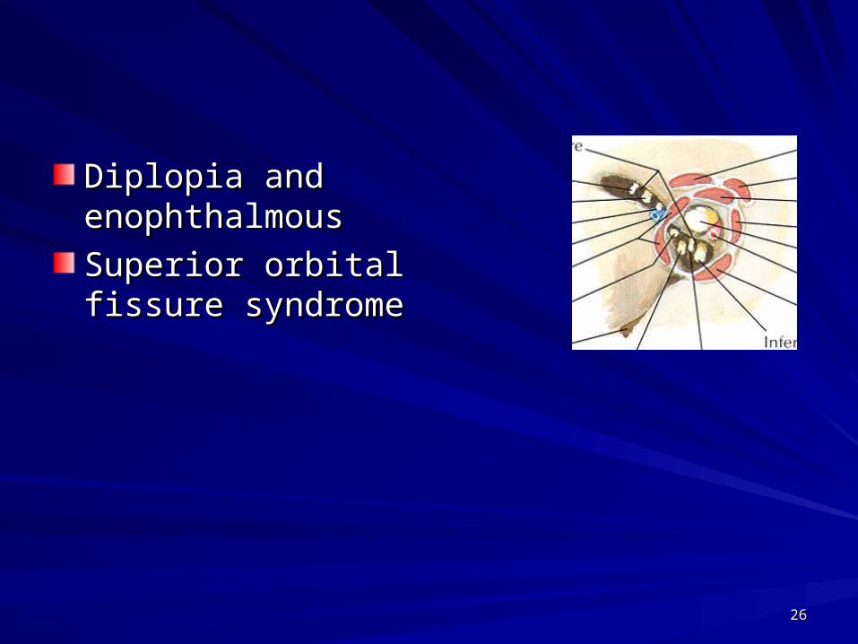
Diplopia and Diplopia and enophthalmousenophthalmous
Superior orbital Superior orbital fissure syndromefissure syndrome
2727
Treatment Treatment
Rational for intervention:Rational for intervention:
Small defect with no clinical consequence Small defect with no clinical consequence may not warrant the surgical intervention.may not warrant the surgical intervention.
Large defect with handicapping symptoms Large defect with handicapping symptoms should be operated.should be operated.
2828
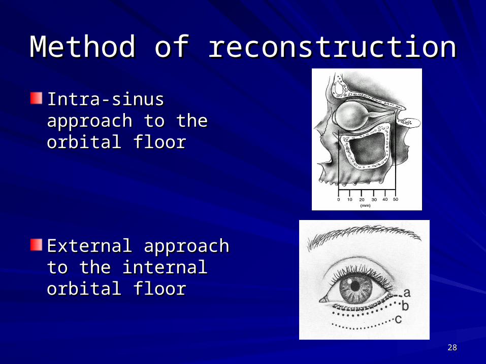
Method of reconstructionMethod of reconstruction
Intra-sinus approach Intra-sinus approach to the orbital floorto the orbital floor
External approach to External approach to the internal orbital the internal orbital floorfloor
2929
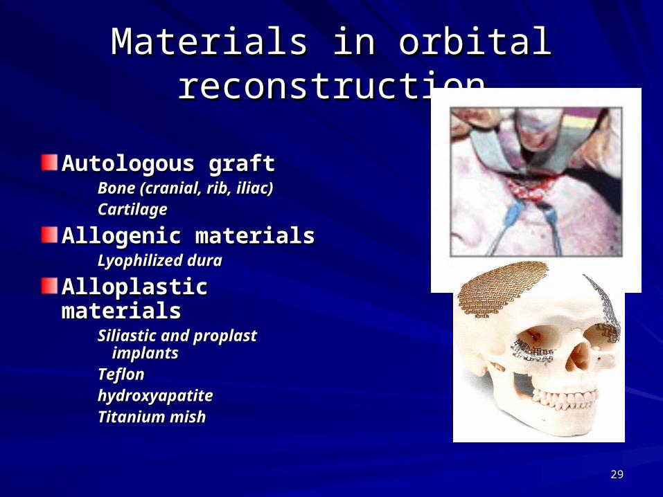
Materials in orbital reconstructionMaterials in orbital reconstruction
Autologous graftAutologous graftBone (cranial, rib, iliac) Bone (cranial, rib, iliac) CartilageCartilage
Allogenic materialsAllogenic materialsLyophilized duraLyophilized dura
Alloplastic materialsAlloplastic materialsSiliastic and proplast Siliastic and proplast
implantsimplantsTeflonTeflonhydroxyapatitehydroxyapatiteTitanium mishTitanium mish
3030
Nasal-orbital ethmoid injuriesNasal-orbital ethmoid injuries
They represent a wide spectrum of injuriesThey represent a wide spectrum of injuries
Simple nasal fracture with involvementOf orbital bones
Grossly comminuted and compound naso-orbital ethmoid fracture involving the base
of skull with significant displacement

3131
Diagnosis Diagnosis
Clinical examination:Clinical examination:Obliterating swellingObliterating swellingCanthus detachmentCanthus detachmentLacrimal apparatus damageLacrimal apparatus damageDeformity of nasal bridgeDeformity of nasal bridgeCSF leakCSF leak
Radiographical examinationRadiographical examination::Occipitomental viewsOccipitomental viewsLateral skull viewsLateral skull viewsCT and 3D CTCT and 3D CT
3232
Fracture classificationFracture classificationNasal-orbital ethmoid fracturesNasal-orbital ethmoid fractures
Type IType IUnilateral or bilateral, involves only one portion of the Unilateral or bilateral, involves only one portion of the
medial orbital rim with the attached canthal tendonmedial orbital rim with the attached canthal tendon
Type IIType IIUnilateral or bilateral, may be large segments of Unilateral or bilateral, may be large segments of
comminuted type and the canthus remains attached comminuted type and the canthus remains attached to the large central segmentto the large central segment
Type IIIType IIIUnilateral or bilateral, comminution involves the Unilateral or bilateral, comminution involves the
central segment of the attached tendon results in central segment of the attached tendon results in avulsion of medial canthusavulsion of medial canthus

3333
Management of nasal-orbital Management of nasal-orbital ethmoid fracturesethmoid fractures
Examination for Examination for determination of the extent determination of the extent of the injury (surgical of the injury (surgical exploration)exploration)
Nasal boneNasal boneOrbital and ethmoidalOrbital and ethmoidalFrontal boneFrontal bone
Debridement and closure of Debridement and closure of open woundsopen wounds
Reduction and stabilization Reduction and stabilization of bone fractureof bone fracture
3434
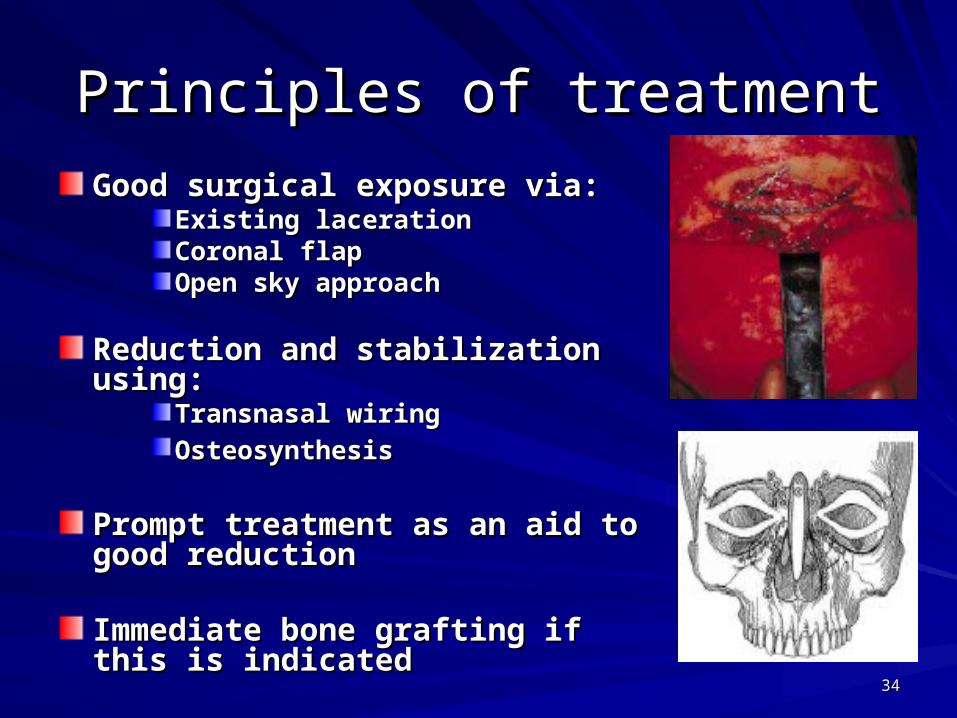
Principles of treatmentPrinciples of treatment
Good surgical exposure via:Good surgical exposure via:Existing lacerationExisting lacerationCoronal flapCoronal flapOpen sky approachOpen sky approach
Reduction and stabilization using:Reduction and stabilization using:Transnasal wiringTransnasal wiring
OsteosynthesisOsteosynthesis
Prompt treatment as an aid to good Prompt treatment as an aid to good reductionreduction
Immediate bone grafting if this is Immediate bone grafting if this is indicatedindicated
3535
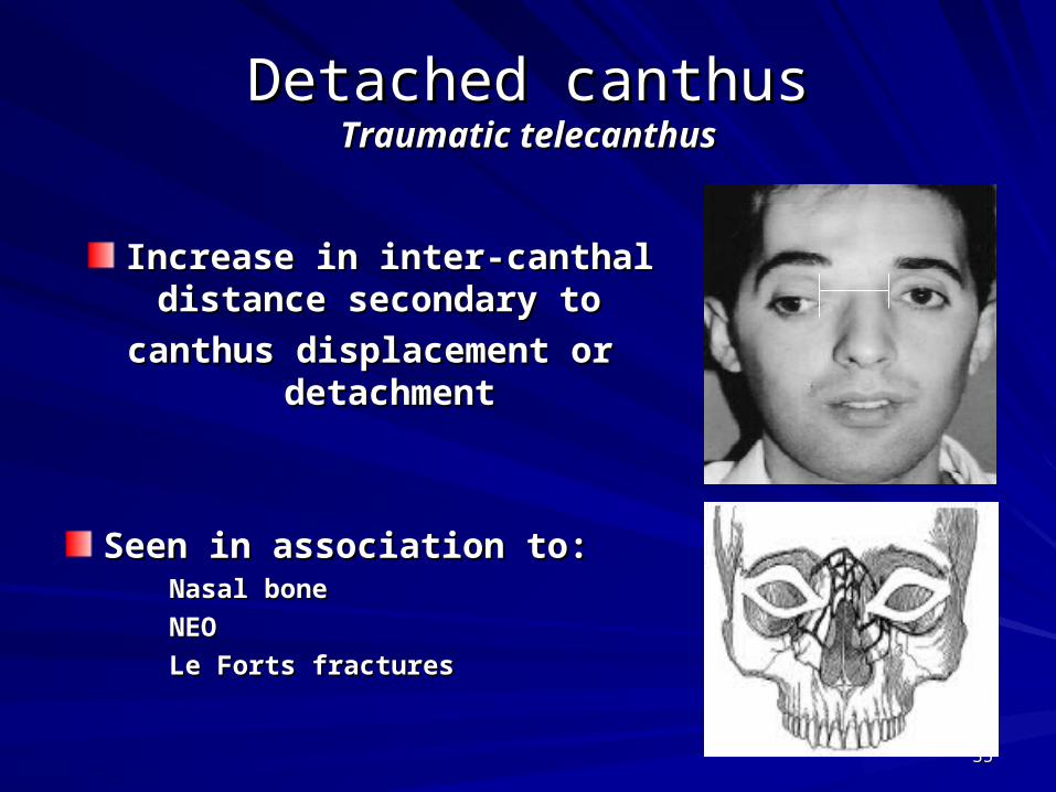
Detached canthusDetached canthusTraumatic telecanthusTraumatic telecanthus
Increase in inter-canthal distance Increase in inter-canthal distance secondary to secondary to
canthus displacement or canthus displacement or detachmentdetachment
Seen in association to:Seen in association to:Nasal boneNasal bone
NEONEO
Le Forts fracturesLe Forts fractures

3636
Surgical management of detached Surgical management of detached canthuscanthus
Transnasal wiring Transnasal wiring technique technique (unilateral (unilateral type)type)
Canthopexy Canthopexy – Identification of the Identification of the
ligamentligament– Liberation of the Liberation of the
periorbital tissueperiorbital tissue– Liberation of the lacrimal Liberation of the lacrimal
pathwaypathway– Nasal transfixationNasal transfixation– Contralateral fixationContralateral fixation
3737
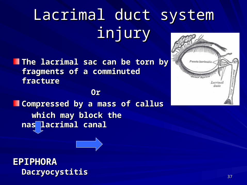
Lacrimal duct system injuryLacrimal duct system injury
The lacrimal sac can be torn by The lacrimal sac can be torn by fragments of a comminuted fracturefragments of a comminuted fracture
OrOr
Compressed by a mass of callus Compressed by a mass of callus
which may block the nasolacrimal canalwhich may block the nasolacrimal canal
EPIPHORAEPIPHORA DacryocystitisDacryocystitis

3838
Reconstitution of the lacrimal passagesReconstitution of the lacrimal passages
Done at the same time of canthopexy viaDone at the same time of canthopexy via– The original scarsThe original scars– Lateral nasal incision (Lynch) Lateral nasal incision (Lynch) – Bi-coronal incisionBi-coronal incision
Dacryocystorhinostomy Dacryocystorhinostomy If the sac remains intact, drainage of lacrimal fluid by probing If the sac remains intact, drainage of lacrimal fluid by probing
or removing of surrounded bone to allow drainage into the or removing of surrounded bone to allow drainage into the nosenose
Conjunctivo-rhinostomyConjunctivo-rhinostomyimplantation of a duct-like polythene tube or glass in case of implantation of a duct-like polythene tube or glass in case of
duct damageduct damage
3939
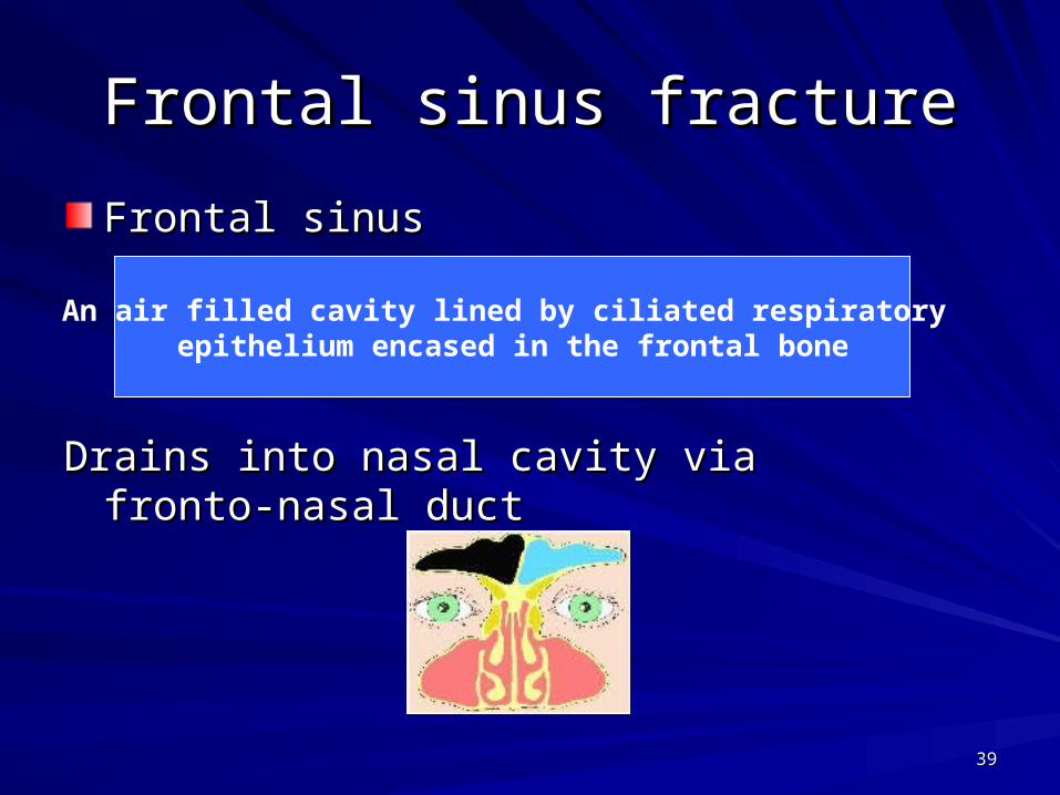
Frontal sinus fractureFrontal sinus fracture
Frontal sinusFrontal sinus
Drains into nasal cavity via fronto-nasal ductDrains into nasal cavity via fronto-nasal duct
An air filled cavity lined by ciliated respiratory epithelium encased in the frontal bone
4040
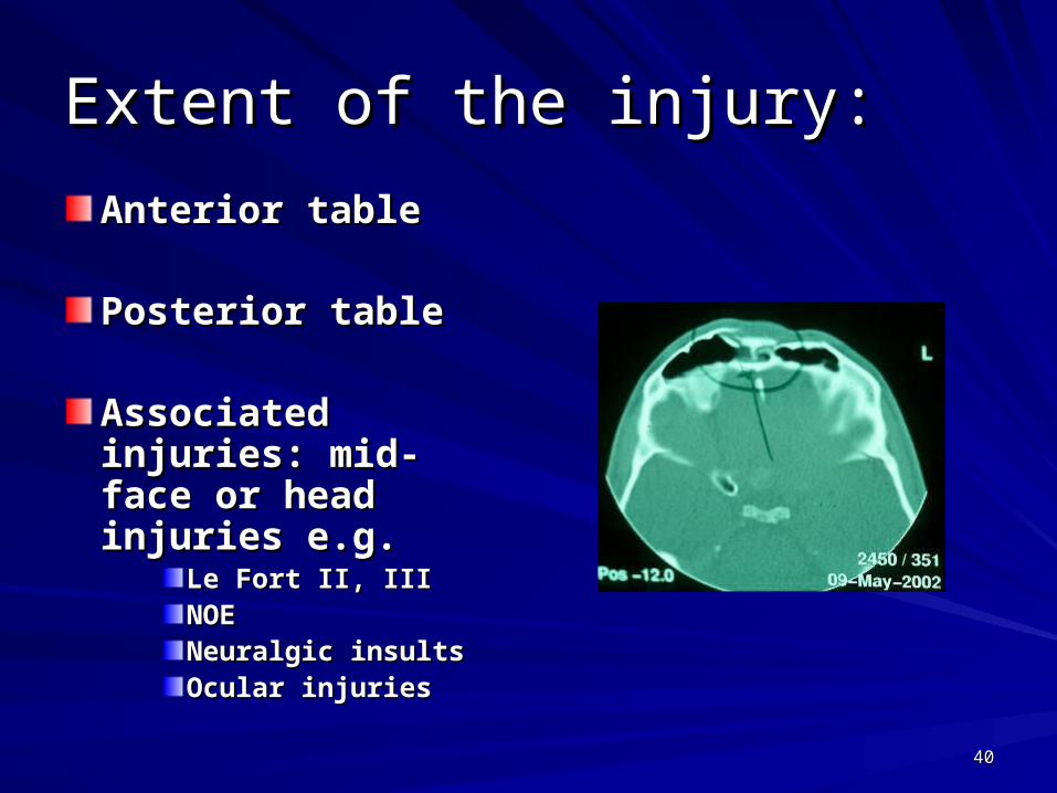
Extent of the injury:Extent of the injury:
Anterior tableAnterior table
Posterior tablePosterior table
Associated injuries: Associated injuries: mid-face or head mid-face or head injuries e.g.injuries e.g.
Le Fort II, IIILe Fort II, IIINOENOENeuralgic insultsNeuralgic insultsOcular injuriesOcular injuries
4141
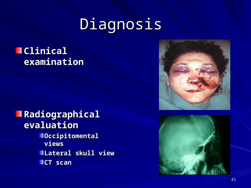
Diagnosis Diagnosis
Clinical examinationClinical examination
Radiographical Radiographical evaluationevaluation
Occipitomental viewsOccipitomental views
Lateral skull viewLateral skull view
CT scanCT scan
4242
Classification of fracturesClassification of fractures
Anterior table fractureAnterior table fracture– LinearLinear– DisplacedDisplaced
Posterior table fracturePosterior table fracture– LinearLinear– DisplacedDisplaced
Outflow tract injury Outflow tract injury (naso-lacrimal duct)(naso-lacrimal duct)
4343
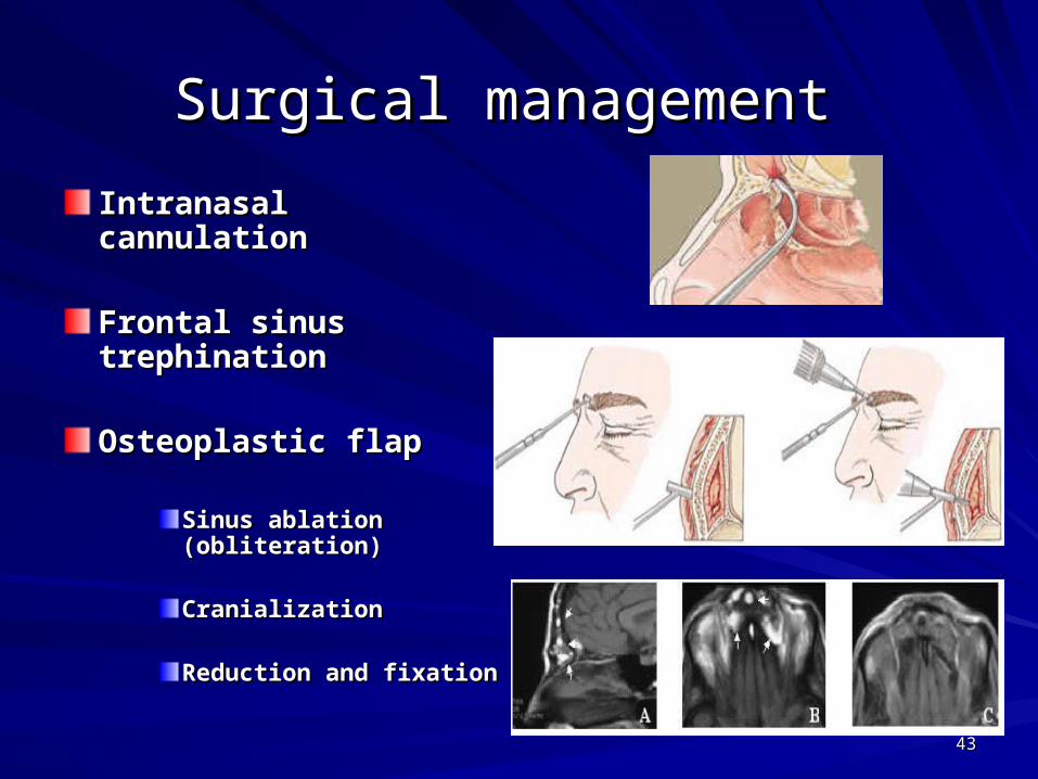
Surgical management Surgical management
Intranasal cannulationIntranasal cannulation
Frontal sinus Frontal sinus trephinationtrephination
Osteoplastic flap Osteoplastic flap
Sinus ablation Sinus ablation (obliteration)(obliteration)
Cranialization Cranialization
Reduction and fixationReduction and fixation
4444
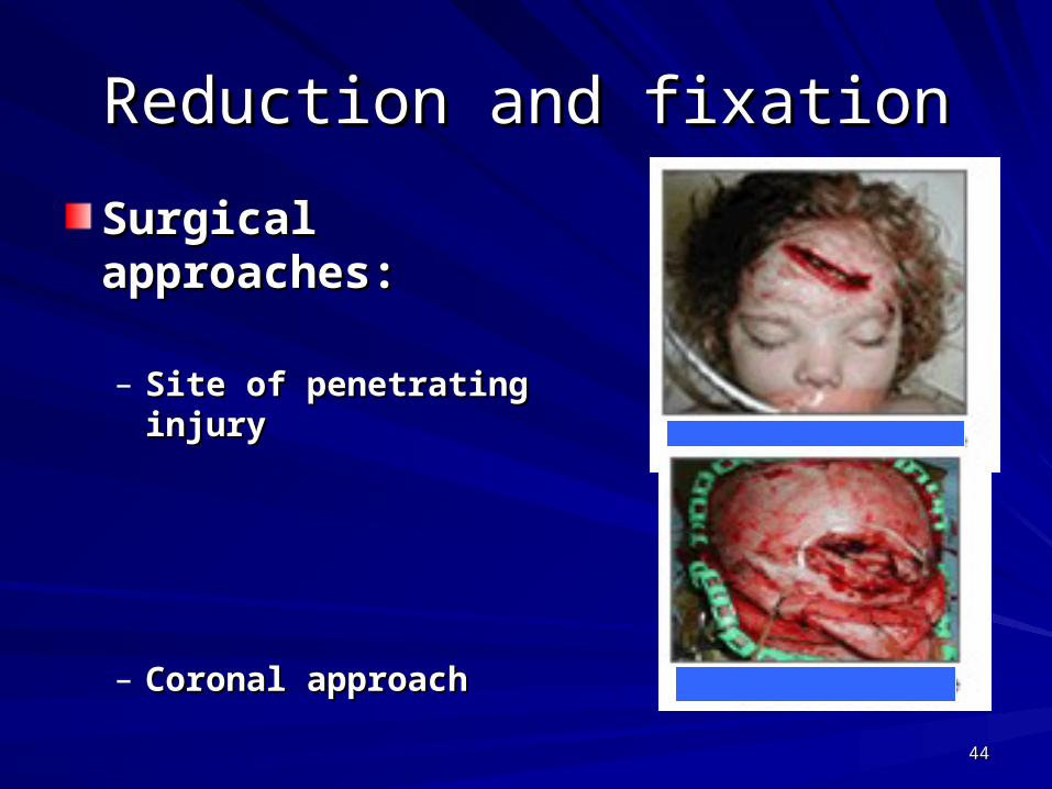
Reduction and fixationReduction and fixation
Surgical approaches:Surgical approaches:
– Site of penetrating injurySite of penetrating injury
– Coronal approachCoronal approach
4545
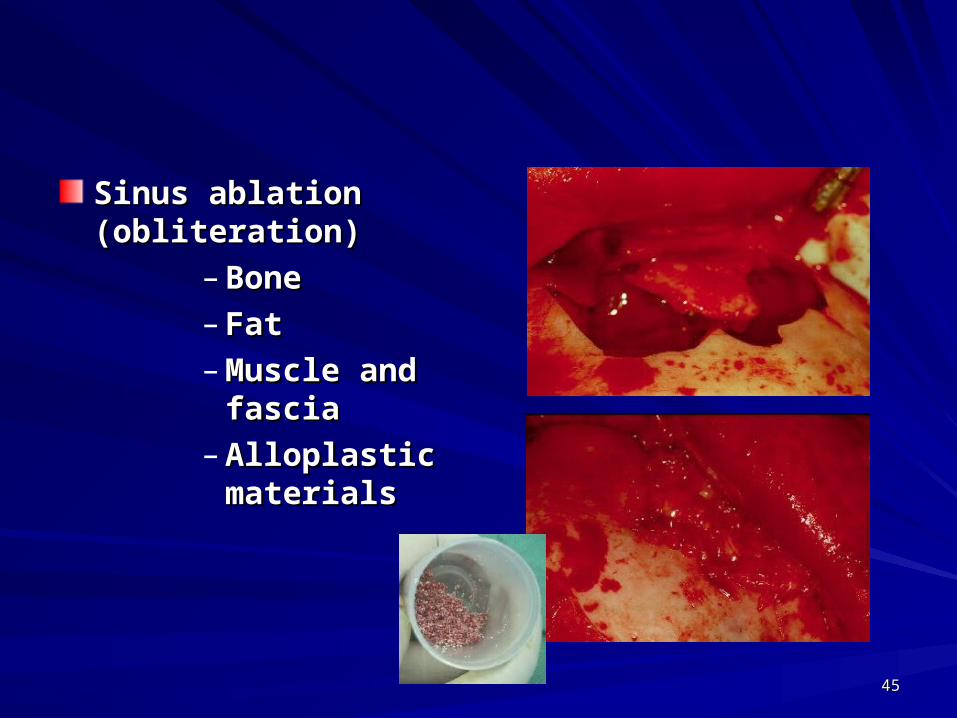
Sinus ablation Sinus ablation (obliteration)(obliteration)
– BoneBone– FatFat– Muscle and Muscle and
fasciafascia– Alloplastic Alloplastic
materialsmaterials
4646
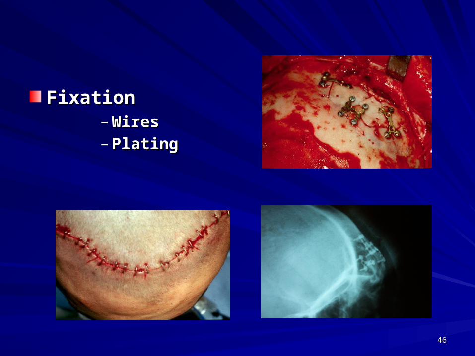
FixationFixation– WiresWires– PlatingPlating
4747
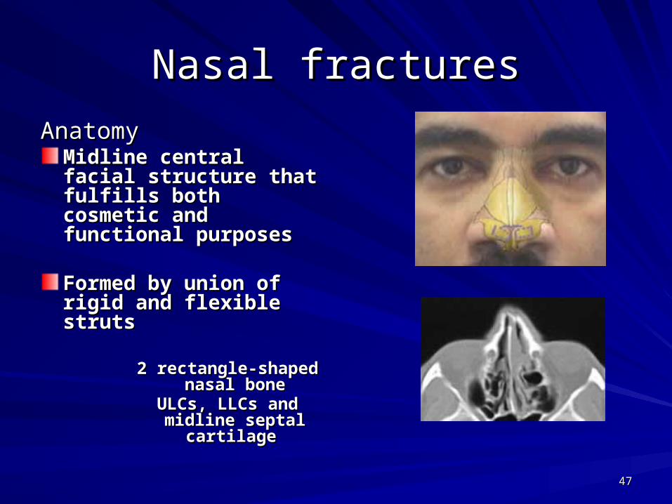
Nasal fracturesNasal fractures
AnatomyAnatomyMidline central facial Midline central facial structure that fulfills structure that fulfills both cosmetic and both cosmetic and functional purposesfunctional purposes
Formed by union of Formed by union of rigid and flexible strutsrigid and flexible struts
2 rectangle-shaped 2 rectangle-shaped nasal bonenasal bone
ULCs, LLCs and ULCs, LLCs and midline septal midline septal
cartilagecartilage
4848
Classification of injuriesClassification of injuriesLow energy injuriesLow energy injuries
Simple injury caused by low velocity trauma (simple Simple injury caused by low velocity trauma (simple noncomminuted)noncomminuted)
High energy injuriesHigh energy injuriesSevere injury with comminution of nasal facial Skelton due to Severe injury with comminution of nasal facial Skelton due to
higher amount of energyhigher amount of energy
Patterns of injury
•Lateral injury (from the side)•Sagittal injury (from the front)•Inferior injury (from below)

4949
Treatment Treatment
Low energy injuriesLow energy injuriesReduction (close Reduction (close manipulation, open manipulation, open reduction) and stabilizationreduction) and stabilization
Nasal packingNasal packing
External nasal splintExternal nasal splint
Adjunct septoplastyAdjunct septoplasty
Postoperative carePostoperative care
5050
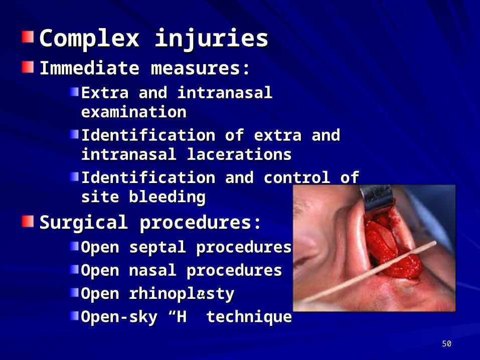
Complex injuriesComplex injuriesImmediate measures:Immediate measures:
Extra and intranasal examinationExtra and intranasal examination
Identification of extra and intranasal Identification of extra and intranasal lacerationslacerations
Identification and control of site Identification and control of site bleedingbleeding
Surgical procedures:Surgical procedures:Open septal proceduresOpen septal procedures
Open nasal proceduresOpen nasal procedures
Open rhinoplastyOpen rhinoplasty
Open-sky “H” techniqueOpen-sky “H” technique


![Oral & Maxillofacial Surgeryopenaccessebooks.com/oral-maxillofacial-surgery/condylar-fractures.pdftreatment of mandibular condylar process fractures [10]. It was found that treatment](https://static.fdocuments.in/doc/165x107/5e27326a457720282958fba6/oral-maxillofacial-sur-treatment-of-mandibular-condylar-process-fractures.jpg)