1. The images have been made by three different types of microscopes. How do the images differ? 2....
45
1. The images have been made by three different types of microscopes. How do the images differ? 2. What kinds of information might scientists gather with each type of microscope? Magnified 41500x Magnified 110x LM Magnified 1500x SEM TEM
-
Upload
juniper-conley -
Category
Documents
-
view
216 -
download
2
Transcript of 1. The images have been made by three different types of microscopes. How do the images differ? 2....
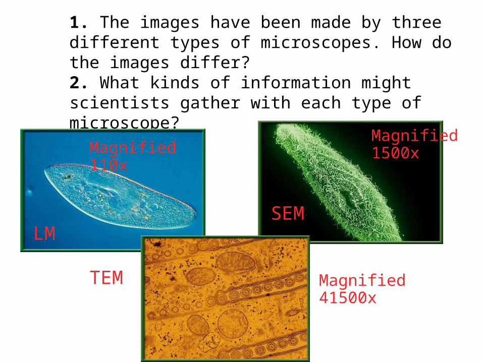
1. The images have been made by three different types of microscopes. How do the images differ?2. What kinds of information might scientists gather with each type of microscope?
Magnified 41500x
Magnified 110x
LM
Magnified 1500x
SEM
TEM

The Life of a Cell
A View of the Cell
The Discovery of Cells
The History of the Cell TheoryThe History of the Cell Theory• Before microscopes were invented, people
believed that diseases were caused by curses and supernatural spirits.
• Microscopes enabled scientists to view and study cells, the basic units of living organisms.
• As scientists began using microscopes, they quickly realized they were entering a new world–one of microorganisms.
• The first person to record looking at water under a microscope was Anton van Leeuwenhoek.
• The microscope van Leeuwenhoek used is considered a simple light microscope because it contained one lens and used natural light to view objects.
Development of Light MicroscopesDevelopment of Light Microscopes
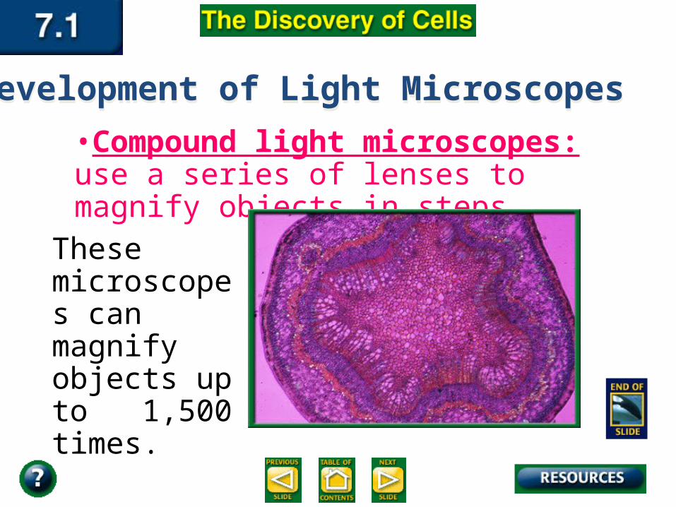
•Compound light microscopes: use a series of lenses to magnify objects in steps.
These microscopes can magnify objects up to 1,500 times.
Development of Light MicroscopesDevelopment of Light Microscopes
• Robert Hooke was an English scientist who lived at the same time as van Leeuwenhock.
The Cell TheoryThe Cell Theory
• Hooke used a compound light microscope to study cork, the dead cells of oak bark.
• Cells : the basic building blocks of all living things.
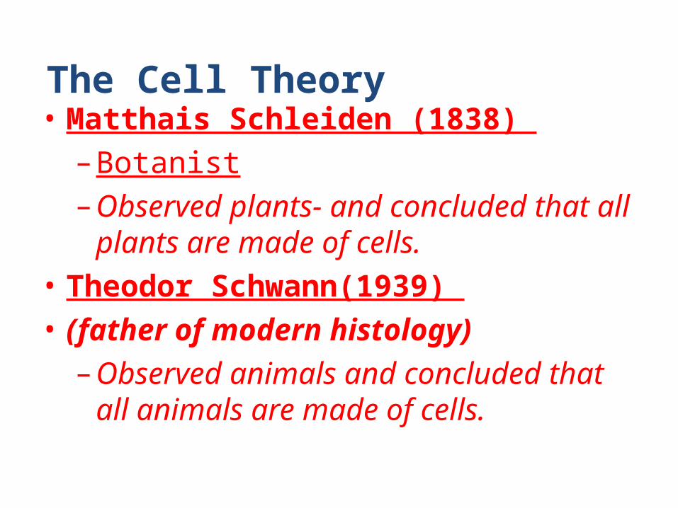
The Cell Theory• Matthais Schleiden (1838) –Botanist–Observed plants- and concluded that all
plants are made of cells. • Theodor Schwann(1939) • (father of modern histology)–Observed animals and concluded that all
animals are made of cells.

The Cell Theory
• Rudolf Virchow - 1852– Concluded that the nucleus was responsible for
cell division
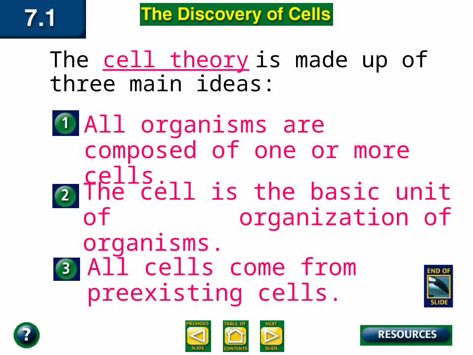
The cell theory is made up of three main ideas:
All cells come from preexisting cells.
The cell is the basic unit of organization of organisms.
All organisms are composed of one or more cells.
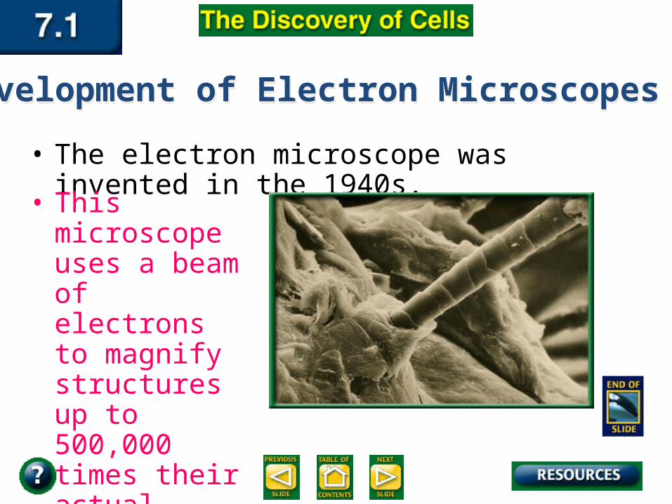
• The electron microscope was invented in the 1940s.
• This microscope uses a beam of electrons to magnify structures up to 500,000 times their actual size.
Development of Electron MicroscopesDevelopment of Electron Microscopes

There are two basic types of electron microscopes.
The transmission electron microscope allows scientists to study the structures contained within a cell.
The scanning electron microscope scans the surface of cells to learn their three dimensional shape.
Development of Electron MicroscopesDevelopment of Electron Microscopes
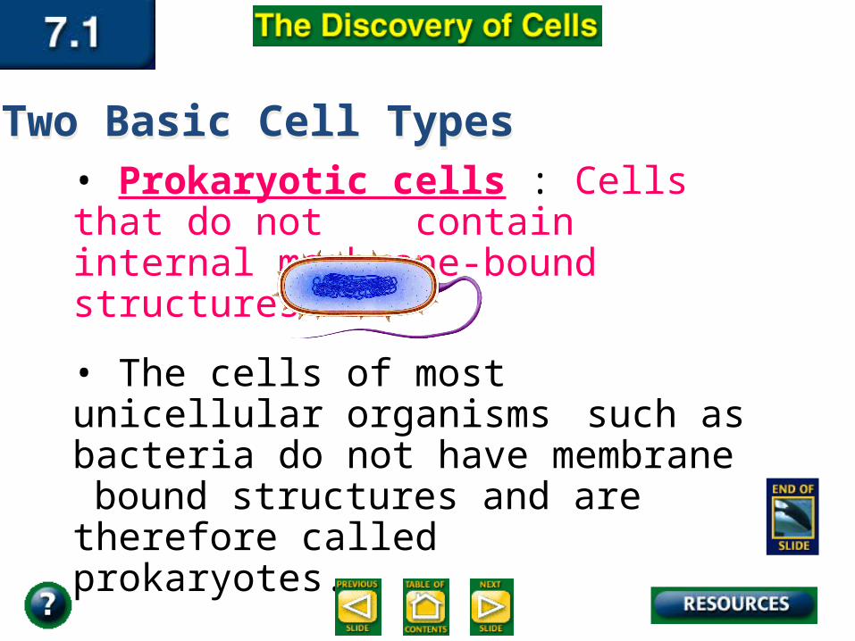
• Prokaryotic cells : Cells that do not contain internal membrane-bound structures
• The cells of most unicellular organisms such as bacteria do not have membrane bound structures and are therefore called prokaryotes.
Two Basic Cell TypesTwo Basic Cell Types
• Most of the multi-cellular plants and animals we know are made up of cells containing membrane-bound structures
and are therefore called eukaryotes.
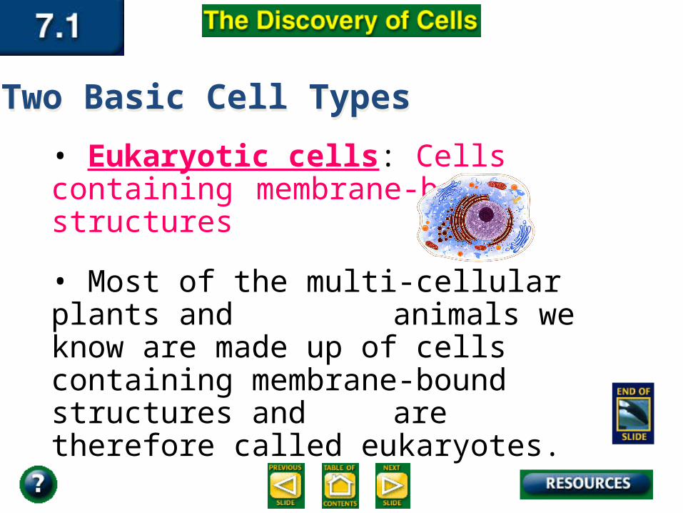
• Eukaryotic cells: Cells containing membrane-bound structures
7.17.1
Two Basic Cell TypesTwo Basic Cell Types
• Organelles: the membrane-bound structures within eukaryotic cells
• Each organelle has a specific function that contributes to cell survival.
Two Basic Cell TypesTwo Basic Cell Types

•Nucleus: the central membrane-bound organelle that manages cellular functions.
• Separation of organelles into distinct compartments benefits the eukaryotic cells.
Two Basic Cell TypesTwo Basic Cell Types
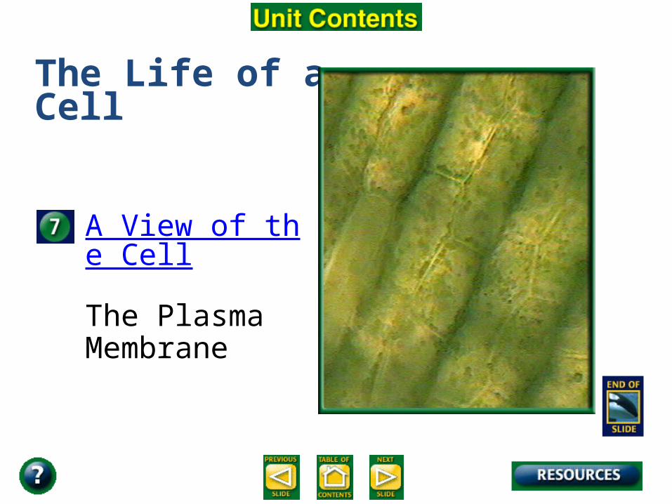
The Life of a Cell
A View of the Cell
The Plasma Membrane
All living cells must maintain a balance regardless of internal and external conditions. Survival depends on the cell’s ability to maintain the proper conditions within itself.
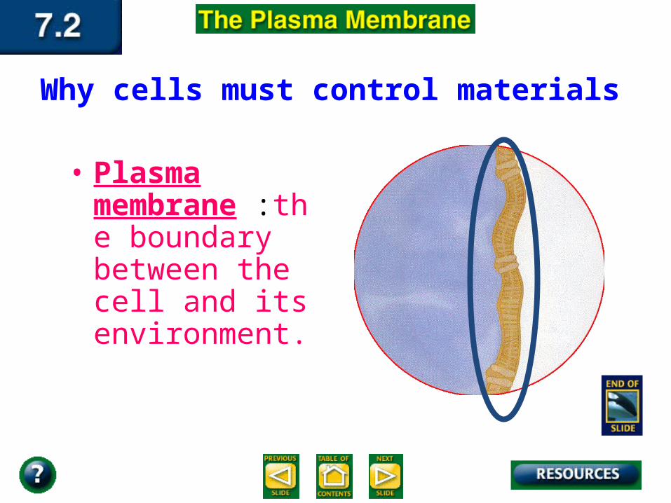
Why cells must control materials
• Plasma membrane :the boundary between the cell and its environment.
It is the plasma membrane’s job to:
• allow waste and other products to leave the cell.
• remove excess amounts of these nutrients when levels get so high that they are harmful.
• allow a steady supply of glucose, amino acids, and lipids to come into the cell no matter what the external conditions are.
• Selective permeability : a process used to maintain homeostasis in which the plasma membrane allows some molecules into the cell while keeping others out.
• Homeostasis: the process of maintaining the cell’s environment
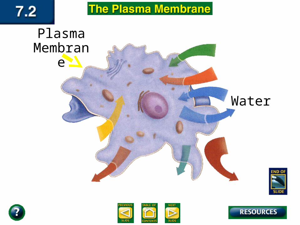
Water
Plasma Membrane
Structure of the Plasma Membrane
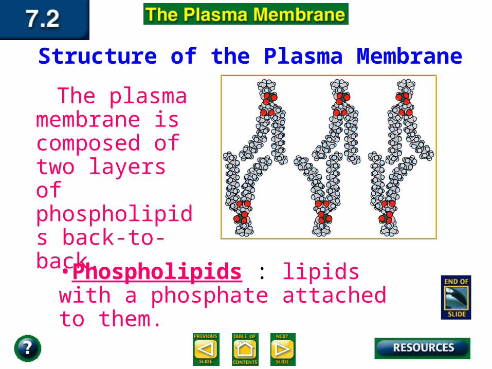
The plasma membrane is composed of two layers of phospholipids back-to-back.
•Phospholipids : lipids with a phosphate attached to them.
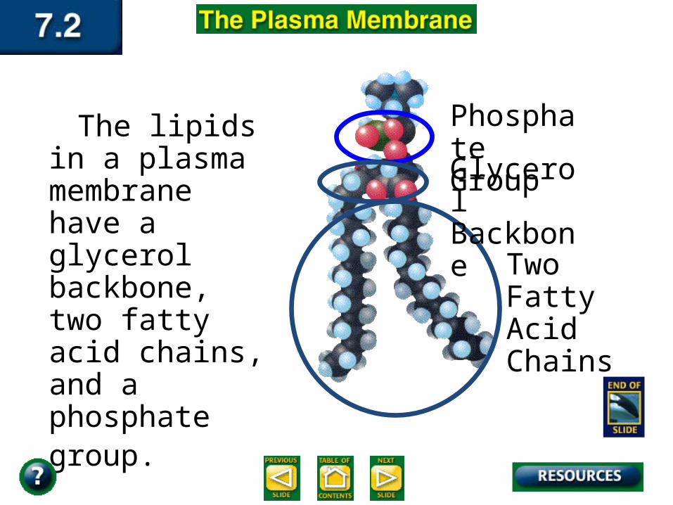
The lipids in a plasma membrane have a glycerol backbone, two fatty acid chains, and a phosphate group.
Glycerol Backbone
Two Fatty Acid Chains
Phosphate Group
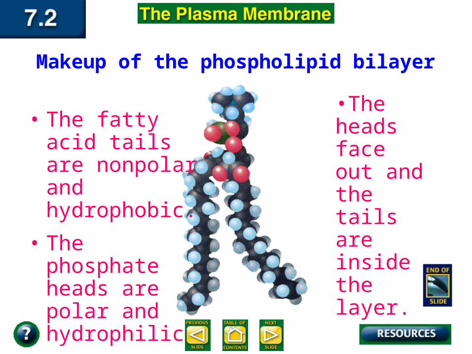
Makeup of the phospholipid bilayer
• The fatty acid tails are nonpolar and hydrophobic.
• The phosphate heads are polar and hydrophilic.
•The heads face out and the tails are inside the layer.
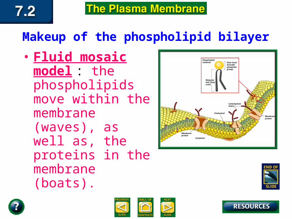
Makeup of the phospholipid bilayer• Fluid mosaic model :
the phospholipids move within the membrane (waves), as well as, the proteins in the membrane (boats).
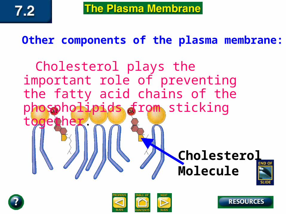
Other components of the plasma membrane:
Cholesterol plays the important role of preventing the fatty acid chains of the phospholipids from sticking together.
CholesterolMolecule
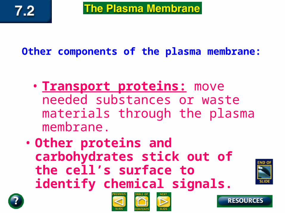
Other components of the plasma membrane:
• Transport proteins: move needed substances or waste materials through the plasma membrane.
• Other proteins and carbohydrates stick out of the cell’s surface to identify chemical signals.
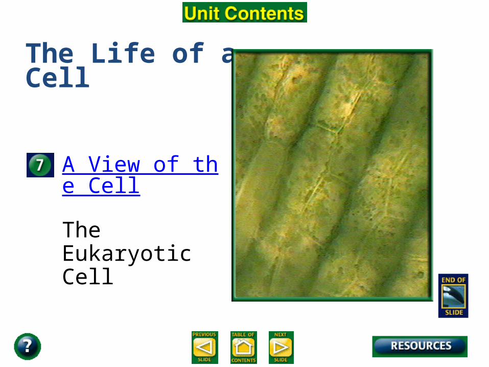
The Life of a Cell
A View of the Cell
The Eukaryotic Cell
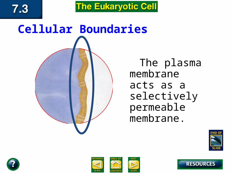
The plasma membrane acts as a selectively permeable membrane.
Cellular Boundaries
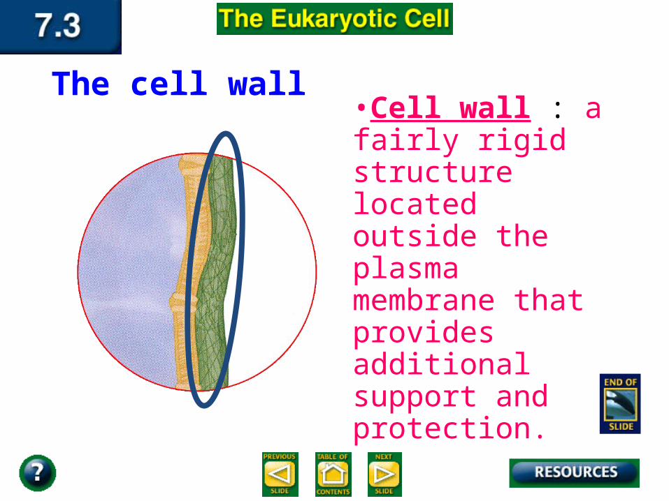
The cell wall •Cell wall : a fairly rigid structure located outside the plasma membrane that provides additional support and protection.
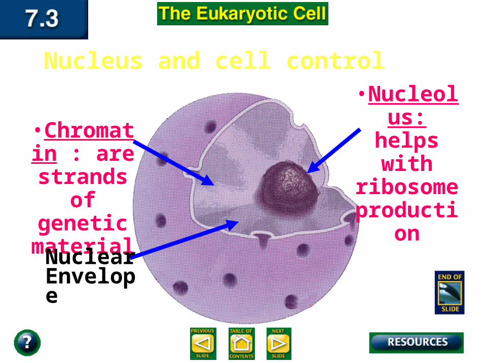
Nucleus and cell control
•Chromatin : are
strands of genetic
material
•Nucleolus: helps with ribosome
production
Nuclear Envelope

Inside the Eukaryotic Cell
•Cytoplasm: the gelatin-like material inside every cell; it constantly flows inside the cell
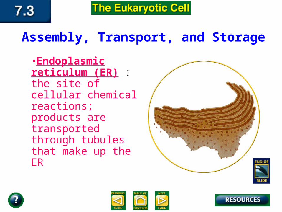
Assembly, Transport, and Storage
•Endoplasmic reticulum (ER) : the site of cellular chemical reactions; products are transported through tubules that make up the ER

Assembly, Transport, and Storage Endoplasmic
Reticulum (ER)
•Ribosomes : the smallest organelles that are not
membrane bound and make proteins
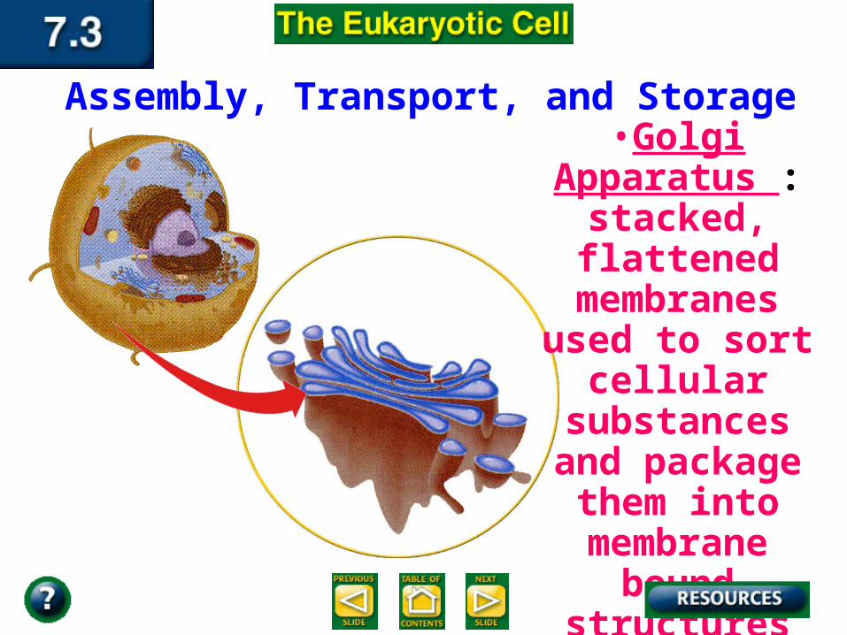
Assembly, Transport, and Storage •Golgi
Apparatus : stacked, flattened
membranes used to sort cellular substances and package them
into membrane bound structures
called vesicles
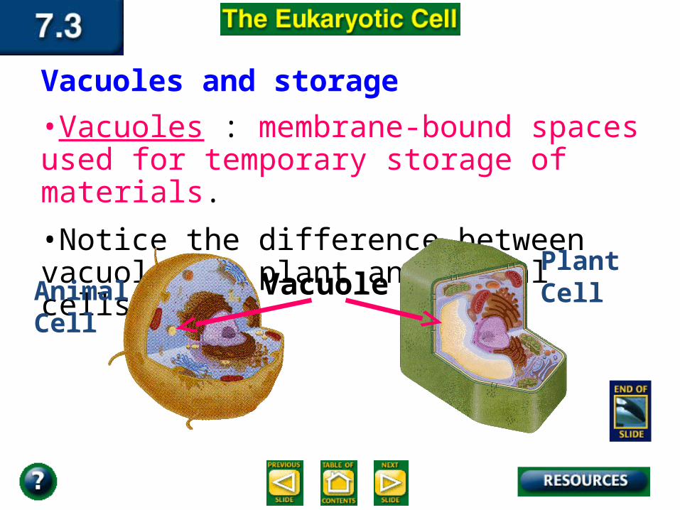
Vacuoles and storage •Vacuoles : membrane-bound spaces used for temporary storage of materials.
•Notice the difference between vacuoles in plant and animal cells.
VacuoleAnimalCell
PlantCell
Lysosomes and recycling
•Lysosomes : organelles that contain digestive enzymes.
•They digest excess or worn out organelles, food particles, and engulfed viruses or bacteria.

Energy Transformers:
•Chloroplasts : cell organelles that capture light energy and produce food to store for a later time.
Chloroplasts and energy
•The chloroplasts belongs to a group of plant organelles called plastids, which are used for storage.
•Chloroplasts contain green pigment called chlorophyll. Chlorophyll traps light energy and gives leaves and stems their green color.
Chloroplasts and energy
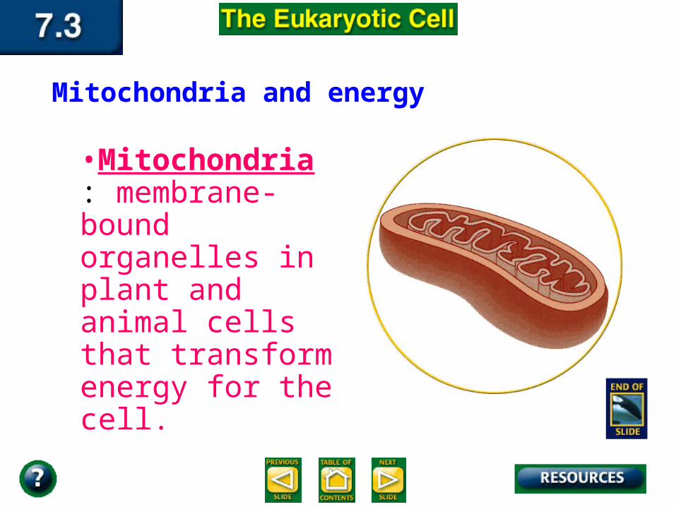
•Mitochondria : membrane-bound organelles in plant and animal cells that transform energy for the cell.
Mitochondria and energy
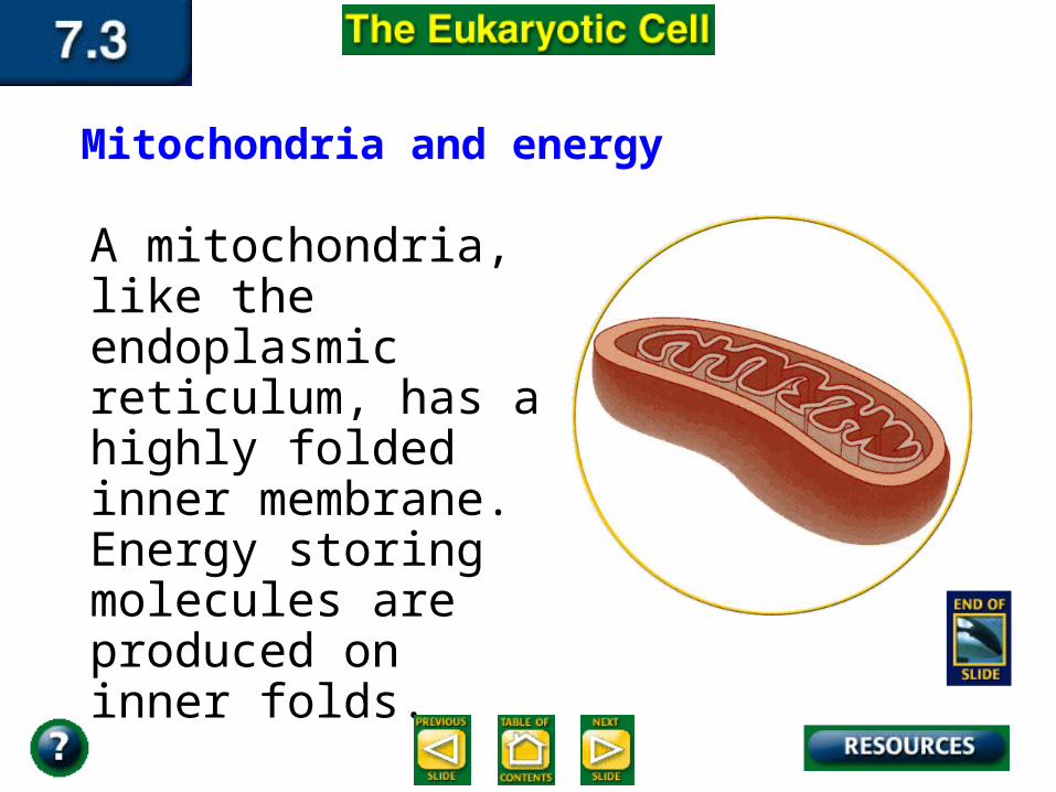
A mitochondria, like the endoplasmic reticulum, has a highly folded inner membrane. Energy storing molecules are produced on inner folds.
Mitochondria and energy
•Cytoskeleton : support structure composed of microtubules and microfilaments.
•Microtubules are thin, hollow cylinders made of protein and microfilaments are thin solid protein fibers.
Structures for Support and Locomotion
Some cell surfaces have cilia and flagella, which are structures that aid in locomotion or feeding. Cilia and flagella can be distinguished by their structure and by the nature of their action.
Cilia and flagella
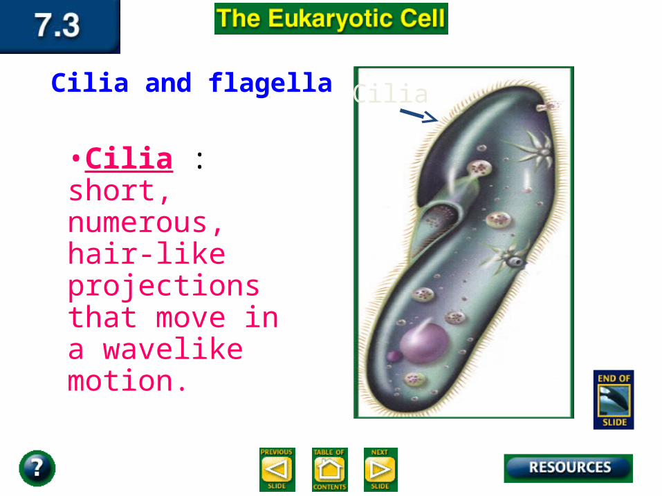
•Cilia : short, numerous, hair-like projections that move in a wavelike motion.
Cilia and flagella Cilia
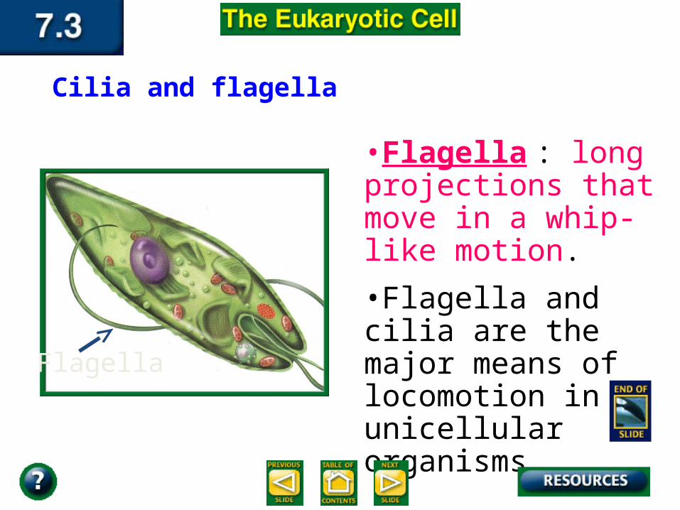
•Flagella : long projections that move in a whip-like motion.
•Flagella and cilia are the major means of locomotion in unicellular organisms.
Cilia and flagella
Flagella