1. The following image demonstrates the axis … following image demonstrates the axis vertebra in...
12
1. The following image demonstrates the axis vertebra in oblique lateral projection. What is the “*” ? A. Articular process B. Pars interarticularis C. Spinous process D. Odontoid process 1. Ans. D. Odontoid process The axis (C2) bears the dens (odontoid process) on the superior aspect of its body, representing the detached centrum of C1. Nodding and lateral flexion movements occur at the atlanto-occipital joint, whereas rotation of the skull occurs at the atlanto-axial joint around the dens, which acts as a pivot. 2. The elongation of fatty acids occurs in which of the diagrammatic structures shown below? A. Structure A B. Structure B C. Structure C D. Structure D 2. Ans. C. Structure C Fatty acid synthesis in the cytosol terminates at palmitate, a C16 saturated fatty acid. Elongation of acyl groups can occur from palmitate as well as from other dietary saturated and unsaturated fatty acids of lengths C10 and greater. Longer-chain elongation of fatty acids occurs in the endoplasmic reticulum (structure C) using malonyl CoA as the acetyl donor and NADPH as the reductant. Very-long-chain and long-chain fatty acids are preferentially catabolized in peroxisomes (structure J). Other structures diagrammed in the figure are the plasma membrane (A), mitochondrion (B), nucleoplasm (D), nucleolus (E), Golgi apparatus (F), secretory vesicles (G), caveolae such as those taking up low-density lipoprotein from its receptor (H), and lysosomes (I). 3. A 37-year-old newly married man presents with multiple blister-like lesions on the glans of his penis, appearing over the past 2 days. On questioning, he recalls similar episodes over the past 2 years. Examination is remarkable for tender, 3-4 mm vesicular lesions on the shaft of his penis with no apparent crusting, drainage, or bleeding. There is also slight bilateral inguinal adenopathy. During the asymptomatic period between outbreaks, where would the causative agent likely have been found?
Transcript of 1. The following image demonstrates the axis … following image demonstrates the axis vertebra in...

1. The following image demonstrates the axis vertebra in oblique lateral projection. What is the “*” ?
A. Articular process B. Pars interarticularis C. Spinous process D. Odontoid process
1. Ans. D. Odontoid process The axis (C2) bears the dens (odontoid process) on the superior aspect of its body, representing the detached centrum of C1. Nodding and lateral flexion movements occur at the atlanto-occipital joint, whereas rotation of the skull occurs at the atlanto-axial joint around the dens, which acts as a pivot.
2. The elongation of fatty acids occurs in which of the diagrammatic structures shown below?
A. Structure A B. Structure B C. Structure C D. Structure D
2. Ans. C. Structure C Fatty acid synthesis in the cytosol terminates at palmitate, a C16 saturated fatty acid. Elongation of acyl groups can occur from palmitate as well as from other dietary saturated and unsaturated fatty acids of lengths C10 and greater. Longer-chain elongation of fatty acids occurs in the endoplasmic reticulum (structure C) using malonyl CoA as the acetyl donor and NADPH as the reductant. Very-long-chain and long-chain fatty acids are preferentially catabolized in peroxisomes (structure J). Other structures diagrammed in the figure are the plasma membrane (A), mitochondrion (B), nucleoplasm (D), nucleolus (E), Golgi apparatus (F), secretory vesicles (G), caveolae such as those taking up low-density lipoprotein from its receptor (H), and lysosomes (I).
3. A 37-year-old newly married man presents with multiple blister-like lesions on the glans of his penis, appearing over the past 2 days. On questioning, he recalls similar episodes over the past 2 years. Examination is remarkable for tender, 3-4 mm vesicular lesions on the shaft of his penis with no apparent crusting, drainage, or bleeding. There is also slight bilateral inguinal adenopathy. During the asymptomatic period between outbreaks, where would the causative agent likely have been found?

A. Fibroblasts B. Lymphocytes C. Neurons of the sacral ganglia D. Mucoepithelial cells
3. Ans. (C) This is the history of genital herpes. HSV2 produces latent infection in sacral ganglia.
Clinical Features of Genital Ulcers
Feature Syphilis Herpes Chancroid Lymphogranuloma Venereum
Donovanosis
Incubation period 9–90 days 2–7 days 1–14 days 3 days–6 weeks 1–4 weeks (up to 6 months)
Early primary lesions
Papule Vesicle Pustule Papule, pustule, or vesicle Papule
No. of lesions Usually one Multiple Usually multiple, may coalesce
Usually one; often not detected, despite lymphadenopathy
Variable
Diameter 5–15 mm 1–2 mm Variable 2–10 mm Variable
Edges Sharply demarcated, elevated, round, or oval
Erythematous Undermined, ragged, irregular
Elevated, round, or oval Elevated, irregular
Depth Superficial or deep Superficial Excavated Superficial or deep Elevated
Base Smooth, nonpurulent, relatively nonvascular
Serous, erythematous, nonvascular
Purulent, bleeds easily
Variable, nonvascular Red and velvety, bleeds readily
Induration Firm None Soft Occasionally firm Firm
Pain Uncommon Frequently tender Usually very tender Variable Uncommon
Lymphadenopathy Firm, nontender, bilateral
Firm, tender, often bilateral with initial episode
Tender, may suppurate, loculated, usually unilateral
Tender, may suppurate, loculated, usually unilateral
None; pseudobuboes
4. A 54-year-old man with a chronic cough has a squamous cell carcinoma diagnosed in his right lung. While performing a pneumonectomy, the thoracic surgeon notes that the hilar lymph nodes are small, 0.5 to 1.0 cm in size, and jet black in color throughout( see picture below). Which of the following is the most likely cause for this appearance to the hilar nodes?
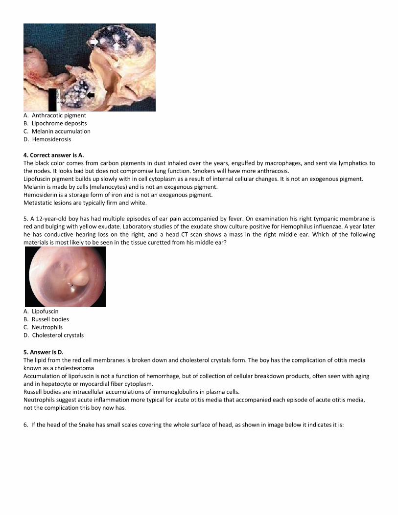
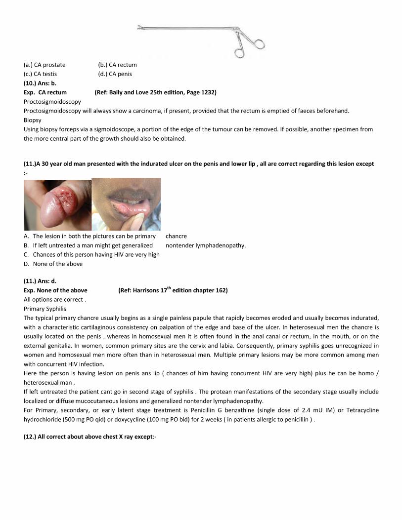
A. Anthracotic pigment B. Lipochrome deposits C. Melanin accumulation D. Hemosiderosis 4. Correct answer is A. The black color comes from carbon pigments in dust inhaled over the years, engulfed by macrophages, and sent via lymphatics to the nodes. It looks bad but does not compromise lung function. Smokers will have more anthracosis. Lipofuscin pigment builds up slowly with in cell cytoplasm as a result of internal cellular changes. It is not an exogenous pigment. Melanin is made by cells (melanocytes) and is not an exogenous pigment. Hemosiderin is a storage form of iron and is not an exogenous pigment. Metastatic lesions are typically firm and white. 5. A 12-year-old boy has had multiple episodes of ear pain accompanied by fever. On examination his right tympanic membrane is red and bulging with yellow exudate. Laboratory studies of the exudate show culture positive for Hemophilus influenzae. A year later he has conductive hearing loss on the right, and a head CT scan shows a mass in the right middle ear. Which of the following materials is most likely to be seen in the tissue curetted from his middle ear?
A. Lipofuscin B. Russell bodies C. Neutrophils D. Cholesterol crystals
5. Answer is D. The lipid from the red cell membranes is broken down and cholesterol crystals form. The boy has the complication of otitis media known as a cholesteatoma Accumulation of lipofuscin is not a function of hemorrhage, but of collection of cellular breakdown products, often seen with aging and in hepatocyte or myocardial fiber cytoplasm. Russell bodies are intracellular accumulations of immunoglobulins in plasma cells. Neutrophils suggest acute inflammation more typical for acute otitis media that accompanied each episode of acute otitis media, not the complication this boy now has.
6. If the head of the Snake has small scales covering the whole surface of head, as shown in image below it indicates it is:

A. Viper B. Krait C. Cobra D. Nonpoisonous
6. Ans. A. Viper If the head of the snake has large scales over the head, it could be- poisonous or non-poisonous, both. But if the head of snake has small scales, it is most likely- poisonous, commonly viper.
7. A middle aged female presents with chronic pain in right side of abdomen with intermittent fever. Clinical examination revealed mild hepatomegaly and hence a contrast enhanced CT abdomen was performed. Based on the imaging characteristics of this focal lesion the most likely diagnosis is ?
A. Liver abscess B. Hydatid cyst C. HCC D. Hepatic adenoma
7. Ans is B. Hydatid cyst REF : Adam: Grainger & Allison's Diagnostic Radiology, 5th ed.Chap 35 The Portal phase CT demonstrates a large cystic structure with a discrete wall, separated internal membranes and several ‘daughter cysts’ (arrowheads This is an infection of the liver with Echinococcus granulosus, a parasitic tapeworm present worldwide and transmitted from sheep, foxes and other wild animals to humans as part of its lifecycle. Larvae migrate from the gut and embed in the liver, where they encyst and develop, slowly provoking a surrounding inflammatory reaction. The disease may remain occult for several years. On imaging there is a wide range of appearances, from a simple cyst indistinguishable from a true hepatic cyst to a complicated cyst with any or all of the following features: debris (hydatid ‘sand’ made up of dead scolices, which may calcify), daughter cysts, membrane separation, and wall calcification. The lesions may be multiple and vary widely in size. Serological testing can be employed to confirm the presence of infection prior to any therapy or intervention. Although the risk of anaphylaxis following aspiration or surgery of these lesions is well recognized, it is less than previously thought, and uncomplicated aspiration following medical treatment has been described. US demonstrates clearly not only the simple cyst form but also the more complex cyst features, such as the dependent debris, daughter cysts (cyst within a cyst appearance), membrane separation and wall calcification. CT defines all these features as well and

is helpful where wall calcification obscures the view on US. MRI will define the cystic structure and internal anatomy but is insensitive to the calcification.
(8.) A 42-year-old man has had increasing difficulty with activities of daily living for the past year, mainly because of involuntary
choreiform movements. His family has also noted that he has exhibited behavioral changes, though his memory remains intact.
His brother is similarly affected. He has the appearance of the coronal section of brain shown here. Which of the following is the
most likely diagnosis?
(a.) Huntington disease
(b.) Parkinson disease
(c.) Alzheimer disease
(d.) Amyotrophic lateral sclerosis
(8.) Ans: a.
Exp. Huntington disease
Huntington disease is an autosomal dominant condition that presents with choreiform movements beginning in adult life. There are
increased CAG tandem repeat sequences in the huntingtin gene. Note the marked atrophy of the caudate nucleus seen in picture .
(9.) A 2-year-old child is noted to have abdominal enlargement on a visit to the pediatrician. His abdominal MR image reveals a lesion above the left kidney. Which of the following laboratory test findings is most likely to be present in this child?
(a.) Elevated serum cortisol
(b.) Elevated serum estrogen
(c.) Increased urine homovanillic acid
(d.) Increased plasma renin
(9.) Ans: c.
Exp. Increased urine homovanillic acid
A neuroblastoma is one of the more common childhood malignant neoplasms, and it often arises in the adrenal. Neuroblastoma,
a malignant tumor of childhood, originates from precursor cells of sympathetic nerve tissue or adrenal medulla. These cells
produce excessive quantities of dopa, dopamine, and norepinephrine. These are converted to two major terminal urinary
metabolites, homovanillic acid (HVA) and vanilmandelic acid (VMA).
Plain radiographs of the abdomen may show a flank mass. Stippled calcifications are present on up to 30% of radiographs.
(10.) The below shown biopsy forceps is used most commonly in which condition?
(a.) CA prostate (b.) CA rectum
(c.) CA testis (d.) CA penis
(10.) Ans: b.
Exp. CA rectum (Ref: Baily and Love 25th edition, Page 1232)
Proctosigmoidoscopy
Proctosigmoidoscopy will always show a carcinoma, if present, provided that the rectum is emptied of faeces beforehand.
Biopsy
Using biopsy forceps via a sigmoidoscope, a portion of the edge of the tumour can be removed. If possible, another specimen from
the more central part of the growth should also be obtained.
(11.)A 30 year old man presented with the indurated ulcer on the penis and lower lip , all are correct regarding this lesion except
:-
A. The lesion in both the pictures can be primary chancre
B. If left untreated a man might get generalized nontender lymphadenopathy.
C. Chances of this person having HIV are very high
D. None of the above
(11.) Ans: d.
Exp. None of the above (Ref: Harrisons 17th edition chapter 162)
All options are correct .
Primary Syphilis
The typical primary chancre usually begins as a single painless papule that rapidly becomes eroded and usually becomes indurated,
with a characteristic cartilaginous consistency on palpation of the edge and base of the ulcer. In heterosexual men the chancre is
usually located on the penis , whereas in homosexual men it is often found in the anal canal or rectum, in the mouth, or on the
external genitalia. In women, common primary sites are the cervix and labia. Consequently, primary syphilis goes unrecognized in
women and homosexual men more often than in heterosexual men. Multiple primary lesions may be more common among men
with concurrent HIV infection.
Here the person is having lesion on penis ans lip ( chances of him having concurrent HIV are very high) plus he can be homo /
heterosexual man .
If left untreated the patient cant go in second stage of syphilis . The protean manifestations of the secondary stage usually include
localized or diffuse mucocutaneous lesions and generalized nontender lymphadenopathy.
For Primary, secondary, or early latent stage treatment is Penicillin G benzathine (single dose of 2.4 mU IM) or Tetracycline
hydrochloride (500 mg PO qid) or doxycycline (100 mg PO bid) for 2 weeks ( in patients allergic to penicillin ) .
(12.) All correct about above chest X ray except:-

A. Diffuse reticulonodular pattern is seen
B. The appearance is Caused by a decrease in the gas to soft tissue ratio
C. Main cause is thickening of any of the interstitial compartments
D. None
(12.) Ans: d.
Exp.None. (Ref:- http://radiopaedia.org/articles/reticular-and-linear-pulmonary-opacification)
All options are correct .
A reticulonodular interstitial pattern is produced by either, overlap of reticular shadows, or by the presence of reticular shadowing
and pulmonary nodules. While this is a relatively common appearance on a chest radiograph, very few diseases are confirmed to
show this patten pathologically. Examples include: silicosis, pulmonary sarcoidosis, lymphangitis carcinomatosis.
In chest radiology, reticular and linear opacification refers to a broad sub-group of pulmonary opacification caused by a decrease in
the gas to soft tissue ratio caused by a pathological process centred in and around the pulmonary interstitium. This includes
thickening of any of the interstitial compartments by blood, water, tumour, cells, fibrous disease or any combination thereof. The
thickening of the interstitium can be reticular, reticulonodular, linear where the predominant pattern is a result of the underlying
pathological process.
(13.) A (27-year-old woman is brought to the emergency department after she suddenly collapsed. On physical examination her temperature is 37.)1 C, pulse 100/minute, respiratory rate (20/minute, and blood pressure 80/40 mm Hg. Laboratory studies show a positive pregnancy test. Her peripheral blood smear is shown here. Which of the following is the most likely diagnosis?
(a.) Septic shock (b.) Mycobacterium tuberculosis infection (c.) Acute renal failure (d.) Disseminated intravascular coagulation (13.) Ans: d.
Exp. Disseminated intravascular coagulation
The fragmented erythrocytes, or schistocytes, shown here are indicative of a microangiopathic hemolytic anemia. Obstetrical
disasters (such as ruptured ectopic pregnancy), trauma with blood loss, sepsis, and many other conditions can lead to
DIC(Disseminated intravascular coagulation).

(14.) 70-year-old man who died suddenly has the finding shown here on external examination at autopsy. Which of the following
is the most likely diagnosis?
(a.) Accidental electrocution
(b.) Suicidal gunshot wound
(c.) Homicidal stab wounds
(d.) Fall from a height
14. Ans: b.
Exp. Suicidal gunshot wound
This is a contact range gunshot wound with sooting to produce the central dark discoloration. The star-shaped lacerations result
from gases from the gunshot causing laceration to the skin over the skull.
15. This is the X-ray of a 9 year old girl, this type of bone finding is most likely seen in which condition?
A. Hyperthyroid B. Hyperparathyroid C. Hypoparathyroid D. Hypothyroid 15. Ans. D. Hypothyroid
1. In hypothyroid Retardation of osseous development can be shown radiographically at birth in about 60% of congenitally hypothyroid infants and indicates some deprivation of thyroid hormone during intrauterine life.
2. The distal femoral epiphysis, normally present at birth, is often absent. In undetected and untreated patients, the discrepancy between chronological age and osseous development increases.

3. The epiphyses often have multiple foci of ossification (epiphyseal dysgenesis); deformity (“beaking”) of the 12th thoracic or 1st or 2nd lumbar vertebra is common.
4. Roentgenograms of the skull show large fontanels and wide sutures; intersutural (wormian) bones are common. 5. The sella turcica is often enlarged and round; in rare instances, there may be erosion and thinning. Delays in formation and
eruption of teeth may occur. Cardiac enlargement or pericardial effusion may be present. 16. Following lesion in the brain is seen in which condition?
(A). Parkinsonism (B). Rabies (C). Creutzfeldt–Jakob disease (D). Tuberculosis 16. Ans. (B). Rabies
a. These are Negri body. b. Negri bodies are eosinophilic, sharply outlined, pathognomonic inclusion bodies (2–10 µm in diameter) found in the
cytoplasm of certain nerve cells containing the virus of rabies, especially in Ammon's horn of the hippocampus. c. They are also often found in the cerebellar cortex of postmortem brain samples of rabies victims.
17. In the PV loop below, the EDV is shown at what point?
A) A B) B C) C D) D 17. The answer is B. B
AB = LV filling
BC = Isovolumetric contraction
CD = LV ejection
DA = Isovolumetric relaxation
Point-A = Coincides with MV opening, and represents LV end-systolic volume and early diastolic pressure
Point- B = Coincides with MV closure, and represents LV end diastolic pressure (LV EDP) and volume (EDV)
Point-C= Represents opening of Aortic valve and coincides with systemic, aortic diastolic pressure
Point-D= is the closure of the Aortic valve and represents LV end systolic pressure and volume, coinciding with the dicrotic notch in
the Aortic pressure tracing
Segment AB => LV compliance is defined by the slope of the filling phase or segment AB Preload or EDV. The compliance is
decreased when the ventricles become stiff or unable to fill properly eg MI, constrictive pericarditis, pericardial effusion etc and the
PV loop(baseline shifts up.
Therefore PV loops analysis gives information about - LV compliance, Preload, contractility. Stroke volume (SV) [SV = EDV - ESV],
Ejection Fraction (EF) and various valvular lesions.
Remember: The curve shifts to right side in case of increased preload, Upside in case of increased afterload and to Left & upside
in incase of increased myocardial contractility
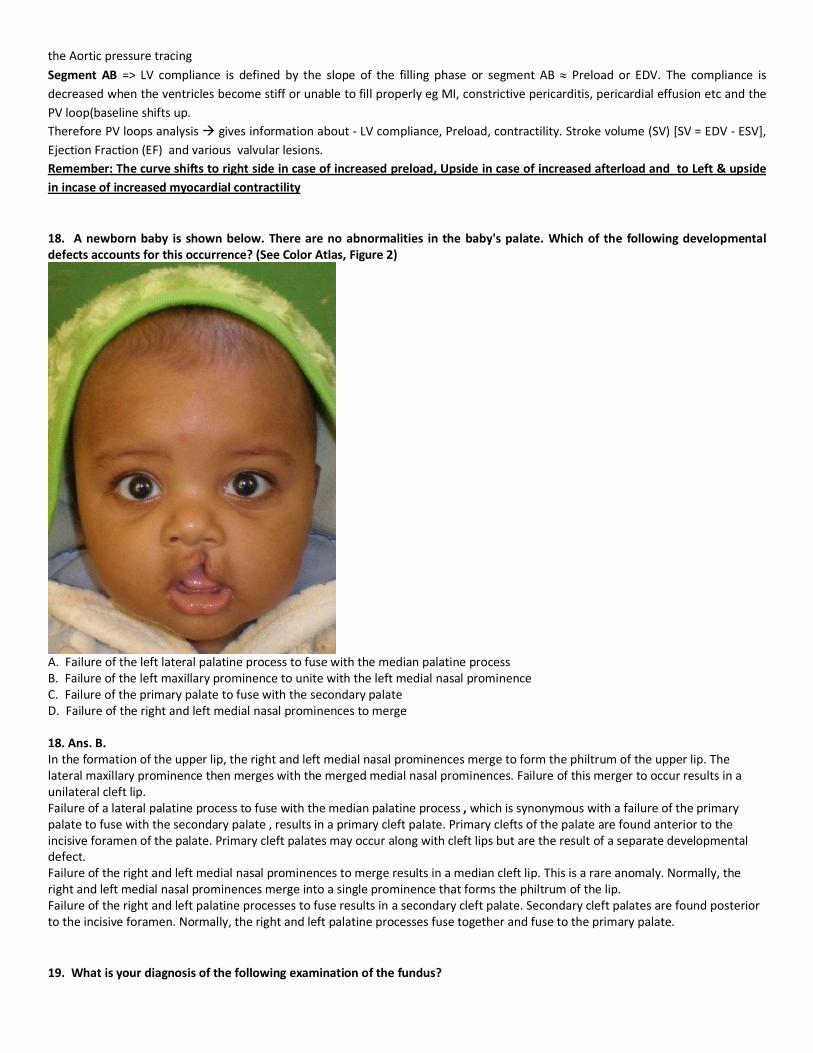
18. A newborn baby is shown below. There are no abnormalities in the baby's palate. Which of the following developmental defects accounts for this occurrence? (See Color Atlas, Figure 2)
A. Failure of the left lateral palatine process to fuse with the median palatine process B. Failure of the left maxillary prominence to unite with the left medial nasal prominence C. Failure of the primary palate to fuse with the secondary palate D. Failure of the right and left medial nasal prominences to merge 18. Ans. B. In the formation of the upper lip, the right and left medial nasal prominences merge to form the philtrum of the upper lip. The lateral maxillary prominence then merges with the merged medial nasal prominences. Failure of this merger to occur results in a unilateral cleft lip. Failure of a lateral palatine process to fuse with the median palatine process , which is synonymous with a failure of the primary palate to fuse with the secondary palate , results in a primary cleft palate. Primary clefts of the palate are found anterior to the incisive foramen of the palate. Primary cleft palates may occur along with cleft lips but are the result of a separate developmental defect. Failure of the right and left medial nasal prominences to merge results in a median cleft lip. This is a rare anomaly. Normally, the right and left medial nasal prominences merge into a single prominence that forms the philtrum of the lip. Failure of the right and left palatine processes to fuse results in a secondary cleft palate. Secondary cleft palates are found posterior to the incisive foramen. Normally, the right and left palatine processes fuse together and fuse to the primary palate.
19. What is your diagnosis of the following examination of the fundus?

(A) Diabetes retinopathy.
(B) Optic atrophy
(C) Acute glaucoma
(D) Optic disc drusen
19Ans. (C) Acute glaucoma Glaucoma results in "cupping" as the neural rim is destroyed and the central cup becomes enlarged and excavated. The cup-to-disc ratio is about 0.7/1.0 in this patient. Glaucoma Glaucoma is a slowly progressive, insidious optic neuropathy that usually is associated with chronic elevation of intraocular pressure. The mechanism by which raised intraocular pressure injures the optic nerve is not understood. Axons entering the inferotemporal and superotemporal aspects of the optic disc are damaged first, producing typical nerve fiber bundle or arcuate scotomas on perimetric testing. As fibers are destroyed, the neural rim of the optic disc shrinks and the physiologic cup within the optic disc enlarges. This process is referred to as pathologic "cupping." The cup-to-disc diameter is expressed as a ratio (e.g., 0.2/1). The cup-to-disc ratio ranges widely in normal individuals, making it difficult to diagnose glaucoma reliably simply by observing an unusually large or deep optic cup. Careful documentation of serial examinations is helpful. In a patient with physiologic cupping the large cup remains stable, whereas in a patient with glaucoma it expands relentlessly over the years. Detection of visual field loss by computerized perimetry also contributes to the diagnosis. Finally, most patients with glaucoma have raised intraocular pressure. However, many patients with typical glaucomatous cupping and visual field loss have intraocular pressures that apparently never exceed the normal limit of 20 mmHg (so-called low-tension glaucoma). 20.What is your diagnosis for a patient having following fundus examination?
(A)Diabetic retinopathy
(B) Hypertension retinopathy
(C) Optic atrophy
(D) Central retinal vein occlusion
20. Ans. (B) Hypertension retinopathy This is case of Hypertensive retinopathy with scattered flame (splinter) hemorrhages and cotton-wool spots Hypertensive retinopathy Patient with systemic hypertension has frequent headaches.

Vasoconstriction of the retinal arterioles is primary response to the raised blood pressure. Grading of hypertensive retinopathy (Keith and Wegner) Grade I - Consists of mild arterial attenuation, broadening of the arteriolar light reflex. Grade II - Marked generalized narrowing and focal attenuation of arterioles associated with deflection of
Veins at arteriovenous crossings (Salus sign) Grade III - Grade n + Copper wiring of arterioles flame shaped hemorrhages. cotton wool spots, hard exudates. Grade IV - Grade Ill + Silver wiring + papilloedema.