1. OVERVIEW OF THE LUDWIG BOLTZMANN INSTITUTE FOR … Report...1 1. overview of the ludwig boltzmann...
40
Transcript of 1. OVERVIEW OF THE LUDWIG BOLTZMANN INSTITUTE FOR … Report...1 1. overview of the ludwig boltzmann...


1
1. OVERVIEW OF THE LUDWIG BOLTZMANN INSTITUTE FOR CLINICAL-FORENSIC
IMAGING...................................................................................................................... 3
1.1 AIMS ............................................................................................................................................................. 3
1.2 QUANTITY STRUCTURE................................................................................................................................. 4
1.3 INSTITUTIONAL PARTNERS............................................................................................................................ 5
1.4 SUPERVISORY BOARD UND SCIENTIFIC ADVISORY BOARD ........................................................................ 7
1.4.1 Supervisory Board.................................................................................................................................. 7
1.4.2 Scientific Advisory Board ...................................................................................................................... 8
1.5 HUMAN RESOURCES AND DEVELOPMENT .................................................................................................... 8
1.5.1 Human resources ................................................................................................................................... 8
1.5.2 Career development ............................................................................................................................ 11
1.5.3 Teamevents........................................................................................................................................... 15
1.6 INFRASTRUCTURE ...................................................................................................................................... 16
1.7 HIGHLIGHTS OF THE YEAR.......................................................................................................................... 16
1.8 PUBLIC RELATIONS ..................................................................................................................................... 17
1.8.1 Media contacts and reports................................................................................................................. 17
1.8.2 Public presentations............................................................................................................................. 18
2. RESEARCH PROGRAM AND RESULTS...................................................................18
2.1 PROJECTS .................................................................................................................................................. 18
2.1.1 Hematomas and blunt force injury of soft tissues............................................................................ 19
2.1.2 Forensic aspects of traumatic brain injury ........................................................................................ 20
2.1.3 Radiologic evidence in forensic reconstruction and age estimation ............................................. 20
2.1.4 Computer-aided tools for forensic case analysis: preparation and presentation ........................ 22
2.1.5 Juridical issues of radiological methods in clinical forensic medicine .......................................... 23
2.1.6 Clinical Forensic Care Unit ................................................................................................................. 23
2.2 PUBLICATIONS ............................................................................................................................................ 25
2.2.1 Publication policies and intellectual property rights......................................................................... 25
2.2.2 Publications ........................................................................................................................................... 25
2.3 PARTICIPATION IN SCIENTIFIC CONFERENCES ........................................................................................... 25
3. OTHER ACTIVITIES ...................................................................................................27
3.1 COOPERATIONS.......................................................................................................................................... 27
3.1.1 Scientific cooperations......................................................................................................................... 27
3.1.2 Non-scientific cooperations................................................................................................................. 29
3.1.3 Third party projects .............................................................................................................................. 29
3.2 MEMBERSHIP IN SCIENTIFIC ASSOCIATIONS .............................................................................................. 30
3.3 LECTURE SERIES AND WORKSHOPS .......................................................................................................... 30
3.4 TEACHING AND TRAINING ACTIVITIES ......................................................................................................... 31
3.4.1 Teaching activities................................................................................................................................ 31
3.5 REVIEWING ACTIVITIES............................................................................................................................... 33

2
4. OUTLOOK...................................................................................................................34
5. LIST OF PUBLICATIONS ...........................................................................................36
5.1 PEER-REVIEWED PAPERS........................................................................................................................... 36
5.2 BOOKS, BOOK CHAPTERS AND OTHER PUBLICATIONS ............................................................................... 36
5.3 ABSTRACTS AND CONFERENCE PRESENTATIONS...................................................................................... 37
5.4 DIPLOMA, BACHELOR, AND MASTER THESES ............................................................................................. 39

3
1. Overview of the Ludwig Boltzmann Institute for Clinical-forensic Imaging
1.1 Aims
The main goals of the LBI are
1. to provide specific forensic imaging studies to form the scientific basis for a clinical-
forensic routine application of radiological methods
2. to establish the juridical basics for the implementation of clinical-forensic imaging (CFI)
into the forensic routine examination of living persons.
Objectives regarding the establishment of the fundament for clinical forensic imaging are
pursued relying on the dedication of all team members. Based on an interdisciplinary
Ad 1)

4
discussion of the studies and their current state at regular meetings the different research
areas constantly grow together to form a coherent entity.
To ensure the interaction of the juridical research with the daily forensic routine of the
Clinical-Forensic Care Unit (CFCU) forming the basis for the juridical questions one member
of the law team is present at the daily morning meetings of the CFCU team where all
examined cases are presented. Additionally, specific legal questions regarding clinical
forensic examinations are discussed which helps directing the juridical research to practical
issues.
Ad 2)
Generally, the research strategy of the LBI comprises
1. Daily case work in forensic medicine and, particularly, clinical forensic medicine as a
basis for the definition of the areas of research and the specific research questions
2. Logical and systematic approach to scientific questions aiming at an increase of
knowledge and understanding in the different areas of research
3. Hypothesis driven and mainly prospective study design with a clear intention
regarding the methodology of data analysis
4. Ethical correctness at all study stages, and the approval of the studies by the local
ethics committee
5. Encouragement and promotion of the scientific and personal skills of young
researchers concerning posing of scientific questions, study design, performance of
the study, and data analysis, but also regarding scientific writing and presentation of
results
6. Backflow of the achieved study results into forensic routine and the implementing
institutions such as the prosecution authorities.
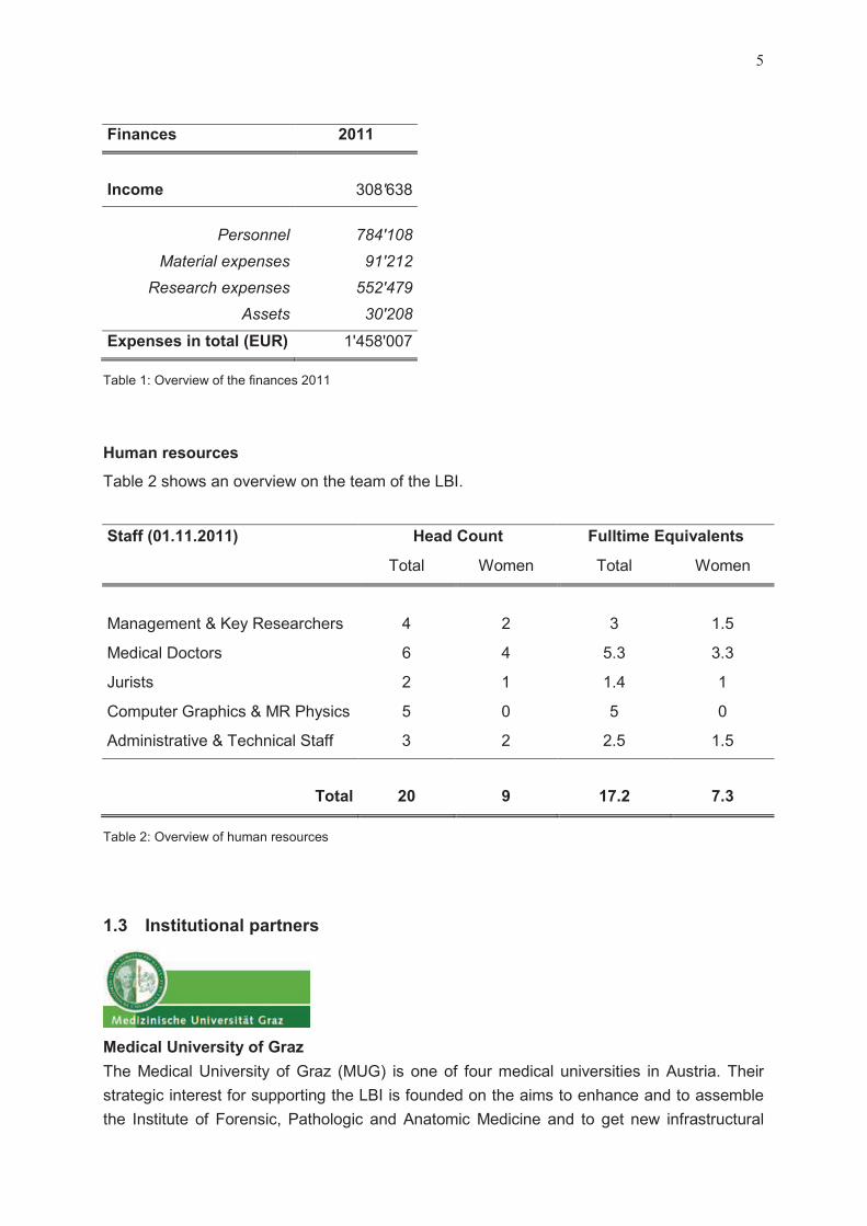
1.2 Quantity structure
Finances
Table 1 presents an overview of the finances 2011. The income consisting of cash and in
kind contributions of the LBG GmbH and the institutional partners was approximately
309.000 Euro. The expenses amounted to 1,46 Mio. Euro (cash and in kind contributions),
and the additional expenditures were covered by the surplus of the last years. The expenses
for the personnel increased to about 54% of the total costs reflecting the completion of the
team. The higher research expenses compared to the previous year accounting for about
38% of the total costs can mainly be attributed to the increasing number of imaging
examinations.
5
Finances 2011
Income 308'638
Personnel 784'108
Material expenses 91'212
Research expenses 552'479
Assets 30'208
Expenses in total (EUR) 1'458'007
Table 1: Overview of the finances 2011
Human resources
Table 2 shows an overview on the team of the LBI.
Staff (01.11.2011) Head Count Fulltime Equivalents
Total Women Total Women
Management & Key Researchers 4 2 3 1.5
Medical Doctors 6 4 5.3 3.3
Jurists 2 1 1.4 1
Computer Graphics & MR Physics 5 0 5 0
Administrative & Technical Staff 3 2 2.5 1.5
Total 20 9 17.2 7.3
Table 2: Overview of human resources
1.3 Institutional partners
Medical University of Graz
The Medical University of Graz (MUG) is one of four medical universities in Austria. Their
strategic interest for supporting the LBI is founded on the aims to enhance and to assemble
the Institute of Forensic, Pathologic and Anatomic Medicine and to get new infrastructural

6
possibilities in the field of forensic, pathologic and anatomic imaging, particularly for research
purposes. As the LBI, after seven years of run time, will have a lot of technical expertise and
project experience the LBI staff is intended to work as a nucleus in the field of forensic,
pathologic and anatomic imaging at the MUG.
The role of the MUG in the LBI is to offer the availability of the research cases together with
the Superior Court of Appeal of Styria and Carinthia, and to provide the infrastructure.
Siemens AG Österreich
Siemens Medical Solutions of Siemens AG is one of the world’s largest suppliers of the
healthcare industry. In helping to reach the research targets, Siemens will primarily be
implicated in the technical and methodological aspects of the research regarding magnetic
resonance imaging. The main strategic interest of Siemens for the participation in the LBI is
the chance to develop a new, relevant and realistic market right from its beginning. By
participating and supporting the research of the LBI and by developing new MRI tools and
solutions Siemens gets a unique selling proposition in this market.
Their role in the LBI is to support the MRI and CT based research to adapt sequences,
protocols and new tools by contributing experts of different areas.
Karl-Franzens-University of Graz
The Institute of Criminal Law, Criminal Law Procedure and Criminology of the Karl-Franzens-
University of Graz (KFUG) will provide the scientific background for a legal framework in
which the validity and applicability of forensic radiological imaging as evidence in criminal
procedures can be evaluated. A long-term accompanying study is planned to evaluate the
expected advantages of modern imaging techniques as evidence in legal procedures.
Therefore, the close cooperation between forensic doctors, the judges and prosecutors from
the Superior Court of Appeal of Styria and Carinthia (OLG) is of primary importance. One of
the basic scopes of this partner is to participate in national and international scientific
research and discussions about criminal law and criminal law procedures. The partnership
with the LBI allows having influence in the development and implementation process with
respect to possible legal implications from the very beginning.
Their role is to support the evaluation of the impact of clinical forensic imaging in routine
juridical work in national and international legal systems and to support the LBI in the
evaluation of the advantages and disadvantages of imaging techniques in criminal
proceedings.

7
Superior Court of Appeal of Styria and Carinthia
Currently, forensic expert opinion is often commissioned weeks or months after the incidence
which makes the forensic assessment very difficult. The OLG and the BMJ will support the
LBI with the aim of ensuring that the LBI will be commissioned and integrated as early as
possible into the clinical forensic cases in Styria and Carinthia to be able to conduct the
planned studies. Their strategic interest is to improve the evidence situation based on an
entire collection of findings and objective documentation. Moreover, their intention is to
reduce the length of legal proceedings and revisions, and, therefore, to obtain less costs.
Their role is to cover the user´s perspectives, to provide the knowledge of legal proceedings,
to grant the access to the court files, and to support the appliance of clinical forensic imaging
within the investigation procedure. The OLG and the BMJ do not dispose of funds to directly
finance research.
1.4 Supervisory Board und Scientific Advisory Board
1.4.1 Supervisory Board
The Supervisory Board of the LBI CFI consists of representatives of the four partner
institutions (MUG, Siemens, KFUG and OLG) and the management of the LBG GmbH. The
Board monitors the performance of the LBI, but also allows the partners to make
propositions, to decide together and to commission the director of the LBI with the
implementation of the decisions. Equally, the director can submit proposals or change
requests, which are then decided on by the Board.
The members of the Supervisory Board are:
Vicerector Univ.Prof. Dr. Irmgard Lippe (Medical University Graz) as chair
Ing. Harald Kauders (Siemens Austria, Healthcare)
Mag. Gerd Obetzhofer (Oberlandesgericht Graz)
Vicerector Univ.Prof. Dr. Martin Polaschek (Karl-Franzens University Graz)
Mag. Claudia Lingner (Ludwig Boltzmann Gesellschaft GmbH)
Dr. Erich Heiss (Ludwig Boltzmann Gesellschaft GmbH)
Mag. Marisa Radatz (Ludwig Boltzmann Gesellschaft GmbH)
The meetings of the Supervisory Board took place on 29.6.2011 and 7.12.2011 at the LBI for
Lung Vascular Research in Graz.

8
1.4.2 Scientific Advisory Board
The Scientific Advisory Board consists of 5 experts representing the different disciplines
involved in the LBI CFI and the management of the LBG GmbH.
The members of the Scientific Advisory Board are:
Prof. Dr. Dorothee Auer (University of Nottingham, Queen´s Medical Centre Campus)
Univ.Prof. Dr. Walter Bär (Institut für Rechtsmedizin der Universität Zürich)
Univ.Prof. Dr. Hansjürgen Bratzke (Zentrum der Rechtsmedizin der Johann Wolfgang
Goethe Universität Frankfurt am Main)
Univ.Prof. Dr. Karl-Olof Lövblad (HCUG, Unité de Neuroradiologie Genf)
Prof. Dr. Gustav Strijkers (Department of Biomedical Engineering, Eindhoven University of
Technology)
The meeting of the Scientific Advisory Board took place on 11.4.2011 at the ZMF, Medical
University in Graz.
1.5 Human resources and development
1.5.1 Human resources
Human resources of the LBI CFI consist of a director, three key researchers covering
forensic medicine, radiology, and law, and two senior researchers in computer graphics,
being responsible for the work in their research areas, 11 researchers (i.e., 8 postdoctoral
researchers and 3 doctoral students) with various educational backgrounds (i.e. forensic
medicine, physics, law and IT), one physician working mainly at the Clinical-Forensic Care
Unit, two team assistants for the administrative support and one technologist for performing
the radiologic scans. All employees are located in Graz.
The staff is organized in four content-based teams:
1. The team Forensic Medicine I covers the two research areas “hematomas and blunt
force injury of soft tissues” and “traumatic brain injury” and consists of one key
researcher (Eva Scheurer) and 4 researchers forensic medicine and MR physics.
2. The team Forensic Medicine II is responsible for the research area “radiologic evidence
in forensic reconstruction and age estimation” and consists of two key researchers
(Florian Fischer and Thomas Ehammer) one in forensic medicine and one radiologist,
and 3 researchers forensic medicine.
9
3. The team Computer Graphics is responsible for the research area “computer-aided tools
for forensic Case Analysis: Preparation and Presentation” and consists of two senior
researchers in computer graphics and one software developer.
4. The team Law is responsible for the research area “juridical issues of radiological
methods in clinical forensic medicine” and consists of one key researcher (Reingard
Riener-Hofer), one researcher law and one expert in law.
The technologist in radiology, the physician, and the two team assistants do not belong to a
specific research team, but support all teams in taking responsibility for efficient
organizational and administrative processes
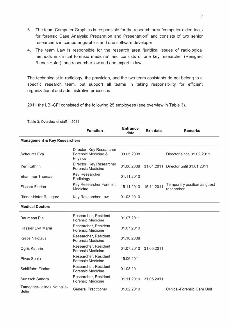
2011 the LBI-CFI consisted of the following 25 employees (see overview in Table 3).
Table 3: Overview of staff in 2011
FunctionEntrance
dateExit date Remarks
Management & Key Researchers
Scheurer EvaDirector, Key Researcher Forensic Medicine & Physics
09.05.2008 Director since 01.02.2011
Yen KathrinDirector, Key Researcher Forensic Medicine
01.06.2008 31.01.2011 Director until 31.01.2011
Ehammer ThomasKey Researcher Radiology
01.11.2010
Fischer FlorianKey Researcher Forensic Medicine
15.11.2010 15.11.2011Temporary position as guest researcher
Riener-Hofer Reingard Key Researcher Law 01.03.2010
Medical Doctors
Baumann PiaResearcher, Resident Forensic Medicine
01.07.2011
Hassler Eva MariaResearcher, Resident Forensic Medicine
01.07.2010
Krebs NikolausResearcher, Resident Forensic Medicine
01.10.2008
Ogris KathrinResearcher, Resident Forensic Medicine
01.07.2010 31.05.2011
Pivec SonjaResearcher, Resident Forensic Medicine
15.06.2011
Schillfahrt FlorianResearcher, Resident Forensic Medicine
01.08.2011
Sunitsch SandraResearcher, Resident Forensic Medicine
01.11.2010 31.05.2011
Tamegger-Jelinek Nathalie-Belin
General Practitioner 01.02.2010 Clinical-Forensic Care Unit

10
Webhofer MagdalenaResearcher, Resident Forensic Medicine
04.10.2010 30.04.2011
Jurists
Kainz SimoneResearcher, Doctoral Candidate Law
01.04.2011
Schick Peter Scientific Expert, Law 01.10.2008 Em. Professor at KFU Graz
Computer Graphics & MR Physics
Bornik AlexanderSenior Researcher, Visualization Specialist
01.12.2008
Höller JohannesSoftware Engineer, Diploma Student IT
15.11.2010
Langkammer ChristianResearcher, PhD-Student MR Physics
15.11.2009 15.11.2011
Petrovic AndreasResearcher, PhD-Student MR Physics
06.07.2009
Urschler MartinSenior Researcher, Segmentation Specialist
01.12.2008
Administrative and Technical Staff
Habersatter Stefanie Teamassistant 13.04.2010Maternity leave since 16.09.2011
Reisner Evelyn Executive Teamassistant 01.10.2008
Schachner Silvia Teamassistant 14.03.2011 Maternity leave substitution
Widek Thomas Technologist Radiology 03.05.2011
On February 1st, 2011, Kathrin Yen left the LBI for the post of department head of the
Institute of Forensic Medicine in Heidelberg.
Eva Scheurer has become the new director of the LBI since February 2011.
Simone Kainz has been a part-time assistant lawyer in the legal branch since April, and
since May has become a full-time employee of the LBI.
Magdalena Webhofer left the LBI at the end of April in order to complete her
specialization in traumatic surgery.
Thomas Widek is a radiology technician and has become part of the radiology branch
since May.
Stefanie Habersatter took her maternity leave in May; Silvia Schachner has taken her
replacement in March.
Kathrin Ogris left the LBI at the end of May in order to complete her specialization in
forensic medicine in Salzburg.
Sandra Sunitsch left the LBI at the end of May in order to complete her residency in
general medicine.

11
In June Eva Scheurer submitted her habilitation in forensic medicine to the Medical
University Graz. A committee has been founded at the beginning of November.
Christian Langkammer, graduate student in MR technology has completed his doctoral
thesis by the end of the year. He left the LBI in November for a postdoctoral position at
the Department of Neurology in Graz, and will further collaborate with the LBI in the
research area of traumatic brain injury.
In the course of a temporary exchange program of experienced specialists in forensic
medicine as guest researches, Florian Fischer from Munich stayed at the LBI for one
year until November.
1.5.2 Career development
Concerning continuing education and training of the researchers of the LBI, the following
internal trainings have been organized in 2011:
Internal education and training
Educational presentation on manner of death, cause of death and causality by Kathrin
Yen (24.01.2011)
Educational presentation on applied statistics by Eva Scheurer (02.02.2011)
Training in professional photography for the medical employees of the Clinical Forensic
Care Unit by Peter Hausleitner (Opernfoto) (02.02.2011)
Introduction to the Leonardo Workstation by Thomas Widek (Siemens) (10.03.2011)
Journal club meetings of all researchers with presentations of current scientific papers
and discussion (in February, March, August, and October)

12
In April 2011 a team development workshop was hold under the guidance of an external
specialist, Bernd Peters. The aim was a self assessment of the team and the internal
processes in order to improve coordination and communication and to motivate the
employees to participate in taking responsibility in view of the evaluation of the LBI in
November.
At the end of the year talks of the director with all employees take place with the aim to
discuss personal aims related to career development. In 2011 the talks were postponed to
the beginning of 2012 due to the evaluation of the institute in November.
In regular meetings every two weeks the entire team of the LBI meets for information of the
others on the current state and recent developments of their research and for the
interdisciplinary discussion of the studies to ensure an optimal communication and
cooperation between the researchers.
Daily morning meetings of all team members concerned with the Clinical Forensic Care Unit
take place where all examined cases are presented including the demonstration of
photographs taken from characteristic morphologic findings. Additionally, legal questions of
the forensic doctors regarding clinical forensic examinations are discussed with the present
member of the law team.
Team members of the LBI regularly participated in educational lectures and meetings, PhD
and diploma presentations as well as workshops of Graz University of Technology and
Medical University Graz, and in the context of scientific conventions (e.g., at ISMRM, ISALM,
ESMRMB).
External education and training
Examples of externally organized activities with educational aspects with participation of
researchers of the LBI in 2011 are:
“Meet the expert” with Prof. Tim Skern, organized by the LBG GmbH in Vienna
(15.02.2011)
9. Internationale Kasseler Fortbildung: „Medizinische Diagnostik bei
Kindesmisshandlung“, organized by the Arbeitsgemeinschaft Kinder- und
Jugendgynäkologie e.V. (18.-19.03.2011)
Symposium „Tod im Gefängnis“, organized by the University Zurich (13.09.2011)
Workshop “Projekt- und Antragsmanagement”, organized by the Institute of Forensic
Medicine, Düsseldorf (07.-08.10.2011)
Informationday on COMET – K-Projects, organized by FFG, Vienna (18.10.2011)
Medien- und Präsentationstraining, organized by science2public, Vienna (04.-
05.11.2011)
Presentation and visit of IST Austria, Klosterneuburg (08.11.2011)
Workshop “How to write a scientific paper?” in Graz (19.11.2011)

13
Intensive training workshop “Kinder- und Jugendgynäkologie”, organized by the
Arbeitsgemeinschaft Kinder- und Jugendgynäkologie e.V. (23.-26.11.2011)
Eva Scheurer took part in the annual management workshop of the LBG (07.-09.09.2011) in
Pöllauberg where the key areas “public relations” and “human resources and development”
were discussed.
The LBI and career development: examples of opinions of actual employees
“Working at the LBI gives me the chance to obtain expert knowledge and experience in
the field of clinical forensic medicine. Furthermore it gives me the possibility to achieve
experience in applied research. Both points are important for my further career as a
forensic doctor.”
“Benefits from working at the LBI are the development of a scientific profile focusing on
forensic imaging, both postmortem and clinical, getting experience in the supervision of
other persons, contact to MR imaging research, and the knowledge of how to establish
cooperating working groups in the field of forensic imaging.”
“In addition to a big number of education and development opportunities (i.e. exclusive
MRI tutorials at the Technical University Graz, various workshops, contacts to public
media, active participation in multidisciplinary network groups) research and publication
facilities offer gaining experience and getting eligible for habilitation.”
„I appreciate the diversity of my work and the associated diversion of my duties.
Particularly the disposal of the secured evidence after the time of storage was new to me,
and I find it very interesting. The issues of research associated with my work, e.g., child
abuse or child neglect and the opportunities of the imaging methods are a challenge, and
make me mature in my profession. We are a good team.“
“The main advantage of our employment at the LBI is to work together with people from
different fields and learn about the open problems and questions in forensic medicine

14
from their specific points of view. This includes physicians, forensic scientists,
radiologists, electrical engineers, physicists, jurists, and others. The field of forensic
imaging is a rather new one, which we think will grow in importance over the coming
years. Therefore, from a scientific point of view, we have the opportunity to work on the
forefront of a research discipline. Combined with the contacts to the relevant industrial
partners like Siemens, this fact underlines our enthusiasm to work in this field. As an
excellent side effect, we are also becoming experts in a highly specialized niche, i.e.
software development for forensic applications.”
“Working at the Ludwig Boltzmann Institute is an enjoyable, interdisciplinary experience
which helps me to gain insights into various disciplines other than my own. This enables
me to enrich and complete my dissertation with additional qualities. I consider it as a solid
practical basis for working in other areas thereafter.”
“After working for quite a long time in clinical medicine it is a totally new and great
experience to work in a forensic research team. I can contribute my know-how and even
more I can deepen my knowledge. This will be very useful in my further employments.”
“Working in the domain of clinical forensic imaging and the Ludwig Boltzmann Institute
offers good perspectives for my personal development. The interdisciplinary approach
between the quite diverse fields of expertise such as medicine, physics, IT and law
challenges the personal view of the world and vice versa. Therefore, it is a good breeding
ground for extending the individual horizon while helping people in very practical
problems and concerns. This is even truer when looking at the various fields the LBG
GmbH is engaged in.”
„This position offered me to actively participate in the built-up of an institute. Additionally,
for the first time I was assigned management tasks by being responsible for another
employee. The delegation of duties is a new challenge which I am happy with. Due to the
variety of my duties I have grown and matured in my job. This is of great importance for
my life and a huge plus. In my job at the LBI I appreciate particularly the possibility of
scheduling my work freely and to work on my own. The collaboration in a research
institute and to participate in a „good thing“ is exactly what I was wishing for in my
career.”

15
1.5.3 Teamevents
At the end of January we had to take leave of Kathrin Yen, director and founder of the LBI,
which left for her new position as a director of the Institute of Forensic and Traffic Medicine at
the University of Heidelberg. Her farewell party with guests from the institutional partners and
cooperating institutions was on 27.01.2011 at the Café Purberg.
At the beginning of March we were happy to inaugurate our additional office rooms at
Elisabethstrasse.
On 09.03.2011 the LBI skiing day took place in the skiing area of Lachtal. The sun was
shining all day, and the snow was perfect. In the evening the day was rounded up by a
dinner at the restaurant “Weisses Kreuz”.

16
On 05.05.2011 the LBI started at the Business Marathon at Schwarzl See with a team of 8
consisting of Alexander Bornik, Nikolaus Krebs, Martin Urschler and his partner, Florian
Fischer, Eva Hassler and her partner, and Andreas Petrovic with the result of being 149th (of
390).
On 31.05.2011 the team of the LBI visited the exposition of Nick Veasey “X-Ray” at the
Atelier Jungwirth in Graz with a personally guided tour by the artist.
After the site-visit related to the evaluation of the LBI, the team met on 18.11.2011 at
Kitchen12 to celebrate the positive feedback of the evaluation with a cooking evening. In four
teams a heavenly four-course menu was created and enjoyed.
On 01.12.2011 LBI team members tested their luck at the Casino Graz where they had been
invited on the occasion of the business marathon in May.
The LBI christmas dinner took place on 13.12.2011 at the Restaurant San Pietro in Graz.
1.6 Infrastructure
The institute is located at the second floor of Universitätsplatz 4 neighbouring the Institute for
Forensic Medicine of the Medical University Graz, with which some rooms are shared (e.g.,
the kitchen, examination room, autopsy rooms). Additionally, since April 2011, two office
rooms at Elisabethstrasse 27, which is in walking distance, about 7 minutes from the main
office, were allocated by the Karl Franzens University Graz.
The scientific MR and CT scans are performed at the Department of Radiology, LKH Graz,
while in cases of age estimation routine CT scans are performed at the Privatklinik der
Kreuzschwestern (CT/MR Zentrum Graz-Geidorf), and X-rays at the Radiologiepraxis Dr.
Uranitsch, Graz.
1.7 Highlights of the year
The most important and also most challenging highlight in 2011 was the interim evaluation of
the LBI CFI which was organized by the LBG. The internal schedule already started in April
with the team building workshop (see section 1.5.2) where all team members discussed
important issues concerning communication and internal processes, and thought about
possible improvements. In a next step an evaluation report was written giving insights on the
structure of the LBI and the performed research of the years 2008 – 2011 as a basis for the
evaluation. The site-visit of the Evaluation Panel which consisted of Prof. Volker Dittmann
(University of Basel, Switzerland), Prof. Alan Colchester (University of Kent, United
Kingdom), Prof. Jean Jacques Le Jeune (Angers University, France), and Dr. Lothar Behlau
(Fraunhofer-Gesellschaft, Germany) took place on 16.-17.11.2011. After two days of
presentations of the researchers on the performed studies, and various meetings with

17
researchers and key researchers, the director of the LBI as well as representatives of the
institutional partners and the Scientific Advisory Board the Evaluation Panel stated that they
acknowledge the importance of the core research area on clinical forensic imaging
established by the LBI CFI and recommend to the Ludwig Boltzmann Gesellschaft to
continue the financing of the institute. Specifically, they acknowledged the high motivation of
all the employees. The recommendations of the Evaluation Panel which mostly are advices
to continue or strengthen certain activities which are already planned or on the way will be
implemented into the structure and program of the LBI. Additionally, some specific scientific
remarks will be discussed with the Scientific Advisory Board and integrated into the future
research program.
Another highlight in 2011 was the successful initiation of an LBI lecture series “The
interdisciplinary world of forensic imaging” for which national and international renowned
speakers are invited to talk about their specialist field comprising forensic medicine, MR
physics and methodology, radiology and imaging, computer graphics, law enforcement,
prosecution and criminal law as well as victim support. In 2011 the series started with 5
lectures given by Prof. Bratzke (Frankfurt a. M., Germany), Prof. Eder (Salzburg, Austria), Dr.
Keplinger (Linz, Austria), Dr. Zebedin (Graz, Austria), and Prof. Boesch (Bern, Switzerland)
(see section 3.3). The lectures attracted an interdisciplinary and very interested audience.
The series will be continued next year.
As a practical interdisciplinary basis for the research at the LBI CFI regular Jour Fixe -
meetings with representatives from the hospital (LKH Graz), police and prosecution have
been initiated in February 2011. The aim of these meetings is to optimize communication and
cooperation of the institutions being concerned with incidents of violence. Five meetings
have been held and several issues concerning processes and cooperation as well as real
cases have been discussed and analyzed. The results from these discussions have been
incorporated into the activities of the Clinical Forensic Care Unit and will also be a central
part in the juridical key area of research.
1.8 Public relations
1.8.1 Media contacts and reports
The Ludwig Boltzmann Institute for Clinical-Forensic Imaging received numerous requests
for TV and radio interviews as well as for interviews for printed media reports which were
accepted whenever possible.
A selection of contributions released to the public is listed below:

18
TV interview K. Yen & E. Scheurer about expert witnesses in „Thema“ - ORF 2, Mar 22th
2011
Magazine article about Clinical-Forensic out-patient clinics in „Clinicum“ with the title „Der
forensische Blick“, Jun 15th 2011
TV broadcasts and interviews of E. Scheurer about „K.O.-Tropfen“ in
o „Steiermark heute“, ORF 2, Sept 20th 2011
o „ATV Aktuell“, ATV, Sept 20th 2011
Magazine article about body imaging in „Öffentliche Sicherheit“, issue Sep/Oct 2011
Magazine article about forensic age estimating based on an interview with F. Fischer in
„Die Furche“, title: „Minderjährigen aus der Hand gelesen“, Oct 13th 2011
Radio interview with F. Fischer about forensic age estimation in „Journal Panorama“ -
OE1, title: "Unbegleitete minderjährige Flüchtlinge“, Oct 17th 2011
TV report with an interview of F. Fischer on the occasion of the 20 year anniversary of
Austrian coordination of Asylant-seekers in „Heimat Fremde Heimat“ - ORF 2, Oct 23th
2011
1.8.2 Public presentations
The scientific work and other activities of the LBI were presented to the non-scientific public
at the following research exhibitions:
200 years celebration of Graz University of Technology 2011, Dom im Berg, Graz
Multimedia Exhibition “Der Mensch” in cooperation with the Science-Center Graz,
(starting October 2011)
2. Research program and results
2.1 Projects
The research program at the LBI during the year 2011 was performed within five main
focuses each comprising different studies which are shortly described in the following
sections.
Within the total of studies of which some were conducted in parallel 237 MRI scans in living
and deceased subjects and 1 CT scan of an antique bowl in cooperation with the LBI for
Archaeological Prospection and Virtual Archaeology were performed. Table 4 shows an
overview of the studies in which MRI scans have been performed.

19
MRI 2011
Blunt injury of the subcutaneous tissue 21
Subcutaneous blood in living persons 118
Diffusion in muscle tissue 12
Traumatic brain injury 8
Strangulation study 8
Dental MRI 18
Age estimation using MRI 52
Total 237
Table 4: Overview on research MRI scans 2011
2.1.1 Hematomas and blunt force injury of soft tissues
This study aimed at the evaluation of the rate of detection, the quality of localization and the
potential of grading of subcutaneous lesions in living persons using clinical standard MRI
sequences.
Evaluation of subcutaneous hematomas in clinically used MRI-sequences
25 persons with at least one visible hematoma of the thigh underwent exterior forensic
examination with documentation of its size and localization, and subsequent 3T MRI using a
clinical protocol with standard TSE sequences. Radiological data were evaluated by a board
certified radiologist blinded to the results of the exterior documentation.
The sensitivity of the used sequences for the detection of subcutaneous hematomas was
58%-74% depending on the specific sequence. Grading of the lesions was difficult due to
artefacts and inhomogeneous fat saturation as well as limited image resolution. However, in
addition to the hematomas muscle injuries were detected in 32% in MRI which were not
diagnosed in the external examination. In conclusion, the sensitivity of typical clinical
sequences to detect subcutaneous hemorrhage is limited, and not appropriate for being used
in court. Clinical sequences have to be adapted to allow for reliable diagnosis, including
further grading of the lesion. However, in contrast to the current gold standard, i.e, the
external examination only, MRI permits to get an overview with additional information on
lesions of deeper tissues and organs.
The aim of this study which will be completed in 2012 is to evaluate the MR characteristics of
injected blood volumes in the subcutaneous fatty tissue of healthy living volunteers regularly
over a time course of several weeks using MRI and IR photography. Additionally, the quality
of volume estimation by automated segmentation is evaluated.
Artificial hematomas in subcutaneous fatty tissue of living volunteers: temporal changes of
MR parameters, volume estimation by automated segmentation, and contrast enhancement
using IR photography

20
The aim of this ongoing prospective study is the characterization of traumatic lesions and the
following regeneration of skeletal muscle tissue using MRI. A group of subjects with a trauma
of the musculature and a group of healthy controls are examined using MRI. The results are
analyzed regarding specific radiological signs and are correlated with the exact traumatic
incident.
Diffusion weighted (DWI) and diffusion tensor MRI (DTI) for the evaluation of muscle lesions
2.1.2 Forensic aspects of traumatic brain injury
The ongoing study “Blunt head trauma: correlation of postmortem MRI and histological
analysis of the brain” includes deceased subjects with a blunt head trauma and a control
group of deceased subjects without brain trauma. The aim of the study is to detect specific
differences in terms of MRI-detectable lesions caused by trauma between the two groups.
Aspects as the characteristics of visible lesions in MRI, the estimation of impact power, the
direction of the force of impact or the age of the findings are core issues in forensic
evaluation. Validation and correlation of MRI findings with macroscopic, histological,
chemical as well as physical methods might improve to draw forensic conclusions.
Abnormal high iron deposition in the human brain has been shown to play a role in several
neurological disorders and is linked to the process of neurodegeneration. The rupture of
nerve fibers leads to a rapid loss of white matter tissue which might be paralleled by iron
deposition. However, so far this has not been investigated as a consequence of traumatic
brain injury. Therefore, tissue specimens are taken from pre-specified white and gray matter
regions for microscopic examination and chemical determination of trace element
concentrations (iron, calcium, zinc and others).
2.1.3 Radiologic evidence in forensic reconstruction and age estimation
After survived strangulation important forensic questions are whether the strangulation
incident is objectively verifiable and how the incident can be qualified and quantified. At
present, this question can only be answered based on the statement of the victim. However,
this information, e.g. regarding loss of consciousness, voiding of urine or stool, and
psychological aspects is highly subjective. The aim of this ongoing study is to evaluate
whether radiologically detectable internal lesions of the neck and throat allow the
differentiation between subjects with and without strangulation and whether the injury
patterns can be used to characterize the incident. The study involves the examination of
living and deceased victims of strangulation incidents, and of living and deceased persons
without injuries as controls using MRI. First results in living persons show that
Detection and forensic interpretation of soft tissue findings in living and postmortem subjects
after strangulation using MRI
The enhanced use of radiological techniques offers new possibilities of documentation in
clinical-forensic medicine. In order to promote image techniques regarding the use of injury
documentation in crime victims a discussion based on ethical and judicial principles seems
Acceptance of radiological examinations for forensic purposes: evaluation of the influence of
radiation exposure and duration of the examination

21
reasonable. This study aimed at evaluating the approval of radiology based documentation
methods for forensic purposes. A questionnaire with 12 questions particularly accounting for
the influence of radiation exposure and the duration of the examination was answered by a
group of crime victims as well as by a control group. The evaluation of the answers showed a
high acceptance rate of imaging techniques by both groups. Overall, all of the victims and
94% of the controls approved to at least one of the imaging modalities. Most of the
interviewees did not consider the duration of the examination to be relevant; however, the
radiation exposure was relevant for almost half of the controls, but only for about 20% of the
victims. These results need to be considered for establishing imaging techniques as a
standard procedure in clinical-forensic medicine.
Dental age estimation is one part of forensic age estimation of living persons and is usually
based on the evaluation of the development of the third molars in an orthopantomogram
(OPG). However, the use of ionizing radiation without medical indication is not permitted in
many countries. Thus, the aim was to define an MRI scan protocol for the use of MRI as a
non-invasive method without radiation exposure providing high resolution images of the
teeth.
Dental age estimation: development and evaluation of MR sequences for the imaging of
tooth development
For the optimization of the protocol 6 healthy volunteers were scanned on a 3T scanner
using a pair of CPC coils. Three different sequence types were evaluated and imaging
parameters were adjusted to yield optimal image quality. 3D acquisitions with similar
acquisition time yielded either less image intensity or enhanced blurring, and were prone to
motion artifacts. Our results show that high quality images of the third molars can be
acquired in a standard clinical MRI setup.
In an ongoing study the diagnostic value of dental MRI for the evaluation of developmental
criteria of wisdom teeth is assessed by comparing the results yielded on the basis of the
OPG are performed for dental reasons with those of the.
In this ongoing study healthy males between 13 and 26 years undergo an MRI examination
of the wrist, the clavicles, and the wisdom teeth. The aim of the study is 1) to investigate all
of the information currently used for forensic age estimation in living persons in the same
individuals, and, thus, get an insight into developmental differences between the three
parameters, and 2) to determine statistically relevant reference values for middle European
males which can be used for forensic age estimation by performing a single MRI examination
of three body regions. As the use of radiographic examinations such as the OPG, the
radiograph of the wrist and the CT of the clavicles (which constitute the current gold
standard) associated with radiation exposure is a recurrent issue of public and political
debate an adequate or even better alternative using MRI would be highly appreciated.
Validation study: forensic age estimation of living persons using MR imaging of wrist,
clavicles, and wisdom teeth

22
2.1.4 Computer-aided tools for forensic case analysis: preparation and presentation
The research to support forensic analysis by computer-aided preparation and presentation
tools addressed three main research areas: visualization, segmentation, and the
development of an interactive tool making both available through an intuitive user interface
tailored to a forensic task at hand.
Visualization:
For the task of visualization, a novel flexible rendering algorithm was developed, which
supports visualization of multiple arbitrarily overlapping volumetric datasets and polyhedral
models [Kainz2009]. In the sequel the rendering algorithm was enhanced to improve its
applicability in forensic tasks. Detail scans of body regions can be visualized in the context of
a transparent polyhedral reference model. Moreover a dynamic level of detail (LOD)
algorithm enables visualization of large datasets, which do not fit into GPU or system
memory by maintaining a subset of the data trying to maximize visualization quality for the
given memory constraints. This way it is possible to visualize even high resolution full body
scans, which are necessary to see, e.g. fine bone fractures. The data model supporting
arbitrary clipping operations based on constructive solid geometry (CSG) operations allows
focus and context visualization with the possibility to produce explosion diagrams of the
human body in analogy to technical illustrations [Chen2011]. In the forensic context such
views are useful to depict complex anatomical relationships. In order to enhance depth
perception and realism the rendering algorithm was extended to support shadows
[Bornik2011, Knecht2011].
Segmentation:
To support extraction of forensically relevant structures from 3D CT/MRI data, we have
investigated a number of image segmentation methods. Image segmentation is an important
topic in 2D computer vision [Donoser2009, Donoser2011], we are extending these concepts
to 3D for our forensic application. The research on segmentation algorithms focused on
techniques like adaptive region growing, level-set segmentation, geodesic active contour
models, and enhancement of vascular structures. With the goal of an integrated computer-
aided forensic tool in mind, we developed algorithms which allow hardware-accelerated
parallel execution on current GPUs. Close integration with the visualization side led to a first
prototype of an interactive segmentation and visualization tool [Urschler2009].
Interactive Forensic Tool:
Our research to incorporate visualization and segmentation into a single integrated
framework led to the development of a second prototype with improved visualization quality
and speed [Urschler2011]. The new framework allows flexible ad hoc composition of forensic
scenes through a graphical editor, where segmentation algorithms are available as intelligent
3D selection tools in analogy to common 2D image processing software. The framework
readily supports presentation of forensic cases in court, where polyhedral reference manikin
models can be used. These manikins are chosen from a set of pre-defined models of
different age or gender, for model creation we cooperate with the company RISC Software
GmbH from Hagenberg. A chosen model may be modified to support different poses, and it
allows registration of 2D photographs and 3D clinical forensic data onto it [Hoeller2011].

23
Furthermore, the reference models allow for interactive markup, e.g. painting of injury
regions or overlay from photographs. Cases processed this way may be stored in a case
database, which is one key requirement for the implementation of a digital reference atlas.
Another necessary step is the research on non-linear 3D registration techniques to represent
typical injury variations [Urschler2010, Murphy2011]. The developed tool is being tested with
real-world scenarios throughout the project.
2.1.5 Juridical issues of radiological methods in clinical forensic medicine
Juridical research on clinical forensic imaging means to examine the specific juridical
framework by analyzing and investigating legal regulations of criminal law, medical law, but
also of public law. The results of this research work are being summarized in a compilation
on the Austrian juridical framework of clinical forensic medicine. A disposition of this
compilation has been generated already.
A study on the value of expert reports in criminal court records has been conducted. 1500
case files from the years 2007 to 2010 have been examined at the Regional Criminal Court
Graz during June/July 2010. The resulting database has been evaluated and used to answer
actual questions in the legal context of clinical forensic medicine already. It will continue to be
an important source of information for future research. A follow-up study on the sentencing of
bodily injury was conducted by Simone Kainz in 2011.
2.1.6 Clinical Forensic Care Unit
The first Austrian Forensic Care Unit was established in October 2008 by the Ludwig
Boltzmann Institute for Clinical-Forensic Imaging in Graz as a facility of the LBI together with
its institutional partner Medical University of Graz. The Clinical-Forensic Care Unit offers
medico-legal examinations for living persons after incidents of suspected physical or sexual
violence as well as forensic age estimations in living persons, which are a traditional duty of
clinical forensic medicine. The medico-legal documentation of injuries after acts of violence
helps to improve not only the quality of the medico-legal expert opinion, but also the quality
of the juridical decision-making in court by having a greater legal security.

24
The service of the Clinical-Forensic Care Unit is available to all persons having suffered
physical violence including accidents or sexual violence at no personal costs and
independent of whether a report to the police has been made. An on-call service available
24/7 guarantees the availability of a medico-legal examination in the greater Graz area
(hospitals, police stations, detention centres, and organizations offering help to victims, etc.)
within 35 to 45 minutes. Concerning examinations outside this geographical radius medico-
legal assistance can be given by phone and email to ensure a successful examination by
other physicians. Examinations in cases with suspected sexual assault or maltreatment in
adults and children are usually conducted in the corresponding hospital departments in
cooperation with a gynaecologist or specialized paediatrician. Additionally, two residents are
members of the clinical child protection groups of the Department of Pediatric Medicine and
the Department of Pediatric Surgery with weekly meetings and interdisciplinary discussions
of suspected child abuse cases.
Selected persons examined at the Clinical-Forensic Care Unit, who match the inclusion
criteria of current studies of the LBI-CFI are asked, if they are willing to participate in the
respective study.
To our knowledge the LBI is the only institution in Austria performing forensic age estimation
examinations based on the guidelines issued by the German working group on age
diagnostics (AGFAD). These examinations are an excellent example for applying clinical
forensic imaging as a modern tool in forensic medicine.
For the enhancement of the communication between prosecution, police, clinical and
forensic medicine the Jour Fixe-meetings (see section 1.7) provide substantial progress with
respect to the optimization of processes for victims of violence.
Table 5 presents an overview of the cases which have been seen at the Clinical-Forensic
Care Unit in 2011. The age estimations are performed on behalf of the Federal Office for
Migration and Refugees; the other cases are examined by order of the Office of Public
Prosecution or on a consultation basis for clinicians. In total, 23% of all cases were not
financially rewarded.
Table 5: Overview of the cases seen at the Clinical-Forensic Care Unit 2011
Clinical-Forensic Care Unit 2011
Physical violence (against adults > 18 years) 34
Sexual violence (> 18 y) 34
Physical child abuse (< 18 y) 65
Sexual child abuse (< 18 y) 35
Forensic age estimation in living persons 372
Other cases 4
Cases in total 544

25
2.2 Publications
2.2.1 Publication policies and intellectual property rights
Regarding publications rules for the regulation of the authorships have been defined
according to good scientific practice. These are supervised and executed for each study by
the key researcher or the responsible researcher of the corresponding team. The authorships
are regulated under consideration of the general principles for author contributions as
outlined in the “instructions for authors” of main scientific journals.
The acquired radiological scanning data are owned by the LBI which is responsible for its
acquisition and has to comply with the national law regulations. Requests or the usage of
these data for other purposes than scientific or those outlined in the research program are
evaluated and decided by the Supervisory Board of the LBI. The whole output of the
research activities is published in scientific journals, since it is the main target of the LBI to
implement clinical forensic imaging in legal practice. The IPR of the institutional partners are
handled according to the contract of 2008.
2.2.2 Publications
Summary of publications 2011:
6 Peer-reviewed journal papers
5 Books, book chapters and other publications
21 Abstracts and conference presentations
The scientific output in the form of publications, proceedings and abstracts (see the complete
list of publications in section 5) achieved in 2011 is satisfactory. However, the already
observable annual increase of the number of publications (journal papers & other
publications: 2008: 7, 2009: 9, 2010: 10, 2011: 11) and abstracts in the last years suggests
that publication activities will be enhanced in the next years.
2.3 Participation in scientific conferences
As an image of the interdisciplinarity of the LBI numerous contributions to international
scientific meetings and conferences within the scientific communities of forensic medicine,
magnetic resonance in medicine, and computer graphics have been made. In summary, 9
oral presentations and 8 poster presentations were given by researchers of the LBI.

26
An overview of the contributions to the different scientific communities 2011 is given here:
International Symposium on Advances in Legal Medicine (ISALM) in combination with the
Annual Scientific Meeting of the German Association of Forensic Medicine (DGRM), 26.-
30.09.2011, Frankfurt a. Main, Germany
Forensic medicine
7 oral presentations and 1 poster presentation
Annual Scientific Meeting of the International Society of Magnetic Resonance in Medicine
(ISMRM), 07.-13.05.2011, Montreal, Canada
Magnetic resonance in medicine and biomedical engineering
5 poster presentations
1 educational oral presentation (invited lecture given by Eva Scheurer)
Scientific Meeting of the European Society for Magnetic Resonance in Medicine and
Biology (ESMRMB), 06.-08.10.2011, Leipzig, Germany
1 oral presentation and 2 poster presentations
Additionally, the following meetings and symposia were attended by team members of the
LBI:
14.-15.01.2011 1st International MRI workshop on MRI phase contrast and quantitative
susceptibility mapping, Jena
22.02.2011 Tag der Kriminalitätsopfer, Bundesministerium für Inneres, Wien
18.03.2011 AGFAD - Meeting, Berlin
20. - 21.05.2011 20. Frühjahrstagung der DGRM Süd, München

27
13.09.2011 Symposium „Tod im Gefängnis“, University of Zurich
19.09.2011 15 Jahre Gewaltschutzgesetz und Gewaltschutzarbeit in Österreich,
Wien
04.11.2011 Annual Meeting Österreichische Kinderschutzgruppen, Wien
21.11.2011 „Die vielen Gesichter der Gewalt“, Wien
25.11.2010 Workshop of the Criminalistic Study Association: „Rechtsmedizin und
Bildgebung“, Landespolizeikommando für Steiermark, Graz
3. Other activities
3.1 Cooperations
3.1.1 Scientific cooperations
For the different fields and areas of research involved in the LBI scientific collaborations with
national and international institutions have been established. These cooperations are
beneficial both for the LBI and for the cooperating institutions as the conjointly performed
studies result in an enhancement of the scientific output and in an increase of knowledge in
the different fields.
A scientific cooperation has been established with the Institute for Forensic medicine of the
Ludwig-Maximilians-University Munich during the last year. This cooperation is intended to
provide a larger case collective needed in order to obtain statistically significant results in
certain studies. Clinical-forensic study cases can be recruited by the out-patient service for
assault victims of the Munich Institute as well as from victims being referred to the Institute
by the police. Embedded in this cooperation is the collaboration with the Institute for Clinical
Radiology of the Technical University Munich (TUM) with the use of a 3T MRI scanner
(Verio, Siemens Healthcare) and the collaboration with its radiologists.
Cooperations for the focus in forensic medicine
For the research in dental MRI a cooperation has been established with Prof. Norbert Jakse,
Department for Oral and Maxillofacial Surgery, Medical University Graz, Julian Boldt,
Department for Oral and Maxillofacial Surgery, University of Würzburg, and Andreas
Hopfgartner, Department for Experimental Physics, University of Würzburg, as well as with
Dr. Heiko Merkens, dentist with a private practice in Aachen. The main benefit for all
collaborators in this field is the facilitated demonstration of wisdom teeth and other dental
structures by MRI.
Within the research area “traumatic brain injury” regional distribution of iron-, copper-,
calcium-, manganese-, magnesium- and zinc-concentrations in the human brains were
determined in cooperation with Prof. Walter Goessler, Department of Chemistry, University of

28
Graz. These results in correlation with the results of quantitative MRI have impact for the field
of brain trauma research, but also for the research of neurodegenerative disorders.
The neuroimaging research unit under the direction of PD Dr. Stefan Ropele, Department of
Neurology, Medical University Graz is specialized in quantitative MRI for assessing brain
tissue changes. Myelin integrity, blood and iron are the most notable factors impacting MR
relaxation behavior and MRI contrast and, thus, have to be differentiated when drawing
conclusions about the origins of blunt force.
Dr. Michael Scarpatetti, Department of Pathology, Medical University Graz, is our
collaborator for the examination of microscopic traumatic changes of white matter tracts and
for the detection of iron deposits for the correlation with MRI.
Prof. Kathrin Yen, Institute of Forensic and Traffic Medicine, University of Heidelberg, has a
long track in establishing MRI and CT in forensic applications. As former head of the LBI she
was substantially included in the beginning phase of most studies of the LBI, and is included
in the interpretation of the results.
Further good and long existing scientific cooperations exist with:
Prof. Gerhard Ranner, CT/MR Zentrum Graz, Prof. Rainer Rienmüller and Prof. Franz Ebner,
Department of Radiology and Neuroradiology, respectively, at Medical University Graz, as
well as Dr. Karl Fritz, Krankenhaus der Barmherzigen Brüder, Graz-Eggenberg, which
support the LBI with radiologic reading and interpretation, and concerning organizational
procedures of radiological examinations.
Prof. Andrea Berghold, und Dr. Franz Quehenberger, Institute for Medical Informatics,
Statistics and Documentation, Medical University Graz, which support and cooperate with
the LBI concerning data analysis and statistical training of the researchers.
The most important cooperation in this field since the foundation of the LBI is with Rudolf
Stollberger and his team of the Institute for Medical Engineering, Graz University of
Technology.
MR physics
A cooperation is currently established with DI Tilman J Sumpf, Biomedizinische NMR
Forschungs GmbH, Max-Planck-Institut for Biophysical Chemistry, Göttingen, for the field of
T2-mapping.
The institute of Prof. Jürg Reichenbach, Department of Diagnostic and Interventional
Radiology, Jena University Hospital cooperates in the field of traumatic brain injury by
performing quantitative MR susceptibility mapping. This technique which is used to assess
the basic magnetic property of brain tissue helps differentiating between hemorrhages and
calcifications for example, which are not distinguishable from each other in conventional MRI,
and is, therefore, important for forensic aspects.
The former group of Prof. Klaus Scheffler, Radiological Physics, University of Basel Hospital,
Basel, is specialized in assessing disease induced tissue changes in multiple sclerosis. Their
method for the mapping of myelin integrity (based on bSSFP sequences) is applied in our
study of traumatic brain injury to investigate ruptures and detracting of nerve fibers.

29
Prof. P. Boesiger, Swiss Federal Institute of Technology (ETH), Zürich, and his group are
biomechnically modeling the choroid plexus and CSF pulsation. Due to CSF pulsation this
model cannot be validated in-vivo and was therefore supported by volumetric data of our
postmortem MRI study.
Our main cooperation is with the Institute for Computer Graphics and Vision (ICG), Graz
University of Technology. Prof. Horst Bischof and Prof. Dieter Schmalstieg are world-
renowned experts in their respective fields of computer vision and computer graphics. By the
constellation of financing two post-docs via the ICG the knowledge transfer in both directions
is optimally established. Martin Urschler works together in depth with two other post-docs
from ICG, Michael Donoser, an expert in 3D image segmentation, and Thomas Pock, an
expert in variational methods for low-level image processing. Alexander Bornik works
together with post-doc Bernhard Kainz from ICG, who is an expert in volume rendering
techniques. This tight collaboration is also visible in the co-authorships of methodological
computer vision and computer graphics papers (see publication list in section 5).
Computer graphics
Another cooperation has been established with RISC Software GmbH in Hagenberg, with its
research unit for medical informatics around Dr. Michael Giretzlehner. Johannes Höller is
working in tight collaboration with them, who are the developers of the BurnCase 3D
software. This software is used for the documentation of burns, and their concepts of generic
3D surface models of different genders, ages, and obesity levels as well as their methods for
painting injuries on this 3D model are also important aspects in our 3D reference manikin
model.
The master thesis of Wolfgang Knecht on the topic of shadows in volume rendering was
supervised by Alexander Bornik in cooperation with Vienna University of Technology, and
with Markus Hadwiger from the King Abdullah University of Science and Technology, Saudi
Arabia, who is one of the leading international experts in medical volume visualization.
3.1.2 Non-scientific cooperations
Valuable cooperations concerning the Clinical Forensic Care Unit exist with the Departments
of Pediatric Medicine and Pedicatric Surgery and their child protection groups, as well as with
the Department of Obstetrics and Gynaecology, both at the Medical University Graz.
3.1.3 Third party projects
While some financing of the activities of the LBI has slowly been evolving from the Clinical-
Forensic Care Unit, to date no third party financing of research has taken place. From the
point of view of the management this was not a priority for the institute for the first three
years as the limiting factor concerning the performance of the planned research program
were not finances but mainly the availability of experienced researchers. However, in the

30
upcoming period third party financing will become relevant particularly for projects involving
cooperations with clinical fields and for certain aspects of technical advances.
3.2 Membership in scientific associations
Team members of the LBI-CFI are members of the following scientific associations:
ISMRM – International Society of Magnetic Resonance in Medicine
ESMRMB – European Society of Magnetic Resonance in Medicine and Biology
RSNA – Radiological Society of North America
DGRM – Deutsche Gesellschaft für Rechtsmedizin
SGRM – Schweizerische Gesellschaft für Rechtsmedizin
ÖGGM – Österreichische Gesellschaft für Gerichtliche Medizin
AGFAD – Arbeitsgruppe für Forensische Altersdiagnostik der DGRM
Kriminalistische Studiengemeinschaft Steiermark
Forschungsfeld Neurowissenschaften der Medizinischen Universität Graz
Active participation of LBI team members was in the following working groups of scientific
associations:
Klinische Rechtsmedizin der DGRM (Nikolaus Krebs)
AGFAD (Eva Scheurer, Pia Baumann)
3.3 Lecture series and workshops
In 2011 the LBI started with a lecture series “The interdisciplinary world of forensic imaging”
which is open to the public and for which national and international renowned speakers are
invited to talk about their specialist field comprising forensic medicine, MR physics and
methodology, radiology and imaging, computer graphics, law enforcement, prosecution and
criminal law as well as victim support. 2011 lectures have been given by

31
Prof. Hansjürgen Bratzke, “Verletzungen des Gehirns aus Sicht des
Rechtsmediziners”, 11.04.2011
Prof. Maria Eder, “Forensische Fragestellungen an den Sachverständigen bezüglich
Körperverletzungen”, 29.06.2011
Dr. Rudolf Keplinger, “Die polizeilichen Möglichkeiten bei Gewalt in der Wohnung”,
05.09.2011
Dr. Doris Zebedin, „Bildgebung bei Verdacht auf Kindesmisshandlung“, 14.11.2011
The planned lecture of Prof. Chris Boesch on in vivo MR spectroscopy had to be postponed
to January 2012.
3.4 Teaching and training activities
3.4.1 Teaching activities
Teaching appointment of Martin Urschler at Graz University of Technology,
for the lecture „Medical Image Analysis” as a part of the computer science and
biomedical engineering curricula, since SS 2008
Academic teaching
Teaching appointment of Eva Scheurer at Medical University Graz,
for the lecture “Gerichtsmedizin am Lebenden – Kindesmisshandlung, Sexualdelikte,
häusliche Gewalt”, since WS 2010
Seminar Medizinstrafrecht “Medizinrechtliche Differentialdiagnostik: kurative –
forensische – forschende Medizin” at Karl-Franzens-University of Graz, given by Peter
Schick and Stefan Schumann in cooperation with Reingard Riener-Hofer, WS 2010/11.
Seminar „Der Rechtsmedizinische Sachverständige“ at Karl-Franzens-University of Graz,
given by Peter Schick and Eva Scheurer in cooperation with Reingard Riener-Hofer, WS
2011/12
Tutorial “Bioimaging laboratory” as a part of the biomedical engineering curriculum at
Graz University of Technology, given by Andreas Petrovic, WS 2011
Teaching and training topics of clinical forensic medicine to different kinds of trainees, e.g,
police officers, social workers, nursery teachers, is considered as important for the
implementation of clinical forensic knowledge in Austria. It is, thus, a strategic policy of the
management to support such activities and to encourage particularly the residents in forensic
medicine to acquire experience in teaching. Table 6 shows the numbers of lectures given in
the different fields; and below some examples of individual presentations are given.
Non-academic teaching

32
Table 6: Number of non-academic lectures given by team members 2011
Number of lectures 2011
Hospitals, Clinical Units, Medical Doctors
2
Victim Support Institutions, Child Protective Services
1
Police, Prosecution authorities, Court, Jurists
19
Training (nursery, medical care, social work, etc.)
4
Total 26
Examples of non-academic teaching:
Lecture on forensic radiology given by Kathrin Yen in the context of the European School
of Radiology (ESOR), in January 2011, organized by Prof. Rienmüller, Institute for
Radiology, Medical University Graz
Lectures for the Police Training College, Graz and Vienna, given by Eva Scheurer,
Kathrin Ogris, Florian Fischer, Nikolaus Krebs, Eva Hassler, Belin Tamegger-Jelinek,
Magdalena Webhofer, Sonja Pivec, Pia Baumann, Florian Schillfahrt, in February,
September and November 2011
Workshop on “Radiological Imaging in Forensic Medicine” of the Kriminalistische
Studiengesellschaft, Graz, with lectures given by Eva Scheurer, Florian Fischer, and
Thomas Ehammer, 2011
Lectures concerning clinical-forensic medicine and the Clinical-Forensic Care Unit of the
LBI in hospitals, victim support institutions, and training in various fields were given by
Kathrin Yen, Eva Scheurer, Eva Hassler, Belin Tamegger-Jelinek, Nikolaus Krebs, Zoe
Voigt, and Astrid Krauskopf, regularly since 2008
Ongoing student projects
Bettina Pucher is working on her bachelor theses with the working title „IR Photography
of subcutaneous hematomas“, at Graz University of Technology in cooperation with the
LBI (Supervision by Hermann Scharfetter, Institute for Medical Engineering, in
cooperation with Andreas Petrovic and Eva Scheurer, LBI).
Julia Kruisz writes her diploma thesis with the working title „Temperature dependence of
relaxation parameters and optimization of contrast for post-mortem MRI“ at Graz
University of Technology in cooperation with the LBI (Supervision by Rudolf Stollberger,
Institute for Medical Engineering, in cooperation with Andreas Petrovic and Eva
Scheurer, LBI).

33
Christoph Sirk works on his bachelor thesis “Fast GPU implementation of advanced fitting
algorithms for MRI” at the Institute for Medical Engineering, Graz University of
Technology in cooperation with the LBI (Supervision by Rudolf Stollberger, Institute for
Medical Engineering, in cooperation with Andreas Petrovic, LBI)
Michael Steiner, diploma student at Graz University of Technology investigated the
impact of varying brain temperature as a consequence of different postmortem intervals
for MRI. The temperature dependence of MR relaxation parameters and diffusion derived
parameters where investigated in phantoms and by using data of the LBI study of
postmortem brain. Furthermore, this work included the development of strategies for
correcting temperature mediated changes – essential when comparing MR relaxation and
diffusion parameters in postmortem MRI. (Supervision by Rudolf Stollberger, Institute for
Medical Engineering, and Stefan Ropele, Departement of Neurology, in cooperation with
Christian Langkammer, LBI).
Marc Steiner is working on a master thesis dealing with statistical shape models to
represent strong prior shape knowledge for 3D image segmentation, which is an
important part of our interactive real-time segmentation software (Institute for Computer
Graphics and Vision, Graz University of Technology, in cooperation with the LBI).
Michael Schneeberger is a master student who writes his master thesis at Joanneum
Research on the topic of synthetic wound simulation. Martin Urschler supervises this
thesis to deepen the relationships to the group for human centered image analysis at the
DIGITAL institute of Joanneum Research, which is lead by Dr. Heinz Mayer.
Marlene Vukmanic is working on a literature study on transfer functions and transfer
function design in the context of volume rendering. In this bachelor thesis she is also
supposed to implement selected methods in an encapsulated software component.
Marlene Vukmanic is jointly supervised by Martin Urschler, LBI, and Alexander Bornik,
LBI.
Georg Poier is currently working on a paid project to evaluate and
efficiently implement a number of tubular structure segmentation approaches on the
graphics processing unit. The algorithms were published in the PhD thesis of Christian
Bauer from the Institute for Computer Graphics and Vision at Graz University of
Technology. Georg Poier is supervised by Martin Urschler, LBI.
3.5 Reviewing activities
International Journal of Legal Medicine (Eva Scheurer)
Journals
Journal of Forensic and Legal Medicine (Eva Scheurer)
European Radiology (Eva Scheurer)
Medical Image Analysis (Martin Urschler)
IEEE Transactions on Medical Imaging (Martin Urschler)

34
Pattern Recognition (Martin Urschler)
Computer Vision and Image Understanding (Martin Urschler)
International Journal of Human-Computer Studies (Alexander Bornik)
Stroke (Christian Langkammer)
Journal of Neuroimaging (Christian Langkammer)
CVPR (Computer Vision and Pattern Recognition) (Martin Urschler)
Conferences
ECCV (European Conference on Computer Vision) (Martin Urschler)
ICCV (International Conference on Computer Vision) (Martin Urschler)
ISMAR 2011 (Alexander Bornik)
High Performance Graphics 2011 (Alexander Bornik)
Wiley, review of book proposal 2011 (Eva Scheurer)
Book proposals and scientific grant proposals
Estonian Science Foundation, evaluation of grant proposal 2011 (Eva Scheurer)
4. Outlook
Institutional partners
The collaboration with our institutional partners Medical University Graz (MUG), Siemens AG
Österreich, the Institute of Criminal Law, Criminal Law Procedure and Criminology at the
Karl-Franzens University Graz (KFUG) as well as the Superior Court of Syria and Carinthia
(OLG) shall be continued. The combination of these institutions is an exceptional opportunity
for the LBI and its interdisciplinary area of research, and is a main key to achieving its
challenging goals. Similarly, there are scientific and strategic benefits for these partners.
However, on the basis of the difficulties in the recruitment of postmortem cases for scientific
studies an additional institutional partnership with the Institute of Forensic and Traffic
Medicine at the University of Heidelberg, Germany, is currently being established. The
director of the institute is Kathrin Yen, the former director of the LBI. A scientific cooperation
has already been established. We are confident that a partnership with the University of
Heidelberg adds a stable fundament for conducting studies involving the examination of
postmortem cases, and complements the ensemble of partner institutions in an ideal way.
Scientific cooperations
In addition to the ongoing and valuable scientific cooperations listed in section 3.1
collaborations are planned with a number of institutions. While some of them are currently
been established others are still being evaluated.
35
For the research to be conducted in the fields of Hematomas and blunt force injury of soft
tissues and Radiologic evidence in forensic reconstruction and age estimation
Additionally, a closer cooperation with radiologists will be established to assist in the process
of the definition of criteria for the diagnosis of forensically relevant lesions. Experience shows
that both specialties, i.e., forensic medicine and radiology, lack specific knowledge for
reliable diagnoses. The need for the highest possible certainty in forensic medicine, however,
needs to be well funded before being implemented in routine case work.
cooperations
which allow for higher numbers and a higher variety of study cases shall be intensified, e.g.,
the Institutes of Forensic Medicine in Munich and in Heidelberg.
A focus on a closer cooperation between informatics and medical specialists may provide
new tools for forensic diagnosing, reconstructing and - often crucial – measuring of distances
and volumes.
For the area of research Forensic aspects of brain injury and function
The group of Prof. Krenn, Department of Physics, University of Graz, has been evaluated for
a cooperation and the first test measurements on a SQUID-Magnetometer have already
been conducted. The SQUID-Magnetometry offers a unique possibility to characterize basic
magnetic properties in a magnetic field with varying field strength and at varying temperature.
This will improve the understanding of field dependent effects and help to establish the
results on other MRI systems as well as will further allow investigating MRI contrast behavior
when the body temperature is not equal to in vivo conditions.
the recruitment of
postmortem cases is essential. In addition to the current methods of recruitment, corpses
could also be obtained from other institutions. A corresponding cooperation has already been
approved by the Department of Anatomy at the Medical University of Graz, and shall be
discussed with the Department of Pathology as well.
Still ongoing is the evaluation of partners concerning the trauma-induced tissue changes on
a histological level. This requires an extremely specialized expertise which could be acquired
by establishing a scientific cooperation with a corresponding specialist. We will establish a
collaboration with an expert in neurotrauma-pathology that will assist and supervise the
analysis of histological data from e.g. APP and MFB stainings.
It would be interesting to work together with people who already investigated automatic
transfer function generation techniques, e.g., Carlos D. Correa (UC Davis, USA), or in the
general “Virtopsy” context with pioneers in the area, e.g., the group of A. Ynnerman
(Linköping, Sweden).
Computer-aided Tools for Forensic Case Analysis: Preparation and Presentation
Teaching and training
Also in the next period the LBI lecture series “The interdisciplinary world of forensic imaging”
for which national and international renowned speakers are invited to talk about their
specialist field comprising forensic medicine, MR physics and methodology, radiology and
imaging, computer graphics, law enforcement, prosecution and criminal law as well as victim
support will be continued. For 2012 lectures are currently being planned. An outlook on
planned speakers involves Mag. Cornelia Koller (Office of Public Prosecution Graz), PD Dr.

36
Christian Jackowski (University of Zurich), Prof. Dr. Andreas Schmeling, Prof. Dr. Christian
Kopetzki (University of Vienna).
Similarly, the lecture on “Gerichtsmedizin am Lebenden – Kindesmisshandlung,
Sexualdelikte, häusliche Gewalt” at the Medical University Graz, and seminars at the Institute
of Criminal Law at the Karl Franzens University Graz will be given in the next year.
5. List of publications
5.1 Peer-reviewed papers
Décombaz J, Jentjens R, Ith M, Scheurer E, Buehler T, Jeukendrup A, Boesch C. Fructose
and galactose enhance human liver glycogen synthesis post-exercise. Med Sci Sports Exe,
43(10): 1964-71 (2011)
Ith M, Scheurer E, Kreis R, Thali MJ, Dirnhofer R, Boesch C. Estimation of the postmortem
interval by means of 1H magnetic resonance spectroscopy of decomposing brain tissue:
influence of ambient temperature. NMR Biomed, 24(7): 791-8 (2011)
Langkammer C, Krebs N, Goessler W, Scheurer E, Yen K, Fazekas F, Ropele S.
Susceptibility induced gray-white matter MRI contrast in the human brain. Neuroimage, Aug
26 Epub ahead of print (2011)
Scheurer E, Quehenberger F, Mund MT, Merkens H, Yen K. Validation of reference data on
wisdom tooth mineralization and eruption for forensic age estimation in living persons. Int J
Legal Med, 125(5):707-15 (2011)
Scheurer E, Loevblad KO, Kreis R, Maier SE, Boesch C, Dirnhofer R, Yen K. Forensic
application of postmortem diffusion-weighted and diffusion tensor MRI of the human brain in
situ. AJNR Am J Neuroradiol, 32(8): 1518-24 (2011)
Murphy K, van Ginneken B, Reinhardt J, Kabus S, Ding K, Deng X, Cao K, Du K,
Christensen G, Garcia V, Vercauteren T, Ayache N, Commowick O, Malandain G, Glocker B,
Paragios N, Navab N, Gorbunova V, Sporring J, de Bruijne M, Han X, Heinrich M, Schnabel
J, Jenkinson M, Lorenz C, Modat M, McClelland J, Ourselin S, Muenzing S, Viergever M, De
Nigris D, Collins D, Arbel T, Peroni M, Li R, Sharp G, Schmidt-Richberg A, Ehrhardt J,
Werner R, Smeets D, Loeckx D, Song G, Tustison N, Avants B, Gee J, Staring M, Klein S,
Stoel B, Urschler M, Werlberger M, Vandemeulebroucke J, Rit S, Sarrut D, and Pluim J.
Evaluation of Registration Methods on Thoracic CT: The EMPIRE10 Challenge. IEEE Trans
Med Imaging May 31 Epub ahead of print (2011)
5.2 Books, book chapters and other publications
Donoser M, Urschler M, Riemenschneider H, and Bischof H. Highly Consistent Sequential
Segmentation. In: Scandinavian Conference on Image Analysis; Heyden A and Kahl F (Eds.)
Springer Berlin/Heidelberg 6688:48-58 (2011).

37
Krebs N, Riener-Hofer R, Scheurer E, Schick PJ, Yen K. Rechtsmedizin an Lebenden: Die
erste österreichische „klinisch-forensische Ambulanz“. Österreichische Richterzeitung 89:6-
12 (2011)
Riener-Hofer R, Krebs N, Scheurer E, Schick P, Yen K. Die Ambulanz des Ludwig
Boltzmann Instituts für klinisch-forensische Bildgebung in Graz. SIAK Journal - Zeitschr für
Polizeiwissenschaften und Polizeiliche Praxis, 2:76-82 (2011)
Riener-Hofer R, Krebs N, Scheurer E, Schick P, Yen K. Forensic outpatient clinic at the
Ludwig Boltz-mann Institute for Clinical-Forensic Imaging in Graz. SIAK Scientific Series,
Forensic Perspectives 6:105-114 (2011)
Riener-Hofer R, Fischer F. Gerichtsmedizin am Lebenden: Klinische Rechtsmedizin. Die
Rufbereitschaft der klinisch-forensischen Ambulanz in Graz. Öffentliche Sicherheit (2011)
5.3 Abstracts and conference presentations
Bottan S, Fritz A, Kurtcuoglu V, Schmid Daners M, Knobloch V, Langkammer C, Krebs N,
Gloor M, Scheurer E, Scheffler K, Ropele S, Boesiger P, Poulikakos D, Soellinger M.
Investigating the role of choroid plexus in CSF pulsation by combining in-vivo and
postmortem MRI. 19th Annual Meeting ISMRM, Montréal, 6.-13. May, Proc. Intl. Soc. Mag.
Reson. Med. 19: 2578 (2011)
Buehler T, Ith M, Scheurer E, Décombaz J, Boesch C. MRI based volume determination of
single organs using the Versatile, Interactive Sparse Sampling (VISS) method. 28th Annual
Meeting ESMRMB, Leipzig, 6.-8. Oct., 575 (2011)
Buehler T, Bruce S, Breton I, Ith M, Scheurer E, Boesch C, Décombaz J. Effect of different
sugars after exercise on liver glycogen synthesis, liver volume and metabolic profile using 13C-MRS, MRI and GC-TOFMS. Annual meeting SGMS, Lausanne, 20.-21. Oct., Schweiz
Zschrift Sportmed Sporttraumat 59(3): 137 (2011)
Ehammer T, Malli N, Yen K, Scheurer E. Correlation of forensic examination and radiology
for the detection and characterization of traumatic scalp injuries using clinical cerebral MSCT
and multiplanar reconstruction. 8th ISALM, Frankfurt/Main, 26.-30. Sept., Rechtsmedizin
21(4): 353 (2011)
Fischer F, Urschler M, Kirchhoff S, Ehammer T, Herzog P. Systematic approach to liver gas
volume and distribution in post-mortem CT by computer aided density analysis. 8th ISALM,
Frankfurt/Main, 26.-30. Sept., Rechtsmedizin 21(4):392 (2011)
Hassler E, Ehammer T, Petrovic A, Yen K, Scheurer E. Evaluation of subcutaneous
hematomas in clinically used MRI-sequences. 8th ISALM, Frankfurt/Main, 26.-30. Sept.,
Rechtsmedizin 21(4):361 (2011)
Hassler E, Ehammer T, Petrovic A, Yen K, Scheurer E. Evaluation of subcutaneous
hematomas in clinically used MRI-sequences. 28th Annual Meeting ESMRMB, Leipzig, 6.-8.
Oct., 316 (2011)
Krebs N, Soellinger M, Scarpatetti M, Langkammer C, Gloor M, Ropele S, Fazekas F, Yen K,
and Scheurer E. Detecting histological changes in traumatic brain injury with magnetization

38
transfer imaging. 19th Annual Meeting ISMRM, Montréal, 6.-13. May, Proc. Intl. Soc. Mag.
Reson. Med. 19: 4090 (2011)
Krebs N, Soellinger M, Scarpatetti M, Langkammer C, Gloor M, Scheffler K, Ropele S,
Fazekas F, Yen K, Scheurer E. Alterations of Magnetization Transfer Ratio in Traumatic
Brain Injury. 8th ISALM, Frankfurt/Main, 26.-30. Sept., Rechtsmedizin 21(4):361 (2011)
Langkammer C, Krebs N, Goessler W, Scheurer E, Soellinger M, Yen K, Fazekas F, Ropele
S. Determinants of T2* relaxation in white matter: insights from postmortem analyses. 19th
Annual Meeting ISMRM, Montréal, 6.-13. May, Proc. Intl. Soc. Mag. Reson. Med. 19: 4512
(2011).
Langkammer C, Krebs N, Goessler W, Scheurer E, Yen K, Fazekas F, Ropele S.
Quantification of brain iron with conventional double echo sequences. 28th Annual Meeting
ESMRMB, Leipzig, 6.-8. Oct., 115 (2011)
Ogris K, Urschler M, Petrovic A, Yen K, Scheurer E. Artificial hematomas in subcutaneous
fatty tissue: volume estimation by using different MR sequences and manual segmentation of
pork belly phantoms, 19th Annual Meeting ISMRM, Montréal, 6.-13. May, Proc. Intl. Soc.
Mag. Reson. Med. 2581 (2011)
Petrovic A, Scheurer E, Yen K, Stollberger R. Monte Carlo analysis of T1-mixing errors for
MSE T2 mapping. 19th Annual Meeting ISMRM, Montréal, 6.-13. May, Proc. Intl. Soc. Mag.
Reson. Med. 19: 2750 (2011)
Petrovic A, Scheurer E, Yen K, Stollberger R. Improved T2-quantification with slice selective
MSE-Sequences. 19th Annual Meeting ISMRM, Montréal, 6.-13. May, Proc. Intl. Soc. Mag.
Reson. Med. 19: 2749 (2011)
Petrovic A, Ogris K, Hassler E, Yen K, Stollberger R, Scheurer E. In-vitro blood. 28th Annual
Meeting ESMRMB, Leipzig, 6.-8. Oct., 566 (2011)
Petrovic A, Sunitsch S, Stollberger R, Scheurer E. Dental age estimation: evaluation of MR
sequences for the imaging of tooth development. 8th ISALM, Frankfurt/Main, 26.-30. Sept.,
Rechtsmedizin 21 (4): 364 (2011)
Riener-Hofer R, Kainz S, Scheurer E. Are you allowed to scan? Legal requirements for
clinical-forensic imaging. 8th ISALM, Frankfurt/Main, 26.-30. Sept., Rechtsmedizin 21(4):353
(2011)
Scheurer E. Postmortem Neuro-MRI: Forensic and Research Indications. 19th Annual
Meeting ISMRM, Montréal, 6.-13. May, Proc. Intl. Soc. Mag. Reson. Med. 19: (2011)
Scheurer E, Quehenberger F, Mund M, Merkens H, Yen K. Dental age estimation in living
persons: validation of reference data on mineralization and eruption. 8th ISALM,
Frankfurt/Main, 26.-30. Sept., Rechtsmedizin 21(4):364 (2011)
Schweser F, Langkammer C, Deistung A, Krebs N, Goessler W, Scheurer E, Yen K,
Fazekas F, Reichenbach JR, Ropele S. Is iron the source of post mortem susceptibility
contrast in the brain? 19th Annual Meeting ISMRM, Montréal, 6.-13. May, Proc. Intl. Soc.
Mag. Reson. Med. 19: 232 (2011)

39
Urschler M, Bornik A, Yen K, Scheurer E. Comparison of a Generic 3D Reference Model with
a Person-Dependent Whole-Body MRI-Scan for Presentation of Cinical-Forensic Data. 8th
ISALM, Frankfurt/Main, 26.-30. Sept., Rechtsmedizin 21(4):362 (2011)
5.4 Diploma, bachelor, and master theses
Carolin Krenz (2011). „Der postmortale Zersetzungsprozess des menschlichen Körpers
und die Verfahren zur Eingrenzung der Leichenliegezeit, unter besonderer
Berücksichtigung des späten Postmortalintervalls“. Diploma thesis, Hochschule für
Polizei Villingen-Schwenningen, Germany (Supervision by Frank Wehner, Tübingen,
Mark Benecke, Köln, and Eva Scheurer, LBI)
Stefanie Schölzke (2011). „Akzeptanz von bildgebenden Verfahren bei Gewaltopfern”.
Bachelor thesis, University of Applied Sciences Joanneum Graz (Supervision by Eva
Scheurer, LBI, and University of Applied Sciences Joanneum Graz)
Johannes Höller (2011). „A 3D Reference Model for Presentation of 2D/3D Image Based
Forensic Data“. Master thesis, Institute for Computer Graphics and Vision, Graz
University of Technology (Supervision by Martin Urschler, LBI and Horst Bischof, Institute
for Computer Graphics and Vision)
Wolfgang Knecht (2011). “Clustered Deep Shadow Maps for Multiple Volumes and
Geometry Using CUDA”. Master thesis, Faculty of Informatics, Vienna University of
Technology (Supervision by Alexander Bornik, LBI, and Markus Hadwiger, King Abdullah
University of Science and Technology, Saudi-Arabia)
Qiang Chen (2011). “Interactive CSG Rendering of Polyhedral Bounded Objects”.
Master Thesis, Institute for Computer Graphics and Vision, Graz University of
Technology (Supervision by Alexander Bornik, LBI, Dieter Schmalstieg and Markus
Grabner, Graz University of Technology)
Norbert Malli (2011). „Detection of traumatic scalp injuries for forensic reconstruction
using computed tomography“. Diploma thesis, Medical University Graz (Supervision by
Kathrin Yen and Eva Scheurer, LBI)
Lisa Maria Perner (2011). „Die Leiche als Tatobjekt im Sinne des Strafrechts“. Master
thesis, Institute of Criminal Law, Criminal Law Procedure and Criminology of the Karl-
Franzens-University of Graz. Appr. 14.7.2011 (Supervision by Peter Schick, LBI and
Institute of Criminal Law)