1 On the menu at top click on “Slide Show” and then click on “From Beginning”, If this opens...
36
1 On the menu at top click on “Slide Show” and then click on “From Beginning” , If this opens in PowerPoint, otherwise just click to start.
-
Upload
collin-wigglesworth -
Category
Documents
-
view
214 -
download
0
Transcript of 1 On the menu at top click on “Slide Show” and then click on “From Beginning”, If this opens...

1
On the menu at top click on “Slide Show” and then click on
“From Beginning” , If this opens in PowerPoint, otherwise just
click to start.
Southwestern Illinois EMS System
Introduction to Cardiac
Anatomy and Physiology
3
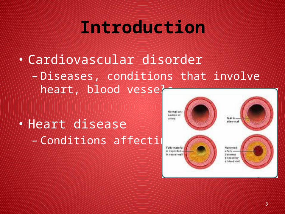
Introduction
• Cardiovascular disorder– Diseases, conditions that involve heart, blood
vessels
• Heart disease– Conditions affecting heart
4
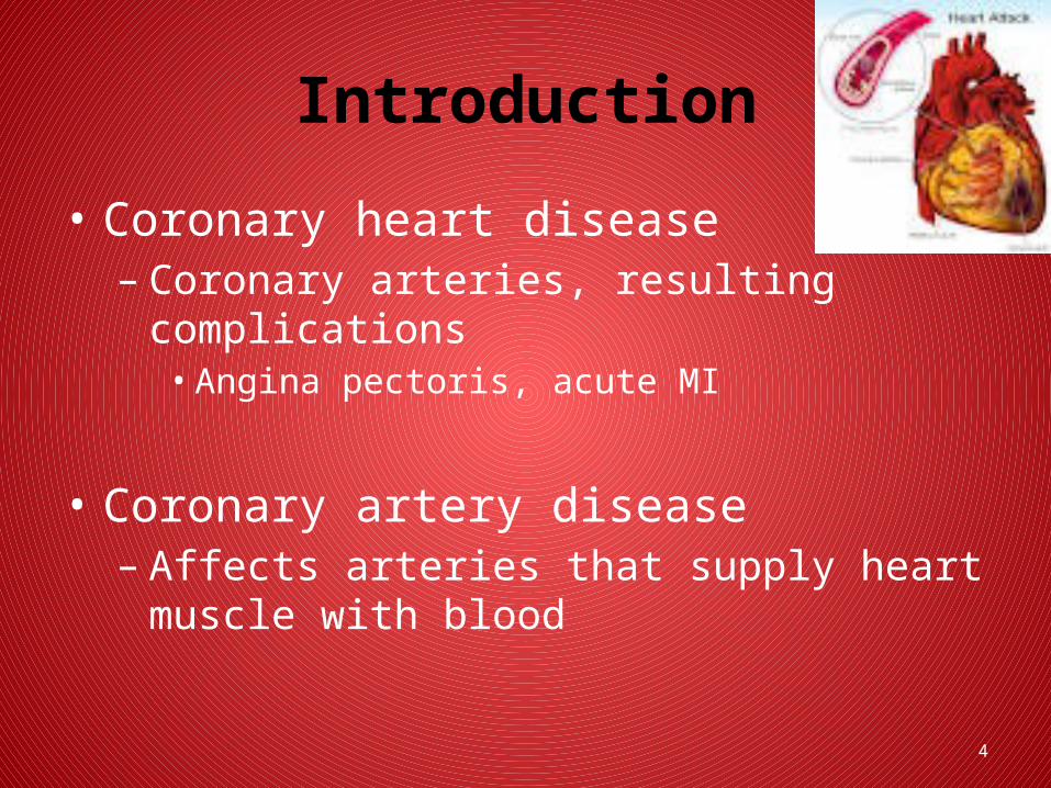
Introduction
• Coronary heart disease– Coronary arteries, resulting complications
• Angina pectoris, acute MI
• Coronary artery disease– Affects arteries that supply heart muscle with
blood
5
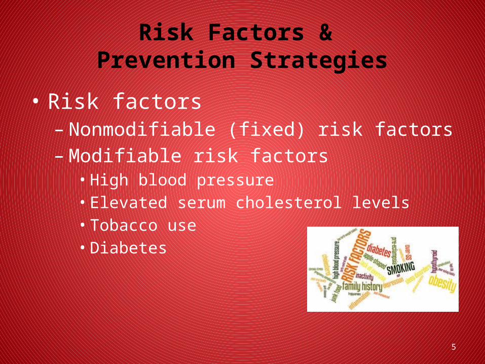
Risk Factors & Prevention Strategies
• Risk factors– Nonmodifiable (fixed) risk factors– Modifiable risk factors
• High blood pressure• Elevated serum cholesterol levels• Tobacco use• Diabetes
6
Risk Factors & Prevention Strategies
• Risk factors– Modifiable risk factors
• Physical inactivity• Obesity, body fat distribution• Metabolic syndrome
7
Risk Factors & Prevention Strategies
• Risk factors– Contributing risk factors
• Stress• Inflammatory markers• Psychosocial factors• Alcohol intake

8
Cardiovascular Anatomy & Physiology

9
Anatomy Review
• Blood vessels– Arteries– Arterioles– Capillaries– Venules– Veins

10
Anatomy Review
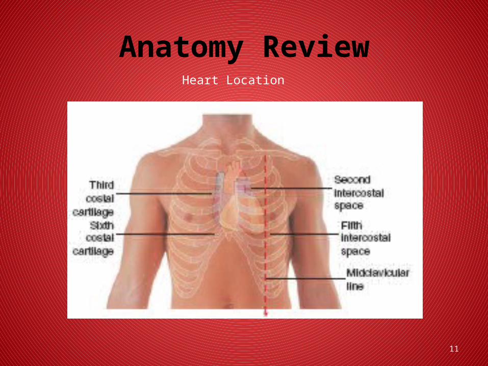
• Heart anatomy– Location
• Mediastinum• Behind sternum, above diaphragm• Base• Apex
11
Anatomy ReviewHeart Location
12
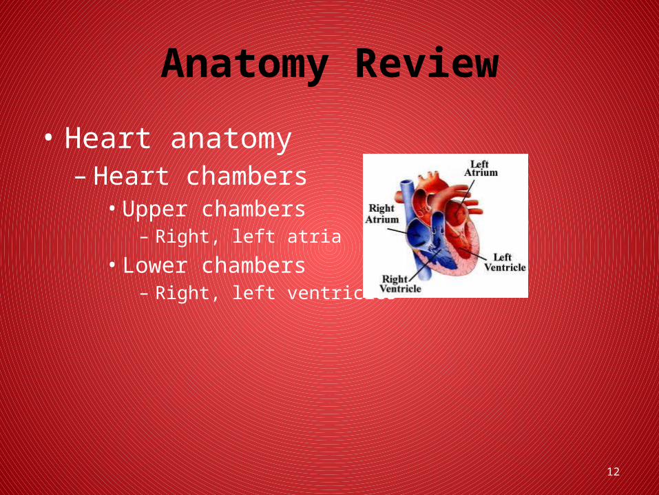
Anatomy Review
• Heart anatomy– Heart chambers
• Upper chambers– Right, left atria
• Lower chambers– Right, left ventricles
13
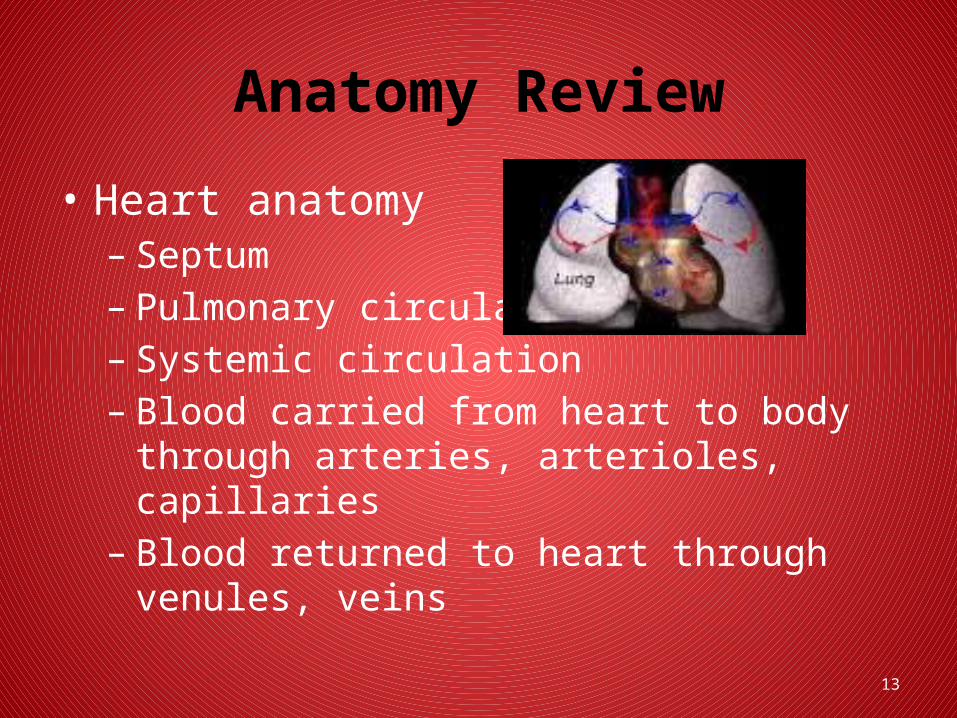
Anatomy Review
• Heart anatomy– Septum– Pulmonary circulation– Systemic circulation– Blood carried from heart to body through arteries,
arterioles, capillaries– Blood returned to heart through venules, veins
14
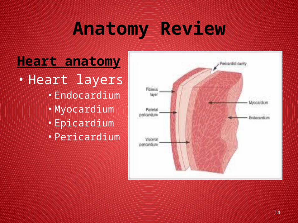
Anatomy Review
Heart anatomy• Heart layers
• Endocardium• Myocardium• Epicardium• Pericardium
15
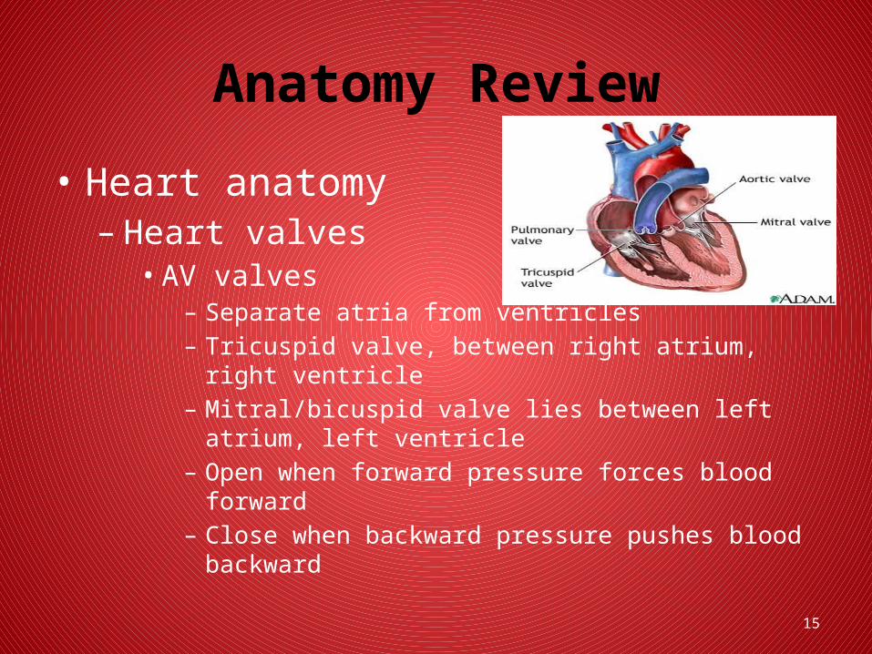
Anatomy Review
• Heart anatomy– Heart valves
• AV valves– Separate atria from ventricles– Tricuspid valve, between right atrium, right ventricle – Mitral/bicuspid valve lies between left atrium, left ventricle – Open when forward pressure forces blood forward– Close when backward pressure pushes blood backward
16
Anatomy Review
• Heart anatomy– Atrial kick
• Blood flows continuously into atria• 70% flows directly through, into ventricles before atria
contract• When atria contract, additional 30% added to filling of
ventricles• When ventricles contract (systole), pressure rises• Tricuspid, mitral valves close when pressure within
ventricles exceeds that of atria
17
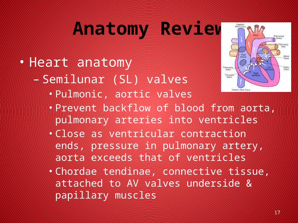
Anatomy Review
• Heart anatomy– Semilunar (SL) valves
• Pulmonic, aortic valves • Prevent backflow of blood from aorta, pulmonary
arteries into ventricles• Close as ventricular contraction ends, pressure in
pulmonary artery, aorta exceeds that of ventricles• Chordae tendinae, connective tissue, attached to AV
valves underside & papillary muscles
18
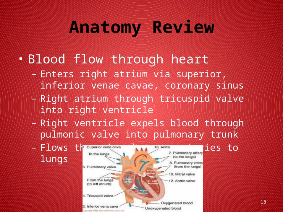
Anatomy Review
• Blood flow through heart– Enters right atrium via superior, inferior venae cavae,
coronary sinus– Right atrium through tricuspid valve into right ventricle– Right ventricle expels blood through pulmonic valve into
pulmonary trunk– Flows through pulmonary arteries to lungs
19
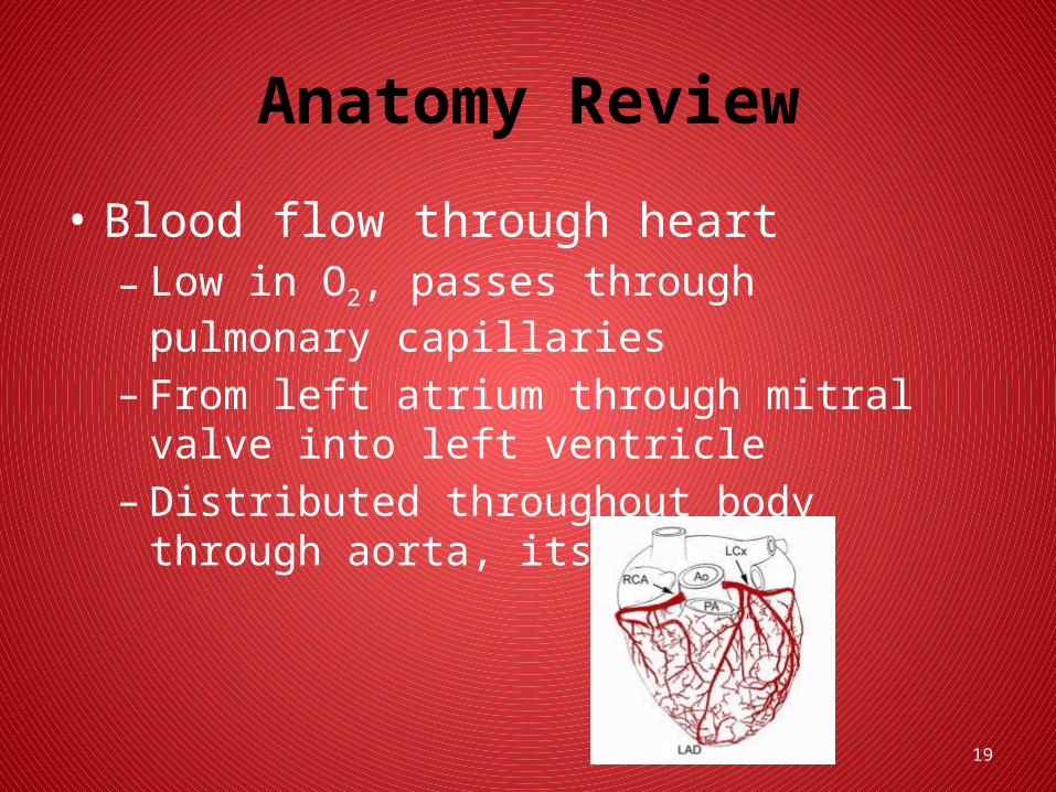
Anatomy Review
• Blood flow through heart– Low in O2, passes through pulmonary capillaries– From left atrium through mitral valve into left
ventricle– Distributed throughout body through aorta, its
branches

20
Anatomy Review
• Blood flow through heart– Tissues of head, neck, upper extremities via
superior vena cava– Lower body via inferior vena cava– Superior, inferior vena cava carry contents into
right atrium
21
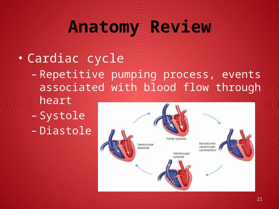
Anatomy Review
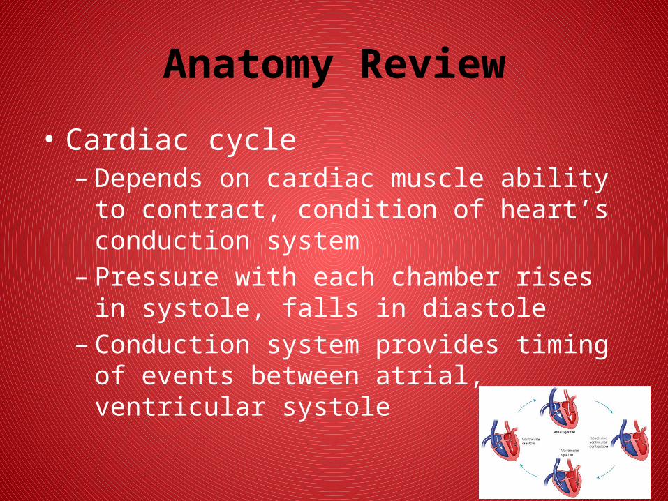
• Cardiac cycle– Repetitive pumping process, events associated
with blood flow through heart– Systole– Diastole
22
Anatomy Review
• Cardiac cycle– Depends on cardiac muscle ability to contract,
condition of heart’s conduction system– Pressure with each chamber rises in systole, falls
in diastole– Conduction system provides timing of events
between atrial, ventricular systole

23
Anatomy Review
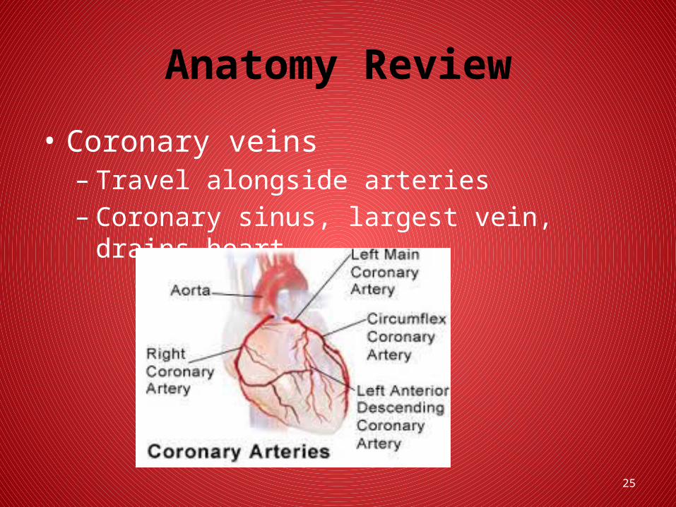
• Coronary arteries– Right, left – Main arteries– Left anterior descending (LAD), left circumflex
(LCX), right coronary artery (RCA)– Lie on outer surface of heart

24
Anatomy ReviewCoronary Arteries
25
Anatomy Review
• Coronary veins– Travel alongside arteries– Coronary sinus, largest vein, drains heart

26
Anatomy Review
• Heart rate– Affected by sympathetic, parasympathetic ANS– Chronotropic effect– Inotropic effect– Dromotropic effect

27
Anatomy Review
• Heart rate– Baroreceptors
• Specialized nerve tissue (sensors)• Found in internal carotid arteries, aortic arch• Detect changes in blood pressure• When stimulated cause sympathetic/parasympathetic
response • Will “reset” to new “normal” after few days of
exposure to specific pressure

28
Anatomy Review
• Heart rate– Chemoreceptors
• In internal carotid arteries, aortic arch, medulla detect changes in concentration of hydrogen ions (pH), O2, carbon dioxide in blood
29
Anatomy Review
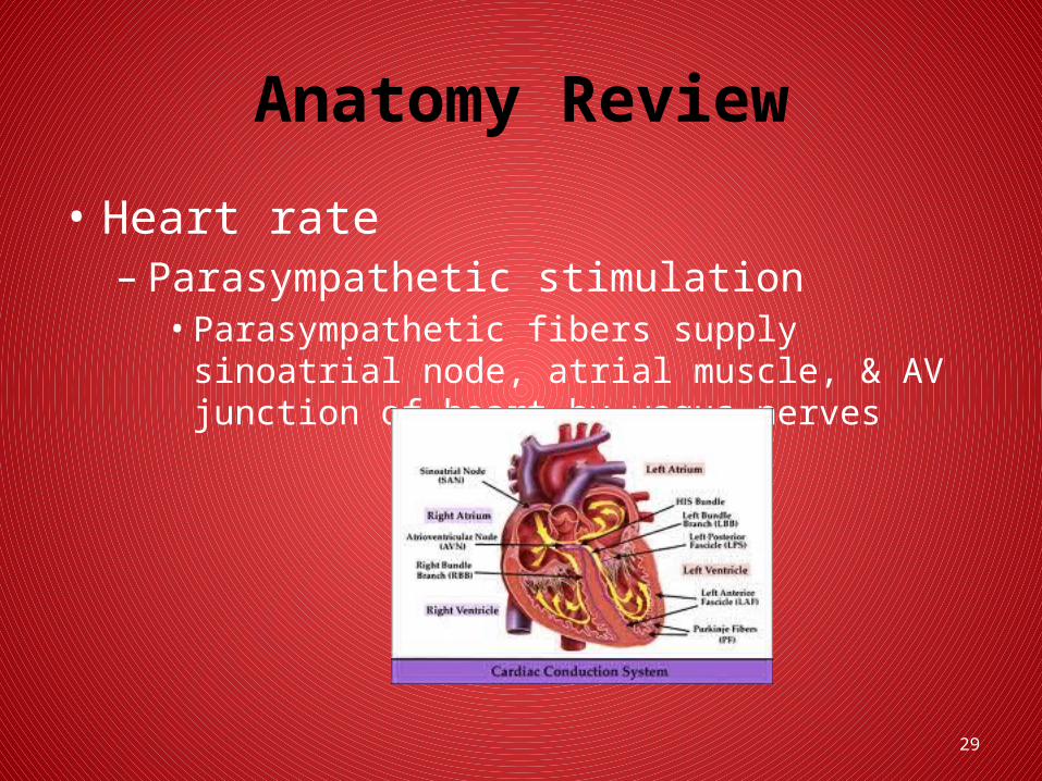
• Heart rate– Parasympathetic stimulation
• Parasympathetic fibers supply sinoatrial node, atrial muscle, & AV junction of heart by vagus nerves

30
Anatomy Review
• Heart rate– Sympathetic stimulation
• Sympathetic nerves supply specific areas of heart’s electrical system, atrial muscle, ventricular myocardium
• When stimulated, norepinephrine released• Increases in heart rate shorten all phases of cardiac
cycle
31
Anatomy Review
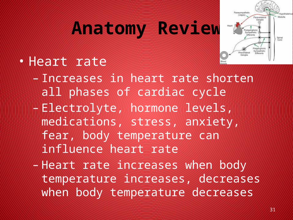
• Heart rate– Increases in heart rate shorten all phases of
cardiac cycle– Electrolyte, hormone levels, medications, stress,
anxiety, fear, body temperature can influence heart rate
– Heart rate increases when body temperature increases, decreases when body temperature decreases
32
Heart as Pump
• Venous return– Most important factor determining amount of
blood pumped out by heart is amount of blood flowing into right heart
33
Heart as Pump
• Cardiac output– Amount of blood pumped into the aorta each
minute by heart– Defined as stroke volume x heart rate– Stroke volume determined by
• Preload• Afterload

34
Heart as Pump
• Cardiac output– Frank–Starling’s law
• Greater the volume of blood in heart during diastole (preload), the more forceful cardiac contraction & more blood ventricle will pump (stroke volume)
• Important that heart adjust its pumping capacity in response to changes in venous return
• During exercise, heart muscle fibers stretch in response to increased volume (preload) before contracting

35
Heart as Pump
• Cardiac output– Frank–Starling’s law
• Factors that increase cardiac output include increased body metabolism, exercise, age & size of body
• Factors that may decrease cardiac output include shock, hypovolemia, heart failure
36
Conclusion
Please complete the 10 question online exam and submit when completed.
Thank You