1 Molecular Biology of Cancer Signal Transduction 2.
59
1 Molecular Biology of Molecular Biology of Cancer Cancer Signal Transduction 2
-
date post
20-Dec-2015 -
Category
Documents
-
view
217 -
download
0
Transcript of 1 Molecular Biology of Cancer Signal Transduction 2.

1Molecular Biology of Molecular Biology of CancerCancer
Signal Transduction
2

2Molecular Biology of Molecular Biology of CancerCancer
Signal Transduction by the Mitogenic Signal Transduction by the Mitogenic PathwaysPathways
Cells in an organism receive a variety of extracellular stimuli for cell proliferation.
Signal transduction is the intracellular event that convey extracellular stimuli into specific cellular responsesprotein-protein interactionPhosphorylation
3Molecular Biology of Molecular Biology of CancerCancer
The MAPK signalling pathwaysThe MAPK signalling pathways
The best characterized mitogenic pathway is:the mitogen-activated protein kinase
(MAPK) cascadesalso called extracellular signal-regulated
kinase 1 and 2 (ERK1 and ERK2)Many growth stimulation converges on
the kinase cascade that activates the MAPK
4Molecular Biology of Molecular Biology of CancerCancer
The RAS-activated MAPK pathway
The first example where all the steps in a complete signalling cascade from the cell surface receptor PTK, to the nuclear transcription is known
RAS RAF MEK MAPK

5Molecular Biology of Molecular Biology of CancerCancer
Ras (from Ras (from rarat t ssarcoma) is a GEFarcoma) is a GEF
guanine nucleotide exchange factor
GTPase activity

6Molecular Biology of Molecular Biology of CancerCancer
GDPGDP
Ligand binds receptor PTK Autophosphorylation on tyrosine
GRB2 (a SH2- and SH3-containing protein): binds to the receptor phosphotyrosine via
its SH2 domain
constitutively binds via its SH3 to the proline-rich sequence in the C-terminus of SOS (a guanine nucleotide exchange factor)
SOS is recruited to the close proximity of RAS in the membrane RAS becomes activated by exchanging
GDP for GTP
The active RAS-GTP: interacts with the N-terminal regulatory
region of the RAF (serine/threonine protein kinase)
RAF recruited to the membrane and changes its conformation
phosphorylation of RAF and binding to the scaffold protein 14-3-3
P
P
P
P SH
2 SH
3
GRB2
SOS RAF P
GTPGTP14-3-3
RAS

7Molecular Biology of Molecular Biology of CancerCancer

8Molecular Biology of Molecular Biology of CancerCancer
MAPK
Activated RAF: activates MEK (also called MAPK
kinase; a dual specificity kinase) by phosphorylation on two conserved serine residues in MEK.
Activated MEK: activates MAPK (a serine/threonine
protein kinase) by phosphorylation of conserved threonine and tyrosine residues.
Activated MAPK: phosphorylates a number of
substrates in the plasma membrane and the cytoplasm;
it is also translocated into the nucleus (within minutes) where it phosphorylates nuclear transcription factors.
Transcription of genes important for cell proliferation.
GDPGDPP
P
P
P SH
2 SH
3
GRB2
SOS RAF P
GTPGTP14-3-3
RAS
MEK
P P
P PSubstrates
Substrates
P
P
SubstratesPMAPK
P P

9Molecular Biology of Molecular Biology of CancerCancer
Homework
Write no more that 2
papers about the
regulation of Raf
activity.

10Molecular Biology of Molecular Biology of CancerCancer
Substrates of MAPKSubstrates of MAPK
MAPK phosphorylates:In cytoplasm: MAPK phosphorylates its upstream
components in a negative feedback loop MAPK phosphorylates SOS, RAF, MEK inhibition of MAP
kinase pathway.
In nucleus: MAPK phosphorylates a number of transcription factors (e.g. Elk1) increase transcription (e.g. of c-Fos mRNA).
Many other substrates: of MAPK probably unknown - identification is difficult as in the case of CDKs
11Molecular Biology of Molecular Biology of CancerCancer
The MAPK signalling pathwaysThe MAPK signalling pathways
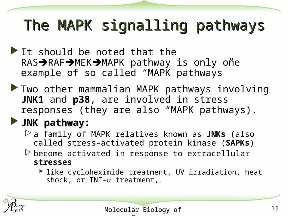
It should be noted that the RASRAFMEKMAPK pathway is only one example of so called “MAPK pathways”
Two other mammalian MAPK pathways involving JNK1 and p38, are involved in stress responses (they are also “MAPK pathways).
JNK pathwayJNK pathway: a family of MAPK relatives known as JNKs (also called
stress-activated protein kinase (SAPKs) become activated in response to extracellular stresses
like cycloheximide treatment, UV irradiation, heat shock, or TNF- treatment,.

12Molecular Biology of Molecular Biology of CancerCancer
RAC1/CDC42GTPGTP
STRESS
PAK
MEKK1-3P P
MEK4P P
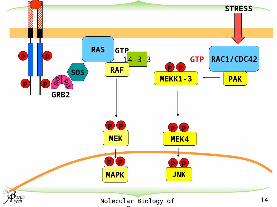
RAC1 and CDC42 are two members of the RHO family of GTP-binding proteins. RAC1 and CDC42 are mainly
activated by stress response independent of RAS
RAC1 can also be activated by RAS (minor pathway) explaining why receptor PTK can sometimes contribute to JNK activation.
GTP-bound form of RAC1 and CDC42 bind and activate the serine/threonine protein kinase PAK, PKN, and PtdIns kinases.
these kinases phosphorylate and activate MEKK1-3
MEKK1-3 phosphorylate and activate MEK4 (also called JNKK) (~ MEK in MAPK pathway; 45% identical in sequence with MEK)
PAK: p21-activated protein kinasePKN: protein kinase NPtdIns kinase: phosphatidylinositol kinase

13Molecular Biology of Molecular Biology of CancerCancer
RAC1/CDC42GTPGTP
STRESS
PAK
MEKK1-3P P
MEK4P P
JNKP P
c-JUNP
JNKP P
MEK4 phosphorylates JNK at two similar sites as in ERK (but T-P-Y in JNK instead of T-E-Y in MAPK) i.e. conservation between the ERK
and JNK pathways at the level of proteins and mode of regulation!
JNK translocation into the nucleus phosphorylation of the transcription factor c-JUN at the N-terminal residues (Ser63 and Ser73) activation of transcription by c-JUN
14Molecular Biology of Molecular Biology of CancerCancer
RAC1/CDC42GTP
STRESS
PAKMEKK1-3P P
14-3-3P
P
P
P SH
2 SH
3
GRB2
SOS
RAS GTP
RAF
MEK4P P
MEK
P P
JNKP P
MAPK
P P
15Molecular Biology of Molecular Biology of CancerCancer
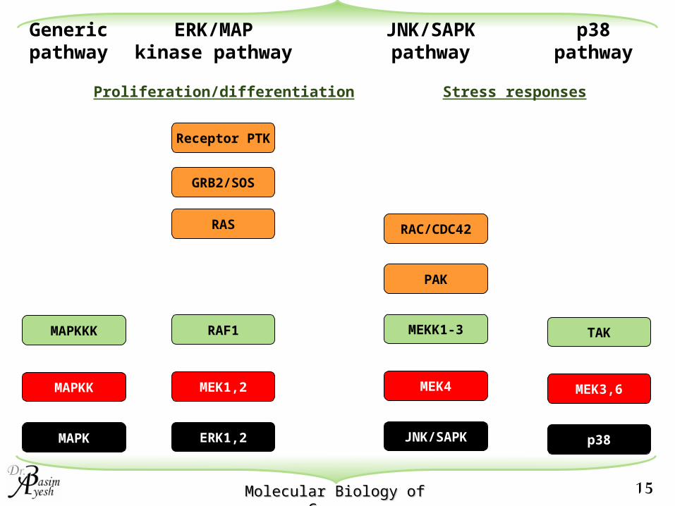
Genericpathway
MAPKKK
MAPKK
MAPK
ERK/MAPkinase pathway
RAF1
MEK1,2
ERK1,2
RAS
GRB2/SOS
Receptor PTK
JNK/SAPKpathway
RAC/CDC42
MEKK1-3
MEK4
JNK/SAPK
TAK
MEK3,6
p38
p38pathway
PAK
Stress responsesProliferation/differentiation
16Molecular Biology of Molecular Biology of CancerCancer
Specificity of MAP kinase pathwaysSpecificity of MAP kinase pathways
It seems JNK and ERK pathways are biologically distinct. However, they are both protein kinases with similar substrate specificity most in vitro substrates are the same for both.Yet these pathways must result in unique
transcriptional activity - because stress and mitogen must elicit different responses
There are at least five parallel MAP kinase pathways in mammalian cells.
How is specificity achieved?

17Molecular Biology of Molecular Biology of CancerCancer
•Specificity of MAP kinase pathwaysSpecificity of MAP kinase pathways
One way is by scaffold proteins.
18Molecular Biology of Molecular Biology of CancerCancer
G Protein-Linked ReceptorsG Protein-Linked Receptors
G protein-linked receptors compose the largest family of cell-surface receptors: >100 members in mammals include:
light-activated receptors (rhodopsins) in the eyeodorant receptors in the nosereceptors for various hormones and
neurotransmitters
19Molecular Biology of Molecular Biology of CancerCancer
G Protein-Linked ReceptorsG Protein-Linked Receptors
A number of different hormones mediate biological responses by binding to G protein-linked receptors - and -adrenergic receptors Muscarinic cholinergic receptors Vasopressin (ADH) Angiotensin II Serotonin Substance P Dopamine Lutenizing hormone (LH) Follicle-stimulating hormone (FSH) Thyroid stimulating hormone (TSH) Platelet-activating factor Prostaglandins Rhodopsin
20Molecular Biology of Molecular Biology of CancerCancer
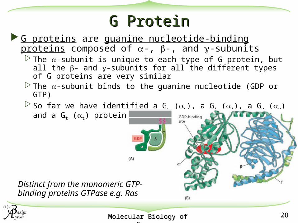
G ProteinG Protein G proteins are guanine nucleotide-binding proteins
composed of -, -, and -subunits The -subunit is unique to each type of G protein, but all the
- and -subunits for all the different types of G proteins are very similar
The -subunit binds to the guanine nucleotide (GDP or GTP) So far we have identified a Gs (s), a Gi (i), a Gq (q) and a Gt
(t) protein
Distinct from the monomeric GTP-binding proteins GTPase e.g. Ras

21Molecular Biology of Molecular Biology of CancerCancer
22Molecular Biology of Molecular Biology of CancerCancer
G Protein-Linked ReceptorsG Protein-Linked Receptors
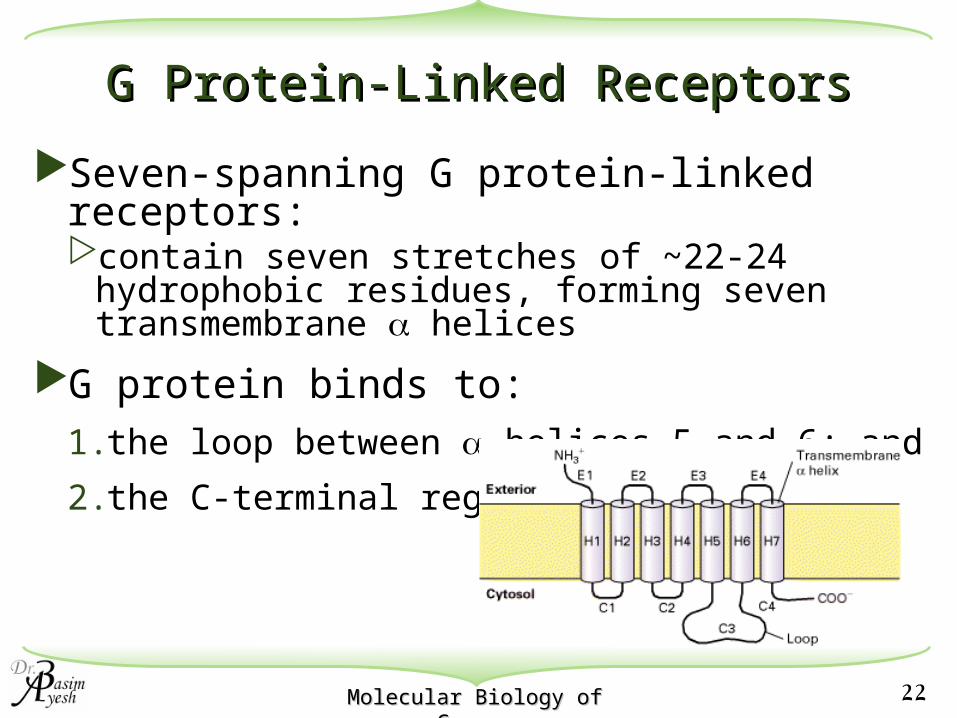
Seven-spanning G protein-linked receptors:contain seven stretches of ~22-24
hydrophobic residues, forming seven transmembrane helices
G protein binds to: 1.the loop between helices 5 and 6; and
2.the C-terminal region

23Molecular Biology of Molecular Biology of CancerCancer
G protein acts as an on/off switchG protein acts as an on/off switch
No ligand G protein binds GDP inactive
Ligand binding to receptor G protein binds GTP active
Activated G protein binds to and activates an effector enzyme, which catalyzes the formation of a secondary messenger.
Hydrolysis of GTP to GDP converts G protein back to inactive state.

24Molecular Biology of Molecular Biology of CancerCancer
Example of G protein-linked receptorExample of G protein-linked receptorAdrenaline receptorAdrenaline receptor
1. Hormone binding to - and -adrenergic receptors.
2. The receptor interacts with G protein
3. Activation/inhibition of adenylate cyclase (effector enzyme).
4. Increase/decrease in intracellular cAMP (secondary messenger).
25Molecular Biology of Molecular Biology of CancerCancer
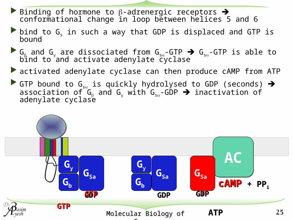
Binding of hormone to -adrenergic receptors conformational change in loop between helices 5 and 6
bind to Gs in such a way that GDP is displaced and GTP is bound
G and G are dissociated from Gs-GTP Gs-GTP is able to bind to and activate adenylate cyclase
activated adenylate cyclase can then produce cAMP from ATP
GTP bound to Gs is quickly hydrolysed to GDP (seconds) association of G and G with Gs-GDP inactivation of adenylate cyclase
G
Gb
GSa
GDPGDP
ACACG
Gb
GSa
GDPGDP
GTPGTP
GTPGTP
ATPATP
cAMPcAMP + PP+ PPi i
GSa
GTPGTPGDGDPP

26Molecular Biology of Molecular Biology of CancerCancer
Amplification of signalAmplification of signal
1. Activated Gs-GTP can diffuse rapidly
one activated receptor can activate many Gs.
2. One Gs-GTP can bind to only one adenylate cyclase
but this can catalyze the synthesis of many cAMP.

27Molecular Biology of Molecular Biology of CancerCancer
Some bacterial toxins irreversibly Some bacterial toxins irreversibly modify G proteinsmodify G proteins
Cholera toxin: a peptide produced by the bacterium Vibrio cholerae, causes serious diarrhea death by dehydration.
Irreversibly modifies Gs (at Arg174, which is located near the GTP-binding site in Gs)
modified Gs can bind GTP but cannot hydrolyze it to GDP permanent activation of adenylate cyclase sustained high cAMP level; in intestinal epithelial cells
this sustained increase in cAMP causes membrane proteins to allow water efflux into the intestine.
28Molecular Biology of Molecular Biology of CancerCancer
Gi may inhibit adenylate cyclase by two mechanisms:
1. The i-GTP complex interacts with adenylyl cyclase, inhibiting its activity
2. Adenylyl cyclase activity is further reduced by increasing the amount of -subunits; this allows them to interact with s-subunits preventing activation of adenylyl cyclase

29Molecular Biology of Molecular Biology of CancerCancer
cAMP as a Second MessengercAMP as a Second Messenger
The main target of cAMP in the cell is cAMP-dependent kinase (PKA).
PKA is a serine/threonine protein kinase. Inactive conformation: a dimer of PKA binding to two regulatory subunits. Each regulatory subunit contains two cAMP binding sites When cAMP binds cooperatively to the regulatory subunits
regulatory subunits dissociate from the PKA PKA becomes activated.

30Molecular Biology of Molecular Biology of CancerCancer
PKA substratesPKA substrates
PKA catalyzes phosphorylation and activation of hormone-sensitive lipase, cholesteryl esterase, & glycogen phosphorylase, and inhibits glycogen synthase
cAMP also (through PKA) regulates gene transcriptionPhosphoenolpyruvate carboxykinaseTyrosine aminotransferaseHuman glycoprotein hormone -subunit genePreprosomatostatinVasoactive intestinal polypeptideA surfactant protein, SP-ASeveral isoforms of cytochrome P450
31Molecular Biology of Molecular Biology of CancerCancer
Examples of PKA Examples of PKA substratessubstrates
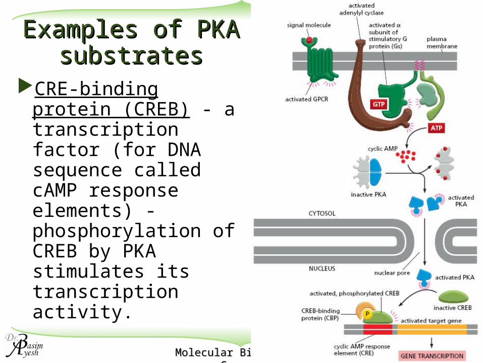
CRE-binding protein (CREB) - a transcription factor (for DNA sequence called cAMP response elements) - phosphorylation of CREB by PKA stimulates its transcription activity.

32Molecular Biology of Molecular Biology of CancerCancer
The GThe Gqq protein-linked receptors and Caprotein-linked receptors and Ca2+2+
Ca2+ is an important intracellular second messenger.[Ca2+] in the cytosol is low (10-7 M)
[Ca2+] outside the cell is high (10-3 M)
[Ca2+] in ER also high.
Extracellular signals open Ca2+ channels in plasma / ER membranesCa2+ rushes into the cytosol increase Ca2+
Ca2+-dependent responses.
33Molecular Biology of Molecular Biology of CancerCancer
Control of cytosolic calciumControl of cytosolic calcium
Ca2+-ATPase in plasma membrane and ER membrane pumps Ca2+ out of the cytosol (use ATP as energy) into the extracellular space and the ER respectively.Normally, free [Ca2+] changed from ~10-7 M in
resting cells to ~5x10⁻⁶ M in stimulated cells.
If Ca2+ pumps are defective and the free [Ca2+] in the cytosol gets to >10-5 M, a low affinity, high capacity Ca2+ pump in the inner mitochondrial membranes kicks in and pump Ca2+ into the mitochondria (uses electrochemical gradient as energy).

34Molecular Biology of Molecular Biology of CancerCancer
Adapted from Molecular Biology of the Cell

35Molecular Biology of Molecular Biology of CancerCancer
G
G
Gq
GDPGDP
PLC-
IP3 :Inositol triphosphate
Release Ca2+ from ER
DAG: Diacylglycerol Activates PKC
OverviewOverview
1. Extracellular signaling molecules binds to G protein-linked receptor in the plasma membrane.
Activation of a G protein Gq
2. Activation of phospholipase C- 3. Cleaves phosphatidylinositol bisphosphate (PIP2)
into two products: 2 different signal transduction pathways

36Molecular Biology of Molecular Biology of CancerCancer
Phosphatidylinositol (PI) is a minor phospholipid in cell Phosphatidylinositol (PI) is a minor phospholipid in cell membranes; PIPmembranes; PIP22 is a is a phosphorylated derivative of PIphosphorylated derivative of PI - located in - located in the inner half of the plasma membrane lipid bilayer.the inner half of the plasma membrane lipid bilayer.
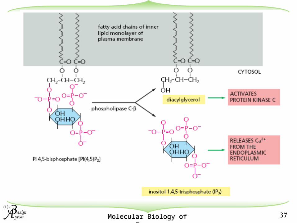
37Molecular Biology of Molecular Biology of CancerCancer
38Molecular Biology of Molecular Biology of CancerCancer
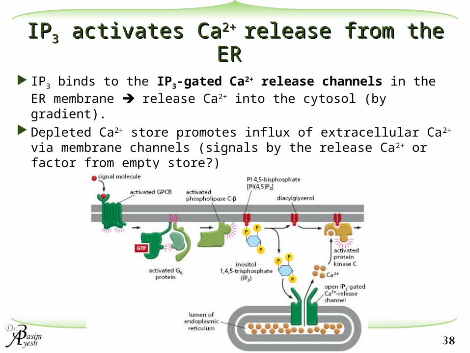
IPIP33 activates Ca activates Ca2+ 2+ release from the ER release from the ER
IP3 binds to the IP3-gated Ca2+ release channels in the ER membrane release Ca2+ into the cytosol (by gradient).
Depleted Ca2+ store promotes influx of extracellular Ca2+ via membrane channels (signals by the release Ca2+ or factor from empty store?)

39Molecular Biology of Molecular Biology of CancerCancer
IP3
DAG
ER
IP3-gated Ca2+ release channels
Ca2+
Calmodulin
4 high-affinity Ca2+-binding sites
Enzymes (e.g. myosin light-chain kinase,phosphorylase kinase, Ca2+-calmodulin kinase II etc)
Membrane transport proteins (e.g. Ca2+-ATPase on plasma membrane

40Molecular Biology of Molecular Biology of CancerCancer
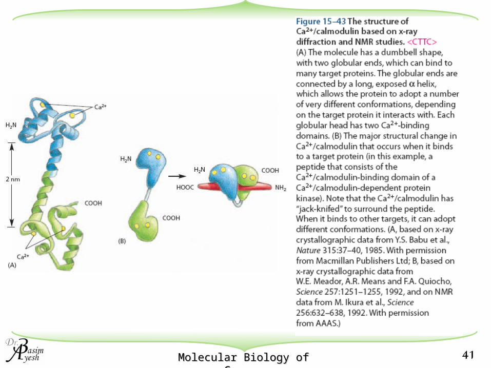
Calmodulin
Calmodulin is a polypeptide that undergoes a conformational change when it binds to calcium
The conformational change allows the calmodulin effect on cellular proteins
Many effects of Ca2+ are mediated by Ca2+/calmodulin-dependent kinases (CaM-kinases).The best studied example of CaM-kinase is CaM-
kinase II. CaM-kinase II is found in all animal cells but is
especially enriched in the nervous system.
41Molecular Biology of Molecular Biology of CancerCancer
42Molecular Biology of Molecular Biology of CancerCancer
CaM kinasesCaM kinases
Functions of CaM-kinase II: Molecular memory device switching to active state when exposed to
Ca2+/calmodulin.remains active by autophosphorylation (i.e.
remains active even when Ca2+ is removed)inactivated only when the phosphatase
overwhelms the autophosphorylation)important in memory (mice lacking CaM-kinase II
have defects in remembering where things are in space)

43Molecular Biology of Molecular Biology of CancerCancer

44Molecular Biology of Molecular Biology of CancerCancer
Termination of CaTermination of Ca2+ 2+ responseresponse
1. Breakdown of DAG
2. Further phosphorylation of PIP2
3. IP3 is dephosphorylated and inactivated by phosphatases. (sometimes it is further phosphorylated to IP4 to mediate other responses)
4. Ca2+ is pumped out of the cell by Ca2+-ATPase
5. Phosphatases which inactivate CaM-kinase II
45Molecular Biology of Molecular Biology of CancerCancer
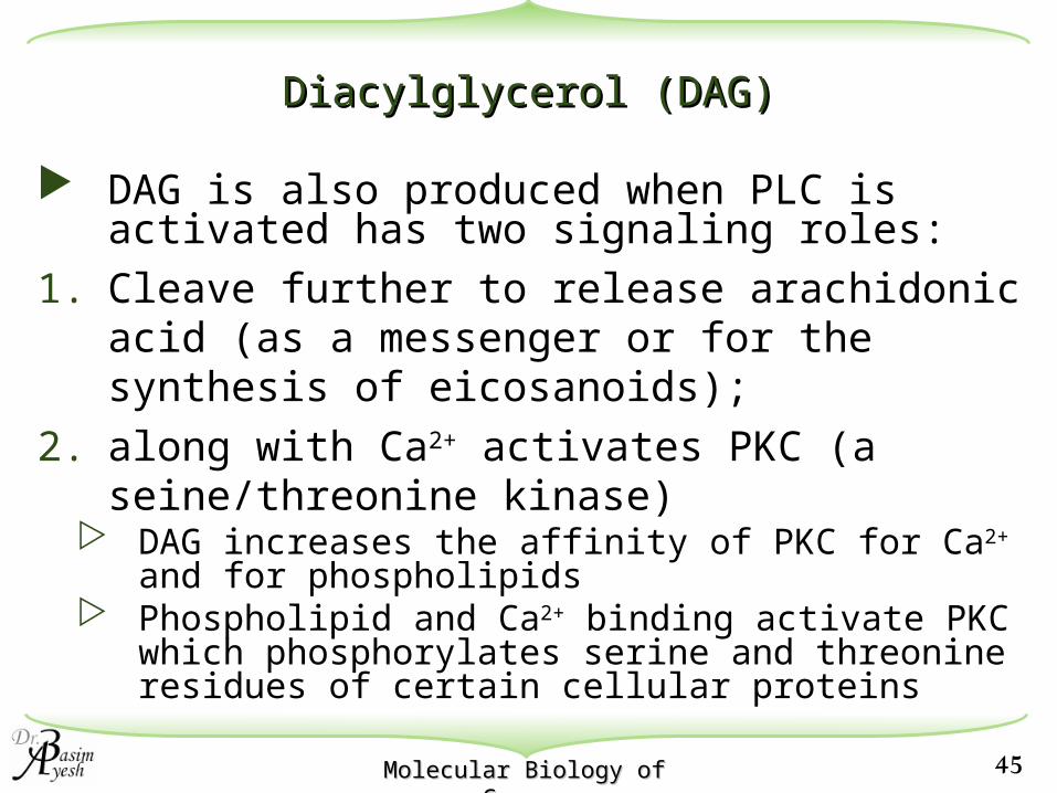
Diacylglycerol (DAG)Diacylglycerol (DAG)
DAG is also produced when PLC is activated has two signaling roles:
1. Cleave further to release arachidonic acid (as a messenger or for the synthesis of eicosanoids);
2. along with Ca2+ activates PKC (a seine/threonine kinase)
DAG increases the affinity of PKC for Ca2+ and for phospholipids
Phospholipid and Ca2+ binding activate PKC which phosphorylates serine and threonine residues of certain cellular proteins
46Molecular Biology of Molecular Biology of CancerCancer
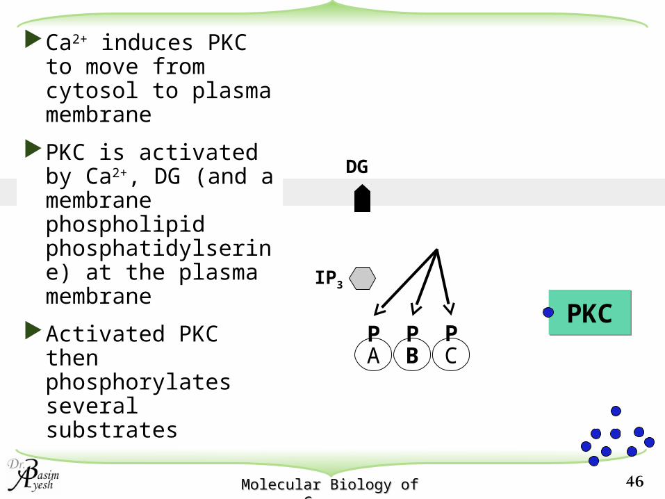
IP3
DG
PKCPKC
Ca2+ induces PKC to move from cytosol to plasma membrane
PKC is activated by Ca2+, DG (and a membrane phospholipid phosphatidylserine) at the plasma membrane
Activated PKC then phosphorylates several substrates
A B CP P P
47Molecular Biology of Molecular Biology of CancerCancer
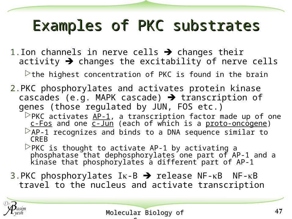
Examples of PKC substratesExamples of PKC substrates
1.Ion channels in nerve cells changes their activity changes the excitability of nerve cells
the highest concentration of PKC is found in the brain
2.PKC phosphorylates and activates protein kinase cascades (e.g. MAPK cascade) transcription of genes (those regulated by JUN, FOS etc.)
PKC activates AP-1, a transcription factor made up of one c-Fos and one c-Jun (each of which is a proto-oncogene)
AP-1 recognizes and binds to a DNA sequence similar to CREBPKC is thought to activate AP-1 by activating a phosphatase that
dephosphorylates one part of AP-1 and a kinase that phosphorylates a different part of AP-1
3.PKC phosphorylates I-B release NF-B NF-B travel to the nucleus and activate transcription

48Molecular Biology of Molecular Biology of CancerCancer
MAPK IkB NFkB
Gene 1
Gene 2
DG
PKC
PPPP
Nucleus
PP
Activates transcription
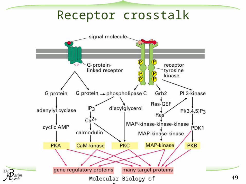
49Molecular Biology of Molecular Biology of CancerCancer
Receptor crosstalk

50Molecular Biology of Molecular Biology of CancerCancer
Receptor-linked Tyr kinases
This is a common motif. It is called the Jak/STAT pathway for gene regulation.
51Molecular Biology of Molecular Biology of CancerCancer
Signal transduction by nuclear Signal transduction by nuclear receptorsreceptors

52Molecular Biology of Molecular Biology of CancerCancer
e.g.Glucocorticoid receptor response element:5’-AGAACA(N)3TGTTCT-3’3’-TCTTGT(N)3ACAAGA-5’
Steroid Hormone ReceptorsSteroid Hormone Receptors
The consensus sequence of DNA binding sites of glucocorticoid-receptors (called response elements) = 6 bp inverted repeats separated by any 3 bp.
This suggests that these steroid receptors bind to DNA as symmetrical dimers (later confirmed by X-ray crystallography).

53Molecular Biology of Molecular Biology of CancerCancer
Glucocorticoid receptor - a C4 zinc-finger homodimerGlucocorticoid receptor - a C4 zinc-finger homodimer

54Molecular Biology of Molecular Biology of CancerCancer
N C
1 2 3
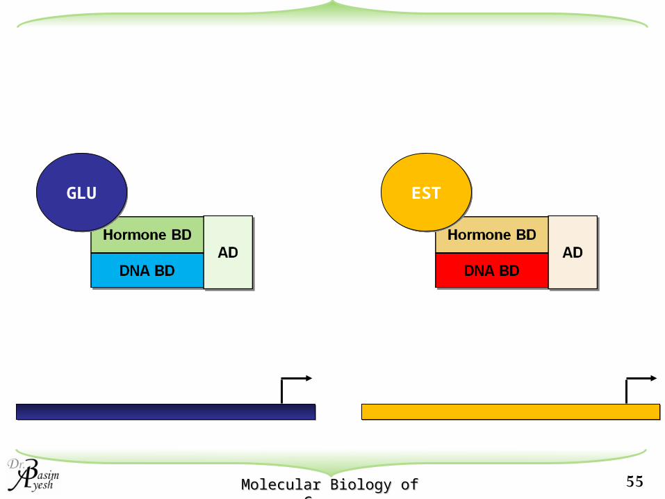
Steroid Hormone ReceptorsSteroid Hormone Receptors
Different hormone receptors are conserved in their amino acid sequences and functional domains - all contain:
1. An unique N-terminal region that contains the activation region
2. DNA binding domain
3. Hormone binding domain
55Molecular Biology of Molecular Biology of CancerCancer
GLUGLU ESTEST
56Molecular Biology of Molecular Biology of CancerCancer
GLUGLU
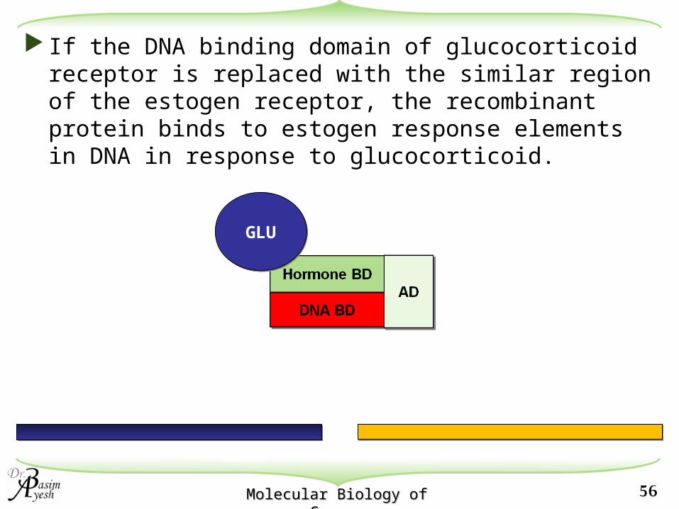
If the DNA binding domain of glucocorticoid receptor is replaced with the similar region of the estogen receptor, the recombinant protein binds to estogen response elements in DNA in response to glucocorticoid.
57Molecular Biology of Molecular Biology of CancerCancer
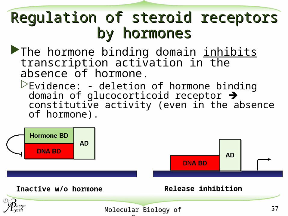
Inactive w/o hormone
Regulation of steroid receptors by Regulation of steroid receptors by hormoneshormones
The hormone binding domain inhibits transcription activation in the absence of hormone.Evidence: - deletion of hormone binding domain
of glucocorticoid receptor constitutive activity (even in the absence of hormone).
Release inhibition
58Molecular Biology of Molecular Biology of CancerCancer
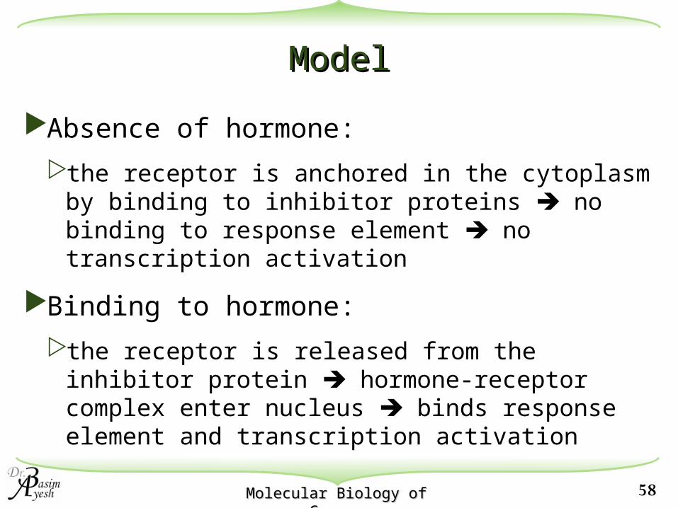
ModelModel
Absence of hormone:
the receptor is anchored in the cytoplasm by binding to inhibitor proteins no binding to response element no transcription activation
Binding to hormone:
the receptor is released from the inhibitor protein hormone-receptor complex enter nucleus binds response element and transcription activation

59Molecular Biology of Molecular Biology of CancerCancer
The proteins that retain hormone receptor in the cytoplasm are likely to be proteins known as molecular chaperones - which includes heat-shock protein (HSP90)HSP90 masked the nuclear localization signal
(NLS) in the absence of hormone
Hormone BD
DNA BDDNA BDADAD
NLS
Hsp90
GLUGLU Nuclear membrane


![[VII]. Regulation of Gene Expression Via Signal Transduction Reading List VII: Signal transduction Signal transduction in biological systems.](https://static.fdocuments.in/doc/165x107/56649e385503460f94b28319/vii-regulation-of-gene-expression-via-signal-transduction-reading-list-vii.jpg)