1 Membrane Structure and Function. 2 Plasma Membrane boundary Is the boundary that separates the...
53
1 Membrane Structure and Function
-
Upload
hubert-martin -
Category
Documents
-
view
224 -
download
3
Transcript of 1 Membrane Structure and Function. 2 Plasma Membrane boundary Is the boundary that separates the...
1
Membrane Structure and Function
2
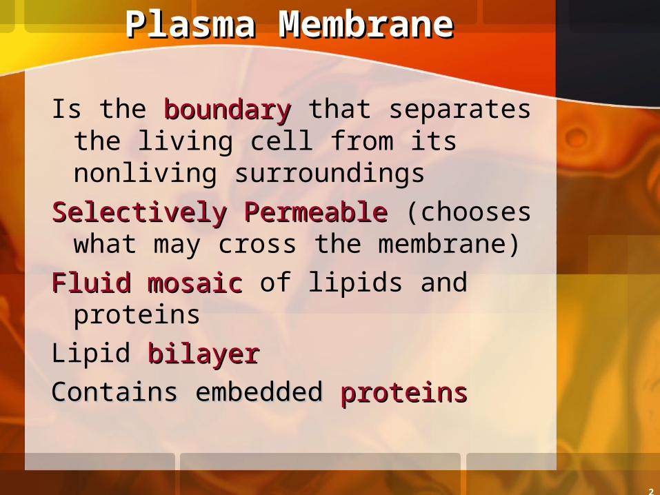
Plasma MembranePlasma Membrane
Is the boundaryboundary that separates the living cell from its nonliving surroundings
Selectively PermeableSelectively Permeable (chooses what may cross the membrane)
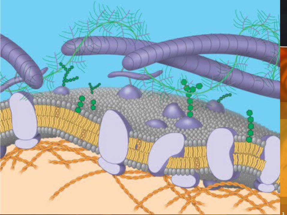
Fluid mosaicFluid mosaic of lipids and proteinsLipid bilayerbilayer
Contains embeddedContains embedded proteins proteins
3
4
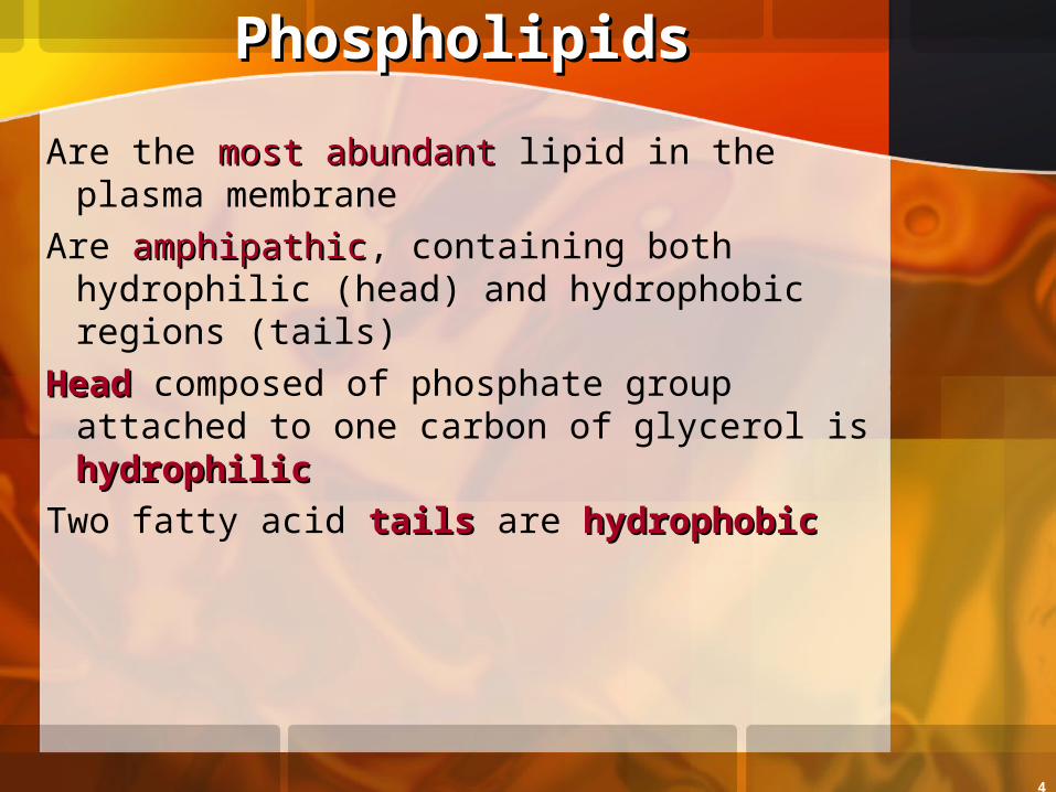
PhospholipidsPhospholipids
Are the most abundantmost abundant lipid in the plasma membrane
Are amphipathicamphipathic, containing both hydrophilic (head) and hydrophobic regions (tails)
HeadHead composed of phosphate group attached to one carbon of glycerol is hydrophilichydrophilic
Two fatty acid tailstails are hydrophobichydrophobic
5
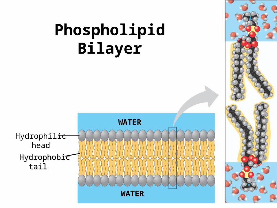
Hydrophilichead
HydrophobicHydrophobic tail tail
WATER
WATER
Phospholipid Bilayer
6
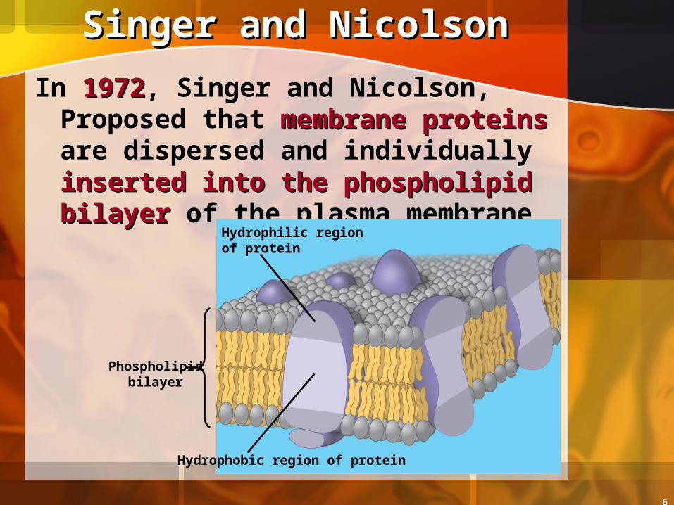
Singer and NicolsonSinger and Nicolson
In 19721972, Singer and Nicolson, Proposed that membrane membrane proteinsproteins are dispersed and individually inserted into the inserted into the phospholipid bilayer phospholipid bilayer of the of the plasma membraneplasma membrane
Phospholipidbilayer
Hydrophilic region of protein
Hydrophobic region of protein
7
Fluid Mosaic ModelFluid Mosaic Model
A membrane is a fluid structurefluid structure with a “mosaic” of various proteins embedded in it when viewed from the top
PhospholipidsPhospholipids can move laterallylaterally a small amount and can “flex” their tails
Membrane proteinsMembrane proteins also move side to side or lateralllaterally making the membrane fluid

8
Freeze-fractureFreeze-fracture studies of the plasma membrane support the fluid mosaic model of membrane structure
A cell is frozen and fractured with a knife. The fracture fracture plane often follows the hydrophobic interior of a plane often follows the hydrophobic interior of a membranemembrane, splitting the phospholipid bilayer into two two separated layersseparated layers. The membrane proteins go wholly membrane proteins go wholly with one of the layerswith one of the layers.
9
The Fluidity of Membranes
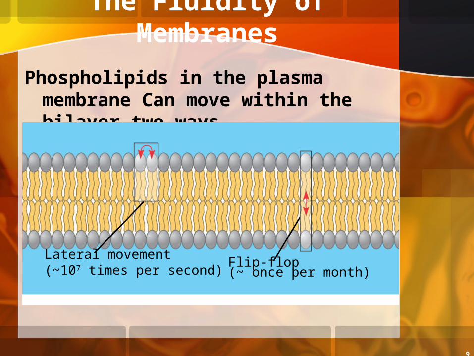
Phospholipids in the plasma membrane Can move within the bilayer two ways
Lateral movement(~107 times per second)
Flip-flop(~ once per month)
10
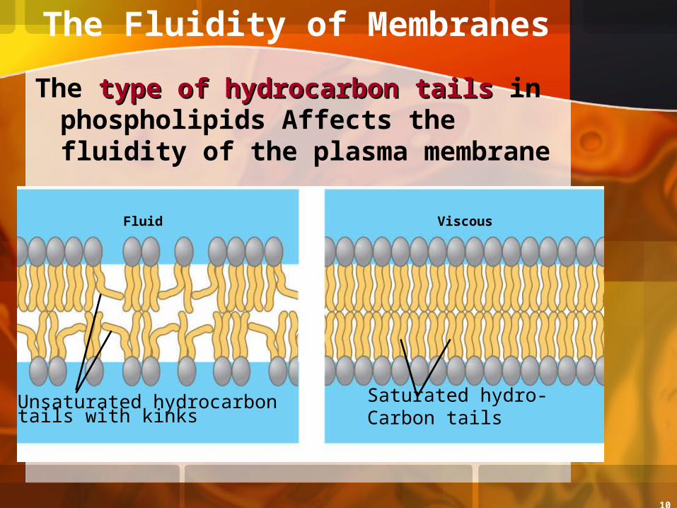
The type of hydrocarbon tailstype of hydrocarbon tails in phospholipids Affects the fluidity of the plasma membrane
Fluid Viscous
Unsaturated hydrocarbontails with kinks
Saturated hydro-Carbon tails
The Fluidity of Membranes
11
The Fluidity of Membranes
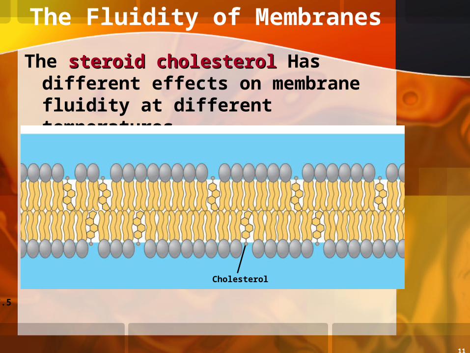
The steroid cholesterolsteroid cholesterol Has different effects on membrane fluidity at different temperatures
Figure 7.5
Cholesterol
12
Membrane Proteins and Their Functions
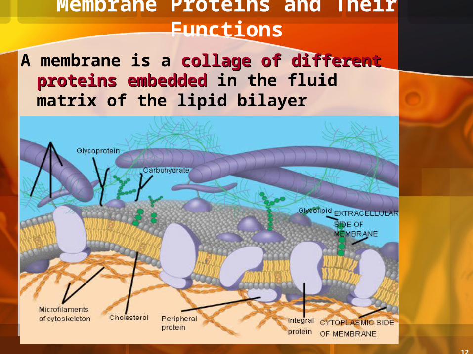
A membrane is a collage of different collage of different proteins embeddedproteins embedded in the fluid matrix of the lipid bilayer
Fibers of
extracellularmatrix (ECM)

13
Types of Membrane Proteins
Integral proteinsIntegral proteinsPenetrate the hydrophobic core of
the lipid bilayerAre often transmembrane proteinstransmembrane proteins,
completely spanning the membrane
EXTRACELLULARSIDE
14
Types of Membrane Proteins
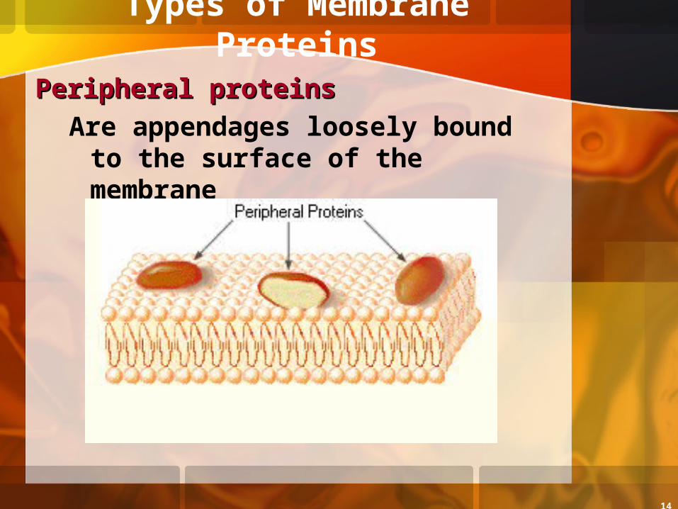
Peripheral proteinsPeripheral proteinsAre appendages loosely bound
to the surface of the membrane
15
Six Major Functions of Membrane Proteins
Figure 7.9
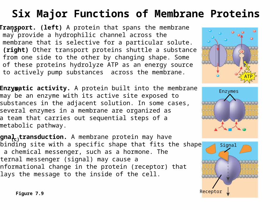
Transport. (left) A protein that spans the membrane may provide a hydrophilic channel across the membrane that is selective for a particular solute. (right) Other transport proteins shuttle a substance from one side to the other by changing shape. Some of these proteins hydrolyze ATP as an energy source to actively pump substances across the membrane.
Enzymatic activity. A protein built into the membranemay be an enzyme with its active site exposed tosubstances in the adjacent solution. In some cases,several enzymes in a membrane are organized asa team that carries out sequential steps of ametabolic pathway.
Signal transduction. A membrane protein may havea binding site with a specific shape that fits the shapeof a chemical messenger, such as a hormone. Theexternal messenger (signal) may cause aconformational change in the protein (receptor) thatrelays the message to the inside of the cell.
(a)
(b)
(c)
ATP
Enzymes
Signal
Receptor
16
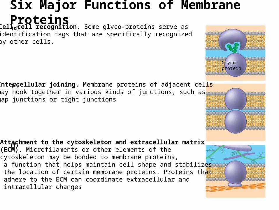
Cell-cell recognition. Some glyco-proteins serve as identification tags that are specifically recognized by other cells.
Intercellular joining. Membrane proteins of adjacent cellsmay hook together in various kinds of junctions, such asgap junctions or tight junctions
Attachment to the cytoskeleton and extracellular matrix(ECM). Microfilaments or other elements of thecytoskeleton may be bonded to membrane proteins, a function that helps maintain cell shape and stabilizes the location of certain membrane proteins. Proteins that adhere to the ECM can coordinate extracellular and intracellular changes
(d)
(e)
(f)
Glyco-protein
Six Major Functions of Membrane Proteins
17
The Role of Membrane Carbohydrates in Cell-Cell
RecognitionCell-cell recognitionCell-cell recognition IIs a cell’s ability to distinguish
one type of neighboring cell from another
Membrane carbohydratesMembrane carbohydratesInteract with the surface
molecules of other cells, facilitating cell-cell recognition
18
Synthesis and Sidedness of Membranes
Membranes have distinct distinct inside and outside facesinside and outside faces
This affects the movementaffects the movement of proteins synthesizedproteins synthesized in the endomembrane system endomembrane system (Golgi and ER)(Golgi and ER)
19
Synthesis and Sidedness of Membranes
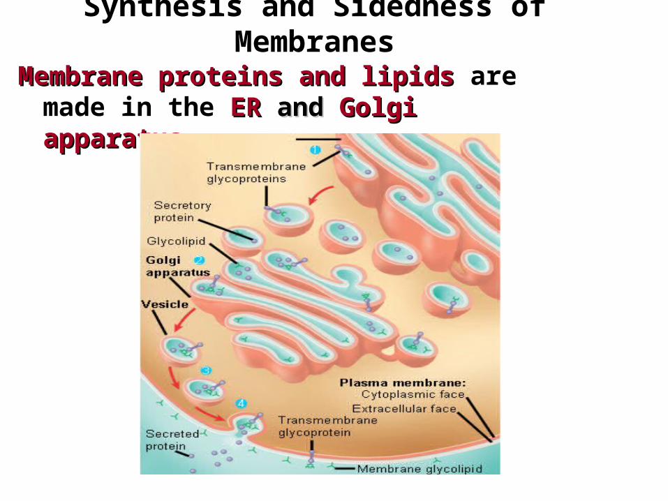
Membrane proteins and lipidsMembrane proteins and lipids are made in the ER ER andand Golgi Golgi apparatusapparatus
ER
20
Membrane Permeability
Membrane structurestructure results in selective permeabilityselective permeability
A cell must exchange cell must exchange materials with its materials with its surroundingssurroundings, a process controlled by the plasma membrane
21
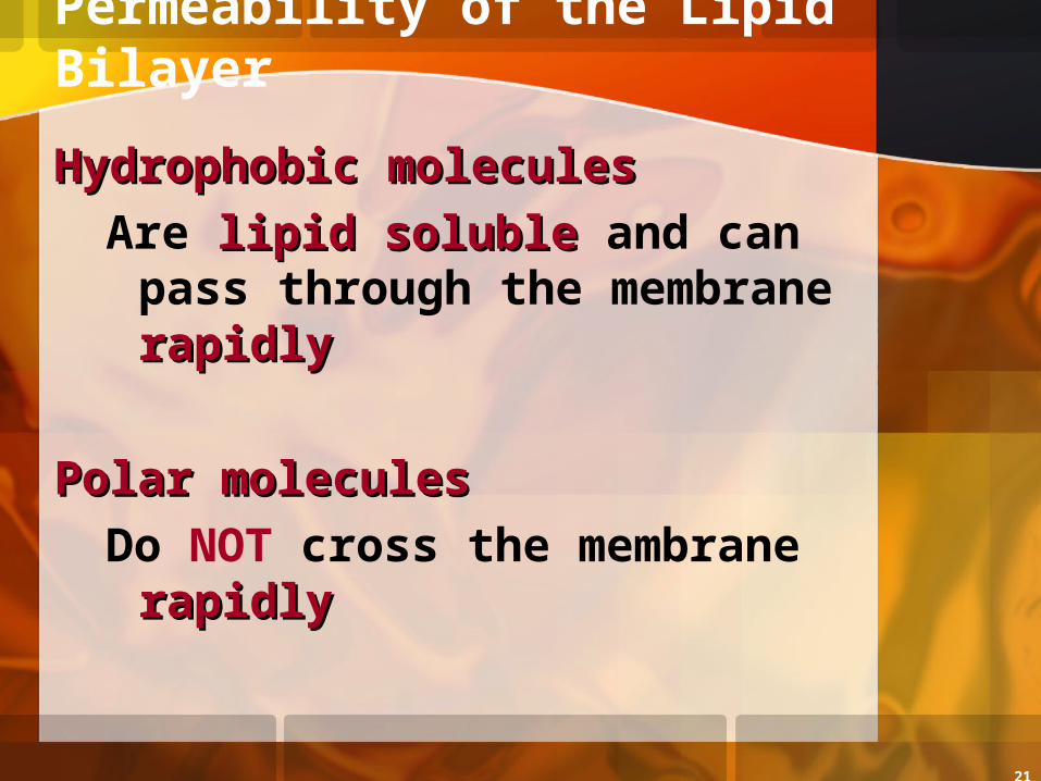
Permeability of the Lipid Bilayer
Hydrophobic moleculesHydrophobic moleculesAre lipid solublelipid soluble and can
pass through the membrane rapidlyrapidly
Polar moleculesPolar moleculesDo NOT cross the membrane
rapidlyrapidly
22
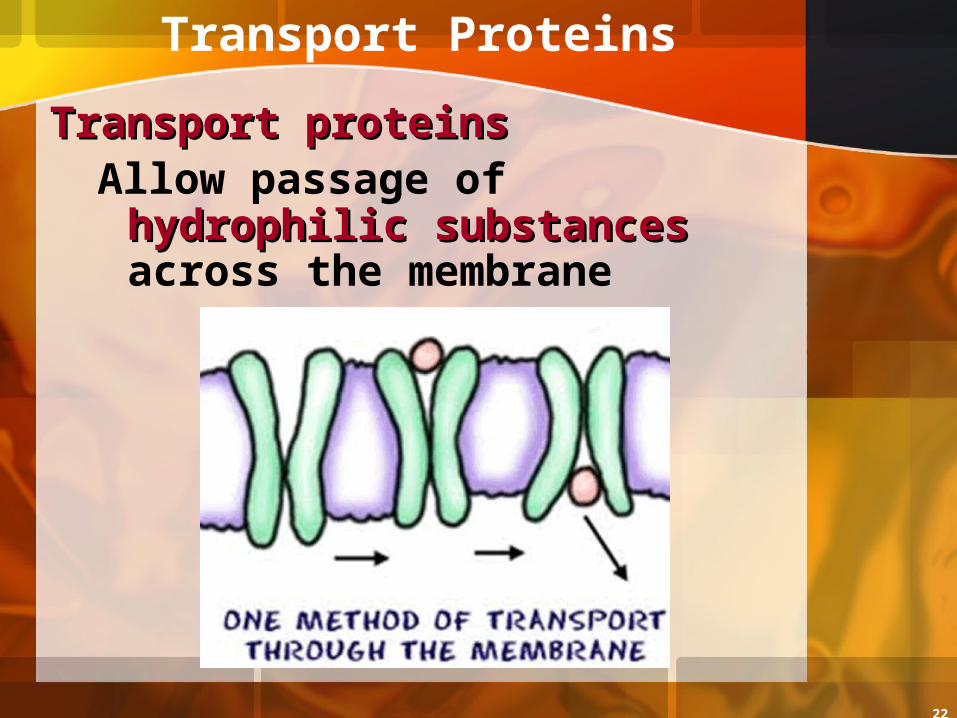
Transport Proteins
Transport proteinsTransport proteinsAllow passage of hydrophilic hydrophilic
substancessubstances across the membrane
23
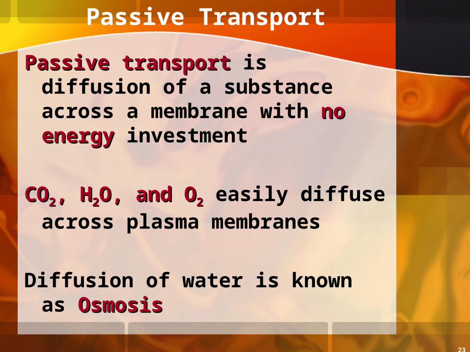
Passive Transport
Passive transportPassive transport is diffusion of a substance across a membrane with no energyno energy investment
COCO22, H, H22O, and OO, and O22 easily diffuse across plasma membranes
Diffusion of water is known as OsmosisOsmosis
24
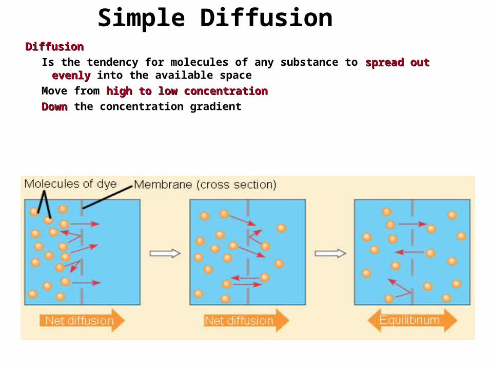
Simple DiffusionDiffusionDiffusion
Is the tendency for molecules of any substance to spread out evenlyspread out evenly into the available space
Move from high to low concentrationhigh to low concentration
DownDown the concentration gradient
25
Effects of Osmosis on Water Balance
OsmosisOsmosisIs the movement of water
across a semipermeable membrane
Is affected affected by theby the concentration gradient of concentration gradient of dissolved substances dissolved substances called thecalled the solution’ssolution’s tonicitytonicity
26
Water Balance of Cells Without WallsTonicity
Is the ability of a solution to cause a cell to gain or lose water
Has a great impact on cells impact on cells without wallswithout walls
27
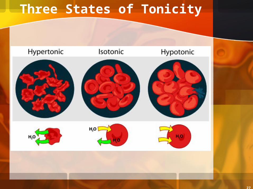
Three States of Tonicity
28
Isotonic Solutions
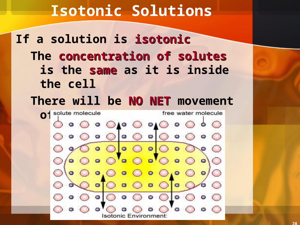
If a solution is isotonicisotonicThe concentration of solutesconcentration of solutes is
the samesame as it is inside the cellThere will be NO NETNO NET movement
of WATER
29
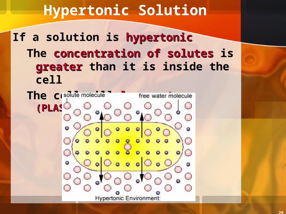
Hypertonic Solution
If a solution is hypertonichypertonicThe concentration of solutesconcentration of solutes is
greatergreater than it is inside the cellThe cell will lose water lose water
(PLASMOLYSIS)(PLASMOLYSIS)
30
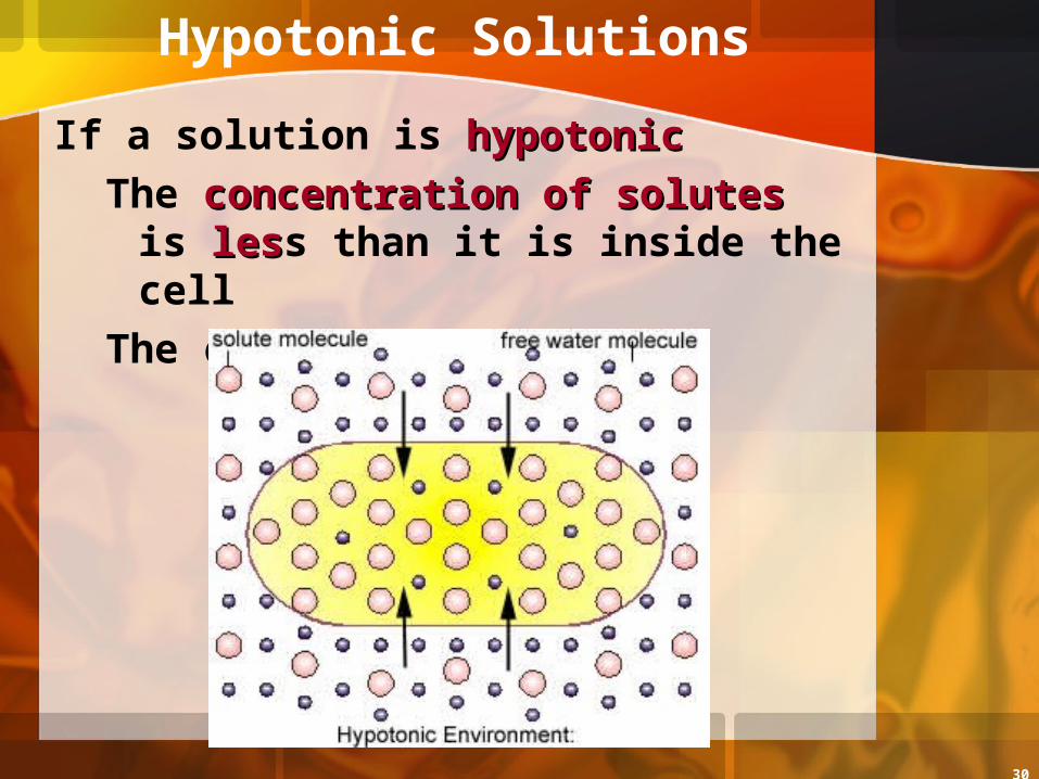
Hypotonic Solutions
If a solution is hypotonichypotonicThe concentration of solutesconcentration of solutes is
lesless than it is inside the cellThe cell will gain watergain water
31
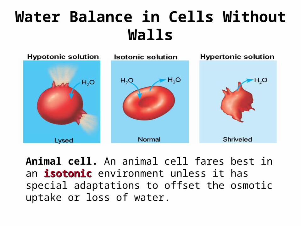
Water Balance in Cells Without Walls
Animal cell. An animal cell fares best in an isotonic isotonic environment unless it has special adaptations to offset the osmotic uptake or loss of water.
32
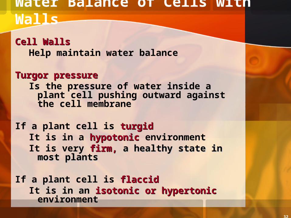
Water Balance of Cells with Walls
Cell WallsCell WallsHelp maintain water balance
Turgor pressureTurgor pressureIs the pressure of water inside a plant
cell pushing outward against the cell membrane
If a plant cell is turgidturgidIt is in a hypotonichypotonic environmentIt is very firm, firm, a healthy state in most a healthy state in most
plantsplants
If a plant cell is flaccidflaccidIt is in an isotonic or hypertonicisotonic or hypertonic
environment
33
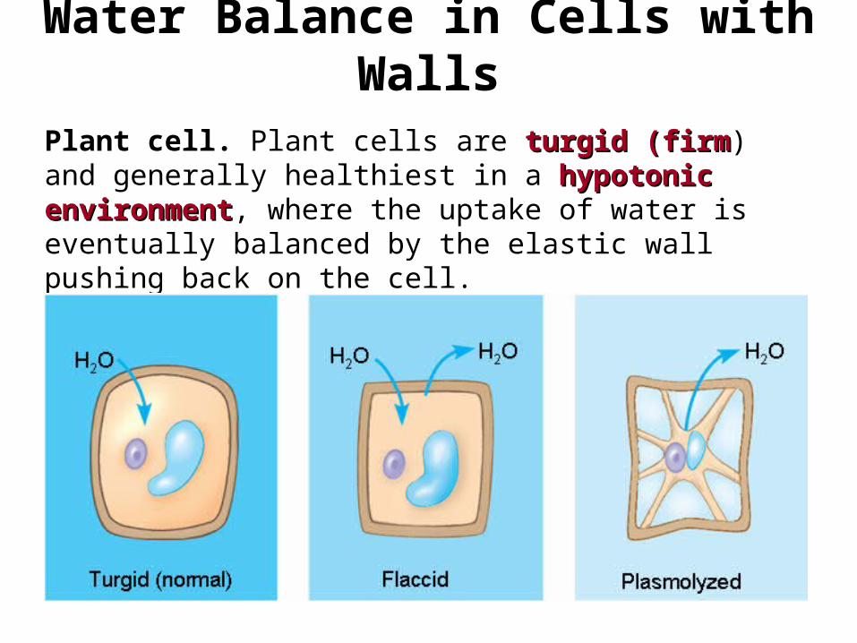
Water Balance in Cells with Walls
Plant cell. Plant cells are turgid (firmturgid (firm) and generally healthiest in a hypotonic environmenthypotonic environment, where the uptake of water is eventually balanced by the elastic wall pushing back on the cell.
34
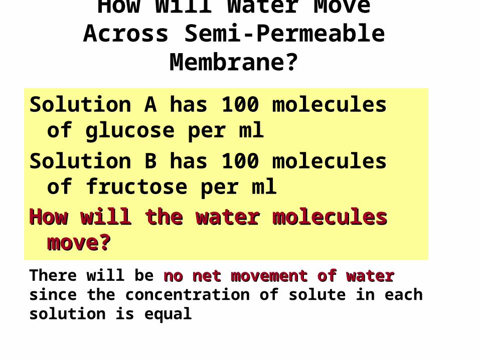
How Will Water Move Across Semi-Permeable
Membrane?
Solution A has 100 molecules of glucose per ml
Solution B has 100 molecules of fructose per ml
How will the water molecules How will the water molecules move?move?
There will be no net movement of waterno net movement of water since the concentration of solute in each solution is equal
35
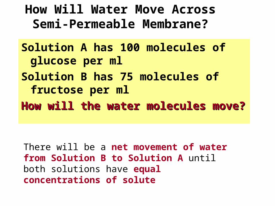
How Will Water Move Across Semi-Permeable Membrane?
Solution A has 100 molecules of glucose per ml
Solution B has 75 molecules of fructose per ml
How will the water molecules How will the water molecules move?move?
There will be a net movement of water from Solution B to Solution A until both solutions have equal concentrations of solute
36
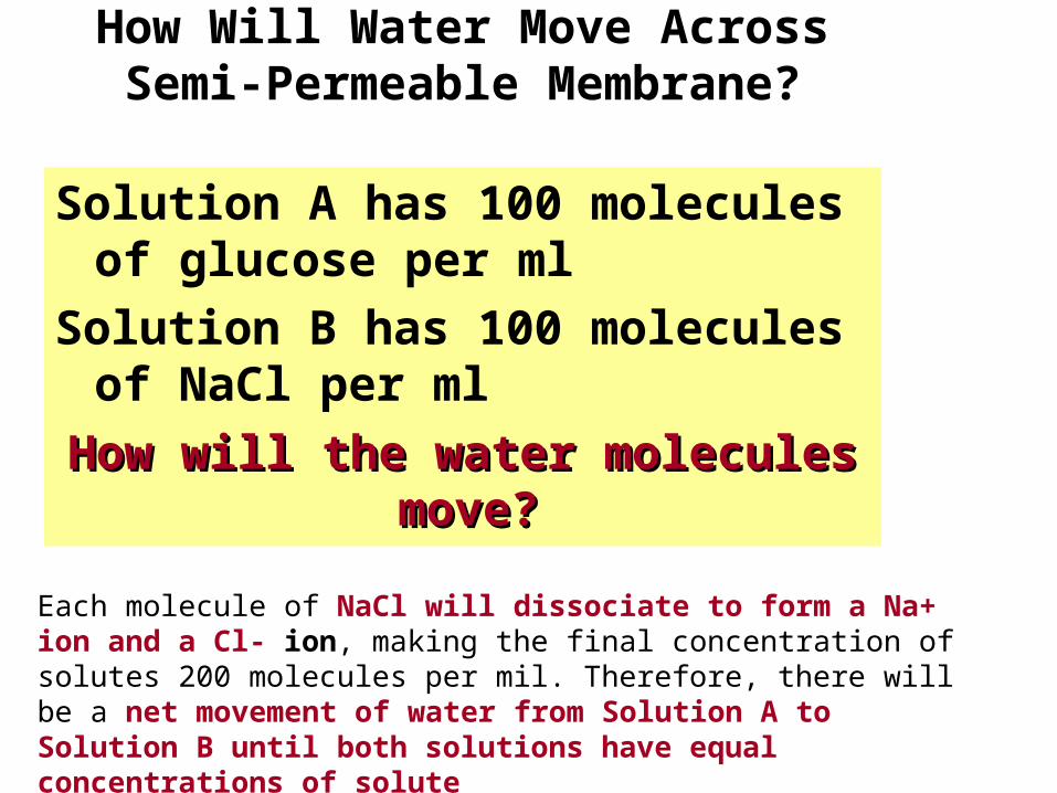
How Will Water Move Across Semi-Permeable Membrane?
Solution A has 100 molecules of glucose per ml
Solution B has 100 molecules of NaCl per ml
How will the water molecules How will the water molecules move?move?
Each molecule of NaCl will dissociate to form a Na+ ion and a Cl- ion, making the final concentration of solutes 200 molecules per mil. Therefore, there will be a net movement of water from Solution A to Solution B until both solutions have equal concentrations of solute
37
Facilitated Diffusion
Facilitated diffusionFacilitated diffusionIs a type of PassivePassive Transport Aided by
ProteinsProteins
In facilitated diffusion
Transport proteinsTransport proteins speed the movement of molecules across the plasma membrane
38
Facilitated Diffusion & Proteins
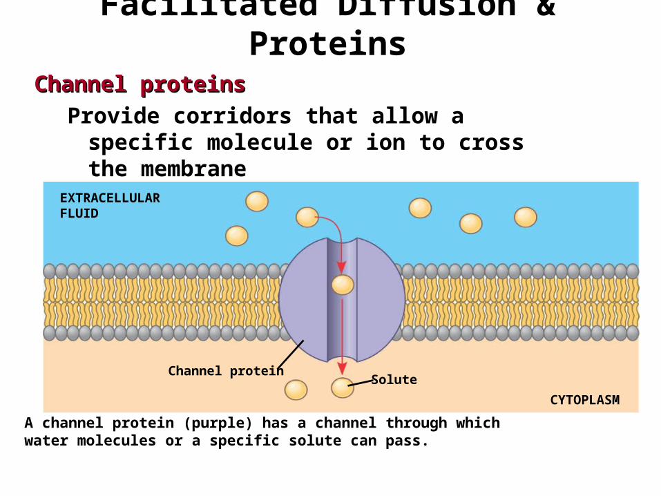
Channel proteinsChannel proteinsProvide corridors that allow a specific
molecule or ion to cross the membrane
EXTRACELLULARFLUID
Channel proteinSolute
CYTOPLASM
A channel protein (purple) has a channel through which water molecules or a specific solute can pass.
39
Facilitated Diffusion & Proteins
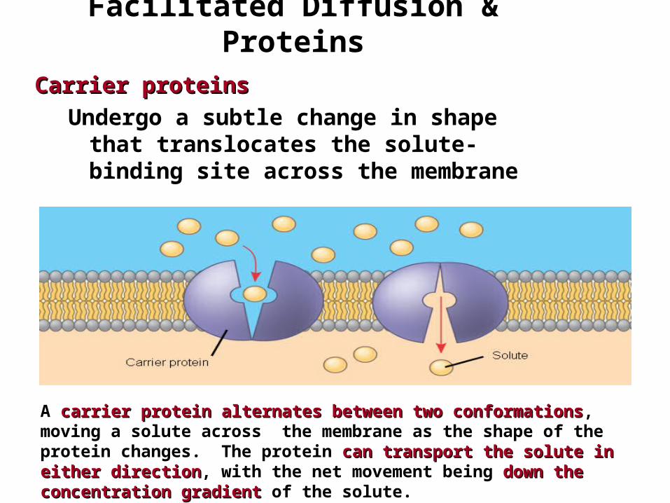
Carrier proteinsCarrier proteinsUndergo a subtle change in shape
that translocates the solute-binding site across the membrane
A carrier proteincarrier protein alternates between two conformationsalternates between two conformations, moving a solute across the membrane as the shape of the protein changes. The protein can transport the solute in either directioncan transport the solute in either direction, with the net movement being down the concentration gradientdown the concentration gradient of the solute.
40
Active Transport
Active transportUses energyUses energy to move solutes againstagainst their concentration gradients
Requires energy, usually in the form of ATPATP
41
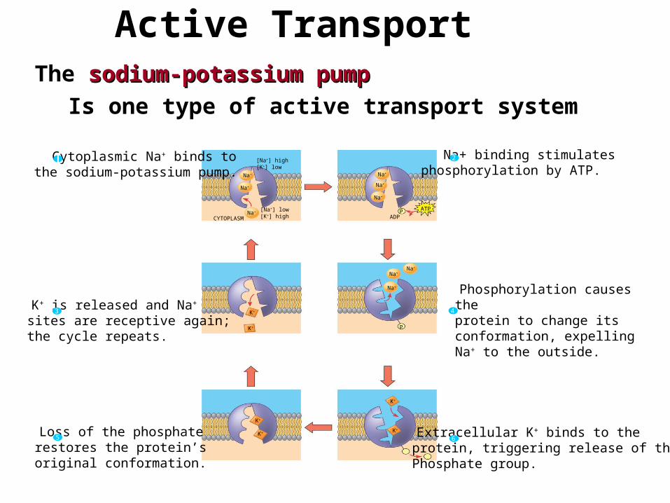
The sodium-potassium pumpsodium-potassium pumpIs one type of active transport system
Active Transport
PP i
EXTRACELLULARFLUID
Na+ binding stimulatesphosphorylation by ATP.
2
Na+
Cytoplasmic Na+ binds tothe sodium-potassium pump.1
K+ is released and Na+
sites are receptive again; the cycle repeats.
3
Phosphorylation causes the protein to change its conformation, expelling Na+ to the outside.
4
Extracellular K+ binds to the protein, triggering release of the Phosphate group.
6 Loss of the phosphaterestores the protein’s original conformation.
5
CYTOPLASM
[Na+] low[K+] high
Na+
Na+
Na+
Na+
Na+
P ATP
Na+
Na+
Na+
P
ADP
K+
K+
K+
K+ K+
K+
[Na+] high[K+] low
42
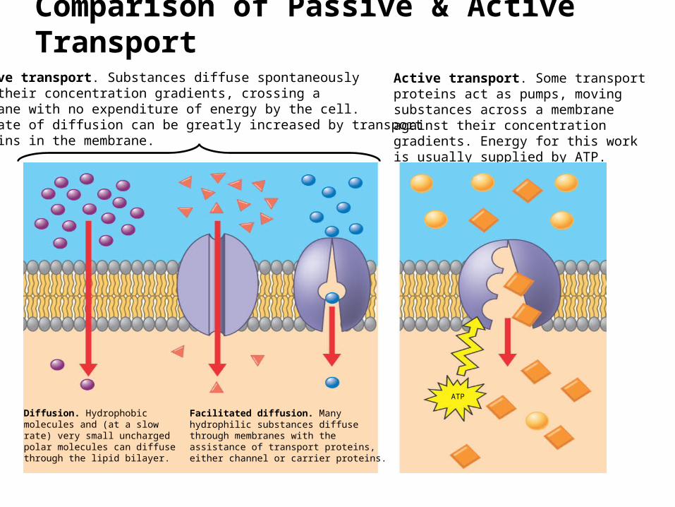
Comparison of Passive & Active Transport
Passive transport. Substances diffuse spontaneously down their concentration gradients, crossing a membrane with no expenditure of energy by the cell. The rate of diffusion can be greatly increased by transport proteins in the membrane.
Active transport. Some transport proteins act as pumps, moving substances across a membrane against their concentration gradients. Energy for this work is usually supplied by ATP.
Diffusion. Hydrophobicmolecules and (at a slow rate) very small uncharged polar molecules can diffuse through the lipid bilayer.
Facilitated diffusion. Many hydrophilic substances diffuse through membranes with the assistance of transport proteins,either channel or carrier proteins.
ATP
43
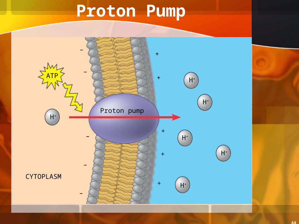
Maintenance of Membrane Potential by Ion Pumps
Membrane potentialMembrane potentialIs the voltage difference across a membrane
An electrochemical gradientelectrochemical gradientIs caused by the concentration electrical gradient of ions
across a membrane
An electrogenic pumpelectrogenic pumpIs a transport protein that generates the voltage across a
membrane
44
Proton Pump
EXTRACELLULARFLUID
+
H+
H+
H+
H+
H+
H+Proton pump
ATP
CYTOPLASM
+
+
+
+
–
–
–
–
–
+
45
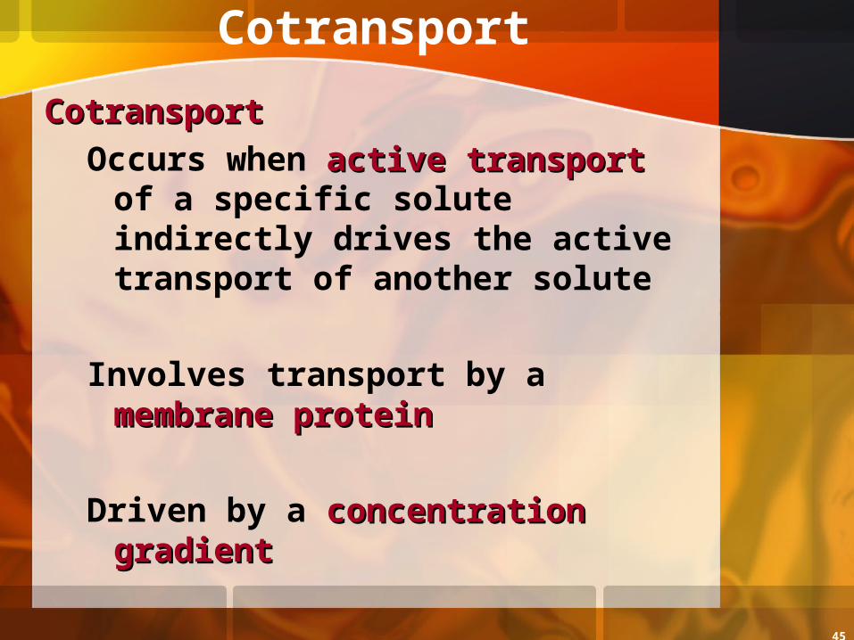
Cotransport
CotransportCotransportOccurs when active transportactive transport of
a specific solute indirectly drives the active transport of another solute
Involves transport by a membrane protein membrane protein
Driven by a concentration concentration gradientgradient
46
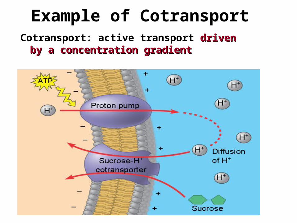
Example of CotransportCotransport: active transport driven driven
by a concentration gradientby a concentration gradient
47
Bulk Transport
Bulk transport across the plasma membrane occurs by exocytosis and endocytosisexocytosis and endocytosis
Large proteinsCross the membrane by
different mechanisms
48
Exocytosis & Endocytosis
In exocytosisexocytosis
Transport vesiclesTransport vesicles migrate to the plasma membrane, fuse with it, and release their contents
In endocytosisendocytosisThe cell takes in macromolecules
by forming new vesicles from forming new vesicles from the plasma membranethe plasma membrane
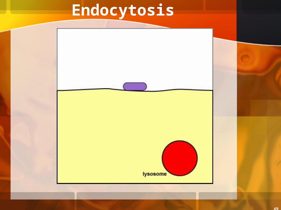
49
Endocytosis
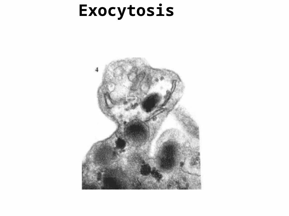
50
Exocytosis
51
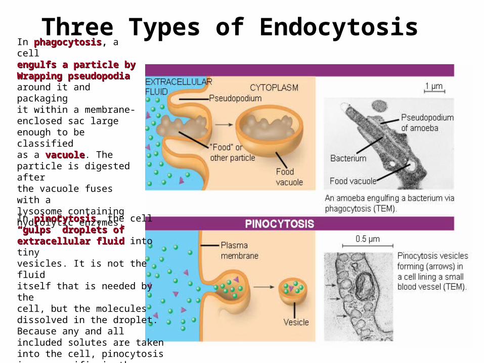
In phagocytosisphagocytosis, a cellengulfs a particle by engulfs a particle by Wrapping pseudopodiaWrapping pseudopodia around it and packaging it within a membrane-enclosed sac large enough to be classified as a vacuole vacuole. The particle is digested after the vacuole fuses with a lysosome containing hydrolytic enzymes.
Three Types of Endocytosis
PHAGOCYTOSIS
In pinocytosispinocytosis, the cell ““gulps” droplets of gulps” droplets of extracellular fluidextracellular fluid into tinyvesicles. It is not the fluiditself that is needed by the cell, but the molecules dissolved in the droplet. Because any and all included solutes are taken into the cell, pinocytosisis nonspecific in the substances it transports.
52
0.25 µm
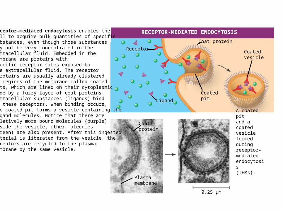
RECEPTOR-MEDIATED ENDOCYTOSIS
Receptor
Ligand
Coat protein
Coatedpit
Coatedvesicle
A coated pitand a coatedvesicle formedduringreceptor-mediatedendocytosis(TEMs).
Plasmamembrane
Coatprotein
Receptor-mediated endocytosis enables the cell to acquire bulk quantities of specific substances, even though those substances may not be very concentrated in the extracellular fluid. Embedded in the membrane are proteins with specific receptor sites exposed to the extracellular fluid. The receptor proteins are usually already clustered in regions of the membrane called coated pits, which are lined on their cytoplasmic side by a fuzzy layer of coat proteins. Extracellular substances (ligands) bind to these receptors. When binding occurs, the coated pit forms a vesicle containing the ligand molecules. Notice that there are relatively more bound molecules (purple) inside the vesicle, other molecules (green) are also present. After this ingested material is liberated from the vesicle, the receptors are recycled to the plasma membrane by the same vesicle.

53