1 Lipoproteins Seminar No. 2 - Chapter 13 -. 2 Lipids of Blood Plasma LipidPlasma concentration...
45
1 Lipoproteins Seminar No. 2 - Chapter 13 -
-
Upload
sandra-lawson -
Category
Documents
-
view
219 -
download
0
Transcript of 1 Lipoproteins Seminar No. 2 - Chapter 13 -. 2 Lipids of Blood Plasma LipidPlasma concentration...

1
LipoproteinsSeminar No. 2
- Chapter 13 -
2
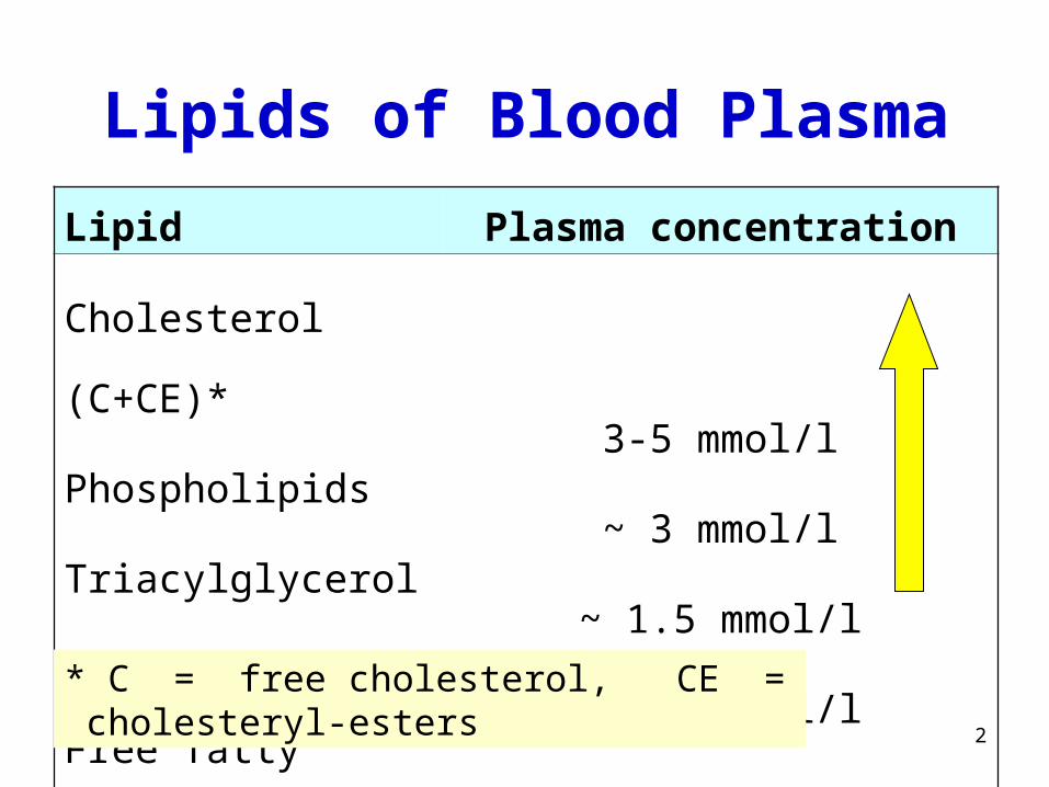
Lipids of Blood Plasma
Lipid Plasma concentration
Cholesterol (C+CE)*
Phospholipids
Triacylglycerols
Free fatty acids
3-5 mmol/l
~ 3 mmol/l
~ 1.5 mmol/l
~ 0.5 mmol/l
* C = free cholesterol, CE = cholesteryl-esters

3
Q. 1 (p. 78)
Which natural tensides participate in micelle formation
in intestine (GIT) ?
What is a tenside?
4
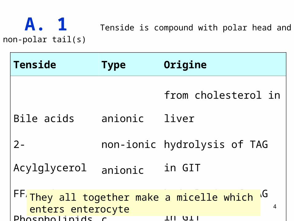
A. 1 Tenside is compound with polar head and non-polar tail(s)
Tenside Type Origine
Bile acids
2-Acylglycerol
FFA anions
Phospholipids
anionic
non-ionic
anionic
amphoteric
from cholesterol in liver
hydrolysis of TAG in GIT
hydrolysis of TAG in GIT
food
They all together make a micelle which enters enterocyte
5
Q. 2 (p. 78)
In which form are FFA transported in blood?
6
A. 2
FFA are non-polar species, insoluble in water.
They are bound to albumin, which is the main
transport protein in plasma.
7
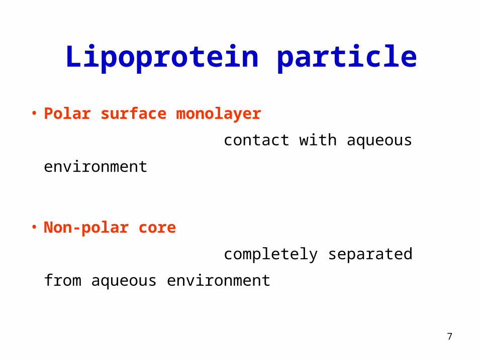
Lipoprotein particle
• Polar surface monolayer
contact with aqueous environment
• Non-polar core
completely separated from aqueous environment
8
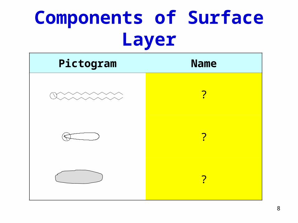
Components of Surface Layer
Pictogram Name
?
?
?
9
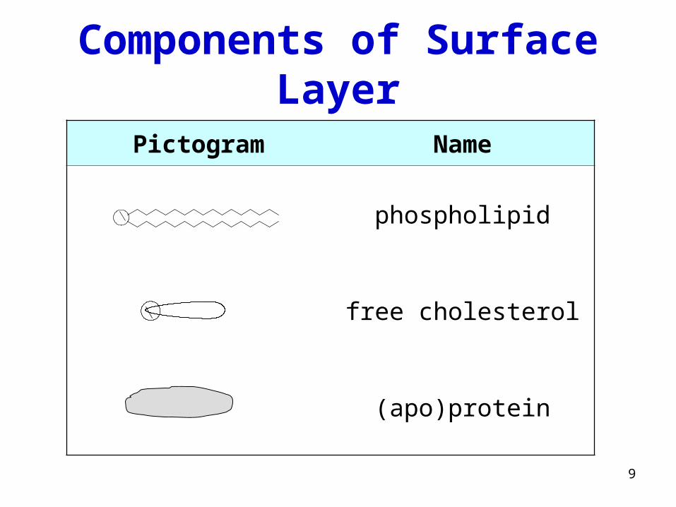
Components of Surface Layer
Pictogram Name
phospholipid
free cholesterol
(apo)protein
10
Draw a general structure of phospholipid
or
phosphatidylcholine

11
Structure of phospholipid
C
O
CH
CH2O
O
C
O
CH2 O P
O
O
O CH2 CH2 N
CH3
CH3
CH3
phosphatidylcholine (lecithine)
12
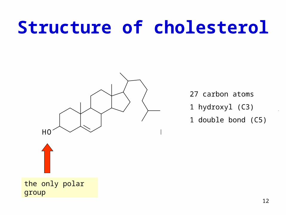
Structure of cholesterol
HO HO
27 carbon atoms
1 hydroxyl (C3)
1 double bond (C5)
the only polar group
13
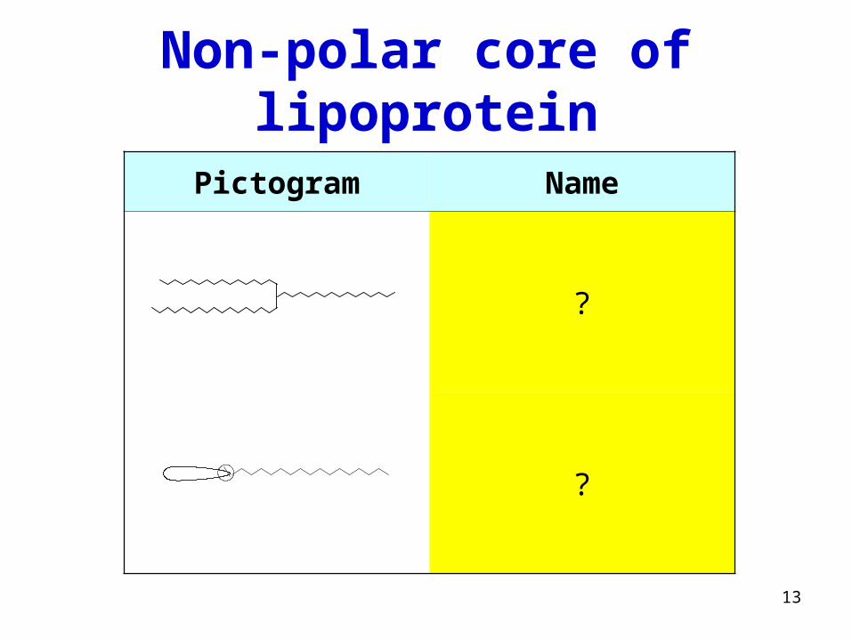
Non-polar core of lipoprotein
Pictogram Name
?
?
14
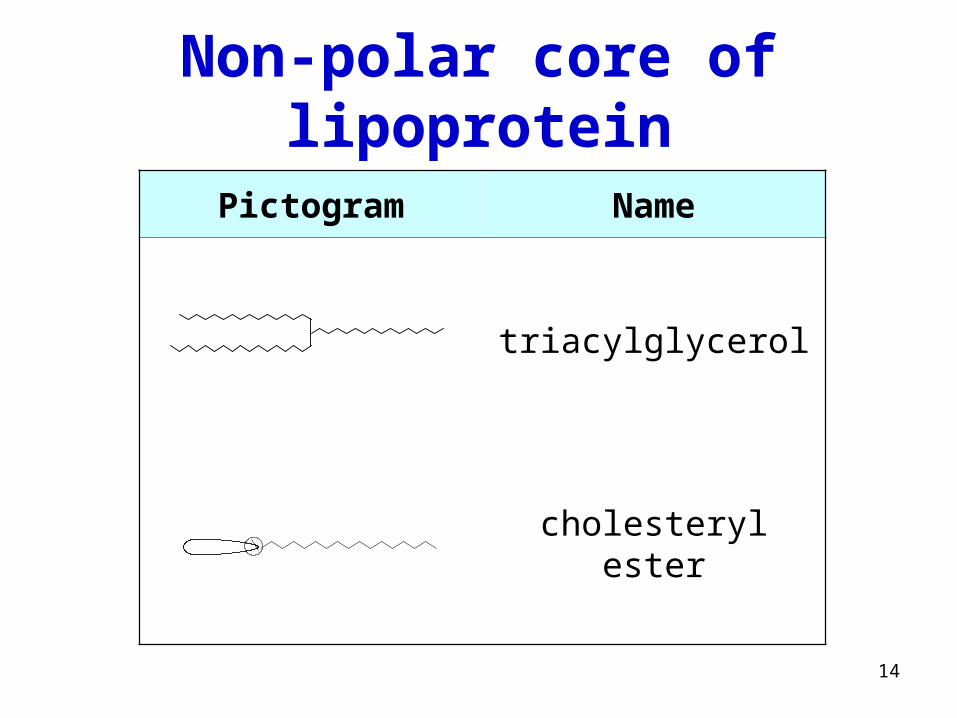
Non-polar core of lipoprotein
Pictogram Name
triacylglycerol
cholesteryl ester
15
Draw a structure of TAG

16
CH2
CH
O C
O
CH2
OC
O
O
C
O
triacylglycerol (TAG)
17
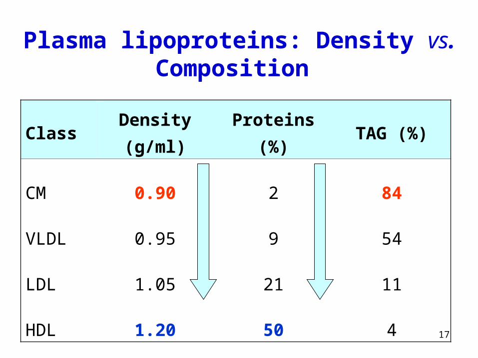
Plasma lipoproteins: Density vs. Composition
Class Density (g/ml) Proteins (%) TAG (%)
CM
VLDL
LDL
HDL
0.90
0.95
1.05
1.20
2
9
21
50
84
54
11
4
18
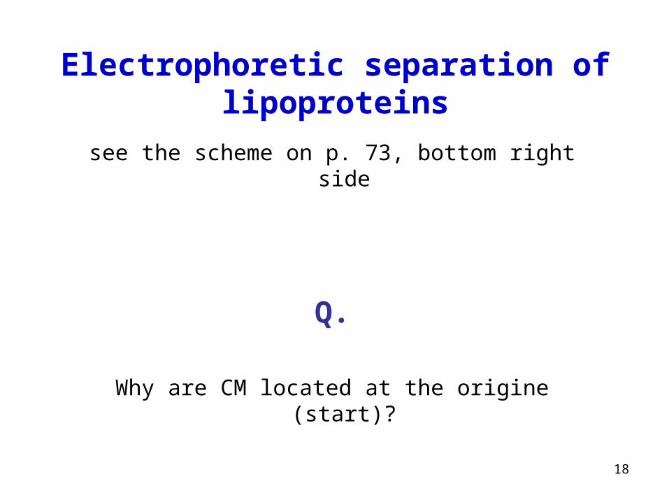
Electrophoretic separation of lipoproteins
see the scheme on p. 73, bottom right side
Q.
Why are CM located at the origine (start)?
19
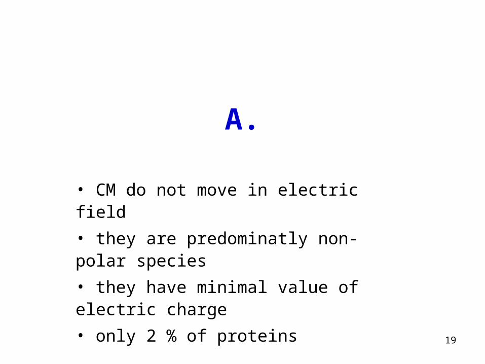
A.
• CM do not move in electric field
• they are predominatly non-polar species
• they have minimal value of electric charge
• only 2 % of proteins
20
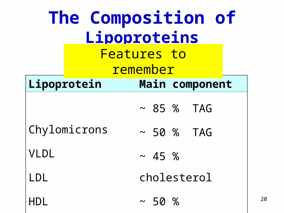
The Composition of Lipoproteins
Lipoprotein Main component
Chylomicrons
VLDL
LDL
HDL
~ 85 % TAG
~ 50 % TAG
~ 45 % cholesterol
~ 50 % proteins
Features to remember
21
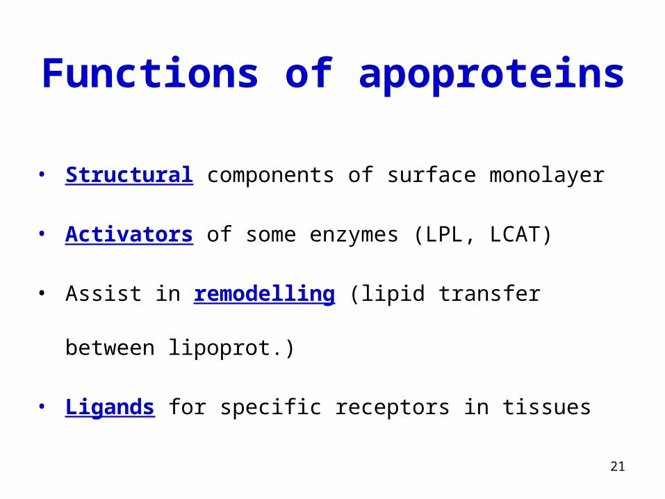
Functions of apoproteins
• Structural components of surface monolayer
• Activators of some enzymes (LPL, LCAT)
• Assist in remodelling (lipid transfer between lipoprot.)
• Ligands for specific receptors in tissues
22
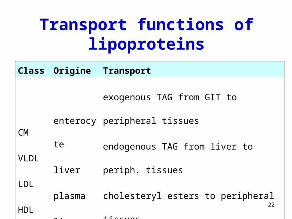
Transport functions of lipoproteins
Class Origine Transport
CM
VLDL
LDL
HDL
enterocyte
liver
plasma
liver
exogenous TAG from GIT to peripheral tissues
endogenous TAG from liver to periph. tissues
cholesteryl esters to peripheral tissues
cholesterol from tissues to liver
23
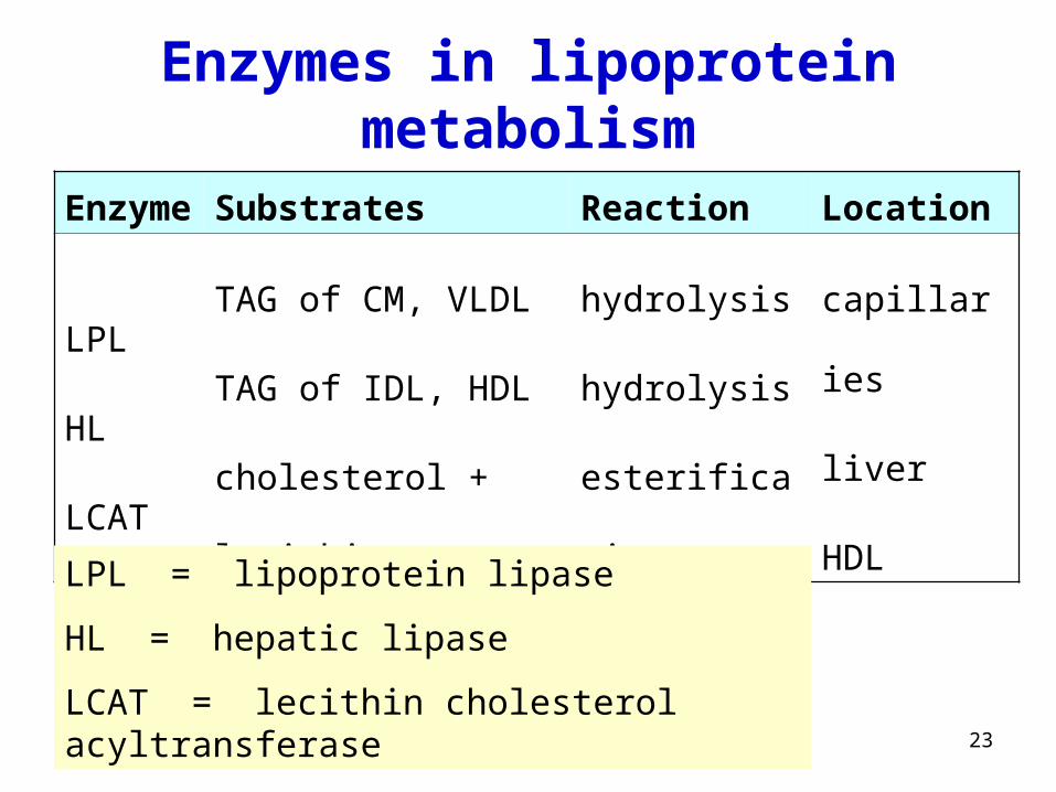
Enzymes in lipoprotein metabolism
Enzyme Substrates Reaction Location
LPL
HL
LCAT
TAG of CM, VLDL
TAG of IDL, HDL
cholesterol + lecithin
hydrolysis
hydrolysis
esterification
capillaries
liver
HDL
LPL = lipoprotein lipase
HL = hepatic lipase
LCAT = lecithin cholesterol acyltransferase
24
Write the equation of reaction
catalyzed by LPL
25
CH2
CH
O C
O
CH2
OC
O
O
C
O
3 H2O H+
CH2
CH
OH
CH2 OH
HO HOOC3
LPL
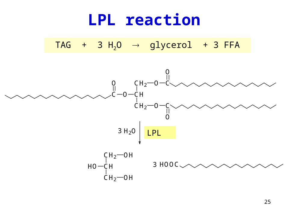
LPL reaction
TAG + 3 H2O glycerol + 3 FFA
26
Q.
What is acyl?
Write the equation of reaction
catalyzed by LCAT
(lecithin cholesterol acyltransferase)
27
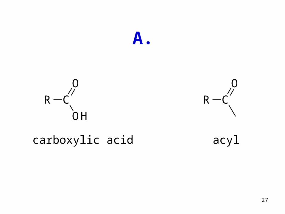
OHCR
OCR
O
carboxylic acid acyl
A.
28
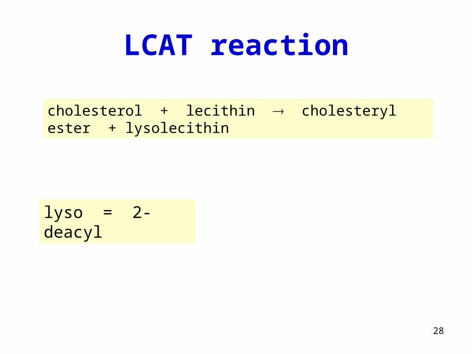
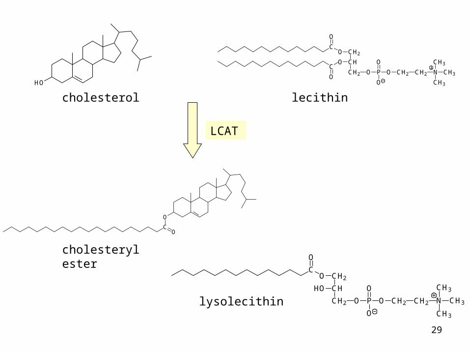
LCAT reaction
cholesterol + lecithin cholesteryl ester + lysolecithin
lyso = 2-deacyl
29
HO
C
O
CH
CH2O
O
C
O
CH2 O P
O
O
O CH2 CH2 N
CH3
CH3
CH3
O
CO
CH
CH2O
HO
C
O
CH2 O P
O
O
O CH2 CH2 N
CH3
CH3
CH3
cholesterol
cholesteryl ester
lecithin
lysolecithin
LCAT
30
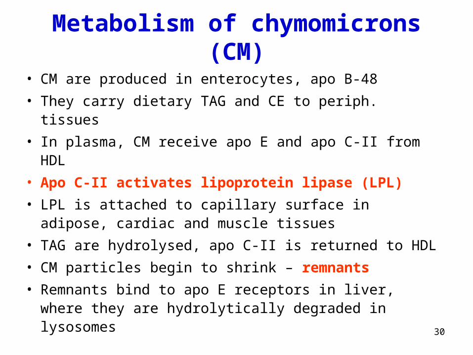
Metabolism of chymomicrons (CM)
• CM are produced in enterocytes, apo B-48
• They carry dietary TAG and CE to periph. tissues
• In plasma, CM receive apo E and apo C-II from HDL
• Apo C-II activates lipoprotein lipase (LPL)
• LPL is attached to capillary surface in adipose, cardiac and muscle tissues
• TAG are hydrolysed, apo C-II is returned to HDL
• CM particles begin to shrink – remnants
• Remnants bind to apo E receptors in liver, where they are hydrolytically degraded in lysosomes
31
Q.5 (p. 78)
What will be result of deficient synthesis of apo B-48?
32
A.5
No CM will be produced, dietary fat remains in stool (steatorrhoea)
33
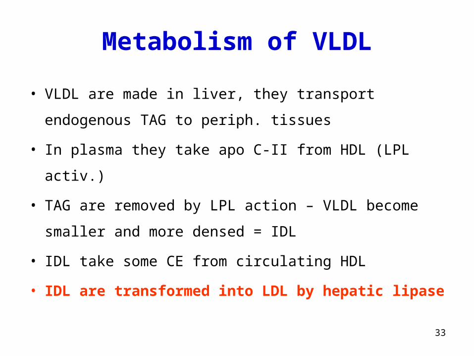
• VLDL are made in liver, they transport endogenous TAG to
periph. tissues
• In plasma they take apo C-II from HDL (LPL activ.)
• TAG are removed by LPL action – VLDL become smaller
and more densed = IDL
• IDL take some CE from circulating HDL
• IDL are transformed into LDL by hepatic lipase
Metabolism of VLDL
34
Q. (p. 76)
Name some dietary factors which may affect the
synthesis of VLDL in the liver.
35
A.
Food rich in lipids (fat) and saccharides (sugars)

36
Q. (p. 76)
How are utilized fatty acids released by LPL?
37
A.
Depending on energy status in tissues FFA are:
• either utilized for energy
(β-oxidation acetyl-CoA CAC)
• or substrates for TAG synthesis (making energy reserves)
38
Three pathways of LDL
1. LDL provide cholesterol to peripheral tissues via LDL receptors
2. The rest of LDL is taken up by liver and degraded
3. Small amount of LDL (chemically modified by oxidative stress)
enters to some cells (endothelial) by non-specific endocytosis
and alters them to „foam cells“
39
• HDL particles are made in liver
• Nascent HDL are disc-shaped (bilayer of PL + proteins)
• HDL take free cholesterol (C) from cell membranes
• Once C is taken up, it is esterified by LCAT
• After this process HDL becomes spherical
• Spherical HDL are taken up by liver and CE are degraded
Metabolism of HDL
40
Cellular uptake of LDL
• LDL receptors are in clathrin-coated pits
• After binding, LDL+receptor are internalized by endocytosis
• Vesicle loses its clathrin coat and becomes endosome
• Receptor is removed and recycled
• LDL is hydrolyzed after fusing with lysosome
• Free cholesterol is released to make cholesterol pool
41
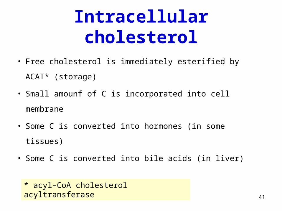
Intracellular cholesterol
• Free cholesterol is immediately esterified by ACAT*
(storage)
• Small amounf of C is incorporated into cell membrane
• Some C is converted into hormones (in some tissues)
• Some C is converted into bile acids (in liver)
* acyl-CoA cholesterol acyltransferase
42
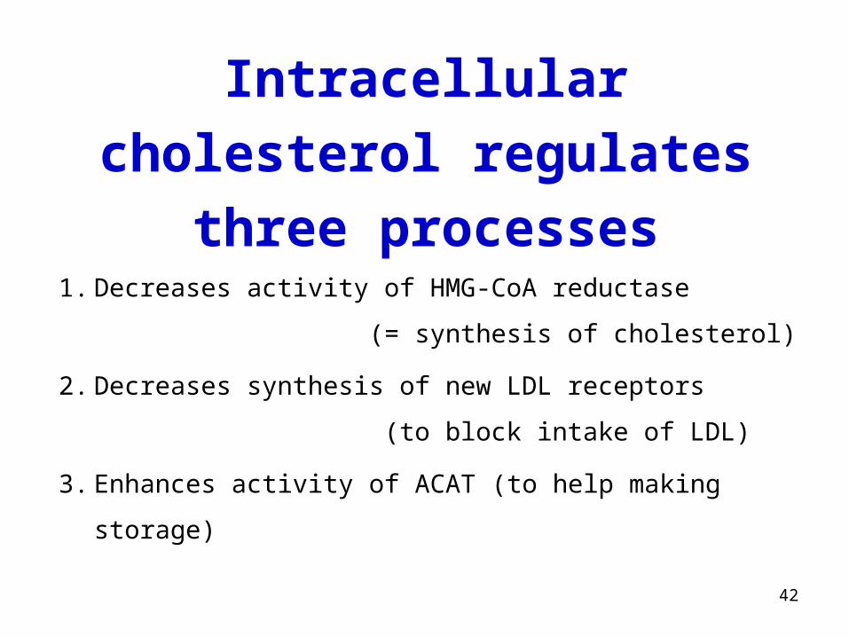
1. Decreases activity of HMG-CoA reductase
(= synthesis of cholesterol)
2. Decreases synthesis of new LDL receptors
(to block intake of LDL)
3. Enhances activity of ACAT (to help making storage)
Intracellular cholesterol
regulates three processes
43
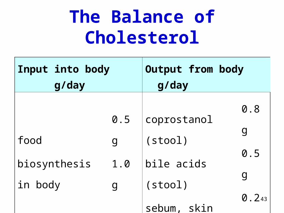
The Balance of Cholesterol
Input into body g/day Output from body g/day
food
biosynthesis in body
Total:
0.5 g
1.0 g
1.5 g
coprostanol (stool)
bile acids (stool)
sebum, skin etc.
Total:
0.8 g
0.5 g
0.2 g
1.5 g
44
Which food is the main source
of cholesterol?
Q.
45
A.
• only animal fats (including fish)
lard, butter, bacon, egg yolk, mayonnaise, fat meat, fat cheese
• plant oils and margarines are cholesterol free