1. Introduction - Freie Universität · Transmucosal routes of drug delivery ... Parenteral...
50
___________________________________________________________________________ 1 1. Introduction The aim of this work was the development and characterization of in situ gelling nasal inserts, a new, bioadhesive, solid dosage form for the prolonged systemic drug delivery via the nasal route. In this chapter conventional and alternative mucosal routes for systemic drug delivery are compared (section 1.1) and a basic introduction to the anatomy and physiology of the human nose (section 1.2) and to nasal immunology (section 1.4.1) is given. Further on, the concept of bioadhesion will be introduced (section 1.3) as a major approach to increase the nasal residence time of drug delivery systems. The current state of research concerning nasal drug delivery systems and nasal immunization approaches (sections 1.5 and 1.4.2) is presented. Finally, the objectives of this work are laid out. 1.1. Transmucosal routes of drug delivery Drugs for systemic medication are administered traditionally and routinely by oral and by parenteral routes. Although generally convenient, both routes have a number of disadvantages, especially for the delivery of peptides and proteins, a class of drugs that has been rapidly emerging over the last decades (Zhou and Li Wan Po, 1991a). Oral administration results in the exposure of the drug to the harsh environment of the gastrointestinal tract and thus to potential chemical and enzymatic degradation (Langguth et al., 1997). After gastrointestinal absorption the drug has to pass the liver, where, dependent on the nature of the drug, extensive first pass metabolism can take place with subsequent rapid clearance from the blood stream (Lalka et al., 1993; Taki et al., 1998). Low permeability across the gastrointestinal mucosa is also often encountered for macromolecular drugs (Yamamoto et al., 2001; Pauletti et al., 1997). Parenteral administration avoids drug degradation in the gastrointestinal tract and hepatic first pass clearance but due to pain or discomfort during injection, patient compliance is poor, particularly if multiple daily injections are required as e.g. in the insulin therapy (Hinchcliffe and Illum, 1999). Also injection related side effects like tissue necrosis and thrombophlebitis lead to low patient
-
Upload
truongtruc -
Category
Documents
-
view
216 -
download
2
Transcript of 1. Introduction - Freie Universität · Transmucosal routes of drug delivery ... Parenteral...

___________________________________________________________________________
1
1. Introduction
The aim of this work was the development and characterization of in situ gelling nasal inserts,
a new, bioadhesive, solid dosage form for the prolonged systemic drug delivery via the nasal
route. In this chapter conventional and alternative mucosal routes for systemic drug delivery
are compared (section 1.1) and a basic introduction to the anatomy and physiology of the
human nose (section 1.2) and to nasal immunology (section 1.4.1) is given. Further on, the
concept of bioadhesion will be introduced (section 1.3) as a major approach to increase the
nasal residence time of drug delivery systems. The current state of research concerning nasal
drug delivery systems and nasal immunization approaches (sections 1.5 and 1.4.2) is
presented. Finally, the objectives of this work are laid out.
1.1. Transmucosal routes of drug delivery
Drugs for systemic medication are administered traditionally and routinely by oral and by
parenteral routes. Although generally convenient, both routes have a number of
disadvantages, especially for the delivery of peptides and proteins, a class of drugs that has
been rapidly emerging over the last decades (Zhou and Li Wan Po, 1991a). Oral
administration results in the exposure of the drug to the harsh environment of the
gastrointestinal tract and thus to potential chemical and enzymatic degradation (Langguth et
al., 1997). After gastrointestinal absorption the drug has to pass the liver, where, dependent on
the nature of the drug, extensive first pass metabolism can take place with subsequent rapid
clearance from the blood stream (Lalka et al., 1993; Taki et al., 1998). Low permeability
across the gastrointestinal mucosa is also often encountered for macromolecular drugs
(Yamamoto et al., 2001; Pauletti et al., 1997). Parenteral administration avoids drug
degradation in the gastrointestinal tract and hepatic first pass clearance but due to pain or
discomfort during injection, patient compliance is poor, particularly if multiple daily
injections are required as e.g. in the insulin therapy (Hinchcliffe and Illum, 1999). Also
injection related side effects like tissue necrosis and thrombophlebitis lead to low patient
1. Introduction ___________________________________________________________________________
2
acceptability (Zhou, 1994). In addition, administration by injection requires trained personnel
which adds to the relatively high costs of parenteral medication.
Figure 1. 1 Various potential mucosal pathways for systemic delivery of therapeutic
agents, which bypass the hepatic first pass clearance associated with oral
administration. The venous drainage system involved in the systemic delivery
of therapeutic agents following the transmucosal permeation is illustrated
(Zhou and Li Wan Po, 1991b).
1. Introduction ___________________________________________________________________________
3
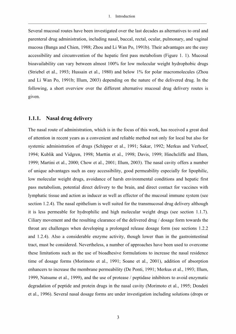
Several mucosal routes have been investigated over the last decades as alternatives to oral and
parenteral drug administration, including nasal, buccal, rectal, ocular, pulmonary, and vaginal
mucosa (Banga and Chien, 1988; Zhou and Li Wan Po, 1991b). Their advantages are the easy
accessibility and circumvention of the hepatic first pass metabolism (Figure 1. 1). Mucosal
bioavailability can vary between almost 100% for low molecular weight hydrophobic drugs
(Striebel et al., 1993; Hussain et al., 1980) and below 1% for polar macromolecules (Zhou
and Li Wan Po, 1991b; Illum, 2003) depending on the nature of the delivered drug. In the
following, a short overview over the different alternative mucosal drug delivery routes is
given.
1.1.1. Nasal drug delivery
The nasal route of administration, which is in the focus of this work, has received a great deal
of attention in recent years as a convenient and reliable method not only for local but also for
systemic administration of drugs (Schipper et al., 1991; Sakar, 1992; Merkus and Verhoef,
1994; Kublik and Vidgren, 1998; Marttin et al., 1998; Davis, 1999; Hinchcliffe and Illum,
1999; Martini et al., 2000; Chow et al., 2001; Illum, 2003). The nasal cavity offers a number
of unique advantages such as easy accessibility, good permeability especially for lipophilic,
low molecular weight drugs, avoidance of harsh environmental conditions and hepatic first
pass metabolism, potential direct delivery to the brain, and direct contact for vaccines with
lymphatic tissue and action as inducer as well as effector of the mucosal immune system (see
section 1.2.4). The nasal epithelium is well suited for the transmucosal drug delivery although
it is less permeable for hydrophilic and high molecular weight drugs (see section 1.1.7).
Ciliary movement and the resulting clearance of the delivered drug / dosage form towards the
throat are challenges when developing a prolonged release dosage form (see sections 1.2.2
and 1.2.4). Also a considerable enzyme activity, though lower than in the gastrointestinal
tract, must be considered. Nevertheless, a number of approaches have been used to overcome
these limitations such as the use of bioadhesive formulations to increase the nasal residence
time of dosage forms (Morimoto et al., 1991; Soane et al., 2001), addition of absorption
enhancers to increase the membrane permeability (De Ponti, 1991; Merkus et al., 1993; Illum,
1999, Natsume et al., 1999), and the use of protease / peptidase inhibitors to avoid enzymatic
degradation of peptide and protein drugs in the nasal cavity (Morimoto et al., 1995; Dondeti
et al., 1996). Several nasal dosage forms are under investigation including solutions (drops or

1. Introduction ___________________________________________________________________________
4
sprays), gels, suspensions and emulsions, liposomal preparations, powders and microspheres,
as well as inserts (see section 1.5).
1.1.2. Buccal drug delivery
The oral cavity is lined by a stratified squamous epithelium. The epithelium has a cornified
surface in regions subject to mechanical forces during mastication, which resembles that of
the upper epidermis in the skin. Non-keratinized epithelium occupies approximately 60% of
the total oral cavity including the buccal, lingual, and sublingual mucosa (Chien, 1995;
Hoogstraate and Wertz, 1998) and is of interest for systemic drug delivery. Although non-
keratinized, the buccal mucosa contains intercellular lipids which are responsible for its
physical barrier properties (Hoogstraate and Wertz, 1998; Shojaei, 1998), resulting in poor
permeability for larger drugs, especially for peptides and proteins (Junginger et al., 1999;
Veuillez et al., 2001). Transfer of peptides with molecular weights above 500 - 1000 Da
through buccal mucosa would require use of an absorption enhancer (Merkle et al., 1992).
Another limitation in buccal drug delivery is the mucosal enzyme activity, especially of
proteases (Bird et al., 2001; Veuillez et al., 2001; Walzer et al., 2002). The reduced retention
of the dosage form at the buccal surface due to constant washing with saliva can be overcome
by the use of bioadhesive formulations (Shojaei, 1998; Veuillez et al., 2001; Langoth et al.,
2003). The influence of food intake and mastication on the residence time of bioadhesive
buccal formulations is so far not clear. Thiolated polymers can be used simultaneously as
bioadhesive carrier and protease inhibitors (Langoth et al., 2003). The sublingual epithelium
is more permeable than the buccal one but more handicapped by the saliva washing and
constant mobility (Shojaei, 1998). It is therefore more suitable for immediate release
products.
Dosage forms for buccal drug delivery include tablets, patches, films, lozenges, sprays,
hydrogels, lollypops, chewing gums, powders, solutions (Hoogstraate and Wertz, 1998), a
freeze-dried sublingual dosage form (Vaugelade et al., 2001), wafers (Kalra et al., 2001), and
liposomal formulations (Veuillez et al., 2001).
1.1.3. Ocular drug delivery
Ocular delivery of drugs is typically for the treatment of ocular inflammation, corneal
wounds, and glaucoma. In addition, this route has been investigated for the systemic delivery

1. Introduction ___________________________________________________________________________
5
of peptides and proteins. Already in 1931, ocular administration of insulin produced sustained
lowering of the blood glucose level in proportion to the dose instilled (Christie and Hanzal,
1931). However, today it is known that the majority of the systemic drug absorption after
ocular instillation takes place across the nasal mucosa after drainage via the nasolachrymal
duct (Lee et al., 2002). Some absorption occurs also from the conjunctival sac. Drug
absorption via the cornea is relatively low due to the lipophilicity of the corneal epithelium,
dilution of the drug in the tear fluid (reflex tearing and reflex blinking) and drug binding to
proteins in tear fluid (Zhou and Li Wan Po, 1991b). In addition, the corneal and conjunctival
tissues act also as enzymatic barrier, which contain e.g. proteases (Zhou and Li Wan Po,
1991b). Therefore, the eye offers no additional advantage over the nose as systemic drug
delivery site and is of higher interest only for drug administration for local (ophthalmic)
therapy. However, also the local drug delivery is restricted by the dynamics of the lachrymal
drainage system, which is the natural defense mechanism of the eye. This system introduces
tear fluid to the eye and rapidly drains the fluid together with any instilled formulation from
the precorneal area to the nasal cavity and throat. The high elimination rate results in short
duration of contact of the drug with its absorption sites and consequently in a low local
bioavailability. Increased ocular bioavailability can be achieved by the use of viscosity
enhanced aqueous eye drops, suspensions, oily drops and unguents, mucoadhesive ocular
delivery systems such as solutions and microparticle suspensions, in-situ gelling systems
triggered by pH, temperature, or ions, colloidal delivery systems such as liposomes and
nanoparticles, and ocular inserts (Le Bourlais et al., 1995). Ocular inserts can be divided into
non-erodible (Chetoni et al., 1998; Kawakami et al., 2001) and erodible inserts. Erodible
ocular inserts, which do not need to be removed mechanically from the eye, have been
prepared by powder compression from poly(ethylene oxide) (Di Colo et al., 2001), from
bioadhesive mixtures of poly(ethylene oxide) with chitosan hydrochloride (Di Colo et al.,
2002), and from mixtures of Carbopol® 974P with drum dried waxy maize starch (Ceulemans
et al., 2001; Weyenberg et al., 2003). Also absorbable gelatin sponge (Gelfoam®) soaked with
an organic drug solution and subsequent solvent removal has been used as erodible ocular
insert with improved bioavailability compared to eye drops and gels (Simomara et al., 1998).
Finally, ocular inserts have also been prepared by freeze-drying aqueous solutions of water
soluble polymers such as HPMC resulting in a sponge-like structure (Diestelhorst et al., 1999;
Lux et al., 2003).

1. Introduction ___________________________________________________________________________
6
1.1.4. Pulmonary drug delivery
Pulmonary drug delivery has traditionally been used for the systemic administration of drugs
such as anesthetic gases and nicotine (tobacco smoke). Direct delivery of drugs to the lung by
inhalation for the local treatment of respiratory diseases grew rapidly in the second half of the
20th century as a result of the availability of effective asthma drugs in convenient, portable
devices (Gonda, 2000). The lung offers a number of advantages which render it also a suitable
organ for systemic drug delivery: a large surface area of about 150 m2 and an extremely well
vascularized, thin epithelium. Thus, various drugs including peptides and proteins (e.g.
insulin, human growth hormone, luteinizing hormone releasing hormone analogue, glucagon,
calcitonin) have efficiently been delivered via the lung (Qiu et al., 1997; Adjei and Gupta,
1998; Edwards et al., 1998). A number of technologies for the delivery of drug formulations
have been developed (Martini et al., 2000): (i) pressurized metered dose inhalers using
propellants to deliver micronized drug suspensions (Autohaler®, Spacehaler®), (ii) dry powder
inhalers which dispense micronized drug particles with / without carrier (lactose) by
inhalation activation (Spinhaler®, Rotohaler®, Diskhaler®), and (iii) nebulizers and aqueous
mist inhalers which aerosolize drug solutions using compressed air or ultrasound (AERx®,
Respimat®). Although the pulmonary route of administration is very promising and the
available delivery technologies are highly sophisticated, systemic drug delivery via the lung is
still a challenging area of research. A key issue is the achievement of high delivery efficiency
to the alveolar region. However, this is handicapped by the 90° bend in the oropharynx and
the concomitant branching and narrowing of the bronchial tree (Malcolmson and Embleton,
1998). The particle size should be in the aerodynamic diameter window of 0.5 - 5 µm, ideally
2 - 3 µm, for deep lung delivery to avoid loss of delivered particles by impaction onto the
mucus lined epithelia. The aerodynamic diameter relates the geometric particle diameter and
the particle mass density. Thus, large porous particles are effective means for drug delivery to
the alveolar region (Edwards et al., 1997; Vanbever et al., 1999; Crowder et al., 2002). Even
optimized aerosol particles can be deposited in mouth and throat by inertia when delivered
with too high a velocity (Edwards et al., 1998). In addition, the high humidity in the airways
furthers particle agglomeration, thus decreasing the delivery efficiency due to hygroscopic
growth (Malcolmson and Embleton, 1998; Crowder et al., 2002). Once in the lung, the
particles must release the therapeutic substance at a desired rate and, in some case, escape the
lung’s natural cleaning mechanisms (mucociliary transport in the conducting airways and
phagocytosis by macrophages in the alveoli) until their therapeutic payload has been delivered
(Kim and Folinsbee, 1997). Prolonged drug action after pulmonary delivery is another

1. Introduction ___________________________________________________________________________
7
challenge in pulmonary drug delivery and is approached by polymeric particle formulations
(Kawashima et al., 1999; Zhang et al., 2001), mucoadhesive formulations (Takeuchi et al.,
2001), and protein crystal formulations (Tam et al., 2001). However, in that case
accumulation of polymeric material in the alveoli has to be taken into consideration as well as
the possible delivery related development of fibrosis. Finally, the lung contains high levels of
hydrolytic and other enzymes, which can become significant absorption barriers to drugs,
although the metabolic activity of the lung is much lower than in the gastrointestinal tract.
Numerous endoproteases and exoproteases were identified in lung tissue and in the bronchial
lavage fluid (Adjei, 1997).
1.1.5. Rectal drug delivery
The lower digestive tract is less harmful to administered drugs than the stomach and the small
intestine due to the lower enzymatic activity and neutral pH. Also the rectal route of drug
administration is safe and convenient. In several countries it is generally accepted, especially
for infants (Lejus et al., 1997; Jensen and Matsson, 2002), although the acceptance can be low
in other states, particularly among adults. This may be overcome by the use of colon-specific
drug targeting via the peroral route, which is under intensive investigation (Sinha and Kumria,
2001; Raghavan et al., 2002) but not within the scope of this work. The adult’s lower intestine
has also been shown to be relatively impermeable for macromolecules such as high molecular
weight protein drugs and heparin (Warshaw et al., 1977; Zhou and Li Wan Po, 1991b;
Lohikangas et al., 1994). Also a considerable protease activity still exists in the rectum and is
still enhanced by the presence of bacterial flora (Lewin et al., 1986; Hacker et al., 1991; Zhou
and Li Wan Po, 1991b). Additionally, the circumvention of the hepatic first pass metabolism
by rectal administration is only partial and depends on the positioning and / or spreading of
the drug formulation (de Boer and Breimer, 1997; Kurosawa et al., 1998).
Traditional rectal dosage forms are suppositories, unguents and cremes, as well as enemas.
More recent studies have evaluated thermogelling dosage forms (Miyazaki et al., 1998), gels
(de Leede et al., 1986), osmotic mini pumps (Teunissen et al., 1985), and hard gelatin
capsules (Eerikainen et al., 1996) as rectal drug delivery systems. Strategies to improve the
rectal bioavailability of peptide and protein drugs include the use of absorption enhancers, the
use of protease inhibitors and structural modifications of peptide and protein drugs
(Yamamoto and Muranishi, 1997).

1. Introduction ___________________________________________________________________________
8
1.1.6. Vaginal drug delivery
It has been known for decades that a number of therapeutic agents, such as steroids, can be
effectively absorbed through the vaginal mucosa (Ho et al., 1976; Alvarez et al., 1983).
Traditionally, the vagina has been used for the delivery of locally acting drugs such as
antibacterial, antifungal, antiprotozoal, antiviral, labor-inducing, and spermicidal agents,
prostaglandins, and steroids (Vermani and Garg, 2000). The large surface area, rich blood
supply and permeability to a wide range of compounds including peptides and proteins make
the vagina also attractive for systemic drug administration (Benziger and Edelson, 1983;
Honkanen et al., 2002; Valenta et al., 2002). The vaginal route has also the potential for
uterine targeting of active agents such as progesterone and danazol (Bulletti et al., 1997;
Cicinelli et al., 1998). Commonly used dosage forms are creams, gels, tablets, capsules,
pessaries, foams, films, tampons, vaginal rings, and douches (Vermani and Garg, 2000). The
vagina as drug delivery site has a number of unique features which have to be considered
during the development of dosage forms. The vaginal pH of usually 4 - 5 is maintained by
lactobacilli which convert glycogen into lactic acid. However, it changes with age, stage of
menstrual cycle, infections, and sexual arousal (Vermani and Garg, 2000). The variation in
vaginal pH and secretions may affect the absorption of pH-sensitive and / or solubility-
dependent therapeutic agents (Chien, 1995). The vaginal microflora is also influenced by a
number of factors (glycogen content of epithelial cells, pH, hormone levels, birth control
method etc.) and can potentially contribute to enzymatic drug degradation in addition to the
membrane-bound enzymes of the vaginal mucosa (Chien, 1995; Vermani and Garg, 2000).
The changes in the hormone levels during the menstrual cycle vary also the enzyme activity
of the mucosa as well as the thickness of the epithelial layer and width of the intercellular
channels (Vermani and Garg, 2000). Limitations of systemic vaginal drug delivery next to the
physiological barriers are also the gender specificity and the relatively low convenience.
1.1.7. Comparison of transmucosal drug delivery routes
With nasal, buccal, pulmonary, ocular, rectal, and vaginal mucosa as potential drug delivery
sites, it is hard to identify the most suitable for clinical use. Only few studies were conducted
to directly evaluate the different membrane permeabilities between these mucosal sites.
Rojanaskul et al. (1992) measured the electrical membrane resistance and the flux of the
hydrophilic probe 6-carboxyfuorescein at various mucosal sites and found a good correlation
between these two parameters (Table 1. 1). The data indicates that nasal and pulmonary

1. Introduction ___________________________________________________________________________
9
epithelia are equally or only slightly less permeable than that of the intestine. The high
permeability values of the respiratory tissue are a result of the presence of numerous aqueous
pores through which water-soluble molecules can diffuse. Both large and small pores were
reported in the nasal and pulmonary epithelium. The aqueous pores in the nasal epithelium,
particularly those of small size (0.4 - 0.8 nm), were found to be more abundant than those
observed in the jejunum (0.7 - 1.6 nm) (Hayashi et al., 1985). In the pulmonary epithelium,
pores of 0.6 - 1.0 nm size and large pores of ≥ 8 nm were reported (Taylor and Gaar, 1970).
Table 1. 1 Membrane electrical resistance and flux of 6-carboxyfluorescein of various
mucosal sites (mean ± SD, n = 6)(Rojanaskul et al., 1992).
Membrane
tissue
Membrane electrical resistance,
Ω·cm2
Steady state flux of 6-
carboxyfluorescein, 106 µg/cm2·h
Skin 9703 ± 175 0.5 ± 0.4
Buccal 1803 ± 175 3.0 ± 1.3
Corneal 1012 ± 106 5.1 ± 1.7
Rectal 406 ± 70 9.9 ± 2.3
Vaginal 372 ± 85 12.4 ± 4.1
Tracheal 291 ± 65 14.2 ± 5.4
Colonic 288 ± 72 16.3 ± 6.8
Bronchial 266 ± 97 16.7 ± 4.5
Ileal 266 ± 95 19.6 ± 3.9
Nasal 261 ± 55 16.8 ± 1.8
Jejunal 224 ± 104 21.1 ± 6.2
Duodenal 211 ± 91 21.0 ± 3.9
1. Introduction ___________________________________________________________________________
10
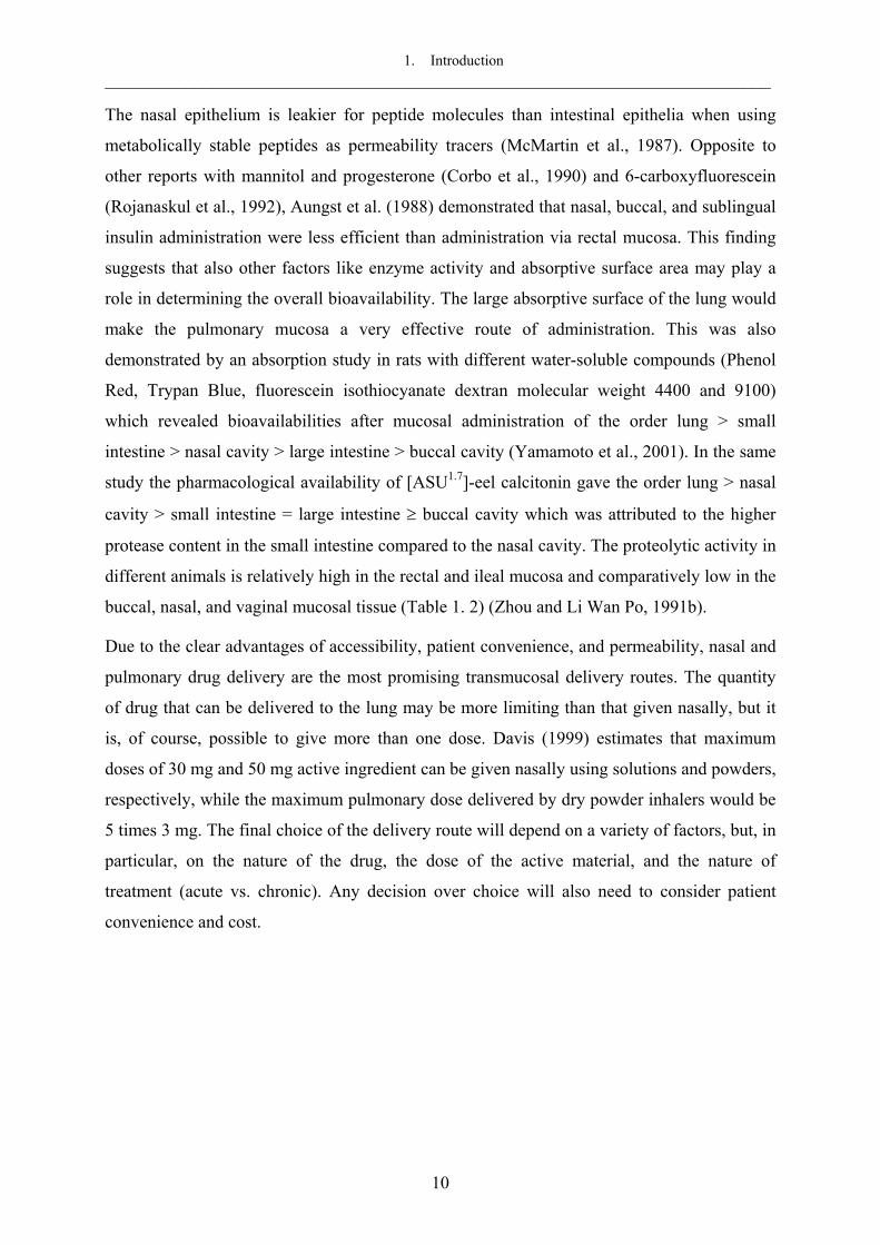
The nasal epithelium is leakier for peptide molecules than intestinal epithelia when using
metabolically stable peptides as permeability tracers (McMartin et al., 1987). Opposite to
other reports with mannitol and progesterone (Corbo et al., 1990) and 6-carboxyfluorescein
(Rojanaskul et al., 1992), Aungst et al. (1988) demonstrated that nasal, buccal, and sublingual
insulin administration were less efficient than administration via rectal mucosa. This finding
suggests that also other factors like enzyme activity and absorptive surface area may play a
role in determining the overall bioavailability. The large absorptive surface of the lung would
make the pulmonary mucosa a very effective route of administration. This was also
demonstrated by an absorption study in rats with different water-soluble compounds (Phenol
Red, Trypan Blue, fluorescein isothiocyanate dextran molecular weight 4400 and 9100)
which revealed bioavailabilities after mucosal administration of the order lung > small
intestine > nasal cavity > large intestine > buccal cavity (Yamamoto et al., 2001). In the same
study the pharmacological availability of [ASU1.7]-eel calcitonin gave the order lung > nasal
cavity > small intestine = large intestine ≥ buccal cavity which was attributed to the higher
protease content in the small intestine compared to the nasal cavity. The proteolytic activity in
different animals is relatively high in the rectal and ileal mucosa and comparatively low in the
buccal, nasal, and vaginal mucosal tissue (Table 1. 2) (Zhou and Li Wan Po, 1991b).
Due to the clear advantages of accessibility, patient convenience, and permeability, nasal and
pulmonary drug delivery are the most promising transmucosal delivery routes. The quantity
of drug that can be delivered to the lung may be more limiting than that given nasally, but it
is, of course, possible to give more than one dose. Davis (1999) estimates that maximum
doses of 30 mg and 50 mg active ingredient can be given nasally using solutions and powders,
respectively, while the maximum pulmonary dose delivered by dry powder inhalers would be
5 times 3 mg. The final choice of the delivery route will depend on a variety of factors, but, in
particular, on the nature of the drug, the dose of the active material, and the nature of
treatment (acute vs. chronic). Any decision over choice will also need to consider patient
convenience and cost.

1. Introduction ___________________________________________________________________________
11
Table 1. 2 Proteolytic activities in different experimental animal tissue homogenates using
peptides and proteins as substrate (Zhou and Li Wan Po, 1991b).
Substrate T1/2, min, in various tissues Animal
Ileal Rectal Vaginal Buccal Nasal
Met-enkepahlin 15.1 11.3 22.2 12.0 16.3 Rabbit
Leu-enkephalin 226.5 114.3 183.7 153.2 162.0 Rabbit
Substance P 5.8 5.9 10.9 8.7 11.6 Rabbit
Insulin 98.1 71.6 106.0 318.5 29.2 Rabbit
Proinsulin 55.7 122.0 163.2 528.3 86.2 Rabbit
L-leucin-β-naphtylamine HCl ----- 53.0 ---- 50.2 60.4 Rat

1. Introduction ___________________________________________________________________________
12
1.2. Nasal anatomy and physiology
The nose as drug delivery site has a number of unique features related to its anatomy and
physiology. These features have to be taken into consideration when developing a nasal drug
delivery system. The following sections will therefore give an introduction to the anatomy
and physiology of the human nose.
1.2.1. Anatomy and air passage
The nose is part of the upper respiratory system and is the main route by which ambient air
enters the body. The apparent external nose surrounds the nostrils and one third of the nasal
cavity. The entire human nasal cavity is an approximately 5 cm high and 10 cm long dual
chamber with a total surface area of about 150 cm2 and a total volume of about 15 - 20 ml.
The nasal cavity is divided by the nasal septum into two halves of approximately equal size,
beginning anteriorly at the nares and extending posteriorly to the nasopharynx where the two
halves of the airway join together. Located approximately 1.5 cm from the nares is the
narrowest portion of the entire airway, the internal ostium (or nasal valve) with a cross-
sectional area of about 30 mm2 on each side (Figure 1. 2). The nasal valve accounts for
approximately 50% of the total resistance to respiratory airflow from the nostrils to the alveoli
(Mygind and Dahl, 1998). This high resistance to airflow, the relatively high linear velocity of
the air stream, combined with an almost 90° angle of the flow passage at the ostium, and
turbulences facilitate the impaction of the majority of particles carried in the inspired air
stream in the anterior of the nasal cavity from where they are mainly removed by mucociliary
clearance (see section 1.2.2) (Hinchcliffe and Illum, 1999).
Each half of the nasal cavity is limited by the septal wall and the lateral wall. Bony scroll-like
conchae (or turbinates) are attached to the lateral wall and project into the main part of the
cavities (Figure 1. 2). Although more complex in many animal species, in humans three
conchae, called the inferior, median, and superior, have a relatively simple scroll arrangement,
(Gizurarson, 1990; Illum, 1996). The presence of these chonchae creates a turbulent air flow
through the nasal passages which ensures a better contact between the mucosa and the
inspired air, thus facilitating its humidification and temperature regulation.
Underneath and lateral to each of the turbinates are passages called the inferior, middle, and
superior meatus. The inferior and middle meatus receive the openings of the nasolachrymal
1. Introduction ___________________________________________________________________________
13
duct and the paranasal sinuses. The mucous membrane in a meatus will not be hit by an
ordinary nasal spray (Mygind and Dahl, 1998).
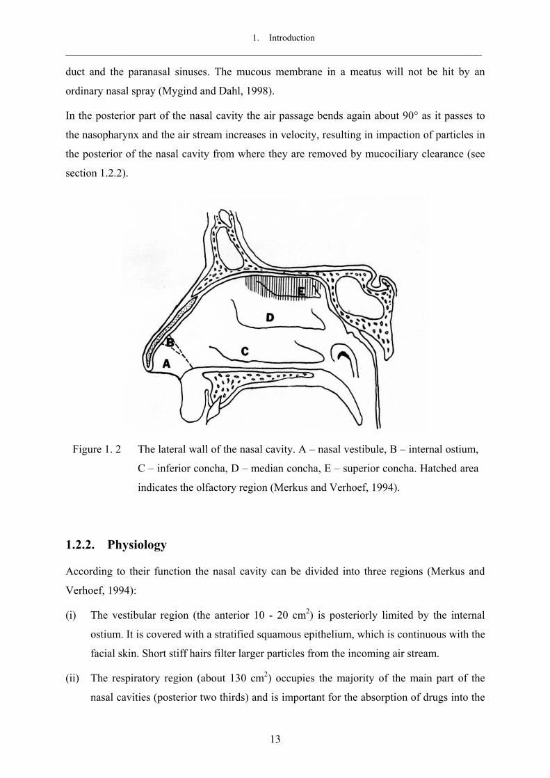
In the posterior part of the nasal cavity the air passage bends again about 90° as it passes to
the nasopharynx and the air stream increases in velocity, resulting in impaction of particles in
the posterior of the nasal cavity from where they are removed by mucociliary clearance (see
section 1.2.2).
1.2.2. Physiology
According to their function the nasal cavity can be divided into three regions (Merkus and
Verhoef, 1994):
(i) The vestibular region (the anterior 10 - 20 cm2) is posteriorly limited by the internal
ostium. It is covered with a stratified squamous epithelium, which is continuous with the
facial skin. Short stiff hairs filter larger particles from the incoming air stream.
(ii) The respiratory region (about 130 cm2) occupies the majority of the main part of the
nasal cavities (posterior two thirds) and is important for the absorption of drugs into the
Figure 1. 2 The lateral wall of the nasal cavity. A – nasal vestibule, B – internal ostium,
C – inferior concha, D – median concha, E – superior concha. Hatched area
indicates the olfactory region (Merkus and Verhoef, 1994).

1. Introduction ___________________________________________________________________________
14
systemic circulation. The epithelium consists of pseudostratified columnar epithelial
cells.
(iii) The olfactory region (10 - 20 cm2) at the roof of the nasal cavities comprises of the
small patch of columnar cells containing the smell receptors (Figure 1. 2).
Respiratory surface epithelium. The respiratory epithelium has a thickness of
approximately 100 µm (Merkus and Verhoef, 1994). It consists of four major cell types
(Figure 1. 3) (Mygind and Dahl, 1998).
(i) Basal cells, which are progenitors of the other cell types, lie on the basement membrane
and do not reach the airway lumen.
(ii) Columnar cells are related to neighboring cells by tight junctions. The cytoplasm
contains numerous mitochondria in the apical part, as a sign of an active metabolism.
Figure 1. 3 Cell types of the nasal respiratory epithelium. I – nonciliated columnar cell
with microvilli, II – goblet cell with mucous granules and Golgi apparatus,
III – basal cell, IV – ciliated columnar cell with many mitochondria in the
apical part, DM – double membrane, CTM – connective tissue membrane
(Merkus and Verhoef, 1994).
1. Introduction ___________________________________________________________________________
15
All columnar cells are covered by about 300 microvilli, uniformly distributed to the
entire apical surface. These short and slender fingerlike cytoplasmic expansions increase
the surface area of the epithelial cells, thus promoting exchange processes across the
epithelium. The microvilli also prevent drying of the surface by retaining moisture
essential for ciliary function. Columnar cells can be divided into non-ciliated and
ciliated cells. Cilia are fingerlike protrusions (0.2 - 0.3 µm wide and 5 µm in length) on
the apical surface of cells, which have a typical ultrastructure and are larger than
microvilli. Each ciliated cell contains about 100 - 300 cilia (Petruson et al., 1984). The
anterior third of the nasal cavity is non-ciliated. Cilia start occurring just behind the
front edge of the inferior turbinate. The posterior part of the nasal cavity as well as the
paranasal sinuses are densely covered with cilia.
(iii) Goblet cells are mucous-containing and secreting cells typical for a respiratory
epithelium. Their number is slightly larger in the posterior than in the anterior part of the
nasal cavity with a mean concentration of goblet cells (4,000 - 7,000 / mm2) similar to
the trachea and the main bronchi (Tos, 1983). The contribution of goblet cells to the
volume of nasal secretion is probably small compared to that of the submucosal glands.
Little is known about the release mechanism from goblet cells, which in contrast to
submucosal glands are not under parasympathetic control. Goblet cells probably respond
to physical and chemical irritants in the microenvironments.
Submucosal glands, mucus, and mucociliary clearance. Below the respiratory epithelium
is a thick lamina propria, composed of a loose mesh of fibroelastic tissue with many blood
vessels, nerves, and glands. These submucosal glands possess both serous and mucous
secretory cells and release directly onto the surface of the epithelium. The majority of what is
referred to as ‘nasal secretion’ is produced by the glands. Other minor contributors are goblet
cells and plasma exudation, especially during inflammatory processes.
A thin, clear, and continuous layer of fluid, called mucus, covers the entire nasal epithelial
surface. Approximately 20 - 40 ml of mucus are produced from the normal ‘resting’ nose each
day (Quraishi, 1998). This mucus is composed of water (95 - 97%), mucus glycoproteins (2.5
- 3%), electrolytes (1%), proteins (1%), and other macromolecules (Kaliner et al., 1984). The
baseline pH in the human nasal cavity is approximately 6.3, ranging from 5.2 - 8.1
(Washington et al., 2000a). The mucus glycoproteins (mucins) consist of a protein core (20%)
with oligosaccharide side chains (80%), cross-linked by disulfide and hydrogen bonds

1. Introduction ___________________________________________________________________________
16
(Kaliner et al., 1984). These glycoproteins are responsible for the characteristic viscoelastic
properties of the mucus, which are related to its function of providing a protective coating to
the nasal epithelium and mucociliary clearance. Mucus consists of two fluid layers, each
approximately 5 µm thick: a viscous gel layer (mucus or epiphase) floats on a less viscous sol
layer (periciliary fluid or hypophase) immediately adjacent to the epithelial surface. The cilia
of the columnar cells move with regular, symmetric beats at a frequency of about 10 Hz in the
lower sol phase (Duchateau et al., 1985). During this process, the ciliary tips make contact
with and propel the gel layer whilst the sol layer remains relatively stationary (Sleigh et al.,
1988). During the recovery stroke the cilia move backward exclusively through the sol layer.
By this action the upper mucus layer, together with deposited particles, is transported towards
the nasopharynx from where it is swallowed. The velocity of mucous transport is
approximately 5 - 8 mm/min (Procter et al., 1973; Andersen and Procter, 1983), thus
renewing the nasal mucus layer every 10 - 20 min. The combined action of mucus layer and
cilia is called mucociliary clearance. It is an important nonspecific physiological defense
mechanism of the respiratory tract to protect the body against noxious inhaled materials.
Inhibition of the mucociliary clearance by drugs and drug delivery systems results in longer
contact times of the nasal mucosa with inhaled bacteria, viruses, carcinogens etc.. On the
other hand, the mucociliary clearance is responsible for the generally observed rapid clearance
of nasally administered drugs from the nasal cavity to the nasopharynx. It forms, therefore, an
opposing mechanism in the absorption process of drugs following intranasal delivery. To
overcome the rapid removal of nasally administered drugs the concept of bioadhesion can be
applied (see section 1.3).
Vasculature and innervation. The lamina propria under the nasal epithelium and the
basement membrane is rich in blood vessels and has an extensive blood supply (about
40 ml/min/100g, Bende et al., 1983) as well as a large lymph drainage system, particularly in
the respiratory region of the nasal cavity (Hinchcliffe and Illum, 1999). These blood vessels
differ from the vasculature of the tracheobronchial tree in three ways (Mygind and Dahl,
1998):
(i) Cavernous venous sinusoids are specialized vessels adapted to the functional demands
of the nose with respect to heating and humidification of inhaled air. When they distend
with blood the mucosa will swell and block the airway lumen.

1. Introduction ___________________________________________________________________________
17
(ii) Arterio-venous anastomoses allow the blood to bypass the capillaries. Their role is
probably related to the temperature and water control. At least 50% of the blood flow is
normally shunted through arterio-venous anastomoses (Anggard, 1974).
(iii) Nasal vasculature shows cyclic changes of congestion (nasal cycle, every 3 – 7 h).
Engorgement of venous plexuses with blood leads to a swelling of the mucosa which
can temporarily occlude the airway and make the tissue appear erectile. This occlusion
of the airway is thought to occur alternately between the two sides of the nasal cavity
preventing the drying-out of the mucous membrane. (Hinchcliffe and Illum, 1999). The
effect of the nasal cycle on the absorption of nasally administered drugs is still unclear.
The total clearance of radiolabelled saline was not affected by the nasal patency, even
though the initial clearance was higher in the more patent half of the nasal cavity
(Washington et al., 2000b).
Different to the gastrointestinal tract, the venous blood draining from the nose passes directly
into the systemic circulation, thereby circumventing hepatic first pass elimination.
The lamina propria of the nasal mucosa embeds also nerves. Afferent nerve fibers run in the
trigeminal nerve. Stimulation of the trigeminus in the nasal mucosa results in the sneezing
reflex (Faller, 1988). There is a rich parasympathetic innervation of the glands. Nervous
stimulation of the glandular cholinoceptors causes marked hypersecretion and is often part of
the reflex arc. Nasal blood vessels are both sympathetically and parasympathetically
innervated, but are mainly controlled by sympathetic fibers (Mygind and Dahl, 1998).
Function of the nose. Humans breath in about 12 - 24 times / min, thereby inhaling daily
approximately 10,000 liters of air of differing temperature and humidity, containing dust and
organisms. As the main entrance for inspired air, the nose has the following functions (Faller,
1988; Jones, 2001):
(i) Olfaction: Humans can detect more than 10,000 different odors and discriminate
between about 5,000. The olfactory epithelium at the roof of the nasal cavity has several
million olfactory sensory neurons (Jones and Rog, 1998). Odorant binding proteins bind
and solubilize hydrophobic molecules, increasing their concentration up to 10,000 times
that in ambient air. Olfactory transduction is then mediated by a cascade of transmitters
resulting in a depolarization of the olfactory neurons. The precise mechanism by which
1. Introduction ___________________________________________________________________________
18
different smells are recognized and discriminated remains still unclear, but possible
theories include specific odorant - receptor interactions.
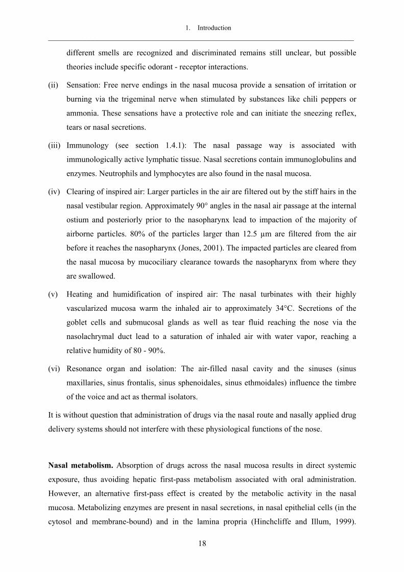
(ii) Sensation: Free nerve endings in the nasal mucosa provide a sensation of irritation or
burning via the trigeminal nerve when stimulated by substances like chili peppers or
ammonia. These sensations have a protective role and can initiate the sneezing reflex,
tears or nasal secretions.
(iii) Immunology (see section 1.4.1): The nasal passage way is associated with
immunologically active lymphatic tissue. Nasal secretions contain immunoglobulins and
enzymes. Neutrophils and lymphocytes are also found in the nasal mucosa.
(iv) Clearing of inspired air: Larger particles in the air are filtered out by the stiff hairs in the
nasal vestibular region. Approximately 90° angles in the nasal air passage at the internal
ostium and posteriorly prior to the nasopharynx lead to impaction of the majority of
airborne particles. 80% of the particles larger than 12.5 µm are filtered from the air
before it reaches the nasopharynx (Jones, 2001). The impacted particles are cleared from
the nasal mucosa by mucociliary clearance towards the nasopharynx from where they
are swallowed.
(v) Heating and humidification of inspired air: The nasal turbinates with their highly
vascularized mucosa warm the inhaled air to approximately 34°C. Secretions of the
goblet cells and submucosal glands as well as tear fluid reaching the nose via the
nasolachrymal duct lead to a saturation of inhaled air with water vapor, reaching a
relative humidity of 80 - 90%.
(vi) Resonance organ and isolation: The air-filled nasal cavity and the sinuses (sinus
maxillaries, sinus frontalis, sinus sphenoidales, sinus ethmoidales) influence the timbre
of the voice and act as thermal isolators.
It is without question that administration of drugs via the nasal route and nasally applied drug
delivery systems should not interfere with these physiological functions of the nose.
Nasal metabolism. Absorption of drugs across the nasal mucosa results in direct systemic
exposure, thus avoiding hepatic first-pass metabolism associated with oral administration.
However, an alternative first-pass effect is created by the metabolic activity in the nasal
mucosa. Metabolizing enzymes are present in nasal secretions, in nasal epithelial cells (in the
cytosol and membrane-bound) and in the lamina propria (Hinchcliffe and Illum, 1999).

1. Introduction ___________________________________________________________________________
19
Although much of the literature concerning nasal enzymes relates to studies in animals (rat,
rabbit, Syrian hamster, dog, and monkey), the profile of nasal enzymes in humans is
considered similar, despite inter-species variations (Sakar, 1992).
Monooxygenases, reductases, transferases, esterases and proteolytic enzymes were identified
in the nasal mucosa (Irwin et al., 1995). Oxidative phase I enzymes such as cytochrome P-450
dependent monooxygenase are recognized as a potential first line defense of the upper
respiratory tract against airborne xenobiotics. Numerous compounds are metabolized in vitro
by nasal cytochrome P-450, e.g. nasal decongestants, essences, anesthetics, alcohols, nicotine,
and cocaine (Sakar, 1992). The specific content of P-450 in the nasal mucosa is relatively
high, second only to that of the liver. The catalytic activity is more pronounced in the
olfactory region of the nose than in the respiratory region (Brittebo, 1982). Also phase II
enzymes such as glutathione transferase are present in the nasal mucosa.
Alternative mucosal routes of administration, such as the nasal mucosa, are especially
interesting with regards to protein and peptide delivery. However, the nasal epithelium and
the nasal secretions contain various peptidase and protease activities, including exopeptidases
as well as endopeptidases (Lee and Yamamoto, 1990). Aminopeptidases are the principle
proteolytic enzymes in the nasal mucosa (Kashi and Lee, 1986; Audus and Tavakoli-Saberi,
1991), with almost half of the aminopeptidase activity being membrane bound (Lee and
Yamamoto, 1990). Inhibition of proteolytic enzymes is also discussed as a contributing
mechanism for some penetration enhancers, e.g. sodium glycocholate, which has an inhibitory
effect on aminopeptidase activity (Hirai et al., 1981).
In general, nasal administration of drugs has to consider a pseudo-first-pass effect caused by
enzymes in the nasal cavity. Naturally the metabolic clearance of substances from the nose
into the blood is highly variable, depending on the particular compound under investigation.
Whether a nasally administered drug is subject to a nasal first-pass metabolism depends on
the presence of specific isozymes and the contact time. Propranolol for instance, a drug which
suffers extensive gastrointestinal and hepatic first-pass metabolism after oral administration
(Walle et al., 1985) is rapidly absorbed unmetabolized after nasal administration (Hussain et
al., 1980).

1. Introduction ___________________________________________________________________________
20
1.2.3. Nasal pathology with relevance to nasal drug absorption
The nose may be affected by a number of pathological conditions. It is important to consider
the effect that these may have on nasal drug absorption.
The pH of the nasal fluid lays normally around 5.5 - 6.5 but depends on air temperature,
sleep, emotions, and food ingestion (Junginger et al., 1991a; Washington et al., 2000a). An
increase in pH to 7 - 9 during acute and allergic rhinitis, rhinorrhoea, and chronic and acute
sinusitis can be observed (Berendes et al., 1977; Junginger et al., 1991a). Also diabetes
mellitus has been shown to influence the nasal pH (Sachdeva et al., 1993).
Inhalation of cold, dry air can act as a physical stimulus inducing symptoms of rhinitis that
are associated with an increase in osmolality from 280 - 290 to approximately 310 mosmol/kg
(Togias et al., 1988). Stimulation of the nasal gland secretion with chili powder reduces the
osmolality to approximately 238 mosmol/l, with simultaneous reduction of the sodium and
potassium ion content of the nasal secretion (Knowles et al., 1997). Pathological conditions
also affect the viscosity and viscoelasticity of the nasal mucus as well as the ciliary beat
frequency (Atsuta and Majima, 1998; Majima et al., 1999). This variability in composition
and properties of nasal fluid can greatly influence the performance of a nasally administered
drug delivery system, especially when it relies on nasal fluid uptake for activation.
The clearance of drug formulations from the nasal mucosa may be reduced in patients with
pathological conditions, which tend to impair the ciliary function. The ciliary function is
influenced by the pH of the surrounding fluids, being optimal between 7 - 10 for tracheal and
bronchial tissue (van de Donk et al., 1980a; Luk and Dulfano, 1983). pH values below 6 and
equal or above 11 result in severe decreases in the ciliary beat frequency. Isotonic conditions
preserve the ciliary activity best (van de Donk et al., 1980a; Luk and Dulfano, 1983).
Bacteria, e.g. Haemophilus influenza and Staphylococcus epidermidis, are known to disturb
normal synchronous ciliary motion, causing adjacent cilia to beat at different rates (Ferguson
et al., 1988). Also disruption of epithelial cells with loss of a confluent epithelial field has
been reported. Changes in ciliary structure occur in patients with long-standing allergic
rhinosinusitis and variations in secreted mucus happen at times of acute allergen challenge
(Maurizi et al., 1984).
Rhinorrhoea, a symptom in patients suffering from rhinosinusitis resulting from an allergic
reaction or from infections such as the common cold, is often associated with increased nasal
clearance, while nasal congestion, also a common symptom of rhinosinusitis, leads to a

1. Introduction ___________________________________________________________________________
21
strongly reduced nasal clearance (Bond et al., 1984). However, no influence of rhinorrhoea on
the nasal clearance of interferon was observed (Phillpotts et al., 1984). Increased mucociliary
clearance can also be observed following acute exposure to tobacco smoke (Bascom et al.,
1995). The pattern of nasal deposition from a spray device did not differ between normal
subjects and those with nasal polyposis, although clearance was considerably slower in the
latter (Lee et al., 1984). Also allergic rhinitis, atrophic rhinitis and chronic sinusitis lead to a
reduced nasal mucociliary clearance (Sakakura et al., 1983).
Patients with primary ciliary dyskinesia have no or dyskinetic beating cilia and thus a reduced
mucociliary clearance, resulting in frequent infections of the respiratory system (Blouin et al.,
2000). Cystic fibrosis patients exhibit also a reduced mucociliary clearance due to the
abnormality of the mucus, while the cilia function is normal (Middleton et al., 1993).
Mucociliary clearance in diabetes patients is also reduced compared to non-diabetic controls
(Sachdeva et al., 1993).
1.2.4. The nose as drug delivery site: advantages, barriers, and solutions
The nose as a site of drug administration offers the following advantages:
(i) Easy accessibility and needle free drug application without the necessity of trained
personnel facilitates self-medication, thus improving patient compliance compared to
parenteral routes (Pontiroli et al., 1989).
(ii) Good penetration of, especially lipophilic, low molecular weight drugs through the nasal
mucosa. For instance the absolute nasal bioavailability of fentanyl is about 80%
(Striebel et al., 1993).
(iii) Rapid absorption and fast onset of action due to a relatively large absorptive surface and
high vascularization. Thus the tmax of fentanyl after nasal administration was ≤ 7 min
comparable to i.v. (Striebel et al., 1993). Nasal administration of suitable drugs would
therefore be effective in emergency therapy as alternative to parenteral administration
routes.
(iv) Avoidance of the harsh environmental conditions in the gastrointestinal tract (chemical
and enzymatic degradation of drugs).
(v) Avoidance of hepatic first-pass metabolism and thus potential for dose reduction
compared to oral delivery.

1. Introduction ___________________________________________________________________________
22
(vi) Potential for direct delivery of drugs to the central nervous system via the olfactory
region under bypassing the blood-brain-barrier (Illum, 2000a; Chow et al., 2001; Dahlin
et al., 2001; Dufes et al., 2003).
(vii) Direct delivery of vaccine to lymphatic tissue and secretory immune response at distant
mucosal sites (see section 1.4.1).
Despite this number of advantages of the nose as drug delivery site, certain barriers may be
encountered when developing a nasal drug formulation:
(i) Bioavailabilities of polar drugs are generally low, about 10% for low molecular weight
drugs and not above 1% for peptides such as calcitonin and insulin (Illum, 2003). The
most important factor limiting the nasal absorption of polar drugs and especially large
molecular weight polar drugs such as peptides and proteins is the low membrane
permeability. Drugs can cross the epithelial cell membrane either by the transcellular
route exploiting simple concentration gradients, by receptor mediated or vesicular
transport mechanisms, or by the paracellular route through the tight junctions between
the cells. Polar drugs with molecular weights below 1000 Da will generally pass the
membrane using the latter route (McMartin et al., 1987). Although tight junctions are
dynamic structures and can open and close to a certain degree when needed, the mean
size of the channels is of the order of less than 10 Å and the transport of larger
molecules is considerably more limited (Madara and Dharmsathaphorn, 1985;
McMartin et al., 1987). Larger peptides and proteins are able to pass the nasal
membrane using an endocytotic transport process but only in low amounts (Inagaki et
al., 1985; Grass and Robinson, 1988). Nasal absorption of such polar drugs can be
greatly improved by co-administration of absorption enhancing agents. Agents
described in the literature for nasal drug delivery have included surfactants (laureth-9,
sodium laurylsulfate), bile salts and bile salt derivatives (sodium glycocholate, sodium
deoxycholate, sodium taurodihydrofusidate), fatty acids and fatty acid derivatives
(linoleic acid), phospholipids (lysophosphatidylcholine, DDPC), various cyclodextrins
(dimethyl-β-cyclodextrin, parenteral α-, β-, and γ-cyclodextrin), and cationic compounds
(chitosan and derivatives, poly-L-arginine, poly-L-lysine) (De Ponti, 1991; Merkus et
al., 1993; Illum, 1999, Natsume et al., 1999). These enhancers work by a variety of
mechanisms but generally change the permeability of the epithelial cell layer by
modifying the phospholipid bilayers, leaching of proteins from the membrane or even
stripping off the outer layer of the mucosa. Some of these enhancers also have an effect
1. Introduction ___________________________________________________________________________
23
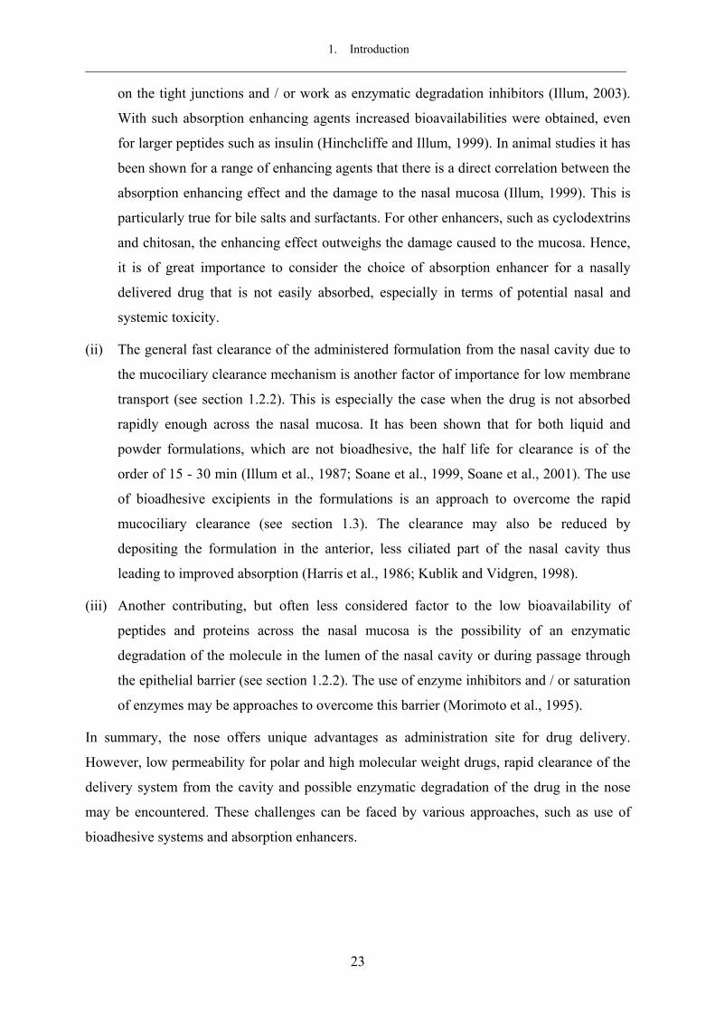
on the tight junctions and / or work as enzymatic degradation inhibitors (Illum, 2003).
With such absorption enhancing agents increased bioavailabilities were obtained, even
for larger peptides such as insulin (Hinchcliffe and Illum, 1999). In animal studies it has
been shown for a range of enhancing agents that there is a direct correlation between the
absorption enhancing effect and the damage to the nasal mucosa (Illum, 1999). This is
particularly true for bile salts and surfactants. For other enhancers, such as cyclodextrins
and chitosan, the enhancing effect outweighs the damage caused to the mucosa. Hence,
it is of great importance to consider the choice of absorption enhancer for a nasally
delivered drug that is not easily absorbed, especially in terms of potential nasal and
systemic toxicity.
(ii) The general fast clearance of the administered formulation from the nasal cavity due to
the mucociliary clearance mechanism is another factor of importance for low membrane
transport (see section 1.2.2). This is especially the case when the drug is not absorbed
rapidly enough across the nasal mucosa. It has been shown that for both liquid and
powder formulations, which are not bioadhesive, the half life for clearance is of the
order of 15 - 30 min (Illum et al., 1987; Soane et al., 1999, Soane et al., 2001). The use
of bioadhesive excipients in the formulations is an approach to overcome the rapid
mucociliary clearance (see section 1.3). The clearance may also be reduced by
depositing the formulation in the anterior, less ciliated part of the nasal cavity thus
leading to improved absorption (Harris et al., 1986; Kublik and Vidgren, 1998).
(iii) Another contributing, but often less considered factor to the low bioavailability of
peptides and proteins across the nasal mucosa is the possibility of an enzymatic
degradation of the molecule in the lumen of the nasal cavity or during passage through
the epithelial barrier (see section 1.2.2). The use of enzyme inhibitors and / or saturation
of enzymes may be approaches to overcome this barrier (Morimoto et al., 1995).
In summary, the nose offers unique advantages as administration site for drug delivery.
However, low permeability for polar and high molecular weight drugs, rapid clearance of the
delivery system from the cavity and possible enzymatic degradation of the drug in the nose
may be encountered. These challenges can be faced by various approaches, such as use of
bioadhesive systems and absorption enhancers.
1. Introduction ___________________________________________________________________________
24
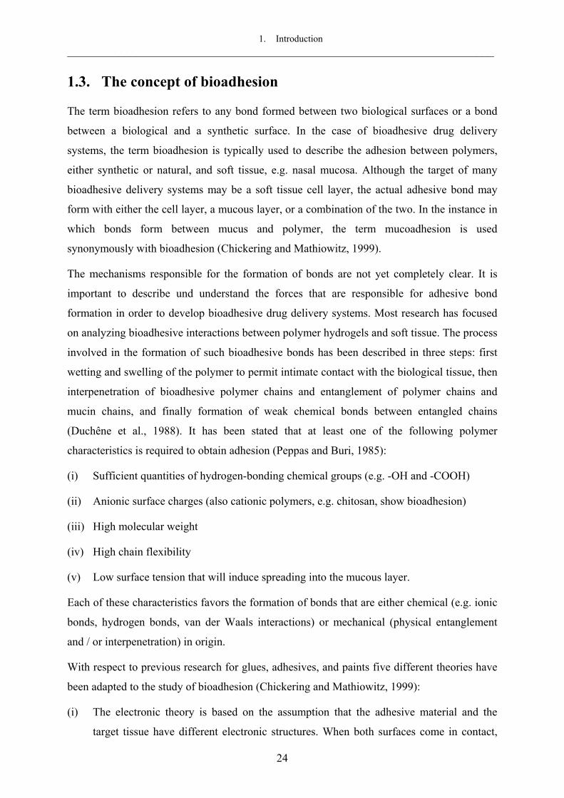
1.3. The concept of bioadhesion
The term bioadhesion refers to any bond formed between two biological surfaces or a bond
between a biological and a synthetic surface. In the case of bioadhesive drug delivery
systems, the term bioadhesion is typically used to describe the adhesion between polymers,
either synthetic or natural, and soft tissue, e.g. nasal mucosa. Although the target of many
bioadhesive delivery systems may be a soft tissue cell layer, the actual adhesive bond may
form with either the cell layer, a mucous layer, or a combination of the two. In the instance in
which bonds form between mucus and polymer, the term mucoadhesion is used
synonymously with bioadhesion (Chickering and Mathiowitz, 1999).
The mechanisms responsible for the formation of bonds are not yet completely clear. It is
important to describe und understand the forces that are responsible for adhesive bond
formation in order to develop bioadhesive drug delivery systems. Most research has focused
on analyzing bioadhesive interactions between polymer hydrogels and soft tissue. The process
involved in the formation of such bioadhesive bonds has been described in three steps: first
wetting and swelling of the polymer to permit intimate contact with the biological tissue, then
interpenetration of bioadhesive polymer chains and entanglement of polymer chains and
mucin chains, and finally formation of weak chemical bonds between entangled chains
(Duchêne et al., 1988). It has been stated that at least one of the following polymer
characteristics is required to obtain adhesion (Peppas and Buri, 1985):
(i) Sufficient quantities of hydrogen-bonding chemical groups (e.g. -OH and -COOH)
(ii) Anionic surface charges (also cationic polymers, e.g. chitosan, show bioadhesion)
(iii) High molecular weight
(iv) High chain flexibility
(v) Low surface tension that will induce spreading into the mucous layer.
Each of these characteristics favors the formation of bonds that are either chemical (e.g. ionic
bonds, hydrogen bonds, van der Waals interactions) or mechanical (physical entanglement
and / or interpenetration) in origin.
With respect to previous research for glues, adhesives, and paints five different theories have
been adapted to the study of bioadhesion (Chickering and Mathiowitz, 1999):
(i) The electronic theory is based on the assumption that the adhesive material and the
target tissue have different electronic structures. When both surfaces come in contact,

1. Introduction ___________________________________________________________________________
25
electron transfer occurs causing the formation of a double layer of electric charge at the
interface. The bioadhesive force is believed to be due to attractive forces across the
electrical double layer.
(ii) The adsorption theory states that the bioadhesive bond is formed due to van der Waals
interactions, hydrogen bonds, and related forces. Although the individual forces are
weak, the high number of interaction sites can produce intense adhesive strength. The
adsorption theory is the most widely accepted theory of bioadhesion.
(iii) The wetting theory was developed predominantly with regard to liquid adhesives. It uses
interfacial tension to predict spreading and in turn adhesion.
(iv) The diffusion theory supports the concept that interpenetration and entanglement of
bioadhesive polymer chains and mucus polymer chains produce semipermanent
adhesive bonds. It is believed that the bond strength increases with the degree of the
polymer chain penetration into the mucus layer. Penetration of polymer chains into the
mucus network, and vice versa, is dependent on the concentration gradients and the
diffusion coefficients. Cross-linking of either component hinders the interpenetration
but small chains and chain ends can still become entangled. For diffusion to occur, it is
important to have good solubility of one component in the other. The bioadhesive and
the mucus should therefore be of similar structure.
(v) The fracture theory analyzes the forces required to separate two surfaces after adhesion.
It is therefore most applicable to studying bioadhesion through mechanical
measurements. When determining fracture properties of an adhesive union from
separation experiments, failure of the adhesive bond must be assumed to occur at the
bioadhesive interface. However, it has been demonstrated that fracture rarely, if ever,
happens at the interface but instead occurs close to the interface (Ponchel et al., 1987).
The largest group of mucosal-adhesive materials are hydrophilic macromolecules containing
numerous hydrogen bond-forming groups. These are called “wet” adhesives as they are
activated by moistening. However, unless water uptake is restricted, they may overhydrate to
from a slippery mucilage (Smart, 1999). These hydrogel forming materials are nonspecific in
action.
A new group of bioadhesive polymers are the thiolated polymer derivatives such as
polyacrylic acid - cystein conjugates, chitosan - 2-iminothiolane conjugates, and others
(Bernkop-Schnürch, 2000; Bernkop-Schnürch et al., 2003; Hornof et al., 2003). For these

1. Introduction ___________________________________________________________________________
26
polymers the formation of disulfide bonds with mucus glycoproteins is discussed as
mechanism of bioadhesion. Thus, for instance, the work of adhesion of alginate tablets was
increased 4-fold from 25.8 ± 0.6 to 101.6 ± 36.1 µJ by conjugating it to cystein (Bernkop-
Schnürch et al., 2001).
Even more specific is the action of lectins as targeting agent of drug delivery systems. Many
epithelial surfaces are extensively glycosylated so that lectins, sugar-binding proteins and
glycoproteins isolated from plants, bacteria, and viruses, can bind specifically to epithelial
cells. Thus, drug delivery systems such as microparticles can be targeted to specific epithelial
cells, e.g. M-cells for the intranasal vaccine delivery (Clark et al., 2000). Also bacterial
adhesins can potentially be used to achieve site-specific bioadhesion (Vasir et al., 2003).
Drug delivery systems based on the concept of bioadhesion have been widely investigated for
various mucosal routes of administration including the nasal cavity. The prolonged residence
time of the delivery system on the mucosa can result in higher drug absorption and
consequently better bioavailability (Illum and Fisher, 1997).
1. Introduction ___________________________________________________________________________
27
1.4. Nasal vaccination
Part of this work deals with the nasal delivery of influenza vaccine incorporated into solutions
and in situ gelling nasal inserts. To understand the aims, advantages, and requirements of
nasal immunization the following section will give a brief introduction into nasal
immunology. Further on, the current state of research with respect to nasal vaccination
approaches will be discussed.
1.4.1. Nasal immunology
The majority of the invading pathogens enter the body via mucosal surfaces (Mestecky et al.,
1997). Therefore, mucosal sites have a potential as first line defense against entering
pathogens, especially the nasal mucosa due to its constant exposure to inhaled air. Pathogens
are filtered from the inspired air by compaction and mucociliary clearance. But the nose with
its nose-associated lymphoid tissue (NALT) is also an inductive as well as an effective site of
the immune system (Kuper et al., 1992). Nasal secretions are known to contain
immunoglobulins (IgA, IgG, IgM, IgE), protective proteins such as complement as well as
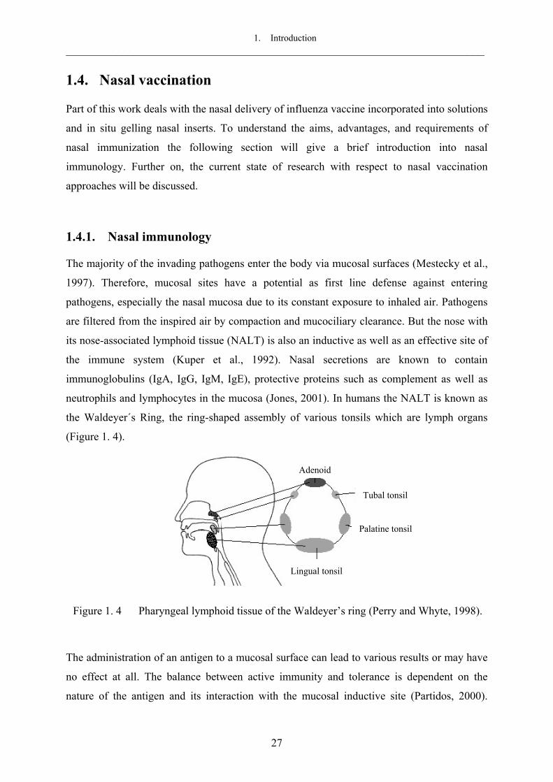
neutrophils and lymphocytes in the mucosa (Jones, 2001). In humans the NALT is known as
the Waldeyer´s Ring, the ring-shaped assembly of various tonsils which are lymph organs
(Figure 1. 4).
The administration of an antigen to a mucosal surface can lead to various results or may have
no effect at all. The balance between active immunity and tolerance is dependent on the
nature of the antigen and its interaction with the mucosal inductive site (Partidos, 2000).
Figure 1. 4 Pharyngeal lymphoid tissue of the Waldeyer’s ring (Perry and Whyte, 1998).
Adenoid
Lingual tonsil
Tubal tonsil
Palatine tonsil
1. Introduction ___________________________________________________________________________
28
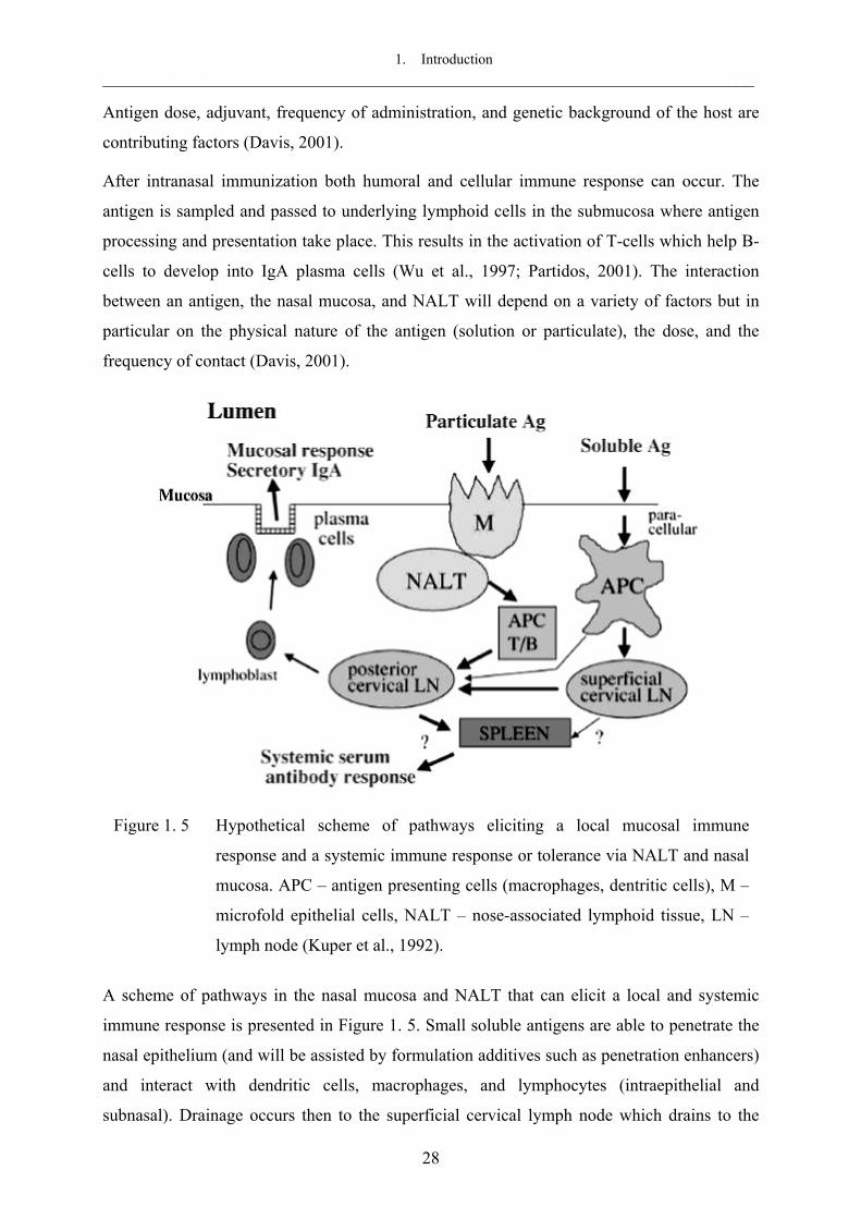
Antigen dose, adjuvant, frequency of administration, and genetic background of the host are
contributing factors (Davis, 2001).
After intranasal immunization both humoral and cellular immune response can occur. The
antigen is sampled and passed to underlying lymphoid cells in the submucosa where antigen
processing and presentation take place. This results in the activation of T-cells which help B-
cells to develop into IgA plasma cells (Wu et al., 1997; Partidos, 2001). The interaction
between an antigen, the nasal mucosa, and NALT will depend on a variety of factors but in
particular on the physical nature of the antigen (solution or particulate), the dose, and the
frequency of contact (Davis, 2001).
A scheme of pathways in the nasal mucosa and NALT that can elicit a local and systemic
immune response is presented in Figure 1. 5. Small soluble antigens are able to penetrate the
nasal epithelium (and will be assisted by formulation additives such as penetration enhancers)
and interact with dendritic cells, macrophages, and lymphocytes (intraepithelial and
subnasal). Drainage occurs then to the superficial cervical lymph node which drains to the
Figure 1. 5 Hypothetical scheme of pathways eliciting a local mucosal immune
response and a systemic immune response or tolerance via NALT and nasal
mucosa. APC – antigen presenting cells (macrophages, dentritic cells), M –
microfold epithelial cells, NALT – nose-associated lymphoid tissue, LN –
lymph node (Kuper et al., 1992).

1. Introduction ___________________________________________________________________________
29
posterior lymph nodes (Tilney, 1971). In contrast, antigens in the form of particles are largely
taken up by M-cells in the NALT. The NALT drains preferentially to the cervical lymph
nodes. The antigen so taken up can elicit a local (and also a distant) mucosal response or lead
to tolerance.
The nasal mucosa and its associated lymphoid structure do not only allow serum and local
(nasal) immune response but they are also inductive sites of the mucosa-associated lymphoid
tissue (MALT). MALT is scattered along mucosal linings and protects the body from antigens
entering via mucosal surfaces. It consists of the NALT, LALT (larynx-associated lymphoid
tissue), BALT (bronchus-associated lymphoid tissue) and GALT (gut-associated lymphoid
tissue = Peyer’s patches, appendix and colonic follicles) as so far identified inductive sites.
These inductive sites can initiate immune responses at various secretory effector sites, such as
the lachrymal, nasal, bronchial, and salivary glands, pharyngeal and middle ear mucosa, small
and large intestinal mucosa, and the mucosa of the urogenital tract, although not all relations
have yet been documented (Davis, 2001).
Precursors of mucosal IgA plasma cells originate mainly from organized lymphoepithelial
structures and are committed to IgA synthesis. These precursors mature in the regional lymph
nodes and enter the circulation via the thoracic duct. They can then lodge in the lamina
propria of distant mucosal sites (e.g. intestines, respiratory tract, genital tract, salivary gland,
etc.) where differentiation can occur. Thus, secretory IgA (sIgA) antibodies can appear in
parallel at different mucosal sites as part of a common mucosal immune system. While most
studies have been performed in animal models such as mice and rats, evidence for the
existence of the common mucosal immune system in man has been strengthened in recent
years (Mestecky et al., 1997).
Thus, an immune response following nasal exposure to antigens is not restricted to the nose
and the systemic circulation but can also be conveyed as good secretory immune response to
distant mucosal sites such as intestine, lung and vagina (Gallichan and Rosenthal, 1995;
Rudin et al., 1999; Isaka et al., 2001).
1.4.2. Nasal vaccine delivery
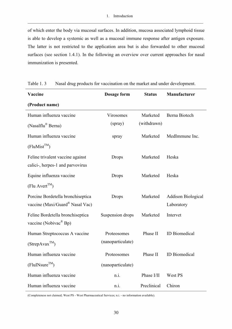
Nasal vaccination has gained a lot of interest over the last decades resulting in the
development and market launch of several nasally applied vaccines for human and animal use
(Table 1. 3). The underlying rational is to generate a first line defense against pathogens, most
1. Introduction ___________________________________________________________________________
30
of which enter the body via mucosal surfaces. In addition, mucosa associated lymphoid tissue
is able to develop a systemic as well as a mucosal immune response after antigen exposure.
The latter is not restricted to the application area but is also forwarded to other mucosal
surfaces (see section 1.4.1). In the following an overview over current approaches for nasal
immunization is presented.
Table 1. 3 Nasal drug products for vaccination on the market and under development.
Vaccine
(Product name)
Dosage form Status Manufacturer
Human influenza vaccine
(Nasalflu® Berna)
Virosomes
(spray)
Marketed
(withdrawn)
Berna Biotech
Human influenza vaccine
(FluMistTM)
spray Marketed MedImmune Inc.
Feline trivalent vaccine against
calici-, herpes-1 and parvovirus
Drops Marketed Heska
Equine influenza vaccine
(Flu AvertTM)
Drops Marketed Heska
Porcine Bordetella bronchiseptica
vaccine (Maxi/Guard® Nasal Vac)
Drops Marketed Addison Biological
Laboratory
Feline Bordetella bronchiseptica
vaccine (Nobivac® Bp)
Suspension drops Marketed Intervet
Human Streptococcus A vaccine
(StrepAvaxTM)
Proteosomes
(nanoparticulate)
Phase II ID Biomedical
Human influenza vaccine
(FluINsureTM)
Proteosomes
(nanoparticulate)
Phase II ID Biomedical
Human influenza vaccine n.i. Phase I/II West PS
Human influenza vaccine n.i. Preclinical Chiron
(Completeness not claimed; West PS - West Pharmaceutical Services; n.i. - no information available).

1. Introduction ___________________________________________________________________________
31
Antigens for nasal delivery can take many forms including whole cells (virus, bacteria),
surface proteins, synthetic peptides, protein polysaccharide conjugates, and DNA. As a
consequence, there will be no single system that fits all applications. Instead it is important to
choose a system that addresses the clinical need and the nature of the antigen. Most of the
antigens require some form of adjuvant that will amplify the immune response or provide a
degree of selectivity (Illum, 2001). A wide range of materials is now available as mucosal
adjuvants (Mestecky et al., 1997; Jenkins, 1999; Partidos, 2000).
Live bacterial vectors (e.g. avirulent Salmonella mutants) and live viral vectors (e.g. highly
attenuated Vaccinia virus or canary pox virus) were investigated as mucosal adjuvants
(Mestecky et al., 1997). Similarly inactivated meningococci and pertussis bacteria were
successfully used as adjuvants for intranasal influenza vaccination (Berstad et al., 2000). A
highly researched area for mucosal adjuvants is the use of bacterial toxins and derivatives
thereof. The most potent are the cholera toxin (Vibrio cholerae) and heat-labile enterotoxin
(Escherichia coli). To avoid toxicological adverse effects mutants and subunits of these
bacterial toxin were developed. Various heat-labile enterotoxin mutants prepared by specific
amino acid substitution were tested with respect to their adjuvant effect for nasally
administered influenza vaccine (Komase et al., 1998). Some of the mutants exceed even the
adjuvanticity of the wild-type toxin. Studies with papillomavirus type 6b virus-like particles
showed similar effectiveness of the mutant LTR72 with wild-type heat-labile enterotoxin after
nasal vaccination (Greer et al., 2001). One µg of LT-K63, another mutant of heat-labile
enterotoxin, increased the serum IgG response to influenza subunit vaccine after nasal
administration in mice 3-fold, while enhancing the nasal sIgA response 4.5-fold compared to
vaccine without adjuvant (Barchfeld et al., 1999). Other studies showed a significantly
decreased toxicity of the mutants (LT H44A) and almost no pathological changes of the nasal
mucosa three days after immunization (Hagiwara et al., 2001). Similar promising results were
found for cholera toxin B subunit adjuvant for hepatitis B and influenza vaccination (Matsuo
et al., 2000; Isaka et al., 2001). The first marketed human nasal vaccine (Nasalflu® Berna
against influenza), which was developed by Berna Biotech AG, Bern, Switzerland, used the
heat-labile enterotoxin of E.coli as adjuvant in combination with virosomes (Eich et al.,
2001). However, occurrence of facial paralysis led to the withdrawal of the product from the
market (Berna Biotech AG press release, 14.09.2001). Although there is no evidence of a
connection between the observed side effect and the adjuvant, this case underlines the
potential toxicological risk of new mucosal adjuvants.
1. Introduction ___________________________________________________________________________
32
Proteosomes are vaccine adjuvant systems consisting of nanoparticles formed from purified
bacterial outer membrane proteins. Nasal proteosomal influenza immunization has been
shown to induce potent immune response in a number of human and animal studies (Fries et
al., 2001; Plante et al., 2001) and led to several patents by the company ID Biomedical,
Vancouver, Canada, former Intellivax International (Burt et al., 2001; Jones et al., 2002).
Emulsions also act as adjuvants for nasal immunization, e.g. MF59, a microfluidized oil-in-
water emulsion containing 5% squalene, 0.5% Tween® 80, 0.5% Span® 85 in citrate buffer
(Barchfeld et al., 1999; Greer et al., 2001). Barchfeld et al. (1999) postulated an uptake of the
emulsion droplets and soluble antigen by the M-cells of the nasal epithelium with subsequent
transfer to antigen presenting cells. A screening of various surfactants showed the highest
nasal adjuvant effect for gangliosides, polysorbate 20, Cremophor® EL and a mixture of
caprylic / capric glyceride (Gizurarson et al., 1996; Gizurarson et al., 1998).
Nasal immunization with influenza subunit vaccine in a liposomal preparation revealed
strongly increased serum IgG titers in mice over 33 days post immunization compared to
vaccine without liposomes. Likewise nasal and lung sIgA levels were raised with liposomal
vaccine i.n. but not with pure vaccine i.n. and i.m. (de Haan et al., 1995). Administration of
empty liposomes up to 48 hours prior to i.n. vaccination with the subunit antigen also resulted
in immune stimulation. This indicates that the liposomes did not exert their adjuvant effect by
acting as a carrier for the antigen. Another approach is the use of cationic liposomal
formulations. Guy et al. (2001) demonstrated in mice the superior adjuvant effect of cationic
liposomes as compared to neutral liposomes and pure influenza split vaccine in raising the
hemagglutination inhibition titer in serum and nasopharyngeal washes. The positive effect
was explained by mucoadhesion and absorption enhancing properties of the cationic lipid DC-
cholesterol.
Other liposomal approaches to improve nasal immunization are virosomes and immuno-
stimulating complexes (ISCOMs). Virosomes are lipid vesicles which contain viral
glycoproteins. Intranasal vaccination of hamsters with virosomes containing human
parainfluenza virus hemagglutinin, neuraminidase and fusion glycoprotein led to a complete
resistance of the animals to challenge infections. In contrast, only partial protection was
observed in animals immunized subcutaneously with the same dose of antigen (Ray et al.,
1985). ISCOMs are negatively charged liposomal preparations containing the saponin Quil A.
Their most striking feature is the strong induction of cytotoxic T-cell responses (Mowat and

1. Introduction ___________________________________________________________________________
33
Donachie, 1991). However, their major drawback is the haemolytic and cytolytic activity of
Quil A with subsequent toxicity (Partidos, 2000).
Classical polymeric microparticles have also been investigated as nasal delivery systems for
vaccines. Shahin et al. (1995) showed strongly increased serum IgG and bronchoalveolar
lavage fluid sIgA in mice after nasal delivery of 1 µg Bordetella pertussis antigen embedded
in PLGA-microparticles, with subsequent protection against pertussis challenge. In contrast,
Lemoine et al. (1999) found significantly lower serum IgG and nasal sIgA response after
nasal delivery of influenza vaccine PLGA-microparticles compared to the free antigen.
Studies with a fluorescent probe showed that the particles failed to reach the NALT. This
implies that the physicochemical parameters of the particles and the application method may
play a crucial role in the adjuvant effect of microparticulate vaccine formulations for nasal
use. As an alternative microparticulate vaccine delivery system, polymeric lamellar substrate
particles were developed to avoid the loss of integrity and immunogenicity of the antigens
during microparticle preparation due to high shear stresses, organic solvent, and interface
exposure etc.. These particles were produced by simple precipitation of PLA (Jabbal-Gill et
al., 2001). Also microparticles based on other polymers such as hyaluronic acid ester (Singh
et al., 2001), chitosan (van der Lubben et al., 2003), and alginate (Rebelatto et al., 2001) were
investigated as nasal vaccine delivery systems.
Finally, also gels and polymeric solutions of carbohydrates increase the systemic and mucosal
immune response of nasally administered vaccines. Sucrose acetate isobutyrate gels were
tested in horses using the heat-resistant M-like protein of Streptococcus equi as antigen (Nally
et al., 2001). The nasal mucosal response of the immunized horses was qualitatively and
quantitatively similar to that observed in convalescent horses. Solutions of chitosan and gellan
gum both enhanced the systemic and mucosal immune response after nasal influenza
immunization of mice (Bacon et al., 2000). The effect was more pronounced for chitosan.
Decreased mucociliary clearance, opening of tight junctions, and activation of components of
the non-specific immune systems were discussed as possible mechanisms.
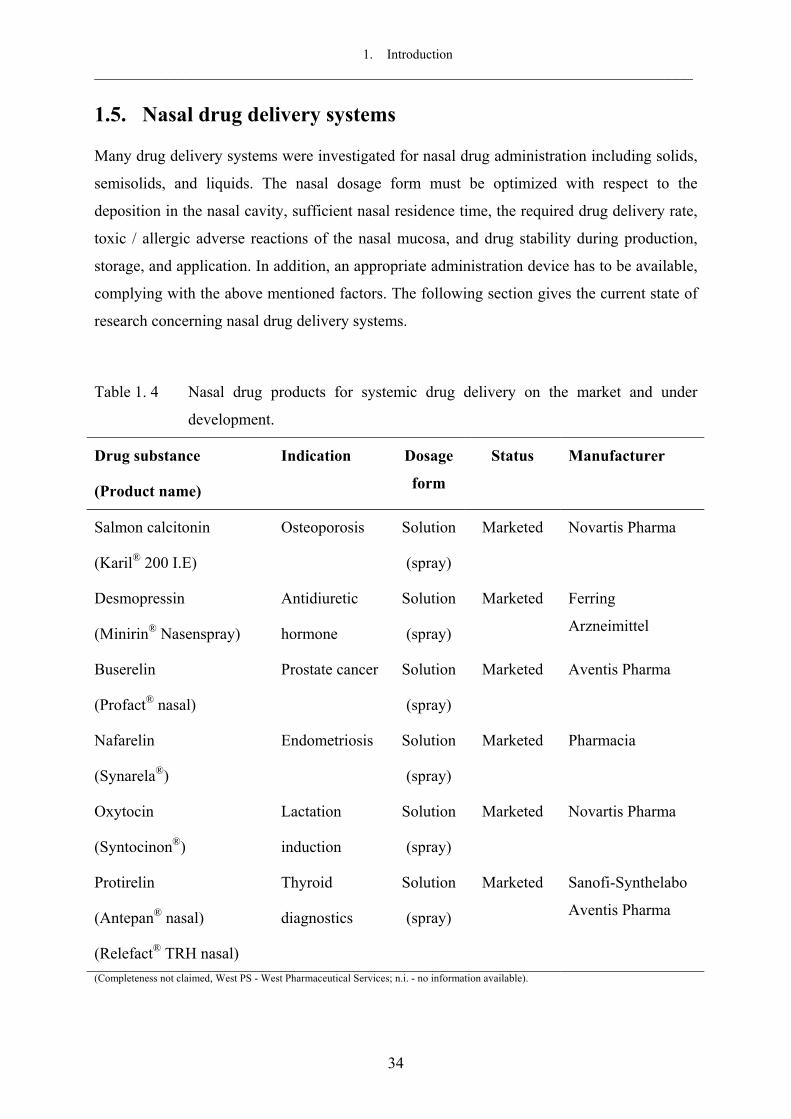
1. Introduction ___________________________________________________________________________
34
1.5. Nasal drug delivery systems
Many drug delivery systems were investigated for nasal drug administration including solids,
semisolids, and liquids. The nasal dosage form must be optimized with respect to the
deposition in the nasal cavity, sufficient nasal residence time, the required drug delivery rate,
toxic / allergic adverse reactions of the nasal mucosa, and drug stability during production,
storage, and application. In addition, an appropriate administration device has to be available,
complying with the above mentioned factors. The following section gives the current state of
research concerning nasal drug delivery systems.
Table 1. 4 Nasal drug products for systemic drug delivery on the market and under
development.
Drug substance
(Product name)
Indication Dosage
form
Status Manufacturer
Salmon calcitonin
(Karil® 200 I.E)
Osteoporosis Solution
(spray)
Marketed Novartis Pharma
Desmopressin
(Minirin® Nasenspray)
Antidiuretic
hormone
Solution
(spray)
Marketed Ferring
Arzneimittel
Buserelin
(Profact® nasal)
Prostate cancer Solution
(spray)
Marketed Aventis Pharma
Nafarelin
(Synarela®)
Endometriosis Solution
(spray)
Marketed Pharmacia
Oxytocin
(Syntocinon®)
Lactation
induction
Solution
(spray)
Marketed Novartis Pharma
Protirelin
(Antepan® nasal)
(Relefact® TRH nasal)
Thyroid
diagnostics
Solution
(spray)
Marketed Sanofi-Synthelabo
Aventis Pharma
(Completeness not claimed, West PS - West Pharmaceutical Services; n.i. - no information available).

1. Introduction ___________________________________________________________________________
35
Table 1. 4 continued.
Drug substance
(Product)
Indication Dosage
form
Status Manufacturer
Zolmitriptan
(AscoTop® Nasal)
Migraine Solution
(spray)
Marketed AstraZeneca
Sumatriptan
(Imigran® Nasal)
Migraine Solution
(spray)
Marketed GlaxoSmithKline
Dihydroergotamin
(Migranal® Nasal Spray)
Migraine Solution
spray)
Marketed Novartis Pharma
Nicotine
(Nicotrol® NS)
Smoking
cessation
Solution
(spray)
Marketed
Pfizer
Estradiol
(Aerodiol®)
Hormone
replacement
Solution
(spray)
Marketed
Servier
Morphine Pain n.i. Phase II
Phase II
Nastech
West PS
Apomorphine Erectile
Dysfunction
n.i. Phase II Nastech
PH80 Premenstrual
syndrome
n.i. Phase II Pherin
Pharmaceuticals
PH284 Eating disorders n.i. Phase II Pherin
Pharmaceuticals
Triptan Migraine n.i. Phase II
Phase I
West PS
Nastech
Interferon alpha Various n.i. Phase I Nastech
Interferon beta Multiple Sclerosis n.i. Phase I Nastech
Somatropin Growth Failure n.i. Phase I Nastech (Completeness not claimed, West PS - West Pharmaceutical Services; n.i. - no information available).
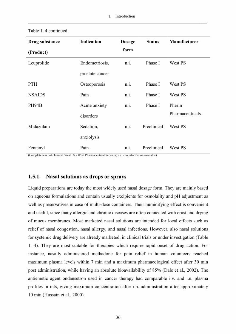
1. Introduction ___________________________________________________________________________
36
Table 1. 4 continued.
Drug substance
(Product)
Indication Dosage
form
Status Manufacturer
Leuprolide Endometriosis,
prostate cancer
n.i. Phase I West PS
PTH Osteoporosis n.i. Phase I West PS
NSAIDS Pain n.i. Phase I West PS
PH94B Acute anxiety
disorders
n.i. Phase I Pherin
Pharmaceuticals
Midazolam Sedation,
anxiolysis
n.i. Preclinical West PS
Fentanyl Pain n.i. Preclinical West PS (Completeness not claimed, West PS - West Pharmaceutical Services; n.i. - no information available).
1.5.1. Nasal solutions as drops or sprays
Liquid preparations are today the most widely used nasal dosage form. They are mainly based
on aqueous formulations and contain usually excipients for osmolality and pH adjustment as
well as preservatives in case of multi-dose containers. Their humidifying effect is convenient
and useful, since many allergic and chronic diseases are often connected with crust and drying
of mucus membranes. Most marketed nasal solutions are intended for local effects such as
relief of nasal congestion, nasal allergy, and nasal infections. However, also nasal solutions
for systemic drug delivery are already marketed, in clinical trials or under investigation (Table
1. 4). They are most suitable for therapies which require rapid onset of drug action. For
instance, nasally administered methadone for pain relief in human volunteers reached
maximum plasma levels within 7 min and a maximum pharmacological effect after 30 min
post administration, while having an absolute bioavailability of 85% (Dale et al., 2002). The
antiemetic agent ondansetron used in cancer therapy had comparable i.v. and i.n. plasma
profiles in rats, giving maximum concentration after i.n. administration after approximately
10 min (Hussain et al., 2000).
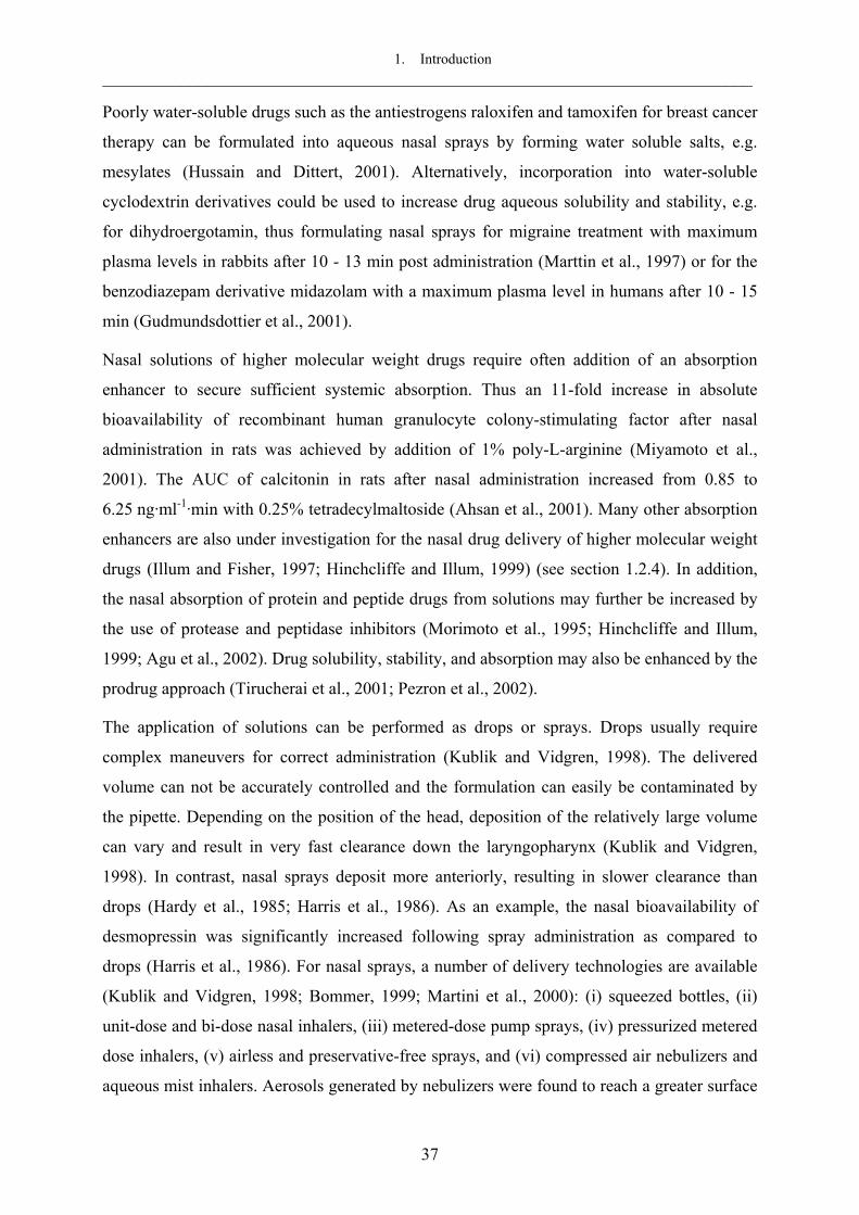
1. Introduction ___________________________________________________________________________
37
Poorly water-soluble drugs such as the antiestrogens raloxifen and tamoxifen for breast cancer
therapy can be formulated into aqueous nasal sprays by forming water soluble salts, e.g.
mesylates (Hussain and Dittert, 2001). Alternatively, incorporation into water-soluble
cyclodextrin derivatives could be used to increase drug aqueous solubility and stability, e.g.
for dihydroergotamin, thus formulating nasal sprays for migraine treatment with maximum
plasma levels in rabbits after 10 - 13 min post administration (Marttin et al., 1997) or for the
benzodiazepam derivative midazolam with a maximum plasma level in humans after 10 - 15
min (Gudmundsdottier et al., 2001).
Nasal solutions of higher molecular weight drugs require often addition of an absorption
enhancer to secure sufficient systemic absorption. Thus an 11-fold increase in absolute
bioavailability of recombinant human granulocyte colony-stimulating factor after nasal
administration in rats was achieved by addition of 1% poly-L-arginine (Miyamoto et al.,
2001). The AUC of calcitonin in rats after nasal administration increased from 0.85 to
6.25 ng·ml-1·min with 0.25% tetradecylmaltoside (Ahsan et al., 2001). Many other absorption
enhancers are also under investigation for the nasal drug delivery of higher molecular weight
drugs (Illum and Fisher, 1997; Hinchcliffe and Illum, 1999) (see section 1.2.4). In addition,
the nasal absorption of protein and peptide drugs from solutions may further be increased by
the use of protease and peptidase inhibitors (Morimoto et al., 1995; Hinchcliffe and Illum,
1999; Agu et al., 2002). Drug solubility, stability, and absorption may also be enhanced by the
prodrug approach (Tirucherai et al., 2001; Pezron et al., 2002).
The application of solutions can be performed as drops or sprays. Drops usually require
complex maneuvers for correct administration (Kublik and Vidgren, 1998). The delivered
volume can not be accurately controlled and the formulation can easily be contaminated by
the pipette. Depending on the position of the head, deposition of the relatively large volume
can vary and result in very fast clearance down the laryngopharynx (Kublik and Vidgren,
1998). In contrast, nasal sprays deposit more anteriorly, resulting in slower clearance than
drops (Hardy et al., 1985; Harris et al., 1986). As an example, the nasal bioavailability of
desmopressin was significantly increased following spray administration as compared to
drops (Harris et al., 1986). For nasal sprays, a number of delivery technologies are available
(Kublik and Vidgren, 1998; Bommer, 1999; Martini et al., 2000): (i) squeezed bottles, (ii)
unit-dose and bi-dose nasal inhalers, (iii) metered-dose pump sprays, (iv) pressurized metered
dose inhalers, (v) airless and preservative-free sprays, and (vi) compressed air nebulizers and
aqueous mist inhalers. Aerosols generated by nebulizers were found to reach a greater surface
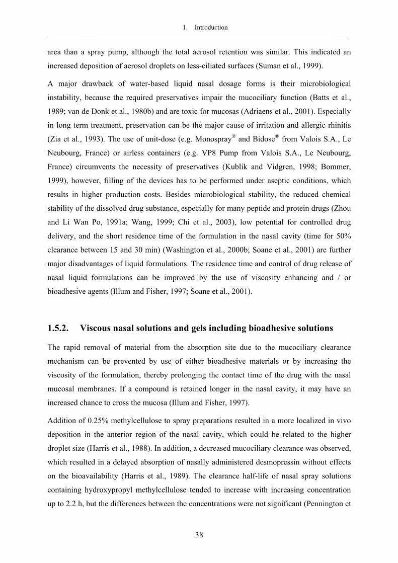
1. Introduction ___________________________________________________________________________
38
area than a spray pump, although the total aerosol retention was similar. This indicated an
increased deposition of aerosol droplets on less-ciliated surfaces (Suman et al., 1999).
A major drawback of water-based liquid nasal dosage forms is their microbiological
instability, because the required preservatives impair the mucociliary function (Batts et al.,
1989; van de Donk et al., 1980b) and are toxic for mucosas (Adriaens et al., 2001). Especially
in long term treatment, preservation can be the major cause of irritation and allergic rhinitis
(Zia et al., 1993). The use of unit-dose (e.g. Monospray® and Bidose® from Valois S.A., Le
Neubourg, France) or airless containers (e.g. VP8 Pump from Valois S.A., Le Neubourg,
France) circumvents the necessity of preservatives (Kublik and Vidgren, 1998; Bommer,
1999), however, filling of the devices has to be performed under aseptic conditions, which
results in higher production costs. Besides microbiological stability, the reduced chemical
stability of the dissolved drug substance, especially for many peptide and protein drugs (Zhou
and Li Wan Po, 1991a; Wang, 1999; Chi et al., 2003), low potential for controlled drug
delivery, and the short residence time of the formulation in the nasal cavity (time for 50%
clearance between 15 and 30 min) (Washington et al., 2000b; Soane et al., 2001) are further
major disadvantages of liquid formulations. The residence time and control of drug release of
nasal liquid formulations can be improved by the use of viscosity enhancing and / or
bioadhesive agents (Illum and Fisher, 1997; Soane et al., 2001).
1.5.2. Viscous nasal solutions and gels including bioadhesive solutions
The rapid removal of material from the absorption site due to the mucociliary clearance
mechanism can be prevented by use of either bioadhesive materials or by increasing the
viscosity of the formulation, thereby prolonging the contact time of the drug with the nasal
mucosal membranes. If a compound is retained longer in the nasal cavity, it may have an
increased chance to cross the mucosa (Illum and Fisher, 1997).
Addition of 0.25% methylcellulose to spray preparations resulted in a more localized in vivo
deposition in the anterior region of the nasal cavity, which could be related to the higher
droplet size (Harris et al., 1988). In addition, a decreased mucociliary clearance was observed,
which resulted in a delayed absorption of nasally administered desmopressin without effects
on the bioavailability (Harris et al., 1989). The clearance half-life of nasal spray solutions
containing hydroxypropyl methylcellulose tended to increase with increasing concentration
up to 2.2 h, but the differences between the concentrations were not significant (Pennington et

1. Introduction ___________________________________________________________________________
39
al., 1988). Hydroxyethylcellulose slightly, but significantly, increased the nasal absorption of
nicardipine (Visor et al., 1986). However, no study has so far demonstrated that the reduced
clearance and enhanced absorption was solely contributable to the increased viscosity. Several
nasal gel compositions using hydroxypropyl methylcellulose or other gelling agents were
patented, e.g. for the delivery of erythropoietin (Shimoda and Igusa, 1986), insulin with
sodium glycocholate as absorption enhancer (Zirinis, 1995), tamoxifen (Hussain and Dittert,
2001), and oxybutynin (Sherrat and Houdi, 2002). pH responsive polymers such as
polyvinylacetal diethylamino acetate (PVADEA) (Aikawa et al., 1998; Aikawa et al., 2002),
temperature responsive polymers such as poly(N-isopropylacrylamide) and
ethyl(hydroxyethyl)cellulose (Ryden and Edman, 1992; Pereswetoff-Morath and Edman,
1995a), and ion-responsive polymers such as pectin (Watts and Illum, 1998a) are also under
investigation. They offer the advantage of easy administration at low pH, low temperature or
low ion content, respectively, but formation of a viscous gel after contact with the nasal
mucosa.
Additionally to the viscosity enhancing effect, numerous hydrophilic polymers display
bioadhesive properties, among others xanthan gum, tamarind gum, sodium alginate,
carrageenan, gelatin, pectin, hyaluronic acid, chitosan, hydroxypropylcellulose, sodium
carboxymethyl cellulose, and polyacrylate-derivatives (Nakamura et al., 1996; Zaman et al.,
1999; Lee et al., 2000). Morimoto et al. (1991) reported the use of bioadhesive sodium
hyaluronate solutions (1%) to enhance the nasal bioavailability of vasopressin and one of its
analogues in rats. The nasal absorption depended on the sodium hyaluronate molecular weight
(55 - 2,000 kDa) and its concentration. No absorption enhancement was detected with the low
molecular weight sodium hyaluronate (55 kDa). No effect of the sodium hyaluronate solution
on the ciliary beat frequency was observed.
Gels of Carbopol®, a bioadhesive polyacrylic acid derivative, were used to enhance the nasal
absorption of insulin in rats (Morimoto et al., 1985). Both, 0.1 and 1.0 % Carbopol® promoted
insulin absorption resulting in a maximum hypoglycemic effect after 30 min and 60 min,
respectively. The delayed action of the latter was attributed to the slower release of insulin
from the 1% gel base due to its higher viscosity. In contrast, a solution of similar viscosity
containing 1% sodium carboxymethylcellulose, also a bioadhesive polymer, showed no
effect, indicating that not only viscosity and bioadhesive properties affected the insulin
absorption. The authors suggested that water movement from the gel base into the mucosa
would drag hydrophilic and macromolecular compounds through intercellular channels. Thus,
1. Introduction ___________________________________________________________________________
40
Carbopol® was suspected to promote drug absorption via the paracellular route rather than the
transcellular route. Borchard et al. (1996) suggested that Carbopol® may bind calcium ions
thus promoting opening of tight junctions in the nasal mucosa. Confocal laser scanning
microscopy studies showed a widening of paracellular spaces in caco-2 cell monolayers after
exposure to Carbopol® which was accompanied by a reduced transepithelial electrical
resistance and enhanced transport of 14C-mannitol and FITC-dextran (MW 4,000 Da) (Kriwet
and Kissel, 1995; Borchard et al., 1996). Carbopol® additionally possesses an inhibitory
potential against proteolytic enzymes, which was also attributed to the calcium-binding
properties of the polymer (Lueßen et al., 1995). Similarly to insulin, calcitonin delivered
nasally in a Carbopol® (0.1%) gel showed a prominent hypocalcaemic effect within 30 min
post administration (Morimoto et al., 1985). Histological studies with excised pig nasal
mucosa showed that Carbopol® gel did not affect the viability of the mucosa over 3.5 h (Östh
et al., 2002).
Another widely investigated bioadhesive polymer is chitosan, a partially deacetylated chitin
with positive charge. The clearance of a solution from the sheep nasal cavity was significantly
reduced by addition of 1% chitosan glutamate, giving a half-life of 43 min compared to
15 min without chitosan (Soane et al., 2001). The reduction in the mucociliary clearance rate
by chitosan on excised human nasal mucosa was molecular weight dependent (Aspden et al.,
1997). Chitosan with a molecular weight of less than 50 kDa was not able to reduce the
clearance rate significantly. The mucociliary clearance measured as saccharine clearance in
human volunteers before and after a 7-day once-daily administration of a 0.25% chitosan
solution was not significantly changed. Also the saccharin clearance immediately before and
1 h after the first dose was unchanged in 9 out of 10 volunteers. Chitosan has a concentration
dependent enhancing effect on the absorption of salmon calcitonin (Sinswat and Tengamnuay,
2003). With 1% chitosan, the AUC of calcitonin was doubled compared to the pure drug
solution and reached an absolute bioavailability of 2.5%. Also the intranasal relative
bioavailability of insulin based on a subcutaneous injection in sheep was significantly
increased by chitosan 0.5% compared to the insulin control solution, 3.6% versus 0.5%,
respectively (Dyer et al., 2002). Chitosan salts were less pH-dependent in their drug
absorption promoting effect than free chitosan likely due to a less coiled conformation of the
polymer chains (Tengamnuay et al., 2000). Equipping chitosan with a permanent positive
charge further increases its drug absorption enhancing properties. Intraduodenal co-
administration of the peptide drug buserelin with N-trimethylchitosan (1%) in rats increased
its absolute bioavailability to 6.3% and 13.0% for chitosan with a degree of N-trimethylation

1. Introduction ___________________________________________________________________________
41
of 40% and 60%, respectively, compared to 1.7% for chitosan chloride (1%) (Thanou et al.,
2000). The bioadhesive properties of chitosan are thought to be in part responsible for the
positive effect of chitosan and its derivatives on drug absorption across mucosal surfaces.
Additionally, chitosan is believed to modulate tight junctions and thus the paracellular
pathway as concluded from the reduced transepithelial electrical resistance of caco-2 cell
monolayers after exposure to 1.5% chitosan and simultaneous enhanced transport of the
marker compound 14C-mannitol (Artursson et al., 1994; Borchard, et al., 1996). Side effects
of chitosan on the nasal mucosa (release of lactate dehydrogenase, morphological evaluation)
were concentration dependent in range of 0.1 - 0.5% and lower than for other common
enhancer such as hydroxypropyl-β-cyclodextrin 5% (Tengamnuay et al., 2000). Chitosan has
therefore a potential as safe absorption enhancer for nasal drug delivery. Due to its highly
promising properties, chitosan formulated as microparticles and as solution is the proprietary
drug delivery platform of West Pharmaceutical Services Inc., Lionville, PA, USA (ChiSysTM).
Absorption enhancer, proteinase inhibitors etc. can be added to bioadhesive polymer solutions
and gels to further increase the drug absorption especially of peptides and proteins (Dondeti et
al., 1996). Although the use of bioadhesives and the increased viscosity enhances the
absorption of many drugs and prolongs the nasal residence time, polymer-containing drug
solutions still have a number of disadvantages such as the need of preservatives and potential
drug instabilities in aqueous solutions. In addition, viscous liquids and gels require
sophisticated delivery devices to ensure administration into the nose with high dosing
accuracy.
1.5.3. Nasal suspensions and emulsions
Suspensions are rarely used or investigated as nasal drug delivery system. Analogous to
marketed aqueous ophthalmic suspensions of the soft corticosteroid loteprednol etabonate
(e.g. Alrex® from Bausch and Lomb Pharmaceuticals Inc., Tampa, FL, USA), a nasal aqueous
suspension of this drug containing microcrystalline sodium carboxymethylcellulose for
stabilization and retention in the nasal cavity was patented by Senju Pharmaceuticals Inc.,
Osaka, Japan (Koji, 1998) intended for the local treatment of allergic rhinitis. A nasal
suspension for the delivery of insulin was investigated by Taeko et al. (1998). Here, soybean-
derived steryl glycoside and sterol mixtures (1%) were used as absorption enhancers and
pharmacological bioavailabilities of 6.7% and 11.3% were achieved. However, for oral drug
delivery, Kararli et al. (1992a) showed that emulsions were superior over suspensions in
1. Introduction ___________________________________________________________________________
42
enhancing the bioavailability of a badly soluble, modified tripeptide renin inhibitor. In
addition, the absolute nasal bioavailability of the same drug was significantly enhanced by an
emulsion formulation compared to a PEG 400 solution: 15 - 27% vs. 3 - 6% (Kararli et al.,
1992b). The enhancing effect of the emulsion can be attributed to the solubilization effect for
the drug and the lipophilic absorption enhancers in the composition, such as monoolein, oleic
acid and sodium taurocholate. Similarly, other low solubility compounds have been
formulated in emulsions to increase the drug solubility, e.g. diazepam in an ethyl-laurate
based microemulsion (Li et al., 2002) and testosterone (Ko et al., 1998). The latter was better
absorbed nasally from emulsions having a positive or negative zeta-potential (55% and 51%
bioavailability, respectively) than from emulsions of near neutral zeta-potential (37%
bioavailability), which was attributed to charge interactions with the mucus. Emulsions
containing calcitonin or parathyroid hormone together with absorption enhancers of the
azacycloalkane-group and glycyrrhizinic acid or its salts were patented by Yamamoto et al.
(1992, 1993).
In summary, nasal suspensions and especially emulsions have a potential as nasal drug
delivery systems for drugs with very low aqueous solubility. However, similar to aqueous
solutions both formulation types have microbiological stability problems and thus require
preservatives. Additionally, the use of surfactants as dispersion stabilizers and / or absorption
enhancers may lead to a potential muco- and ciliotoxicity. Stability problems of drugs in
aqueous environment can not necessarily be circumvented in emulsions and, due to the
existence of oil / water-interfaces, further stability problems especially of peptide and protein
drugs have to be considered (van de Weert et al., 2000).
1.5.4. Nasal micellar and liposomal formulations
Adjuvants are usually required to reach therapeutic plasma levels when hydrophilic
macromolecular drugs such as peptides and proteins are delivered nasally (De Ponti, 1991;
Merkus et al., 1993; Illum, 1999, Natsume et al., 1999). Among other surfactants, bile salts
are often used as enhancers, e.g. as micellar solutions. Tengamnuay and Mitra (1990a; 1990b)
described the use of micelles of sodium glycocholate and micelles thereof mixed with fatty
acids as absorption enhancers for the model dipeptide [D-Arg2]Kyotorphin and for insulin in
rats. The effect of mixed micelles was synergistic and greater compared to the single
enhancers. Mixed micelles of sodium glycocholate and linoleic acid reduced the blood
glucose level after nasal insulin administration to 47% of the glucose level after identical

1. Introduction ___________________________________________________________________________
43
nasal dosage of unenhanced insulin. Pure sodium glycocholate allowed a reduction to 55%.
Regarding the mechanism, different to the membrane solubilizing effect of pure bile salts, the
mixed micelles were discussed to have an effect on the paracellular pathway. Hereby, the bile
salts were considered to act as solubilizing agents for the fatty acids thus making them more
available at the nasal mucosa (Tengamnuay and Mitra, 1990a). The absorption modifying
effect of mixed micelles was reversible after 20 - 40 min and the morphological alterations of
the nasal mucosa were only mild to moderate after 5 h of exposure (Tengamnuay and Mitra,
1990a; Tengamnuay and Mitra, 1990b). However, measurement of marker enzymes in rat
nasal perfusate showed that the damaging effect of mixed micelles on the epithelial membrane
is significantly greater compared to pure sodium glycocholate solution and phosphate
buffered saline after 90 min exposure (carboxylesterase activity of 1545 U/l, 266 U/l and
9 U/l, respectively) (Shao and Mitra, 1994).
Liposomes were also investigated as nasal drug delivery system. Absorption enhancing
effects were found for insulin and calcitonin in in vitro permeability studies with nasal
mucosa (Maitani et al., 1992; Law et al., 2000). The enhancing effect was attributed to
increased nasal retention of peptides. The best carrier effect for calcitonin was demonstrated
with cationic liposomes. They were found to adhere intimately to the nasal mucosa surface
facilitating the penetration of the entrapped drug (Law et al., 2000). Similar observations were
made for desmopressin loaded cationic liposomes which resulted in enhanced antidiuretic
effects in rats compared to anionic liposomes and solutions (Law et al., 2001). Muramatsu et
al. (1999) showed increased nasal absorption of insulin for liposomes of high membrane
fluidity compared to low fluidity liposomes. However, the absorption enhancing effect of
liposomes is difficult to separate from the enhancing effects of the single components such as
phosphatidylcholines and steryl glycosides.
Proliposomes are dry, free-flowing granules composed of sorbitol as carrier and lipids that
form a liposomal dispersion on contact with water. They were investigated as intranasal drug
delivery system that combines fast onset (surface drug) and prolonged drug action
(encapsulated drug) e.g. for propranolol and nicotine (Jung et al., 2000; Jung et al., 2002).
1.5.5. Nasal powders and microparticles
Particulate nasal dosage forms are usually prepared by simply mixing of the drug substance
and the excipients (Takenaga et al., 1998; Ishikawa et al., 2002), by spray-drying or freeze-
1. Introduction ___________________________________________________________________________
44
drying of drug and excipient / preformed microparticles together (Illum et al., 1994a; Ugwoke
et al., 2000; Callens and Remon, 2000; Hascicek et al., 2003) or by direct production of drug
loaded nano- and microparticles (Fernandez-Urrusuno et al., 1999; Caliceti et al., 2000).
The use of dry-powder formulations containing bioadhesive polymers for the nasal delivery of
peptides and proteins was first investigated by Nagai et al. (1984). Water insoluble cellulose
derivatives and Carbopol® 934P were mixed with insulin and the powder mixture was
administered nasally. The powder took up water, swelled, and established a gel with a
prolonged residence time in the nasal cavity. Nasal administration thus reached one third of
the glucose reduction of an i.v. injection using the same insulin dose. Powder formulations for
nasal drug delivery have since been widely investigated, e.g. for a somastatin analogue using
cross-linked dextran, microcrystalline cellulose, and others (Oechslein et al., 1996), for
glucagon using microcrystalline cellulose (Teshima et al., 2002), for leuprolide, calcitonin,
and FITC-dextrans using microcrystalline cellulose in combination with hydroxypropyl
cellulose (Suzuki and Makino, 1999), and for gentamicin sulfate using hydroxypropyl
methylcellulose (Hascicek et al., 2003). A bioadhesive powder containing beclomethasone
dipropionate for local treatment of allergic rhinitis and hydroxypropyl cellulose as carrier had
a significantly enhanced nasal residence time compared to administration of a solution as
drops (Suzuki and Makino, 1999). Ugwoke et al. (2000) compared the nasal retention of
apomorphin freeze-dried with lactose, Carbopol® 971P or sodium carboxymethylcellulose.
After 3 h post insufflation 58%, 12%, and 27%, respectively, of the formulation had been
cleared from the nasal cavity. In all cases, the administered powder reduced the nasal
mucociliary clearance. The difference in nasal residence time led to a sustained plasma peak
level of the Carbopol® formulation of 52 min vs. 11 min for the lactose powder while
maintaining similar bioavailabilities (after dose correction) (Ugwoke et al., 1999). Callens and
Remon (2000) demonstrated nasal insulin delivery with freeze-dried powders of drum-dried
waxy maize starch and Carbopol® 974P 90:10 reaching an absolute bioavailability of 14.4%.
Comparison of different starch / Carbopol® 974P and maltodextrin / Carbopol® 974P mixtures
by oscillatory rheology showed no synergistic increase of the viscosity and elasticity with
mucus, which is often used as an indicator of bioadhesion (Callens et al., 2003a). However,
the formulation with the highest bioavailability provided also the highest storage modulus, i.e.
the most solid-like properties. It was also observed that the insulin bioavailability was
markedly reduced after repeated administration of the powder formulations (Callens et al.,
2003b). Although the reasons remained unclear, it was speculated that the powders were not
completely cleared from the nasal cavity after each delivery but formed a physical barrier on
1. Introduction ___________________________________________________________________________
45
the nasal mucosa inhibiting penetration of the drug on the long run. Thus, bioadhesion seemed
to have reversed into deteriorating the bioavailability.
Also inorganic, water insoluble powder formulations such as calcium phosphates enhanced
the drug absorption in rats after nasal administration (Ishikawa et al., 2002), although they did
not promote the in vitro drug permeability across rabbit nasal mucosa (Ishikawa et al., 2001).
Retardation at the site of administration was mentioned as possible explanation.
Well-characterized microparticles as another way of prolonging the residence time in the
nasal cavity were introduced by Illum et al. (1987). Microspheres of albumin, starch, and
DEAE-dextran absorbed water and formed a gel-like layer which was cleared slowly from the
nasal cavity. Three hours after administration, 50% of the delivered amount of albumin and
starch microspheres and 60% of the DEAE-dextran microspheres were still present at the site
of deposition. It was suggested that an increased contact time could increase the absorption
efficiency of drugs. As proposed, the relative intranasal bioavailability (vs. s.c.) of human
growth hormone in sheep was increased from 0.1% for the solution to 2.7% for the
degradable starch microsphere formulation (Illum et al., 1990). Addition of the absorption
enhancer lysophosphatidylcholine further enhanced the growth hormone absorption (relative
bioavailability 14.4%). Björk and Edman (1990) showed that plasma glucose reduction after
nasal insulin administration was comparable for degradable starch microspheres (cross-linked
with epichlorohydrin) and insoluble starch powder (molecular weight 25 kDa) but
significantly lower for soluble starch powder (molecular weight 11 kDa). It was therefore
concluded that crucial parameters for the absorption promoting effect of microspheres are
water absorption and water insolubility. No alteration of the nasal mucosa was observable by
scanning electron microscopy after 8 weeks of twice daily administration of starch
microspheres, except slight hyperplasia in the septum wall (Edman et al., 1992).
Although DEAE-dextran microspheres were retained strongly in the nasal cavity (Illum et al.,
1987), they were not successful in promoting nasal insulin absorption in rats (Rydén and
Edman, 1992). The insulin was too intensely bound to the DEAE-groups to be released by a
solution with an ionic strength corresponding to physiological conditions. Dextran
microparticles without ion exchange groups induced a 25% decrease in blood glucose level
about 40 min after administration compared to initial levels. In a later study, dextran
microspheres with different insulin distribution were compared (Pereswetoff-Morath and
Edman, 1995b). When insulin was situated on the microsphere surface, a 52% reduction in
plasma glucose was induced 30 min after administration in rats. However, microspheres,
1. Introduction ___________________________________________________________________________
46
which included the insulin in the sphere matrix, reached a maximum plasma glucose level
reduction of 30% after 60 min. Possibly the limited amount of fluid in the nasal cavity is
responsible for the differences observed, because the microspheres needed to be completely
swollen to release the entire amount of incorporated insulin (Pereswetoff-Morath, 1998).
Chitosan has also a high potential as particulate drug delivery system. In vivo studies in sheep
showed a half-life of nasal clearance for chitosan microparticles of 115 min compared to
43 min for a solution of the polymer (Soane et al., 2001). In general, chitosan powder
formulations, whether in form of microparticles or powders, were shown to provide a better
absorption enhancing effect than chitosan solutions (Illum, 2003). Hence, a chitosan
microsphere formulation for a LHRH analogue provided an absolute bioavailability in the
order of 40% in sheep (Illum et al., 2000b). Numerous patents were developed based on
chitosan microparticles for nasal drug delivery (Watts and Illum, 1996; Watts and Illum,
1998b; Watts and Illum, 2001). Also chitosan nanoparticles were exploited as nasal drug
delivery system. Insulin loaded chitosan nanoparticles enhanced the nasal absorption of
insulin to a greater extent than chitosan solution (Fernandez-Urrusuno et al., 1999). The
amount and molecular weight range of chitosan investigated did not have a significant effect
on insulin response.
Among other microparticulate delivery devices, hyaluronic acid ester microspheres were
successfully examined as potential drug delivery system for insulin reaching a relative
bioavailability (vs. s.c.) of 11% in sheep (Illum et al., 1994a). Also polyphosphazene
microspheres incorporating insulin and prepared by different solvent evaporation methods
reduced the blood glucose level of diabetic rats to 20% of the original level (Caliceti et al.,
2000). Bioadhesive microparticles of pH-sensitive co-polymers of polymethacrylic acid and
polyethylene glycol were investigated as potential nasal drug delivery system for budesonide
(Nakamura et al., 1999). Another approach for nasal microparticulate delivery systems is the
use of ion exchange resins. Takenaga et al. (1998) showed superior insulin absorption and
pharmacological effects for preparations of insulin mixed with microparticles of sodium
polystyrene sulfonate (anionic) and styrene-divinylbenzene copolymer (nonionic) compared
to polyacrylester (nonionic) and cholestyramine (cationic). Effects of ionic interactions
between insulin and exchange ions were discussed. Similar systems were also investigated as
a pulsed release system for nicotine (Illum, 1994b).
The mechanism by which dry-powder / particulate carriers enhance the nasal bioavailability
of drugs is only incompletely understood. Several hypotheses have been presented, which are
1. Introduction ___________________________________________________________________________
47
usually quite specific for the physicochemical parameters of the respective delivery system,
and also combinations of these proposed mechanisms seem likely: (i) increased nasal contact
time due to bioadhesion for starch microspheres (Illum et al., 1987), (ii) increased nasal
residence time due to the insolubility of the particulate carrier, e.g. for microcrystalline
cellulose and calcium phosphate (Nagai et al., 1984; Ishikawa et al., 2002), (iii) particulate
uptake via macrophages for PLA-microparticles (Almeida et al., 1993), (iv) opening of tight
junctions by dehydration of mucosal cells due to the connection to the cell cytoskeleton, e.g.
in the case of degradable starch microspheres (Björk et al., 1995), (v) opening of tight
junctions by binding of Ca2+ for cross-linked dextran microspheres, alginic acid, and
microcrystalline cellulose (Oechslein et al., 1996). Oechslein et al. (1996) found a better
correlation between the Ca2+-binding capacity of various powder excipients and the
bioavailability of octreotide in rats than with their water uptake behavior. Although not
described for a nasal powder formulation, thiolated polymers also increased the absorption of
drugs by opening of tight junctions but by a different mechanism (Clausen et al., 2002). Here,
the reducing activity of the thiolated polymer (polycarbophil-cystein conjugate) on oxidized
glutathione led to an increased glutathione level in the mucosa. Glutathione inhibited the
enzyme tyrosine phosphatase, which in turn is responsible for the dephosphorylation of the
tyrosine residues of occludin, a major tight junction protein. This dephosphorylation of
occludin then resulted in opening of the junctions and thus increased paracellular transport.
Although particulate / powder formulations for nasal administration offer numerous
advantages with respect to retention in the nasal cavity, absorption enhancement, and stability
due to the absence of water and lack of preservatives, the delivery of these powder dosage
forms to the nose is complex and so far no product has entered the market. Nasal powders
require sophisticated delivery devices and formulation optimization. Parameters such as
particle size and shape, density, and flow characteristics influence the distribution in the nose
(Kublik and Vidgren, 1998). Insufflators (mainly for study purposes), mono-dose and multi-
dose dry powder inhalers (e.g. from Pfeiffer, Randolfzell, Germany; Valois, Le Neubourg,
France; Orion, Helsinki, Finland; Teijin, Osaka, Japan) have been developed for the nasal
application. Dry powder inhalers ensure correct dosing and achieve better patient compliance.
A new development enables the application of freeze-dried powder, which can be lyophilized
directly in the device.
1. Introduction ___________________________________________________________________________
48
1.5.6. Nasal solid dosage forms
Solid dosage forms are rarely investigated as nasal drug delivery device. Some have been
described in the patent literature for veterinary use (Azria, 1986). Nevertheless, due to
absence of water, easy use of excipients for drug release control, and their single-unit
character, solid nasal dosage forms offer advantages compared to liquid and powder
formulations. Currently known solid preparations suitable for nasal drug delivery are tablets,
pellets, and films. However, foreign body sensation can be expected from all of them due to
slow / incomplete dissolution in the restricted nasal fluid, which would lead to low patient
acceptability.
Thus, attempts have been made to combine the advantages of a solid, single-unit dosage form
during administration with those of viscous, semisolid preparations (Azria, 1986; Bodmeier
and Maincent, 1999). This may be achieved by using carrier systems for nasal drug delivery
which hydrate quickly after contact with the mucosa and disintegrate. Porous inserts based on
water-soluble polymers such as gelatin and hydroxypropyl methylcellulose were described by
Azria (1986) for the nasal administration of calcitonin. Bodmeier and Maincent (1999)
suggested also fast disintegrating / gelling dosage forms for nasal, ocular, rectal or vaginal use
(porous, sponge-structure inserts prepared by lyophilisation, tablets, films, extruded devices).
Opposite to Azria (1986), in this patent the aspect of sustained drug release after
disintegration / gelation is added.
Sponge-structured inserts have the advantage of taking up fluid easily due to their porosity.
The proper choice of polymers can allow bioadhesion and controlled drug release. Due to
dissolution of the gel and / or mucociliary removal towards the nasopharynx, there would be
no need to remove the insert mechanically after it is depleted of drug. Thus, sponge structured
nasal inserts based on bioadhesive, release controlling polymers, which gel in situ, have a
high potential as nasal drug delivery system. They are therefore in the focus of this work.
Similar sponge-structured, gelating inserts have previously been investigated as ocular drug
delivery systems (Diestelhorst et al., 1999; Lux et al., 2003). Drug loaded sponges may also
be used as wound dressing (Lai et al., 2003).

1. Introduction ___________________________________________________________________________
49
1.6. Research objectives
The aim of this work was to develop and characterize a novel, bioadhesive, sponge-structured
solid dosage form for nasal administration: in situ gelling nasal inserts. The principle of the
dosage form was to imbibe nasal fluid from the mucosa after administration and to form a gel
in the nasal cavity to avoid foreign body sensation (Figure 1. 6). This gel should adhere to the
nasal mucosa due to its bioadhesive properties. In addition, it was to act as release controlling
matrix thus allowing sustained drug delivery. Due to dissolution of the gel and / or
mucociliary removal towards the nasopharynx, there would be no need to remove the insert
mechanically after it is depleted of drug. The application of this dosage form would be for the
systemic drug delivery via the nasal route.
Figure 1. 6 Principle behaviour of in situ gelling nasal inserts on the mucosal surface.
The in situ gelling nasal inserts were prepared by lyophilisation of aqueous solutions of drug
and various excipients. Initial studies were conducted to investigate the potential of polymeric
excipients to form the sponge-structure and thus to allow rapid fluid uptake due to capillary
forces as hypothesized. Low molecular weight model drugs were used to assess the in vitro
behavior of in situ gelling nasal inserts. Further on, the application range of in situ gelling
nasal inserts was broadened to a low solubility drug and protein antigens for nasal
vaccination.
Hydrated
mucosal surface
In situ gelling nasal insert
Fluid Drug
1. Introduction ___________________________________________________________________________
50
Following main properties of in situ gelling nasal inserts were identified as critical parameters
and therefore investigated:
(i) Solution viscosity (prior to lyophilisation)
(ii) Bioadhesion potential
(iii) Water uptake behavior and mass loss during hydration
(iv) In vitro drug release
(v) Mechanical properties
The main purposes of this work were:
(i) To investigate the influence of the sponge-forming polymeric excipients on the
properties of nasal inserts.
(ii) To identify means of controlling the in vitro drug release from nasal inserts (polymer
concentration, addition of freely water-soluble additives, polymer molecular weight,
polymer blends) and the effect thereof on nasal insert properties.
(iii) To study the drug release mechanisms of various drugs (low and high solubility,
charged and uncharged) from nasal inserts and to examine the impact of the dissolution
medium composition (osmolality, ion content, pH).
(iv) To incorporate the poorly water-soluble drug estradiol into nasal inserts, to study the
effect of the solubility enhancing excipient on the insert properties, and to verify the in
vitro results with in vivo investigations.
(v) To load nasal inserts with a split influenza vaccine (protein antigens) and to study the
effect of polymers and absorption enhancers in solutions as well as in nasal inserts on
the in vivo immune response.