1 Azilsartan as “add on” Treatment with Methotrexate...
10
Clinical Study Azilsartan as (Add-On) Treatment with Methotrexate Improves the Disease Activity of Rheumatoid Arthritis Naza Mohammed Ali Mahmood, 1 Saad Abdulrahman Hussain , 2 and Hawar Ali Ehsan Kaka Khan 3 1 Department of Pharmacology and Toxicology, College of Pharmacy, University of Sulaimani, Kurdistan Region Sulaimani, Iraq 2 Department of Pharmacology and Toxicology, Faculty of Pharmacy, Al-Rafidain University College, Baghdad, Iraq 3 Specialized Center of Rheumatology, Kurdistan Region, Sulaimani, Iraq Correspondence should be addressed to Saad Abdulrahman Hussain; saad.hussain@coalrafidain.edu.iq Received 23 February 2018; Revised 9 April 2018; Accepted 16 April 2018; Published 15 May 2018 Academic Editor: Fabrizio Montecucco Copyright © 2018 Naza Mohammed Ali Mahmood et al. is is an open access article distributed under the Creative Commons Attribution License, which permits unrestricted use, distribution, and reproduction in any medium, provided the original work is properly cited. Objective. e present study aimed to evaluate the efficacy and safety of azilsartan (Azil) as “add-on” treatment with methotrexate (MTX) in patients with active rheumatoid arthritis (RA). Methods. is single center, randomized, placebo-controlled, double- blind, pilot study included 64 patients with active RA. Patients received either placebo or Azil in addition to their currently used MTX doses for 90 days. e primary outcomes were DAS-28, SDAI, HAQ-DI, CDAI, EGA, and swollen and tender joints count. e secondary outcomes were the changes in the pain visual analogue scale (VAS-100), serum levels of TNF-, IL-1, IL-6, and anti- CCP, the lipid profile, and the markers of kidney and liver functions in the two groups at baseline and aſter 90 days. Results. Aſter 90 days, most clinical scores were significantly better in the Azil-treated group than in the placebo group. All inflammatory biomarkers were significantly improved aſter treatment with MTX + Azil compared to baseline and placebo group. No safety concerns were reported during the study period. Conclusions. Azilsartan improved the effects of methotrexate on the clinical scores and certain inflammatory biomarkers of patients with active RA. Trial Registration. e protocol was registered under the number 507/SA/1024 at the local clinical studies database, College of Medicine, Sulaimani University. 1. Introduction Rheumatoid arthritis (RA) is a chronic, systemic pathological disorder described as persistent inflammation of the synovial joints. Uncontrolled active RA leads to severe joint damage, which may progress to disability, poor quality of life, and other comorbid conditions such as cardiovascular diseases [1]. Although steroids and nonsteroidal anti-inflammatory drugs (NSAIDs) are recommended for the treatment of joint pain and other symptoms of systemic inflammation associated with RA, chronic use of these drugs was not considered as a current practice and was associated with serious adverse effects that were poorly tolerated by most of RA patients [2]. Meanwhile, many therapeutic approaches are currently used in clinical practice to treat active RA, including disease-modifying drugs (DMARDs), like methotrexate (MTX) and biological agents; however, rapport between their long-term use and wide range of side effects, in addition to high cost, limits their use, especially in low-income communities [3, 4]. Added to the central role of the immune system in the inflammatory response, increased expression of type-1 angiotensin II receptors (AT1-R) was found to be involved in various chronic inflammatory disorders [5]. Moreover, stimulation of AT1-R was associated with excessive production of reactive oxygen species (ROS) and enhanced secretion of inflammatory cytokines that accelerate inflammatory cascades [6, 7]. ese findings are supported by the fact that AT1-R blockade attenuated inflammatory response in animal models of inflammatory liver diseases [8, 9]. Furthermore, excessive activation of the renin-angiotensin system (RAS) mediates inflammation and modulates the immune response of T-cells, suggesting a potential influence in autoimmune diseases such as RA [10, 11]. In this regard, several studies Hindawi BioMed Research International Volume 2018, Article ID 7164291, 9 pages https://doi.org/10.1155/2018/7164291
Transcript of 1 Azilsartan as “add on” Treatment with Methotrexate...

Clinical StudyAzilsartan as (Add-On) Treatment with Methotrexate Improvesthe Disease Activity of Rheumatoid Arthritis
Naza Mohammed Ali Mahmood,1 Saad Abdulrahman Hussain ,2
and Hawar Ali Ehsan Kaka Khan3
1Department of Pharmacology and Toxicology, College of Pharmacy, University of Sulaimani, Kurdistan Region Sulaimani, Iraq2Department of Pharmacology and Toxicology, Faculty of Pharmacy, Al-Rafidain University College, Baghdad, Iraq3Specialized Center of Rheumatology, Kurdistan Region, Sulaimani, Iraq
Correspondence should be addressed to Saad Abdulrahman Hussain; [email protected]
Received 23 February 2018; Revised 9 April 2018; Accepted 16 April 2018; Published 15 May 2018
Academic Editor: Fabrizio Montecucco
Copyright © 2018 Naza Mohammed Ali Mahmood et al. This is an open access article distributed under the Creative CommonsAttribution License, which permits unrestricted use, distribution, and reproduction in any medium, provided the original work isproperly cited.
Objective. The present study aimed to evaluate the efficacy and safety of azilsartan (Azil) as “add-on” treatment with methotrexate(MTX) in patients with active rheumatoid arthritis (RA). Methods. This single center, randomized, placebo-controlled, double-blind, pilot study included 64 patients with active RA. Patients received either placebo or Azil in addition to their currently usedMTX doses for 90 days. The primary outcomes were DAS-28, SDAI, HAQ-DI, CDAI, EGA, and swollen and tender joints count.The secondary outcomes were the changes in the pain visual analogue scale (VAS-100), serum levels of TNF-𝛼, IL-1𝛽, IL-6, and anti-CCP, the lipid profile, and themarkers of kidney and liver functions in the two groups at baseline and after 90 days. Results.After 90days, most clinical scores were significantly better in the Azil-treated group than in the placebo group. All inflammatory biomarkerswere significantly improved after treatment with MTX + Azil compared to baseline and placebo group. No safety concerns werereported during the study period. Conclusions. Azilsartan improved the effects of methotrexate on the clinical scores and certaininflammatory biomarkers of patients with active RA. Trial Registration.The protocol was registered under the number 507/SA/1024at the local clinical studies database, College of Medicine, Sulaimani University.
1. Introduction
Rheumatoid arthritis (RA) is a chronic, systemic pathologicaldisorder described as persistent inflammation of the synovialjoints. Uncontrolled active RA leads to severe joint damage,which may progress to disability, poor quality of life, andother comorbid conditions such as cardiovascular diseases[1]. Although steroids and nonsteroidal anti-inflammatorydrugs (NSAIDs) are recommended for the treatment ofjoint pain and other symptoms of systemic inflammationassociated with RA, chronic use of these drugs was notconsidered as a current practice and was associated withserious adverse effects that were poorly tolerated bymost of RA patients [2]. Meanwhile, many therapeuticapproaches are currently used in clinical practice to treatactive RA, including disease-modifying drugs (DMARDs),like methotrexate (MTX) and biological agents; however,
rapport between their long-term use and wide range of sideeffects, in addition to high cost, limits their use, especiallyin low-income communities [3, 4]. Added to the centralrole of the immune system in the inflammatory response,increased expression of type-1 angiotensin II receptors(AT1-R) was found to be involved in various chronicinflammatory disorders [5]. Moreover, stimulation of AT1-Rwas associated with excessive production of reactive oxygenspecies (ROS) and enhanced secretion of inflammatorycytokines that accelerate inflammatory cascades [6, 7]. Thesefindings are supported by the fact that AT1-R blockadeattenuated inflammatory response in animal models ofinflammatory liver diseases [8, 9]. Furthermore, excessiveactivation of the renin-angiotensin system (RAS) mediatesinflammation and modulates the immune response ofT-cells, suggesting a potential influence in autoimmunediseases such as RA [10, 11]. In this regard, several studies
HindawiBioMed Research InternationalVolume 2018, Article ID 7164291, 9 pageshttps://doi.org/10.1155/2018/7164291

2 BioMed Research International
Screened RA patients n = 80
Accepted participation
n = 64
MTX + Placebo n = 32
Complete the study n = 25
Lostfollow-up
Not eligible n = 16
MTX + Azil n = 32
n = 7
Missed Azil doses
n = 2Complete the
study n = 30
RA: Rheumatoid arthritis MTX: MethotrexateAzil: Azilsartann: Number of patients
Figure 1: Flowchart of the study.
revealed the beneficial role of angiotensin receptor blockers(ARBs) and angiotensin-converting enzyme inhibitors(ACEIs) in experimental animal models of arthritis [12, 13]. Itis noteworthy that newly approved ARBs, such as telmisartanand azilsartan, demonstrated potent anti-inflammatoryactivity in animal models through mechanisms not relatedto RAS blockade [14, 15]. Yet, there are no data available fromrandomized clinical trials to support this concept. In lightof this indirect evidence, blockade of AT1-R results in dualantihypertensive and anti-inflammatory effects; this may bean effective therapeutic choice. Even though the use of ARBsis not expected to replace antirheumatic drugs such as MTXand biological agents, they may be suggested as an adjuncttherapy to improve response in RA patients. Accordingly, wedesign this pilot clinical study to assess, for the first time, theclinical beneficial effects of the ARB azilsartan as an adjuvanttreatment with MTX in patients with active RA.
2. Materials and Methods
2.1. Patient Recruitment and Study Design. We performed adouble-blinded, placebo-controlled, randomized pilot clini-cal study with treatment duration of 90 days over 14 months(from April 2016 to June 2017) at the Specialized Centerof Rheumatology, Sulaimani City, Kurdistan Region, Iraq.Eighty patients with active RA, who routinely went to theSpecialized Rheumatology Center for treatment follow up,were screened for eligibility. Based on the 2010 ACR/EULARcriteria [16] and 28-joint Disease Activity Score (DAS-28) ≥3.2, only 64 patients with active moderate to severe RA wereenrolled in the study (age: range 20–70 years). All the enrolledpatients demonstrate poor response to the currently used oral
methotrexate (MTX) (doses ranged between 7.5mg/week and25mg/week for at least 3 months) at the time of screeningeligibility for inclusion and were candidate for initiation oftreatment with biologic DMARDs as “add-on” approach.Only 55 patients completed the study: the evaluators lostthe contact with 7 patients in the MTX-placebo groupduring week 3 and week 5 for unknown reasons, while2 patients in the MTX-Azil group were excluded due tomissing more than 2 doses of azilsartan (Figure 1). In adouble-blinded pattern, the patients were randomly assignedto either of two treatment groups in an approximate 2 : 2ratio: Methotrexate plus placebo treated (MTX + placebo;𝑛 = 32) or to methotrexate plus Azilsartan-treated (MTX+ Azil; 𝑛 = 32). Methotrexate (Ebewe Pharma, Austria)was already administered as an oral tablet (7.5–25mg perweek) as a part of their treatment program before inclusion,while azilsartan was administered as a single oral daily dose(20mg/day). The azilsartan (Apollo Healthcare Resources,Singapore) doses were prepared as a capsule dosage formand administered as “add-on” single daily doses with theregularly usedMTX regimen.The placebo dose was preparedas a capsule dosage form that matches the shape and colorof the test drug formula and administered similarly. Thepatients were advised to keep on their regular drug treatmentschedule and were regularly observed clinically every 15days for proper compliance and occurrence of any unusualadverse effects. Before inclusion, all randomized patientswere asked to sign informed consent form according to theprinciples of the Declaration of Helsinki. The local scientificethics committee of the University of Sulaimani, College ofMedicine approved the study protocol (Certificate number507/SA/1024).

BioMed Research International 3
2.2. Outcome Measurement and Follow Up. At the time ofinclusion, patients with one of the following characteristicswere excluded: patients with mild or inactive RA, patientsusing nonsteroidal anti-inflammatory drugs 2 days beforeinclusion, hypersensitivity or severe adverse effects to thetested drugs, impaired renal or hepatic function, preg-nant and breastfeeding women, juvenile RA, patients usingdisease-modifying anti-rheumatic drugs other than MTX,biologics or high-dose steroids (>10mg/day prednisolone orequivalent), hypertensive patients using ACEIs, ARBs or anydrug that interfere with RAS for treatment of hypertension,and coexistence of other connective tissue diseases. Forassessment of the clinical endpoints at baseline and at the endof 90-day treatment, 4 instruments of clinical outcome evalu-ation were used including the Disease Activity Score-28 joint(DAS-28) [17], simplified disease activity index (SDAI) [18],clinical disease activity index (CDAI), and the health assess-ment questionnaire disease index (HAQ-DI) that assessesfunctional ability for eight subscales: arising, common dailyactivities, dressing, eating, grip, hygiene, reach, and walking[19]. Additionally, tender joints count (TJC), swollen jointscount (SJC), pain severity using visual analogue scale (VAS-100), evaluator global assessment (EGA), and duration ofmorning stiffness (measured in minutes) were also evaluatedto support the clinical assessment primary outcomes. Bloodsamples (10ml) were obtained from each patient by veinpuncture at baseline and the end of the treatment period. Ofthe blood collected, 3ml was kept in an ethylene diaminetetra-acetic acid tube to be used for measurement of ery-throcyte sedimentation rate (ESR) and hematology markers.The remaining blood was kept in a plain tube, left to clot atroom temperature for 30min, and centrifuged for 10min at4000 rpm to get the serum.Using ready-made enzyme-linkedimmunosorbent assay kits, the resultant serum was utilizedfor the measurement of highly sensitive C-reactive protein(hs-CRP) (Demeditec, Germany), tumor necrosis factor-𝛼(TNF-𝛼), interleukins-1𝛽 and -6 (IL-1𝛽, IL-6), and anticycliccitrullinated peptide (anti-CCP) (Beckman Coulter, USA).Additionally, the lipid profile (Triglycerides: TG, total choles-terol: TC, low-density lipoprotein cholesterol: LDL-c andhigh-density lipoprotein cholesterol: HDL-c) and the mark-ers of hepatic and renal functions (urea, creatinine, aspartateaminotransferase: AST and alanine aminotransferase: ALT)were analyzed spectrophotometrically using ready-made kits(Biomerieux, France).
2.3. Statistical Analysis. Theresults were statistically analyzedutilizing Graph Pad Prism 5.1 software (Graph Pad SoftwareInc., California, US). Continuous variables were presented asmean ± S.D, while discrete variables presented as numbersand frequencies. The Chi-square and Wilcoxon-rank testswere used to test the significance of the association betweendiscrete variables. Paired 𝑡-test was used to evaluate the dif-ference between pre- and posttreatment values. Additionally,one-way ANOVA was used to evaluate the significance ofthe difference between means and supported by Bonferroni’spost hoc analysis. Values with 𝑃 < 0.05 were consideredsignificantly different.
3. Results
3.1. Primary Outcome: Clinical Scores. Before initiation of thetreatment, the demographic characteristics of the patientswere recorded; the results revealed no significant differencesbetween the two randomized patients groups as shown inTable 1. Although no significant difference was reported forDAS-28 score at baseline, our data showed that adjunct use ofAzil withMTX significantly decreased theDAS-28 score after90 days compared with baseline value (5.2 ± 0.8 versus 6.5 ±0.7; 𝑃 < 0.001), and this effect was found to be significantlygreater than that of the placebo (Table 2). Similarly, the SDAIscore was not significantly decreased in the MTX + placebotreated group compared with baseline, while coadministra-tion of Azil with MTX decreases significantly the SDAI scorecompared with both the baseline and the placebo group(43.3±15.8 versus 62.8±13.4 and 61.5.5±19.6, resp.;𝑃 < 0.01)at the end of the treatment period. Regarding the effects onHAQ-DI score, Table 2 showed that adjunct use of Azil withMTX decreases significantly the HAQ-DI score after 90 daysof treatment (1.3 ± 0.6 versus 1.8 ± 0.5; 𝑃 = 0.001), andthis value was significantly greater than that of the placebogroup as well. However, the addition of the placebo didnot significantly change this score compared with baseline.Moreover, Table 2 showed that treatment with Azil decreasessignificantly the CDAI score (37.5 ± 15.8 versus 56.3 ± 11.2;𝑃 < 0.001) after 90 days of treatment, and this effect wassignificantly greater than that reported in the placebo group.Table 2 also showed that Azil significantly improved the TJCscore compared with the baseline value (18.1 ± 6.7 versus25.3 ± 3.1; 𝑃 < 0.01); however, the posttreatment value wasnot significantly different compared to the placebo group.Meanwhile, combination of MTX with Azil decreased SJCsignificantly after 90 days compared with baseline (11.4 ± 8.8versus 17.3 ± 9.7; 𝑃 = 0.0004) and placebo group, whereascoadministration of the placebo did not significantly alter thisscore compared to baseline. The influence of Ang II receptorblockade on the VAS-100 score was also examined, wherecoadministration of Azil with MTX significantly improvedthe VAS-100 score compared to baseline value (49.3 ± 17.5versus 70.0 ± 13.6; 𝑃 < 0.001); this pattern of effect was notrecognized in the placebo treated group (𝑃 > 0.05) after 90days. Table 2 also showed that the EGA score in Azil-treatedgroup was significantly decreased compared to both baselinevalue and placebo group after 90 days (4.1±1.1 versus 6.2±1.1and 6.8± 1.1, resp.; 𝑃 < 0.001), while coadministration of theplacebo formula withMTXdid not significantly influence theEGA score at the end of the treatment period. Moreover, theMTX-Azil combination improved the duration of morningstiffness significantly compared to both the baseline valueand the MTX-placebo effect after 90 days (16.6 ± 5.9 versus26.1 ± 8.5 and 19.2 ± 4.9, resp.; 𝑃 = 0.001), where theplacebo formula did not show significant effect in this regard(Table 2).The ESR value inMTX-Azil group was significantlydecreased after 90 days compared to baseline (28.5 ± 11.0versus 37.6 ± 15.8; 𝑃 = 0.0006), while coadministration ofthe placebo with MTX did not show such effect, and theESR value was significantly greater than that of the MTX-Azil group posttreatment (𝑃 = 0.72) as shown in Table 2.

4 BioMed Research International
Table 1: Demographic data and baseline characteristics of the RApatients treatedwithmethotrexate (MTX) or its combinationwith azilsartan(Azil).
Parameters MTX + placebo MTX + Azil𝑃 value
𝑛 = 25 𝑛 = 30
GenderMale 𝑛 (%) 8 (32) 12 (40) 0.62Female 𝑛 (%) 17 (78) 18 (60) 0.53Age (years) 55.5 ± 12.8 68.1 ± 13.0 0.57
Body weight (Kg) 82.7 ± 10.5 81.2 ± 9.6 0.41
BMI (Kg/m2) 31.9 ± 8.5 30.5 ± 6.9 0.62
Disease duration (year) 10.8 ± 9.1 8.7 ± 5.3 0.44
MTX treatment (months) 28.5 ± 12.8 25.4 ± 11.3 0.45
ESR (mm/hr) 36.6 ± 8.6 37.6 ± 15.8 0.84
hsCRP (𝜇g/ml) 6.5 ± 3.2 5.7 ± 3.9 0.55
DAS-28 score (4 values) 6.4 ± 0.96 6.5 ± 0.77 0.96
SDAI score 63.2 ± 20.1 62.8 ± 13.4 0.94
HAQ-DI score 1.77 ± 0.6 1.83 ± 0.58 0.7
CDAI score 51.6 ± 15.3 56.3 ± 11.2 0.33
Joint deformities 𝑛 (%) 4 (16.0) 5 (16.7) 0.48Use of steroids 𝑛 (%) 3 (12) 4 (13.3) 0.5Use of NSAIDs 𝑛 (%) 2 (8) 2 (6.7) 0.61Associated diseasesHypertension 𝑛 (%) 4 (16.0) 6 (20.0) 0.12Diabetes mellitus 𝑛 (%) 3 (12.0) 2 (6.7) 0.11Values are presented as mean ± S.D or percentage; n: number of patients; MTX: methotrexate; Azil: azilsartan; NSAIDs: nonsteroidal anti-inflammatory drugs.
Table 2: Effect of azilsartan (Azil) and placebo on ESR and hs-CRP levels and the clinical evaluation scores of patients with active RAmaintained on methotrexate (MTX).
Clinical Score MTX + placebo, 𝑛 = 25 MTX + Azil, 𝑛 = 30Baseline After 90 days Baseline After 90 days
DAS-28 6.44 ± 0.9a 6.49 ± 0.8a 6.45 ± 0.7a 5.2 ± 0.8∗b
SDAI 63.2 ± 20.1a 61.5 ± 19.6a 62.8 ± 13.4a 43.3 ± 15.8∗b
HAQ-DI 1.8 ± 0.6a 2.0 ± 0.6∗a 1.8 ± 0.5a 1.3 ± 0.6∗b
CDAI 51.6 ± 15.3a 50.0 ± 15.3a 56.3 ± 11.2a 37.5 ± 15.8∗b
TJC-28 23.2 ± 7.6 22.9 ± 7.5 25.8 ± 3.1 18.1 ± 6.7∗
SJC-28 16.0 ± 9.3a 15.8 ± 9.4a 17.3 ± 9.7a 11.4 ± 8.8∗b
Pain VAS-100 (mm) 71.9 ± 12.2a 69.8 ± 12.9a 70.0 ± 13.6a 49.3 ± 17.5∗b
EGA (cm) 6.7 ± 1.3a 6.8 ± 1.1a 6.2 ± 1.1a 4.1 ± 1.5∗b
Morning stiffness (min) 21.5 ± 11.6a 19.2 ± 4.9a 26.1 ± 8.5a 16.6 ± 5.9∗b
ESR (mm/hr) 36.6 ± 8.6a 37.2 ± 8.7a 37.6 ± 15.8a 28.5 ± 11.0∗b
hsCRP (𝜇g/ml) 6.5 ± 3.3 6.02 ± 3.6 5.7 ± 3.9 4.9 ± 2.8
Values are presented as mean ± SD; 𝑛 = number of patients; ∗significantly different compared to pretreatment within the same group (𝑃 < 0.050); values withdifferent superscripts (a, b) among groups are significantly different (𝑃 < 0.05). RA: rheumatoid arthritis; TJC: tender joint count; SJC: swollen joint count;VAS: visual analogue scale; EGA: evaluator global assessment; ESR: erythrocyte sedimentation rate; hsCRP: highly sensitive C-reactive protein; DAS-28: 28-joint disease activity score; SDAI: simple disease activity index; HAQ-DI: health assessment questionnaire disability index; CDAI: clinical disease activity index.
Furthermore, serum hs-CRP levels did not significantlychange (𝑃 = 0.62) in both treatment approaches comparedto baseline values.The influence of coadministration of eitherAzil or placebo with MTX on different functional areas ofthe HAQ-DI score was demonstrated in Table 3. Althoughall areas of the HAQ-DI score were significantly improvedin the MTX-Azil-treated group compared to baseline (𝑃 <0.001), only those that represent arise, eat, walk, and hygiene
areas demonstrated significant differences compared to thatreported in the MTX-placebo treated group posttreatment.
3.2. Secondary Outcome: Biomarkers of Inflammation. InTable 4, the results indicated that coadministration of Azilwith MTX significantly reduced the serum concentration ofTNF-𝛼 after 90 days compared to the baseline value (14.2 ±

BioMed Research International 5
Table 3: Effect of azilsartan (Azil) on different functional areas of HAQDI score of patients with active RA maintained on methotrexate(MTX).
HAQDI Areas MTX + placebo (𝑛 = 25) MTX + Azil (𝑛 = 30)Baseline After 90 days Baseline After 90 days
Dress 1.6 ± 1.1 1.7 ± 1.0 1.4 ± 0.9 0.8 ± 0.8∗
Arise 2.0 ± 0.5a 2.2 ± 0.4∗a 1.7 ± 0.9a 1.1 ± 0.7∗b
Eat 2.0 ± 0.5a 2.3 ± 0.6∗a 2.3 ± 0.9a 1.5 ± 0.9∗b
Walk 2.2 ± 0.7a 2.5 ± 0.5∗a 2.1 ± 0.8a 1.4 ± 0.9∗b
Hygiene 1.6 ± 0.8a 1.7 ± 0.7a 1.3 ± 0.7a 1.0 ± 0.6∗b
Reach 2.1 ± 0.6 2.2 ± 0.7 2.3 ± 0.8 1.8 ± 0.8∗
Grip 1.4 ± 0.5 1.7 ± 0.8∗ 1.1 ± 1.9 0.7 ± 1.5∗
Daily activity 1.9 ± 0.8 2.0 ± 0.8 2.1 ± 0.9 1.5 ± 0.9∗
Values are expressed as mean ± S.D; n: number of patients; ∗significantly different compared to pretreatment (𝑃 < 0.05); posttreatment values with differentsuperscripts (a, b) within each parameter are significantly different (𝑃 < 0.05). HAQ-DI: health assessment questionnaire disability index; RA: rheumatoidarthritis.
Table 4: Effect of azilsartan (Azil) and placebo on TNF-𝛼, IL-1𝛽, IL-6 and anti-CCP levels and the clinical evaluation scores of patients withactive RA maintained on methotrexate (MTX).
Inflammatory marker MTX + placebo, 𝑛 = 25 MTX + Azil, 𝑛 = 30Baseline After 90 days Baseline After 90 days
TNF-𝛼 (pg/ml) 17.8 ± 5.1a 16.7 ± 5.6a 18.4 ± 5.5a 14.2 ± 4.2∗b
IL-1𝛽 (pg/ml) 15.3 ± 2.9a 16.9 ± 4.4a 16.1 ± 3.9a 11.6 ± 5.4∗b
IL-6 (pg/ml) 29.1 ± 5.3a 30.3 ± 7.1a 23.9 ± 5.6b 16.7 ± 4.2∗c
Anti-CCP (IU/ml) 90.4 ± 10.0a 84.5 ± 8.3∗b 88.6 ± 9.3a 69.9 ± 9.6∗c
Values are presented as mean ± SD; 𝑛 = number of patients; ∗significantly different compared to pretreatment within the same group (𝑃 < 0.050); values withdifferent superscripts (a, b, and c) among groups are significantly different (𝑃 < 0.05).
Table 5: Effect of azilsartan (Azil) on serum lipid profile of patients with active RA maintained on methotrexate (MTX).
Parameters MTX + placebo (𝑛 = 25) MTX + Azil (𝑛 = 30)Baseline After 90 days Baseline After 90 days
Triglycerides (mg/dl) 143.3 ± 31.7 147.6 ± 36.6 153.2 ± 32.9 122.0 ± 34.4∗
Cholesterol (mg/dl) 170.5 ± 32.0a 181.9 ± 25.5a 165.1 ± 23.8a 154.3 ± 29.4a
LDL-c (mg/dl) 100.9 ± 29.3 105.8 ± 24.7 105.4 ± 19.2 94.4 ± 15.8∗
HDL-c (mg/dl) 39.4 ± 9.6 38.7 ± 7.9 33.3 ± 9.4 38.0 ± 11.9∗
Values are presented as mean ± S.D; 𝑛: number of patients; ∗significantly different compared with baseline within the same group (𝑃 < 0.05); values withdifferent superscripts (a, b) within each parameter are significantly different (𝑃 < 0.05). LDL-c: low-density lipoprotein cholesterol; HDL-c: high-densitylipoprotein cholesterol.
4.2 versus 18.4 ± 5.5 pg/ml; 𝑃 < 0.001), and this levelwas significantly lower than that reported in the placebogroup posttreatment; however, no significant differenceswerereported for serum TNF-𝛼 levels between the two groupsat baseline. Similarly, serum concentration of IL-1𝛽 was notsignificantly changed in the MTX + placebo treated groupcompared to the baseline, while coadministration of Azilwith MTX significantly decreases serum IL-1𝛽 compared toboth baseline and MTX + placebo treated group (11.6 ± 5.4versus 16.1 ± 3.9 and 16.9 ± 4.4 pg/ml, resp.; 𝑃 = 0.001)at the end of the treatment period. Table 4 also revealedthat the serum concentrations of IL-6 in the MTX-placebogroup were nonsignificantly changed compared to baselinevalues (𝑃 = 0.26), while coadministration of Azil withMTX resulted in significant decrease in serum IL-6 levelscompared to both baseline values and MTX + placebo group
posttreatment (16.7±4.2 versus 23.9±5.6 and 30.3±7.1 pg/ml,resp.; 𝑃 < 0.001). Serum anti-CCP concentrations weresignificantly decreased in the MTX + placebo treated groupcompared to baseline; likewise, coadministration of Azil withMTX resulted in significant decrease in serum anti-CCPconcentrations compared to both baseline values and MTX+ placebo treated group (69.9 ± 9.6 versus 88.6 ± 9.3 and84.5 ± 8.3 𝜇g/ml, resp.; 𝑃 = 0.001) at the end of the treatmentperiod.
3.3. Lipid Profile. The results indicated that coadministrationof Azil with MTX produced significant decrease in TG andLDL-c concentrations, associated with significant increase inHDL-c levels compared to baseline values; however, thesechanges were not significantly different compared to theMTX + placebo group posttreatment (Table 5).

6 BioMed Research International
Table 6: Effect of azilsartan (Azil) on the liver and kidney function markers of patients with active RA maintained on methotrexate (MTX).
Parameters MTX + placebo (𝑛 = 25) MTX + Azil (𝑛 = 30)Baseline After 90 days Baseline After 90 days
Serum AST (U/L) 19.9 ± 3.8a 30.6 ± 7.2∗b 19.8 ± 3.7a 17.6 ± 3.2∗c
Serum ALT (U/L) 19.7 ± 7.4a 30.3 ± 12.1∗b 17.0 ± 4.6a 15.3 ± 5.4a
Serum creatinine (mg/dl) 0.63 ± 0.1a 0.67 ± 0.1a 0.61 ± 0.2a 0.54 ± 0.1a
Serum urea (mg/dl) 27.4 ± 7.7a 29.6 ± 7.2a 23.9 ± 9.3a 21.3 ± 6.8a
Values were presented as mean ± S.D; 𝑛: number of patients; ∗significantly different compared with baseline within the same group (𝑃 < 0.05); values withdifferent superscripts (a, b, and c) within each parameter were significantly different (𝑃 < 0.05).
Table 7: Effect of azilsartan (Azil) on the hematopoietic function markers of patients with active RA maintained on methotrexate (MTX).
Parameters MTX + placebo (𝑛 = 25) MTX + Azil (𝑛 = 30)Baseline After 90 days Baseline After 90 days
Hb (g/dL) 12.4 ± 1.1a 12.1 ± 1.2a 12.4 ± 1.2a 12.3 ± 1.1a
WBC count ×103 cells/𝜇L 7.1 ± 1.4 6.7 ± 1.3 7.0 ± 1.8 6.2 ± 1.5∗
Platelets count ×109 cells/L 272.2 ± 41.2 265.8 ± 43.6∗ 265.6 ± 51.1 233.3 ± 40.2∗
Values were presented as mean ± S.D; 𝑛: number of patients; ∗significantly different compared with baseline within the same group (𝑃 < 0.05); values withdifferent superscripts (a, b) within each parameter were significantly different (𝑃 < 0.05).
3.4. Safety Profile. The influence of using Azil with MTXon the hepatic and renal functions in RA patients wasshown in Table 6. Serum levels of AST and ALT weresignificantly elevated in the MTX + placebo group com-pared to baseline, while adjunct use of Azil with MTXresulted in significant decrease in the AST levels comparedto baseline. However, serum urea and creatinine levels werenot significantly changed in the two groups. Regarding thehematopoietic system, coadministration of Azil with MTXdid not significantly change Hb levels, while WBC count wassignificantly decreased (𝑃 < 0.05) in the MTX + Azil groupcompared to baseline; however, it was comparable to MTX +placebo group (Table 7). Although both groups demonstratedsignificant decrease in the platelets count posttreatment,these changes were comparable (𝑃 > 0.05).
4. Discussion
Increasing evidence emerged regarding the role of RASactivation in inflammatory disorders, where upregulation ofAng II type-1 receptors in the synovium of RA patients wasfound to play a role in this context [20].Thebeneficial effect ofAzil on some inflammatory disorders was previously demon-strated, as both prophylactic and therapeutic administrationsignificantly decreased inflammatory consequences in animalmodels of inflammation [15]. However, the coadministrationof MTX and Azil was not evaluated clinically or in animalmodels of RA. Apart from the cardiovascular modulatoryeffects, certain ARBs, like losartan and telmisartan, have well-characterized anti-inflammatory activities [21, 22]. Basedon data that suggested the anti-inflammatory activity ofazilsartan [15], we assessed the influence of adjunct useof azilsartan with MTX on the clinical and biochemicalmarkers in RA patients. The results of this pilot clinicalstudy demonstrated, for the first time, that use of Azil as“add-on” option with MTX in the treatment of patients with
active RA augments the effects of the latter in improvingthe biochemical and clinical progression of the disease. Ourresults regarding the beneficial effects of Azil, as adjuncttreatment with MTX, in improving the clinical scores andthe inflammatory cytokines levels in RA patients were intune with the previously reported data about the anti-inflammatory properties of several RAS modulators in vitroand in vivo [23, 24]. We have recently reported the anti-inflammatory effect of Azil, when used alone or in combi-nation with the direct renin inhibitor (aliskiren), to attenuatethe production of TNF-𝛼 and IL-1𝛽 in a rat model of highfat diet-induced steatohepatitis [25]. Based on our previousresults and others that evaluated coadministration of losartanwithMTX in animalmodel of RA [25, 26], we accelerated ourefforts toward conducting the present pilot clinical study. Ourstudy revealed that Azil may improve the inhibitory effectsof MTX on tissue destruction attributed to the excessiveproduction of damaging inflammatory mediators within theinflamed joint tissues. The results of the present study areconsistent with previously reported data, where Azil or otherARBs decreased synthesis of the proinflammatory cytokinesby various mechanisms including Ang II type-1 receptorblockade and/or activation of PPAR-𝛾 nuclear receptors [27].In our study, Azil enhanced the antirheumatic effect of MTX,manifested as a significantly greater improvement in theclinical and biochemical markers compared to the use ofMTXwith the placebo formula. Although the benefit of usingAzil alone was not assessed due to ethical considerations, itsconcomitant use with other antiarthritic agents like biologicagents could show promising results (unpublished data),and future studies with larger patients sample and longerduration are proposed to elucidate its therapeutic role in thisregard. In the present study, coadministration of Azil withMTX demonstrated quantitatively different outcomes in thedisease activity indices. Although all scores were significantlyimproved compared to placebo, the level of changes at the

BioMed Research International 7
end of the treatment was varied and significantly better thanthat reported in the MTX + placebo group. The differencesin the result of these scores and the effect of Azil dependon multiple factors including the tender and swollen jointcounts, pain severity, and the ESR value.The antiarthritic roleof Azil in RA patients could be linked to the attenuation ofcytokines production including TNF-𝛼. This effect supportsother previously reported data regarding the use of RASmodulators (ACEIs and ARBs) in inflammatory conditions[28, 29]. Moreover, Azil improves the ability of MTX todecrease the levels of inflammatory markers like TNF-𝛼 andanti-CCP antibodies.Thedetection of anti-CCP antibodies inthe serum of RA patients depends on their immunoreactivitywith many cyclic citrullinated peptide fragments of naturalhuman proteins and correlated with the severity of RA andsynovial tissue damage [30, 31]. It has been demonstrated thatelevation of anti-CCP antibodies is significantly correlatedwith the excessive activation of phospholipase A2 and C-reactive protein at the preclinical period of RA [32]. Inthe present study, the reported improvement of anti-CCPlevels in the Azil-treated group possibly attributed to theblockade of Ang II-mediated increase of phospholipase A2activity during excessive RAS activation [33]. Consistentwith previous studies, our results addressed the role of RASblockade in RA treatment; however, concomitant use of Azilwith MTX was not evaluated with regard to the efficacyand safety of the MTX in animal models or human studies[34, 35]. Coadministration of ARBs, like Azil, with MTXcould be of value in limiting the adverse effects of MTXor other DMARDs drugs used for the RA treatment andmay influence the increasing cost and inadequate patientcompliance related to emergence of the adverse effects. Thepresent study revealed that the extent of changes in theclinical scores was not associated with improving all theinflammatory biomarkers. This behavior is quite usual whenusing drugs that target TNF-𝛼 during RA treatment [36], andthe relation between the biochemical and clinical outcomesof the treatment approach used in the present study wasaffected by the study limitations. The relatively moderatebenefits of Azil reported in this pilot study, which involvedIraqi patients with active RA, could be due to its pleiotropiceffects that interfere with multiple pathological events of RAincluding anti-inflammatory and immunomodulatory activ-ities. Meanwhile, other studies reported the dose-dependentantioxidant and anti-inflammatory activities of Azil, andmany of these activities were produced by using lower dailydoses, compared to the dose used in our study [23, 37].Moreover, the sample size limitation and short duration oftreatment should be considered as important factors thatvariably influence clinical and biochemical outcomes of thepresent study. Larger sample and longer clinical trial durationare highly recommended.
5. Conclusion
For the first time, we reported that azilsartan may improvethe effects of methotrexate on the clinical scores andcertain inflammatory biomarkers of patients with activeRA.
Abbreviations
ACR: American College of RheumatologyAng II: Angiotensin IIAnti-CCP: Anti-cyclic citrullinated peptideAT1-R: Angiotensin type-1 receptorAzil: AzilsartanCDAI: Clinical disease activity indexDAS-28: Disease Activity Score-28 JointsDMARDs: Disease-modifying antirheumatic drugsEGA: Evaluator global assessmentEULAR: European League Against RheumatismHAQ-DI: Health assessment questionnaire disease indexIL-1𝛽: Interleukin-1-betaIL-6: Interleukin-6MTX: MethotrexateRA: Rheumatoid arthritisRAS: Renin-angiotensin systemROS: Reactive oxygen speciesSDAI: Simplified disease activity indexTNF-𝛼: Tumor necrosis factor-alphaVAS: Visual analogue scale.
Data Availability
The data used to support the findings of this study areavailable from the corresponding author upon request.
Ethical Approval
The study protocol was approved by the Institutional ReviewCommittee of Clinical Research Ethics, College of Medicine,University of Sulaimani (507/SA/1024), and can be officiallyaccessed there.
Disclosure
The presented data were abstracted from a Ph.D. thesissubmitted by N. M. Mahmood to the College of Medicine,University of Sulaimani.
Conflicts of Interest
The authors declare no conflicts of interest.
Authors’ Contributions
Naza Mohammed Ali ([email protected]) carried outthe experiments and participated in writing the manuscript.Saad A. Hussain ([email protected]) de-signed the protocol, analyzed the data, and wrote the man-uscript. Hawar Ali Ehsan Kaka Khan ([email protected])helped in patient recruitment and clinical evaluation.
Acknowledgments
The authors sincerely thank the Department of Pharma-cology, College of Medicine, the University of Sulaimani,

8 BioMed Research International
for supporting the project. Thanks are due to the staff ofthe Specialized Center of Rheumatology and Dr. Hussain S.Abdulrahman for the technical assistance.
References
[1] A. L. Alessandri, L. P. Sousa, C. D. Lucas, A. G. Rossi, V. Pinho,and M. M. Teixeira, “Resolution of inflammation: mechanismsand opportunity for drug development,” Pharmacology &Ther-apeutics, vol. 139, no. 2, pp. 189–212, 2013.
[2] J. W J Bijlsma and F. Buttgereit, “Adverse events of glucocor-ticoids during treatment of rheumatoid arthritis: lessons fromcohort and registry studies,” Rheumatology, vol. 55, pp. ii3–ii5,2016.
[3] D. L. Scott andG.H.Kingsley, “Tumor necrosis factor inhibitorsfor rheumatoid arthritis,”TheNew England Journal of Medicine,vol. 355, no. 7, pp. 704–712, 2006.
[4] T. Bongartz, A. J. Sutton, M. J. Sweeting, I. Buchan, E. L.Matteson, and V. Montori, “Anti-TNF antibody therapy inrheumatoid arthritis and the risk of serious infections andmalignancies: systematic review and meta-analysis of rareharmful effects in randomized controlled trials,” Journal of theAmerican Medical Association, vol. 295, no. 19, pp. 2275–2285,2006.
[5] R. Bataller, P. Sancho-Bru, P. Gines et al., “Activated humanhepatic stellate cells express the renin-angiotensin system andsynthesize angiotensin II,” Gastroenterology, vol. 125, no. 1, pp.117–125, 2003.
[6] D. A. Walsh, J. Catravas, and J. Wharton, “Angiotensin convert-ing enzyme in human synovium: increased stromal [125I]351Abinding in rheumatoid arthritis,” Annals of the RheumaticDiseases, vol. 59, no. 2, pp. 125–131, 2000.
[7] R. Bataller, E. Gabele, R. Schoonhoven et al., “Prolongedinfusion of angiotensin II into normal rats induces stellatecell activation and proinflammatory events in liver,” AmericanJournal of Physiology-Gastrointestinal and Liver Physiology, vol.285, no. 3, pp. G642–G651, 2003.
[8] L. N. Ramalho, F. S. Ramalho, S. Zucoloto et al., “Effect oflosartan, an angiotensin II antagonist on secondary biliarycirrhosis,” Hepatogastroenterology, vol. 49, no. 48, pp. 1499–1502, 2002.
[9] H. Yoshiji, J. Yoshii, Y. Ikenaka et al., “Inhibition of renin-angiotensin system attenuates liver enzyme-altered preneo-plastic lesions and fibrosis development in rats,” Journal ofHepatology, vol. 37, no. 1, pp. 22–30, 2002.
[10] T. Sakuta, Y. Morita, M. Satoh, D. A. Fox, and N. Kashihara,“Involvement of the renin–angiotensin system in the develop-ment of vascular damage in a rat model of arthritis: effect ofangiotensin receptor blockers,”Arthritis & Rheumatism, vol. 62,no. 5, pp. 1319–1328, 2010.
[11] K. Sagawa, K. Nagatani, Y. Komagata, and K. Yamamoto,“Angiotensin receptor blockers suppress antigen-specific T cellresponses and ameliorate collagen-induced arthritis in mice,”Arthritis & Rheumatology, vol. 52, no. 6, pp. 1920–1928, 2005.
[12] N. Dalbeth, J. Edwards, S. Fairchild, M. Callan, and F. C.Hall, “The non-thiol angiotensin-converting enzyme inhibitorquinapril suppresses inflammatory arthritis,” Rheumatology,vol. 44, no. 1, pp. 24–31, 2005.
[13] K. D. Silveira, F. M. Coelho, A. T. Vieira et al., “Mechanisms ofthe anti-inflammatory actions of the angiotensin type1 receptorantagonist losartan in experimental models of arthritis,” Pep-tides, vol. 46, pp. 53–63, 2013.
[14] W. K. Al-Hejjaj, I. T. Numan, R. Z. Al-Sa’ad, and S. A. Hussain,“Anti-inflammatory activity of telmisartan in rat models ofexperimentally-induced chronic inflammation: Comparativestudy with dexamethasone,” Saudi Pharmaceutical Journal, vol.19, no. 1, pp. 29–34, 2011.
[15] A. A. de Araujo, H. Varela, C. A. de Medeiros et al., “AzilsartanReduced TNF-𝛼 and IL-1𝛽 Levels, Increased IL-10 Levels andUpregulated VEGF, FGF, KGF, and TGF-𝛼 in an Oral MucositisModel,” PLoS ONE, vol. 10, no. 2, p. e0116799, 2015.
[16] I. Biliavska, T. A. Stamm, J.Martinez-Avila et al., “Application ofthe 2010 ACR/EULAR classification criteria in patients withvery early inflammatory arthritis: Analysis of sensitivity, speci-ficity and predictive values in the SAVE study cohort,”Annals ofthe Rheumatic Diseases, vol. 72, no. 8, pp. 1335–1341, 2013.
[17] M. L. Prevoo, M. A. van’t Hof, H. H. Kuper, M. A. van Leeuwen,L. B. van de Putte, and P. L. van Riel, “Modified disease activityscores that include twenty-eight-joint counts: development andvalidation in a prospective longitudinal study of patients withrheumatoid arthritis,” Arthritis & Rheumatology, vol. 38, no. 1,pp. 44–48, 1995.
[18] J. S. Smolen, F. C. Breedveld, M. H. Schiff et al., “A simplifieddisease activity index for rheumatoid arthritis for use in clinicalpractice,” Rheumatology, vol. 42, no. 2, pp. 244–257, 2003.
[19] A. Ghosh, B. Ghosh, S. Pain et al., “Comparison betweenDAS28, CDAI and HAQ-DI as tools to monitor early rheuma-toid arthritis patients in eastern India,” Indian Journal ofRheumatology, vol. 6, no. 3, pp. 116–122, 2011.
[20] D.A.Walsh, T. Suzuki, G. A. Knock,D. R. Blake, J.M. Polak, andJ. Wharton, “AT1 receptor characteristics of angiotensin ana-logue binding in human synovium,” British Journal of Pharma-cology, vol. 112, no. 2, pp. 435–442, 1994.
[21] X.-M. Gao, A. Tsai, A. Al-Sharea et al., “Inhibition of theRenin-Angiotensin SystemPostMyocardial Infarction PreventsInflammation-Associated Acute Cardiac Rupture,” Cardiovas-cular Drugs and Therapy, vol. 31, no. 2, pp. 145–156, 2017.
[22] S. Prathab Balaji, C. Vijay Chand, A. Justin, and M. Ramanat-han, “Telmisartan mediates anti-inflammatory and not cogni-tive function through PPAR-𝛾 agonism via SARM and MyD88signaling,” Pharmacology Biochemistry & Behavior, vol. 137, pp.60–68, 2015.
[23] H. Liu, P. Mao, J. Wang, T. Wang, and C.-H. Xie, “Azilsartan, anangiotensin II type 1 receptor blocker, attenuates tert-butylhydroperoxide-induced endothelial cell injury through inhi-bition of mitochondrial dysfunction and anti-inflammatoryactivity,” Neurochemistry International, vol. 94, pp. 48–58, 2016.
[24] S. Matsumoto, M. Shimabukuro, D. Fukuda et al., “Azilsartan,an angiotensin II type 1 receptor blocker, restores endothelialfunction by reducing vascular inflammation and by increasingthe phosphorylation ratio Ser1177/Thr497 of endothelial nitricoxide synthase in diabetic mice,” Cardiovascular Diabetology,vol. 13, no. 1, article no. 30, 2014.
[25] S. Hussain, R. Utba, and A. Assumaidaee, “Effects of Azilsartan,Aliskiren or their Combination onHigh Fat Diet-inducedNon-alcoholic Liver DiseaseModel in Rats,”Medical Archives, vol. 71,no. 4, p. 251, 2017.
[26] R. Refaat, M. Salama, E. Abdel Meguid, A. El Sarha, and M.Gowayed, “Evaluation of the effect of losartan andmethotrexatecombined therapy in adjuvant-induced arthritis in rats,” Euro-pean Journal of Pharmacology, vol. 698, no. 1-3, pp. 421–428,2013.
[27] K. Matsushita, H. Yang, M. M. Mysore et al., “Effects ofcombination PPAR𝛾 agonist and angiotensin receptor blocker
BioMed Research International 9
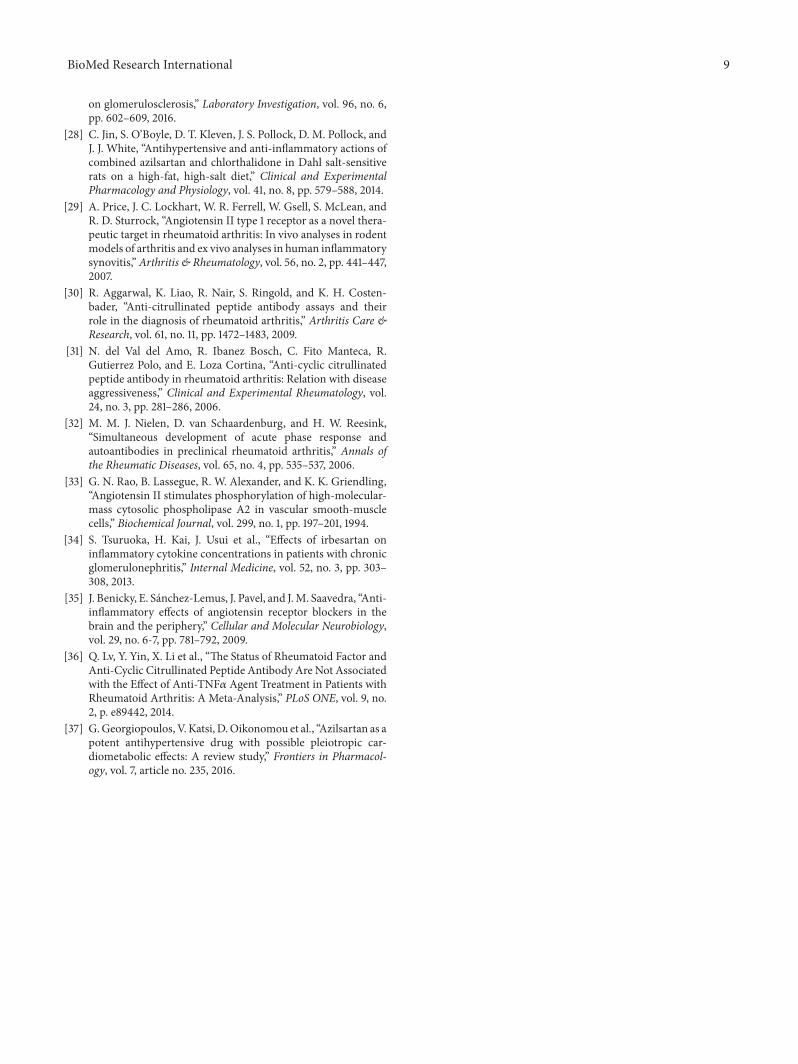
on glomerulosclerosis,” Laboratory Investigation, vol. 96, no. 6,pp. 602–609, 2016.
[28] C. Jin, S. O’Boyle, D. T. Kleven, J. S. Pollock, D. M. Pollock, andJ. J. White, “Antihypertensive and anti-inflammatory actions ofcombined azilsartan and chlorthalidone in Dahl salt-sensitiverats on a high-fat, high-salt diet,” Clinical and ExperimentalPharmacology and Physiology, vol. 41, no. 8, pp. 579–588, 2014.
[29] A. Price, J. C. Lockhart, W. R. Ferrell, W. Gsell, S. McLean, andR. D. Sturrock, “Angiotensin II type 1 receptor as a novel thera-peutic target in rheumatoid arthritis: In vivo analyses in rodentmodels of arthritis and ex vivo analyses in human inflammatorysynovitis,” Arthritis & Rheumatology, vol. 56, no. 2, pp. 441–447,2007.
[30] R. Aggarwal, K. Liao, R. Nair, S. Ringold, and K. H. Costen-bader, “Anti-citrullinated peptide antibody assays and theirrole in the diagnosis of rheumatoid arthritis,” Arthritis Care &Research, vol. 61, no. 11, pp. 1472–1483, 2009.
[31] N. del Val del Amo, R. Ibanez Bosch, C. Fito Manteca, R.Gutierrez Polo, and E. Loza Cortina, “Anti-cyclic citrullinatedpeptide antibody in rheumatoid arthritis: Relation with diseaseaggressiveness,” Clinical and Experimental Rheumatology, vol.24, no. 3, pp. 281–286, 2006.
[32] M. M. J. Nielen, D. van Schaardenburg, and H. W. Reesink,“Simultaneous development of acute phase response andautoantibodies in preclinical rheumatoid arthritis,” Annals ofthe Rheumatic Diseases, vol. 65, no. 4, pp. 535–537, 2006.
[33] G. N. Rao, B. Lassegue, R. W. Alexander, and K. K. Griendling,“Angiotensin II stimulates phosphorylation of high-molecular-mass cytosolic phospholipase A2 in vascular smooth-musclecells,” Biochemical Journal, vol. 299, no. 1, pp. 197–201, 1994.
[34] S. Tsuruoka, H. Kai, J. Usui et al., “Effects of irbesartan oninflammatory cytokine concentrations in patients with chronicglomerulonephritis,” Internal Medicine, vol. 52, no. 3, pp. 303–308, 2013.
[35] J. Benicky, E. Sanchez-Lemus, J. Pavel, and J.M. Saavedra, “Anti-inflammatory effects of angiotensin receptor blockers in thebrain and the periphery,” Cellular and Molecular Neurobiology,vol. 29, no. 6-7, pp. 781–792, 2009.
[36] Q. Lv, Y. Yin, X. Li et al., “The Status of Rheumatoid Factor andAnti-Cyclic Citrullinated Peptide Antibody Are Not Associatedwith the Effect of Anti-TNF𝛼 Agent Treatment in Patients withRheumatoid Arthritis: A Meta-Analysis,” PLoS ONE, vol. 9, no.2, p. e89442, 2014.
[37] G.Georgiopoulos, V.Katsi, D.Oikonomou et al., “Azilsartan as apotent antihypertensive drug with possible pleiotropic car-diometabolic effects: A review study,” Frontiers in Pharmacol-ogy, vol. 7, article no. 235, 2016.

Medicinal ChemistryInternational Journal of
Hindawiwww.hindawi.com Volume 2018
ToxicologyJournal of
Hindawiwww.hindawi.com Volume 2018
PainResearch and TreatmentHindawiwww.hindawi.com Volume 2018
Hindawiwww.hindawi.com Volume 2018
Arthritis
Neurology Research International
Hindawiwww.hindawi.com Volume 2018
StrokeResearch and TreatmentHindawiwww.hindawi.com Volume 2018
Drug DeliveryJournal of
Hindawiwww.hindawi.com Volume 2018
Hindawiwww.hindawi.com Volume 2018
Advances in Pharmacological Sciences
Tropical MedicineJournal of
Hindawiwww.hindawi.com Volume 2018
AddictionJournal of
Hindawiwww.hindawi.com Volume 2018
Hindawiwww.hindawi.com Volume 2018
BioMed Research International
Emergency Medicine InternationalHindawiwww.hindawi.com Volume 2018
Hindawiwww.hindawi.com Volume 2018
Anesthesiology Research and Practice
Journal of
Hindawiwww.hindawi.com Volume 2018
Pharmaceutics
Hindawi Publishing Corporation http://www.hindawi.com Volume 2013Hindawiwww.hindawi.com
The Scientific World Journal
Volume 2018
Infectious Diseases and Medical Microbiology
Hindawiwww.hindawi.com Volume 2018
Canadian Journal of
Hindawiwww.hindawi.com Volume 2018
Autoimmune DiseasesScienti�ca
Hindawiwww.hindawi.com Volume 2018
Hindawiwww.hindawi.com Volume 2018
MEDIATORSINFLAMMATION
of
Submit your manuscripts atwww.hindawi.com