1. ADAMTS13 in Arterial Thrombosis
160
ADAMTS13 in Arterial Thrombosis Tamara Bongers
-
Upload
sukma-effendy -
Category
Documents
-
view
20 -
download
0
description
nnn
Transcript of 1. ADAMTS13 in Arterial Thrombosis

ADAMTS13 inArterial Thrombosis
Tamara Bongers

ADAMTS13 in Arterial Thrombosis
© 2010 Tamara Bongers, Rotterdam, The Netherlands
No part of this thesis may be reproduced, stored in a retrieval system or transmitted in any form or by any means without permission from the author or, when appropriate, from publishers of the publications.
ISBN: 978-90-9025798-3
Cover design: Tamara BongersLayout: Henri Wijnbergen and Tamara BongersPrinting: Ipskamp Drukkers, Enschede

ADAMTS13 in Arterial Thrombosis
ADAMTS13 in arteriële trombose
Proefschrift
ter verkrijging van de graad van doctor aan deErasmus Universiteit Rotterdam
op gezag van de rector magnificus
Prof.dr. H.G. Schmidt
en volgens besluit van het College voor Promoties.De openbare verdediging zal plaatsvinden op
donderdag 9 december 2010 om 11:30 uur
doorTamara Natascha Bongers
geboren te Zevenaar

Promotiecommissie
Promotor: Prof.dr. F.W.G. Leebeek
Overige leden: Prof.dr. M.M.B. Breteler Prof.dr. D.W.J. Dippel Dr. T. Lisman Copromotor: Dr. M.P.M. de Maat
The work described in this thesis was performed at the Deparment of Hematology of Erasmus University Medical Center, Rotterdam, The Nether-lands. This work was partly funded by MRACE Translational Research Grant ErasmusMC 2004 as a clinical fellow to F.W.G. Leebeek.
Financial support by the Netherlands Heart Foundation for publication of this thesis is gratefully acknowledged.
Printing of this thesis was financially supported by Baxter, Erasmus University Rotterdam, Jurriaanse Stichting, Kordia and Pfizer.

“ The World is a book, and those who do not travel, only read a page”
Saint Augustine (354-430)
Aan mijn ouders


Table of contents
1 Introduction and outline thesis 11
Part 1 ADAMTS13 in arterial thrombosis
2 High Von Willebrand factor levels increase the risk of first ischemic stroke: influence of ADAMTS13, inflammation and genetic variability
29
3 Lower levels of ADAMTS13 are associated with cardiovascular disease in young patients
43
4a Absence of Pro475Ser polymorphism in ADAMTS13 in Caucasians
59
4b Frequency of the Tyr1584Cys polymorphism in arterial thrombosis
63
Part 2 Role of ADAMTS13 in other pathological conditions
5 Reduced ADAMTS13 in children with severe meningococcal sepsis is associated with severity and outcome
71
6 Elevated levels of Von Willebrand factor in cirrhosis support platelet adhesion despite reduced functional capacity
87
7 Measurement of ADAMTS13: assay comparison in TTP, liver cirrhosis patients and healthy controls
107
8 General Discussion 119
Summary / Samenvatting 135
Acknowledgements 145Curriculum vitae 151Publications 153Portfolio 157Abbreviations 159


Chapter 1
Introduction


11
Chapter 1
Atherosclerosis and atherothrombosisAtherothrombotic complications, including acute myocardial infarction, stroke and peripheral arterial diseases, are a major cause of morbidity and mortality in Western industrialized countries. Already in childhood changes occur in the vessel wall, such as thickening, reor-ganization of the tunica intima, collagenous matrix protein and the deposition of lipids.1 The lesions may progress by migration of smooth muscle cells from the media to the intima of the vessel wall. These cells proliferate and synthesize several matrix components. Some of the cells will go into apoptosis and form a necrotic core. The lesion is covered by a fibrous cap, which plays an important role in plaque stabilization. Rupture of this fibrous cap allows contact between coagulation factors in the blood and the thrombogenic tissue factor expressed by macrophage foam cells. This triggers platelet activation and aggregation. One of the main factors that initiates platelet aggregation and thrombus formation at sites of lesions of the endothelium is Von Willebrand Factor (VWF).
Figure 1 Process of atherosclerosis and thrombus formation in a blood vessel.
Von Willebrand factorVon Willebrand Factor is a glycoprotein that is important for platelet adhesion and aggregation and acts as a bridging molecule between subendothelial collagen molecules and circulating platelets at the site of vascular injury. Furthermore, it serves as a chaperon molecule for Factor VIII. VWF is synthesized in endothelial cells and megakaryocytes. A monomer of proVWF is synthesized in the endoplasmatic reticulum and forms dimers via di-sulfide bonds.2 These dimers subsequently form multimers, which are stored in
Fig 1 Process of atherosclerosis and thrombus formation in a blood vessel

12
Weibel-Palade bodies in the endothelial cells and α-granules of platelets. A small fraction of VWF is constitutively secreted into the circulation. Upon stimula-tion, ultra large VWF (ULVWF) is secreted from the Weibel-Palade bodies and forms string-like structures that are attached to the endothelial cell.3, 4 Under conditions of high shear stress, ULVWF strings are cleaved by the metallopro-tease A disintegrin-like and metalloprotease with thrombospondin type-1 motifs 13 (ADAMTS13) at the Tyr1605-Met1606 bond to form multimers of a size that normally circulate. The cleavage of multimers is important because ULVWF in the microvasculature are thrombogenic.The plasma levels of VWF are regulated by genetic and non-genetic factors. The most important genetic factor is ABO blood group. Individuals with blood group O have around 25% lower levels of VWF than individuals with non-O blood group. 5, 6 In addition, several common single nucleotide polymorphisms (SNPs) in the VWF gene influence the levels of VWF. For example, carriers of the minor G-allele of the –1793 C/G polymorphism in the promotor region, have 20% higher VWF levels.7 The GG genotype was also associated with a higher risk of coronary heart disease.8 Also other SNPs in VWF gene have been reported to determine VWF levels or the risk of cardiovascular disease.9, 10
VWF plasma levels are also influenced by a large number of non-genetic factors. Age is strongly associated with plasma levels of VWF.11, 12 The rigidity of the arterial wall and thereby the increased pulse pressure may stimulate VWF secretion. VWF levels are elevated in several inflammatory diseases, vasculitis and autoimmune diseases, which suggests that indeed cytokines and other markers of inflammation induce the secretion of VWF.13-15 Also in diabetic patients elevated levels of VWF are found.16
ADAMTS13The ADAMTS13 gene is located on chromosome 9q34. The gene is approximately 37 kb long and comprises 29 exons. The primary gene product is a protein of 1427 amino acids with a molecular mass of 145 kDa.This VWF cleaving protease has been identified as the 13th member of the ADAMTS family. This family shares the homology of domain structures. They all start with a signal peptide that is followed by a propeptide. The mature ADAMTS13 is build up by a catalytic domain, a disintegrin domain, TSP1-motif, a cystein-rich domain, a spacer domain, seven other TSP-1 domains and two CUB-domains. These two CUB-domains are specific for ADAMTS13 and are not seen in other members of the ADAMTS family (Figure 2). The function

13
Chapter 1
of these domains is not completely known, but they may play a role in protein-protein interactions.17
Figure 2 Domain alignment of ADAMTS13S: signal peptide, P:propeptide, TSP-1: thrombospondin-1 motif, CUB:complement compo-nents C1r/C1s, Uegf (epidermal growth factor) bone morphogenic protein-1
The expression of ADAMTS13 has been observed in various cell types and tissues. The transcript of approximately 4.7 kb is primarily expressed in the stel-late cells in the liver.18, 19 It has been suggested that the stellate cells may play a role in the regulation of plasma levels of ADAMTS13.20 A transcript of 2.4 kb was detectable in multiple tissues, including placenta, skeletal muscle and certain tumor cell lines.18, 21-23 Also vascular endothelial cells have been shown to syn-thesize and secrete ADAMTS13.24, 25 The expression of ADAMTS13 has also been observed in platelets.26 Upon activation, ADAMTS13 may be released from platelets. It has been reported that ADAMTS13 is also expressed in renal tissue in glomeruli and tubuli.27
After initiation of hemostasis platelets adhere to ULVWF, which is anchored to the activated endothelial cell or the exposed subendothelium. Shear stress forces and the binding of the platelet receptor Glycoprotein Ibα to the A1 do-main of VWF cause a conformational change of VWF. Hereby the ADAMTS13 binding site and the scissile bond in the A2 domain in VWF become exposed. ADAMTS13 from the circulation binds to the A3 domain, which has been identified as the major docking site for the metalloprotease. Subsequently, the metalloprotease binds to the A2 domain. Once attached, ADAMTS13 cleaves VWF at the Tyr1605-Met1606 peptide bond, predominantly at or close to the anchoring sites of the ULVWF multimers, where the tensile forces are the great-est.28 The remaining strings of VWF that are released after cleavage are less biologically active and less susceptible for cleavage.29 Hereby, the proteolysis of VWF by ADAMTS13 appears to be critical in preventing thrombosis in the microvasculature (Figure 3).

14
Figure 3 Cleavage of VWF by ADAMTS13 in a normal subject and a TTP patient (NEJM 2002 Moake). Upon stimulation unusually large VWF multimers are released from the Weibel Palade bodies from the endothelial cell. ADAMTS13 (also known as VWF-cleaving protease) attaches to the endothelial cell via one of its thrombospondin-1-like domains. It than cleaves the unusually large multimers into smaller multimers and prevents thereby that VWF can induce adhesion and aggregation of platelets during normal flow.
Thrombotic Thrombocytopenic PurpuraThe physiological importance of ADAMTS13 is examplified by the rare disease entity thrombotic thrombocytopenic purpura (TTP). In TTP, ADAMTS13 activity is strongly reduced or absent which results in the inability to cleave ULVWF into smaller fragments. This results in platelet aggregation and eventually in micro-vascular thrombus formation (Figure 3).TTP is, if untreated, a life-threatening disorder, which is characterized by throm-bocytopenia, micro-angiopathic hemolytic anemia, neurological symptoms, renal dysfunction and fever. Typical for TTP is the occurrence of microvascular hyaline thrombi in organs such as the brain and kidneys. These thrombi are VWF- and platelet-rich, but fibrin-poor. Platelets are consumed which leads to severe thrombocytopenia and thereby to hemorrhage, ischemia and organ dysfunction.

15
Chapter 1
TTP can occur in two forms: a congenital form and an acquired form. The congenital form is caused by mutations in the ADAMTS13 gene, resulting in absence of ADAMTS13 activity, while the acquired form is caused by the formation of auto-antibodies against the metalloprotease.18, 30, 31 Most of the auto-antibodies are inhibitory, but also non-inhibitory antibodies have been described. These are suggested to interfere with the binding of ADAMTS13 to VWF or to accelerate the clearance of ADAMTS13, which results in low levels of ADAMTS13 in plasma.
ADAMTS13 in other diseasesDecreased levels of ADAMTS13 have also been reported in several other disease states including disseminated intravascular coagulation (DIC), sepsis, inflammatory diseases, in the post-operative state, hemolytic uremic syndrome (HUS) and liver disease.32-37
Sepsis and DIC are associated with a prothrombotic and systemic inflammatory state, in which many factors can influence ADAMTS13 activity. It is not known whether ADAMTS13 plays a role in inflammation, but studies have reported reduced ADAMTS13 activity in patients with acute systemic inflammation or sepsis.38-40 In sepsis plasma levels of acute phase proteins, such as VWF are strongly increased. It has been suggested that the release of VWF is so strongly increased that ADAMTS13 is no longer able to cleave all ULVWF molecules. Pro-inflammatory cytokines like Interleukin-8 (IL-8), tumor necrosis factor-α (TNF-α) and Interleukin-6 (IL-6) have been shown to have an effect on the release of ULVWF and its processing.14 Chauhan et al. previously demonstrated that ADAMTS13 cleaves platelet-VWF strings, regulates platelet interaction and modulates the thrombotic response in injured arterioles.41, 42 These studies have shown that ADAMTS13 may be a new link between inflammation and throm-bosis. This link is interesting, since inflammation is associated with a prothrom-botic state43 and ADAMTS13 may be an intermediate or pathogenetic factor. In this thesis we will focus on the association between ADAMTS13 and cardiovas-cular disease, such as acute myocardial infarction, transient ischemic attack and ischemic stroke. Furthermore, we will investigate ADAMTS13 in other disease states that are associated with a prothrombotic phenotype.
Genetics of ADAMTS13Since the discovery of a genetic defect of ADAMTS13 in patients with con-genital TTP, genetic analyses have been performed in many patients, and various

16
mutations in the ADAMTS13 gene have been reported. Often, carriers of such mutations are known to develop TTP during childhood, but also cases with an adult onset have been reported.18 Genetic variations occur throughout the ADAMTS13 gene without clustering. The majority of the mutations are missense mutations, but also splice site, non-sense and frame-shift mutations have been described.18, 44-53
In recent years genetic variation in the ADAMTS13 gene has been further inves-tigated. To date 46 mutations and 19 non-synonymous SNPs have been found in the ADAMTS13 gene.54 It is not completely clear whether these genetic variations influence the activity of ADAMTS13. When the activity of ADAMTS13 activity is reduced by the genetic variation, this may result in larger VWF multimers. For example, in individuals with the P475S polymorphism, ADAMTS13 is normally secreted, but has a low proteolytic activity toward VWF.45 This SNP is very common in Japanese subjects. Nearly 10% of the population is heterozygous for this polymorphism, whereas P475S is rare in Chinese.55 The frequency in Caucasians is yet unknown. The ADAMTS13 variant Q449X is secreted normally, but is characterized by a very low activity of ADAMTS13. Also combinations of genetic variations may result in low ADAMTS13 activity.56
Some mutations that cause premature truncations have shown to impair secre-tion or reduce ADAMTS13 activity. The frameshift mutation 4143-4144insA results in deletion of the second CUB-domain and results in impaired ADAMTS13 secretion.44 Since some of the polymorphisms are influencing the activity of ADAMTS13 one can imagine that these polymorphisms may influ-ence VWF levels and thereby the risk of arterial thrombosis. In this thesis several single nucleotide polymorphisms (SNP) in ADAMTS13 will be studied. The SNPs have been chosen according to the Seattle SNP site (http://pga.gs.washington.edu/ ). In this database, tools are provided to select certain tagging SNPs to cover the whole genetic variation in a gene of inter-est. The selected SNPs will be studied in relation to the levels and activity of ADAMTS13 and to the relative risk of arterial thrombosis.
Assays for ADAMTS13Since Furlan and Tsai reported the discovery of ADAMTS13, several assays to determine ADAMTS13 activity or concentration have been developed.31, 57 The assays that measure ADAMTS13 activity are based on two principles: (1) detec-tion of VWF cleavage fragments or (2) VWF residual activity after cleavage by

17
Chapter 1
ADAMTS13.58
The first described assay was the Gerritsen assay and was based on the principle of proteolysis of VWF by ADAMTS13 and quantifying the amount of VWF digestion products. A disadvantage of this method is that it is very time- consuming and has a high coefficient of variation.59
The cleavage products of VWF can be detected by VWF multimer structure analysis under non-reducing conditions57 or SDS-PAGE under non-reducing conditions (140 kDa and 176 kDa) or two site immuno-radiometric assays with antibodies against the C- and N-terminal VWF epitopes. Cleavage is detected by an apparent loss of VWF antigen.60
A limitation of these assays is that they are performed under static conditions. They require urea or guanidine HCL to denaturate and unfold VWF, which is time-consuming. Furthermore, Ba2+ or Ca2+ must be added to stimulate ADAMTS13 activity. Newer assays have become available for the measurement of ADAMTS13. Some of them use recombinant VWF fragments that do not need the denaturing conditions, such as the recombinant VWF A2 domain or the VWF73 peptide. These assays are less complex and save time. The VWF73 peptide has been reported to be the minimal substrate to be recognized and cleaved by ADAMTS13.61
One more recent developed assay can detect cleavage products by immunoblotting, ELISA or fluorescence.62-65 Furthermore, another assay uses more physiological conditions and measures ADAMTS13 under flow and quantifies the amount of ULVWF-platelet strings that are cleaved.29 This assay is reliable primarily for discriminating between levels of ADAMTS13 higher or lower than 20%.66 As an alternative one can use the detection of VWF residual activity after proteolysis of VWF to measure ADAMTS13 activity. The VWF residual activity correlates with the multimeric size. The residual activity of VWF can be determined by the ability of VWF to bind to collagen or to induce platelet aggregation in the presence of ristocetin.58, 59
Outline of this thesisThe aim of this thesis is to further unravel the physiological and pathophysiological role of ADAMTS13 in several disease states. In the first part we will study ADAMTS13 in cardiovascular disease, with a special focus on the genetic variation in both ADAMTS13 and VWF. The second part of the thesis studies the role of ADAMTS13 in other pathological conditions known to be

18
associated with a prothrombotic state. In addition several assays for ADAMTS13 antigen and activity levels are compared both in healthy controls and patients.In chapter 2 a study on the relationship between ADAMTS13 and VWF in patients with ischemic stroke included in the COCOS study is presented. This study is previously described.67 We hypothesize that low levels of ADAMTS13 may result in higher VWF levels and thereby increase the risk of arterial throm-bosis. Subsequently another, independent study is performed in the ATTAC study population, a case-control study of young patients who have recently suffered a first acute myocardial infarction, ischemic stroke or peripheral arterial disease. We include young individuals, males before the age of 45 and women before the age of 55, because it is known from previous studies that in younger patients the contribution of genetic variation is more pronounced than in elderly subjects. The role of genetic variation, its relationship with levels of ADAMTS13 and the risk of arterial thrombosis are assessed (Chapter 3). The role of genetic variation in ADAMTS13 is further investigated in Chapter 4, in which we study a genetic variant that is previously reported to be associated with a reduced activity of ADAMTS13 in Japanese subjects. Genetic variations in VWF gene have been described that lead to less or increased cleavage of VWF. We hypothesized that common SNPs in VWF gene maybe be associated with the risk of arterial thrombosis by influencing the cleavage by ADAMTS13. Therefore, we study a genetic variant in the VWF gene that is previously reported to be associated with increased proteolysis of VWF (Chapter 4b).68
The second part of the thesis describes the role of ADAMTS13 in other pathological conditions associated with microvascular thrombosis, such as liver cirrhosis (Chapter 5) and sepsis (Chapter 6). The liver is the main site of synthesis of many coagulation factors. Also ADAMTS13 is synthesized in the liver predominantly in stellate cells. In patients with cirrhosis of the liver several alterations of the haemostatic system do occur.69 Most of the coagulation factors are reduced in cirrhosis, mainly due to decreased synthesis. However, these alterations do not necessarily lead to a bleeding disorder, but are now even thought to be associated with a prothrombotic state.70 We study the role of ADAMTS13 and VWF in patients with liver cirrhosis to further unravel primary hemostasis in these individuals (Chapter 5). Another pathological condition in which we study the role of ADAMTS13 is meningococcal sepsis. Patients with sepsis often suffer from thrombotic

19
Chapter 1
complications. Especially patients with meningococcal sepsis frequently have severe disseminated intravascular coagulation (DIC) and microthrombi forma-tion, even resulting in limb ischemia and skin necrosis. Therefore we investigated the levels of ADAMTS13 in children with meningococcal sepsis which were referred to our hospital to study its potential pathogenetic role in hemostatic complications and outcome of these patients. (Chapter 6)The last part of this thesis comprises a study on various assays of ADAMTS13 that are currently available. Some of the assays only measure activity of ADAMTS13 while others measure the antigen levels of ADAMTS13. Initially these assays where designed for the diagnosis of TTP. We will investigate the correlation between these assays in patients with TTP, but also in patients with liver cirrhosis. The results of this study are reported in Chapter 7.
References
1. Napoli C, D'Armiento FP, Mancini FP, et al. Fatty streak formation occurs in human fetal aortas
and is greatly enhanced by maternal hypercholesterolemia. Intimal accumulation of low density
lipoprotein and its oxidation precede monocyte recruitment into early atherosclerotic lesions. The
Journal of clinical investigation 1997;100;11:2680-90.
2. Zheng XL, Sadler JE. Pathogenesis of thrombotic microangiopathies. Annual review of pathology
2008;3:249-77.
3. Vischer UM, Wagner DD. von Willebrand factor proteolytic processing and multimerization pre-
cede the formation of Weibel-Palade bodies. Blood 1994;83;12:3536-44.
4. van Mourik JA, Romani de Wit T, Voorberg J. Biogenesis and exocytosis of Weibel-Palade bodies.
Histochemistry and cell biology 2002;117;2:113-22.
5. de Lange M, Snieder H, Ariens RA, et al. The genetics of haemostasis: a twin study. Lancet
2001;357;9250:101-5.
6. Matsui T, Fujimura Y, Nishida S, et al. Human plasma alpha 2-macroglobulin and von Willebrand
factor possess covalently linked ABO(H) blood group antigens in subjects with corresponding
ABO phenotype. Blood 1993;82;2:663-8.
7. Harvey PJ, Keightley AM, Lam YM, et al. A single nucleotide polymorphism at nucleotide -1793
in the von Willebrand factor (VWF) regulatory region is associated with plasma VWF:Ag levels.
British journal of haematology 2000;109;2:349-53.
8. van der Meer IM, Brouwers GJ, Bulk S, et al. Genetic variability of von Willebrand factor and risk
of coronary heart disease: the Rotterdam Study. British journal of haematology 2004;124;3:343-7.
9. Di Bitondo R, Cameron CL, Daly ME, et al. The -1185 A/G and -1051 G/A dimorphisms in the
von Willebrand factor gene promoter and risk of myocardial infarction. British journal of haema-

20
tology 2001;115;3:701-6.
10. Dai K, Gao W, Ruan C. The Sma I polymorphism in the von Willebrand factor gene associated
with acute ischemic stroke. Thromb Res 2001;104;6:389-95.
11. Thompson SG, Kienast J, Pyke SD, et al. Hemostatic factors and the risk of myocardial infarction
or sudden death in patients with angina pectoris. European Concerted Action on Thrombosis and
Disabilities Angina Pectoris Study Group. The New England journal of medicine 1995;332;10:635-
41.
12. Danesh J, Wheeler JG, Hirschfield GM, et al. C-reactive protein and other circulating markers of
inflammation in the prediction of coronary heart disease. The New England journal of medicine
2004;350;14:1387-97.
13. Blann AD, Herrick A, Jayson MI. Altered levels of soluble adhesion molecules in rheumatoid ar-
thritis, vasculitis and systemic sclerosis. British journal of rheumatology 1995;34;9:814-9.
14. Bernardo A, Ball C, Nolasco L, et al. Effects of inflammatory cytokines on the release and cleav-
age of the endothelial cell-derived ultralarge von Willebrand factor multimers under flow. Blood
2004;104;1:100-6.
15. Bhatia R, Matsushita K, Yamakuchi M, et al. Ceramide triggers Weibel-Palade body exocytosis.
Circulation research 2004;95;3:319-24.
16. Stehouwer CD, Gall MA, Twisk JW, et al. Increased urinary albumin excretion, endothelial dys-
function, and chronic low-grade inflammation in type 2 diabetes: progressive, interrelated, and
independently associated with risk of death. Diabetes 2002;51;4:1157-65.
17. Levy GG, Motto DG, Ginsburg D. ADAMTS13 turns 3. Blood 2005;106;1:11-7.
18. Levy GG, Nichols WC, Lian EC, et al. Mutations in a member of the ADAMTS gene family cause
thrombotic thrombocytopenic purpura. Nature 2001;413;6855:488-94.
19. Zheng X, Chung D, Takayama TK, et al. Structure of von Willebrand factor-cleaving protease
(ADAMTS13), a metalloprotease involved in thrombotic thrombocytopenic purpura. The Journal
of biological chemistry 2001;276;44:41059-63.
20. Kume Y, Ikeda H, Inoue M, et al. Hepatic stellate cell damage may lead to decreased plasma AD-
AMTS13 activity in rats. FEBS letters 2007;581;8:1631-4.
21. Soejima K, Mimura N, Hirashima M, et al. A novel human metalloprotease synthesized in the liver
and secreted into the blood: possibly, the von Willebrand factor-cleaving protease? Journal of bio-
chemistry 2001;130;4:475-80.
22. Zhou W, Inada M, Lee TP, et al. ADAMTS13 is expressed in hepatic stellate cells. Laboratory in-
vestigation; a journal of technical methods and pathology 2005;85;6:780-8.
23. Uemura M, Tatsumi K, Matsumoto M, et al. Localization of ADAMTS13 to the stellate cells of
human liver. Blood 2005;106;3:922-4.
24. Turner N, Nolasco L, Tao Z, et al. Human endothelial cells synthesize and release ADAMTS-13. J
Thromb Haemost 2006;4;6:1396-404.

21
25. Shang D, Zheng XW, Niiya M, et al. Apical sorting of ADAMTS13 in vascular endothelial cells and
Madin-Darby canine kidney cells depends on the CUB domains and their association with lipid
rafts. Blood 2006;108;7:2207-15.
26. Suzuki M, Murata M, Matsubara Y, et al. Detection of von Willebrand factor-cleaving pro-
tease (ADAMTS-13) in human platelets. Biochemical and biophysical research communications
2004;313;1:212-6.
27. Manea M, Kristoffersson A, Schneppenheim R, et al. Podocytes express ADAMTS13 in normal
renal cortex and in patients with thrombotic thrombocytopenic purpura. British journal of haema-
tology 2007;138;5:651-62.
28. Wu JJ, Fujikawa K, McMullen BA, et al. Characterization of a core binding site for ADAMTS-13
in the A2 domain of von Willebrand factor. Proceedings of the National Academy of Sciences of
the United States of America 2006;103;49:18470-4.
29. Dong JF, Moake JL, Nolasco L, et al. ADAMTS-13 rapidly cleaves newly secreted ultralarge
von Willebrand factor multimers on the endothelial surface under flowing conditions. Blood
2002;100;12:4033-9.
30. Furlan M, Lammle B. Deficiency of von Willebrand factor-cleaving protease in familial and ac-
quired thrombotic thrombocytopenic purpura. Bailliere's clinical haematology 1998;11;2:509-14.
31. Tsai HM, Lian EC. Antibodies to von Willebrand factor-cleaving protease in acute thrombotic
thrombocytopenic purpura. The New England journal of medicine 1998;339;22:1585-94.
32. Mannucci PM, Canciani MT, Forza I, et al. Changes in health and disease of the metalloprotease
that cleaves von Willebrand factor. Blood 2001;98;9:2730-5.
33. Feys HB, Canciani MT, Peyvandi F, et al. ADAMTS13 activity to antigen ratio in physiological and
pathological conditions associated with an increased risk of thrombosis. British journal of haema-
tology 2007;138;4:534-40.
34. Bianchi V, Robles R, Alberio L, et al. Von Willebrand factor-cleaving protease (ADAMTS13) in
thrombocytopenic disorders: a severely deficient activity is specific for thrombotic thrombocyto-
penic purpura. Blood 2002;100;2:710-3.
35. Martin K, Borgel D, Lerolle N, et al. Decreased ADAMTS-13 (A disintegrin-like and metallopro-
tease with thrombospondin type 1 repeats) is associated with a poor prognosis in sepsis-induced
organ failure. Critical care medicine 2007;35;10:2375-82.
36. Kremer Hovinga JA, Zeerleder S, Kessler P, et al. ADAMTS-13, von Willebrand factor and related
parameters in severe sepsis and septic shock. J Thromb Haemost 2007;5;11:2284-90.
37. Loof AH, van Vliet HH, Kappers-Klunne MC. Low activity of von Willebrand factor-cleaving
protease is not restricted to patients suffering from thrombotic thrombocytopenic purpura. British
journal of haematology 2001;112;4:1087-8.
38. Ono T, Mimuro J, Madoiwa S, et al. Severe secondary deficiency of von Willebrand factor-cleaving
protease (ADAMTS13) in patients with sepsis-induced disseminated intravascular coagulation: its

22
correlation with development of renal failure. Blood 2006;107;2:528-34.
39. Nguyen TC, Liu A, Liu L, et al. Acquired ADAMTS-13 deficiency in pediatric patients with severe
sepsis. Haematologica 2007;92;1:121-4.
40. Reiter RA, Varadi K, Turecek PL, et al. Changes in ADAMTS13 (von-Willebrand-factor-cleaving
protease) activity after induced release of von Willebrand factor during acute systemic inflamma-
tion. Thrombosis and haemostasis 2005;93;3:554-8.
41. Motto DG, Chauhan AK, Zhu G, et al. Shigatoxin triggers thrombotic thrombocytopenic pur-
pura in genetically susceptible ADAMTS13-deficient mice. The Journal of clinical investigation
2005;115;10:2752-61.
42. Chauhan AK, Motto DG, Lamb CB, et al. Systemic antithrombotic effects of ADAMTS13. The
Journal of experimental medicine 2006;203;3:767-76.
43. Levi M, van der Poll T. Inflammation and coagulation. Critical care medicine 38;2 Suppl:S26-34.
44. Pimanda JE, Maekawa A, Wind T, et al. Congenital thrombotic thrombocytopenic purpura in as-
sociation with a mutation in the second CUB domain of ADAMTS13. Blood 2004;103;2:627-9.
45. Kokame K, Matsumoto M, Soejima K, et al. Mutations and common polymorphisms in AD-
AMTS13 gene responsible for von Willebrand factor-cleaving protease activity. Proceedings of the
National Academy of Sciences of the United States of America 2002;99;18:11902-7.
46. Antoine G, Zimmermann K, Plaimauer B, et al. ADAMTS13 gene defects in two brothers with
constitutional thrombotic thrombocytopenic purpura and normalization of von Willebrand factor-
cleaving protease activity by recombinant human ADAMTS13. British journal of haematology
2003;120;5:821-4.
47. Assink K, Schiphorst R, Allford S, et al. Mutation analysis and clinical implications of von Wil-
lebrand factor-cleaving protease deficiency. Kidney international 2003;63;6:1995-9.
48. Matsumoto M, Kokame K, Soejima K, et al. Molecular characterization of ADAMTS13 gene mu-
tations in Japanese patients with Upshaw-Schulman syndrome. Blood 2004;103;4:1305-10.
49. Savasan S, Lee SK, Ginsburg D, et al. ADAMTS13 gene mutation in congenital thrombotic throm-
bocytopenic purpura with previously reported normal VWF cleaving protease activity. Blood
2003;101;11:4449-51.
50. Schneppenheim R, Budde U, Oyen F, et al. von Willebrand factor cleaving protease and AD-
AMTS13 mutations in childhood TTP. Blood 2003;101;5:1845-50.
51. Licht C, Stapenhorst L, Simon T, et al. Two novel ADAMTS13 gene mutations in thrombot-
ic thrombocytopenic purpura/hemolytic-uremic syndrome (TTP/HUS). Kidney international
2004;66;3:955-8.
52. Uchida T, Wada H, Mizutani M, et al. Identification of novel mutations in ADAMTS13 in an adult
patient with congenital thrombotic thrombocytopenic purpura. Blood 2004;104;7:2081-3.
53. Noris M, Bucchioni S, Galbusera M, et al. Complement factor H mutation in familial thrombotic
thrombocytopenic purpura with ADAMTS13 deficiency and renal involvement. J Am Soc Nephrol

23
2005;16;5:1177-83.
54. Lotta LA, Garagiola I, Palla R, et al. ADAMTS13 mutations and polymorphisms in congenital
thrombotic thrombocytopenic purpura. Human mutation 2010 31;1:11-9.
55. Ruan C, Dai L, Su J, et al. The frequency of P475S polymorphism in von Willebrand factor-
cleaving protease in the Chinese population and its relevance to arterial thrombotic disorders.
Thrombosis and haemostasis 2004;91;6:1257-8.
56. Plaimauer B, Fuhrmann J, Mohr G, et al. Modulation of ADAMTS13 secretion and specific ac-
tivity by a combination of common amino acid polymorphisms and a missense mutation. Blood
2006;107;1:118-25.
57. Furlan M, Robles R, Galbusera M, et al. von Willebrand factor-cleaving protease in thrombotic
thrombocytopenic purpura and the hemolytic-uremic syndrome. The New England journal of
medicine 1998;339;22:1578-84.
58. Bohm M, Vigh T, Scharrer I. Evaluation and clinical application of a new method for measuring
activity of von Willebrand factor-cleaving metalloprotease (ADAMTS13). Annals of hematology
2002;81;8:430-5.
59. Gerritsen HE, Turecek PL, Schwarz HP, et al. Assay of von Willebrand factor (vWF)-cleaving
protease based on decreased collagen binding affinity of degraded vWF: a tool for the diagnosis of
thrombotic thrombocytopenic purpura (TTP). Thrombosis and haemostasis 1999;82;5:1386-9.
60. Obert B, Tout H, Veyradier A, et al. Estimation of the von Willebrand factor-cleaving protease in
plasma using monoclonal antibodies to vWF. Thrombosis and haemostasis 1999;82;5:1382-5.
61. Kokame K, Matsumoto M, Fujimura Y, et al. VWF73, a region from D1596 to R1668 of von Wil-
lebrand factor, provides a minimal substrate for ADAMTS-13. Blood 2004;103;2:607-12.
62. Cruz MA, Whitelock J, Dong JF. Evaluation of ADAMTS-13 activity in plasma using recom-
binant von Willebrand Factor A2 domain polypeptide as substrate. Thrombosis and haemostasis
2003;90;6:1204-9.
63. Whitelock JL, Nolasco L, Bernardo A, et al. ADAMTS-13 activity in plasma is rapidly measured by
a new ELISA method that uses recombinant VWF-A2 domain as substrate. J Thromb Haemost
2004;2;3:485-91.
64. Zhou W, Tsai HM. An enzyme immunoassay of ADAMTS13 distinguishes patients with throm-
botic thrombocytopenic purpura from normal individuals and carriers of ADAMTS13 mutations.
Thrombosis and haemostasis 2004;91;4:806-11.
65. Kokame K, Nobe Y, Kokubo Y, et al. FRETS-VWF73, a first fluorogenic substrate for AD-
AMTS13 assay. British journal of haematology 2005;129;1:93-100.
66. Tripodi A, Chantarangkul V, Bohm M, et al. Measurement of von Willebrand factor cleaving pro-
tease (ADAMTS-13): results of an international collaborative study involving 11 methods testing
the same set of coded plasmas. J Thromb Haemost 2004;2;9:1601-9.
67. Leebeek FW, Goor MP, Guimaraes AH, et al. High functional levels of thrombin-activatable fibri-

24
nolysis inhibitor are associated with an increased risk of first ischemic stroke. J Thromb Haemost
2005;3;10:2211-8.
68. Bowen DJ, Collins PW, Lester W, et al. The prevalence of the cysteine1584 variant of von Wil-
lebrand factor is increased in type 1 von Willebrand disease: co-segregation with increased sus-
ceptibility to ADAMTS13 proteolysis but not clinical phenotype. British journal of haematology
2005;128;6:830-6.
69. Lisman T, Leebeek FW. Hemostatic alterations in liver disease: a review on pathophysiology, clini-
cal consequences, and treatment. Digestive surgery 2007;24;4:250-8.
70. Pereboom IT, Adelmeijer J, van Leeuwen Y, et al. Development of a severe von Willebrand factor/
ADAMTS13 dysbalance during orthotopic liver transplantation. Am J Transplant 2009;9;5:1189-
96.


ADAMTS13 in arterial thrombosis
Part 1


High von Willebrand Factor levels increase the risk of first ischemic stroke: influence of ADAMTS13, inflammation and genetic variability
T.N. Bongers, M.P.M. de Maat, M.P.J. van Goor, V. Bhagwanbali, H.H.D.M. van Vliet, E.B. Gómez García, D.W.J. Dippel, F.W.G. Leebeek
Stroke 2006 37; 2672-2677
Chapter 2
30
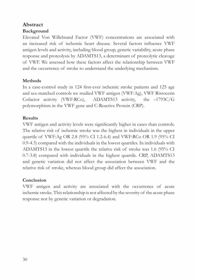
Abstract BackgroundElevated Von Willebrand Factor (VWF) concentrations are associated with an increased risk of ischemic heart disease. Several factors influence VWF antigen levels and activity, including blood group, genetic variability, acute phase response and proteolysis by ADAMTS13, a determinant of proteolytic cleavage of VWF. We assessed how these factors affect the relationship between VWF and the occurrence of stroke to understand the underlying mechanism.
MethodsIn a case-control study in 124 first-ever ischemic stroke patients and 125 age and sex-matched controls we studied VWF antigen (VWF:Ag), VWF Ristocetin Cofactor activity (VWF:RCo), ADAMTS13 activity, the –1793C/G polymorphism in the VWF gene and C-Reactive Protein (CRP).
ResultsVWF antigen and activity levels were significantly higher in cases than controls. The relative risk of ischemic stroke was the highest in individuals in the upper quartile of VWF:Ag OR 2.8 (95% CI 1.2-6.4) and VWF:RCo OR 1.9 (95% CI 0.9-4.3) compared with the individuals in the lowest quartiles. In individuals with ADAMTS13 in the lowest quartile the relative risk of stroke was 1.6 (95% CI 0.7-3.8) compared with individuals in the highest quartile. CRP, ADAMTS13 and genetic variation did not affect the association between VWF and the relative risk of stroke, whereas blood group did affect the association.
ConclusionVWF antigen and activity are associated with the occurrence of acute ischemic stroke. This relationship is not affected by the severity of the acute phase response nor by genetic variation or degradation.

31
Chapter 2
IntroductionVon Willebrand Factor (VWF) is a plasma glycoprotein that is a mediator of platelet adhesion to the vascular subendothelial collagen, which becomes available when the endothelium is damaged. VWF is released as ultra-large multimers, which form spontaneous high-strength bonds with the platelet glycoprotein 1b-IX-V complex, resulting in platelet aggregation and thrombus formation.1 The concentration and activity of VWF are influenced by several factors, including blood group, inflammation and proteolysis by ADAMTS13 (A Disintegrin and Metalloprotease with ThromboSpondin motif). Genetic variations in the VWF gene are associated with VWF levels, for example the –1793 C/G polymorphism in the promotor region of the VWF gene. The G-allele is associated with higher levels of VWF antigen than the C-allele.2
Proteolysis by ADAMTS13 also determines VWF activity. ADAMTS13 is a recently discovered metalloprotease that specifically cleaves the bond between tyrosine 842 and methionine 843 in the A2 domain of VWF multimers, resulting in 2 fragments of 176-kDa and 140-kDa.3, 4 These fragments are less active in platelet aggregation compared with the uncleaved Ultra Large VWF multimers (ULVWF). A deficiency of ADAMTS13 is seen in patients with thrombotic thrombocytopenic purpura (TTP), characterized by cerebral ischemia caused by platelet thrombosis in the cerebral microcirculation.5, 6 Several studies have shown that high VWF antigen levels are a risk factor for arterial thrombosis.7-9 However, information on VWF and cerebrovascular disease is limited and not consistent.8, 10 We hypothesized that ADAMTS13 plays a role in arterial thrombosis, because reduced ADAMTS13 activity will result in less degradation of ULVWF multimers and thereby in an increased VWF activity. The aim of our study is to further investigate the role of VWF in ischemic stroke and to assess how the factors mentioned above can affect the relationship between VWF and the occurrence of ischemic stroke.
MethodsStudy designWe performed a case-control study, where cases were consecutively recruited patients with first-ever acute ischemic stroke, admitted to the department of Neurology of the Erasmus University Medical Center. Population controls were partners, friends or neighbours of the patients. They were age-and sex matched, of the Caucasian race, without a history of stroke and not related to the patient. The study design has previously been described in more detail elsewhere.11

32
Plasma level measurementsVon Willebrand Factor antigen (VWF:Ag) was determined with an in-house ELISA assay using polyclonal rabbit antihuman VWF antibodies (DakoCytoma-tion, Glostrop, Denmark). Von Willebrand Factor ristocetin cofactor activity (VWF:RCo) was measured by an aggregometric method using formaline-fixed platelets and ristocetin (Diagnostica Stago, Asnières, France).
ADAMTS13 activity was measured using a rapid functional assay that deter-mines the digestion of VWF by ADAMTS13, based on the method described by Gerritsen with minor modifications as described before.12 Normal Pooled plasma (NPP) obtained from plasma of 40 healthy volunteers was used for cali-bration.
The concentration of CRP was determined with a sensitive in-house enzyme immunoassay using rabbit antibodies against human CRP as catching antibodies and goat antibodies against human CRP as tagging antibodies (DAKO, Den-mark).
-1793 C/G Polymorphism in Von Willebrand Factor geneThe DNA fragment containing the polymorphism was amplified by PCR with forward primer 5’AGCCCAGCGGACAGTGCGAG-3, and reverse primer: 5’ TACAAGAATGGGCAGTGCAG-3. The PCR comprised 4 min at 95°C of initial denaturation followed by 32 cycles of 1 min at 94°C, 1 min at 65°C and 2 min at 72°C. Subsequently the PCR product of 264 bp was digested by AciI, which cleaves the G-allele into 2 fragments of 180 and 84 bp, while the common C-allele was not cleaved. The fragments were separated using a 2% agarose gel and visualized using UV.13
Statistical analysisThe levels of VWF:Ag, VWF:RCo and ADAMTS13 were compared in cases and controls and in subgroups regarding blood group with a Student’s t-test. VWF:Ag, VWF:RCo and ADAMTS13 activity were divided into quartiles based on the distribution in the control group. The relationship between the levels of these variables and ischemic stroke was estimated using logistic regression with the lowest quartile as reference, except for the ADAMTS13 activity where the highest quartile is the reference group. The analyses were adjusted for pos-sible confounders such as blood group and vascular risk factors, adjustment for

33
Chapter 2
age and sex was not performed because the controls and cases were matched for these variables. A p-value of <0.05 was considered to indicate statistical significance. In the subgroup analysis for blood group, logistic regression was performed using quartiles of VWF, which were calculated for each subgroup separately. Correlations were estimated using the Spearman correlation co- efficient.The relationship between the levels of VWF and the –1793 C/G polymorphism was studied with analysis of variances (ANOVA) with adjustment for age, sex and blood group. The association of the –1793 C/G polymorphism with the occurrence of ischemic stroke was estimated by logistic regression and is presented with the 95% confidence intervals (CI). The statistical analysis was carried out using SPSS software version 11.
ResultsThe study included 124 patients and 125 sex- and age-matched controls and the characteristics are presented in Table 1. Plasma levels were available for all individuals. Genetic analysis could be performed for 109 patients and 119 controls.
Table 1 Baseline characteristics Cases
(N=124)Controls(N=125)
P-value
DemographicsAge (y) 56 (±12) 56 (±12) n.s
Female sex 58 (47%) 59 (47%) n.s
Blood groupO 54 (44%) 63 (50%) 0.37Non O 68 (56%) 62 (50%)
Risk factorsSmoking 61 (49%) 37 (30%) 0.004Hypertension 60 (48%) 24 (19%) 0.000Diabetes 18 (14%) 5 (4%) 0.004Hypercholesterolemia 78 (61%) 84 (67%) 0.70CRP 2.01 1.42 0.008
Data for age are presented as mean (±sd). Data for CRP are presented as median (25-75% range). Other data are counts (percentages). CRP= C-Reactive Protein.

34
Table 2 Levels of Von Willebrand Factor (VWF) and ADAMTS13 in cases and controlsCases Controls p-value OR (95% CI)
VWF:Ag 1.47±0.66 1.23±0.50 0.002 2.2 (1.3-3.7)VWF:RCo 1.37±0.74 1.13±0.47 0.002 1.9 (1.2- 3.0)ADAMTS13 0.96±0.41 1.03±0.44 0.23 0.7 (0.4-1.4)
All levels are in U/ml and indicate mean (±sd). ORs (95% CI) per U/ml are adjusted for blood group.P-values indicate the significance of the t-test for the differences between cases and controls.
Table 3 Relationship between Von Willebrand Factor (VWF) levels, ADAMTS13 and strokeQ1 Q2 Q3 Q4
VWF:Ag (U/ml)OR (95% CI)
<0.841 (reference)
0.84-1.171.8 (0.8-4.0)
1.17-1.522.4 (1.0-5.5)
>1.523.2 (1.4-7.5)
VWF:RCo (U/ml)OR (95% CI)
<0.781 (reference)
0.78-1.061.6 (0.7-3.4)
1.06-1.461.4 (0.7-3.2)
>1.462.1 (0.9-4.8)
ADAMTS13 (U/ml)OR (95% CI)
<0.701.7 (0.71-3.9)
0.70-0.951.9 (0.9-4.3)
0.95-1.342.0 (0.9-4.5)
>1.341 (reference)
Given are the odds ratios for the quartiles with 95% CI and p-value, adjusted for blood group.
Table 4 Levels of VWF and ADAMTS13 in individuals with blood group O versus non-OCases ControlsBloodgroup O
Bloodgroup non-O
P Blood group O
Blood group non-O
P
VWF:Ag 1.30±0.62 1.59±0.66 0.017 1.00±0.39 1.47±0.50 <10-3
VWF:RCo 1.15±0.69 1.54±0.74 0.004 0.93±0.38 1.33±0.47 <10-3
ADAMTS13 0.96±0.41 0.97±0.41 0.89 1.11±0.44 0.95±0.43 0.06
Presented are VWF antigen and activity and ADAMTS13 in U/ml and indicate mean ±sd for the cases and controls and blood group O versus non O.VWF:Ag= VWF antigen. VWF:Rco= VWF Ristocetin cofactor activity
VWF:Ag levels were significantly higher in the cases (mean ± sd, 1.47 U/ml ±0.66) compared with the controls (1.23 U/ml ±0.50), p=0.002. Levels of VWF:RCo were also higher in the cases than in the controls (1.37 U/ml ± 0.74 versus 1.13 U/ml ±0.47, p=0.002). The relative risk of stroke (OR) for VWF:Ag

35
Chapter 2
as a continuous variable was 2.20 (95% CI 1.3-3.7) (Table 2). Individuals with the highest levels of VWF:Ag (highest quartile) had a significantly increased rela-tive risk 3.21 (95% CI 1.4-7.5) compared to individuals in the lowest quartiles (Table 3). In the highest quartiles of VWF:RCo the relative risk of stroke was 2.06 (95% CI 0.9-4.8).As expected, lower levels of VWF:Ag were found in all individuals with blood group O (1.14 U/ml ± 0.53) compared with individuals with blood group non-O (1.53 U/ml ± 0.59). The levels of VWF:RCo were also lower in individuals with blood group O compared with individuals with blood group non O. Within cases and controls higher levels were found in the ones with blood group non-O (Table 4).The relative risk of stroke in individuals increased with increasing VWF levels (p for trend 0.002) and in the highest quartile of VWF:Ag with blood group O the relative risk was 5.9 (95% CI 1.6-21.5) while in subjects with blood group non O the relative risk was 1.6 (95% CI 0.5-5.2) (p for trend 0.69) (Figure 1).
Figure 1a Odds ratio for the quartiles of VWF:Ag. The white bars represent blood group O (p for trend 0.002) and the black bars represent blood group non-O (p for trend 0.69).
Figure 1b Odds ratio for the quartiles of ADAMTS13. The white bars represent blood group O and the black bars represent blood group non-O.
Levels of ADAMTS13 were slightly, but not significantly, lower in the cases (0.96 U/ml ±0.41) than in the controls (1.03 U/ml ± 0.44, p=0.23). The OR for ADAMTS13 in the lowest quartile was 1.7 (95% CI 0.7-3.9) with the highest quartile as a reference group. A weak but significant correlation was found between ADAMTS13 and VWF:Ag (rs= -0.14, p=0.05) and VWF:RCo (rs= -0.18, p=0.01). Adjustment for ADAMTS13 had only a minor effect on the relationship between VWF parameters and stroke risk.
odds
ratio
odds
ratio
36
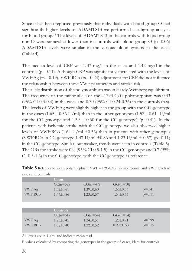
Since it has been reported previously that individuals with blood group O had significantly higher levels of ADAMTS13 we performed a subgroup analysis for blood group.14 The levels of ADAMTS13 in the controls with blood group non-O were somewhat lower than in controls with blood group O (p=0.06) ADAMTS13 levels were similar in the various blood groups in the cases (Table 4).
The median level of CRP was 2.07 mg/l in the cases and 1.42 mg/l in the controls (p=0.11). Although CRP was significantly correlated with the levels of VWF:Ag (rs= 0.19), VWF:RCo (rs= 0.24) adjustment for CRP did not influence the relationship between these VWF parameters and stroke risk.The allele distribution of the polymorphism was in Hardy-Weinberg equilibrium. The frequency of the minor allele of the –1793 C/G polymorphism was 0.33 (95% CI 0.3-0.4) in the cases and 0.30 (95% CI 0.24-0.36) in the controls (n.s). The levels of VWF:Ag were slightly higher in the group with the GG-genotype in the cases (1.65± 0.56 U/ml) than in the other genotypes (1.52± 0.61 U/ml for the CC-genotype and 1.39 ± 0.60 for the CG-genotype) (p=0.41). In the patients with ischemic stroke with the GG-genotype we also observed higher levels of VWF:RCo (1.64 U/ml ±0.56) than in patients with other genotypes (VWF:RCo in CC-genotype 1.47 U/ml ±0.86 and 1.23 U/ml ± 0.57) (p=0.11) in the CG-genotype. Similar, but weaker, trends were seen in controls (Table 5). The ORs for stroke were 0.9 (95% CI 0.5-1.5) in the CG genotype and 0.7 (95% CI 0.3-1.6) in the GG-genotype, with the CC genotype as reference.
Table 5 Relation between polymorphism VWF –1793C/G polymorphism and VWF levels in cases and controls
CasesCC(n=52) CG(n=47) GG(n=10)
VWF:Ag 1.52±0.61 1.39±0.60 1.65±0.56 p=0.41VWF:RCo 1.47±0.86 1.23±0.57 1.64±0.56 p=0.11
ControlsCC(n=51) CG(n=54) GG(n=14)
VWF:Ag 1.23±0.45 1.24±0.51 1.25±0.71 p=0.99VWF:RCo 1.08±0.40 1.22±0.52 0.99±0.53 p=0.15
All levels are in U/ml and indicate mean ±sd. P-values calculated by comparing the genotypes in the group of cases, idem for controls.

37
Chapter 2
DiscussionThe present study shows a significant relationship between increased VWF:Ag levels and the occurrence of ischemic stroke. A similar relationship was observed for VWF:RCo activity. In this study VWF levels were associated with genetic variation, ADAMTS13 activity, inflammation and blood group. However, after adjustment for these factors, the relationship between VWF and stroke remained similar.It is well known that VWF levels are dependent upon ABO blood groups. Previously it has been reported that individuals with blood group non-O have higher levels of VWF.15 It has been reported that individuals with blood group non-O also have a higher risk of arterial thrombosis. 16, 17
We also observed higher VWF levels in blood group non-O individuals and therefore performed subgroup analysis for blood group. Interestingly, we found a higher relative risk of stroke in individuals in the highest quartile of VWF levels with blood group O OR 5.9 (95% CI 1.6-21.5) compared with individ-uals in the highest quartile of VWF levels with blood group non-O OR 1.6 (95% CI 0.5-5.2). The underlying mechanism for this finding is not clear. Some mechanisms have been proposed through which blood group affects VWF, such as the clearance of the VWF, which differs in the various blood groups due to varying carbohydrate structure of plasma glycoproteins.18 Furthermore gene loci were suggested to have a quantitative effect on plasma levels of VWF. ABO blood group locus is one of these, located on chromosome 9q34. Since the gene is located very close to ADAMTS13 we investigated with HAPMAP (http://www.hapmap.org) if there was any linkage disequilibrium, but the genes were in different Linkage Disequilibrium-blocks.Inflammation is an important determinant of VWF levels19 and it has therefore been suggested that raised VWF levels measured shortly after stroke are merely a marker of the acute phase response rather than the cause of the event. CRP and VWF levels were indeed correlated in our study. However, after adjusting for CRP, VWF levels remained significantly associated with the risk of ischemic stroke. This indicates that inflammation may play a role in determining levels of VWF, but it will not be the only important regulatory mechanism since VWF levels are independently of inflammation also associated with the relative risk of ischemic stroke. The -1793 C/G polymorphism in the promoter region of the VWF gene is as-sociated with VWF levels in plasma.2 We have previously shown that carriers of the G-allele have a significantly increased risk of acute myocardial infarction.13

38
This indicates that genetic variation in the VWF gene, resulting in higher VWF levels, may be causally related with the relative risk of arterial thrombosis. In the present study, the highest levels of VWF (VWF:Ag and VWF:RCo) were also found in individuals with the GG- genotype (n.s.). However, there was no indication that carriers of the G-allele have a higher relative risk of ischemic stroke.Proteolytic cleavage of ULVWF by ADAMTS13 into less active smaller multimers determines the activity levels of VWF in plasma. We therefore hypothesized that ADAMTS13 activity may be associated with the risk of stroke. This is the first study that determines levels of ADAMTS13 in patients with ischemic stroke and indeed we observed that levels of ADAMTS13 activity are somewhat lower in ischemic stroke patients compared with the controls. In individuals with the lowest levels of ADAMTS13 the risk of ischemic stroke was almost doubled OR 1.6 (95% CI 0.7-3.8) compared to individuals in the highest quartile of ADAMTS13 levels. However, these results were not statistically significant and these findings should be confirmed in studies with a larger patient population. Our study shows that the levels of VWF and ADAMTS13 are negatively associated, similar to what been found in a variety of conditions in previous studies.14 Therefore we can only speculate about the underlying mechanism: low levels of ADAMTS13 resulting in higher VWF levels, or an increase of VWF levels resulting in lower ADAMTS13 levels. In addition, ADAMTS13 levels in our control group were also dependent upon blood group, individuals with blood group O having higher levels than non-O, as has been reported earlier.14, 20 This was not seen in our patient group, which may indicate that the increase in VWF levels found ischemic stroke patients, overrules the difference between in VWF due to blood group. In conclusion, this study shows clearly that high levels of VWF antigen and VWF activity are associated with an increased risk of stroke. Our results indicate that inflammation, genetic variation and degradation by ADAMTS13 partly determine VWF levels, however we did not demonstrate an interaction with the effect of VWF on ischemic stroke risk.
References1. Nishio K, Anderson PJ, Zheng XL, Sadler JE. Binding of platelet glycoprotein Ibalpha to von
Willebrand factor domain A1 stimulates the cleavage of the adjacent domain A2 by ADAMTS13.
Proc Natl Acad Sci U S A. Vol. 101, 2004:10578-83.
2. Harvey PJ, Keightley AM, Lam YM, Cameron C, Lillicrap D. A single nucleotide polymorphism at

39
nucleotide -1793 in the von Willebrand factor (VWF) regulatory region is associated with plasma
VWF:Ag levels. Br J Haematol. Vol. 109, 2000:349-53.
3. Zheng X, Chung D, Takayama TK, Majerus EM, Sadler JE, Fujikawa K. Structure of von Wil-
lebrand factor-cleaving protease (ADAMTS13), a metalloprotease involved in thrombotic throm-
bocytopenic purpura. J Biol Chem. Vol. 276, 2001:41059-63.
4. Levy GG, Nichols WC, Lian EC, et al. Mutations in a member of the ADAMTS gene family cause
thrombotic thrombocytopenic purpura. Nature. Vol. 413, 2001:488-94.
5. Moake JL, Rudy CK, Troll JH, et al. Unusually large plasma factor VIII:von Willebrand factor
multimers in chronic relapsing thrombotic thrombocytopenic purpura. N Engl J Med. Vol. 307,
1982:1432-5.
6. Tsai HM, Lian EC. Antibodies to von Willebrand factor-cleaving protease in acute thrombotic
thrombocytopenic purpura. N Engl J Med. Vol. 339, 1998:1585-94.
7. Thompson SG, Kienast J, Pyke SD, Haverkate F, van de Loo JC. Hemostatic factors and the risk
of myocardial infarction or sudden death in patients with angina pectoris. European Concerted
Action on Thrombosis and Disabilities Angina Pectoris Study Group. N Engl J Med. Vol. 332,
1995:635-41.
8. Folsom AR, Rosamond WD, Shahar E, et al. Prospective study of markers of hemostatic function
with risk of ischemic stroke. The Atherosclerosis Risk in Communities (ARIC) Study Investigators.
Circulation. Vol. 100, 1999:736-42.
9. Catto AJ, Carter AM, Barrett JH, Bamford J, Rice PJ, Grant PJ. von Willebrand factor and factor
VIII: C in acute cerebrovascular disease. Relationship to stroke subtype and mortality. Thromb
Haemost. Vol. 77, 1997:1104-8.
10. Smith FB, Lee AJ, Fowkes FG, Price JF, Rumley A, Lowe GD. Hemostatic factors as predictors of
ischemic heart disease and stroke in the Edinburgh Artery Study. Arterioscler Thromb Vasc Biol.
Vol. 17, 1997:3321-5.
11. Leebeek FW, Goor MP, Guimaraes AH, et al. High functional levels of thrombin-activatable fibri-
nolysis inhibitor are associated with an increased risk of first ischemic stroke. J Thromb Haemost.
Vol. 3, 2005:2211-8.
12. Loof AH, van Vliet HH, Kappers-Klunne MC. Low activity of von Willebrand factor-cleaving
protease is not restricted to patients suffering from thrombotic thrombocytopenic purpura. Br J
Haematol. Vol. 112, 2001:1087-8.
13. van der Meer IM, Brouwers GJ, Bulk S, et al. Genetic variability of von Willebrand factor and risk
of coronary heart disease: the Rotterdam Study. Br J Haematol. Vol. 124, 2004:343-7.
14. Mannucci PM, Capoferri C, Canciani MT. Plasma levels of von Willebrand factor regulate AD-
AMTS-13, its major cleaving protease. Br J Haematol. Vol. 126, 2004:213-8.
15. Gill JC, Endres-Brooks J, Bauer PJ, Marks WJ, Jr., Montgomery RR. The effect of ABO blood
group on the diagnosis of von Willebrand disease. Blood. Vol. 69, 1987:1691-5.

40
16. von Beckerath N, Koch W, Mehilli J, et al. ABO locus O1 allele and risk of myocardial infarction.
Blood Coagul Fibrinolysis. Vol. 15, 2004:61-7.
17. Suadicani P, Hein HO, Gyntelberg F. Airborne occupational exposure, ABO phenotype and risk
of ischaemic heart disease in the Copenhagen Male Study. J Cardiovasc Risk. Vol. 9, 2002:191-8.
18. Mohlke KL, Purkayastha AA, Westrick RJ, et al. Mvwf, a dominant modifier of murine von Wil-
lebrand factor, results from altered lineage-specific expression of a glycosyltransferase. Cell. Vol.
96, 1999:111-20.
19. Feng D, Lindpaintner K, Larson MG, et al. Platelet glycoprotein IIIa Pl(a) polymorphism, fibrino-
gen, and platelet aggregability: The Framingham Heart Study. Circulation. Vol. 104, 2001:140-4.
20. Sanchez-Luceros A, Farias CE, Amaral MM, et al. von Willebrand factor-cleaving protease AD-
AMTS13 activity in normal non-pregnant women, pregnant and post-delivery women. Thromb
Haemost. Vol. 92, 2004:1320-6.


Lower Levels of ADAMTS13 are associated with cardiovascular disease in young patients
T.N. Bongers, E.L.E. de Bruijne, D.W.J. Dippel, A.J. de Jong, J.W. Deckers, D. Poldermans, M.P.M. de Maat, F.W.G. Leebeek
Atherosclerosis 207, 2009 250-254
Chapter 3

44
AbstractBackground ADAMTS13 may play a role in arterial thrombosis by cleaving the highly active and thrombogenic ultralarge VWF multimers into less active VWF multimers. The aim was to investigate the relationship between plasma levels of ADAMTS13, VWF and genetic variation in the ADAMTS13 gene with cardio-vascular disease.
MethodsWe performed a case-control study in 374 patients with a first-ever arterial thrombosis before the age of 45 years in males and 55 years in women. We included 218 patients with coronary heart disease (CHD), 109 patients with ischemic stroke (IS) and 47 patients with peripheral arterial disease (PAD) and 332 healthy population-based controls. ADAMTS13 and VWF levels were measured 1-3 months after the event.
ResultsADAMTS13 levels were associated with cardiovascular disease (OR antigen 5.1 (95% CI 3.1-8.5, p <0.001) and OR activity 4.4 (95% CI 2.5-7.5, p< 0.001), in the lowest quartiles). VWF levels were associated with cardiovascular disease (OR antigen 2.1 (95% CI 1.3-3.3, p=0.001) and OR activity 2.0 (95% CI 1.3-3.1, p=0.003), in the highest quartile). Patients with combined low ADAMTS13 levels and high VWF levels had an odds ratio of 7.7 (95% CI 3.3-17.7) (p for trend <0.0001). No association was found between genetic variation in the ADAMTS13 gene with levels of ADAMTS13 or with risk of cardiovascular disease.
ConclusionIn conclusion, levels of ADAMTS13 and VWF are strongly associated with the risk of cardiovascular disease.

45
Chapter 3
IntroductionVon Willebrand Factor (VWF) is a glycoprotein that has an important function in the haemostatic process. VWF binds the glycoprotein Ib-IX-V-complex on platelets, which stimulates platelet adhesion and aggregation. VWF is synthe-sized in endothelial cells and stored in Weibel-Palade bodies, in which ultra large VWF multimers (ULVWF) are formed. These very active ULVWF multimers are released into the circulation upon stimulation of the endothelium and easily form platelet aggregates. In the regulation of the size of the VWF multimers an important role is played by ADAMTS13 (A Disintegrin And Metalloprotease with ThromboSpondin motifs).1
ADAMTS13 is a metalloprotease that consists of 1427 amino acids and that cleaves the bond between amino acids Tyr1605 and Met1606 in the A2 domain of VWF.2, 3 The ADAMTS13 gene contains 29 exons and is located on chromo-some 9q34.1, 4 To date, little is known about the regulation of ADAMTS13, but it is known that plasma levels of ADAMTS13 are partially determined by genetic factors, since several amino acid variants have been described that correlate with ADAMTS13 activity.5, 6
Extremely low levels of ADAMTS13 are seen in thrombotic thrombocytopenic purpura (TTP). TTP is characterized by intravascular platelet aggregation and microvascular thrombi, which can result in life threatening cerebral ischemia and arterial thrombosis.9 The antithrombotic role of ADAMTS13 is also shown in less extreme situations, in animal studies and in-vitro studies.7, 8
Because of this antithrombotic role, we hypothesized that low levels of ADAMTS13 will result in an increased risk of cardiovascular disease. However, only limited, conflicting data are available on the role of ADAMTS13 in the pathogenesis of cardiovascular disease.10, 11
The genetic component in the pathogenesis of cardiovascular disease is stronger in young patients than in elderly patients. In elderly patients, the contri-bution of the classical age-related cardiovascular risk factors is more important.12 For this reason we studied the relationship between the genetic variation in the ADAMTS13 gene and the levels and risk of cardiovascular disease in men before 45 years and women before 55 years old.The aim of this study was to investigate the relationship between ADAMTS13, VWF activity, the genetic variation in ADAMTS13 and the risk of cardiovascular disease in young individuals.

46
Patients and MethodsStudy designWe performed a case-control study, in which cases were consecutive patients who presented with a first acute ischemic event in the Erasmus MC. Patients from the Erasmus MC were included if they were 45 years or younger for males or 55 years or younger for females. In these age groups cardiovascular disease are rare (frequency 2% in the Netherlands)13 and genetic factors are known to play a larger role at young ages.12 The cohort consisted of three subgroups: (i) coronary heart disease (CHD), containing patients with acute myocardial infarction (AMI) and unstable angina pectoris (UAP), (ii) Ischemic Stroke, containing patients with an ischemic stroke or transient ischemic attack (TIA), (iii) peripheral arterial diseases (PAD). We used population-based controls, i.e. neighbors or friends of the patients, fulfilling the same age criteria and without a history of cardiovascular diseases. The study was approved by the Medical Ethical Committee of the Erasmus MC and written informed consent was obtained from all patients and controls.
DefinitionsAMI was defined as typical chest pain, with elevated cardiac markers (CK, MB, troponin T) or characteristic electrocardiographic findings (ICD 410). UAP was defined as typical chest pain at rest (ICD 413). TIA was defined as suddenly occurring focal cerebral deficit, which could not be explained otherwise than by local cerebral ischemia (ICD 435). Symptoms had to be temporary and last less than 24 hours after onset. Ischemic Stroke (IS) was defined as suddenly occurring cerebral deficits, which could not be explained other than by local cerebral ischemia (ICD 434). Brain imaging by CT or MRI was compatible with the diagnosis. The diagnosis PAD was defined as peripheral arterial stenosis resulting in ischemia, classified according to the Rutherford criteria.14
Blood sampling Blood was collected 1-3 months after the event in citrate (0.105 M) using a Vacutainer System (Beckton-Dickson, Plymouth, UK). and centrifuged at 2000 g for 10 min at 4˚C, followed by 10 min at 20,000 g for 10 min at 4˚C and plasma was stored in aliquots at -80˚C Genomic DNA was isolated according to standard salting-out procedures. DNA was available from 350 patients and 310 controls due to logistic reasons. Technicians were not aware of the case-control status of the samples.

47
Chapter 3
MethodsADAMTS13 activity and antigen were measured using the Technozym ADAMTS13 kit (Technoclone, Vienna, Austria) as described by the manu-facturer. Fluoresence was measured at 360/460 nm (Biotek reader FLX 800, Austria). The intra-assay variation was 10% for ADAMTS13 activity and 18% for ADAMTS13 antigen. Levels in pooled plasma of 40 healthy men was defined as 100%VWF antigen (VWF:Ag) was determined in duplicate with an in-house ELISA assay, using polyclonal rabbit anti-human VWF and horseradish peroxidase con-jugated anti-human VWF (DakoCytomation, Glostrop, Denmark) for catching and tagging, respectively. VWF collagen binding (VWF:CB) was measured in duplicate by an in-house ELISA using type I collagen (Sigma, St Louis, USA) for catching and horseradish peroxidase conjugated anti-human VWF for tagging. The intra-assay variations of VWF:Ag and VWF:CB were 14.5% and 6.4% respectively.
Genotyping of ADAMTS13The haplotype-tagging SNPs (htSNP) in this study were selected based on the SeattleSNPs database (http://pga.gs.washington.edu/) (16468 (rs 2301612), 19925 (rs 2073932), 25504 (rs 652600), 21576 (rs 603551)). We considered only SNPs with a minor allele frequency of at least 5% in the Caucasian population (build 125, http://www.ncbi.nlm.nih.gov/SNP). Polymerase chain reaction (PCR) was performed using allele- specific Taqman analysis (Applied Biosystems, Foster City, USA). Haplostats (http://cran.r-project.org/src/contrib/Descriptions/haplo.stats.html) was used to compute maximum likelihood estimates of haplotypes probabilities. Only the four haplotypes with a frequency >5% were included in the statistical analysis. The most frequent haplotype was used as reference. Genotypes with less then 10 homozygotes for the minor allele are combined with the heterozygotes.
Statistical analysisThe medians (25th-75th percentile) were presented because the levels are not normally distributed. The levels of ADAMTS13 antigen, ADAMTS13 activity, VWF:Ag and VWF:CB were compared in cases and controls and in the genotypes using an analysis of variance (ANOVA) with adjustment for age and sex. Levels of ADAMTS13 antigen, ADAMTS13 activity, VWF:Ag and VWF:CB were divided in quartiles based on the distribution in the control group. The

48
relationship between the levels of these variables and the occurrence of a cardio-vascular event was determined using logistic regression, using the lowest quartile as reference, except for ADAMTS13, where the highest quartile constituted the reference. In the subgroup analysis, we used tertiles of the distribution instead of quartiles, because of the small numbers of patients. We adjusted for age, sex, smoking, hypertension, diabetes mellitus and hypercholesterolemia. The association between ADAMTS13 haplotypes and risk of arterial thrombosis was determined by weighted logistic regression analysis using HaploStats software.15 Briefly, Haplotstats calculates the posterior probabilities for each possible haplotype of an individual and assigns an appropriate weight to the risk estimate of the corresponding haplotype. This statistical method increases the accuracy of risk estimates that are calculated from phase- ambiguous genotype data. With a power calculation considering an α=0.05 and a β=0.80 and with the allele frequencies of the SNPs, we calculated to find odds ratios between 1.2-2.4. A p-value of <0.05, was considered to indicate statistical significance. All statistical analyses were two-sided and carried out using SPSS software version 12. ResultsThe study included 374 patients and 332 controls. As expected, we observed more risk factors, such as smoking, hypertension, diabetes and statin use in patients (Table 1).The ADAMTS13 activity was significantly lower in cases 96.4% (70.3-112.2) than in controls 109.5% (93.0-123.8) (median (25th-75th percentile) (p<0.001)). ADAMTS13 antigen levels were also significantly lower in cases (78.6% (56.0-102.3) than in controls 97.4% (81.7-111.1) (p<0.001). The specific activity of ADAMTS13 (ratio activity to antigen) was higher in cases (1.11 (1.0-1.3) compared to controls (1.08 (0.9-1.2) p=0.01). Individuals with the lowest levels of ADAMTS13 antigen and activity had increased relative risks OR 5.1 (95% CI 3.1-8.5, p< 0.001, p<0.001*) and OR 4.4 (95% CI 2.5-7.5, p< 0.001, p<0.001*), respectively *(after Bonferroni correction) compared with the individuals in the highest quartile of ADAMTS13. The individuals with the highest specific activity of ADAMTS13 had the highest risk OR 1.9 (95% CI 1.2-3.2, p=0.012, p=0.048*) compared with individuals with the lowest ratio.VWF:Ag levels were significantly higher in cases (1.20 (0.9-1.6) U/ml than in controls 1.04 (0.8-1.4) U/ml; p<0.001). Levels of VWF:CB were also higher incases (1.32 (1.0-1.7) than in controls 1.16 (0.9-1.5) U/ml, p< 0.001. Individuals

49
Chapter 3
Table 1 Baseline characteristicsCases Controls P-value
DemographicsAge, yrs 42.8± 7.0 38.5 ± 8.0 <0.001Male, n (%) 162 (43.3%) 122 (36.7%) 0.08Body mass index, kg/m2 26.4 ± 4.6 25.1 ± 4.2 <0.001Systolic blood pressure, mm Hg 128 ± 24 125 ± 17 0.09Diastolic blood pressure, mm Hg 80 ± 14 80 ± 11 0.70
Index eventUAP/AMI 49/169Stroke/TIA 52/57PAD 47
Risk factorsSmoking (former+ current) (%) 297 (82%) 162 (51%) <0.0001Hypertension or anti-hypertensive drugs
293 (78%) 62 (19%) <0.0001
Diabetes 33 (10%) 5 (2%) <0.0001
MedicationBetablockers 223 (60%) 7 (1%)Diuretics 44 (12%) 6 (1%)ACE inhibitors 150 (40%) 3 (1%)Lipid lowering drugs 303 (81%) 2 (0.6%)Clopidogrel 184 (49%) 0 (0%)
with levels of VWF:Ag and VWF:CB in the highest quartile had an increased risk OR 2.1 (95% CI 1.3-3.3, p=0.001, p=0.004*) and 2.0 (95% CI 1.3-3.1, p=0.003, p=0.012*), respectively) compared with individuals in the lowest quartiles (Table 2).

50
Table 2 Relationship between levels of VWF, ADAMTS13 and risk on cardiovascular diseaseQ1 Q2 Q3 Q4
VWF:Ag (U/ml) <0.8 0.9-1.0 1.1-1.4 > 1.5OR (95% CI) 1 (reference) 1.1 (0.7-1.1) 1.4 (0.9-2.2) 2.1 (1.3-3.3)VWF:CB act (U/ml) <0.9 1.0-1.2 1.3-1.5 > 1.6OR (95% CI) 1 (reference) 0.9 (0.5-1.4) 1.4 (0.9-2.1) 2.0 (1.3-3.1)ADAMTS13 antigen (% of NPP)
<81.6 81.7-97.3 97.4-111.0 > 111.1
OR (95% CI) 5.1 (3.1-8.5) 2.2 (1.3-3.9) 2.1 (1.3-3.7) 1 (reference)ADAMTS13 activity (% of NPP)
<93.0 93.1-109.5 109.6-123.8 > 123.9
OR (95% CI) 4.4 (2.5-7.5) 2.3 (1.3-4.0) 1.8 (1.0-3.2) 1 (reference)
Values presented are the ORs for quartiles with 95% CI and P values, adjusted for age and sex.
Figure 1 The relation between ADAMTS13, VWF and cardiovascular disease. Individuals who were both in the highest tertile of ADAMTS13 and in the lowest tertile of VWF were used as reference.* p<0.05;** p<0.001

51
Chapter 3
We specifically assessed the risk of cardiovascular disease in the individuals with both high levels of VWF and low levels of ADAMTS13 (Figure 1). These have a very high risk of cardiovascular disease (OR 7.7, 95% CI 3.3-17.7) compared to individuals with low VWF levels and high levels of ADAMTS13 (P for trend < 0.001, p<0.001*)(Figure 1)
Subgroup analysisThe decrease in ADAMTS13 levels was most clearly observed in the CHD subgroup. The median levels of ADAMTS13 activity in the CHD group were significantly lower compared to controls; 88.54% (56.5-106.3) vs. 109.5% (93-123.8) p<0.001. In addition, ADAMTS13 antigen levels were lower in the CHD group 74.5% (50.6-98.2) than in the controls 97.38% (81.7-111.0), p<0.001. The specific activity of ADAMTS13 was significantly higher in the CHD (1.1 (0.9-1.3) group compared to the controls (1.08 (0.9-1.2) p=0.002. In the other subgroups, the differences between cases and controls were smaller and not significant.Individuals in the lowest tertile for ADAMTS13 antigen have an eight times increased risk for CHD compared with individuals in the highest tertile (OR 8.2, 95% CI 4.5-14.7 p< 0.001, p<0.001*). Individuals in the intermediate tertile have an (OR 2.2 (95% CI 1.2-3.9 p<0.001, p<0.001*). For ischemic stroke and for PAD this was not observed.
Genetic variation of ADAMTS13The genotype distributions of the ADAMTS13 SNPs were in Hardy- Weinberg equilibrium. None of the SNPs was individually associated with levels of ADAMTS13 activity or antigen (Table 3). Haplotype analysis showed that haplotype GAAT was associated with lower levels of ADAMTS13 activity (14% lower in the controls and 8% lower in the cases compared with the reference haplotype CGAT, p=0.05), but no association with ADAMTS13 antigen was observed. We did not observe a significant association between individual SNPs or haplotypes with the risk of arterial thrombosis (Table 3). Haplotype GAAT was associated with a decreased risk of PAD (OR 0.5, 95% CI 0.3-1.0, p= 0.06), There might be a reduced risk of PAD in individuals with haplotype, but this estimate comes from a small group and needs to be considered with care and also needs replication in a larger cohort.

52
DiscussionIn this study, we show that levels of ADAMTS13 are lower and levels of VWF are higher in young patients with cardiovascular disease compared to healthy individuals. Individuals with low levels of ADAMTS13 had five times more risk on cardiovascular disease than subjects with normal levels of ADAMTS13. The relationship was strongest in the subgroup of patients with coronary heart disease. This may be of importance for identifying individuals who are prone to have a cardiovascular event.We observed that high levels of VWF are a risk factor for both myocardial infarction and stroke, which confirms previous studies.16-20
Table 3 ADAMTS13 gene polymorphisms in cases and controlsCases ADAMTS13 Controls ADAMTS13 OR(95% CI)N= Act Ag N= Act Ag
RS2301612GG 96 (28.7%) 94.9 77.9 97 (32.7%) 109.0 96.9 ReferenceGC 174 (51.9%) 100.5 82.1 148 (49.8%) 110.5 98.1 1.21 (0.8-1.8)CC 65 (19.4%) 90.7 78.7 52 (17.5%) 111.2 91.4 1.28 (0.8-2.1)
RS2073932GG 107 (30.8%) 94.0 78.7 87 (28.8%) 111.4 97.8 ReferenceGA 175 (50.4%) 99.4 82.1 156 (51.7%) 110.5 98.3 0.93 (0.6-1.3)AA 65 (18.7%) 88.3 77.8 59 (19.5%) 102.5 94.1 0.85 (0.5-1.4)
RS652600AA 167 (47.7%) 98.1 78.5 140 (46.8%) 112.0 97.8 ReferenceAG 149 (42.6%) 98.5 82.8 134 (44.8%) 100.9 92.8 0.97 (0.7-1.4)GG 34 (9.7%) 84.0 75.4 25 (8.4%) 107.2 98.2 1.10 (0.6-2.0)
RS603551TT 302 (86.8%) 96.6 80.1 265 (85.5%) 110.9 97.6 ReferenceTC+ CC 46 (13.2%) 98.7 87.1 45 (14.5%) 107.8 97.7 0.86 (0.5-1.4)
ADAMTS13 activity and antigen are in percentage of Normal Pooled Plasma.Logistic regression analysis with adjustment for age and sex.Act = Activity, Ag = Antigen.

53
Chapter 3
The largest VWF multimers are the most thrombogenic and ADAMTS13 can cleave these ULVWF into smaller, less active forms, which suggests a role for ADAMTS13 in cardiovascular disease. Indeed, our study indeed showed that low levels of ADAMTS13 are associated with a higher risk of cardiovascular disease. In our study, we noticed that the relationship was stronger in coro-nary heart disease than stroke, TIA and peripheral arterial diseases. These results are in accordance with two recent studies in elderly patients with myocardial infarction.11, 21 In our case-control study, we cannot prove a causal role for the lower levels of ADAMTS13. A prospective study could demonstrate whether ADAMTS13 is the cause or consequence of the cardiovascular event. In contrast to our lower levels, one earlier study reported an increased risk of AMI in men in the three highest quartiles of ADAMTS13 antigen compared to the lowest quartile.10 The difference results may be explained by age and gender composition. Another difference between these studies and our study is the ADAMTS13 assay that is used. In our study we used an assay that measures both antigen and activity levels of ADAMTS13 (Technoclone), and we observed similar associations with cardiovascular disease for ADAMTS13 activity and antigen. The other studies used an activity assay (FRETS assay)21 or an in-house ELISA based on monoclonal antibodies.21
We also showed that those individuals who have the lowest levels of ADAMTS13 combined with the highest levels of VWF have the highest risk of cardiovascu-lar disease and that there is no correlation between ADAMTS13 and VWF levels (Rs= -0.12). An inverse correlation between ADAMTS13 and VWF was recently described in patients with Von Willebrand Disease.22
We observed an increased specific activity of ADAMTS13 in patients compared with controls. The reduced ADAMTS13 activity levels are therefore not attributable to dysfunctional ADAMTS13. Despite the slightly higher specific activity levels of ADAMTS13, both activity and antigen levels of ADAMTS13 were lower in patients than in control individuals, thereby explaining the pro-thrombotic risk in these individuals. Feys et al. reported recently a lower specific activity of ADAMTS13 in patients in pathologic conditions predisposing to thrombosis, but cardiovascular disease was not included in that study.23
A reduced activity of ADAMTS13 in the Japanese population was seen in subjects with the 475Ser genotype with multimer degradation assays.24 How-ever, the P475S polymorphism, associated with a striking reduction of ADAMTS13 activity, is not observed in Caucasians.6 We investigated whether other genetic variation in the ADAMTS13 gene is associated with ADAMTS13

54
levels, for which we studied four haplotype-tagging SNPs. The SNPs separately were not associated with levels of ADAMTS13. The lowest levels of ADAMTS13 were seen in haplotype GAAT Haplotype GAAT was associated with the risk of PAD, however the number of patients in this subgroup is small and larger studies are required to confirm this association. Since we did not observe associations between genetic variation and cardiovascular disease in the other groups, our study indicates that genetic variation in ADAMTS13 does not play a major role in the reduction of ADAMTS13 levels found in our patients with cardiovascular disease. This study has some limitations. The controls that we included were healthy subjects without a history of cardiovascular diseases. However, we did not screen for occult cardiovascular disease. Furthermore, for haplotype anaylsis, the group is somewhat small, but also single SNP analysis showed no strong associations. Significant associations need to be replicated and tested for clinical relevance in other, larger studies with independently collected cases and controls. Results from a case-control study do not prove causality and levels may be different because of the event. However, since we collected samples one to three months after the event, the acute phase effect on the levels will be minimized. The ATTAC study consists mainly of young, white Europeans and it is not clear whether the results of our study can be extrapolated to other population.In conclusion, we have shown that reduced levels of ADAMTS13 are associated with an increased risk of cardiovascular disease, but that genetic variation does not play a major role.
References1. Levy GG, Nichols WC, Lian EC, Foroud T, McClintick JN, McGee BM, Yang AY, Siemieniak DR,
Stark KR, Gruppo R, Sarode R, Shurin SB, Chandrasekaran V, Stabler SP, Sabio H, Bouhassira EE,
Upshaw JD Jr, Ginsburg D Tsai HM, Mutations in a member of the ADAMTS gene family cause
thrombotic thrombocytopenic purpura, Nature 2001; 413: 488-494.
2. Levy GG, Motto DG, Ginsburg D, ADAMTS13 turns 3. Blood 2005; 106: 11-17.
3. Zheng X, Majerus EM, Sadler JE, ADAMTS13 and TTP. Curr Opin in Hematology 2002; 9: 389-
394.
4. Soejima K, Mimura N, Hirashima M, Maeda H, Hamamoto T, Nakagaki T Nozaki C, A novel
human metalloprotease synthesized in the liver and secreted into the blood: possibly, the von Wil-
lebrand factor-cleaving protease? J Biochem (Tokyo) 2001; 130: 475-480.
5. Plaimauer B, Fuhrmann J, Mohr G, Wernhart W, Bruno K, Ferrari S, Konetschny C, Antoine G,
Rieger M, Scheiflinger F, Modulation of ADAMTS13 secretion and specific activity by a combina-

55
tion of common amino acid polymorphisms and a missense mutation. Blood 2006; 107: 118-125.
6. Bongers TN, De Maat MP, Dippel DW, Uitterlinden AG, Leebeek FW, Absence of Pro475Ser
polymorphism in ADAMTS-13 in Caucasians. J Thromb Haemost 2005; 3: 805.
7. Dong JF, Cleavage of ultra-large von Willebrand factor by ADAMTS-13 under flow conditions, J
Thromb Haemost 2005; 3: 1710-1716.
8. Chauhan AK, Motto DG, Lamb CB, Bergmeier W, Dockal M, Plaimauer B, Scheiflinger F, Gins-
burg D, Wagner DD, Systemic antithrombotic effects of ADAMTS13. J Exp Med 2006; 203: 767-
776.
9. Moake JL, Thrombotic microangiopathies, N Engl J Med: 2002: 347: 589-600.
10. Chion CK, Doggen CJ, Crawley JT, Lane DA, Rosendaal FR, ADAMTS13 and von Willebrand
factor and the risk of myocardial infarction in men. Blood 2007; 109: 1998-2000.
11. Kaikita K, Soejima K, Matsukawa M, Nakagaki T, Ogawa H, Reduced von Willebrand factor-
cleaving protease (ADAMTS13) activity in acute myocardial infarction. J Thromb Haemost 2006;
4: 2490-2493.
12. Marenberg ME, Risch N, Berkman LF, Floderus B, de Faire U, Genetic susceptibility to death from
coronary heart disease in a study of twins. N Engl J Med 1994; 330: 1041-1046.
13. Koek HL, V. L. L., Verschuren WMM, Bots ML, Hart- en vaatziekten in Nederland 2004, cijfers
over leefstijl- en risicofactoren, ziekte en sterfte, In, Den Haag, Nederlandse Hartstichting, 2004;
19-31.
14. Dormandy JA, Rutherford RB, Management of peripheral arterial disease (PAD). TASC Working
Group. TransAtlantic Inter-Society Consensus (TASC), J Vasc Surg 2000; 31: S1-S296.
15. Schaid DJ, Rowland,CM, Tines DE, Jacobson RM, Poland GA, Score tests for association between
traits and haplotypes when linkage phase is ambiguous. American journal of human genetics 2002;
70: 425-434.
16. Bongers TN, de Maat,MP, van Goor ML, Bhagwanbali V, van Vliet HH, Gomez Garcia EB, Dippel
DW, Leebeek FW, High von Willebrand factor levels increase the risk of first ischemic stroke: influ-
ence of ADAMTS13, inflammation, and genetic variability. Stroke 2006; 37: 2672-2677.
17. Folsom AR, Rosamond WD, Shahar E, Cooper LS, Aleksic N, Nieto FJ, Rasmussen ML, Wu KK,
Prospective study of markers of hemostatic function with risk of ischemic stroke. The Atheroscle-
rosis Risk in Communities (ARIC) Study Investigators. Circulation 1999; 100: 736-742.
18. Whincup PH, Danesh J, Walker M, Lennon L, Thomson A, Appleby P, Rumley A, Lowe GD, von
Willebrand factor and coronary heart disease: prospective study and meta-analysis, Eur Heart J
2002; 23: 1764-1770.
19. van der Meer IM, Brouwers GJ, Bulk S, Leebeek FW, van der Kuip DA, Hofman A, Witteman JC,
Gomez Garcia EB, Genetic variability of von Willebrand factor and risk of coronary heart disease:
the Rotterdam Study. Br J Haematol 2004; 124: 343-347.
20. Spiel AO, Gilbert JC, Jilma B, von Willebrand factor in cardiovascular disease: focus on acute coro-

56
nary syndromes. Circulation 2008; 117: 1449-1459.
21. Crawley JT, Lane DA, Woodward M, Rumley A, Lowe GD, Evidence that high von Willebrand
factor and low ADAMTS-13 levels independently increase the risk of a non-fatal heart attack. J
Thromb Haemost 2008; 6: 583-588.
22. Mannucci PM, Capoferri C, Canciani MT, Plasma levels of von Willebrand factor regulate AD-
AMTS-13, its major cleaving protease. Br J Haematol 2004; 126: 213-218.
23. Feys HB, Canciani MT, Peyvandi F, Deckmyn H, Vanhoorelbeke K, Mannucci PM, ADAMTS13
activity to antigen ratio in physiological and pathological conditions associated with an increased
risk of thrombosis. Br J Haematol 2007; 138:534-540.
24. Kokame K, Matsumoto M, Soejima K, Yagi H, Ishizashi H, Funato M, Tamai H, Konno M, Ka-
mide K, Kawano Y, Miyata T, Fujimura Y, Mutations and common polymorphisms in ADAMTS13
gene responsible for von Willebrand factor-cleaving protease activity, Proc Natl Acad Sci U S A
2002; 99: 11902-11907.


Absence of Pro475Ser polymorphism in ADAMTS-13 in Caucasians
T.N. Bongers, M.P.M. de Maat, D.W.J. Dippel, A.G. Uitterlinden, F.W.G. LeebeekJ Thromb Haemost 2005 3 805
Chapter 4a

60
A Disintegrin and Metalloprotease with ThromboSpondin type 1 repeats (ADAMTS13) is a recently characterized metalloprotease that cleaves Ultra Large Von Willebrand Factor (ULVWF)1. A deficiency of ADAMTS13 results in an accumulation of ULVWF, and leads to microangiopathic thrombosis, which is characteristic of thrombotic thrombocytopenic purpura (TTP)2. The microangiopathic arterial thrombotic complications of TTP occur predominantly in the brain.
It is hypothesized that low ADAMTS13 levels may be a risk factor for arterial thrombosis, because less ULVWF cleavage may result in an excess of high VWF multimers, resulting in more adhesion and aggregation of platelets.3
Recently, the proline (Pro) to serine (Ser) polymorphism in codon 475 of the ADAMTS13 gene has been identified. This Pro475Ser polymorphism, caused by a base substitution of C1423 to T in exon 12, is reported to impair the activity of ADAMTS13. In a Japanese study the frequency of the rare 475Ser allele of this single nucleotide polymorphism (SNP) was 5.1% in 364 healthy controls and it has been suggested that the 475Ser allele may increase the risk of arterial thrombosis because of the reduced activity of ADAMTS13.3, 4
To study whether this ADAMTS13 polymorphism contributes to arterial thrombosis in Caucasians, we investigated the distribution of the Pro475Ser polymorphism in a Caucasian population of 125 patients suffering from ischemic stroke and 125 age- and gender-matched healthy controls.
The genotype was determined by polymerase chain reaction, digestion with RsaI and separation on a 2% agarose gel. We found that none of the 250 individuals carried the rare 475Ser allele.
We also investigated the Pro475Ser polymorphism in other populations, including 110 Chinese (HD02 and HD100 of the Human Variation Panel, Coriell Institute for Medical Research, Camden, NJ, USA) where we found one heterozygous subject, giving a frequency for the 475Ser allele of 0.5% [95% confidence interval (CI) 0–2.9]. This confirms the results of Ruan et al. who reported a frequency of 1.7% (95% CI 0.6–4.2) in healthy Chinese and 1.9% (95% CI 0.2–3.8) in Chinese patients who had had a myocardial infarction.5 Also in Afro-Americans (HD50), the 475Ser allele was not present.

61
Chapter 4a
Since no 475Ser carrier for the polymorphism was identified in our study (95% CI 0–1.5%) we conclude that the Pro475Ser polymorphism, which is associated with a reduced ADAMTS-13 activity, is not an important contributor to the risk of ischemic stroke in Caucasians. Our data suggest that outside the Japanese population, the 475Ser allele is very rare.
References1. Zheng X, Chung D, Takayama TK,Majerus EM, Sadler JE, Fujikawa K. Structure of von Wil-
lebrand factor-cleaving protease (ADAMTS-13), a metalloprotease involved in thrombotic throm-
bocytopenic purpura. J Biol Chem 2001; 276: 41059–63.
2. Bell WR, Brain HG, Ness PM, Kickler TS. Improved survival in thrombotic thrombocytopenic
purpura–hemolytic uremic syndrome. Clinical experience in 108 patients. N Engl J Med 1991; 325:
398–403.
3. Sadler JE. A new name in thrombosis, ADAMTS-13. Proc Natl Acad Sci U S A 2002; 99: 11552–4.
4. Kokame K, Tamai H, Konno M, Kamide K, Kawano Y, Miyata T, Fugimura Y. Mutations and
common polymorphisms in ADAMTS-13 gene responsible for von Willebrand factor-cleaving
protease activity. Proc Natl Acad Sci U S A 2002; 99: 11902–7.
5. Ruan CH. The frequency of P475S polymorphism in von Willebrand factor-cleaving protease in
the Chinese population and its relevance to arterial thrombotic disorders. Thromb Haemost 2004;
91: 1258–9.

Frequency of the Von Willebrand Factor Tyr1584Cys polymorphism in arterial thrombosis
T.N. Bongers, M.P.M. de Maat, D.W.J. Dippel, J.W. Deckers, F.W.G Leebeek British Journal of haematology 2007, 140 578-79
Chapter 4b

64
IntroductionIn case of a damaged blood vessel Von Willebrand Factor (VWF) binds to collagen in the subendothelium and mediates the adhesion of platelets at the site of vascular damage. Ultra Large VWF (ULVWF), secreted upon stimula-tion, is degraded to a less active form by the metalloprotease ADAMTS13. The role of VWF in arterial thrombosis has been studied intensively. In coronary heart disease high levels of VWF are associated with an increased risk of arte-rial thrombosis.1, 2 We recently confirmed that elevated levels of VWF antigen and activity are also a risk factor for ischemic stroke.3 Normally, the ULVWF multimers are cleaved by ADAMTS13 into smaller fragments that are less ac-tive and less thrombogenic. It has been postulated that ADAMTS13 levels might also be associated with the risk of arterial thrombosis. In a recent study Ginsburg et al. showed that ADAMTS13 has a direct anti-thrombotic effect.4 One of the factors that determines the proteolysis of VWF by ADAMTS13 is the recently reported amino acid polymorphism Tyr1584Cys in exon 28 of the VWF gene, which encodes the A2 domain of VWF. This polymorphism is associated with an increased susceptibility of VWF for proteolysis by ADAMTS13. Individuals who are homozygous for Tyr1584 have a normal proteolysis, while heterozygotes Tyr184Cys have an increased proteolysis. In Canadian patients with Von Willebrand Disease (VWD) type I the frequency of this polymorphism was 14%, whereas in healthy Caucasian populations the estimate is around 1%.5-10 (Table 1).An increased susceptibility to proteolysis of VWF will lead to a decrease in less active circulating VWF molecules. The resulting anti-thrombotic effect could be a protective mechanism for arterial thrombotic disease. It has not yet been studied whether the Tyr1584Cys polymorphisms is associated with the risk of arterial thrombosis. Therefore we investigated the Tyr1584Cys polymorphism in two different case-control studies in patients with well- documented arterial thrombosis.
Patients and MethodsRecently, we have performed two case-control studies on arterial thrombo-sis. The first, the COCOS-study, has been described previously.11 Briefly, this study comprises 124 patients with a first-ever ischemic stroke and 125 controls without a history of stroke with an age range between 18-75 years. The second study, the ATTAC-study, is a case-control study that comprises 374 young patients (males ≤ 45 years and females ≤ 55 years) with a first-ever arterial thrombotic

65
Chapter 4b
Table 1 Allele frequencies of the rare 1584C allelePopulation N= Carriers Cys1584
(95%CI)Reference
Type 1 VWDCanadianUK affectedMCMDM-1VWD index casesMCMDM-1VWD index cases
unknown76104153
14% 25% 7% 8%
5789
Healthy controlCanadian healthy controlsMCMDM-1VWD controlsCOCOS healthy controlsATTAC healthy controls
2001070125332
1% 0.4% 3.2% 0.6%
58This studyThis study
Arterial thrombosisCOCOS (ischemic stroke patients) 124 0.8% This studyATTAC (first arterial thrombosis) 374 0.5% This study
Vitamin K antagonistFACTORS studypatients with bleedingcontrols without bleeding
110217
1.8% 0 %
1010
event, including unstable angina pectoris, acute myocardial infarction, transient ischemic attack, ischemic stroke or peripheral arterial disease and 332 young population controls without a cardiovascular event. The Tyr1584Cys polymorphism in the VWF gene results from an A/G Single Nucleotide Polymorphism at 24/1584 (rs1800386). The relevant region of exon 28 was amplified by PCR, using the forward primers: 5-‘AAGCCG-GATTAGAACC-‘3 and reverse primer: 5’-AACTCCATGGTTGTGGAT-‘3. The primers contain 3 mismatches to avoid amplifying the VWF pseudogene. The PCR comprised 95°C for 4 minutes followed by 35 cycles of 94°C for 30 sec, 65°C for 1 minutes, 72°C for 1 min and finally 72°C for 4 minutes. The PCR product (682bp) was digested with Kpn1, which cleaves the A-allele in two

66
fragments of 276 and 406, while the G-allele was not cleaved. The fragments were separated using 2% agarose gels and visualized using UV.
ResultsIn the COCOS study (mean age 56 years ± 12 (sd)) we observed similar carrier frequencies, 1 patient (0.8%) and 4 (3.2%) controls were heterozygote for Cys1584, p=0.48. No homozygotes for the 1584Cys allele were identified. In the ATTAC population (mean age 43± 7 (sd)) we observed no differences in carrier frequency between patients and controls (2 patients (0.5%) and 2 controls (0.6%) who were heterozygotes, p=0.88. Also in this population no homozygotes were identified (Table 1.)
DiscussionOur study is the first to show that the Tyr1584Cys polymorphism has a similar low frequency for carriers of the minor 1584Cys allele in patients with arterial thrombosis in two different study cohorts, and healthy controls.The study cohorts included patients with coronary heart disease, ischemic stroke and peripheral arterial disease. Our findings are consistent with the frequencies in several healthy Caucasian populations.5,8 Due to the limited size of the study and the low frequency of the Tyr1584Cys polymorphism we were not able to obtain a reliable estimate of the contribution of the polymorphism to the risk on arterial thrombosis.3 However, our data suggest that the Tyr1584Cys polymorphism has no protective effect on the occurrence of arterial thrombosis. More and larger studies are required to assess the importance of changes of VWF proteolysis by ADAMTS13 as a potential pathogenic factor in arterial thrombosis.
References1. Thompson SG, Kienast J, Pyke SD, Haverkate F, van de Loo JC. Hemostatic factors and the risk of
myocardial infarction or sudden death in patients with angina pectoris. European Concerted Ac-
tion on Thrombosis and Disabilities Angina Pectoris Study Group. N Engl J Med 1995;332:635-41.
2. Jansson JH, Nilsson TK, Johnson O. von Willebrand factor in plasma: a novel risk factor for recur-
rent myocardial infarction and death. Br Heart J 1991;66:351-5.
3. Bongers TN, de Maat MP, van Goor ML, Bhagwanbali V, van Vliet HH, Gomez Garcia EB, Dip-
pel DW, Leebeek FW. High von Willebrand factor levels increase the risk of first ischemic stroke:
influence of ADAMTS13, inflammation, and genetic variability. Stroke 2006;37:2672-7.
4. Chauhan AK, Motto DG, Lamb CB, Bergmeier W, Dockal M, Plaimauer B, Scheiflinger F, Gins-

67
burg D, Wagner DD. Systemic antithrombotic effects of ADAMTS13. J Exp Med 2006;203:767-76.
5. O’Brien LA, James PD, Othman M, Berber E, Cameron C, Notley CR, Hegadorn CA, Sutherland
JJ, Hough C, Rivard GE, O’Shaunessey D, Lillicrap D. Founder von Willebrand factor haplotype
associated with type 1 von Willebrand disease. Blood 2003;102:549-57.
6. Bowen DJ, Collins PW. An amino acid polymorphism in von Willebrand factor correlates with
increased susceptibility to proteolysis by ADAMTS13. Blood 2004;103:941-7.
7. Bowen DJ, Collins PW, Lester W, Cumming AM, Keeney S, Grundy P, Enayat SM, Bolton-Maggs
PH, Keeling DM, Khair K, Tait RC, Wilde JT, Pasi KJ, Hill FG. The prevalence of the cysteine1584
variant of von Willebrand factor is increased in type 1 von Willebrand disease: co-segregation
with increased susceptibility to ADAMTS13 proteolysis but not clinical phenotype. Br J Haematol
2005;128:830-6.
8. Hashemi M, Peake, I, Goodeve A, Rodeghiero F, Castaman G, Federici A, Budde U, Ingerslev J,
Vorlova Z, Habart D, Holmberg L, Lethagen LS, Pasi J, Hill F. The incidence and Penetrance of
common VWF Gene Mutations in a Cohort of Type 1 VWD Patients: Results from the EU funded
MCMDM-1 Project. J Thromb Haemost 2005;3.
9. Goodeve A, Hashemi, M, Castaman, G, Baronciani, L, Oyen, F, Dudlova, J, Halder, C, Guilliat, A,
Peake, I, Rodeghiero, F, Federici, A, Batlle, J, Meyer, D, Mazurier, C, Goudemand, J, Eikenboom,
J, Schneppenheim, R, Budde, U, Ingerslev, J, Vorlova, Z, Holmberg, L, Lethagen, S, Pasi, J, Hill, F.
Mutation Profile in Patients Diagnosed with Type 1 von Willebrand Disease in the European Study,
Molecular and Clinical Markers for the Diagnosis and Management of Type 1 VWD (MCMDM-
1VWD). J Thromb Haemost 2005;3.
10. Tjernberg P, Van Der Heijden JF, Eikenboom JC, Reitsma PH. Evaluation of the von Willebrand
factor Y1584C polymorphism as a potential risk factor for bleeding in patients receiving anticoagu-
lant treatment with vitamin K antagonists. J Thromb Haemost 2005;3:797-8.
11. Leebeek FW, Goor MP, Guimaraes AH, Brouwers GJ, Maat MP, Dippel DW, Rijken DC. High
functional levels of thrombin-activatable fibrinolysis inhibitor are associated with an increased risk
of first ischemic stroke. J Thromb Haemost 2005;3:2211-8.


Role of ADAMTS13 in other pathological conditions
Part 2


Reduced ADAMTS13 in children with severe meningococcal sepsis is associated with severity and outcome
T.N. Bongers, M. Emonts, M.P.M. de Maat, R. de Groot, T. Lisman, J.A. Hazelzet, F.W.G. Leebeek
Thromb Haemost 2010; 103 1181-1187
Chapter 5

72
AbstractBackgroundMultiple organ failure is a common feature of pediatric meningococcal sepsis and is associated with an imbalance of coagulation and fibrinolysis. This is partly due to an increased secretion of prothrombotic Ultra Large Von Willebrand Factor (VWF) as the result of vascular endothelial damage. Another factor that may contribute is ADAMTS13, which converts VWF into smaller, less active, VWF multimers and thus influences VWF activity in plasma. We investigated the role of ADAMTS13 and VWF in the severity and outcome of sepsis.
MethodsIn 58 children with severe meningococcal sepsis we measured ADAMTS13 activity and antigen, Von Willebrand Factor collagen binding activity (VWF:CB) and antigen levels (VWF:Ag), VWF propeptide and Factor VIII at different time points during their stay in the pediatric intensive care unit.
ResultsIn the acute phase, both ADAMTS13 activity and antigen were decreased (median 23.4% and 33.7% of normal, respectively) and VWF:CB and VWF:Ag levels were strongly increased (325% and 348%, respectively.) ADAMTS13 antigen (23.9% vs 34.6%; p=0.06) and VWF:CB (240% and 340% p<0.001) were lower in non-survivors than in survivors. ADAMTS13 activity and VWF:CB were both correlated with the severity of the disease, as indicated by the Pediatric Risk of Mortality score (rs= –0.38 and rs= -0.50, p=0.01 respectively, p<0.001).
ConclusionIn the acute phase of severe sepsis decreased levels of ADAMTS13 and increased levels of VWF are observed, and the changes are related to severity of disease and outcome. This may contribute to the formation of microthrombi and the severity of thrombotic sequelae of sepsis.
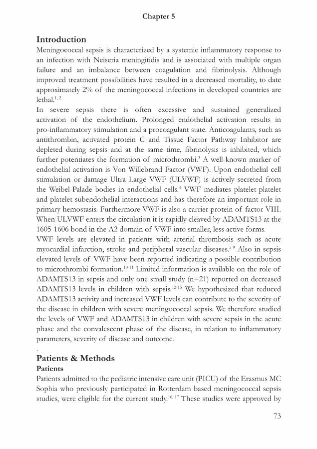
73
Chapter 5
IntroductionMeningococcal sepsis is characterized by a systemic inflammatory response to an infection with Neiseria meningitidis and is associated with multiple organ failure and an imbalance between coagulation and fibrinolysis. Although improved treatment possibilities have resulted in a decreased mortality, to date approximately 2% of the meningococcal infections in developed countries are lethal.1, 2
In severe sepsis there is often excessive and sustained generalized activation of the endothelium. Prolonged endothelial activation results in pro-inflammatory stimulation and a procoagulant state. Anticoagulants, such as antithrombin, activated protein C and Tissue Factor Pathway Inhibitor are depleted during sepsis and at the same time, fibrinolysis is inhibited, which further potentiates the formation of microthrombi.3 A well-known marker of endothelial activation is Von Willebrand Factor (VWF). Upon endothelial cell stimulation or damage Ultra Large VWF (ULVWF) is actively secreted from the Weibel-Palade bodies in endothelial cells.4 VWF mediates platelet-platelet and platelet-subendothelial interactions and has therefore an important role in primary hemostasis. Furthermore VWF is also a carrier protein of factor VIII. When ULVWF enters the circulation it is rapidly cleaved by ADAMTS13 at the 1605-1606 bond in the A2 domain of VWF into smaller, less active forms.VWF levels are elevated in patients with arterial thrombosis such as acute myocardial infarction, stroke and peripheral vascular diseases.5-9 Also in sepsis elevated levels of VWF have been reported indicating a possible contribution to microthrombi formation.10-11 Limited information is available on the role of ADAMTS13 in sepsis and only one small study (n=21) reported on decreased ADAMTS13 levels in children with sepsis.12-15 We hypothesized that reduced ADAMTS13 activity and increased VWF levels can contribute to the severity of the disease in children with severe meningococcal sepsis. We therefore studied the levels of VWF and ADAMTS13 in children with severe sepsis in the acute phase and the convalescent phase of the disease, in relation to inflammatory parameters, severity of disease and outcome.. Patients & MethodsPatientsPatients admitted to the pediatric intensive care unit (PICU) of the Erasmus MC Sophia who previously participated in Rotterdam based meningococcal sepsis studies, were eligible for the current study.16, 17 These studies were approved by

74
the medical ethical committee. Children were enrolled after obtaining informed consent of the parents or guardians. In 58 of total of 71 patients, citrate blood was available.Inclusion criteria were presentation with tachycardia, tachypnea, rectal temperature <36ºC or > 38.5ºC, and petechiae. Data on all patients were collected at various time-points in the course of the disease (t=0, t=0.5 day, t=1 day and t=90 days). The pediatric risk of mortality (PRISM) score (based on blood pressure, heart rate, bilirubin, calcium, Pa O2, PaCo2, potassium, HCO3, glucose and Glasgow Coma Scale18, predicted death rate based on the Rotterdam score (based on serum potassium, base excess, platelet count and level of C-reactive protein)19, and the presence of disseminated intravascular coagulation (DIC) were collected as markers of severity of disease on admission. DIC was defined as having a score of 5 or more on the DIC score, which included platelets, FDP or D-dimers levels, PT and fibrinogen.20 Within 4 hours after admission to the PICU a baseline plasma sample was collected (t=0, n=58). Additionally blood was drawn at t=1 day, (n=50). Differences in number of patients at different time-points are the result of the fact that patients, with similar baseline characteristics, were included in several studies that did not all include a blood sample at t=0.5 day. Furthermore no samples were drawn when an arterial line was no longer available, when a patient was transported back to the referral hospital or when the patient died. Blood was drawn after 90 days to study the levels in the convalescent phase, but could only be obtained from 6 cooperative children. Some of the children needed to be treated with coagulation factor concentrates, such as protein C. However, concentrates were given after the first blood draw, so all t=0 samples are without any influence of coagulation factor concentrates. Until analysis the samples were stored at –80 degrees Celsius.
Plasma measurementsADAMTS13 activity and antigen were measured using the Technozym ADAMTS13 kit (Technoclone, Vienna, Austria) as described by the manu-facturer. Fluoresence was measured at 360/460 nm (Biotek reader FLX 800, Austria). The inter-assay variation was 10% for ADAMTS13 activity and 18% for ADAMTS13 antigen. Levels of both ADAMTS13 antigen and activity in pooled plasma of 40 healthy men were defined as 100%Von Willebrand Factor antigen (VWF:Ag) was determined with an in-house ELISA assay, using polyclonal rabbit anti-human VWF and horseradish

75
Chapter 5
peroxidase conjugated anti-human VWF (DakoCytomation, Glostrop, Denmark) for catching and tagging, respectively. Von Willebrand Factor collagen binding (VWF:CB) activity was measured by an in-house ELISA using type I collagen (Sigma, St Louis, USA) for catching and horseradish peroxidase conjugated anti-human VWF. The inter-assay variations of VWF:Ag and VWF:CB were 14.5% and 6.4% respectively.VWF propeptide levels were determined by an ELISA using a polyclonal an-tibody against recombinant VWF propeptide as capture and detection anti-body, as described previously.21 Factor VIII was determined according to the one-stage clotting assay method on the Sysmex CA-1500 (Dade-Behring, Leusden, the Netherlands) with the APTT reagent Triniclot (Biomerieux, Boxtel, the Netherlands) and FVIII-deficient plasma (Biopool, Kordia, the Netherlands)The concentrations of C-reactive protein, Interleukin (IL-6), thrombin-anti-thrombin complex (TAT) and plasminogen activator inhibitor I (PAI-I) were determined as described previously.22-24
Statistical analysisContinuous parameters were not normally distributed. Therefore data were presented as median and analyzed using the Mann-Whitney U test. Binomial variables were analyzed using Pearson’s chi-square test or Fisher’s exact test when appropriate. Spearman correlation coefficient was determined to assess the correlation between ADAMTS13 levels and severity scores or other labora-tory parameters. All ADAMTS13 levels were expressed as a percentage relative to normal pooled plasma. VWF levels were expressed in percentages of normal pooled plasma. The statistical analysis was performed using SPSS version 11.0. P-values less than or equal to 0.05 were considered to be statistical significant. ResultsPatient characteristicsIn total 71 patients with meningococcal sepsis were included in this study of whom from 58 patients citrated blood was available. The median age in this population was 5.5 years. The mortality rate was 12.6%. The baseline patient characteristics are presented in Table 1.
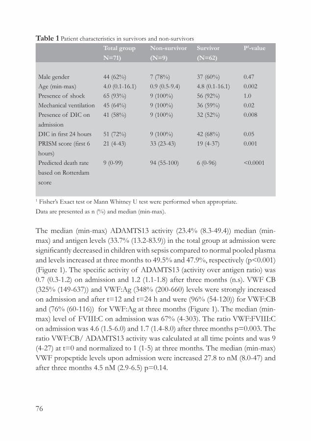
76
Table 1 Patient characteristics in survivors and non-survivorsTotal groupN=71)
Non-survivor(N=9)
Survivor(N=62)
P1-value
Male gender 44 (62%) 7 (78%) 37 (60%) 0.47Age (min-max) 4.0 (0.1-16.1) 0.9 (0.5-9.4) 4.8 (0.1-16.1) 0.002Presence of shock 65 (93%) 9 (100%) 56 (92%) 1.0Mechanical ventilation 45 (64%) 9 (100%) 36 (59%) 0.02Presence of DIC on admission
41 (58%) 9 (100%) 32 (52%) 0.008
DIC in first 24 hours 51 (72%) 9 (100%) 42 (68%) 0.05PRISM score (first 6 hours)
21 (4-43) 33 (23-43) 19 (4-37) 0.001
Predicted death rate based on Rotterdam score
9 (0-99) 94 (55-100) 6 (0-96) <0.0001
1 Fisher’s Exact test or Mann Whitney U test were performed when appropriate.Data are presented as n (%) and median (min-max).
The median (min-max) ADAMTS13 activity (23.4% (8.3-49.4)) median (min-max) and antigen levels (33.7% (13.2-83.9)) in the total group at admission were significantly decreased in children with sepsis compared to normal pooled plasma and levels increased at three months to 49.5% and 47.9%, respectively (p<0.001) (Figure 1). The specific activity of ADAMTS13 (activity over antigen ratio) was 0.7 (0.3-1.2) on admission and 1.2 (1.1-1.8) after three months (n.s). VWF CB (325% (149-637)) and VWF:Ag (348% (200-660) levels were strongly increased on admission and after t=12 and t=24 h and were (96% (54-120)) for VWF:CB and (76% (60-116)) for VWF:Ag at three months (Figure 1). The median (min-max) level of FVIII:C on admission was 67% (4-303). The ratio VWF:FVIII:C on admission was 4.6 (1.5-6.0) and 1.7 (1.4-8.0) after three months p=0.003. The ratio VWF:CB/ ADAMTS13 activity was calculated at all time points and was 9 (4-27) at t=0 and normalized to 1 (1-5) at three months. The median (min-max) VWF propeptide levels upon admission were increased 27.8 to nM (8.0-47) and after three months 4.5 nM (2.9-6.5) p=0.14.

77
Chapter 5
Plasma levels in survivors and non-survivorsOn admission ADAMTS13 antigen was slightly higher (34.5% (14.5-83.8)) in survivors on admission, than in non-survivors (24.0% (13.2-41.8)) (p=0.06) (Table 2). ADAMTS13 activity in survivors was 24.3% (11.5-49.4) versus 19.4% (8.3-27.6) in non-survivors. (p=0.19) In survivors the levels of VWF:Ag were higher than in non-survivors (370% (240-660)) vs (240% (200-330)), p<0.001). Also the VWF:CB was higher in survivors than in non-survivors, (340% (160-640)) vs. (240% (150-320)), p<0.001) (Table 2). In survivors the levels of FVIII:C (0.20 U/ml (0-0.6) were significantly lower than in non- survivors (0.8 U/ml (0-3.0)). The ratio VWF/FVIII:C on admission was 4.3 (1.5-46.2) in survivors and 9.7 (3.4-60) in non-survivors, p=0.003. The ratio VWF:CB/ ADAMTS13 activity was higher in survivors than in non-survivors after 0.5 day. The propeptide levels were not significantly different in survivors compared to non-survivors (28.7 nM (8.0-47.0) versus 22.2 nM (21.9-34.6), respectively. At 90 days the VWF propeptide levels were normal in the survivors.
Figure 1 ADAMTS13 and VWF levels in the total study population.Median ADAMTS13 and VWF levels (% relative to normal pooled plasma for ADAMTS13 and for VWF) on admission, after 0.5, 1 and 90 days are listed. The bars depict the ranges.
ADAMTS13 antigen
0 10
50
100
90Time from admission (days)
AD
AM
TS13
ant
igen
(%)
ADAMTS13 activity
0 10
50
100
90Time from admission (days)
AD
AM
TS13
act
ivity
(%)
VWF antigen
0 10
100
200
300
400
500
600
700
90Time form admission (days)
VWF
antig
en (%
)
VWF:CB
0 10
100
200
300
400
500
600
700
90Time from admission (days)
VWF:
CB
(%)

78
Table 2 ADAMTS13 and VWF levels in survivors and non-survivorsDay ADAMTS13 antigen P-
valueADAMTS13 activity P-
valueSurvivors Non-survivors Survivors Non-survivors
t=0 34.5 (14.5-83.8) 24.0 (13.2-41.8) 0.06 24.3 (11.5-49.4) 19.4 (8.3-27.6) 0.19t=0,5 35.6 (29.5-67) 46.2 (45.8-46.6) 0.10 26.7 (15.5-54.9) 35.3 (27.7-43.1) 0.38t=1 36.0 (19.7-66.2) 41.7 (25.7-57.9) 0.88 33.2 (13.9-65.3) 24.5 (20.1-28.9) 0.24t=90 47.9 (6.0-57.2) 49.5 (16.8-70.9)
Day VWF antigen P-value
VWF: CB P-value
Survivors Non-survivors Survivors Non-survivorst=0 370 (240-660) 240 (200-330) <10-3 340 (160-640) 240 (150-320) <10-3
t=0,5 310 (140-400) 240 (230-240) 0.21 280 (80-530) 130 (110-120) 0.06t=1 320 (110-490) 210 (180-240) 0.04 300 (120-450) 120 (110-120) 0.02t=90 80 (60-130) 100 (50-120)
Day Ratio VWF:CB/ADAMTS13 activity
P-value
Survivors Non-survivorst=0 10 (5-27) 8 (4-19) 0.33t=0,5 7 (1-15) 3 (2-3) 0.04t=1 6 (2-16) 3 (3-4) 0.07t=90 1 (1-5)
All values are presented in percentages of normal pooled plasma
Plasma levels in patients with DIC and without DICFor ADAMTS13 antigen no statistical difference between patients with DIC and without DIC was observed (Figure 2). However, ADAMTS13 activity (min-max) on admission was lower in patients with DIC 19.4% (8.3-35.2) than in individuals without DIC 27.3% (11.5-49.4); (p=0.01). On admission VWF:Ag was higher in the patients without DIC (390% (250-590)) than in patients with DIC (330% (200-660)) p=0.03 (Figure 2). For VWF:CB there was no significant difference in levels at the various time points. FVIII:C levels on admission were significantly higher in patients without DIC (1.3 U/ml (0.5-3.0)) than in patients with DIC (0.4 U/ml (0.0-1.45) (p<0.001).

79
Chapter 5
ADAMTS13 antigen
0 10
50
100
90Time from admission (days)
AD
AM
TS13
ant
igen
(%)
ADAMTS13 activity
0 10
50
100
90Time from admission (days)
AD
AM
TS13
act
ivity
(%)
VWF antigen
0 10
100
200
300
400
500
600
700
90Time form admission (days)
VWF
antig
en (%
)
VWF:CB
0 10
100
200
300
400
500
600
700
90Time from admission (days)
VWF:
CB
(%)
Figure 2 ADAMTS13 and VWF antigen levels in patients with and without DIC.Median levels of ADAMTS13 and VWF in patients with and without DIC are plotted. The closed squares represent the patients without DIC, the open squares represent the patients with DIC. The bar represents the range.
The ratio VWF/FVIII:C on admission was 2.7 (1.5-5.6) in patient without DIC and 8.3 (3.4-60) in patients with DIC, p=0.001. There was no difference in the ratio VWF:CB/ADAMTS13 activity in patients with DIC or without DIC. The VWF propeptide levels were similar in patients with DIC (28.5 (8.0-43.0) nM) compared to patients without DIC (26.5 nM (9.0-47.0)) p=0.57.
Correlations with severity of diseaseAll correlations were calculated at t=0. Both ADAMTS13 and VWF were nega-tively correlated with the PRISM score and the predicted death rate based on the Rotterdam score (Table 3). There was no correlation between ADAMTS13 antigen or activity and DIC parameters, such as thrombin-antithrombin (TAT) complexes and plasminogen activator inhibitor I (PAI-1). A positive significant correlation was seen for VWF with the levels of CRP and age. Another inflam-matory marker, IL-6, was negatively correlated with VWF activity (Table 3).

80
Tabl
e 3:
Cor
rela
tions
of
AD
AM
TS13
and
VW
F on
adm
issio
nLa
bora
tory
par
amet
erA
DA
MT
S13
antig
enP-
valu
eA
DA
MT
S13
activ
ityP-
valu
eV
WF:
Ag
P-va
lue
VW
F:C
BA
P-va
lue
Clin
ical
par
amet
ers
Age
in y
ears
0.22
0.10
0.08
0.58
0.40
0.00
20.
280.
03PR
ISM
scor
e (6
hr)
-0.3
10.
02-0
.38
0.01
-0.4
60.
00-0
.50
0.00
Pred
icte
d de
ath
rate
(R
otte
rdam
scor
e)-0
.26
0.05
-0.3
30.
02-0
.56
0.00
-0.5
80.
00
IL 6
(ng/
ml)*
-0.2
10.
280.
020.
94-0
.37
0.05
-0.5
20.
01C
RP (m
g/L)
0.29
0.03
0.05
0.72
0.39
0.00
0.29
0.03
Hae
mos
tasis
Plat
elet
cou
nt
(*10
9/l)
0.17
0.20
0.34
0.01
0.51
0.00
00.
390.
00
TAT
(ng/
ml)*
-0.2
50.
21-0
.03
0.87
-0.3
50.
07-0
.48
0.01
PAI-
1(ng
/ml)*
-0.2
60.
19-0
.05
0.81
-0.4
10.
03-0
.38
0.05
Fact
or V
III:C
(U/m
l)0.
210.
110.
480.
000.
430.
000.
590.
00
Due
to m
ultip
le c
ompa
rison
s a p
-val
ue <
0.00
5 w
as c
onsid
ered
stat
istic
ally
sign
ifica
nt.
* Fo
r the
mea
sure
men
t of
IL-6
, TA
T, P
AI-
1 bl
ood
from
onl
y 27
pat
ient
s was
ava
ilabl
e, al
l oth
er p
aram
eter
s wer
e de
term
ined
in 5
8 pa
tient
s

81
Chapter 5
DiscussionIn the present study, we showed that the plasma levels of ADAMTS13 and VWF on admission were associated with the outcome of meningococcal sepsis in children. Furthermore, a strong decrease of ADAMTS13 and increase of VWF were seen in the first 24 hours of the disease and both correlated with disease severity. We observed a strong correlation between ADAMTS13 and markers for disease severity, such as the PRISM score, and the predicted death rate based on the Rotterdam score.Since ADAMTS13 cleaves ultra large VWF multimers into smaller, less thrombogenic multimers, it is expected that low levels of ADAMTS13 will result in more ULVWF in plasma and is therefore expected to result in a more thrombogenic state. Recently Bockmeyer et al. indeed showed that inflammation-induced ADAMTS13 deficiency is associated with the appearance of ULVWF in plasma.25 In sepsis patients in whom VWF levels are strongly increased this pro-thrombotic state may further worsen the outcome in these patients. In our study in a relatively large population of children with meningococcal sepsis, we have shown a reduction in the levels of ADAMTS13 activity and antigen in the acute phase of the disease. These results are in concordance with previous studies in sepsis patients that reported that the levels of ADAMTS13 antigen and ADAMTS13 activity are decreased and levels of VWF are increased.12-15 However, most of these studies were in adult patients with sepsis-induced disseminated intravascular coagulation and it was not yet known whether the contribution of ADAMTS13 to sepsis in children is similar to that in adults. Only one study had been performed so far in children which is in agreement with our study.15 In our study we showed for the first time that there is a relationship between ADAMTS13 and DIC and with outcome and sever-ity of the disease in children. In addition, lower levels of ADAMTS13 were inversely related to outcome scores (Rotterdam score and PRISM score), which indicate that low ADAMTS13 levels are associated with poor prognosis. Levels of ADAMTS13 antigen on admission were slightly lower (n.s) in the non-survivors than in survivors. ADAMTS13 was not correlated with age, but VWF was (Table 3). Age may play a role, since an immature state of clotting system may cause a more severe coagulation state in young children.26
Interestingly, higher levels of VWF were observed in the survivors com-pared to the non-survivors. One explanation might be that this is caused by a decreased release of VWF in the non-survivors, but it is expected that a more

82
severe disease results in increased endothelial damage and higher VWF lev-els. An alternative hypothesis is that differences in VWF levels result from an increased consumption of VWF in the non-survivors. To discriminate between these possible mechanisms, we measured the VWF propeptide, which is a marker for the secretion of VWF. VWF propeptide was significantly increased in the first 24 hours and was normal after three months. No difference was seen between survivors and non-survivors concerning the propeptide levels. This indicates that a decreased release of VWF is not the cause of the lower VWF levels in the non-survivors. The second mechanism, more consumption of VWF, is more likely since patients with DIC had significantly lower VWF levels than patients without DIC. This is further substantiated by the negative correlation between VWF with TAT, a marker of coagulation activation. Also exhaustion of storage pools could be an explanation for the differences in non-survivor and survivor.A limitation of the study was that we compared the levels of ADAMTS13 in septic children with the normal levels of ADAMTS13 in adults. Nguyen et al. used as a control group children that were critically ill but did not have sepsis, and showed that in their control group of children ADAMTS13 levels were around 85% of adult reference values.15 Another limitation of the study could be that we have not screened for ADAMTS13 inhibitors. This might influence the activity of ADAMTS13. However, no influence on the antigen levels is expected.Some patients with severe sepsis are treated with coagulation factor concentrates, such as protein C, which may improve the prognosis.27 However, patients were not treated with concentrates, such as protein C concentrate, before the first blood collection. Furthermore, we determined ADAMTS13 and VWF post-hoc. This can be seen as a limitation; however if we had decided to measure these levels immediately, the study design would not have been changed and we would still have had the same end-point.Our results show that ADAMTS13 is significantly correlated with the severity of the disease. Infusion of recombinant human-ADAMTS13 (r-hu-ADAMTS13) has previously been shown to inhibit the thrombus growth and thereby the thrombus formation in ADAMTS13 deficient mice.28 It would be an interesting subject to investigate in future studies whether ADAMTS13 may be used to prevent thrombus formation. However, first, additional studies on ADAMTS13 levels in both children and adults with severe sepsis should be performed to confirm our findings before this approach is further explored.

83
Chapter 5
In conclusion, our study shows that levels of ADAMTS13 are strongly reduced and levels of VWF are strongly increased in children with meningococcal sepsis and that these levels are associated with severity and outcome of the disease, likely by promoting the formation of microthrombi in these affected children.
References1. Booy R, Habibi P, Nadel S, et al. Reduction in case fatality rate from meningococcal disease associ-
ated with improved healthcare delivery. Arch Dis Child 2001;85;5:386-90.
2. Maat M, Buysse CM, Emonts M, et al. Improved survival of children with sepsis and purpura: ef-
fects of age, gender, and era. Critical care (London, England) 2007;11;5:R112.
3. O’Brien JM, Jr., Ali NA, Aberegg SK, et al. Sepsis. The American journal of medicine
2007;120;12:1012-22.
4. Blann AD. Plasma von Willebrand factor, thrombosis, and the endothelium: the first 30 years.
Thromb Haemost 2006;95;1:49-55.
5. Thompson SG, Kienast J, Pyke SD, et al. Hemostatic factors and the risk of myocardial infarction
or sudden death in patients with angina pectoris. European Concerted Action on Thrombosis and
Disabilities Angina Pectoris Study Group. The New England journal of medicine 1995;332;10:635-
41.
6. Folsom AR, Rosamond WD, Shahar E, et al. Prospective study of markers of hemostatic function
with risk of ischemic stroke. The Atherosclerosis Risk in Communities (ARIC) Study Investigators.
Circulation 1999;100;7:736-42.
7. Catto AJ, Carter AM, Barrett JH, et al. von Willebrand factor and factor VIII: C in acute cerebrov-
ascular disease. Relationship to stroke subtype and mortality. Thromb Haemost 1997;77;6:1104-8.
8. Bongers TN, de Maat MP, van Goor ML, et al. High von Willebrand factor levels increase the risk
of first ischemic stroke: influence of ADAMTS13, inflammation, and genetic variability. Stroke
2006;37;11:2672-7.
9. Miura M, Kaikita K, Matsukawa M, et al. Prognostic value of plasma von Willebrand factor-cleav-
ing protease (ADAMTS13) antigen levels in patients with coronary artery disease. Thromb Hae-
most 2010 103;3;623-629.
10. van Mourik JA, Boertjes R, Huisveld IA, et al. von Willebrand factor propeptide in vascular
disorders: A tool to distinguish between acute and chronic endothelial cell perturbation. Blood
1999;94;1:179-85.
11. Claus RA, Bockmeyer CL, Budde U, et al. Variations in the ratio between von Willebrand factor and
its cleaving protease during systemic inflammation and association with severity and prognosis of
organ failure. Thromb Haemost 2009;101;2:239-47.
12. Martin K, Borgel D, Lerolle N, et al. Decreased ADAMTS-13 (A disintegrin-like and metallopro-
tease with thrombospondin type 1 repeats) is associated with a poor prognosis in sepsis-induced

84
organ failure. Crit Care Med 2007;35;10:2375-82.
13. Kremer Hovinga JA, Zeerleder S, Kessler P, et al. ADAMTS-13, von Willebrand factor and related
parameters in severe sepsis and septic shock. J Thromb Haemost 2007;5;11:2284-90.
14. Ono T, Mimuro J, Madoiwa S, et al. Severe secondary deficiency of von Willebrand factor-cleaving
protease (ADAMTS13) in patients with sepsis-induced disseminated intravascular coagulation: its
correlation with development of renal failure. Blood 2006;107;2:528-34.
15. Nguyen TC, Liu A, Liu L, et al. Acquired ADAMTS-13 deficiency in pediatric patients with severe
sepsis. Haematologica 2007;92;1:121-4.
16. de Kleijn ED, de Groot R, Hack CE, et al. Activation of protein C following infusion of protein
C concentrate in children with severe meningococcal sepsis and purpura fulminans: a randomized,
double-blinded, placebo-controlled, dose-finding study. Crit Care Med 2003;31;6:1839-47.
17. den Brinker M, Joosten KF, Visser TJ, et al. Euthyroid sick syndrome in meningococcal sepsis: the
impact of peripheral thyroid hormone metabolism and binding proteins. J Clin Endocrinol Metab
2005;90;10:5613-20.
18. Pollack MM, Ruttimann UE, Getson PR. Pediatric risk of mortality (PRISM) score. Crit Care Med
1988;16;11:1110-6.
19. Kornelisse RF, Hazelzet JA, Hop WC, et al. Meningococcal septic shock in children: clinical and lab-
oratory features, outcome, and development of a prognostic score. Clin Infect Dis 1997;25;3:640-
6.
20. Taylor FB, Jr., Toh CH, Hoots WK, et al. Towards definition, clinical and laboratory criteria, and a
scoring system for disseminated intravascular coagulation. Thromb Haemost 2001;86;5:1327-30.
21. Borchiellini A, Fijnvandraat K, ten Cate JW, et al. Quantitative analysis of von Willebrand factor
propeptide release in vivo: effect of experimental endotoxemia and administration of 1-deamino-
8-D-arginine vasopressin in humans. Blood 1996;88;8:2951-8.
22. den Brinker M, Joosten KF, Liem O, et al. Adrenal insufficiency in meningococcal sepsis: bioavail-
able cortisol levels and impact of interleukin-6 levels and intubation with etomidate on adrenal
function and mortality. J Clin Endocrinol Metab 2005;90;9:5110-7.
23. Pralong G, Calandra T, Glauser MP, et al. Plasminogen activator inhibitor 1: a new prognostic
marker in septic shock. Thromb Haemost 1989;61;3:459-62.
24. Boermeester MA, van Leeuwen PA, Coyle SM, et al. Interleukin-1 blockade attenuates me-
diator release and dysregulation of the hemostatic mechanism during human sepsis. Arch Surg
1995;130;7:739-48.
25. Bockmeyer CL, Claus RA, Budde U, et al. Inflammation-associated ADAMTS13 deficiency pro-
motes formation of ultra-large von Willebrand factor. Haematologica 2008;93;1:137-40.
26. Hazelzet JA, Risseeuw-Appel IM, Kornelisse RF, et al. Age-related differences in outcome
and severity of DIC in children with septic shock and purpura. Thrombosis and haemostasis
1996;76;6:932-8.

85
27. Bernard GR, Vincent JL, Laterre PF, et al. Efficacy and safety of recombinant human activated
protein C for severe sepsis. The New England journal of medicine 2001;344;10:699-709.
28. Chauhan AK, Motto DG, Lamb CB, et al. Systemic antithrombotic effects of ADAMTS13. J Exp
Med 2006;203;3:767-76.


Elevated levels of von Willebrand factor in cirrhosis support platelet adhesion despite reduced functional capacity
T.N. Bongers, T. Lisman, J. Adelmeijer, H.L.A. Janssen, M.P.M. de Maat, P.G. de Groot, F.W.G. Leebeek
Hepatology 2006; 44; 53-61
Chapter 6

88
AbstractBackgroundCirrhosis of the liver is frequently accompanied by complex alterations in the hemostatic system, resulting in a bleeding tendency. Although many hemostatic changes in liver disease promote bleeding, also compensatory mechanisms are found, including high levels of the platelet adhesive protein von Willebrand Factor (VWF). However, conflicting reports on the functional properties of VWF in cirrhosis have appeared in literature.
MethodsWe have measured a panel of VWF parameters in plasma from patients with cirrhosis of varying severity and etiology. Furthermore, we assessed the contri-bution of VWF to platelet adhesion, by measuring the ability of cirrhotic plasma to support adhesion of normal platelets under flow conditions.
ResultsVWF antigen levels were strongly increased in patients with cirrhosis. In contrast, the relative collagen binding activity, as well as the relative ristocetin cofactor activity was significantly lower in patients as compared to controls, indicating loss of function. Accordingly, patients had a reduced fraction of high molecular weight VWF multimers. Both strongly elevated and reduced activity and antigen levels of the VWF cleaving protease ADAMTS13 were found in individual patients. Adhesion of either normal or patient platelets to a collagen surface was substantially increased when these platelets were resuspended in plasma of patients with cirrhosis, as compared to control plasma.
ConclusionIn conclusion, highly elevated levels of VWF in patients with cirrhosis contribute to the induction of primary hemostasis despite reduced functional properties of the molecule. This phenomenon might compensate for defects in platelet number and function in patients with cirrhosis.

89
Chapter 6
IntroductionCirrhosis of the liver is often accompanied by extensive alterations in the hemostatic system. A decreased platelet count, impaired platelet function, a decreased thrombin generating capacity, defective fibrin formation due to dysfibrinogenemia, and defects in the fibrinolytic system may all be encountered in these patients (reviewed in 1). The net effect of these hemostatic changes is a bleeding tendency, which may be particularly manifested during invasive procedures.Although many of the hemostatic changes found in patients with cirrhosis result in a reduced hemostatic capacity, compensatory mechanisms are also found. For example, the reduction in thrombin generating capacity caused by reduced levels of procoagulant proteins is, in part, compensated by the concomitant reduction of the natural anticoagulants.1 In fact, tissue factor-induced thrombin genera-tion, which is substantially depressed in a prothrombin-like assay, is completely normalized on addition of thrombomodulin which allows activation of the anticoagulant protein C system.2 Similarly, the reduction in profibrinolytic proteins may (in part) be compensated by reduction of antifibrinolytics.3, 4 A mild to moderate thrombocytopenia and a poorly defined thrombocytopathia are often present in patients with cirrhosis, but its clinical relevance is unclear. A possible compensatory mechanism for these platelet abnormalities is the presence of abnormally high plasma levels of von Willebrand factor (VWF).5 VWF is a large, multimeric protein with a crucial role in primary hemostasis, as evidenced by the severe bleeding tendency associated with complete VWF deficiency.6 Conversely, elevated levels of VWF are associated with (arterial) thrombosis.7 Platelet-VWF interaction is the first step in platelet adhesion. After vessel wall damage, VWF from the circulation binds to exposed collagen fibers in the subendothelium. Once bound to collagen, VWF is able to interact with platelet glycoprotein Ib. Through transient VWF-glycoprotein Ib interactions, the platelet slows down, allowing subsequent stable interaction of the platelet with collagen through the collagen receptors a2b1 and glycoprotein VI.The elevated levels of VWF in cirrhosis may be a consequence of endothe-lial perturbation, possibly caused by bacterial infection.5 Another possible mechanism of elevated VWF in cirrhosis is induction of synthesis of VWF in the cirrhotic liver itself 8, or reduced liver-mediated clearance. Although it is established that VWF antigen levels are increased in patients with cirrhosis, relatively little is known on the functional capacity of the protein in these patients. Conflicting reports on the capacity of VWF to bind platelets (as

90
measured by ristocetin or botrocetin induced platelet aggregation) have appeared in literature, and patient numbers in these studies were small.5, 9, 10 An important qualitative aspect of VWF is its multimeric composition. The high molecular weight multimers are more potent in supporting hemostasis, as they have a higher affinity for both glycoprotein Ib and collagen. The multimeric composition of VWF is partly regulated by the VWF cleaving protease ADAMTS13. Reduced levels of ADAMTS13 have been found in patients with liver disease, suggesting that elevated amounts of high molecular weight multimers may circulate in these patients.11 However, the multimeric structure has been reported as either normal 5 or reduced 9, which may indicate that VWF proteolysis in liver disease is also accomplished by other proteases such as plasmin or elastase.12
In this study, qualitative and quantitative parameters of VWF were measured in a large group of patients with cirrhosis of varying severity and etiology. Parameters examined were VWF antigen levels, VWF ristocetin cofactor activity, which is a measurement for the ability of VWF to interact with platelet glyco-protein Ib, the VWF collagen binding activity, the VWF cleaving protease AD-AMTS13 antigen and activity, the multimeric structure of VWF, and the levels of the propeptide of VWF. In addition, the ability of cirrhotic plasma to support adhesion of normal or patient platelets under flow conditions was examined. The combined results of these assays provide more insight to what extent elevated VWF levels in these patients contribute to induction of hemostasis. Materials and MethodsPatientsFifty-four patients with biopsy-proven cirrhosis of various etiology – alcohol abuse 16, viral hepatitis 19, autoimmune hepatitis 2, primary biliary cirrhosis 4, cryptogenic cirrhosis 8, and others 5 – were included in this study. The patients were classified according to Pugh’s modification of the Child classification.13 Nineteen patients with Child A cirrhosis, 17 patients with Child B cirrhosis, and 18 patients with Child C cirrhosis were studied. In addition, 5 patients with acute liver failure were studied. Most patients were hospitalized for a short period as part of a liver transplant screening program in a single institution (Erasmus MC, Rotterdam, The Netherlands) at the time of inclusion. None of the patients received transfusion of platelet or plasma and did not use aspirin or other non-steroidal anti-inflammatory drugs in two weeks prior to the blood draw. One patient in the Child A and three in the Child C group were on antibiotics at the time of the blood draw. One patient in the Child C group was admitted

91
Chapter 6
to the hospital with sepsis and one was admitted for bacterial peritonitis. One patient in the Child C group had diabetes mellitus and hepatorenal syndrome.A group of 40 healthy volunteers from our laboratory was used to establish reference values for all assays used. Pooled normal plasma was obtained by combining plasma from these healthy volunteers. In the platelet adhesion experiments pooled cirrhosis plasma was used. For this, plasma from 4 patients with Child’s A, 4 patients with Child’s B, and 4 patients with Child’s C (Pooled Child’s C) cirrhosis was combined. For the platelet adhesion experiments, we recruited 3 additional patients with cirrhosis (all 3 had Child’s C cirrhosis of alcoholic origin) and 3 healthy volunteers.Blood samples were obtained by venipuncture from the antecubital vein into 3.2% sodium citrate (9:1, v/v). To obtain platelet poor plasma, samples were centrifuged twice at 2000g for 15 minutes, after which the samples were stored at -70°C until use.
AssaysVWF antigen (VWF:Ag) levels were determined using the STA Liatest from Roche (Almere, The Netherlands) in the Behring Coagulation System (BCS, Dade Behring, Marburg, Germany). VWF ristocetin cofactor activity (VWF:RCo) was determined using the BC von Willebrand reagent (Dade Behring) on the BCS apparatus. VWF:Ag and VWF:RCo levels in pooled normal plasma were set at 100%.
VWF collagen binding activity (VWF:CBA) was determined with an in-house assay. In short, collagen type III (Sigma, St. Louis, MO, USA) was dissolved in 50 mM acetic acid, and dialyzed against phosphate-buffered saline to obtain fibrillar collagen. ELISA plates were coated with collagen (10 mg/well), blocked with PBS containing 3% bovine serum albumin and 0.1% tween-20. Plasma samples were diluted according to their VWF:Ag content. Two dilutions, corresponding to 5 and 2.5% of the amount of VWF present in pooled normal plasma were prepared and added to the wells. Bound VWF was visualized using a horseradish peroxidase(HRP)-conjugated polyclonal antibody against VWF (DAKO, Glostrup, Denmark). The collagen binding activity of pooled normal plasma was set at 100%, and the values obtained in patient samples were expressed as % of pooled normal plasma.

92
The VWF cleaving protease ADAMTS13 activity was measured using a rapid functional assay that determines the digestion of VWF by ADAMTS13, based on the method described by Gerritsen.14 The activity of ADAMTS13 in pooled normal plasma was arbitrarily defined as 1 U/ml. The values of ADAMTS13 in tested plasma samples are read from a calibration curve achieved by incubating the VWF substrate with dilutions of a normal plasma pool. The upper detection limit was 3 U/ml. ADAMTS13 antigen levels were measured by a commercially available ELISA according to the instructions of the manufacturer (American Diagnostica, Stamford, CT).
VWF multimer analysis was performed by sodium dodecylsulphate agarose gelelectroforesis followed by western blotting according to Brosstad et al.15 The blots were scanned and analyzed by densitometric analysis (ImageQuant 5.2., Molecular Dynamics, Sunnyvale, CA). The first 5 bands were considered as low molecular weight multimers, whereas the other bands were designated as high molecular weight multimers.
VWF propeptide levels were determined by ELISA using a polyclonal antibody raised against recombinant VWF propeptide which was purified as described.16 The catching and detecting antibody were the same, except that the detection antibody was conjugated with HRP. The propeptide level measured in pooled normal plasma was set at 100%.
Platelet adhesion experimentsThe ability of plasma from patients with cirrhosis to support platelet adhesion was studied under flow conditions in a reconstituted blood model. Red cells were isolated from whole blood from healthy volunteers who had blood group O as described previously.17 Platelets were isolated from 3 patients with cirrhosis and 3 controls according to previously published methods.17 After the final washing step, platelets from patients or controls were resuspended in pooled normal plasma, or in pooled cirrhosis plasma. The platelets were mixed with red cells to obtain reconstituted blood with a platelet count of 200,000/ml and a hematocrit of 40%. In selected experiments, reconstituted blood with a reduced platelet count (25.000 or 100.000 platelets/μl) and a hematocrit of 40% was prepared. The reconstituted blood was perfused over a collagen type III-coated surface using a single-pass perfusion chamber(18) at a shear rate of 1600s-1.

93
Chapter 6
Platelet adhesion under these conditions is completely dependent on VWF. After 2 minutes of perfusion, the coverslips were stained with May-Grünwald and Giemsa as described previously.19 Platelet adhesion was evaluated using computer-assisted analysis with OPTIMAS 6.0 software (Dutch Vision Systems [DVS], Breda, The Netherlands), and was expressed as the percentage of the surface covered with platelets.
Statistical analysisStatistical analysis was performed using the GraphPad InStat software package (GraphPad, San Diego, USA). Differences in VWF parameters were examined by standard one-way analysis of variance (ANOVA) using the Tukey post-test, except for differences in VWF propeptide, ADAMTS13 activity and antigen levels, and VWF multimer analysis which were assayed using the Kruskal Wallis ANOVA with Dunn’s post-test as in these measurements standard deviations were substantially different between groups, and values were not normally distributed. P values <0.05 were considered statistically significant.
ResultsVWF:Ag levels substantially elevated in cirrhosis and acute liver failureAs shown in figure 1A, VWF:Ag levels were strongly elevated in plasma from patients with Child A (380% [165-980]; median [range]), Child B (500% [130-1455]), and Child C (760% [385-1855]) cirrhosis compared to the reference group in which the median VWF:Ag level was 107% [38-180]) (P<0.001 for mild, moderate, and severe cirrhosis compared to control). In the 5 patients with acute liver failure, median VWF:Ag level was 790% [650-890] (p<0.01 compared to control). When patients were classified according to the model for end-stage liver disease (MELD) score, we also observed a strong correlation between VWF:Ag levels and severity of the disease as assessed by the MELD score. (r=0.448, p<0.001). For calculation of the MELD score, we used the modified MELD score as employed by UNOS for organ allocation: MELD = [0.957 × loge (creatinine) + 0.378 × loge(bilirubin) + 1.12 × loge (international normalized ratio) + 0.64] × 10 (Available at: http://www.unos.org/resources). In accordance, mean MELD scores paralleled the Child classification. The MELD score was 10 [6-18] (median [range]) in the Child A group, 13 [7-23] in the Child B group, 18 [13-36] in the Child C group, and 28 [22-36] in the acute liver failure group.

94
Contro
l
Child A
Child B
Child C
Acute
0
1000
2000
VWF:
Ag
(%)
Contro
l
Child A
Child B
Child C
Acute
0
500
1000
1500
VWF:
RC
o (%
)
Contro
l
Child A
Child B
Child C
Acute
0
1
2
VWF:
RC
o/VW
F:A
g ra
tio
A
B
C
* * * * *
* * * *
* * * *
Figure 1 VWF:Ag levels (A), VWF:RCo levels (B), and VWF:RCo/VWF:Ag ratio (C) in pa-tients with Child A, B and C cirrhosis and in patients with acute liver failure compared with VWF parameters as measured in healthy controls. VWF:Ag and VWF:RCo levels are expressed as a percentage of pooled normal plasma. Horizontal lines represent medians. * p<0.001, ** p<0.01

95
Chapter 6
Substantially elevated VWF:RCo levels, but depressed VWF:RCo/VWF:Ag ratio in cirrhosis and acute liver failureAs shown in figure 1B, VWF:RCo levels were found substantially elevated in patients with Child A (300% [121-630]; median [range]), Child B (290% [40-900]), and Child C (610% [240-1005]) cirrhosis compared to the reference group in which the median VWF:RCo level was 105% [33-222]) (P<0.001 for mild, moderate, and severe cirrhosis compared to control). In the 5 patients with acute liver failure, median VWF:RCo level was 505% [285-540] (p<0.001 compared to control). However, the VWF:RCo levels are not elevated to the same extent as compared to the antigen levels. In other words, although the amount of VWF is substantially elevated, it appears less functional with respect to glyco-protein Ib binding. This is demonstrated by a significantly depressed VWF:RCo/VWF:Ag ratio in all groups: Child A (0.75 [0.48-1.064]; median [range]), Child B (0.60 [0.31-1.02]), and Child C (0.78 [0.41-0.93]) cirrhosis compared to the reference group in which the median ratio was 0.96 [0.67-1.538]) (P<0.001 for mild, moderate, and severe cirrhosis compared to control, see figure 1C). In the 5 patients with acute liver failure, median VWF:RCo/VWF:Ag ratio was 0.61 [0.38-0.70] (p<0.001 compared to control). The functionality of VWF was reduced to the same extent in all patients as shown by a strong correlation between VWF:RCo and VWF:Ag levels (r=0.812 vs r=0.852 in the control group) (p<0.0001).
Reduced collagen binding capacity of VWF in patients with cirrhosis and acute liver failureAs shown in figure 2, VWF collagen binding activity decreased with increasing severity of the disease in patients with cirrhosis. VWF collagen binding activity was 95% [80-113] (median [range]) in Child A, 83% [42-98] in Child B, and 78 [61-90] in Child C cirrhosis, while in the reference group in the median collagen binding activity was 98% [0.87-107] (P<0.001 for moderate and severe cirrhosis compared to control, p>0.05 for mild cirrhosis compared to control). In the 5 patients with acute liver failure, the VWF collagen binding activity was slightly decreased (90% [86-99]), but this difference did not reach statistical significance.

96
Figure 2 VWF collagen binding capacity in patients with cirrhosis of varying severity and in patients with acute liver failure as compared with the activity in plasma from healthy controls. The collagen binding capacity was measured at equal antigen levels of VWF. The collagen bind-ing capacity of pooled normal plasma was set at 100%. Horizontal lines represent medians. * p<0.001
ADAMTS13 activity and antigen levels are highly variable in patients with liver diseaseADAMTS13 activity levels were determined using the collagen binding assay as described by Gerritsen.14 Compared to controls, ADAMTS13 levels in patients with cirrhosis showed a high variability, and both substantially elevated and substantially depressed ADAMTS13 levels were found in all patient groups as shown in figure 3A. Some patients had ADAMTS13 activity levels exceeding 3 U/ml on repeated testing, even after a further predilution of the sample prior to the assay. Mean ADAMTS13 levels in Child A and Child B cirrhosis were elevated compared to controls, but this difference did not reach statistical significance. As ADAMTS13 activity levels were previously reported to be decreased in patients with cirrhosis using the same assay used here 11, we decided to measure ADAMTS13 antigen levels as well. ADAMTS13 antigen levels also showed a high variability in the patient samples compared to the variation ob-served in control samples as shown in figure 3B. ADAMTS13 antigen levels were significantly elevated in Child B (P<0.05) cirrhosis, and in patients with acute liver
Contro
l
Child A
Child B
Child C
Acute
0
50
100
150
Col
lage
n bi
ndin
g ac
tivity
(% o
f NPP
)
ns * * ns

97
Chapter 6
failure (p<0.05) compared to controls, whereas the mean ADAMTS13 antigen levels found in Child A and Child C cirrhosis were not significantly different from the levels in the control group. The activity and antigen levels showed a weak, but statistically significant correlation (r=0.39, p=0.0029). Both ADAMTS13 activity and antigen levels were negatively correlated with the VWF:RCo/VWF:Ag ratio (r=0.29, p=0.29 for activity, r=0.61, p<0.0001 for antigen levels).
Figure 3 ADAMTS13 activity (A) and antigen (B) levels in patients with cirrhosis of varying severity and in patients with acute liver failure as compared with the levels measured in plasma from healthy controls. ADAMTS13 activity levels were calibrated to pooled normal plasma, in which the activity was set at 1 U/ml. ADAMTS13 antigen levels were calibrated using recombinant ADAMTS13 according to the instructions of the manufacturer. Horizontal lines represent medians. * p<0.05, ns is not significant.
Reduced high molecular weight VWF multimers in plasma from patients with liver diseaseWe determined the multimeric pattern of VWF in a subset of the patients (12 with Child A, 10 with Child B, 8 with Child C, and 3 with acute liver failure). Consistent with the collagen binding data, we observed a significantly decreased proportion of high molecular weight multimers in patients with Child A (63% [32-74]; median [range]), Child B (66% [56-72]), and Child C (58% [52-77]) cirrhosis, compared to the amount of high molecular weight multimers in the controls (73% [61-81]) (P<0.01 for mild and severe cirrhosis, and acute liver failure compared to control, p<0.05 for moderate cirrhosis compared to control) (figure 4). In the 5 patients with acute liver failure, also a decreased proportion of high molecular weight multimers was observed (60% [33-61], p<0.01). The fraction of high molecular weight multimers was negatively correlated with the

98
ADAMTS13 activity (r=-0.55, p<0.001), and with ADAMTS13 antigen levels, which almost reached statistical significance (r=-0.34, p=0.051). Also, a correla-tion between the fraction of high molecular weight multimers and VWF:RCo/VWF:Ag ratio was observed (r=0.38, p=0.03).
Figure 4 The proportion of high molecular weight (HMW) VWF multimers in patients with cirrhosis of varying severity and in patients with acute liver failure as compared with the propor-tion of high molecular weight multimers in plasma from healthy controls. The proportion of high molecular weight multimers was estimated from densitometric analysis of western blots of agarose gels. The first 5 visible bands were considered to represent the low molecular weight multimers. Horizontal lines represent medians. * p<0.01, ** P<0.05
Cirrhotic plasma supports VWF-dependent platelet adhesion better than normal plasmaThe ability of pooled cirrhosis plasma (VWF:Ag 402%, VWF:RCo 301%) cirrhosis to support adhesion of normal platelets or platelets from patients with cirrhosis was compared to that of pooled normal plasma (VWF:Ag and VWF:RCo 100%, per definition). The pooled plasma’s were added to isolated red cells and platelets from 3 healthy volunteers or 3 patients with cirrhosis and perfused over collagen type III for 2 minutes. As shown in figure 5A, the amount of platelets isolated from healthy controls or patients adhering to the surface was significantly higher when platelets were resuspended in pooled cirrhotic plasma as compared to resuspension of the platelets in normal plasma.
Contro
l
Child A
Child B
Child C
Acute
0
25
50
75
100
% V
WF
HM
W m
ultim
ers * ** * *

99
Chapter 6
A
B
**
Control p
lt/Contro
l plas
ma
Control p
lt/Cirr
hosis plas
ma
Cirrhosis
plt/Contro
l plas
ma
Cirrhosis
plt/Cirr
hosis plas
ma0
10
20
30
Surf
ace
cove
rage
(%)
Control plt/control plasma Control plt/cirrhosis plasma
Cirrhosis plt/control plasma Cirrhosis plt/cirrhosis plasma
Figure 5 Plasma from patients with cirrhosis better supports platelet adhesion than normal plasma. A) Pooled plasma from patients with cirrhosis or pooled normal plasma was mixed with red cells and platelets either isolated from healthy volunteers or from patients with cirrhosis and perfused over a collagen-coated coverslip for 2 minutes. After May-Grunwald staining, surface coverage was determined (shown are results from experiments with 3 different patients and 3 different controls performed in triplicate, error bars indicate standard error of mean). * P<0.01 B) Morphological appearance of the platelet thrombi on a collagen surface with reconstituted blood with patient or control plasma or platelets as indicated. Shown are representative examples of the experiment presented in panel A. Original magnification is 400.

100
Also, when normal or patient platelets were resuspended in pooled cirrhotic plasma, the aggregates were substantially larger as compared to the aggregate size obtained when platelets were resuspended in pooled normal plasma (figure 5B). The adhesion of platelets isolated from patients with cirrhosis was similar to that of the adhesion of platelets isolated from healthy controls under these conditions of standardized platelet count and hematocrit. When normal platelets were resuspended in either normal or cirrhotic plasma under conditions representing thrombocytopenia (25.000 and 100.000 platelets/ml, we also observed a significant elevation in platelet deposition when cirrhotic plasma was used (data not shown).
VWF propeptide levels As shown in figure 6, VWF propeptide levels were substantially elevated in Child A (488% [234-2942]; median [range]), Child B (711% [261-2190]), and Child C (735% [221-5129]) cirrhosis, while in the reference group in the median VWF propeptide level was 89% [30-237]) (P<0.001 for mild, moderate, and severe cirrhosis compared to control). Also in the patients with acute liver disease, elevated propeptide levels were found (1139% [803-1594], p<0.001).
DiscussionIn this study, we have performed a comprehensive study on VWF in a large group of patients with cirrhosis of varying severity and etiology. Highly elevated levels of VWF were found, which were strongly related to the severity of the disease. However, the functional capacity of the VWF decreased with increa-sing severity of the disease as shown by a reduction in VWF:RCo/VWF:Ag ratio and reduced collagen binding capacity. Despite the suppressed binding capacity to both glycoprotein Ib and collagen, the highly elevated VWF levels in cirrhotic plasma resulted in a substantially elevated platelet deposition to collagen in a VWF-dependent, flow-driven platelet adhesion assay. This indicates that the quantitative increase in VWF in cirrhosis overrules the qualitative defects, and that the elevated levels of VWF might in part compensate for the qualitative and quantitative platelet defects found in these patients. The increased adhesion induced by cirrhotic plasma was observed with both normal and patient platelets, and at normal and thrombocytopenic platelet counts.
Surprisingly, platelet deposition under conditions of standardized platelet count and hematocrit were similar when normal or patient platelets were used,

101
Chapter 6
Contro
l
Child A
Child B
Child C
Acute
0
1000
2000200040006000
vWF
prop
eptid
e(%
of N
PP)
* * * *
Figure 6 VWF propeptide levels in patients with cirrhosis of varying severity and in patients with acute liver failure as compared with the levels in healthy controls. The VWF propeptide level in pooled normal plasma was set at 100%. Horizontal lines represent medians. * p<0.001
suggesting that platelets in cirrhosis are functionally normal. This is in contrastwith experiments describing intrinsic platelet defects in cirrhosis (reviewed in 1). However, the majority of these experiments were performed under static conditions.
The elevated VWF levels in cirrhosis possibly reflect endothelial damage, which has been suggested to be stimulated by endotoxemia (bacterial infection).5 Another possibility is that the synthesis of VWF is increased, since it was recent-ly shown that liver disease induces VWF expression in the liver.8 Alternatively, VWF synthesis may be increased due to the substantially enhanced endothelial surface in patients with cirrhosis as a consequence of extensive collateral forma-tion. Also, the increased levels of vasoconstrictors (notably vasopressin), which arise as a consequence of the hyperdynamic state in these patients, could be responsible for elevated release of VWF from the endothelium.20 Finally it may be possible that reduced VWF clearance contributes to elevated levels, but to our knowledge no experimental evidence has been presented so far. We have measured levels of VWF propeptide in an attempt to investigate whether the elevated VWF levels represent acute endothelial damage, as found for example in patients with sepsis, or chronic endothelial perturbation such as found in patients with diabetes.21 As the half-life of VWF propeptide is much shorter

102
compared to that of VWF itself, only elevated levels of VWF and normal to slightly elevated levels of VWF propeptide are found in patients with chronic endothelial damage.21 The highly elevated propeptide levels found in patients with cirrhosis in our study suggest acute endothelial damage, which may be compatible with the presence of (low-grade) disseminated intravascular coagulation (DIC) in these patients, although the presence of DIC in cirrhosis has been debated.22 On the other hand, the increased propeptide levels may also be explained by a reduced clearance of this molecule in patients with liver disease, or it may reflect persistent enhanced VWF synthesis by the diseased liver.8 The hypothesis that patients with cirrhosis have continuously enhanced VWF release into the bloodstream is supported by analysis of the VWF propeptide/VWF:Ag ratio, which is slightly elevated in patients with Child’s A and B cirrhosis, but not different from controls in patients with Child’s C cirrhosis (data not shown). The VWF propeptide may have physiological relevance in the plasma environment in processes related to inflammation and cell adhesion (reviewed in 23). Whether the highly elevated VWF propeptide levels in patients with cirrhosis interfere with inflammatory or cell adhesion processes is unknown. It has also been shown that the propeptide may bind to collagen resulting in inhibition of collagen-induced platelet aggregation24, 25, but our platelet adhesion studies do not support these observations.
The clearly reduced VWF:RCo/VWF:Ag ratio, the reduced collagen binding activity, and the reduced proportion of circulating high molecular weight multimers measured in this large group of patients resolves the contro- versy of VWF functional capacity in patients with liver disease. Previously, both increased and decreased functional VWF parameters were reported (reviewed in 1). From our study it has become clear that the functional capacity of VWF is reduced in patients with cirrhosis. This may be caused by increased proteolysis by VWF proteases, such as plasmin or elastase.12 Recently a selective VWF cleaving protease has been discovered, which is important for cleavage of large, active VWF multimers into smaller less functional multimers. Surprisingly we found a strong variability in ADAMTS13 levels in patients with cirrhosis. These results are in contrast with a previous report in which significantly reduced levels were demonstrated in a group of patients with Child’s C cirrhosis by Mannucci et al.11 In our patient group we find both elevated and reduced activity and antigen levels of ADAMTS13. However, the measurement of ADAMTS13 activity is difficult, and shows a large coefficient of variation. A lot of controversies on

103
Chapter 6
ADAMTS13 activity measurement still exist, especially in cases with mild or moderately reduced levels.26 This is also illustrated by the relatively poor correla-tion between the two assays used in this study. Moreover, although ADAMTS13 levels would be expected to be decreased in cirrhosis, as it principle site of synthesis is presumably the liver27, it may also be that either reduced clearance, or release of ADAMTS13 from platelets28 (as a result of platelet activation secondary to DIC) lead to the elevated levels observed in some patients in our study. Alternatively, it might be possible that ADAMTS13 synthesis is induced in liver disease, as is has recently been shown that ADAMTS13 is synthesized in hepatic stellate cells29, which are known to show enhanced protein synthe-sis in patients with liver cirrhosis30. Interestingly, ADAMTS13 antigen levels, and, to a lesser extent, ADAMTS13 activity levels were negatively correlated with VWF:RCo/VWF:Ag ratio, indicating that the excess of ADAMTS13 in some patients results in excessive VWF proteolysis, resulting in impaired VWF activity. This is also supported by the negative correlation observed between ADAMTS13 levels and the proportion of high molecular weight VWF multimers. These data also suggest that in those patients with high levels of ADAMTS13, the reduced functional capacity of VWF is a direct consequence of excessive ADAMTS13 proteolysis.
In conclusion, highly elevated levels of VWF in patients with cirrhosis contribute to the induction of primary hemostasis despite reduced functional properties of the VWF molecule. This phenomenon might compensate for defects in platelet number and function, which are present in patients with cirrhosis.
References1. Lisman T, Leebeek FWG, de Groot PG. Haemostatic abnormalities in patients with liver disease.
J Hepatol 2002; 37: 280-7.
2. Tripodi A, Salerno F, Chantarangkul V, Clerici M, Cazzaniga M, Primignani M, Mannuccio MP.
Evidence of normal thrombin generation in cirrhosis despite abnormal conventional coagulation
tests. Hepatology 2005; 41: 553-8.
3. Lisman T, Leebeek FW, Mosnier LO, Bouma BN, Meijers JC, Janssen HL, Nieuwenhuis HK et al.
Thrombin-activatable fibrinolysis inhibitor deficiency in cirrhosis is not associated with increased
plasma fibrinolysis. Gastroenterology 2001; 121: 131-9.
4. Leebeek FWG, Kluft C, Knot EAR, de Maat MPM, Wilson JHP. A shift in balance between profi-
brinolytic and antifibrinolytic factors causes enhanced fibrinolysis in cirrhosis. Gastroenterology

104
1991; 101: 1382-90.
5. Ferro D, Quintarelli C, Lattuada A, Leo R, Alessandroni M, Mannucci PM, Violi F. High plasma
levels of von Willebrand factor as a marker of endothelial perturbation in cirrhosis: relationship to
endotoxemia. Hepatology 1996; 23: 1377-83.
6. Sadler JE, Mannucci PM, Berntorp E, Bochkov N, Boulyjenkov V, Ginsburg D, Meyer D et al.
Impact, diagnosis and treatment of von Willebrand disease. Thromb Haemost 2000; 84: 160-74.
7. Reiner AP, Siscovick DS, Rosendaal FR. Hemostatic risk factors and arterial thrombotic disease.
Thromb Haemost 2001; 85: 584-95.
8. Hollestelle MJ, Geertzen HG, Straatsburg IH, van Gulik TM, van Mourik JA. Factor VIII expres-
sion in liver disease. Thromb Haemost 2004; 91: 267-75.
9. Escolar G, Cases A, Vinas M, Pino M, Calls J, Cirera I, Ordinas A. Evaluation of acquired platelet
dysfunctions in uremic and cirrhotic patients using the platelet function analyzer (PFA-100 ): influ-
ence of hematocrit elevation. Haematologica 1999; 84: 614-9.
10. Beer JH, Clerici N, Baillod P, von Felten A, Schlappritzi E, Buchi L. Quantitative and qualitative
analysis of platelet GPIb and von Willebrand factor in liver cirrhosis. Thromb Haemost 1995; 73:
601-9.
11. Mannucci PM, Canciani MT, Forza I, Lussana F, Lattuada A, Rossi E. Changes in health and dis-
ease of the metalloprotease that cleaves von Willebrand factor. Blood 2001; 98: 2730-5.
12. Federici AB, Berkowitz SD, Lattuada A, Mannucci PM. Degradation of von Willebrand factor in
patients with acquired clinical conditions in which there is heightened proteolysis. Blood 1993; 81:
720-5.
13. Pugh RN, Murray-Lyon IM, Dawson JL, Pietroni MC, Williams R. Transection of the oesophagus
for bleeding oesophageal varices. Br J Surg 1973; 60: 646-9.
14. Gerritsen HE, Turecek PL, Schwarz HP, Lammle B, Furlan M. Assay of von Willebrand factor
(vWF)-cleaving protease based on decreased collagen binding affinity of degraded vWF: a tool
for the diagnosis of thrombotic thrombocytopenic purpura (TTP). Thromb Haemost 1999; 82:
1386-9.
15. Brosstad F, Kjonniksen I, Ronning B, Stormorken H. Visualization of von Willebrand factor mul-
timers by enzyme-conjugated secondary antibodies. Thromb Haemost 1986; 55: 276-8.
16. Borchiellini A, Fijnvandraat K, ten Cate JW, Pajkrt D, van Deventer SJ, Pasterkamp G, Meijer-Hu-
izinga F et al. Quantitative analysis of von Willebrand factor propeptide release in vivo: effect of
experimental endotoxemia and administration of 1-deamino-8-D-arginine vasopressin in humans.
Blood 1996; 88: 2951-8.
17. Lisman T, Moschatsis S, Adelmeijer J, Nieuwenhuis HK, de Groot PG. Recombinant factor VIIa
enhances deposition of platelets with congenital or acquired {alpha}IIb{beta}3 deficiency to en-
dothelial cell matrix and collagen under conditions of flow via tissue factor-independent thrombin
generation. Blood 2003; 101: 1864-70.

105
18. Sixma JJ, de Groot PG, van Zanten H, IJsseldijk M. A new perfusion chamber to detect platelet
adhesion using a small volume of blood. Thromb Res 1998; 92: S43-S46.
19. Zwaginga JJ, Sixma JJ, de Groot PG. Activation of endothelial cells induces platelet thrombus
formation on their matrix. Arteriosclerosis 1990; 10: 49-61.
20. Moller S, Bendtsen F, Henriksen JH. Vasoactive substances in the circulatory dysfunction of cir-
rhosis. Scand J Clin Lab Invest 2001; 61: 421-9.
21. van Mourik JA, Boertjes R, Huisveld IA, Fijnvandraat K, Pajkrt D, van Genderen PJ, Fijnheer R.
von Willebrand factor propeptide in vascular disorders: A tool to distinguish between acute and
chronic endothelial cell perturbation. Blood 1999; 94: 179-85.
22. Ben Ari Z, Osman E, Hutton RA, Burroughs AK. Disseminated intravascular coagulation in liver
cirrhosis: fact or fiction? Am J Gastroenterol 1999; 94: 2977-82.
23. van Mourik JA, Romani dW. Von Willebrand factor propeptide in vascular disorders. Thromb
Haemost 2001; 86: 164-71.
24. Takagi J, Kasahara K, Sekiya F, Inada Y, Saito Y. A collagen-binding glycoprotein from bovine
platelets is identical to propolypeptide of von Willebrand factor. J Biol Chem 1989; 264: 10425-30.
25. Takagi J, Sekiya F, Kasahara K, Inada Y, Saito Y. Inhibition of platelet-collagen interaction by
propolypeptide of von Willebrand factor. J Biol Chem 1989; 264: 6017-20.
26. Studt JD, Bohm M, Budde U, Girma JP, Varadi K, Lammle B. Measurement of von Willebrand
factor-cleaving protease (ADAMTS-13) activity in plasma: a multicenter comparison of different
assay methods. J Thromb Haemost 2003; 1: 1882-7.
27. Zheng X, Chung D, Takayama TK, Majerus EM, Sadler JE, Fujikawa K. Structure of von Wil-
lebrand factor-cleaving protease (ADAMTS13), a metalloprotease involved in thrombotic throm-
bocytopenic purpura. J Biol Chem 2001; 276: 41059-63.
28. Suzuki M, Murata M, Matsubara Y, Uchida T, Ishihara H, Shibano T, Ashida S et al. Detection of
von Willebrand factor-cleaving protease (ADAMTS-13) in human platelets. Biochem Biophys Res
Commun 2004; 313: 212-6.
29. Uemura M, Tatsumi K, Matsumoto M, Fujimoto M, Matsuyama T, Ishikawa M, Iwamoto TA et al.
Localization of ADAMTS13 to the stellate cells of human liver. Blood 2005; 106: 922-4.
30. Bataller R, Brenner DA. Liver fibrosis. J Clin Invest 2005; 115: 209-18.


Measurement of ADAMTS13: assay comparison in TTP, liver cirrhosis patients and healthy controls
T.N. Bongers, S. van Asten, T. Lisman, F.W.G. Leebeek, M.P.M. de Maat
Chapter 7

108
AbstractBackgroundADAMTS13 degrades ultralarge highly active von Willebrand Factor into smaller, less active multimers. Extreme Low levels of ADAMTS13 are associated with thrombotic thrombocytopenic purpura (TTP). Recent studies have indicated that more subtle reduced levels of ADAMTS13 are associated with microthrombi and arterial thrombosis. Therefore it is of utmost importance to precisely measure ADAMTS13 activity and antigen levels of ADAMTS13 in these conditions.Several assays have recently been developed. It is however unclear how to interpret the results of these assays in different patients groups.
MethodsIn this pilot study we compared three activity assays (Gerritsen assay, FRETS, Technozyme activity) and two antigen assays (Technozym antigen and ELISA American Diagnostica) in plasma samples from healthy individuals (n=60), patients with thrombotic thrombocytopenic purpura (TTP, n=41) and patients with liver cirrhosis of different severity (n=50).
ResultsAll activity assays measured very low levels in patients with TTP (levels < 20 %). As expected in patients with acquired TTP, higher levels of ADAMTS13 antigen levels were measured compared to ADAMTS13 activity. In healthy controls, the five assays measured slightly different levels. For all assays the range of the levels measured in cirrhosis patients was very wide and the correlation between the various assays was weak. This could not be explained by bilirubin levels or other patient characteristics and further research is needed to explain these findings.
ConclusionIn conclusion, ADAMTS13 activity assays are all able to identify patients with TTP, but their performance in patients with liver cirrhosis is unclear.

109
Chapter 7
IntroductionPatients with thrombotic thrombocytopenic purpura (TTP) suffer from intravascular platelet aggregation and microthrombi, what can result in ischemia and arterial thrombosis.1 If untreated, TTP is a life-threatening disease and therefore it is of utmost importance to rapidly and accurately diagnose this disease. Platelet aggregation and formation of microthrombi are caused by unusually large Von Willebrand Factor multimers (ULVWF). These ULVWF are the result of the absence of the protease ADAMTS13 (A Disintegrin And Metalloprotease with ThromboSpondin type-1 motifs 13), which is responsible for the cleavage of VWF into smaller, less active, VWF molecules. ADAMTS13 thus regulates the size of VWF multimers and prevents the formation of spontaneous platelet aggregates by ULVWF. The deficiency of ADAMTS13 in TTP patients can be genetic or acquired, and in the latter situation it is caused by antibodies that inhibit the activity of the protease. An inhibitor to ADAMTS13 strongly reduces ADAMTS13 activity but may be associated with normal ADAMTS13 antigen levels. Therefore a discrepancy can be found between the measurement of protein concentration of ADAMTS13 and its activity. ADAMTS13 is primary synthesized in the liver by stellate cells, and we previously showed that ADAMTS13 levels are decreased in patients with liver cirrhosis.2, 3 Surprisingly, we observed a very large variability of ADAMTS13 levels and VWF levels in patients with liver cirrhosis.3 Both very high (>300%) and very low (<30%) ADAMTS13 levels were observed in liver cirrhosis patients, independent of their severity of the cirrhosis.3 The cause for this variability is not explained so far.In recent years a number of assays have been introduced for the measurement of ADAMTS13. These assays are based on different test principles. The first assay developed for the measurement of the activity of ADAMTS13 was described by Gerritsen et al.4 In this assay, purified VWF is incubated with the patient sample and the decrease of VWF collagen binding activity is an indirect measurement of the ADAMTS13 activity. Recently, a number of other assays for the measurement of ADAMTS13 levels and activity have been intro-duced. The FRETS-VWF73 assay is an activity assay that uses a fluorescence- labeled peptide, containing the cleavage site of ADAMTS13.5 Also the ADAMTS13 Technozym assay measures ADAMTS13 activity.6 In this test ADAMTS13 is first added to a microtiter plate coated with anti-ADAMTS13 antibodies, and then in a second step, the activity of the bound ADAMTS13 is

110
determined by measuring the change in fluorescence. This assay also allows the determination of ADAMTS13 antigen via incubation with a tagging monoclonal antibody to ADAMTS13. Another ELISA using specific monoclonal anti-bodies against ADAMTS13 for measurement of ADAMTS13 antigen levels is commercially available (American Diagnostica, Stamford, CT). Each of these assays has its potential advantages and disadvantages. An important difference between the assays is the type of substrate that is used. The Gerritsen assay uses purified VWF as a substrate and theoretically measures all VWF-cleaving-proteases. The FRETS assay and the Technozym ADAMTS13 assay are more specific for ADAMTS13, but use a synthetic peptide as substrate, which is different from the natural substrate VWF. In the FRETS assay, high bilirubin levels may interfere with the FRETS-VWF73 substrate.7 Although the Technozym assay uses the same substrate, this problem is not to be expected, since bilirubin is washed away. A disadvantage of the Gerritsen assay is that it is labour-intensive, time-consuming and has a high assay variability. In contrast, the FRETS assay is fast and reproducible.The aim of our study was to compare the above mentioned assays available for measuring ADAMTS13 in healthy individuals, in patients with TTP and in patients with liver cirrhosis. Patients and MethodsPatientsForty-one patients with TTP were included in the study. These patients were included on admission to the Erasmus MC Rotterdam, the Netherlands in the acute phase of the TTP, before the start of treatment. Patients were diagnosed with TTP when patients had thrombocytopenia, microangiopathic hemolysis and clinical symptoms of TTP. Sixty healthy individuals were included in the study as a reference.In addition, fifty patients with biopsy-proven cirrhosis of the liver were included. These patients were diagnosed at the Erasmus MC. The patients were classified according to Pugh’s modification of the Child classification.8 Nineteen patients had Child A cirrhosis, 16 patients Child B cirrhosis, and 15 patients Child C cirrhosis. The etiology varied from alcohol abuse, viral hepatitis, autoimmune hepatitis, primary biliary cirrhosis to cryptogenic cirrhosis.
Blood samplingBlood samples were obtained by vena puncture from the antecubital vein into

111
Chapter 7
3.2% sodium citrate (9:1, v/v). Samples were centrifuged twice at 2000g for 15 minutes, after which the plasma was stored at -70°C until use.
Gerritsen ADAMTS13 activity assay ADAMTS13 activity according to Gerritsen et al, was performed according to literature, with some minor modifications.4, 9 Briefly, plasma is incubated for two hours with purified VWF as substrate for the protease. The supernatant was then added to a microtiter plate that has been coated with collagen. After incubation, the VWF bound to collagen is measured using a peroxidase-labled anti-human VWF polyclonal antibody and compared to normal pooled plasma. The intra-assay variation was 11 %.
ADAMTS13 FRET-VWF73 assayADAMTS13 activity was measured as previously described by Kokame.5 Briefly, samples were diluted with assay buffer and FRETS-VWF73 substrate (Thermo elektron GmbH, Ulm, Germany; Peptide institute Inc, Osaka, Japan) was added to each well. Fluorescence was measured every 5 minutes at 30ºC in a multi-label counter (Biotek reader FLX 800, Austria) at 340 nm excitation and 450 nm emission. The reaction slope was calculated by linear regression analysis and the ADAMTS13 activity was then calculated by comparing the slope to that of normal pooled plasma (George King). The intra-assay variation was 6 %.
ADAMTS13 assay (activity and antigen)ADAMTS13 activity and antigen were measured using the Technozym ADAMTS13 activity and antigen fluorogenic ELISA kit (Technoclone, Vienna, Austria), performed as described by the manufacturer. Briefly, diluted plasma samples were incubated on micro-titer plates coated with a monoclonal antibody against ADAMTS13. After two hours incubation the plates were washed and an activity substrate was added. The fluorescence was measured for 15 minutes (1 measurement per minute) at 30ºC and ADAMTS13 activity was calculated. The plate was then washed and incubated with a monoclonal antibody against ADAMTS13. A fluorescent labeled antigen substrate was added and after an incubation of 15 min at 37ºC the reaction was stopped and the fluorescence was measured at 340 nm excitation and 450 nm emission (Biotek reader FLX 800, Austria) and ADAMTS13 antigen was calculated. All levels are expressed as percentage of normal pooled plasma (%PP)(George King). The intra-assay variation was 6 % for ADAMTS13 activity and 6% for ADAMTS13 antigen.

112
ADAMTS13 antigenThe ADAMTS13 antigen levels were determined by a commercially available ELISA and were performed as described by the manufacturer (America Diagnostica, Stamford, CT). The assay gives levels in ng/ml, which we expressed in percentage of pooled plasma. ResultsTTP patientsIn the TTP patients, the Gerritsen assay, the FRETS assay and the Technozym assay identified the patients with very low ADAMTS13 levels (Table 1). In the Gerritsen assay, levels were 4 ± 3 % (mean±SD), with the FRETS assay it was 1 ± 3 %, with the Technozym activity assay 3 ± 2 %, with the Technozym antigen assay 34 ± 42 % and with the American Diagnostica ADAMTS13 ELISA 25 ± 31% (156 ± 111 ng/mL). The Spearman rank correlation between the ADAMTS13 activity assays (rs between 0.47 and 0.85) and the antigen assays (rs between 0.43 and 0.93) was good, but weak between activity and antigen assays (Table 2).
Table 1 ADAMTS13 activity and antigen in healthy controls and patients with TTPHealthy controls (N=60) Patients with TTP (N=41)
ADAMTS13 activity assaysGerritsen assay (%PP) 91 ± 15 4 ± 3FRETS assay (%PP) 111 ± 42 1 ± 1Technozym activity (%PP) 136 ± 42 3 ± 2
ADAMTS13 antigen assaysTechnozym antigen (%PP) 135 ± 33 26 ± 41ELISA (%PP) 127 ± 39 25 ± 31
Levels are presented as mean ± SD
Healthy individualsIn the 60 healthy individuals, the levels of ADAMTS13, measured with the different assays, showed a clear variability (Table 1). In the Gerritsen assay, levels were 91 ± 15 % (mean±SD), with the FRETS assay it was 111 ± 42 %, with the Technozym activity assay 136 ± 42 %. ADAMTS13 antigen levels measured with the Technozym antigen assay were 135 ± 33 % and with the American

113
Chapter 7
Diagnostica ADAMTS13 ELISA 126 ± 39% (698 ± 214 ng/mL). The correla-tion between the assays in the healthy controls was good between the FRETS assay and the Technozym activity and antigen assays, but was weak between the other tests (Table 2).
Table 2 Spearman Rank Correlation between the various ADAMTS13 assays in healthy con-trols, patients with TTP and patients with liver cirrhosis. Gerritsen FRETS Technozym:act Technozym:Ag
Total groupGerritsenFRETS 0.79*Technozym:act 0.28* 0.57*Technozym:Ag -0.28* 0.38* 0.87*AD:Ag 0.79* 0.74* 0.27* -0.13
Healthy controlsGerritsenFRETS n.d.Technozym:act -0.38 0.76*Technozym:Ag 0.37 0.62* 0.76*AD:Ag -0.16 n.d. -0.24 0.42
TTPGerritsenFRETS 0.69Technozym:act 0.47 0.85*Technozym:Ag -0.29 n.d. 0.61AD:Ag 0.14 0.30 0.43 0.93*
LiverGerritsenFRETS 0.57*Technozym:act -0.52* -0.35*Technozym:Ag -0.58* -0.28 0.80*AD:Ag 0.54* 0.69* -0.42* -0.38*

114
Liver cirrhosis patientsIn patients with cirrhosis, the range of ADAMTS13 activity and antigen levels varied from very low to very high with all assays (Table 3). In the Gerritsen assay levels of ADAMTS13 were remarkably high 165% (43-361) (median (10th-90th percentile)) in Child A, while this was 45% (14-96) for Technozym activity assay and 50 % (24-112) for the FRETS assay. In Child B the median levels of ADAMTS13 activity with the Gerritsen assay were 136% (43-361), 34% (29-135) for the Technozym activity assay and 51% (4-152) for the FRETS assay. In Child C we found 75% (34-274) in the Gerritsen assay, 59% (17-84) for the Technozym activity assay and 43% (10-118) for the FRETS assay. The levels of ADAMTS13 antigen in Child A were 26% (6-78) with the Technozym antigen assay and 137% (59-364) with the ELISA.In Child B the levels of ADAMTS13 antigen were 41% (14-185) with the Technozym antigen assay and 172% (62-364) with the ELISA. In Child C the levels of ADAMTS13 antigen were 66% (18-138) with the Technozym assay and 121% (55-306) with the ELISA.The correlation between the different activity and antigen assays is variable and weaker than in the healthy controls and the TTP patients (Table 2). Adjustment for bilirubin levels or excluding individuals with high bilirubin levels did not influence our results. Table 3 ADAMTS13 in patients with liver cirrhosis
Child A (N=19) Child B (N=16) Child C (N=15)
ActivityGerritsen 165 (43-361) 136 (43-361) 75 (34-274)
Technozym activity 45 (14-96) 34 (29-135) 59 (17-84)FRETS 50 (24-112) 51 (4-152) 43 (10-118)
AntigenTechnozym antigen 26 (6-78) 41 (14-185) 66 (18-138)ELISA 137 (59-364) 172 (62-364) 121 (55-306)
Levels are given as median (10th-90th percentile).
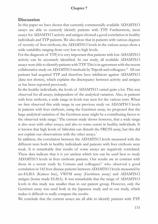
115
Chapter 7
DiscussionIn this paper we have shown that currently commercially available ADAMTS13 assays are able to correctly identify patients with TTP. Furthermore, most assays for ADAMTS13 activity and antigen showed a good correlation in healthy individuals and TTP patients. We also show that in patients with various degrees of severity of liver cirrhosis, the ADAMTS13 levels in the various assays show a wide variability ranging from very low to high levels.For the diagnosis of TTP, it is very important that patients with low ADAMTS13 activity can be accurately identified. In our study, all available ADAMTS13 assays were able to identify patients with TTP. This is in agreement with the recent collaborative study on ADAMTS13 methods by Tripodi and colleagues.10 All our patients had acquired TTP and therefore have inhibitors against ADAMTS13 (data not shown), which explains the discrepancy between activity and antigen as has been reported previously. In the healthy individuals, the levels of ADAMTS13 varied quite a lot. This was observed for all assays, independent of the analytical variation. Also, in patients with liver cirrhosis, a wide range in levels was seen for the various tests. When we first observed this wide range in our previous study on ADAMTS13 levels in patients with liver cirrhosis, using the Gerritsen assay, we proposed that the large analytical variation of the Gerritsen assay might be a contributing factor to the observed wide range.3 The current study shows however, that a wide range is also seen with other assays, and also to some extent in healthy individuals. It is known that high levels of bilirubin can disturb the FRETS assay, but this did not explain our observations with the other assays.7
In addition, the correlation between the ADAMTS13 levels measured with the different tests both in healthy individuals and patients with liver cirrhosis were weak. It is remarkable that results of some assays are negatively correlated. These data indicate that it is yet unclear which test can be used best to study ADAMTS13 levels in liver cirrhosis patients. Our results are in contrast with those in a recent study by Uemura and colleagues11 who observed a good correlation in 142 liver disease patients between ADAMTS13 levels measured by act-ELISA (Kainos Inc), VWFM assay (Gerritsen assay) and ADAMTS13 antigen (home made ELISA). It was remarkable that the range of ADAMTS13 levels in this study was smaller than in our patient group. However, only the Gerritsen assay was used both in the Japanese study and in our study, which makes it difficult to really compare the results.We conclude that the current assays are all able to identify patients with TTP.

116
However, in other study populations than TTP, such as patients with liver disease, the measurement of ADAMTS13 is probably depending on other factors and the optimal test to measure ADAMTS13 in population has to be determined. Also for other disease state associated with alterations in ADAMTS13 levels studies using different ADAMTS13 assays should be performed.
References
1. Moake JL. Thrombotic microangiopathies. The New England journal of medicine 2002;347;8:589-
600.
2. Uemura M, Tatsumi K, Matsumoto M, et al. Localization of ADAMTS13 to the stellate cells of
human liver. Blood 2005;106;3:922-4.
3. Lisman T, Bongers TN, Adelmeijer J, et al. Elevated levels of von Willebrand Factor in cirrho-
sis support platelet adhesion despite reduced functional capacity. Hepatology (Baltimore, Md
2006;44;1:53-61.
4. Gerritsen HE, Turecek PL, Schwarz HP, et al. Assay of von Willebrand factor (vWF)-cleaving
protease based on decreased collagen binding affinity of degraded vWF: a tool for the diagnosis of
thrombotic thrombocytopenic purpura (TTP). Thromb Haemost 1999;82;5:1386-9.
5. Kokame K, Nobe Y, Kokubo Y, et al. FRETS-VWF73, a first fluorogenic substrate for AD-
AMTS13 assay. Br J Haematol 2005;129;1:93-100.
6. Crist RA RG. A comparison of two commercial ADAMTS13 activity assays with a reference labo-
ratory method. Labmedicine 2009;40;4:232-5.
7. Meyer SC, Sulzer I, Lammle B, et al. Hyperbilirubinemia interferes with ADAMTS-13 activ-
ity measurement by FRETS-VWF73 assay: diagnostic relevance in patients suffering from acute
thrombotic microangiopathies. J Thromb Haemost 2007;5;4:866-7.
8. Pugh RN, Murray-Lyon IM, Dawson JL, et al. Transection of the oesophagus for bleeding
oesophageal varices. The British journal of surgery 1973;60;8:646-9.
9. Loof AH, van Vliet HH, Kappers-Klunne MC. Low activity of von Willebrand factor-cleaving
protease is not restricted to patients suffering from thrombotic thrombocytopenic purpura. Br J
Haematol 2001;112;4:1087-8.
10. Tripodi A, Peyvandi F, Chantarangkul V, et al. Second international collaborative study evaluating
performance characteristics of methods measuring the von Willebrand factor cleaving protease
(ADAMTS-13). J Thromb Haemost 2008;6;9:1534-41.
11. Uemura M, Fujimura Y, Matsumoto M, et al. Comprehensive analysis of ADAMTS13 in patients
with liver cirrhosis. Thrombosis and haemostasis 2008;99;6:1019-29.


General discussion
120
The aim of this thesis was to determine the role of ADAMTS13 in the pathogenesis of cardiovascular disease and to study its potential role in 2 other pathological conditions predisposing to thrombosis. We hypothesized that reduced levels of ADAMTS13 will result in higher levels of VWF and more high molecular weight VWF multimers. These high levels of VWF might contribute to a higher risk of arterial thrombosis. To test this hypothesis we measured ADAMTS13 levels in several patient cohorts with cardiovascular disease in chapter 2 and 3. We studied the relationship between ADAMTS13 levels and variations in both VWF and ADAMTS13 gene, in chapter 4a and 4b. Furthermore ADAMTS13 was studied in other pathological conditions, associated with coagulation disturbances and a prothrombotic state, including liver cirrhosis and meningococcal sepsis, in chapter 5 and 6.
ADAMTS13 levels in cardiovascular disease VWF is released into the circulation constitutively and after damage of the endothelium. VWF is released as ultralarge VWF multimers, which form high-strength bonds with the platelet glycoprotein Ib-IX-V complex, resulting in platelet aggregation and thrombus formation. The process of endothelial damage and thereby the release of VWF into the circulation may be a pathoge-netic mechanism in patients with ischemic stroke or myocardial infarction. In two case-control studies we observed that levels of VWF were strongly elevated in patients with arterial thrombosis compared to healthy controls. Individuals with VWF levels in the highest quartile had a 2-fold increased risk of arterial thrombosis.1, 2 This might indicate that increased VWF may result in an increased risk of cardiovascular disease. However, since patients were included after the event occurred, these high levels may be a consequence rather than a cause of the ischemic event. Other groups have also studied VWF levels to assess whether VWF is a risk factor for cardiovascular diseases in previously healthy individuals.3 A meta-analysis of five prospective studies indicated that VWF is a weak independent risk factor for future cardiovascular disease OR 1.2 (95% CI 0.8-1.9).4, 5 However, in persons with pre-existing cardiovascular disease this association is even stronger, also after adjustment for cardiovascular risk factors, in particular in myocardial infarction patients. A meta-analysis of eight prospective studies in pre-existing vascular disease revealed a combined odds ratio of 1.6 (95% CI 1.0-2.5).6 ADAMTS13 is a metalloprotease that cleaves VWF into smaller multimers, which leads to less active VWF. The role of ADAMTS13 in cardiovascular

121
General discussion
Tabl
e 1 S
umm
ary
resu
lts o
f st
udie
s on
arte
rial t
hrom
bosis
in th
is th
esis
Stud
yN
=A
geLe
vels
/ a
ctiv
ityA
ssay
AD
AM
TS13
VW
FA
ctiv
ity (%
)A
ntig
en (%
)A
ctiv
ity (I
U/m
l)A
ntig
en (I
U/m
l)
CO
CO
S-C
ases
(I
sche
mic
stro
ke) /
Con
trols
124/
125
52/5
296
±
4.1
103
± 4
.4O
R 1.
7 (0
.7-3
.0)*
*
Not
per
form
ed1.
37 ±
0.7
41.1
3 ±
0.4
7O
R 2.
1 (0
.9-4
.8)*
**
1.47
± 0
.66
1.23
± 0
.50
OR
3.2
(1.7
-7.5
)***
Ger
ritse
n
ATT
AC
- Cas
es(C
VD
)* /
Con
trols
374/
332
42/3
996
.4 (7
0.3-
112.
2)10
9 (9
3-12
3.8)
OR
5.1
(3.1
-8.5
)**
78.6
(56.
0-10
2.3)
97.4
(81.
7-11
1.1)
OR
4.4
(2.5
-7.5
)**
1.20
(0.9
-1.6
)1.
04 (0
.8-1
.4)
OR
2.1
(1.3
-3.3
)***
1.32
(1.0
-1.7
)1.1
6 (0
.9-1
.5)
OR
2.0
(1.3
-3.1
)***
Tech
nozy
m
* C
VD
=m
yoca
rdia
l inf
arct
ion,
isch
emic
stro
ke o
r per
iphe
ral a
rter
ial d
iseas
e **
Fo
r AD
AM
TS13
the
high
est q
uart
ile w
as u
sed
as re
fere
nce
***
For V
WF
the
low
est q
uart
ile w
as u
sed
as re
fere
nce

122
disease is yet unknown. In the studies described in this thesis we found that the levels of ADAMTS13 were significantly lower in patients with arterial thrombosis than in healthy controls.1, 2 These findings were most significant in the study of young individuals with cardiovascular disease under the age of 45 years for men and under 55 for women. The risk of arterial thrombosis was five-fold higher for individuals in the lowest quartile versus the highest quartile of ADAMTS13 levels OR 5.1 (95% CI 3.1-8.5).2 Only a few other studies have investigated ADAMTS13 levels in patients with cardiovascular disease. The results from these studies show conflicting results. Two small studies determined ADAMTS13 levels in acute myocardial infarction patients shortly after the event, directly on admission or 14 days after the event.7, 8 They reported reduced levels of ADAMTS13 and increased levels of VWF in the acute phase. ADAMTS13 and the ratio of VWF/ADAMTS13 were significant predictors for in-hospital cardiovascular events after acute myocardial infarction. However, the SMILE study reported an increased risk of myocardial infarction in individuals with higher levels of ADAMTS13.9 This is a remarkable finding since all other studies so far reported reduced levels of ADAMTS13 in cardiovascular disease. In this study only men were investigated with an age-range from 18-70 years. Also the moment of blood sampling was different from the other studies. Blood sampling occurred at least 6 months after the event, with a median of 2.6 years after the event, while other studies measured in the acute phase or at the latest 6 months after the event.
Our subgroup analysis in the ATTAC population indicates that the associa-tion of ADAMTS13 with the risk of arterial thrombosis was strongest in the coronary heart disease group. Individuals in the lowest tertile of ADAMTS13 had a eight-fold higher risk of AMI versus individuals in the highest tertile OR 8.2 (95% CI 4.5-14.7). The risk of ischemic stroke patients was slightly higher for individuals with lower level of ADAMTS13 but was not statistical significant in both the COCOS study OR 1.7 (95% CI 0.7-3.9) and ATTAC study OR 1.8 (95% CI 0.9-3.1). An explanation for this finding could be a lack of statistical power due to the relative small number of ischemic stroke patients. Another possibility is the complexity of stroke etiology compared to that of myocardial infarction. Only one experimental study reported about the role of ADAMTS13 in stroke. Zhao et al. showed that in mice with ADAMTS13 deficiency in whom an experimental stroke was induced, the infarct size was increased and that mice

123
General discussion
with reduced or absent VWF had smaller infarct sizes. In addition, infusion of ADAMTS13 in ADAMTS13 knock-out mice reduced the infarct volume and functional outcome, without producing cerebral hemorrhages.10
Genetic variation in ADAMTS13 and VWF in cardiovascular diseaseIt has been hypothesized that genetic predisposition to arterial thrombosis may be associated with variations in a single gene. Within a gene, a single nucle-otide polymorphism (SNP) or a combination of SNPs that inherit together (haplotype) can be studied. In our studies we used a haplotype approach to comprehensively cover the total common variation in the ADAMTS13 gene. By using tools from the HAPMAP consortium and SeattleSNPs database we selected four haplotype tagging SNPs. We studied the possible association of the individual SNPs and the haplotypes with the risk of arterial thrombosis. For none of the SNPs or haplotypes an association was found. The SNPs and haplotypes did no show any correlation with the levels of ADAMTS13. This indicates that other factors may be more important in influencing the levels of ADAMTS13 than genetic variation in the ADAMTS13 gene. Recently, two other studies have been published that have investigated genetic variations in ADAMTS13. The first study, the MASS II study, reported that the minor allele of the genetic variant A900V in patients with coronary artery disease was associated with an increased risk of death OR 1.9 (95% CI 1.1-3.2) and death from cardiac cause OR 2.7 (95% CI 1.6-4.5).11 The second study, the SAHLSIS study, investigated 6 SNPs of the ADAMTS13 gene in patients with ischemic stroke.12 Three minor alleles of these SNPs showed an association with ischemic stroke. One minor allele of these SNPs (rs4962153) showed an increase in risk OR 1.25 (95% CI 1.01–1.54), while the other two minor alleles of rs2285489 and rs2301612 had a decreased risk of ischemic stroke; OR 0.82 (95% CI 0.70–0.97) and OR 0.85 (95% CI 0.73–1.00) respectively.12 Hanson et al. studied two of the four SNPs that we assessed in the ATTAC study. The assay for one of these SNPs (rs2073933) was non-functional and could not be replaced. Another SNP (rs 2301612) was associated with a decreased risk of ischemic stroke. The number of patients in the study by Hanson et al was higher than in our ATTAC study (n=600 versus 109 ischemic stroke patients). In our study the power may have not been sufficient to find an association between the ADAMTS13 SNPs and the risk of arterial thrombosis. Another difference between the studies was

124
that our study population was a more heterogeneous group of patients, since we included not only ischemic stroke patients, but also patients with myocardial infarction and peripheral arterial disease. Another difference was the age dis-tribution of the two studies. Whereas we looked at young patients with CVD, Hanson et al. studied patients with an age of 70 or younger. The selection of the SNPs differed in the various studies because less SNPs were known based on the available databases (HAPMAP and Seattle SNP) at the time that our study was performed. Furthermore, we have selected on the bases of allele frequency of the SNP (> 0.05) and have selected SNPs where possible on the functionality of the SNP. In both of our case-control studies mainly Caucasians were included.It is known that in Japanese the minor allele of the ADAMTS13 polymorphism P475S is present in approximately 10% of the population.13 The minor allele of this polymorphism is associated with reduced activity of ADAMTS13. It was hypothesized that this polymorphism may increase the risk of arterial throm-bosis because of the reduced activity of ADAMTS13. We therefore studied this polymorphism in patients with cardiovascular disease to assess the association between this polymorphism and cardiovascular disease. The minor allele of this polymorphism was however absent in both cases and controls of our Cauca-sian study population. We investigated P475S also in a group of Chinese and Afro-Americans. In this study the minor allele was very rare in Chinese or absent in Afro-Americans, respectively. We therefore concluded that the P475S polymorphism is not an important player in the development of arterial thrombosis in Caucasians. The levels of VWF are influenced both by acquired (environmental) and genetic factors. From twin studies it is known that approximately two-third of all variation in levels of plasma VWF is genetically determined, of which 25-30% can be explained by ABO blood group.14 Individuals with blood group O have lower levels of VWF compared to individuals with blood group non-O. We confirmed this in our study. Subgroup analysis in individuals with blood group O compared with individuals with blood group non-O showed that an increased risk was seen for the levels of VWF antigen. One possible explanation could be the difference in clearance of VWF due to the difference in glycosylation. Another possible explanation could be that there is linkage disequilibrium between ABO blood group and ADAMTS13. The location for ABO blood group is located on chr9q34, which also encodes for ADAMTS13. The locus for

125
General discussion
ABO blood group is indeed found to be in linkage disequilibrium with the locus for ADAMTS13. An increased frequency of the minor allele of the Tyr 1584Cys polymorphism in the VWF gene has been reported in patients with Von Willebrand disease.15 A large study showed increased proteolysis of VWF in heterozygotes.16 We hypothesized that this polymorphism could be protective in patients with arterial thrombosis due to increased proteolysis of VWF. We could not estimate the contribution of the polymorphism to the risk of arterial thrombosis because of the low frequency and the limited number of patients. Therefore, our data suggest that the minor allele of the Tyr1584Cys polymorphism does not have a strong protective effect for arterial thrombosis. More and larger studies are required to assess the importance of changes of VWF proteolysis by ADAMTS13 as a potential pathogenic factor in arterial thrombosis.
Methodological issuesTo investigate the role of ADAMTS13 and VWF in cardiovascular diseases we initiated two case-control studies. An advantage of a case-control study is that it provides a rapid inclusion of individuals in the study. In addition we were able to collect precise data on diagnosis, treatment and prognosis. However, this study design has also some disadvantages and methodological considerations that needs to be discussed. In a case-control study the levels of ADAMTS13 and VWF are determined after the occurrence of the event, making a definite causal inference impossible. The impact of an ischemic event might stimulate patients to change their life style and to live a healthier life. For example, patients may change their exercise pattern, smoking behaviour, which in turn may affect the levels of ADAMTS13 and VWF. In addition, patients will be treated with statins and other drugs that may affect VWF levels. Increased levels of VWF are found in hypertensive individuals and therefore the levels could also be influenced by the use of antihypertensives.17 The effect of these drugs on ADAMTS13 has to be established in future studies. A prospective study does not have these above-mentioned limitations. In our studies however, we were able to adjust for the use of statins and antihypertensives. The first study (COCOS) has included 124 patients with ischemic stroke, while the second case-control study (ATTAC) included, besides stroke patients (n=109) also patients who suffered from an acute myocardial infarction (n=218) or peripheral arterial disease (n=47). Although the total number of stroke

126
patients in both studies is similar, the study populations differ in age. The average age in the COCOS study was 56 years. The ATTAC study was initiated to study the role of coagulation factors in arterial thrombotic disease in the young, and the average age was 43 years. It has been reported that age influences the levels of ADAMTS13.18 ADAMTS13 activity and antigen were lower in healthy individuals of 65 years and above. Feys et al. studied ADAMTS13 levels in both physiological conditions and pathological conditions with prothrombotic state. They divided their patients in various age groups (<35 yrs, 36-50, 51-65, >65 yrs). The levels of ADAMTS13 were not significantly different in the first three age groups. Only individuals that were above 65 years had significantly lower levels of ADAMTS13 activity. Since patients included in our study were all younger than 65 years, we expect no influence of age on the levels of ADAMTS13 activity. An effect of sex on the levels of ADAMTS13 has been described.19 Therefore, we adjusted for sex in both studies, which had only a minor effect on the levels and the risk of arterial thrombosis. Another consideration is that we cannot exclude that our population is a selection of the total group stroke or myocardial infarction patients, since we only included patients that survived the event. We therefore cannot exclude that the studied polymorphisms in our study are associated with a fatal thrombotic event. Also this issue should be investigated in a prospective study.
The levels of VWF and ADAMTS13 can be influenced by several methodological factors. The first factor of influence is the time of sampling after the event. In the COCOS study blood was collected at 7-14 days after the event. Within this time frame an effect of the acute phase may still be present, which is known to increase VWF levels.20 In the ATTAC study, the lag time between the event and the recruitment was one to three months, which ensures that transient inflammatory changes due to the event have disappeared. To further exclude the effect of acute phase in the COCOS study, we corrected for CRP in our analysis. This only had a minor effect on the risk estimate of arterial thrombosis in the COCOS study.It has been hypothesized that even though the levels of ADAMTS13 are not severely decreased in plasma, the levels could be different between the systemic circulation and locally for instance at the site of vascular injury. This implies that the site of blood sampling may be of importance. Horii et al investigated in a study of coronary heart disease patients the levels of ADAMTS13 and VWF in samples from the femoral vein, aortic root and coronary sinus.21 They showed

127
General discussion
in the acute phase shortly after the cardiac event that at all sampling sites, VWF was higher than in the controls. ADAMTS13 was similar at all sampling sites. After six months additional samples were taken at three sites and there was no difference anymore in levels of VWF between cases and controls. This study implies that the hypothesis of locally reduced levels of ADAMTS13 is unlikely, and that our study design with peripheral blood sampling is adequate.Some coagulation factors are known to have a circadian effect. Also for ADAMTS13 this has been investigated.21 It appeared to be a minimal effect during the day. We included the majority of our population in the morning and only few of them in the early afternoon. Therefore we think that this is of minor or no influence on the levels of ADAMTS13 in our study population.
ADAMTS13 in other pathological conditionsThe liver plays a pivotal role in hemostasis since it synthesizes clotting factors, coagulation inhibitors and fibrinolytic proteins. Also ADAMTS13 is synthesized in the liver in hepatic stellate cells.22 In a healthy individual there is a balance between prothrombotic and antithrombotic factors. However, in patients with liver cirrhosis many alterations are seen. A bleeding tendency may be the result of reduced synthesis of clotting factors and thrombocytopenia, but on the other hand patients with liver disease also exhibit thrombotic complications.23 Thus it may be the result of a changed balance in both primary and secondary hemostasis. It is well known that ULVWF is released due to endothelial activa-tion. The role of ADAMTS13 levels in the hemostatic balance in liver cirrhosis is not yet completely understood.18, 24, 25 Some individuals with liver cirrhosis have circulatory levels of endotoxins that may alter VWF and ADAMTS13 levels.26 We hypothesized that reduced synthesis of ADAMTS13 results in low ADAMTS13 levels and thereby more active VWF in circulation. We found in some patients very low levels of ADAMTS13 whereas others had high levels of ADAMTS13. This variation was not caused by the severity of the cirrhosis, but was found in Child A, B and C. Our results are in part in accordance with previous studies.23, 27 Most studies have found decreased levels of ADAMTS13 in all patients with liver cirrhosis. The type of assay that was used in the various studies could explain part of the variation. Therefore we used four different assays, including the Gerritsen assay28, an commercially available ELISA (American Diagnostica,Stamford, CT) a commercially available fluoro-genic assay (Technozym, Vienna, Austria) and the FRETS assay19 and compared these findings in chapter 6. High levels of VWF:Ag, that are often found in

128
patients with liver cirrhosis, may influence the ADAMTS13 assay by Gerritsen et al. Hyperbilirubin has been reported to influence the FRETS assay of ADAMTS13 activity.29 However, in our study we did not find an association between bilirubin and the levels of ADAMTS13 in the various group of patients with liver cirrhosis. Another possibility for the differences could be the aetiology of liver cirrhosis of the patients. Our study has included a limited number of patients to perform subgroup analysis. This should therefore be evaluated in larger studies.
Meningococcal sepsis and disseminated intravascular coagulation (DIC) are associated with a prothrombotic and systemic inflammatory state. Cytokines may influence the activity of ADAMTS13. The role of inflammation on the levels of ADAMTS13 is not clear yet, but some studies have reported reduced ADAMTS13 activity in patients with acute inflammation or sepsis.30-32 In sepsis elevated levels of VWF have been reported indicating a possible contribution of VWF to the formation of microthrombi. Therefore we studied the role of VWF and ADAMTS13 in a large group of children with meningococcal sepsis. We showed that the levels of ADAMTS13 were strongly reduced and VWF levels were elevated at admission and that they were associated with the out-come of meningococcal sepsis in children. Martin et al found that ADAMTS13 was negatively associated with the APACHE score.33 Furthermore they showed an association between ADAMTS13 levels and hemodynamic shock, renal failure and survival.33 Other studies reported no association between ADAMTS13 and the severity of disease or outcome.34, 35. A recent study suggested a role of ADAMTS13 deficiency in the development of sepsis-associated thrombocyto-penia and organ failure. This makes that ADAMTS13 can be seen as a possible link between inflammation, endothelial dysfunction and coagulation.36
However, one should be careful to use the levels of ADAMTS13 already as a diagnostic tool, since the levels show a wide range in a normal healthy population. Therefore there may be overlap can be seen between pathological conditions and healthy controls. Furthermore, it is not clear yet, what is threshold level of ADAMTS13 activity for a true functional deficiency. Further research is needed to investigate ADAMTS13 in a model under flow conditions to see whether ADAMTS13 is consumed in conditions of high shear stress with ULVWF and to study whether the activity of ADAMTS13 is insufficient under these conditions.

129
General discussion
Assays To determine the ADAMTS13 antigen levels and activity of ADAMTS13 several commercial assays nowadays are available. These assays have their advantages and disadvantages. All assays were initially designed to diagnose a severe ADAMTS13 deficiency, as seen in TTP patients. However, it is not yet investigated in other patient groups how these assays perform. Therefore we studied the levels of ADAMTS13 activity and antigen in TTP patients, healthy controls and patients with liver cirrhosis using various assays.We showed that all assays were able to correctly diagnose TTP patients, by measuring a very low ADAMTS13 activity. However, for the other group that we measured, including patients with liver cirrhosis, these assays performed differently and the correlation between the various assays was poor. Therefore we conclude that it is not clear yet which assay should be used in patients with liver cirrhosis. First, more studies should be performed using these various assays to study the levels of ADAMTS13, before the best available assay for certain populations can be indicated. Additional studies are necessary to unravel which factors influence the different assays.
Future perspectiveTo further investigate the role of ADAMTS13 in arterial thrombosis, a study design with a prospective inclusion is required. With baseline values and several time-points before and after the event, the role of ADAMTS13 in time should be studied. For the determination of ADAMTS13 the most ‘ideal’ assay should be used.What is this ‘ideal’ assay? First of all, the coefficient of variation should be low. The Gerritsen assay for example still has a large coefficient of variation and is not suited for these prospective studies. An ideal assay should not be time-consuming or require special equipment.
Recently it has been demonstrated that there is a correlation between cholesterol and ADAMTS13.37 The highest association was observed for HDL-cholesterol that correlated negatively with ADAMTS13 (beta-coefficient = -0.68). For total cholesterol a positive correlation was observed (beta-coefficient = 0.205). This is a remarkable finding given the fact that high cholesterol levels are a strong risk factor for arterial thrombosis. Therefore, in future studies the lipid profile should be assessed in addition to ADAMTS13 levels. Elastases and cytokines are reported to inhibit ADAMTS13 activity.31, 38 It would be helpful in future studies to take also these factors into account. This

130
information could contribute to further unravel the relation between inflammation and thrombosis. Finally, it would be helpful to study more comprehensively the function of VWF and ADAMTS13, by determining ULVWF besides VWF antigen and activity levels by multimer analysis. Using this method the precise amount of very active VWF can be determined.
References
1. Bongers TN, de Maat MP, van Goor ML, et al. High von Willebrand factor levels increase the risk
of first ischemic stroke: influence of ADAMTS13, inflammation, and genetic variability. Stroke; a
journal of cerebral circulation 2006;37;11:2672-7.
2. Bongers TN, de Bruijne EL, Dippel DW, et al. Lower levels of ADAMTS13 are associated with
cardiovascular disease in young patients. Atherosclerosis 2009;207;1:250-4.
3. Folsom AR, Rosamond WD, Shahar E, et al. Prospective study of markers of hemostatic function
with risk of ischemic stroke. The Atherosclerosis Risk in Communities (ARIC) Study Investigators.
Circulation 1999;100;7:736-42.
4. Danesh J, Wheeler JG, Hirschfield GM, et al. C-reactive protein and other circulating markers of
inflammation in the prediction of coronary heart disease. The New England journal of medicine
2004;350;14:1387-97.
5. Whincup PH, Danesh J, Walker M, et al. von Willebrand factor and coronary heart disease: pro-
spective study and meta-analysis. European heart journal 2002;23;22:1764-70.
6. Wiman B, Andersson T, Hallqvist J, et al. Plasma levels of tissue plasminogen activator/plasmino-
gen activator inhibitor-1 complex and von Willebrand factor are significant risk markers for re-
current myocardial infarction in the Stockholm Heart Epidemiology Program (SHEEP) study.
Arteriosclerosis, thrombosis, and vascular biology 2000;20;8:2019-23.
7. Kaikita K, Soejima K, Matsukawa M, et al. Reduced von Willebrand factor-cleaving protease (AD-
AMTS13) activity in acute myocardial infarction. J Thromb Haemost 2006;4;11:2490-3.
8. Matsukawa M, Kaikita K, Soejima K, et al. Serial changes in von Willebrand factor-cleaving pro-
tease (ADAMTS13) and prognosis after acute myocardial infarction. The American journal of
cardiology 2007;100;5:758-63.
9. Chion CK, Doggen CJ, Crawley JT, et al. ADAMTS13 and von Willebrand factor and the risk of
myocardial infarction in men. Blood 2007;109;5:1998-2000.
10. Zhao BQ, Chauhan AK, Canault M, et al. Von Willebrand factor-cleaving protease ADAMTS13
reduces ischemic brain injury in experimental stroke. Blood 2009.
11. Schettert IT, Pereira AC, Lopes NH, et al. Association between ADAMTS13 polymorphisms and
risk of cardiovascular events in chronic coronary disease. Thrombosis research 2009.

131
General discussion
12. Hanson E, Jood K, Nilsson S, et al. Association between genetic variation at the ADAMTS13 locus
and ischemic stroke. J Thromb Haemost 2009.
13. Kokame K, Matsumoto M, Soejima K, et al. Mutations and common polymorphisms in AD-
AMTS13 gene responsible for von Willebrand factor-cleaving protease activity. Proceedings of the
National Academy of Sciences of the United States of America 2002;99;18:11902-7.
14. Orstavik KH, Magnus P, Reisner H, et al. Factor VIII and factor IX in a twin population. Evi-
dence for a major effect of ABO locus on factor VIII level. American journal of human genetics
1985;37;1:89-101.
15. Hashemi M P, I, Goodeve A, Rodeghiero F, Castaman G, Federici A, Budde U, Ingerslev J, Vorlova
Z, Habart D, Holmberg L, Lethagen LS, Pasi J, Hill F The incidence and Penetrance of common
VWF Gene Mutations in a Cohort of Type 1 VWD Patients: Results from the EU funded MC-
MDM-1 Project. J Thromb Haemost 2005; 2005;3, P1469.
16. Bowen DJ, Collins PW, Lester W, et al. The prevalence of the cysteine1584 variant of von Wil-
lebrand factor is increased in type 1 von Willebrand disease: co-segregation with increased sus-
ceptibility to ADAMTS13 proteolysis but not clinical phenotype. British journal of haematology
2005;128;6:830-6.
17. Blann AD, Naqvi T, Waite M, et al. von Willebrand factor and endothelial damage in essential hy-
pertension. Journal of human hypertension 1993;7;2:107-11.
18. Feys HB, Canciani MT, Peyvandi F, et al. ADAMTS13 activity to antigen ratio in physiological and
pathological conditions associated with an increased risk of thrombosis. British journal of haema-
tology 2007;138;4:534-40.
19. Kokame K, Nobe Y, Kokubo Y, et al. FRETS-VWF73, a first fluorogenic substrate for AD-
AMTS13 assay. British journal of haematology 2005;129;1:93-100.
20. Pottinger BE, Read RC, Paleolog EM, et al. von Willebrand factor is an acute phase reactant in
man. Thrombosis research 1989;53;4:387-94.
21. Horii M, Uemura S, Uemura M, et al. Acute myocardial infarction as a systemic prothrombotic con-
dition evidenced by increased von Willebrand factor protein over ADAMTS13 activity in coronary
and systemic circulation. Heart and vessels 2008;23;5:301-7.
22. Uemura M, Tatsumi K, Matsumoto M, et al. Localization of ADAMTS13 to the stellate cells of
human liver. Blood 2005;106;3:922-4.
23. Pereboom IT, Adelmeijer J, van Leeuwen Y, et al. Development of a severe von Willebrand factor/
ADAMTS13 dysbalance during orthotopic liver transplantation. Am J Transplant 2009;9;5:1189-
96.
24. Mannucci PM, Canciani MT, Forza I, et al. Changes in health and disease of the metalloprotease
that cleaves von Willebrand factor. Blood 2001;98;9:2730-5.
25. Lisman T, Bongers TN, Adelmeijer J, et al. Elevated levels of von Willebrand Factor in cirrho-
sis support platelet adhesion despite reduced functional capacity. Hepatology (Baltimore, Md

132
2006;44;1:53-61.
26. Ferro D, Quintarelli C, Lattuada A, et al. High plasma levels of von Willebrand factor as a marker
of endothelial perturbation in cirrhosis: relationship to endotoxemia. Hepatology (Baltimore, Md
1996;23;6:1377-83.
27. Uemura M, Fujimura Y, Matsumoto M, et al. Comprehensive analysis of ADAMTS13 in patients
with liver cirrhosis. Thrombosis and haemostasis 2008;99;6:1019-29.
28. Gerritsen HE, Turecek PL, Schwarz HP, et al. Assay of von Willebrand factor (vWF)-cleaving
protease based on decreased collagen binding affinity of degraded vWF: a tool for the diagnosis of
thrombotic thrombocytopenic purpura (TTP). Thrombosis and haemostasis 1999;82;5:1386-9.
29. Meyer SC, Sulzer I, Lammle B, et al. Hyperbilirubinemia interferes with ADAMTS-13 activ-
ity measurement by FRETS-VWF73 assay: diagnostic relevance in patients suffering from acute
thrombotic microangiopathies. J Thromb Haemost 2007;5;4:866-7.
30. Ono T, Mimuro J, Madoiwa S, et al. Severe secondary deficiency of von Willebrand factor-cleaving
protease (ADAMTS13) in patients with sepsis-induced disseminated intravascular coagulation: its
correlation with development of renal failure. Blood 2006;107;2:528-34.
31. Nguyen TC, Liu A, Liu L, et al. Acquired ADAMTS-13 deficiency in pediatric patients with severe
sepsis. Haematologica 2007;92;1:121-4.
32. Reiter RA, Varadi K, Turecek PL, et al. Changes in ADAMTS13 (von-Willebrand-factor-cleaving
protease) activity after induced release of von Willebrand factor during acute systemic inflamma-
tion. Thrombosis and haemostasis 2005;93;3:554-8.
33. Martin K, Borgel D, Lerolle N, et al. Decreased ADAMTS-13 (A disintegrin-like and metallopro-
tease with thrombospondin type 1 repeats) is associated with a poor prognosis in sepsis-induced
organ failure. Critical care medicine 2007;35;10:2375-82.
34. Kremer Hovinga JA, Zeerleder S, Kessler P, et al. ADAMTS-13, von Willebrand factor and related
parameters in severe sepsis and septic shock. J Thromb Haemost 2007;5;11:2284-90.
35. Bockmeyer CL, Claus RA, Budde U, et al. Inflammation-associated ADAMTS13 deficiency pro-
motes formation of ultra-large von Willebrand factor. Haematologica 2008;93;1:137-40.
36. Levi M, van der Poll T. Inflammation and coagulation. Critical care medicine 38;2 Suppl:S26-34.
37. Crawley JT, Lane DA, Woodward M, et al. Evidence that high von Willebrand factor and low
ADAMTS-13 levels independently increase the risk of a non-fatal heart attack. J Thromb Haemost
2008;6;4:583-8.
38. Cao WJ, Niiya M, Zheng XW, et al. Inflammatory cytokines inhibit ADAMTS13 synthesis in he-
patic stellate cells and endothelial cells. J Thromb Haemost 2008;6;7:1233-5.


Summary / Samenvatting

136
SummaryThe main objective of this thesis was to investigate the role of ADAMTS13 in arterial thrombosis. Arterial thrombosis, as seen in myocardial infarction and ischemic stroke, is an important cause of death in the Western world. Many factors are already known to be a risk factor for cardiovascular diseases. However, other factors are still waiting to be discovered. These other factors both comprise environmental factors and genetic factors. Both of these will be studied in this thesis. Chapter 1 is a general introduction of this thesis. Background information about VWF and ADAMTS13, pathogenesis of arterial thrombosis is reviewed to provide an overview of the current knowledge of ADAMTS13 and VWF. In Chapter 2 we determined whether ADAMTS13 also plays a role in arte-rial thrombosis. We studied a population of 124 patients with ischemic stroke or transient ischemic attacks and 125 healthy controls, the COCOS study. We studied the relationship between the levels of ADAMTS13, VWF and ischemic stroke as well as a polymorphism in VWF gene in order to identify important risk factors for ischemic stroke. In the COCOS study the levels of VWF were significantly higher compared with the controls. Elevated levels of VWF antigen and VWF activity were also associated with a higher risk of ischemic stroke OR 3.2 (95% CI 1.4-7.5) and OR 2.1 (95% CI 0.9-4.8), respectively (Q1 vs Q4). Inflammation (C-reactive protein), ADAMTS13 and genetic variation in the VWF gene did not affect the association between VWF and the odds ratios for ischemic stroke. Secondly, a larger case-control study, ATTAC study, Chapter 3, was performed to study more comprehensively the relationship between ADAMTS13 levels, polymorphisms in the ADAMTS13 gene and the risk of arterial thrombosis. This study comprises 374 young patients with a first myocardial infarction, ischemic stroke or peripheral arterial diseases and 332 healthy young controls. The inclusion criteria for age was 45 years or younger for men and 55 years or younger for women. The levels of ADAMTS13 were associated with the risk of arterial thrombosis OR 5.1 (95% CI, 3.1-8.5) (Q4 vs Q1). Also the levels of VWF were associated with the risk of arterial thrombosis OR 2.1 (95% CI, 1.3-3.3) (Q1 vs Q4). However, the four SNPs did not show a clear relation-ship with the levels or the risk, levels of ADAMTS13 appeared to be strongly associated with arterial thrombosis. In chapter 4, we investigated the role of two polymorphisms in the ADAMTS13 and VWF gene. The first polymorphism, Pro475Ser, in the ADAMTS13 gene

137
Summary
causes a substitution of C1423 into T in exon 12. This polymorphism is known to influence the activity of ADAMTS13. We studied P475S polymorphism in the COCOS study. In our Caucasian population the rare allele was absent. We conclude that the P475S polymorphism is not an important contributor to the risk of ischemic stroke in Caucasians. Furthermore we studied this polymorphism in a panel of Chinese and Afro-American subjects. The frequency for the 475S allele was 0.5% in Chinese and 0% in Afro-Americans. Therefore, our data suggest that except for Japanese the rare allele is very rare.
The second polymorphism studied, is the Tyr1584Cys polymorphism, which is located in VWF gene, exon 28. This polymorphism is in literature associated with an increased proteolysis of VWF. Increased proteolysis of VWF causes detoriation of ultralarge VWF multimers and prevents thereby that VWF remain large and can spontaneously form platelet aggregates and a thrombus. We were interested to study this polymorphism in relation to arterial thrombo-sis. This polymorphism was studied in two different case-control studies with well-documented arterial thrombosis (COCOS and ATTAC). The frequency of the rare allele was 0.5-0.8% in the cases and 0.6-3.2% in the controls. Since the frequency of the Tyr1584Cys polymorphism was low we were not able to obtain a precise estimate of the contribution of the polymorphism to the risk on arterial thrombosis. However, we conclude from our data that the Tyr1584Cys polymorphism does not play an important role in the risk of arterial thrombosis.The aim of chapter 5 was to study the role of ADAMTS13 in other pathological condition than cardiovascular diseases. Patients with sepsis often suffer from thrombotic complication. Therefore we studied in chapter 5 the levels of ADAMTS13 in pediatric patients with meningococcal sepsis. During severe meningococcal sepsis both ADAMTS13 antigen and activity are severely reduced compared to normal (23.4% and 33.7% of normal, respec-tively). Also lower levels of ADAMTS13 (rs=-0.38) and elevated levels of VWF (rs=-0.50) were associated with a severe outcome, indicated by the Pediatric Risk of Mortality Score (PRISM). Our study clearly demonstrates that in the acute phase of severe sepsis the levels of ADAMTS13 are reduced and that the levels of VWF are elevated. Furthermore, these data show that these changes are related to the severity of the disease.Chapter 6 concerns the role of ADAMTS13 and VWF in patients with liver cirrhosis. Cirrhosis of the liver is often accompanied by extensive alterations in the haemostatic system. Most changes result in reduced haemostatic

138
capacity, but also compensatory mechanisms have been found, for example abnormal high levels of VWF. The cleavage of VWF mainly occurs by ADAMTS13, which is primarily synthesized in the liver. Therefore, the aim of this study was to study the relationship of VWF and ADAMTS13 in patients with liver cirrhosis. VWF antigen levels were significantly higher in patients with liver cirrhosis compared with controls (Child A 380%, Child B 500% and Child C 760% versus 107% in controls). The antigen levels of VWF were also strongly correlated with the severity of the disease (assessed by the model for end-stage liver disease (MELD)). VWF:RCo activity levels were also elevated in patients with liver cirrhosis, but not in the same extent as compared with the antigen levels. This means that the amount of VWF is substantially elevated, but appears less functional with respect to the glycoprotein Ib binding. Levels of ADAMTS13 activity in all patients showed a high variability as well as the antigen levels of ADAMTS13 (varying from 0.19-3.0 U/ml). The multimeric pattern of VWF in patients with liver cirrhosis was decreased compared to controls. The proportion high-molecular-weight multimers were in Child A 63%, Child B 66% and Child C 58% compared with the controls 73%. The propep-tide levels of VWF were increased in all patients with liver cirrhosis (Child A 488%, Child b 711% Child C 735% compared to controls 89%). We concluded that the highly elevated levels of VWF contribute to the induction of prima-ry hemostasis. Furthermore, we expected to find a decrease in ADAMTS13 in cirrhosis, since ADAMTS13 is primarily synthesized in the liver. However, we find in some patients reduced levels of ADAMTS13, but in others elevated levels of ADAMTS13. Therefore, we concluded that it is not completely clear yet, whether ADAMTS13 is reduced in liver cirrhosis or not. Reduced clearance or release of ADAMTS13 from platelets may lead to elevated levels of ADAMTS13. Further studies are needed to investigate this on a larger scale.In Chapter 7 several assays for measuring ADAMTS13 activity and/ or antigen are described. A large variety has been observed between the several assays. We compared the levels of ADAMTS13 antigen en activity in patients with TTP and liver cirrhosis. We concluded that for the identification of TTP patients all assays are suitable. However, there no clear answer yet which assay should be used in patients with liver cirrhosis or what caused the large variability in liver cirrhosis patients.In Chapter 8, our findings are discussed and reviewed in a broader context. Overall, we can conclude that the levels of ADAMTS13 play an impor-tant role in arterial thrombosis with an increased risk on arterial thrombosis,

139
Summary
especially in patients with coronary heart disease. This was confirmed in several independent case-control studies. Of course, all results were derived from case-control studies and need to be further studied in prospective studies. Furthermore chapter 8 describes more suggestions for further research.
140
SamenvattingDe belangrijkste doelstelling van dit proefschrift was om de rol van ADAMTS13 in arteriële trombose te onderzoeken. Arteriële trombose (zoals een hartinfarct of herseninfarct) is de belangrijkste oorzaak van sterfte in de Westerse wereld. Door onderzoek zijn er al vele risicofactoren geidentificeerd bij hart- en vaatziekten. Echter, nog steeds zijn niet alle risicofactoren bekend en is er noodzaak om onderzoek te verrichten naar nieuwe risicofactoren. Hierbij richt men zich zowel op omgevingsfactoren als op genetische factoren. Beiden worden in dit proefschrift onderzocht. Hoofdstuk 1 is de algemene inleiding van dit proefschrift. Hierin wordt een overzicht gegeven over de huidige kennis van ADAMTS13 en VWF en de pathogenese bij arteriële trombose.In Hoofdstuk 2 onderzochten wij of ADAMTS13 een rol speelt in arteriële trombose. Wij onderzochten in hoofdstuk twee 124 patienten met een hersen-infarct en 125 gezonde controles uit de COCOS studie. Hierbij werd gekeken naar de relatie tussen de plasmaspiegels van ADAMTS13 en VWF, de genetische variatie in het ADAMTS13 gen en het voorkomen van arteriële trombose. Wij vonden dat de plasmaspiegels van VWF significant hoger waren bij patienten ten opzichte van gezonde controles. Ook de hoeveelheden VWF antigeen en VWF activiteit waren geassocieerd met een hoger risico op arteriële trombose. (OR 3.2, 95%CI 1.4-7.5 and OR 2.1, 95%CI 0.9-4.8, respectievelijk) (Q1 vs Q4). De associatie tussen VWF en arteriële trombose werd niet beinvloed door ontsteking (concentratie-reactief protein), ADAMTS13 of door genetische variatie in het ADAMTS13 gen. De resultaten van deze studie zijn terug te lezen in hoofdstuk 2.Een tweede studie, de ATTAC studie (hoofdstuk 3), werd uitgevoerd om nog uitvoeriger de rol van ADAMTS13, genetica en het voorkomen van arteriële trombose te bestuderen. De studie bevat 374 jonge patienten met hart- of herseninfarct of perifeer vaatlijden en 332 gezonde controles. De mannen werden geincludeerd indien zij 45 jaar of jonger waren, de vrouwen in deze studie konden worden geincludeerd, als zij 55 jaar of jonger waren. De plasmaspiegels van ADAMTS13 waren sterk geassocieerd met het risico op arteri ële trombose OR 5.1 (95% CI, 3.1-8.5) (Q4 vs Q1). Ook de plasmaspiegels van VWF waren geassocieerd met het risico op arteriële trombose OR 2.1 (95%CI 1.3-3.3) (Q1 vs Q4). Daarentegen, lieten de vier bestudeerde SNPs in het ADAMTS13 gen niet een duidelijke relatie met de plasmaspiegels van ADAMTS13 zien.In hoofdstuk 4 onderzochten wij de rol van twee polymorfismen in het

141
Samenvatting
ADAMTS13 en in het VWF gen. Het eerste polymorfisme, P475S, in het ADAMTS13 gen, veroorzaakt een substitutie van C1423 naar een T in exon 12. Van dit polymorfisme is bekend dat het de activiteit van ADAMTS13 kan beinvloeden. Wij onderzochten het P475S polymorfisme in de COCOS studie. Het minor allel van dit polymorfisme bleek niet voor te komen bij Caucasiërs. Wij concludeerden dat het P475S polymorfisme geen belangrijke bijdrage levert aan het risico op een herseninfarct in Caucasiërs. Tevens onderzochten wij dit polymorfisme in een groep van Chinezen en Afro-americanen. Het minor allel van dit polymorfisme kwam in 0.5% bij de Chinezen en in 0% van Afro- americanen voor. Onze data suggereert dat dit polymorfisme waarschijnlijk alleen een rol speelt in Japanners, gezien het feit dat in onze populaties de frequentie van het minor allel zeer laag is.Het tweede polymorfisme dat is bestudeerd, Tyr1584Cys, bevindt zich in het VWF gen in exon 28. Dit polymorfisme is in de literatuur geassocieerd met een verhoogde proteolyse van VWF. Een verhoogde proteolyse van VWF zorgt voor de afbraak van ULVWF multimeren en voorkomt zo dat VWF groot blijft en zo spontaan bloedplaatjes kan aggregeren en daarmee een trombus vormt. Wij onderzochten dit polymorfisme in relatie tot arteriële trombose in twee studies (COCOS en ATTAC) naar arteriële trombose. De frequentie van het minor allel was 0.5-0.8% in de patienten en 0.6-3.2% in de gezonde controles. Gezien deze lage frequentie was het niet mogelijk een schatting te maken van de bijdrage van dit polymorfisme aan het risico op arteriële trombose. Wij concludeerden wel uit onze data dat het Tyr1584Cys polymorfisme geen belangrijke bijdrage levert aan het risico op arteriële trombose.Het doel van hoofstuk 5 was om de rol van ADAMTS13 te bestuderen in pathologische condities anders dan cardiovasculaire ziekten. In patienten met sepsis worden vaak trombotische complicaties gezien. Vandaar dat wij de plasmaspiegels van ADAMTS13 in kinderen met meningococcen sepsis bestudeerden (hoofdstuk 5) . De plasmaspiegels van ADAMTS13 activiteit en antigeen bleken gedurende een periode van meningococcen sepsis sterk gereduceerd te zijn (23.4% en 33.7% t.o.v. normaal, respectievelijk). Verlaagde plasmaspiegels van ADAMTS13 (rs = -0.38) en verhoogde plasmaspiegels van VWF (rs=-.050) waren geassocieerd met een slechtere uitkomstmaat, uitgedrukt in de PRISM score (pediatric risk of mortality). Onze studie laat duidelijk zien dat in de acute fase van ernstige sepsis de plasmaspiegels van ADAMTS13 fors verlaagd zijn. In hoofdstuk 6 onderzochten we de rol van ADAMTS13 in patienten met

142
levercirrose. Levercirrose gaat gepaard met allerlei veranderingen in het hemostatische systeem. Vele verandering leiden tot een verminderde hemostatische capaciteit, maar er worden ook compensatoire mechanismen gevonden, zoals verhoogde VWF plasmaspiegels. ADAMTS13 wordt in de lever gemaakt en is het belangrijkste enzym dat VWF afbreekt. Daarom onder-zochten wij de relatie tussen VWF en ADAMTS13 in lever cirrose patienten. De plasmaspiegels van VWF waren significant hoger in patienten met lever cirrose vergeleken met gezonde controles (Child A 380%, Child B 500%, en Child C 760% versus 107% in controles). VWF antigeen plasmaspiegels waren sterk gecorreleerd met de ernst van de cirrose, bepaald met de MELD score. De MELD score is een score voor de mate van eind-stadium lever cirrose. Ook VWF ristocetine activiteit was verhoogd bij patienten met cirrose, maar niet in dezelfde mate als de VWF antigeen plasmaspiegels. Dit houdt in dat de hoeveelheid VWF substantieel is verhoogd, maar dat het minder functioneel lijkt, wat betreft de binding van glycoproteine-Ib. De plasmaspiegels van ADAMTS13 in alle patienten lieten een grote variabiliteit zien, varierend van 0.19 tot 3 U/ml. Het multimeer patroon van VWF was verlaagd ten opzichte van gezonde controles. De proportie hoog-moleculair-gewichts-multimeren was in Child A 63%, Child B 66%, en Child C 58% vergeleken met controles 73%. De propeptide plasmaspiegels van VWF waren verhoogd in alle patienten met lever cirrose (Child A 488%, Child B 711%, Child C 735% vergeleken met controles 89%).In hoofdstuk 7 worden de verschillende assays voor het meten van ADAMTS13 activiteit en antigeen beschreven. Een grote variatie werd gezien tussen de verschillende assays. We vergeleken de plasmaspiegels van ADAMTS13 in controles, TTP patienten en patienten met lever cirrose. We concludeerden dat voor het identificeren van TTP patienten elke assay geschikt is, maar dat patienten met levercirrose meer onderzoek zal moeten worden gedaan om te bepalen wat de meest geschikt assay is.In hoofdstuk 8 worden tenslotte onze bevindingen bediscussieerd en beschouwd in een groter geheel. Uit de resultaten van de verschillende onderzoeken, concluderen wij dat ADAMTS13 een belangrijke rol speelt bij arteriële trombose en een verhoogd risico geeft op arteriële trombose. Dit is met name gezien in patienten met coronair hart ziekten. Dit is bevestigd in meerdere onafhankelijke case-control studies. Echter, al deze studies waren case-control studies en de resultaten moeten verder onderzocht worden in prospectieve studies. Tevens bevat dit hoofdstuk aanbevelingen voor toekomstig onderzoek.


144

145
DankwoordNa de vele proefschriften die voorbij kwamen, is het dan nu eindelijk ook zover: ik mag eindelijk het dankwoord gaan schrijven. Terugkijkend op de afgelopen jaren, besef ik mij opeens dat dit promotieonderzoek een groot avontuur is geweest. Een reis door de jaren waarbij ik vele mensen heb leren kennen, uitdagingen heb getrotseerd en vriendschappen heb gesloten. Dit onder-zoek was natuurlijk niet mogelijk geweest zonder de steun van patienten, familieleden, vrienden en collega’s. Zonder hen was het niet mogelijk geweest dit promotieonderzoek tot een goed einde te brengen. Mijn dank hiervoor is groot. Enkele mensen wie ik graag persoonlijk noemen, waarbij ik me realiseer nooit volledig te kunnen zijn. Ik hoop dan ook dat de mensen die ik hier oversla, weten dat ik ze minstens zoveel dank verschuldigd ben.
Deelnemende patienten en controlepersonenAls allereerste wil ik alle patienten en gezonde vrijwilligers bedanken voor hun deelname aan het onderzoek. Zonder jullie bijdrage aan het onderzoek zou dit promotieonderzoek niet mogelijk zijn geweest.
Mijn promotor prof. dr. F.W.G. Leebeek, beste Frank. Ik leerde je kennen via het studententeam bij de Hematologie waar ik werkzaam was. Na een onder-zoekje aan de Christopher studie was je degene die kwam met een leuk voorstel voor een nogal ambitieus keuzeonderzoek. Dit bleek later de basis te zijn voor dit proefschrift. Bedankt voor het gestelde vertrouwen en je geduld. Eindelijk nu een volmondig JA, het is af! Het is bewonderenswaardig hoe jij wetenschappelijk onderzoek en kliniek kunt combineren. Tevens bedankt voor alle keren dat je uit het blote hoofd de artikelen met journal en jaartal kon opnoemen welke ik niet meer terug kon vinden! Ik wens je al het beste toe.
Dr M.P.M. de Maat, beste Moniek. Jij was mijn directe aanspreekpunt op de afdeling Hematologie en wat wist ik je te vinden. Je stond altijd voor me klaar met tips, adviezen en met je correcties op artikelen, abstract en presentaties. Zelf in Sydney, toen ik zonder stem aankwam, heb je ervoor gezorgd dat ik op tijd weer “fit” was. Tevens bewonder ik je enorme kennis van programma’s, met in het bijzonder SPSS. Bedankt dat je altijd bereikbaar was, telefonisch of in de weekenden om zo ook de laatste fase van mijn promotieonderzoek af te ronden. Dit was met name erg prettig gedurende mijn “vrije”momenten in mijn coschap-pen en mijn arts-assistentschap. Veel geluk!

146
De overige leden van mijn promotiecommissie prof. dr. H. Boersma, prof. dr. M.M.B. Breteler, dr. J.W. Deckers, prof. dr. H.L.A. Janssen, prof. dr. D.W.J. Dippel, dr. J.A. Lisman, prof. dr. B. Löwenberg, wil ik bedanken voor hun bereidwilligheid om mijn proefschrift te beoordelen.
Mijn paranimf dr. G. Rudež, beste Goran. Jij was mijn partner in crime bin-nen de ATTAC studie. Het was erg prettig om met je samen te werken en je als sparring partner te hebben. Je immer kritisch blik kwam vaak van pas! Bedankt voor alle keren dat ik weer eens de behoefte had om mijn frustaties te ventileren en bedankt als luisterend oor. Je ging samen met Ana en mij het Australische avontuur aan (Victoria Building, kangaroo/cranberry, turbulentie). En dat was slechts een van de vele congresreizen die we samen beleefden. Bedankt dat je samen met mij in Duitsland uit de dip wilde komen. Wat hebben we daar hard gewerkt, maar wat was het leuk! Ik ben erg blij dat je mijn paranimf wilde zijn en hoop dat we nog lang vrienden zullen blijven. Ik wens jou, je ouders en Annelies het allerbeste!
Mijn andere paranimf, dr. A.H. Canas Guimarães, beste Ana. Ook jou ben ik veel dank schuldig. Jij maakte, dat ik me als een vis in het water voelde, bij jullie op de kamer. Wat hebben we veel gelachen. Over radio cotonete, de nederlandse spreekwoorden, onze “ vrije” vertalingen en het speed-shoppen in Edinburgh voor de gala banquette. Met jou vind je altijd wel wat leuks in de stad. Ik bewonder je kookkunsten. Wat kan jij heerlijk koken! Ik hoop dat er nog vele lekkere diners zullen volgen. Tevens wil ik je bedanken voor alle keren dat ik na een drukke week even zin had in een drankje. Jij was praktisch altijd in the mood voor een hapje en een drankje. Bedankt dat je mijn paranimf wilde zijn en tot snel weer bij een borrel of een lekker etentje. Ik wens jou, Winfried en Ella alle goeds. Tot snel!
Verder wil ik natuurlijk de rest van de stollingsgroep bedanken voor alle hulp en gezelligheid. Beste Elisabeth, jij leerde mij een pipet te hanteren! Al gauw had ik alle technieken onder de knie en kon ik beginnen met het echte werk. Beste Emile, zonder jou was mijn onderzoek niet mogelijk geweest. Wat heb jij een werk verricht om de ATTAC studie op te zetten. Hiervoor al mijn dank. Ik vind het bewonderenswaardig hoe je je opleiding, promotieonderzoek en het vervolg in Beverwijk hebt aangepakt. Jij bent ook bijna zover! Veel succes nog met de laatste loodjes! Vinod, met jou ging ik het avontuur aan. Wat hebben

147
we gelachen. Ik hoop dat je ook gevonden hebt wat je wilde. Joyce, bedankt voor al je labondersteuning en je gezellige babbels. Elim, you also good luck with finishing the last phase of your PhD. Eva, bedankt voor alle gezellige en ondersteunende babbels/koffie/diners. Jij hebt mijn interesse voor de tango verder aangewakkerd. Dank hiervoor, tevens voor de tangoschoenen die je dankzij je congres in Buenos Aires voor me kon meenemen! Ik wens jou, Cor, Fien en Wietse alle goeds toe en wie weet weer tot in Cuartito! Marianne, je bent al jaren een goede vriendin van me. Ik vond het fijn om de ATTAC studie in jouw handen achter te laten, wetende dat het vertrouwd was. Ik wens jou ook nog heel veel succes bij het afschrijven van je boekje! Tot snel bij de meiden! Ik wens jou en Jeroen het alle goeds en hopelijk snel het huisje waar jullie van dromen. Simone bedankt voor je gezelligheid en succes met alle proeven en Janine, inmiddels de 4e generatie ATTAC, ook jij bedankt voor je hulp en je succes bij de cardiologie. Steffen en Lamberto, alle twee inmiddels al even weg, maar we hadden veel lol op het lab en bij het salsadansen. Dank hiervoor. Jullie alle twee veel succes met de kleintjes! Jasper, ook jij bedankt voor je gezelligheid tijdens diners en borrels en je mening bij werkdiscussies. Ik wens je ook veel succes toe bij jouw promotietraject! Edith, je was niet direct bij het ATTAC onderzoek betrokken, maar zorgde wel voor goede afleiding op sommige momenten. Je hebt me aan het spinnen gekregen! Daarvoor ben ik je nog altijd dankbaar. Ik wens je nog veel succes met je opleiding. Ard-Jan, ook jou ben ik veel dank verschuldig. Het was prettig samenwerken! Jij hebt je geheel gestort in de taqman analyses in het ADAMTS13 gen. Deze resultaten vormden uiteindelijk de basis voor een mooie paper. Ik wens je heel veel succes bij de kindergeneeskunde. Dick, laatste in de rij van de stollingsmensen, maar zeker niet de minste. Jij was altijd geinteresseerd in wat ik deed en hoe ver ik was. Je was veel op de congressen bij de AIO’s te vinden, iets dat wij altijd erg gezellig vonden. Ik wens je nog veel mooie publicaties met je groep toe.
Alle andere collega’s van de 13e voor de leuke tijd en de nodige afleidende koffies (o.a Annemarie, Karishma, Bas, JO, Onno, Eric, Merel en alle anderen die meededen aan studie-ontwijkend gedrag) en het secretariaat voor alle ondersteuning.
Alle mensen van het hemostase- en transfusielab van de L-vleugel. Dank voor al jullie hulp rondom de ATTAC studie ( het prikken, afdraaien en stickeren). Tevens dank voor het wegwijs maken op het lab tijdens het keuzeonderzoek

148
en het latere promotietraject. Speciale vermelding hierbij voor Sjef, Riekje en Ivanka! Tevens dank aan Huub van Vliet voor zijn altijd enerverende inbreng in de werkdiscussies.
Ook al mijn coauteurs (Marieke Emonts, Jan Hazelzet, Ronald de Groot, Don Poldersmans, Encarna Gómez García, Jelle Adelmeijer, Flip de Groot, Diederik Dippel) wil ik bedanken voor hun bijdragen aan de artikelen die uiteindelijk hebben bijgedragen aan het voltooien van mijn proefschrift.
Natuurlijk wil ik ook mijn cogroepje bedanken voor al hun steun, gezelligheid en begrip. Toen we met zijn allen begonnen beloofde ik een mooi feestje. Jullie heb-ben er even op moeten wachten, maar hoop jullie in grote getalen te zullen zien. Ik denk met recht dat wij de leukste cogroep ever hadden. Erg ondernemend en gezellig was het weekendje weg. De mannen (Jasper en Arjan) bedankt voor jullie luisterend oor en voor het allereerste après-kerstdiner. Dat was een groot succes. Ik wens jullie alle goeds en succes met de kids! De meiden (Anne-Eva, Christien, Emma, Eva, Mirjam, Yin, Yolanda), ik wens jullie alle goeds voor alle carrières. AE, E & E, gezellig dat jullie met me mee wilde schilderen. Een aangename afleiding naast het werk! Yolanda, veel succes in Adelaide, nu ook eindelijk aan de bak!
Al mijn collega’s van de neurologie wil ik bedanken voor de gezellige tijd en de gezellige borrels. Maaike S en Wardell, wanneer gaan we weer eens een drankje doen? Ladbon, bedankt voor alle lekkere hapjes en je luisterend oor ;) Gaan we snel weer eten en dansen? Maaike D, bedankt voor je gezelligheid en al je adviezen. Stefan, ik wens jou alvast een hele goede bruiloft toe! Juscelina en Carina, bedankt voor al jullie ondersteuning.
Al mijn vrienden wil ik bedanken voor hun begrip dat ze tijdens mijn promo-tieonderzoek hebben opgebracht. Bedankt voor alle ontspanning die jullie me brachten tussen het onderzoeken en het schrijven door. Jackelien, jij was en bent nog steeds mijn stok achter de deur om te sporten. Ook koffies in de stad, het samen studeren/ schrijven en de dinertjes samen heb ik enorm gewaardeerd. Marianne, Femke, Mischa en Cynthia, bedankt voor jullie onvoorwaardelijke steun. Ik hoop dat we snel weer eens een weekendje weer in kunnen plannen. Tjeu, bedankt voor alle afleiding tussendoor! Wanneer gaan we weer naar Den Haag? ;) Lien, ook jou zag ik mede door de afstand en het werk veel minder,

149
maar je blijft m’n beste vriendinnetje van thuis! Linda, bedankt dat je zo nu en dan even mijn klaagmuur wilde zijn en voor de nodige ontspanning. Milaan was super! Moeten we snel weer eens doen. Ester, dank voor je steun en alle gezel-lige tijd die we tot nu toe hebben doorgebracht in Nederland en Rome. Wanneer zullen we weer?
Verder wil ik iedereen bedanken die mij onderdak verleende om aan mijn proef-schrift te schrijven. Schrijven in het buitenland is namelijk altijd het leukst en je wordt er niet afgeleid door telefoon. Bjorn, je was amper een maand geëmi-greerd naar Zwitserland, maar nodigde me meteen uit om te komen schrijven. Sterker nog ik vond het een eer om samen met jou mijn verjaardag in Geneve te kunnen vieren. Laten we binnenkort als je weer eens in den lande bent wat afspreken.Inoltre vorrei ringraziare tante persone dall’ Italia/ Roma. La Famiglia Bacci, grazie dell’ospitalità. Siete sempre gentile con me e era sempre divertente di es-sere con voi! Spero di vedere voi anche in Olanda. Mimmo, grazie per tutto tempo che abbiamo passato! Spero che il tuo sogno possa realizzarsi presto! Tutti I dottori che ho conosciuto (Dr Bruno, Onelia, Emanuele, Maria, Alessandra), grazie per accompagnarme in ospedale, per i caffe ;) e a presto! In generale, grazie a tutti per il mio italiano. Ho imparato ancora tanto da voi e sto imparando ancora! A presto!
Henri en Esther, wat had ik zonder jullie gemoeten? Echt super dat jullie me wilde helpen met mijn proefschrift! Dank voor al jullie steun en de nodige aflei-ding gedurende al deze jaren. Wie had gedacht dat ik nog eens zou zweefvliegen? Hopelijk komen er nog vele gezellige avonden. Grazie!
Ten slotte wil ik mijn ouders en zusje bedanken voor hun onvoorwaardelijke steun en liefde. Jullie hebben altijd in mijn geloofd en mij gesteund daar waar nodig en nog steeds. Bedankt voor jullie begrip als ik weer eens moest werken of schrijven. De drukste tijden liggen achter ons, nu weer lekker genieten met zijn vijven! Liefs Tamara

150

151
Curriculum vitae
De auteur van dit proefschrift werd geboren op 16 oktober 1980 te Zevenaar. Na het behalen van het gymnasium diploma aan het Liemers College ging zij vervolgens van het Gelderse Zevenaar naar Utrecht om daar farmacie te studeren na uitgeloot te zijn voor geneeskunde. In 2000 begon zij met de studie geneeskunde aan de Erasmus Universiteit te Rotterdam. In 2004 werd het doctoraal examen afgelegd. Gedurende haar keuze-onderzoek kreeg zij de mogelijkheid om door middel van een AIO constructie klinisch wetenschappelijk onderzoek te verrichten. Dit onderzoek op de afdeling Hematologie van het Erasmus MC was de basis voor dit proef-schrift. Zij behaalde haar artsexamen met lof in 2009. Daarna heeft zij gewerkt op de afdeling Neurologie in het Erasmus MC als arts-assistent. Vanaf december 2010 zal zij gaan werken op de afdeling Geriatrie van het Amphia ziekenhuis te Breda.Buiten werktijd mag zij graag tango dansen, sporten, schilderen en reizen naar haar favoriete Italie.

152

153
List of publications
Reduced ADAMTS13 in children with severe meningococcal sepsis is associated with severity and outcome. Bongers T.N., Emonts M., de Maat M.P., de Groot R., Lisman T., Hazelzet J.A., Leebeek F.W., Thromb Haemost. 2010 Jun;103(6):1181-7.
Lower levels of ADAMTS13 are associated with cardiovascular disease in young patients.Bongers T.N., de Bruijne E.L., Dippel D.W., de Jong A.J., Deckers J.W., Pol-dermans D., de Maat M.P., Leebeek F.W., Atherosclerosis. 2009 Nov;207(1):250-4.
Frequency of the von Willebrand factor Tyr1584Cys polymorphism in arterial thrombosis.Bongers T.N., de Maat M.P., Deckers J.W., Dippel D.W., Leebeek F.W., Br J Haematol. 2008 Mar;140(5):578-9.
High von Willebrand factor levels increase the risk of first ischemic stroke: influ-ence of ADAMTS13, inflammation, and genetic variability.Bongers T.N., de Maat M.P., van Goor M.L., Bhagwanbali V., van Vliet H.H., Gómez García E.B., Dippel D.W., Leebeek F.W., Stroke. 2006 Nov;37(11):2672-7.
Elevated levels of von Willebrand Factor in cirrhosis support platelet adhesion despite reduced functional capacity.Bongers T.N., Lisman T., Adelmeijer J., Janssen H.L., de Maat M.P., de Groot P.G., Leebeek F.W., Hepatology. 2006 Jul;44(1):53-61.
Absence of Pro475Ser polymorphism in ADAMTS-13 in Caucasians.Bongers T.N., De Maat M.P., Dippel D.W., Uitterlinden A.G., Leebeek F.W., J Thromb Haemost. 2005 Apr;3(4):805.

154

155
Awards
Young investigator award of the International Society of Thrombosis and Haemostasis, Sydney, Australia 2005.
Travel Grant, Dutch Society for Thrombosis and Haemostasis- International Congress for Thrombosis and Haemostasis, 2005.
Travel Grant, Van Walree Fonds, The Netherlands, 2005.

156

157

158

159
Abbreviations
ADAMTS13 A Disintegrin-like And Metalloprotease with Thrombo Spondin motifs 13AMI Acute myocardial infarctionANOVA Analysis of variancesATTAC Arterial Thrombosis TAFI and other Coagulation factorsCHD Coronary heart diseaseCI Confidence intervalCK-MB Creatinine kinase MBCRP C-reactive proteinCT Computerized tomographyCys CysteineDIC Disseminated intravascular coagulation.FVIII:C Factor eight activityHUS Hemolytic uremic syndromeIL-6 Interleukin 6IL-8 Interleukine-8IS Ischemic strokeLD Linkage disequilibriumMELD Model for end-stage liver diseaseMRI Magnetic resonance imagingNPP Normal pooled plasmaPAD Peripheral arterial diseasePAI-1 Plasminogen activator inhibitorPCR Polymerase chain reactionPICU Pediatric Intensive Care UnitPRISM Pediatric Risk of Mortalityr-hu-ADAMTS13 Recombinant human ADAMTS13SNP Single nucleotide polymorphismTAT Thrombin-anti-Thrombin complexTIA Transient ischemic attackTNF-α Tumor necrose factor alphaTTP Thrombotic thrombocytopenic purpuraTyr TyrosineUAP Unstable angina pectoris

160
ULVWF Ultra Large Von Willebrand Factor VWD Von Willebrand DiseaseVWF Von Willebrand FactorVWF:Ag Von Willebrand Factor antigenVWF:CB Von Willebrand Factor collagen binding activityVWF:RCo Von Willebrand Factor Ristocetine Cofactor activityVWF:Rco Von Willebrand Factor ristocetine cofactor activity


![Hematological Malignancies and Arterial Thromboembolism · thrombosis in ET has been predicted by thrombosis history, older age, cardiovascular risk factors and JAK2V617F [37]. Extreme](https://static.fdocuments.in/doc/165x107/5e8e8e6f639a7b2284701bf6/hematological-malignancies-and-arterial-thromboembolism-thrombosis-in-et-has-been.jpg)