05 0789735962 ch02.qxd 2/26/07 4:19 PM Page...
38
2 CHAPTER TWO Care of the Client with Cardiovascular Disorders The cardiovascular system comprises the heart and blood vessels, and is responsible for the transport of oxygen and nutrients to organ systems of the body. The heart is a cone-shaped organ made up of four chambers. The right atrium receives blood from the venous system by way of the superior and inferior vena cavae. Most of the venous blood flows through the tricuspid valve and into the right ventricle during the filling phase of cardiac contraction. The remaining venous blood flows into the right ventricle during the atrial systolic or contraction phase of cardiac contraction. The blood then moves to the lungs where carbon dioxide is released and oxygen is taken on. The left side of the heart then pumps the oxygenated blood to the body. During systole the pressure exerted on the ventricle closes the mitral valve to prevent blood from flowing backward into the left atrium and opens the aortic valve to assist the ventricle to pump adequate oxygenated blood out of the heart into the aorta and to the body. Arteries and veins are types of blood vessels. Arteries transport oxygenated blood and veins trans- port deoxygenated blood. Figure 2.1 provides an illustration of the anatomy of the heart for reference throughout the chapter. Aortic arch Superior vena cava Right pulmonary arteries Fossa ovalis Pectinate muscles Right atrium Cusp of right AV (tricuspid) valve Trabeculae carneae Inferior vena cava Right ventricle Moderator band Ligamentum arteriosum Pulmonary trunk Pulmonary semilunar valve Left pulmonary arteries Left pulmonary veins Interatrial septum Cusp of left AV (bicuspid) valve Chordae tendineae Papillary muscles Left ventricle Interventricular septum Descending (thoracic) aorta Aortic semilunar valve Left atrium FIGURE 2.1 Anatomy of the Human Heart
Transcript of 05 0789735962 ch02.qxd 2/26/07 4:19 PM Page...

2C H A P T E R T W O
Care of the Client withCardiovascular Disorders
The cardiovascular system comprises the heart and blood vessels, and isresponsible for the transport of oxygen and nutrients to organ systems of thebody. The heart is a cone-shaped organ made up of four chambers. Theright atrium receives blood from the venous system by way of the superiorand inferior vena cavae. Most of the venous blood flows through thetricuspid valve and into the right ventricle during the filling phase of cardiaccontraction. The remaining venous blood flows into the right ventricleduring the atrial systolic or contraction phase of cardiac contraction. Theblood then moves to the lungs where carbon dioxide is released and oxygenis taken on. The left side of the heart then pumps the oxygenated blood tothe body. During systole the pressure exerted on the ventricle closes themitral valve to prevent blood from flowing backward into the left atrium andopens the aortic valve to assist the ventricle to pump adequate oxygenatedblood out of the heart into the aorta and to the body. Arteries and veins aretypes of blood vessels. Arteries transport oxygenated blood and veins trans-port deoxygenated blood. Figure 2.1 provides an illustration of the anatomyof the heart for reference throughout the chapter.
Aortic arch
Superiorvena cava
Rightpulmonary
arteries
Fossaovalis
Pectinatemuscles
Rightatrium
Cusp ofright AV
(tricuspid)valve
Trabeculaecarneae
Inferiorvena cava
Rightventricle
Moderator band
Ligamentum arteriosum
Pulmonary trunk
Pulmonary semilunar valve
Left pulmonaryarteries
Left pulmonaryveins
Interatrialseptum
Cusp of left AV(bicuspid) valve
Chordaetendineae
Papillarymuscles
Leftventricle
Interventricularseptum
Descending (thoracic)aorta
Aortic semilunarvalve
Leftatrium
FIGURE 2.1Anatomy ofthe HumanHeart
05_0789735962_ch02.qxd 2/26/07 4:19 PM Page 45

46
In this chapter, you will discover diseases that affect the cardiovascular system, treatmentof these diseases, and their effects on the client’s general health status.
HypertensionBlood pressure is the force of blood exerted on the vessel walls. Systolic pressure is thepressure during the contraction phase of the heart and is the top number of a bloodpressure reading. Diastolic pressure is the pressure during the relaxation phase or fillingphase of the heart and is the bottom number of a blood pressure reading. Factors thatalter peripheral resistance, heart rate, and stroke volume affect the blood pressure.Hypertension is defined as a systolic blood pressure greater than or equal to 140 over 90mm Hg. If the client has diabetes or kidney disease, a systolic blood pressure greaterthan 130 mm Hg systolic and a diastolic blood pressure of 80 mm Hg or higher isconsidered hypertension and should be treated. The autonomic nervous system andcirculating blood volume control blood pressure. Blood pressure also directly relates tocirculating hormones such as antidiuretic hormones.
Hypertension is classified as either primary or secondary. Primary or essential hyperten-sion develops without apparent cause; secondary hypertension develops as the result ofanother illness or condition. Some examples of diseases that result in secondary hyper-tension are diabetes, peripheral vascular disease, renal disease, preeclampsia, coarctationof the aorta, adrenal tumors such as pheochromocytomas, brain tumors, encephalitis,and primary aldosteronism. This and other chapters of the book will discuss thesediseases. Obesity and smoking also affect blood pressure. Appropriate treatment of thecontributing illness improves the symptoms associated with secondary hypertension.
Malignant hypertension is an extremely elevated blood pressure that often results in acerebral vascular accident or a myocardial infarction. Secondary hypertension occurswhen another disease process causes the blood pressure to elevate above normal limits.Some examples of causes for secondary hypertension are kidney disease, diabetes,preeclampsia, and pheochromocytoma. Many medications can lead to secondary hyper-tension. Some examples of medications that can lead to hypertension are NSAIDS(nonstreroidal anti-inflammatory drugs), cocaine, amphetamines, bronchodilators andestrogen preparations. The client might complain of a headache, blurred vision, anddyspnea. If renal function is impaired, the client will exhibit signs of uremia. A systolicblood pressure greater than 200 mm Hg and a diastolic blood pressure greater than 150mm Hg is life-threatening. To prevent further deterioration of the client’s condition,medical personnel must implement prompt intervention.
Diagnosing the Client With HypertensionThe accuracy of a BP reading depends on the correct selection of cuff size. The bladderof the blood pressure cuff size should be sufficient to encircle the arm or thigh.
Chapter 2: Care of the Client with Cardiovascular Disorders
05_0789735962_ch02.qxd 2/26/07 4:19 PM Page 46

Hypertension47
According to the American Heart Association, the bladder width should be approxi-mately 40% of the circumference or 20% wider than the diameter of the midpoint ofthe extremity. A too-small blood pressure cuff yields a false high reading, whereas a too-large blood pressure cuff yields a false low reading. For accuracy, the arm being used tocheck the blood pressure should be held at the level of the heart. The blood pressureshould be taken on at least two occasions sitting, standing and in a supine position.Diagnosis of hypertension involves conducting a comprehensive history of illness andstressors in the client’s life and medications taken by the client . Laboratory studies mustbe completed to determine any underlying illness that might be present. Some labora-tory studies indicate the presence of protein in the urine. Others studies measure serumcreatinine levels, blood urea nitrogen, serum corticoids, and 17–ketosteroids in theurine. The presence of serum corticoids and 17-ketosteroids in the urine is diagnostic ofCushing’s disease or increased function of the adrenal glands. A radiography study, suchas an intravenous pyelography (IVP), can confirm renal disease. X-rays to determine thepresence of tumors might also be ordered. An electrocardiogram (ECG) is valuable indetermining the extent of cardiovascular involvement. Ultrasounds of the kidneys or thepresence of adrenal tumors can also assist the physician with making a diagnosis ofsecondary hypertension.
Managing the Client with HypertensionManagement of hypertension includes a program of stress reduction, diet, smokingcessation, and exercise. A diet low in sodium is suggested. If the client’s cholesterol levelis elevated, a low fat, low cholesterol diet is ordered. The normal serum cholesterol levelis 122–200 mg/dL or 3.16–6.5 mmol/L. The normal triglyceride level is 37–286 mg/dLor 0.42–3.23 mmol/L. The National Cholesterol Education Program recommendsscreening guidelines based on
. Total serum cholesterol and high-density lipoprotein (HDL) levels in persons thatdo not show signs of cardiac or peripheral vascular disease
. Total serum cholesterol and HDL levels in clients with risk factors for heartdisease
A desirable high-density lipoprotein level is above 40 mg/dL, and a desirable low-density lipoprotein (LDL) level is below 100 mg/dL. A triglyceride level of 150 mg/dl isconsidered normal. A triglyceride level of 200 mg/dL or higher indicates that the clientis at risk for cardiovascular disease. Scientists recently found that homocysteine, a sulfur-containing amino acid derived from dietary protein, plays a part in the development ofheart disease. A serum homocysteine level greater than 15 µmol/L is considered a riskfactor.
05_0789735962_ch02.qxd 2/26/07 4:19 PM Page 47

48
Current studies show consumption of folic acid can help to lower homocysteine levels.Foods such as meats, eggs, and canola oil are rich in monounsaturated fat. Safflower andsunflower oils are high in polyunsaturated oils. These oils are recommended for individ-uals at risk for coronary disease. The client is taught to avoid palm oil and coconut oil.If a change in diet does not lower the client’s cholesterol level, the doctor mightprescribe hyperlipidemic medications such as simvastatin (Zocor), gemfibrozil (Lopid)or ezetimibe (Zetia).
If diet, weight control, and exercise are unsuccessful in controlling the client’s hyperten-sion, the health care provider might need to treat the client with a diuretic and/or anantihypertensive medication. There are three types of diuretics. Thiazide diuretics suchas Furosemide (Lasix) work by decreasing the amount of sodium, chloride and waterreabsorbed in the distal tubule. These drugs are not potassium-sparing diuretics. Loopdiuretics decrease sodium reabsorption in the ascending loop of Henle and do not sparepotassium. The nurse should assess the client taking non-potassium sparing diuretics forsigns of hypokalemia. Potassium-sparing diuretics work by inhibiting the creation ofantidiuretic hormone, thereby decreasing the amount of sodium ions. Diuretics areusually prescribed to be taken in the morning on a one-time-daily regime. Taking thediuretic in the morning allows the client to sleep comfortably during the night ratherthan experiencing nocturia (night-time voiding).
If diuretics alone are unsuccessful in lowering the blood pressure, the physician mightneed to add an antihypertensive medication. Beta-adrenergic agents lower blood pres-sure by blocking the beta receptors. Bradycardia (a heart rate of less than 60 beats perminute)and congestive heart failure are possible complications of this type of medica-tion. The client should be taught to check his pulse rate daily and report bradycardia tothe physician. Clients with a history of asthma taking beta-adrenergic agents should bebe watched for complications such as bronchospasms. Side effects include fatique, weak-ness, sexual dysfunction, and depression. These drugs might be prescribed in combina-tion with a diuretic.
Calcium channel blockers such as verapamil hydrochloride (Calan) lower the bloodpressure by interfering with calcium ions. This reduction in calcium ions results invasodilation.
Chapter 2: Care of the Client with Cardiovascular Disorders
Calcium channel blockers are more effective for the elderly and African American clients because theyprovide a better control blood pressure without many of the side effects associated with other categoriesof drug.
NOTE
Angiotensin-converting enzyme (ACE) inhibitors are also used alone or in combinationwith a diuretic. ACE inhibitors work by inhibiting angiotensin I to angiotensin II, avery potent vasoconstrictor. An example of an ACE inhibitor is lisinopril (Zestril).When the client starts taking an ACE inhibitor, he should be taught to remain in bedfor three to four hours because it can cause initial postural hypotension in some clients.One of the most common side effects of ACE inhibitors is a chronic cough. If the client
05_0789735962_ch02.qxd 2/26/07 4:19 PM Page 48

Coronary Artery Disease49
experiences chronic coughing he should report to the health care provider. Angioedema,a condition marked by development of edematous and itching areas of the skin ormucous membranes and visceral edema, are signs of a reaction to the medication. If theclient experiences signs of angioedema, the health care provider should be notifiedimmediately.
Angiotensin II receptor antagonists block the binding of angiotensin II while allowingangiotensin-converting enzymes to function normally. This allows vasodilation to occur.An example of an angiotensin II receptor antagonist is losartan (Cozaar). They are anexcellent choice for clients that experience a hacking cough when taking ACE inhibitors.
Central alpha agonists act on the central nervous system and prevent reuptake of norep-inephrine. This results in vasodilation. Two examples of central apha agonists areclonidine (Catapres) and methyldopa (Aldomet). Male clients sometimes experienceimpotence when taking methyldopa (Aldomet). Anemia and liver dysfunction arepossible complications of this category of medication.
Vasodilators such as Nitrobid and Nitropress relax and dilate smooth muscles, thereby,causing a decrease in peripheral vascular resistance.. Alpha-adrenergic receptor agonistsdilate arterioles and veins, therefore lowering the blood pressure quickly. An example ofthis category of drugs is prazosin (Minipress) Most clients with essential hypertensionrequire maintenance with medication and diet for the rest of their life.
Coronary Artery DiseaseCoronary artery disease (CAD) affects the arteries. When narrowing of the coronaryarteries (the large arteries that supply the myocardium with blood) occurs, the result isischemia. Narrowing of the coronary arteries is usually due to atherosclerosis.
Atherosclerosis and ArteriosclerosisThough atherosclerosis and arteriosclerosis are related problems, they are not the same.Atherosclerosis is a type of arteriosclerosis involving cholesterol deposits and triglyceridedeposits. Atherosclerosis is the overgrowth of smooth muscle cells. Narrowing of theblood vessels is the result of an overgrowth of intimal smooth muscle cells with accumu-lation of macrophages and T cells, formation of connective tissue in the vessels, andaccumulation of lipids and cholesterol in the vessels. The narrowing causes decreasedblood flow to heart and major organs. If the client has coronary artery disease stress orexercise can lead to symptoms of ischemia. Arteriosclerosis is the thickening and hard-ening of the arterial walls.
Symptoms of arteriosclerosis and atherosclerosis include intermittent claudication,decreased circulation to the extremities, changes in skin color and coolness of theextremities, headaches, dizziness and loss of memory. Factors that contribute to arte-riosclerosis and atherosclerosis are age, obesity, cigarette smoking, diabetes, and familialpredisposition. Treatment involves weight control with a diet low in fats and cholesterol.Stress reduction and smoking cessation also help to decrease the client’s risk factors.
05_0789735962_ch02.qxd 2/26/07 4:19 PM Page 49

50
Conduction System of the HeartThe normal conduction system of the heart is composed of the sinoatrial (SA) nodelocated at the junction of the right atrium and the superior vena cava. The SA node isthe main pacer of the heart rate. This area contains the pacing cells that initiate thecontraction of the heart. The atrioventricular (AV) node is located in the interventric-ular septum. The AV node receives the impulse and transmits it to the bundle of His,which extends down through the ventricular septum and merges with the Purkinje fibersin the lower portion of the ventricles. Figure 2.2 shows an anatomical drawing of theconduction system of the human heart.
Chapter 2: Care of the Client with Cardiovascular Disorders
Bundle of His
SA Node
AV Node
Left BundleBranch
Right BundleBranch
PurkinjeFibers
FIGURE 2.2 Electricalsystem of the heart.
Heart BlockHeart block can occur as the result of structural changes in the conduction system (suchas myocardial infarctions, coronary artery disease, tumors and infections of the heart) ortoxic effects of drugs (such as digitalis). Heart block occurs when there is a problemwith the conduction system of the heart.
First-degree AV block occurs when the SA node continues to function normally, buttransmission of the impulse fails. Because of the conduction dysfunction and ventriculardepolarization, the heart beats regularly but the P-R interval is slowed. These clientsare usually asymptomatic and all impulses eventually reach the ventricles.
Second-degree heart block is a block in which some impulses reach the ventricles butothers do not.
In third-degree heart block or complete heart block, none of the sinus impulses reachesthe ventricle. This results in erratic heart rates in which the sinus node and the
05_0789735962_ch02.qxd 2/26/07 4:19 PM Page 50

Conduction System of the Heart51
atrioventricular nodes beat independently. The result of this type of heart block can behypotension, seizures, cerebral ischemia, or cardiac arrest. A heart block is detected byassessing an electrocardiogram.
Toxicity to MedicationsToxicity to medications such calcium chanel blockers, betablockers or digitalis can beassociated with heart block. Clients taking betablockers or digoxin (Digitalis) should betaught to check their pulse rate and to return to the physician for regular evaluation oftheir digitalis level. Judious monitoring of the digoxin (Digitalis) blood levels is animportant factor in the care of the client. The therapeutic level for digoxin (Digitalis) is0.9–1.2 ng/mL. If the client’s blood level of digoxin (Digitalis) exceeds 2.0 ng/mL, theclient is considered toxic. Clients with digoxin toxicity often complain of nausea,vomiting, and seeing halos around lights. A resting pulse rate of less than 60 bpm in anadult client, less than 80 bpm in a child, and less than 100 bpm in a neonatal clientshould alert the nurse to the possibility of toxicity. Treatment for digitalis toxicityincludes checking the potassium level because hypokalemia can contribute to digitalistoxicity. The physician often will order potassium be given IV or orally, and that thedigitalis be held until serum levels return to normal. Another medication, such as Isuprelor atropine, is frequently ordered to increase the heart rate. A high fiber diet will also beordered because constipation contributes to digitalis toxicity.
Malfunction of the Conduction SystemBecause a malfunction of the conduction system of the heart is the most common causeof heart block, a pacing mechanism is frequently implanted to facilitate conduction.Pacemakers can be permanent or temporary and categorized as demand or set. A demandpacemaker initiates an impulse if the client’s heart rate falls below the prescribed beatsper minute. A set pacemaker overrides the heart’s own conduction system and delivers animpulse at the rate set by the physician. Pacemakers are frequently combined with aninternal defibrillation device. Figure 2.3 shows a graph that depicts a pacemaker spikewith a normal electrocardiogram.
Artificially induces electronic stimulus that paces the patient’s rhythm causing a blip or spike on the ECG waveform
Electronic Pacemaker Spikes
FIGURE 2.3 Indicates thepacemaker spike with anormal electrocardiogram.
05_0789735962_ch02.qxd 2/26/07 4:19 PM Page 51

52
Cardiac MonitoringAn electrocardiogram provides a tracing of the heart’s electrical currents. Electrodesattach to the client’s chest with adhesive pads and then attach to cables (leads) connectedto the electrocardiograph machine. Leads are made up of positive and negative elec-trodes. The relationship between the positive and negative electrodes is responsible forthe deflections seen on the ECG machine. Figure 2.4 shows the correct placement ofelectrodes.
Chapter 2: Care of the Client with Cardiovascular Disorders
V1
V2
V3
V6
V5
V4
Limb Electrode (outer aspect of wrist)
Limb Electrode (outer aspect of limb)
Limb Electrode
Limb Electrode
FIGURE 2.4 Twelve-leadECG electrode placement.
The most commonly used ECG consists of 12 leads. Six leads are placed on the chestwall (V1–V6). These six leads provide a picture of the heart’s electrical activity from a
05_0789735962_ch02.qxd 2/26/07 4:19 PM Page 52
Cardiac Monitoring53
variety of positions on the chest wall. The chest leads are placed on the horizontal axisof the chest. The limb leads are attached to the arm and legs.
The client should be taught to remain as still as possible during ECG assessment andshould be positioned in a semireclined position. For continuous ECG monitoring, theuse of limb leads is not recommended because limb movement causes an inaccuratereading. Continuous ECG readings are most commonly done using the MCL (modifiedchest lead) system, which incorporates only three leads. The negative electrode is placedjust below the left mid-clavicle area and the positive electrode is placed in the V? posi-tion. The V1 position is located at the fourth intercostals position at the sternal border.V2 is placed at the fourth intercostals space at the left sternal border. V3 is locatedmidway between V2 and V4. V5 is located at the fifth intercostals space at the anterioraxillary line. V6 is located at the fifth intercostals space at the midaxillary line. Theground electrode can be placed anywhere but is usually placed under the right clavicle.For accuracy of chest lead placement, the client’s chest hair should be clipped with scis-sors rather than shaved because shaving can abrade the skin.
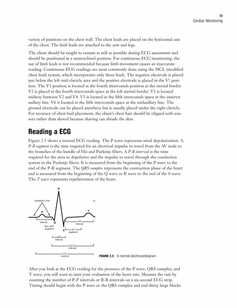
Reading a ECGFigure 2.5 shows a normal ECG reading. The P wave represents atrial depolarization. AP-R segment is the time required for an electrical impulse to travel from the AV node tothe branches of the bundle of His and Purkinje fibers. A P-R interval is the timerequired for the atria to depolarize and the impulse to travel through the conductionsystem to the Purkinje fibers. It is measured from the beginning of the P wave to theend of the P-R segment. The QRS complex represents the contraction phase of the heartand is measured from the beginning of the Q wave or R wave to the end of the S wave.The T wave represents repolarization of the heart.
R
P
Q
S
T
P R segment
Isoelectric line ts
P R interval ST
interval
ST segment
QRS interval
Q T interval
0.8 second FIGURE 2.5 A normal electrocardiogram.
After you look at the ECG reading for the presence of the P wave, QRS complex, andT wave, you will want to start your evaluation of the heart rate. Measure the rate bycounting the number of P-P intervals or R-R intervals on a six-second ECG strip.Timing should begin with the P wave or the QRS complex and end thirty large blocks
05_0789735962_ch02.qxd 2/26/07 4:19 PM Page 53

54
later. The heart rate can be determined looking at a six-second strip, count the cardiaccycles and the number of QRS complexes, and multiply by ten. This method providesan accurate rate analysis whether the rate is regular or irregular.
A normal rhythm is one that originates in the SA node, is regular, has a rate of 60–100beats per minute, has a P wave that is consistent and is followed by a QRS complex.ECG tracing paper measures electrical impulses in duration of time. Each large blockon the paper is 5 mm or 0.20 seconds and contains 25 small blocks. Each small block onthe paper is 1 mm or 0.04 seconds. The normal ECG rhythm has a P-R interval of0.12–0.20 seconds, and has a QRS complex with a duration of 0.04–0.12 seconds.
Cardiac DysrhythmiasCardiac dysrhythmias occur when the heart loses its regular pacing capability. They areclassified according to their origin. These abnormal rhythms can be lethal or of nodanger to the client’s well being. Tachydysrhythmias are characterized by a heart rategreater than 100 bpm. If the client has coronary artery disease, blood flow might bedecreased to the heart. Bradydysrhythmias are characterized by a heart rate less than 60beats per minute. Dizziness and syncopy are often the only symptoms that the clientnotices. The client might tolerate this slow rate or bradydysrhythmias might cause theblood pressure to be subnormal, leading to shock or ischemia. Another alteration in thenormal beat the client might experience is bigeminy, a condition where there arearrhythmias occurring in pairs. The pairs can be junctional, atrial, or ventricular beats. Ajunctional beat is one originating at the AV and bundle of HIS. An atrial dysrhythmiaoriginates in the atria of the heart, while a ventricular dysrhythmia originates in theventricle of the heart.
Unlike tachydysrhythmias and bradydysrhythmias, which usually originate in the atria, ventricular dysrhythmias are life-threatening and their impulse originates in theventricles.
Ventricular TachycardiaVentricular tachycardia is a rapid irregular rhythm with the absence of a P wave. Usuallythe rate exceeds 140–180 bpm. The SA node continues to discharge independently ofthe ventricle. Ventricular tachycardia is often associated with valvular heart disease,heart failure, hypomagnesium, hypotension, and ventricular aneurysms. Figure 2.6shows an ECG reading indicative of ventricular tachycardia.
Chapter 2: Care of the Client with Cardiovascular Disorders
FIGURE 2.6 Evidence of ventricular tachycardia.
05_0789735962_ch02.qxd 2/26/07 4:19 PM Page 54

Cardiac Dysrhythmias55
Ventricular tachycardia is most commonly treated with supplemental oxygen andmedications. Amiodarone (Cordarone), procainamide (Pronestyl), or magnesium sulfateis given to slow the rate and stabilize the rhythm. Lidocaine has long been establishedfor the treatment of ventricular tachycardia; however, it should not be used in an acuteMI client. In addition to the rate and rhythm regulation medications, heparin is oftenordered to prevent further thrombus formation. It is important to note that heparin isnot given to a client receiving streptokinase.
Ventricular FibrillationVentricular fibrillation (V-fib) is the primary mechanism associated with sudden cardiacarrest. This disorganized chaotic rhythm results in a lack of pumping activity of theheart. Without effective pumping, no oxygen is sent to the brain and other vital organs.If this condition is not corrected quickly, the client’s heart stops beating and asystole isseen on the ECG. The client quickly becomes faint, loses consciousness, and becomespulseless. Hypotension, or a lack of blood pressure, and abnormal heart sounds arepresent. Figure 2.7 shows a diagram of the chaotic rhythms typical with V-fib.
Ventricular Fibrillation (V Fib)
“sawtooth” FIGURE 2.7 Ventricular fibrillation diagram.
Treatment of ventricular fibrillation is done with a defibrillator set at approximately 200joules. Three quick, successive shocks are delivered, with the third at 360 joules. If adefibrillator is not readily available, a precordial thump can be delivered. If cardiacarrest occurs, the nurse should initiate cardiopulmonary resuscitation and be ready toadminister first-line drugs such as epinephrine or vasopressin (Pitressin).
Internal Cardiac DefibrillatorsAn internally implanted cardioverter/defibrillator is used to treat ventricular fibrillationand other dysrhythmias. This device is usually implanted on the client’s left side and isconnected to the myocardium with electrical leads. If the client experiences fibrillationor ventricular tachycardia, a shock is automatically delivered to the heart and correctsthe pattern. The internal defibrillator also records dysrhythmias that the client hasexperienced so that the physician is aware of the client’s condition. The client with aninternal cardiac defibrillator or permanent pacemaker should be taught to
. Wear a medic alert stating that a pacemaker/internal defibrillator is implanted.Identification will alert the healthcare worker so that alterations in care can bemade.
. Take pulse for one full minute and report the rate to the physician.
05_0789735962_ch02.qxd 2/26/07 4:19 PM Page 55

56
. Avoid applying pressure over the pacemaker. Pressure on the defibrillator or pace-maker can interfere with the electrical leads.
. Inform the dentist of the presence of a pacemaker because electrical devices areoften used in dentistry.
. Avoid having a magnetic resonance imaging (MRI) test. Magnetic resonance inter-feres with the electrical impulse of the implant.
. Avoid close contact with electrical appliances, electrical or gasoline engines, trans-mitter towers, antitheft devices, metal detectors, and welding equipment becausethey can interfere with conduction.
. Be careful when using microwaves. Microwaves are generally safe for use, but theclient should be taught to stand approximately five feet away from the device whilecooking.
. Report fever, redness, swelling, or soreness at the implantation site.
. If beeping tones are heard coming from the internal defibrillator, immediatelymove away from any electromagnetic source. Stand clear from other peoplebecause shock can affect anyone touching the client during defibrillation.
. Report dizziness, fainting, weakness, blackouts, or a rapid pulse rate. The clientwill most likely be told not to drive a car for approximately 3 months after theinternal defibrillator is inserted to evaluate any dysrhythmias.
. Report persistent hiccupping because this can indicate misfiring of thepacemaker/internal defibrillator.
Cardiopulmonary ResuscitationThe American Heart Association released new guidelines for professionals and thepublic in November of 2005. These guidelines were printed by the American HeartAssociation in the Circulation journal in 2006 and are as follows:
. Unskilled personnel should begin chest compressions and ventilations after deliv-ering two rescue breaths to an unresponsive victim. Lay rescuers are not taught toassess for pulse or signs of circulation for an unresponsive victim.
. Lay rescuers will not be taught to provide rescue breathing without chest compres-sions.
. The lone healthcare provider should alter the sequence of rescue response basedon the most likely etiology of the victim’s problem:
. For sudden collapse in victims of all ages, the lone healthcare providershould telephone the emergency response number and get an AED (whenreadily available) and then return to the victim to begin CPR and use the AED.
Chapter 2: Care of the Client with Cardiovascular Disorders
05_0789735962_ch02.qxd 2/26/07 4:19 PM Page 56

Angina Pectoris57
. For unresponsive victims of all ages with likely asphyxial arrest (for example,drowning), the lone healthcare provider should deliver about five cycles(about two minutes) of CPR before leaving the victim to telephone theemergency response number and get the AED. The rescuer should thenreturn to the victim, begin the steps of CPR, and use the AED.
. After delivery of two rescue breaths, healthcare providers should attempt to feel apulse in the unresponsive, nonbreathing victim for no more than 10 seconds. If theprovider does not definitely feel a pulse within 10 seconds, the provider shouldbegin cycles of chest compressions and ventilations.
. Healthcare providers will be taught to deliver rescue breaths without chestcompressions for the victim with respiratory arrest and a perfusing rhythm (that is,pulses). Rescue breaths without chest compressions should be delivered at a rate ofabout 10 to 12 breaths per minute for the adult, and a rate of about 12 to 20breaths per minute for the infant and child.
. Healthcare providers should deliver cycles of compressions and ventilations duringCPR when there is no advanced airway (for example, endotracheal tube, laryngealmask airway [LMA], or esophageal-tracheal combitube [Combitube]) in place.Once an advanced airway is in place for infant, child, or adult victims, two rescuersno longer deliver “cycles” of compressions interrupted with pauses for ventilation.Instead, the compressing rescuer should deliver 100 compressions per minutecontinuously, without pauses for ventilation. The two rescuers should changecompressor and ventilator roles approximately every two minutes to preventcompressor fatigue and deterioration in quality and rate of chest compressions.When multiple rescuers are present, they should rotate the compressor role aboutevery two minutes. The switch should be accomplished as quickly as possible(ideally in less than five seconds) to minimize interruptions in chest compressions.
Refer to the AHA (American Heart Association) website for current updates.
NOTE
Angina PectorisAngina pectoris is defined as chest pain caused by disruption of the balance and demandfor oxygen by the heart. This disruption results in a lack of oxygen to the myocardium.
Several risk factors predispose the client to cardiac ischemia. These include
. Hypertension
. Hyperlipidemia
. Smoking
. Obesity
. Familial history
05_0789735962_ch02.qxd 2/26/07 4:19 PM Page 57

58
. Anemia
. Stress
. Diabetes
The nurse caring for the client with angina pectoris assesses the type and location ofchest pain. The pain is usually located in the substernal to retrosternal area and radiatesdown the left arm and to the jaw or shoulder. The onset is usually precipitated by alarge meal, exertion, stress, anxiety, smoking, alcohol, or drugs, and might occur imme-diately when the client awakens. The client’s skin is usually warm and dry, but might becool and clammy. He might complain of nausea and vomiting and gripping chest pain.Women frequently do not complain of the typical chest pain associated with angina, butmay complain of fatigue and shortness of breath. An ECG often reveals S-T segmentdepressions and T wave inversion.; there might be S-T depressions. If the client hasPrinzmetal’s angina there might be an elevation in the S-T segment.
Treatment involves the application of oxygen and the administration of nitroglycerinesublingually, topically, or intravenously. The client should be taught to take one nitro-glycerine tablet sublingually every five minutes, not to exceed three tablets. If the firsttablet does not relieve the pain, a second can be taken. If the pain is still not relievedafter taking three tablets the client should go directly to the hospital or call an ambu-lance. The client should be taught to replenish the supply of nitroglycerine every sixmonths and protect the pills from light by leaving them in the brown bottle. It is impor-tant for the client to understand that light decreases the effectiveness of nitroglycerine.Nitroglycerine patches and creams should be applied to dry skin. The site should berelatively free of hair. Most resources suggest that the hair should be clipped and notshaved because shaving might abrade the skin and cause irritation. Nurses should alwayswear gloves when applying nitroglycerine creams or patches to prevent application ofthe medication to themselves. Intravenous nitroglycerine must be administered with aninfusion rate controller.
Myocardial InfarctionWhen there is a disruption in blood supply to the myocardium, the client is consideredto have had a myocardial infarction. Factors contributing to diminished blood flow to theheart include arteriosclerosis, emboli, thrombus, shock, and hemorrhage. If circulation isnot quickly restored to the heart, the muscle becomes necrotic. Hypoxia from ischemiacan lead to vasodilation. Acidosis associated with electrolyte imbalances often occurs,and the client can slip into cardiogenic shock. The most common site for a myocardialinfarction is the left ventricle. Only 10% of clients report the classic symptoms of amyocardial infarction. Women often fail to report chest pain and, if they do, they mighttell the nurse that the pain is beneath the shoulder or in the back. Clients with diabeteshave fewer pain receptors and might report little or no pain.
Chapter 2: Care of the Client with Cardiovascular Disorders
05_0789735962_ch02.qxd 2/26/07 4:19 PM Page 58
Myocardial Infarction59
The most commonly reported signs and symptoms associated with myocardial infarctioninclude
. Substernal pain or pain over the precordium for a duration greater than 15minutes
. Pain that is described as heavy, vise-like, and radiating down the left arm
. Pain that begins spontaneously and is not relieved by nitroglycerin or rest
. Pain that radiates to the jaw and neck
. Pain that is accompanied by shortness of breath, pallor, diaphoresis, dizziness,nausea, and vomiting
. Increased heart rate, decreased blood pressure, increased temperature, andincreased respiratory rate
Diagnosis of Myocardial InfarctionThe diagnosis of a myocardial infarction is made by looking at both the electrocardio-gram and the cardiac profile that consist of the cardiac enzymes. The following are themost commonly used diagnostic tools for determining the type and severity of myocar-dial infarction:
. Electrocardiogram
. Serum enzymes and isoenzymes
Other tests that are useful in providing a complete picture of the client’s condition arewhite blood cell count (WBC), sedimentations rate, and blood urea nitrogen (BUN).
The best serum enzymes used to diagnose myocardial infarction are creatine kinase(CKMB), troponin T and 1, CRP, and LDH. The enzyme CKMB is released whenthere is damage to the myocardium. The troponin T and 1 are specific to striatedmuscle and are often used to determine the severity of the attack. C-reactive protein(CRP) levels are used with the CKMB to determine whether the client has had an acuteMI and the severity of the infarction. Lactic dehydrogenase (LDH) is a nonspecificenzyme that is elevated with any muscle trauma.
Management of a Client with Myocardial InfarctionManagement of a client with myocardial infarction (MI) includes monitoring of bloodpressure, oxygen levels, and pulmonary artery wedge pressures. Because the blood pres-sure can fall rapidly, medication such as dopamine is prescribed. Other medications areordered to relieve pain and to vasodilate the coronary vessels—for example, morphinesulfate IV is ordered for pain. Thrombolytics, such as streptokinase, will most likely beordered. Early diagnosis and treatment significantly improve the client’s prognosis.
05_0789735962_ch02.qxd 2/26/07 4:19 PM Page 59

60
Clients suffering a myocardial infarction can present with dysrhythmias. Ventriculardysrhythmias, such as ventricular tachycardia or fibrillation, can lead to cardiac stand-still and death if not treated quickly.
The client with an MI should be given small, frequent meals. The diet should be low insodium, fat, and cholesterol. Adequate amounts of fluid and fiber are encouraged toprevent constipation. Stool softeners are often ordered to prevent straining during defe-cation. Post-MI teaching should stress the importance of a regular program of exercise,stress reduction, regular bowel elimination, and cessation of smoking. Because caffeinecauses vasoconstriction, caffeine intake should be limited. The client can resume sexualactivity in six weeks or when he is able to climb a flight of stairs without experiencingchest pain. Medications such as sildenafil (Viagra) are discouraged and should not betaken within 24 hours of taking a nitrite. Clients should be taught not to perform theValsalva maneuver or bend at the waist to retrieve items from the floor. Placing items intop drawers helps to prevent increased intrathoracic pressure. The client will probablybe discharged on an anticoagulant such as enoxaparin (Lovenox) or sodium warfarin(Coumadin).
Chapter 2: Care of the Client with Cardiovascular Disorders
Anticoagulants such as heparin are used to decrease the potential for clotting. The nurse should check thepartial thromboplastin time (PTT). The normal control level in the most common laboratory ranges isapproximately 30–60 seconds. The therapeutic bleeding time should be from one and a half to two timesthe control. The medication should be injected in the abdomen 2" from the umbilicus using a tuberculinsyringe. Do not aspirate or massage. The antidote for heparin derivatives is protamine sulfate.Anticoagulants should be stopped at least 24 hours prior to surgery and are usually restarted 12-24 hoursfollowing surgery.
NOTE
If Coumadin (sodium warfarin) is ordered, the nurse should check or prothrombin time (PT). The controllevel for a prothrombin time is 10–12 seconds. The therapeutic level for Coumadin should be from oneand a half to two times the control. The antidote for Coumadin is vitamin K. The international normalizingratio (INR) is done for oral anticoagulants. The therapeutic range is 2–3. If the level exceeds 7, watch forspontaneous bleeding.
NOTE
Exercise ElectrocardiographyAn exercise electrocardiography test, also known as the stress test or exercise tolerancetest, helps to determine the function of the heart during exercise. The client isinstructed to eat a light meal and refrain from smoking or consuming caffeine themorning of the test. Prior to the test, the cardiologist assesses the heart using an ECGtracing and blood pressure monitor. The client then walks on the treadmill or bicycle ata steadily progressing rate of speed of 1 to 10 miles per hour and can also be adjustedfrom flat to inclined. She is asked to report any shortness of breath or chest pain.Abnormalities can then be assessed. The client continues the test until:
05_0789735962_ch02.qxd 2/26/07 4:19 PM Page 60

Myocardial Infarction61
. A rapid heart rate is reached and maintained.
. Signs or symptoms of chest pain, fatigue, or extreme dyspnea, hypotension, orventricular dyshythmias appear on the ECG.
. There are S-T segment depressions noted on the ECG.
The client remains in the unit for approximately 2 hours after the test to assure thatthere are no signs of hypotension or cardiac dyshythmias. Some clients due to mobilityproblems are not able to walk on the treadmill or ride the bicycle. Cardiac stimulantsare then used to induce stress. An example of medications used is dobutamine(Dobutrex).
EchocardiographyEchocardiography is a noninvasive test used to determine the size of the ventricle, thefunctionality of the valves and the size of the heart. There is no special preparation forthe echocardiography and this test takes only 30–60 minutes.
A transesophageal echocardiography is a more invasive method of assessing the struc-tures of the heart. A transducer is placed into the esophagus or stomach in order toexamine the posterior cardiac structures. This test requires that the client be NPO aftermidnight the day of the procedure and the throat be anesthetized to prevent stimulationof the gag reflex. Following the procedure, the client is checked for return of the gagreflex prior to offering food.
The gag reflex is stimulated by placing a tongue blade on the back of the throat. Absence of the gag reflexincreases the chances of aspirating liquids.
NOTE
Cardiac CatheterizationCardiac catheterization is used to detect blockages associated with myocardial infarctionand dysrhythmias. Cardiac catheterization, as with any other dye procedure, requires asigned consent. This procedure can also accompany percutaneous transluminal coro-nary angioplasty. Prior to and following this procedure, the nurse should
. Assess for allergy to iodine or shellfish.
. Maintain the client on bed rest for approximately 8 hours with the leg straight.
. Maintain pressure on the access site for at least five minutes or until no signs ofbleeding are noted. Many cardiologists use a device called an Angio-Seal toprevent bleeding at the insertion site. The device creates a mechanical seal,anchoring a collagen sponge to the site. The sponge absorbs in 60–90 days.
. Use pressure dressing and/or ice packs to control bleeding.
05_0789735962_ch02.qxd 2/26/07 4:19 PM Page 61

62
. Check distal pulses because diminished pulses can indicate a hematoma at thecatheter insertion site and should be reported immediately.
. Force fluids to clear dye from the body.
Percutaneous Transluminal Coronary Angioplasty and Stent PlacementA percutaneous transluminal coronary angioplasty (PTCA) is a less invasive procedurethan coronary artery bypass surgery. Many clients are relieved of chest pain followingthis procedure. Clients with noncalcified lesions, such as plaque, benefit most from aPTCA and recover relatively quickly.
During the procedure, the physician inserts a catheter while visualizing the coronaryvessels. A balloon is used to push plaque into the wall of the vessel. A stent might beplaced in the artery following the balloon procedure. A stent is a mesh tube usually madeof stainless steel. This tube is inserted following an angioplasty to prevent restenosis.
When angiography indicates that the vessel is 50% or more open, the procedure iscomplete. An IV of heparin is administered in a continuous infusion. Nitroglycerin orsublingual nifedipine is often given to prevent spasms of the myocardium.
Coronary Artery Bypass GraftWhen the client does not respond to medical management of a coronary artery occlu-sion and is experiencing chest pain, the physician might perform coronary artery bypassgraft (CABG) surgery. The decision to perform a CABG is based on the results of thecardiac catheterization. If the client has the following symptoms, a CABG might beperformed:
. Angina with greater than 50% blockage of the left anterior descending artery
. Unstable angina with two vessels severely blocked or three vessels moderatelyblocked
. Ischemia of the myocardium
. Has had an acute MI
. Has ischemia following an angiography or PTCA
During a coronary artery bypass a sternal incision is performed and a donor vessel isremoved. A common vessel used to bypass a blockage in the coronary arteries is thesaphenous vein located in the back of the leg. Other vessels, such as the mammary arteryor the radial artery, can also be used to bypass the blockage. When the client is asleep,the team of surgeons goes to work harvesting the donor vessel while another teamprepares to place the client on the cardiopulmonary bypass machine. The cardiopul-monary bypass machine is often used to provide oxygen to the lungs and body duringthe time that the heart is stopped. Blood that is heparinized and oxygenated passesthrough the machine and back into the client by way of the ascending aortic vessel orthe femoral artery. While the client is on the bypass machine, the core body tempera-ture is lowered to approximately 85° F. The rationale for lowering the body temperature
Chapter 2: Care of the Client with Cardiovascular Disorders
05_0789735962_ch02.qxd 2/26/07 4:19 PM Page 62

Myocardial Infarction63
is that the body’s oxygen needs are lowered when the body is cooled. A potassium solu-tion is used to bathe the heart and help prevent dysrhythmias. After the heart is stopped,the surgeon anastomoses the donor vessel to bypass the blockage. When the procedureis finished, the client is warmed and transported to the intensive care unit.
The family should be instructed that the client will return to the intensive care unit withseveral tubes and monitors. The client will have mediastinal tubes to drain fluid fromthe chest cavity. The client might also have chest tubes if reinflation of the lungs wasnecessary. If the client bleeds and the blood is not drained from the mediastinal area,fluid accumulates around the heart and cardiac tamponade results. If this occurs, themyocardium becomes compressed and the accumulated fluid prevents the filling of theventricles and decreases cardiac output.
During surgery, a Swan-Ganz catheter for monitoring central venous pressure—pulmonary artery wedge pressure—is inserted in the pulmonary artery. A radial arterialblood pressure monitor is inserted to measure vital changes in the client’s blood pres-sure. An ECG monitor and oxygen saturation monitor are also used. Other tubes usedto assess and stabilize the client are a nasogastric tube to decompress the stomach, anendotracheal tube to assist in ventilation, and a Foley catheter to measure hourly urinaryoutput.
Some clients experience depression and or recurrent nightmares following coronaryartery bypass graft surgery. The family should be made aware that this is a commonproblem and that this problem might take several months to resolve. It is important totell both the family and the patient to notify the surgeon if these experiences occur.
Cardiac rehabilitation is recommended and includes a plan of exercise, diet, and weightreduction. The client should be taught regarding the needs to stop smoking and tomoderate alcohol consumption. Drugs used to treat sexual dysfunction, such as Viagra,should not be used within 24 hours of taking nitrites such as nitroglycerine.
Congestive Heart FailureThe potential for congestive heart failure (CHF) exists after a myocardial infarction.The nurse must monitor for signs of fluid retention. Left sided congestive heart failureoccurs when fluid backed into the lungs and is indicated by rales and blood-tingedsputum. Distended neck veins are also an indication as well as the client’s report ofneeding to sleep on two or more pillows to breathe. Right-sided congestive heart failureoccurs when the blood backs into the periphery causing peripheral edema, fatigue andasites. Treatment includes diuretics, inotropes, and a diet low in sodium. Other drugsmight be prescribed to decrease preload and afterload. IV nitroprusside, milrinone(Primacor), or nitroglycerine nesiritide (natrecor) are often used to improve cardiaccontractility. Other medications used to support cardiac function are angiotensinreceptor blockers (ARBs), angiotensin-converting enzyme (ACE) inhibitors, and betablockers. These drugs increase the force of cardiac contractions. Morphine is oftengiven to control pain as well as to treat preload.
If the client’s condition deteriorates despite the use of cardiac drugs, an intra-aorticballoon pump (IABP) might be inserted. The IABP is inserted into the aorta. A balloon
05_0789735962_ch02.qxd 2/26/07 4:19 PM Page 63

64
is inflated during diastole and deflates just before systole, reducing the afterload. Thisprocedure improves perfusion to the heart, brain, and lungs and decreases perfusion tothe kidneys and lower extremities.
With use of the IABP, perfusion to the lower extremities and the kidneys could beimpeded during inflation of the pump, so assessment of pulses distal to the pump inser-tion site and assessment of urinary output is essential.
Other management of CHF includes monitoring O2 saturation, pulmonary artery wedgepressure (PAWP) with an attempt to maintain PAWP between 15–20 mm/hg. Centralvenous pressure (CVP) monitoring and frequent checking of vital signs is essentialnursing care for the client with CHF.
Cardiogenic ShockThere are three types of shock: cardiogenic shock, hypovolemic shock, and vasogenic orneurogenic shock. Cardiogenic shock occurs when the heart fails to pump enough blood toperfuse the tissues adequately. This type of shock might be due to a myocardial infarc-tion, congestive heart failure, pericarditis, cardiac tamponade (fluid around the heartthat constricts the heart muscle), severe vascular disease, or rupture of an abdominalaortic aneurysm. Hypovolemic shock occurs when there is insufficient blood flow tomaintain blood pressure. This results in decreased oxygenation to vital organs.Vasogenic or neurogenic shock occurs when there is trauma to the brain or spinal cord.This results in shock secondary to the nervous systems inability to maintain vasocon-striction. Chapter 10, “Care of the Client with Neurological Disorders,” discussses thistype of shock in detail. In cardiogenic shock, there is necrosis of more than 40% of theleft ventricle. Most of the clients experiencing cardiogenic shock complain of chest pain.Other symptoms include
. Hypotension
. Tachycardia
. Tachypnea
. Frothy, pink-tinged sputum
. Restlessness
. Orthopnea
. Oliguria
The mortality rate of cardiogenic shock is extremely high if it is not detected early.Treatment includes oxygen therapy. The physician will order a pain reliever such asmorphine sulfate. Diuretics, nitroglycerin, and other medications to reduce the preloadare also parts of the treatment. In extreme situations, an intra-aortic balloon pumpmight be used to decrease the workload of the heart.
Chapter 2: Care of the Client with Cardiovascular Disorders
05_0789735962_ch02.qxd 2/26/07 4:19 PM Page 64

Aneurysms65
AneurysmsAn aneurysm is ballooning of an artery as illustrated in Figure 2.8. The greatest risk forthese clients is rupture and hemorrhage. Aneurysms can occur in any artery in the bodyand might be the result of congenital malformations, arteriosclerosis, or secondary tohypertension. There are several types of aneurysms:
. Fusiform—Affects the entire circumference of the artery)
. Saccular—An outpouching affecting only one portion of the artery
. Dissecting—Bleeding into the wall of the vessel
The client with an abdominal aortic aneurysm will frequently complain of feeling “myheart beating in my abdomen” or low back pain. Any such complaint should be furtherevaluated. On auscultation of the abdomen, a bruit could be heard. Diagnosis can bemade by ultrasound, computer tomography, arteriogram, or abdominal x-rays. If theaneurysm is found to be 5 centimeters or more, surgery might be scheduled. Duringsurgery, the aorta is clamped above and below and a donor vessel is anastamosed inplace. When the client returns from surgery, pulses distal to the site should be assessed.Because the blood supply is stopped to the kidneys and lower extremities during renalfunction should be evaluated along with pedal pulses. Use of endovascular stints is nowbeing used to relieve pressure on the aneurysm and reinforce the weakened vessel. Thestints are threaded through an incision in the femoral artery. Post-operative care ismuch the same as that of the client that has undergone a cardiac catheterization.
Do not palpate the mass because pressure on the weakened vessel can lead to rupture and hemorage.
CAUTION
FIGURE 2.8 Abdominalaortic aneurysm.
Common carotid arteries
Ascending aorta
Heart
Thoracic aorta
Aortic arch
Thoracic aortic aneurysm
Renal arteries
Abdominal aorta
Abdominal aortic aneurysm
Superior mesenteric artery
Interior mesenteric artery
Common illiac arteries
05_0789735962_ch02.qxd 2/26/07 4:19 PM Page 65

66
Inflammatory Diseases of the HeartInflammatory and infectious diseases of the heart often are a result of systemic infectionsthat affect the heart. Inflammation and infection might involve the endocardium, peri-cardium, valves, or the entire heart.
Infective EndocarditisInfective endocarditis, also known as bacterial endocarditis, is usually the result of a bacterialinfections, collagen diseases, or cancer metastasis. As a result, the heart is damaged andsigns of cardiac decompensation results. The client commonly complains of shortness ofbreath, fatigue, and chest pain. On assessment, the nurse might note distended neckveins a friction rub, or a cardiac murmur.
Treatment involves treating the underlying cause with antibiotics, anti-inflammatorydrugs, and oxygen therapy. Bed rest is recommended until symptoms subside. If thevalve is severely damaged by infection, a valve replacement might have to be performed.Replacement valves are xenograft (bovine [cow] or, procine [pig]), cadaver, or mechan-ical. If the client elects to have a mechanical valve replacement, he will have to take anti-coagulants for life. Following surgery, the nurse must be alert for signs of complications.These include decreased cardiac output or heart failure, infection, and bleeding. Thephysician often will prescribe digoxin, anticoagulants, cortisone, and antibiotics postop-eratively.
PericarditisPericarditis is an inflammatory condition of the pericardium, which is the membrane sacaround the heart. Symptoms include chest pain, difficulty breathing, fever, andorthopnea. Clients with chronic constrictive pericarditis show signs of right-sidedcongestive heart failure. During auscultation, the nurse will likely note a pericardial fric-tion rub. Laboratory findings might show an elevated white cell count. ECG changesconsist of an S-T segment and T wave elevation. The echocardiogram often shows peri-cardial effusion.
Treatment includes use of nonsteroidal anti-inflammatory drugs to relieve pain. Thenurse should monitor the client for signs of pericardial effusion and cardiac tamponadethat include jugular vein distention, paradoxical pulses (systolic blood pressure higher onexpiration than on inspiration), decreased cardiac output, muffled heart sounds. If fluidaccumulates in an amount that causes cardiac constriction, the physician might decide toperform a pericardiocentesis to relieve the pressure around the heart. Using an echocar-diogram or fluoroscopic monitor, the physician inserts a large-bore needle into the peri-cardial sac. After the procedure, the nurse should monitor the client’s vital signs andheart sounds. In severe cases, the pericardium might be removed.
Chapter 2: Care of the Client with Cardiovascular Disorders
05_0789735962_ch02.qxd 2/26/07 4:19 PM Page 66

Inflammatory Diseases of the Heart67
Peripheral Vascular DiseaseThe term peripheral vascular disease (PVD) refers to a group of diseases affecting botharteries and veins. Peripheral arterial disease, the most common type of PVD, oftenresults in amputations, kidney disease, and ulcerations of the extremities.
Signs of PVD include a decrease in pulse rate and strength, coldness of the extremity,intermittent claudication (burning and leg cramps on ambulation), and swelling of theextremity.
Treatment is aimed at restoring blood flow to the extremity. Treatment includes asympathectomy to sever the sympathetic ganglia, thereby resulting in vasodilation,vasodilating drugs, or femoropopliteal bypass graft. Stints can also be used to maintainan open vessel. If circulation to the extremity is not restored, an amputation might berequired.
Femoral Popliteal Bypass GraftWhen blood flow to the lower legs is interrupted, the physician might elect to performto bypass the blockage in the vessel. Grafts can be made of synthetic materials such aspolytetrafluoroethylene, Gore-Tex, and Dacron. Donor vessels can also be used.
Preoperatively, the nurse should assess renal function and the extremity for pulses,swelling, color, and temperature. If a Doppler is used to obtain pulses, it should bedocumented. Dye studies might also be ordered prior to the surgical procedure to deter-mine the extent of the disease. The nurse should assess the client’s potential complica-tions associated with dye procedures such as allergies to iodine.
During the graft procedure, the doctor removes the donor vessel and bypasses the blockvessel. Following the procedure, the nurse should monitor for signs of graft rejection.These include redness at the site and signs of decreased oxygenation to the extremity.Other nursing care includes
. Assessing color, temperature, and pulses
. Assessing for pain and administering medication as ordered
. Monitoring blood pressure
. Instructing the client to keep the affected extremity straight and not to cross thelegs at the knee
. Assessing the incision site
At discharge, the client should be taught to avoid sitting at a 90° angle or crossing thelegs, and to take anticoagulants and vasodilating drugs as ordered. He should also betaught to report signs of decreased oxygenation to the extremity. If graft occlusion doesoccur, a thrombectomy, tissue plasminogen activator, or revision of the graft might berequired.
05_0789735962_ch02.qxd 2/26/07 4:19 PM Page 67
68
Varicose Veins / ThrombophlebitisVaricose veins occur when the valves that serve to push blood back to the heart becomeweak and collapse. This allows blood to pool in the vein. The stagnant blood often clotsand occlusion of the vessel occurs. If a clot breaks loose, it can travel to the heart orlungs resulting in a pulmonary emboli.
Thrombophlebitis occurs when a vein becomes inflamed and a clot forms. Most throm-bophlebitis occurs in the lower extremities, with the saphenous vein being the mostcommonly affected vein. Homan’s sign is an assessment tool used for many years byhealthcare workers to detect deep vein thrombi. It is considered positive if the clientcomplains of pain on dorsiflexion of the foot. Homan’s sign should not be performedroutinely because it can cause a clot to be dislodged and lead to a pulmonary emboli. If adiagnosis of thrombophlebitis is made, the client should be placed on bed rest withwarm, moist compresses to the leg. An anticoagulant such as enoxaparin, heparin, orsodium warfarin is ordered, and the client is monitored for complications such ascellulitis. If cellulitis is present, antibiotics are ordered.
Antithrombolitic stockings or compression devices are ordered to prevent venous stasis.When antithrombolitic stockings are applied, the client should be in bed for a minimumof 30 minutes prior to applying the stockings. The circumference and length of theextremity should be measured to prevent rolling down of the stocking and a tourniqueteffect.
Raynaud’s PhenomenonRaynaud’s phenomenon occurs when there are vascular vasospasms brought on by expo-sure to cold. Raynaud’s is more common in women and has been linked to decreasingestrogen levels. The most commonly affected areas are the hands, nose, and ears.Management includes preventing exposure, stopping smoking, and using vasodilators.The client should be encouraged to wear mittens when outside in cold weather.
Buerger’s DiseaseBuerger’s disease (thromboangiitis obliterans) results when spasms of the arteries andveins occur primarily in the lower extremities. These spasms result in blood clot forma-tions and eventually destruction of the vessels. Symptoms associated with Buerger’sdisease include pallor of the extremities progressing to cyanosis, pain, and paresthesia.As time progresses, trophic changes occur in the extremities. Management of the clientwith Buerger’s disease involves the use of Buerger-Allen exercises, vasodilators, andoxygenation. The client should be encouraged to stop smoking.
Case StudyA 77-year-old male reports to the doctor with complaints of shortness of breath. On examination, thedoctor finds crackles in the base of the lungs, a blood pressure of 190/96, slight tachycardia, and a gain
Chapter 2: Care of the Client with Cardiovascular Disorders
05_0789735962_ch02.qxd 2/26/07 4:19 PM Page 68

Buerger’s Disease69
of 10 pounds since the client’s last check-up. The doctor has prescribed medications to control conges-tive heart failure. Total cholesterol 240 mg/dL, sodium 160 mEq/L, and potassium 3.6 mEq/L.
1. Are the client’s symptoms consistent with right-sided or left-sided congestive heart failure?
2. What medications should the nurse expect the doctor to prescribe?
3. How does the client’s blood pressure affect the client’s cardiac function?
4. What is the correlation between the weight gain and the congestive heart failure?
5. If the client’s congestive heart failure is not treated effectively, what will be the result?
05_0789735962_ch02.qxd 2/26/07 4:19 PM Page 69

70
6. What dietary management should be implemented for this client?
Answers to Case Study1. The client’s symptoms are consistent with both right-sided and left-sided congestive heart failure. In
left-sided congestive heart failure, fluid backs up into the lungs. In right-sided congestive heartfailure, fluid backs up into the extremities. This client has weight gain, shortness of breath, andcrackles heard on auscultation.
2. Treatment of congestive heart failure is threefold. The diet should be low in sodium. Medicationsinclude diuretics such as furosemide (Lasix), milrinone (Primacor), or nesiritide (Natrecor) toincrease cardiac output, and pain management with morphine.
3. The client’s blood pressure is elevated. The peripheral resistance increases the workload on theheart. This further compromises the cardiac condition and leads to worsening congestive heartfailure.
4. The weight gain is a sign of right-sided congestive heart failure.
5. If the client’s congestive heart failure is not treated, the client’s lungs will fill with fluid. Fluid in thelungs prevents oxygenation to the heart and brain. The heart failure will worsen and lead to death.
6. The dietary management is low sodium, low fat, and low cholesterol.
Key ConceptsThis chapter discussed the most common types of cardiovascular problems. The keyconcepts will help the nursing graduate on the NCLEX by focusing on the mostcommonly used key terms, diagnostic exams, and pharmacological agents used to treatthese problems. This section is covered on the NCLEX in the area of physiologicalintegrity.
Key Terms. Aneurysms
. Angina pectoris
. Angioplasty
. Atherosclerosis
. Blood pressure
. Buerger’s disease
. Cardiac catheterization
. Cardiac tamponade
. Cardiopulmonary resuscitation
Chapter 2: Care of the Client with Cardiovascular Disorders
05_0789735962_ch02.qxd 2/26/07 4:19 PM Page 70

Diagnostics71
. Cholesterol
. Conduction system of the heart
. Congestive heart failure
. Coronary artery bypass graft
. Defibrillation
. Diastole
. Electrocardiogram
. Heart block
. Hypertension
. Implantable cardioverter
. Myocardial infarction
. Pacemaker
. Raynaud’s phenomenon
. Systole
. Thrombophlebitis
. Varicose veins
. Ventricular fibrillation
. Ventricular tachycardia
DiagnosticsThe exam reviewer should be knowledgeable of the preparation and care of clientsreceiving exams to diagnose cardiovascular problems. While reviewing these diagnosticexams, the exam reviewer should be alert for information that would be an importantpart of nursing care for these clients. The pertinent labs and exams are as follows:
. Cardiac catheterization
. Cardiac profile
. Central venous pressure monitoring
. Chest x-ray
. Clotting studies
. Complete blood count
. Doppler studies
. Dye studies for cardiac functions
05_0789735962_ch02.qxd 2/26/07 4:19 PM Page 71

72
. Echocardiogram
. Electrophysiologic studies
. Exercise Tolerance Test
. Fluoroscopy
. MRI
. Oxygen saturation levels
. Serum cholesterol and triglycerides
. Serum electrolytes
. Thallium scans
. Ultrasonography
. Vital signs
Pharmacological Agents Used in the Treatment ofClients with Cardiovascular DisordersAn integral part of care to clients with cardiovascular disorders is pharmacological inter-vention. These medications provide an improvement or cure of the clients’ cardiacproblems. The nursing exam reviewer needs to focus on the drugs in Table 2.1. Includedin this table are the most common side and adverse effects and pertinent nursing care.
TABLE 2.1 Pharmacologic Agents Used in the Treatment of Clients with CardiovascularDisordersDrug Action Side Effect Nursing Care
Thiazide Diuretics This category of drugs Electrolyte imbalances, Check potassium Examples of this category of increase excretion of dehydration, can lead levels and teach the drugs are: Chlorothiazide water and sodium by to increases in urea client to increase the (Diuril) and inhibiting resorption and gout. consumption of Hydrochlorothiazide in the early distal potassium rich foods. (Esidrix, HCTZ) tubule. They are used Care should be taken
for hypertension, when administering edema in congestive diuretics to the heart failure, intraocular elderly. The client pressure in glaucoma. should be taught to
take the medication inthe morning toprevent nocturia.
Chapter 2: Care of the Client with Cardiovascular Disorders
(continues)
05_0789735962_ch02.qxd 2/26/07 4:19 PM Page 72

Diagnostics73
Loop Diuretics Loop diuretcs inhibit same sameAn example of this type of resorption of sodium drug is Furosemide (Lasix) and chloride in the
Loop of Henle.
Osmotic Diuretics Osmotic diuretics same sameExamples of this type of increase the osmotic drug are: mannitol (Mannitol, pressure of glomerular Osmitrol, Resectisol), urea filtrate, so decreasing
absorption of sodium.
Potassium Sparing Diuretics Acts on the distal Can cause nausea Because this drug Examples of this type of drug are: tubule to inhibit and vomiting, can category is potassium spironolactone (Aldactone), reabsorption of sodium, lead to electrolyte sparing there is no amiloride, (Midamore)triamterene chloride, and increase imbalances such as need to increase (Dyrenium) potassium retention. hyperkalemia and potassium in the diet.
This drug is used for hyponatremia. Can Teach the client to hypertension and for lead to liver and take the drug with cushings disease. blood dyscrasias. food to decrease
gastrointestinal upset.Teach the client toavoid prolonged expo-sure to the sunlight,photosensitivity mayoccur. This drug cate-gory can turn urineblue.
Beta Adrenergic Blockers Used to treat Orthostatic Teach the client to rise Examples of this category of hypertension, hypotension, slowly. Should be drugs are: Propanolol ventricular bradycardia, used with caution in (Inderal), metopolol dysrhythmias diarrhea, nausea the elderly. Can lead (Lopressor), nadolol (Corgard) and angina and vomiting. to congestive heart
pectoris. failure so the client Nonselective should be taught to blockers produce report signs of edema. a fall in blood Should be used with pressure with caution in the client reflex tachycardia with diabetes, or bradycaria pregnancy or asthma.through a mixture of B-blocking effects. Selective B-blockers compete for stimulation of B-receptors in cardiac smooth muscles.
(continues)
TABLE 2.1 ContinuedDrug Action Side Effect Nursing Care
05_0789735962_ch02.qxd 2/26/07 4:19 PM Page 73

74
Calcium Channel Blockers This category of drugs The most common Teach the client to Examples of this type of drug is used to treat side effects are check his/her pulse, are: Nifedipine (Procardia, hypertension and dysthythmias and and to report signs of Adalat). Verapamil (Calan, dysrhythmias, unstable edema. The client edema such as Isoptin) angina, and stable might experience a shortness of breath
angina. They produce headache, fatigue, and edema. calcium ion influx drowsiness or facial across the cell flushing. These membrane in drugs should not be cardiac and vascular used in clients with smooth muscle. They 2nd and 3rd degree dilate coronary heart block, or arteries, slow the SA cardiogenic shock and AV nodes and since they can dilate peripheral worsen symptoms. arteries. Caution should be
taken when treating the client with congestive heart failure with this category of drugs.
Angiotensin-Converting This type of drugs act The most common The client should be Enzyme Inhibitors by selectively side effects are taught to remain at Examples of this type of suppressing rennin- hypotension. These rest for approximately drug are: captopril (Capoten), angiotensin I to drugs can cause a 30 minutes after lisinopril (Zestril) angiotensin II. They cough, angioedema. taking the first dose to
dilate the arteries The nurse should prevent orthostatic and veins. check creatinine hypotension. The
levels and electrolytes client should be to ensure that the taught to report signs client is not of renal failure.experiencing hyperkalemia.
Central Alpha Agonists (also This type of drug Can lead to Aldomet can cause known as Central Acting acts by inhibiting hypotension, impotence and turn Adrenergics) the sympathetic bradycardia and the urine dark brown Examples of this type of vasomotor center reduce cardiac when it is exposed to drug are: Clinidine hcl in the central nervous output. Assess sunlight. Also can (Catapress), methylodopa system. These blood studies cause photosensitivity.(Aldomet) drugs are used to such as neutrophils, Administer this
treat hypertension. platelets and renal category of drugs function. Can cause prior to meals. dry mouth. Can cause allergic reactions: rash, fever, pruritis, urticaria.
Chapter 2: Care of the Client with Cardiovascular Disorders
TABLE 2.1 ContinuedDrug Action Side Effect Nursing Care
(continues)
05_0789735962_ch02.qxd 2/26/07 4:19 PM Page 74
Diagnostics75
Vasodilators This type of drug is Can lead to nasal Teach the client to An example of this type of used to treat congestion, muscle take with food to drug is Hydralazine (Apresoline) hypertension and cramps, cardiac decrease
congestive heart palpitations, gastrointestinal upset. failure. headaches, dizziness, Notify the health care
nausea, vomiting, provider if they anorexia, diarrhea, or experience fever, constipation. Can severe fatigue or cause a rash or muscle or joint pain. pruritus. Can lead to Rise slowly to prevent bone marrow orthostatic suppression. This hypotension. Notify drug category is the health care contraindicated in the provider if the client client with coronary is pregnant. artery disease and rheumatic fever.
Alpha-receptor blockers Causes pheripheral Can lead to dizziness, Take the first dose at An example of this type of blood vessels to headaches, bedtime to prevent drug is Doxazosin (Cardura) dilate. Lowers drowsiness, vertigo, ortostatic hypotention.
peripheral resistance, weakness. This reduces blood type of drug can pressure. Also used also cause nausea, to increase urinary vomiting and outflow in the client abdominal pain. If with prostate disease. the client is allergic
to quinazolines there might be a cross allergic reaction. Use with caution in the pregnant client.
Angiotensin Receptor Blockers Blocks the Can lead to dizziness, Teach the client to Examples of this type of drugs vasoconstrictor and insomnia, anxiety, notify the health care are: Valsartan (Diovan), aldosterone-sereting diarrhea, dyspepsia, provider if the he/she Losartan (Cozaar), effects of anorexia and develops mouth Candesartan (Atacand), angiotensin II, blocks vomiting. Can cause sores, fever, or Telmisartan (Micardis) the binding of myalgia. Can cause edema.
angiotensin II to the a cough, but this is AT1 receptor found less common in this in tissue. Used to category of drugs treat hypertension. than in the ACE drugs.
Increases digoxin levels. The nurse should check the creatinine levels for renal function.
TABLE 2.1 ContinuedDrug Action Side Effect Nursing Care
(continues)
05_0789735962_ch02.qxd 2/26/07 4:19 PM Page 75
76
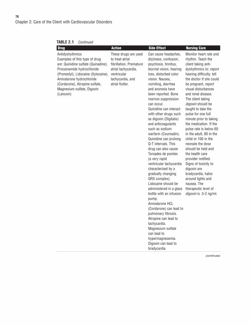
Antidyshythmics These drugs are used Can cause headaches, Monitor heart rate and Examples of this type of drug to treat atrial dizziness, confusion, rhythm. Teach the are: Quinidine sulfate (Quinadine), fibrillation, Premature psychosis, tinnitus, client taking anti-Procainamide hydrochloride atrial tachycardia, blurred vision, hearing dyshythmics to: report (Pronestyl), Lidocaine (Xylocaine), ventricular loss, disturbed color hearing difficulty, tell Amiodarone hydrochloride tachycardia, and vision. Nausea, the doctor if she could (Cordarone), Atropine sulfate, atrial flutter. vomiting, diarrhea be pregnant, report Magnesium sulfate, Digoxin and anorexia have visual disturbances (Lanoxin) been reported. Bone and renal disease.
marrow suppression The client taking can occur. digoxin should be Quinidine can interact taught to take the with other drugs such pulse for one full as digoxin (Digitalis) minute prior to taking and anticoagulants the medication. If the such as sodium pulse rate is below 60 warfarin (Coumadin). in the adult, 80 in the Quinidine can prolong child or 100 in the Q-T intervals. This neonate the dose drug can also cause should be held and Torsades de pointes the health care (a very rapid provider notified. ventricular tachycardia Signs of toxicity to characterized by a digoxin are gradually changing bradycardia, halos QRS complex). around lights and Lidocaine should be nausea. The administered in a glass therapeutic level of bottle with an infusion digoxin is .5-2 ng/ml.pump.Amiodarone HCL (Cordarone) can lead to pulmonary fibrosis.Atropine can lead to tachycardia.Magnesium sulfate can lead to hypermagnesemia. Digoxin can lead to bradycardia.
Chapter 2: Care of the Client with Cardiovascular Disorders
TABLE 2.1 ContinuedDrug Action Side Effect Nursing Care
(continues)
05_0789735962_ch02.qxd 2/26/07 4:19 PM Page 76

Diagnostics77
Anticoagulants Used to treat clients Hemmorage, Teach the client to Examples of anticoagulants with thrombosis agranulocytosis, report to the dentist are: Warfarin sodium Warfarin sodium leucopenia, that he is taking an (Coumadin), Heparin, decreases vitamin K eosinophilia and anticoagulant prior to Enoxaparin (Lovenox) absorption thereby thrombocytopenia. any dental work.
prolonging the These drugs Watch for bleeding bleeding time. interact with during flossing, Heparin and the salicylates (aspirin), toothbrushing, derivatives of heparin steroids, and shaving, and so on. prolong the bleeding NSAIDS (non- Teach the client the time by interfering steroidal anti- correct method of with the clotting chain. inflammatory taking the drug.
drugs). Blood Heparin and heparin studies such as derivatives should be partial prothrombin given in the abdomen time (PTT), protime approximately two (PT should be done inches from periodically during umbilicus. The client the course of should not aspirate treatment. The client after the injection or should report a rash, massage the area. fever, or urticaria. Teach the client The antidote for regarding signs of coumadin is prolonged bleeding vitamin K and the times. If the client is antidote for heparin taking coumadin he is protamine sulfate. should be taught to
limit the intake of darkgreen leafy foods suchas turnip greens.Other example offoods to limit arecabbage, rhubarb, andcauliflower becausethese foods containhigh amounts ofvitamin k. The clientshould report to thedoctor the intake ofherbals, vitamin E, orgreen tea since thesesubstances prolongbleeding times andcan prolong bleedingtimes.Note: enoxaparin(Lovenox) doses arebased on weight.
TABLE 2.1 ContinuedDrug Action Side Effect Nursing Care
(continues)
05_0789735962_ch02.qxd 2/26/07 4:19 PM Page 77

78
Thrombolytics These drugs are Clients with a history Instruct the client to Examples of thrombolytics used to destroy a clot. of streptococcal report signs of are streptokinase They are used to infections may not bleeding. A drug (Streptase), t-PA (Tissue treat coronary respond to treatment history should check Plasminogen Activator), thrombus, acute with streptokinase for previous use of abbokinase (Urokinase) ischemia associated since antibodies streptokinase since
with a cerebro are present. The many physicians do vascular accident, nurse should not recommend that or deep vein thrombus. check the bleeding this drug be repeated
times. only every two years.(This might be a lifelong restriction.)
Apply Your KnowledgeThe nurse reviewing for the licensure exam must be able to apple knowledge to meetclient needs. Utilization of information found in this chapter will help the graduate toanswer questions found on the NCLEX.
Exam Questions1. The client with hypertension has an order for furosemide. Which lab value must
be evaluated during the course of treatment with this medication?
A. Phosphorus
B. Potassium
C. Calcium
D. Magnesium
2. The client is admitted with a diagnosis of heart block. The nurse is aware that thepacemaker of the heart is the:
A. AV node
B. Purkinje fibers
C. SA node
D. Bundle of His
Chapter 2: Care of the Client with Cardiovascular Disorders
TABLE 2.1 ContinuedDrug Action Side Effect Nursing Care
05_0789735962_ch02.qxd 2/26/07 4:19 PM Page 78

Apply Your Knowledge79
3. The client is being treated with nitroprusside (Nitropress). The nurse is aware thatthis medication:
A. Should be protected from light
B. Is a non–potassium-sparing diuretic
C. Causes vasoconstriction
D. Decreases circulation to the extremities
4. The client being treated with lisinopril (Zestril) develops a hacking cough. Thenurse should tell the client to:
A. Take the medication at night to control the problem.
B. Take cough medication to control the problem
C. Stop the medication
D. Report the problem to the doctor
5. The elderly client taking digitalis develops constipation. The nurse is aware thatconstipation in the client taking digitalis might:
A. Develop an elevated digitalis level
B. Have a decrease in the digitalis levels
C. Have alterations in sodium levels
D. Develop tachycardia
6. The client is suspected of having had a myocardium infarction. Which diagnosticfinding is most significant?
A. LDH
B. Troponin
C. Creatinine
D. AST
7. The client with an internally implanted defibrillator should be taught:
A. Avoid driving a car
B. Avoid eating food cooked in a microwave
C. Refrain from using a cellular phone
D. Report swelling at the site
05_0789735962_ch02.qxd 2/26/07 4:19 PM Page 79

80
8. The client is scheduled for a cardiac catheterization. Following the procedure, thenurse should:
A. Assess for allergy to iodine
B. Check pulses proximal to the site
C. Assess the urinary output
D. Check to make sure that the client has a consent form signed
9. The client with Buerger’s disease complains of pain in the lower extremities. Thenurse is aware that Burger’s disease is also called:
A. Pheochromocytoma
B. Intermittent claudication
C. Kawasaki disease
D. Thromboangiitis obliterans
10. The client with an abdominal aneurysm frequently complains of:
A. A headache
B. Shortness of breath only during sleep
C. Lower back pain
D. Difficulty voiding
Answers to Exam Questions1. Answer B is correct. The client taking furosemide is at risk for developing
hypokalemia (decreased potassium) because this drug is a non–potassium-sparingdiuretic. Answers A, C, and D are incorrect because phosphorus, calcium, andmagnesium levels are not directly affected by Lasix. These levels should bechecked, but the lab value that is most significant for the nurse to check is thepotassium. Alteration in the potassium can lead to cardiac conductions problems.
2. Answer C is correct. The pacemaker of the heart is the SA node. The impulsemoves from the SA node to the AV node on to the right and left bundle branchesand finally to the Purkinje fibers. This makes answers A, B, and D incorrect.
3. Answer A is correct. Nitroglycerine preparations should be protected from lightbecause light decreases the effectiveness of this category of medication. Answer Bis incorrect because Nitropress is not a diuretic. Answer C is incorrect becauseNitropress is a vasodilator not a vasoconstrictor. Answer D is incorrect becausenitroglycerine does not decrease circulation to the extremities.
4. Answer D is correct. A hacking cough is a common side effect and should bereported to the doctor. The client should not be told to half the dose since this can
Chapter 2: Care of the Client with Cardiovascular Disorders
05_0789735962_ch02.qxd 2/26/07 4:19 PM Page 80

Apply Your Knowledge81
result in an elevated blood pressure, so answer A is incorrect. Answer B is incorrectbecause taking a cough medication will mask the symptom of the allergy. Answer Cis incorrect because although the client stops taking the medication, this answerstates that the client can report the finding to the doctor at the time of the sched-uled visit. He should report this finding immediately.
5. Answer A is correct. The client taking digitalis should avoid constipation becauseconstipation can lead to digitalis toxicity. Answer B is incorrect because constipa-tion will not lead to a decrease in the digitalis levels. Answer C is incorrect becauseconstipation does not result in alterations in the sodium level. Answer D is incor-rect because digitalis toxicity will result in brachycardia, not tachycardia.
6. Answer B is correct. The best diagnostic tool for confirming that the client hasexperienced a myocardial infarction is the troponin level. Another lab value that isassociated with a myocardial infarction is the CKMB. A is incorrect because theLDH is also elevated in clients with muscle trauma not associated with an MI. C isincorrect because the creatinine level indicates renal function. D is incorrectbecause the AST level is elevated with gallbladder disease as well as muscle inflam-mation.
7. Answer D is correct. The client with an implantable defibrillator should reportredness, pain, and swelling at the site of the implant. Answer A, B, and C areincorrect because the client can drive a car, eat food cooked in a microwave, andcan use a cellular phone. The client probably will be told to wait three months todrive a car. He should put his food in the microwave and step five feet away fromthe microwave during cooking. A cellular phone can be used, but should be held inthe right hand.
8. Answer C is correct. The dye used in the procedure can cause a decrease in renalfunction. The client’s renal function should be assessed and changes reported tothe doctor immediately. Answer A is incorrect because the client’s allergies shouldbe checked prior to the procedure not after the procedure. The femoral artery iscommonly used as the site for a catheterization. Answer B is incorrect because thepulses should be checked distal to the site. Answer D is incorrect because thepermit should be signed prior to the procedure.
9. Answer D is correct. The other name for Buerger’s disease is thromboangiitisobliterans. Answer A is incorrect because pheochromocytoma is an adrenal tumor.Answer B is incorrect because intermittent claudication is pain in an extremitywhen walking. Answer C is incorrect because Kawasaki disease is an acute vasculitisthat can result in an aneurysm in the thoracic area.
10. Answer C is correct. Clients with abdominal aortic aneurysms often complain ofnausea, lower back pain, and feeling their heart beat in the abdomen. Answer A isincorrect because a headache is a symptom of a cerebral aneurysm. Answer B isincorrect because although the client with an abdominal aneurysm might haveshortness of breath, this symptom is not particular to during sleep. Answer D isincorrect because difficulty voiding is not associated with an abdominal aneurysm.
05_0789735962_ch02.qxd 2/26/07 4:19 PM Page 81

82
Suggested Reading and Resources. Ignataviicus, Donna D., Workman, Linda, Medical-Surgical Nursing. Philadelphia:
W.B. Saunders Company, 2005.
. Taber’s Cyclopedic Medical Dictionary. Philadelphia Pennsylvania: F. A. Davis, 2005.
. Vanetzian, Eleanor V. , Critical Thinking: An Interactive Tool for Learning Medical-Surgical Nursing. F.A. Davis, 2005.
. Rinehart, Wilda, Sloan, Diann, Hurd, Clara, NCLEX Exam Cram. Indianapolis:Que Publishing, 2005.
. Deglin, Judith H. , Vallerand, April H., Davis Drug Guide for Nurses. Philadelphia:F. A. Davis, 2006.
Chapter 2: Care of the Client with Cardiovascular Disorders
05_0789735962_ch02.qxd 2/26/07 4:19 PM Page 82