01 Lecture Ppt
38
Tit le Copyright © The McGraw-Hill Companies, Inc. Permission required for reproduction or display. Chapter 1 Lecture Slides
description
anatomy and physiology powerpoints
Transcript of 01 Lecture Ppt
Title
Copyright © The McGraw-Hill Companies, Inc. Permission required for reproduction or display.
Chapter 1LectureSlides
1-2
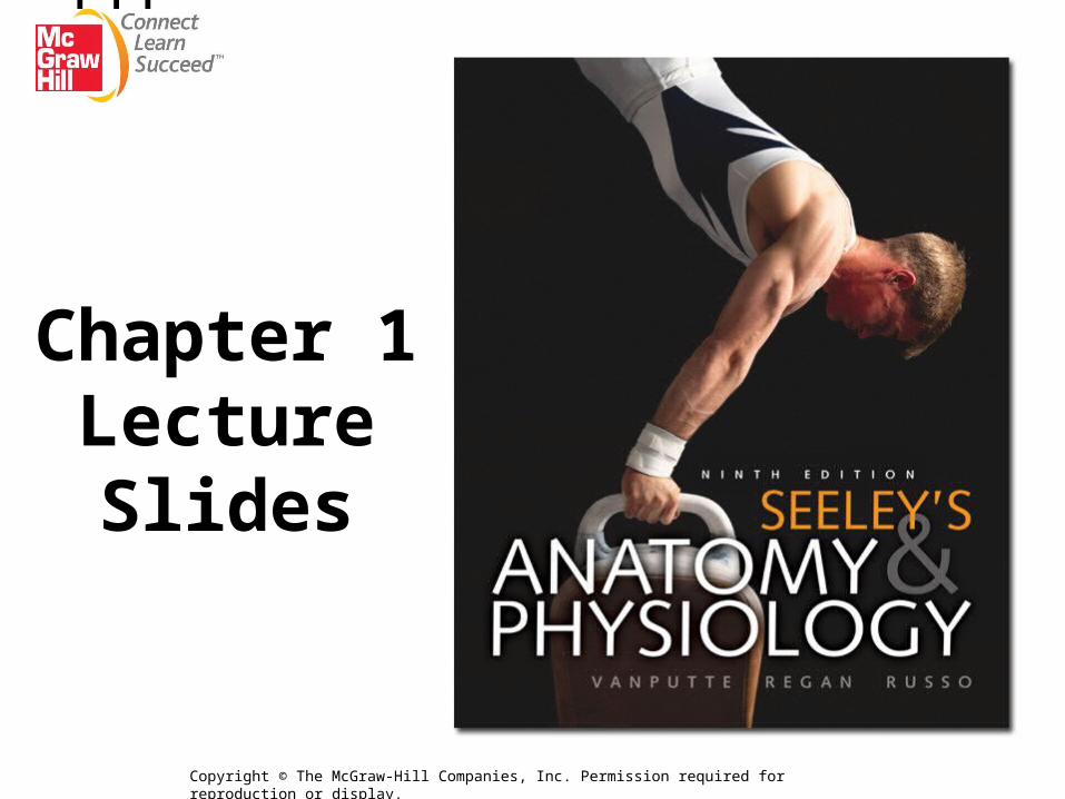
Chapter 1
The Human Organism

1-3
1.1 Anatomy and Physiology
• Anatomy: scientific discipline that investigates the body’s structure
• Physiology: scientific investigation of the processes or functions of living things
1-4
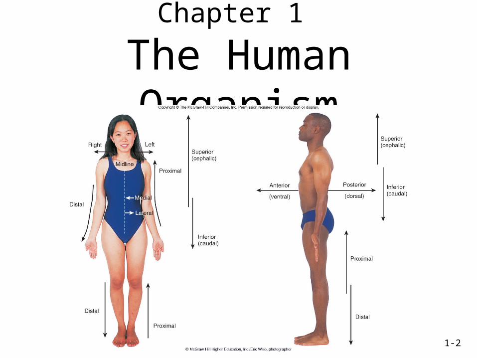
Topics of Anatomy
• Gross or macroscopic: structures examined without a microscope– Regional: studied area by area– Systemic: studied system by system– Surface: external form and relation to deeper
structures as x-ray in anatomic imaging
• Microscopic: structures seen with the microscope– Cytology: cellular anatomy– Histology: study of tissues
1-5
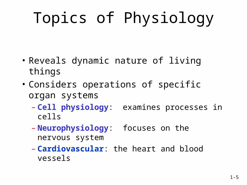
Topics of Physiology
• Reveals dynamic nature of living things
• Considers operations of specific organ systems– Cell physiology: examines processes in cells– Neurophysiology: focuses on the nervous
system– Cardiovascular: the heart and blood vessels
1-6
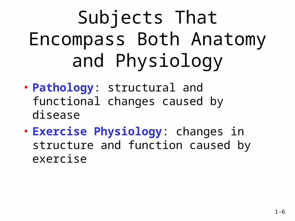
Subjects That Encompass Both Anatomy and Physiology
• Pathology: structural and functional changes caused by disease
• Exercise Physiology: changes in structure and function caused by exercise
1-7
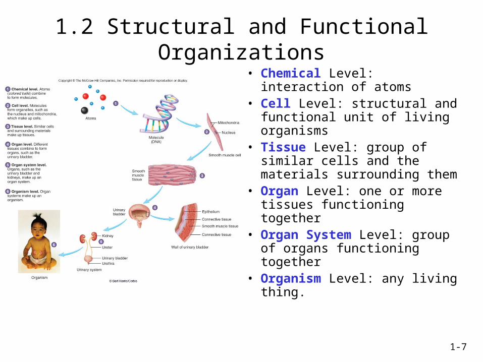
1.2 Structural and Functional Organizations
• Chemical Level: interaction of atoms
• Cell Level: structural and functional unit of living organisms
• Tissue Level: group of similar cells and the materials surrounding them
• Organ Level: one or more tissues functioning together
• Organ System Level: group of organs functioning together
• Organism Level: any living thing.
1-8
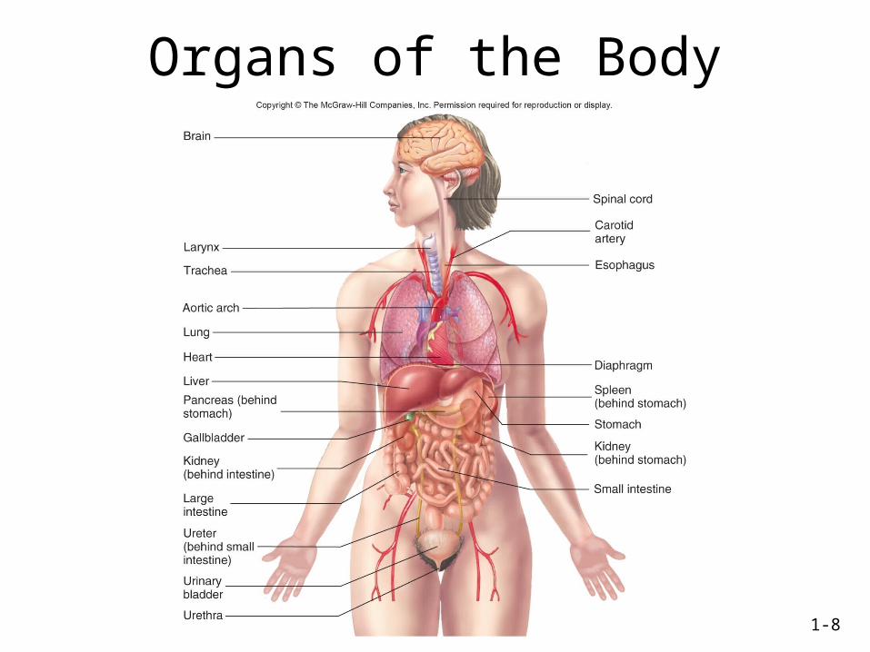
Organs of the Body
1-9
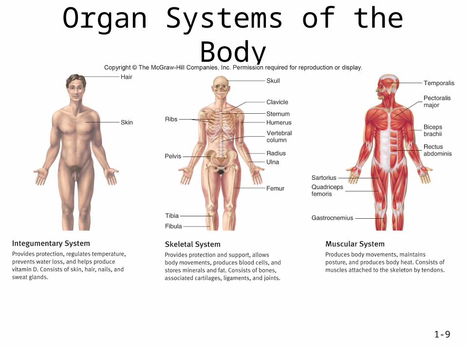
Organ Systems of the Body
1-10
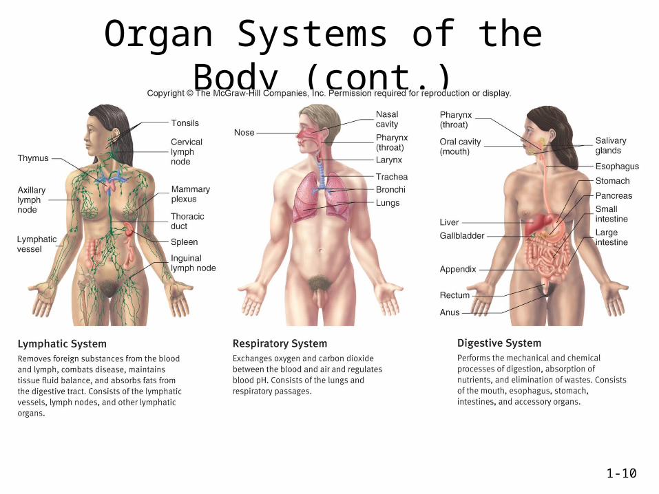
Organ Systems of the Body (cont.)
1-11
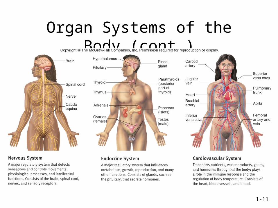
Organ Systems of the Body (cont.)
1-12
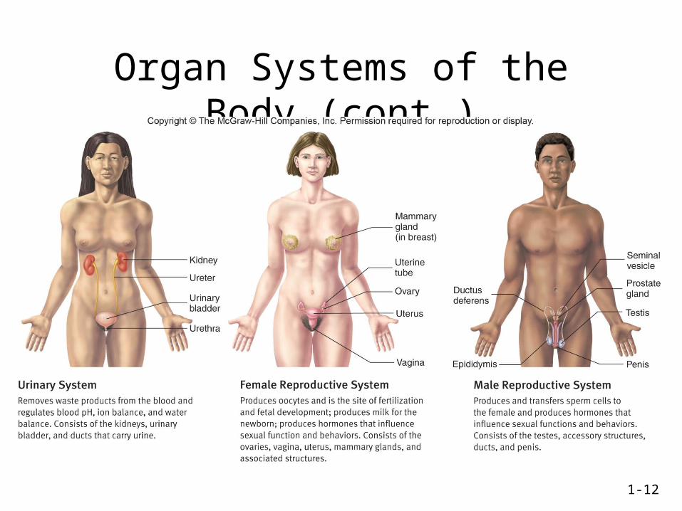
Organ Systems of the Body (cont.)
1-13
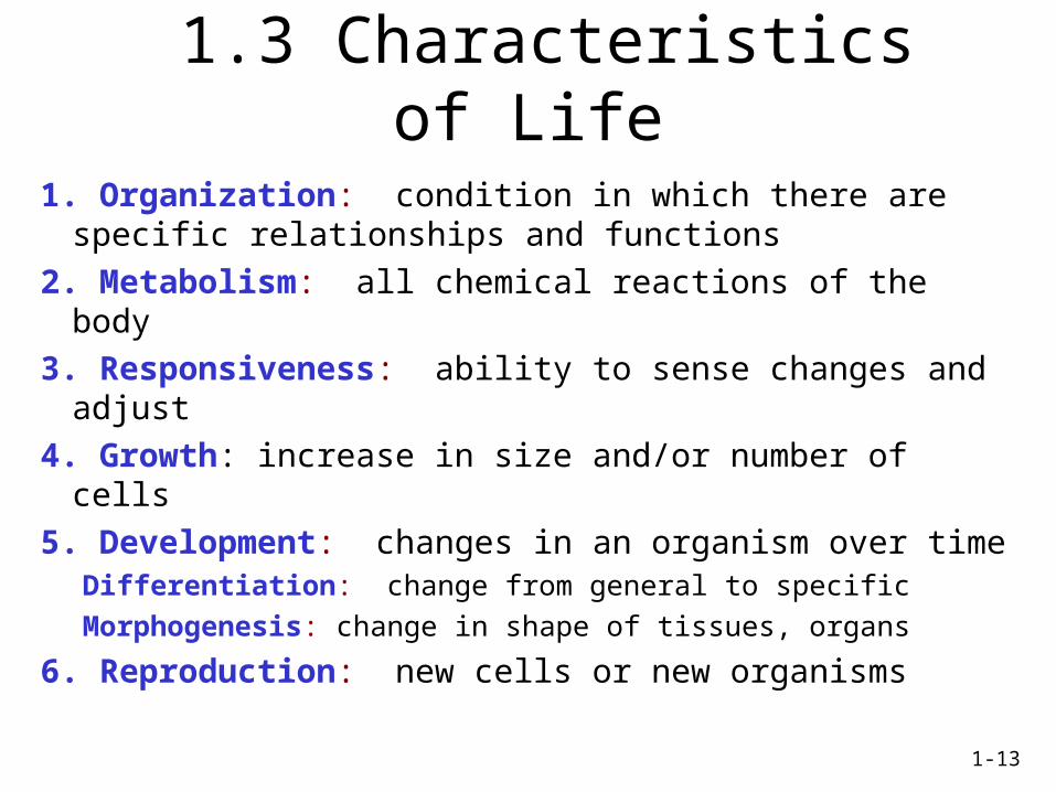
1.3 Characteristics of Life
1. Organization: condition in which there are specific relationships and functions
2. Metabolism: all chemical reactions of the body
3. Responsiveness: ability to sense changes and adjust
4. Growth: increase in size and/or number of cells
5. Development: changes in an organism over timeDifferentiation: change from general to specific
Morphogenesis: change in shape of tissues, organs
6. Reproduction: new cells or new organisms
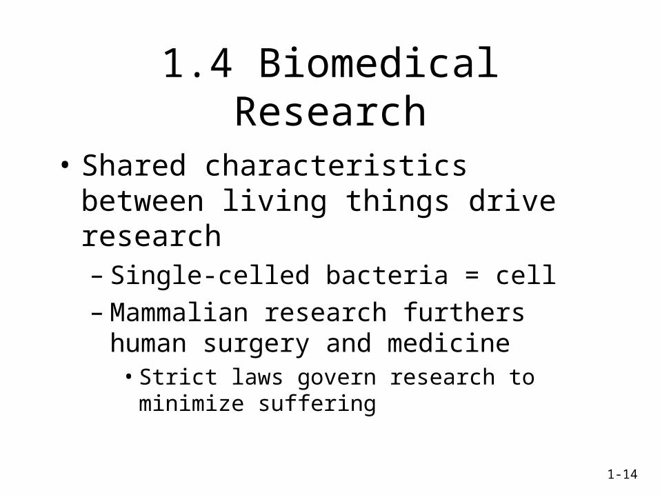
1.4 Biomedical Research
• Shared characteristics between living things drive research– Single-celled bacteria = cell– Mammalian research furthers human surgery
and medicine• Strict laws govern research to minimize suffering
1-14
1-15
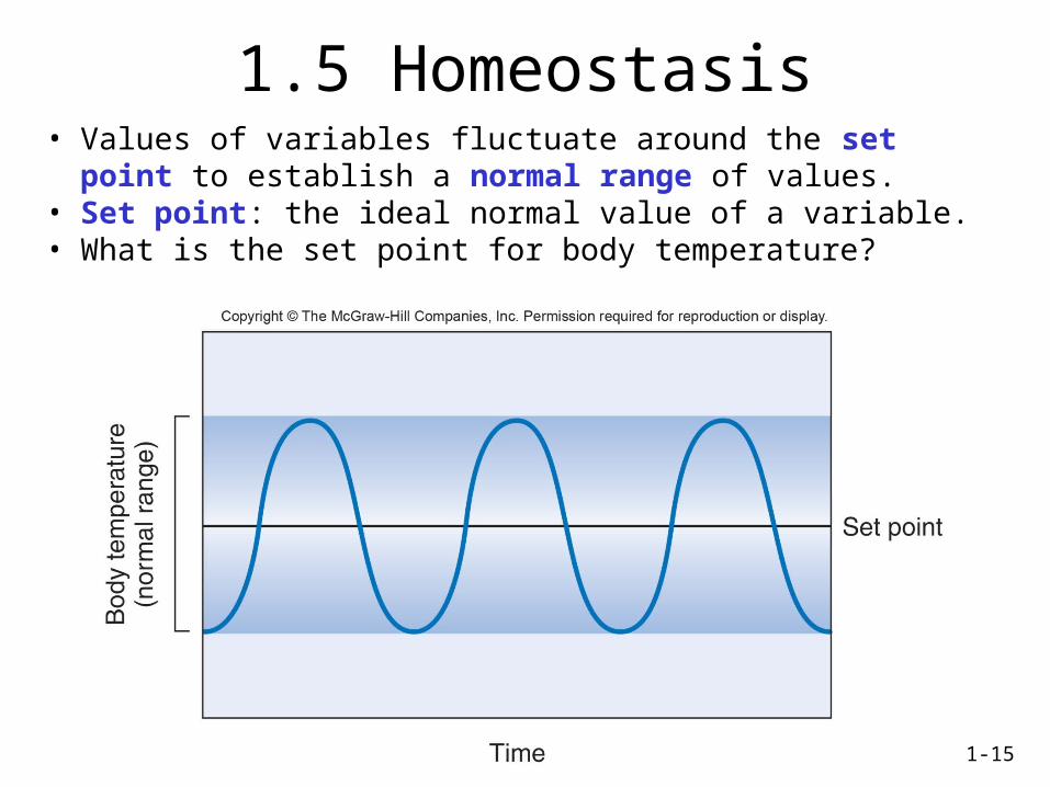
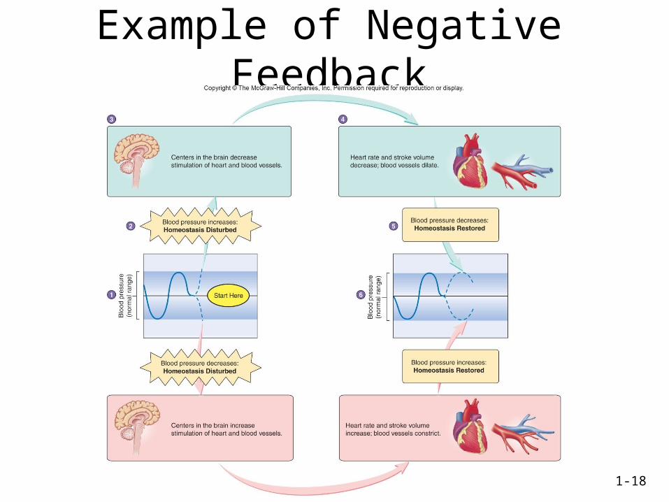
1.5 Homeostasis• Values of variables fluctuate around the set point to establish a
normal range of values.• Set point: the ideal normal value of a variable.• What is the set point for body temperature?
1-16
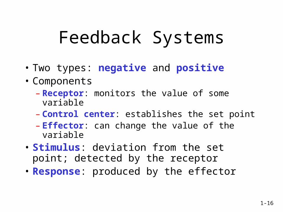
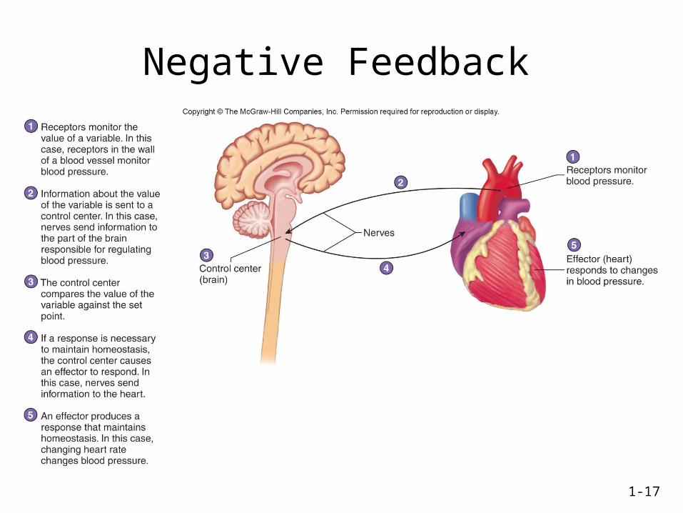
Feedback Systems
• Two types: negative and positive• Components
– Receptor: monitors the value of some variable– Control center: establishes the set point– Effector: can change the value of the variable
• Stimulus: deviation from the set point; detected by the receptor
• Response: produced by the effector
1-17
Negative Feedback
1-18
Example of Negative Feedback
1-19
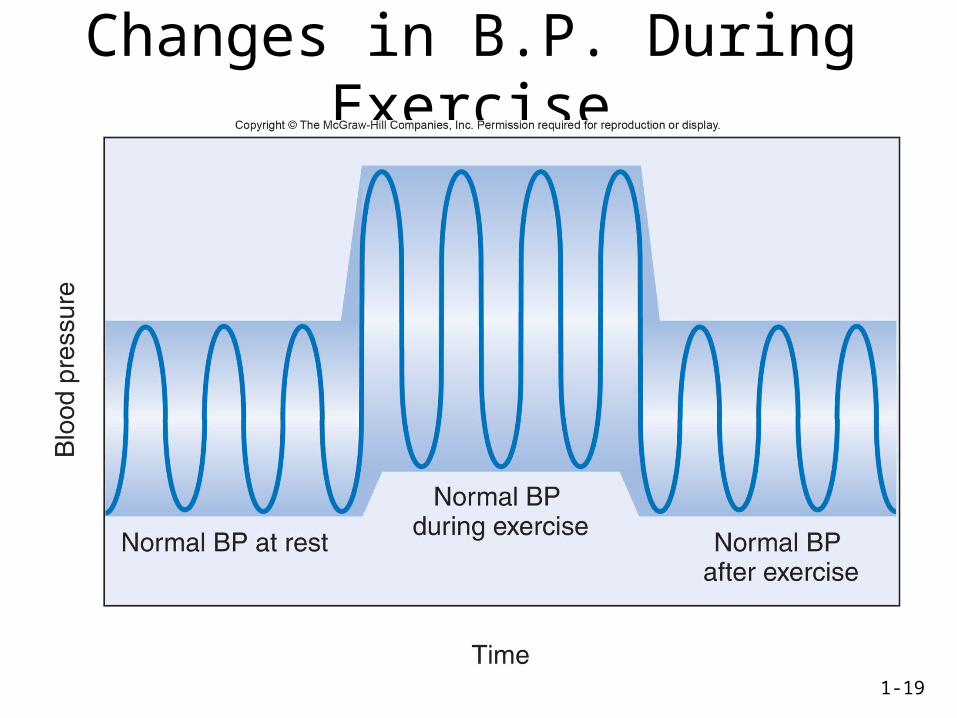
Changes in B.P. During Exercise
1-20
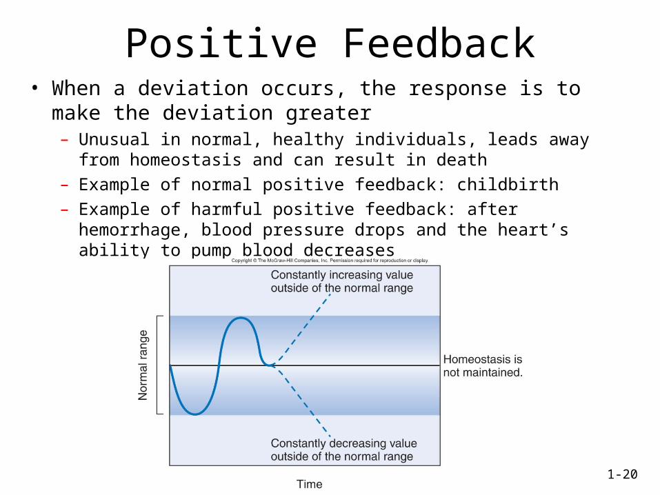
Positive Feedback• When a deviation occurs, the response is to make the deviation
greater– Unusual in normal, healthy individuals, leads away from homeostasis and
can result in death
– Example of normal positive feedback: childbirth
– Example of harmful positive feedback: after hemorrhage, blood pressure drops and the heart’s ability to pump blood decreases
1-21
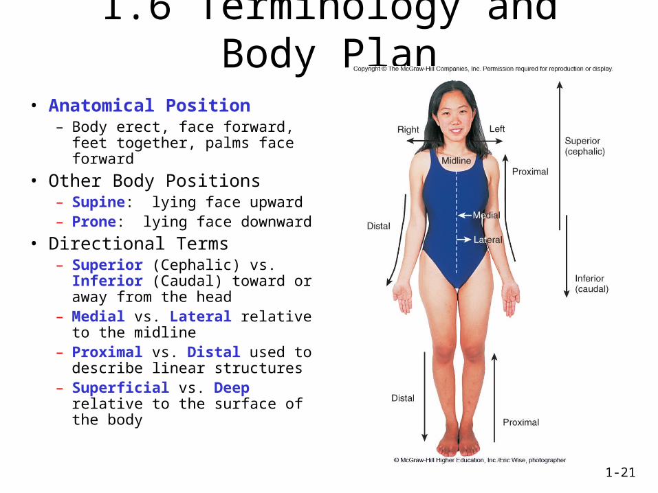
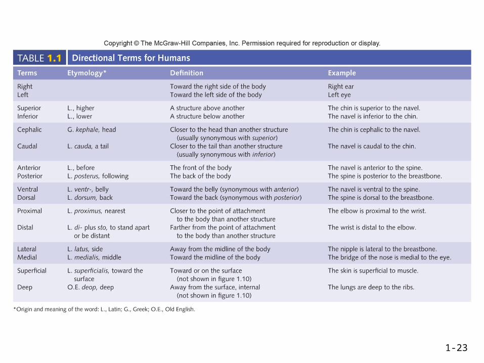
1.6 Terminology and Body Plan
• Anatomical Position– Body erect, face forward, feet
together, palms face forward
• Other Body Positions– Supine: lying face upward– Prone: lying face downward
• Directional Terms– Superior (Cephalic) vs. Inferior
(Caudal) toward or away from the head
– Medial vs. Lateral relative to the midline
– Proximal vs. Distal used to describe linear structures
– Superficial vs. Deep relative to the surface of the body
1-22
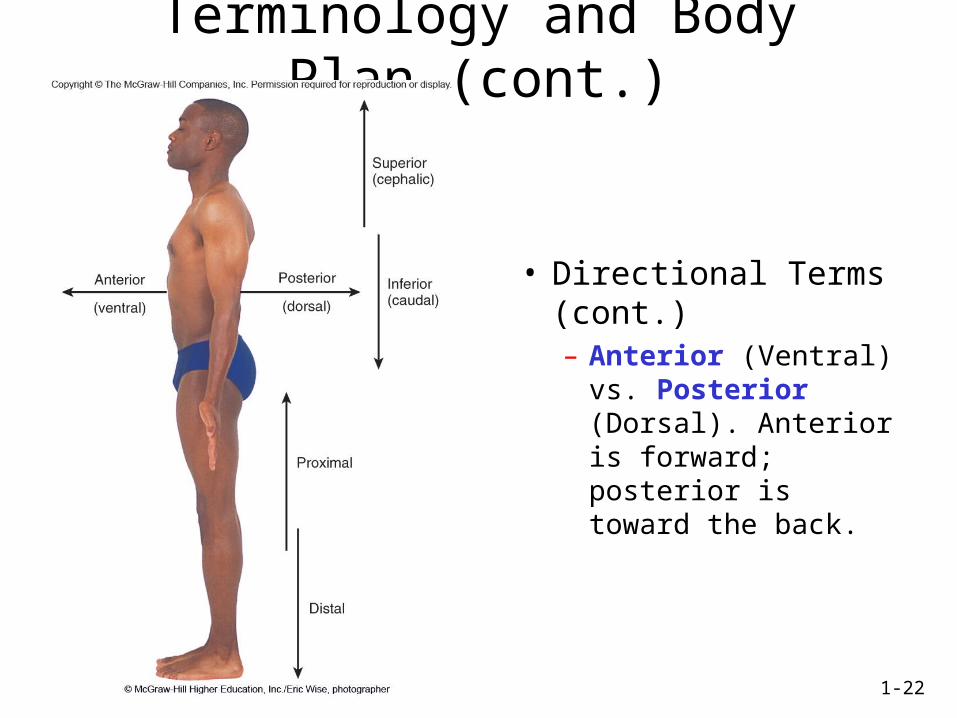
Terminology and Body Plan (cont.)
• Directional Terms (cont.)– Anterior (Ventral) vs.
Posterior (Dorsal). Anterior is forward; posterior is toward the back.
1-23
1-24
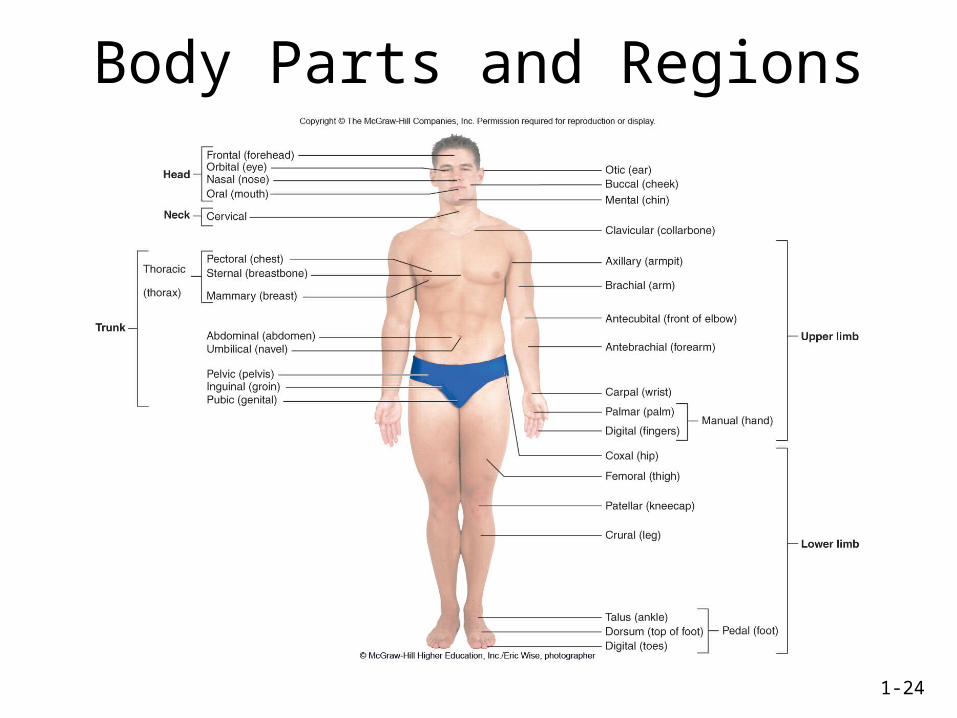
Body Parts and Regions
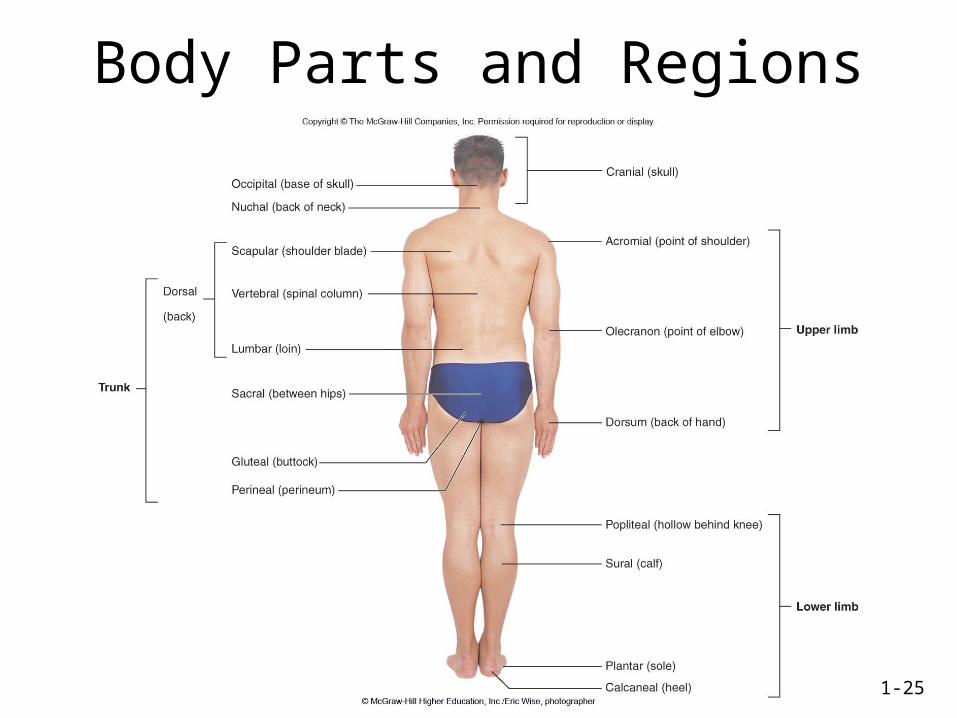
1-25
Body Parts and Regions
1-26
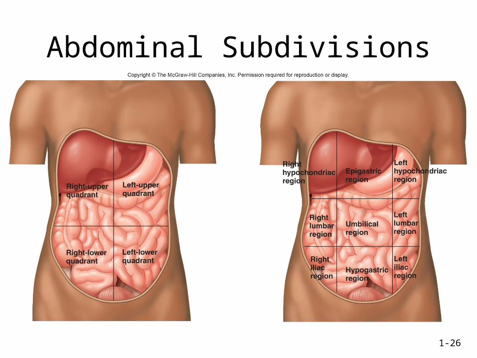
Abdominal Subdivisions
1-27
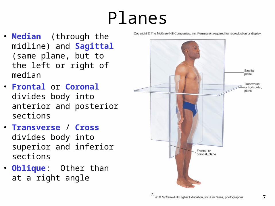
Planes• Median (through the
midline) and Sagittal (same plane, but to the left or right of median
• Frontal or Coronal divides body into anterior and posterior sections
• Transverse / Cross divides body into superior and inferior sections
• Oblique: Other than at a right angle
1-28
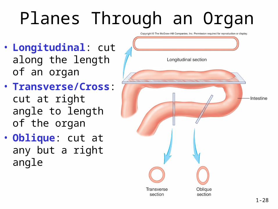
Planes Through an Organ
• Longitudinal: cut along the length of an organ
• Transverse/Cross: cut at right angle to length of the organ
• Oblique: cut at any but a right angle
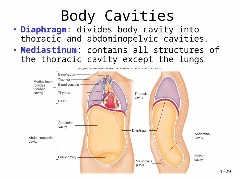
1-29
Body Cavities• Diaphragm: divides body cavity into thoracic and
abdominopelvic cavities.• Mediastinum: contains all structures of the thoracic
cavity except the lungs
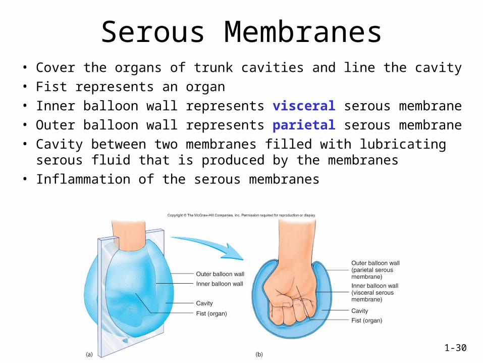
1-30
Serous Membranes• Cover the organs of trunk cavities and line the cavity
• Fist represents an organ
• Inner balloon wall represents visceral serous membrane
• Outer balloon wall represents parietal serous membrane
• Cavity between two membranes filled with lubricating serous fluid that is produced by the membranes
• Inflammation of the serous membranes
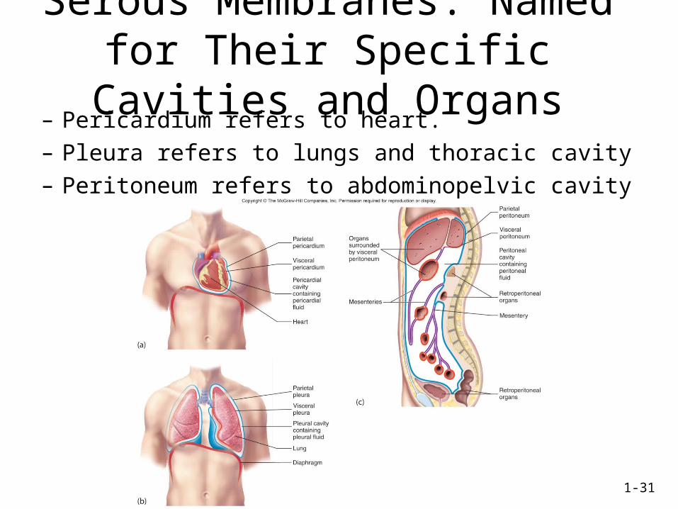
1-31
Serous Membranes: Named for Their Specific Cavities and Organs– Pericardium refers to heart.
– Pleura refers to lungs and thoracic cavity
– Peritoneum refers to abdominopelvic cavity
1-32
Imaging Techniques• Radiography
• Ultrasound (US)
• Computed Tomography (CT)
• Dynamic Spatial Reconstruction (DSR)
• Digital Subtraction Angiography (DSA)
• Magnetic Resonance Imaging (MRI)
• Positron Emission Tomography (PET)
1-33
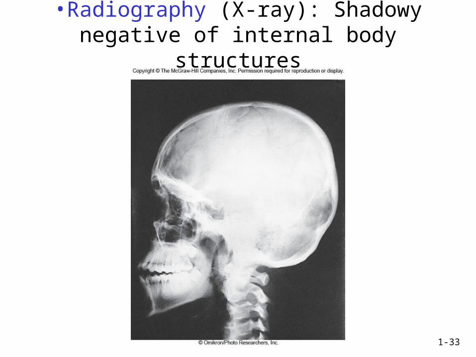
•Radiography (X-ray): Shadowy negative of internal body structures
1-34
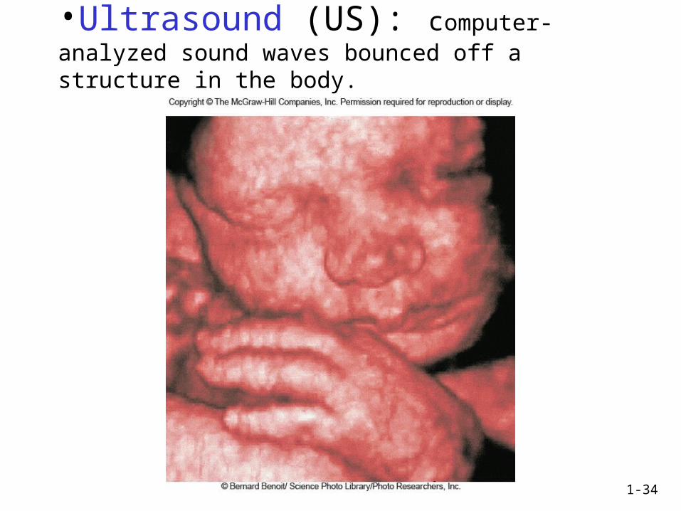
•Ultrasound (US): computer-analyzed sound waves bounced off a structure in the body.
1-35
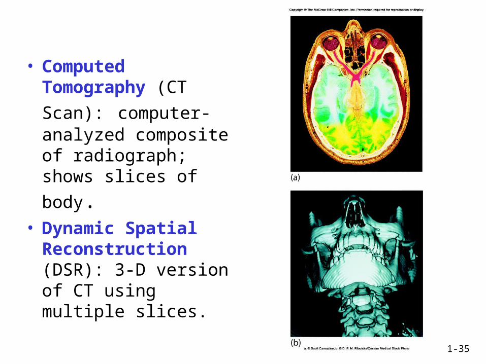
• Computed Tomography (CT
Scan): computer-analyzed composite of radiograph; shows slices
of body.• Dynamic Spatial
Reconstruction (DSR): 3-D version of CT using multiple slices.
1-36
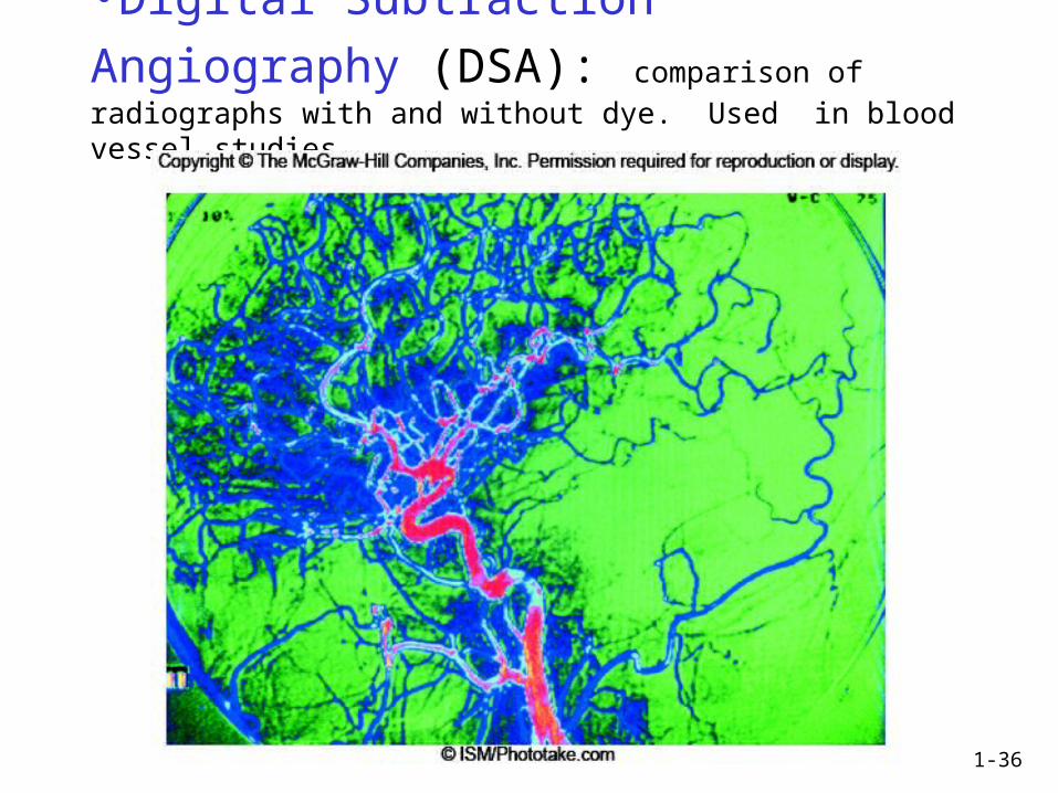
•Digital Subtraction Angiography (DSA): comparison of radiographs with and without dye. Used in blood vessel studies.
1-37
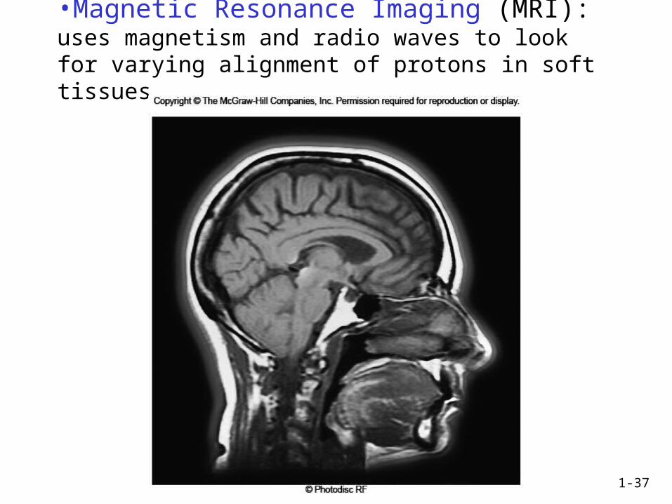
•Magnetic Resonance Imaging (MRI): uses magnetism and radio waves to look for varying alignment of protons in soft tissues.
1-38
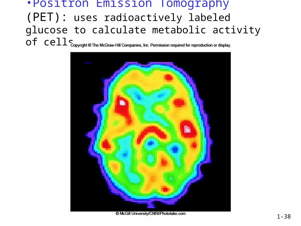
•Positron Emission Tomography (PET): uses radioactively labeled glucose to calculate metabolic activity of cells.