Z5 Vet Diagnostic Ultrasound System...Z5 Vet Diagnostic Ultrasound System ... 1
Upload
abigayle-lynette-pooleCategory
view
223download
0
>> 0 >> 1 >> 2 >> 3 >> 4 >>
Diagnostic Ultrasound
04/19/231 Dr.sai krishna
>> 0 >> 1 >> 2 >> 3 >> 4 >>
Strengths of Ultrasonography
04/19/23
Determining origin of an abdominal mass
Evaluation of organ parenchyma- Liver, spleen, kidneys, adrenals, pancreas,
intestines, prostate, bladder, heart
Fetal viability
Real time scanning – see movement/motion
Performing fine needle aspiration/ biopsy
2 Dr.sai krishna
>> 0 >> 1 >> 2 >> 3 >> 4 >>
Weaknesses of Ultrasonography:
04/19/23
Ultrasound can’t penetrate gas or boneCan’t assess intestinal gas patternsCan’t evaluate some extra abdominal
structures (i.e. spine)Equipment can be expensiveDiagnostic success is user dependentMust know anatomy very well
3 Dr.sai krishna

>> 0 >> 1 >> 2 >> 3 >> 4 >>
Technical considerations:Lesions can be missed in - incorrect transducer - improper TGC settings - poor screen contrast - brightly lit room Use appropriate frequency Highest frequency transducer
appropriate first later lower frequency
04/19/234 Dr.sai krishna

>> 0 >> 1 >> 2 >> 3 >> 4 >>
Appropriate Pressure use the lowest power setting possible to have high
quality image.
If Obese , emaciated , Gas filled leads to images of poor quality.
Cant pass thru Air so clip the hair coat.
Tranquilization needed in biopsies
04/19/235 Dr.sai krishna

>> 0 >> 1 >> 2 >> 3 >> 4 >>
Patient positioningAbdominal scanning: -Dorsal recumbency -V shaped tough -Head towards the machine -Dominant hand scanning & non
dominant hand on control panel
04/19/236 Dr.sai krishna

>> 0 >> 1 >> 2 >> 3 >> 4 >>
Two planes: Longitudinal Transverse
oblique can be used in producing diagnostic images.
Use as many views with as many angles possible.
04/19/237 Dr.sai krishna
>> 0 >> 1 >> 2 >> 3 >> 4 >>
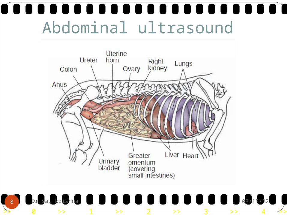
Abdominal ultrasound
04/19/238 Dr.sai krishna

>> 0 >> 1 >> 2 >> 3 >> 4 >>
04/19/239 Dr.sai krishna

>> 0 >> 1 >> 2 >> 3 >> 4 >>
04/19/2310 Dr.sai krishna

>> 0 >> 1 >> 2 >> 3 >> 4 >>
Diaphragm
04/19/2311 Dr.sai krishna
>> 0 >> 1 >> 2 >> 3 >> 4 >>
04/19/2312 Dr.sai krishna
>> 0 >> 1 >> 2 >> 3 >> 4 >>
04/19/2313 Dr.sai krishna

>> 0 >> 1 >> 2 >> 3 >> 4 >>
04/19/2314 Dr.sai krishna

>> 0 >> 1 >> 2 >> 3 >> 4 >>
04/19/2315 Dr.sai krishna

>> 0 >> 1 >> 2 >> 3 >> 4 >>
Basic rules for UltraSonography
Always scan in dim lighted room
Always remove hair
Always position the animal in consistent orientation.
Always position the image on screen with proper orientation
04/19/2316 Dr.sai krishna

>> 0 >> 1 >> 2 >> 3 >> 4 >>
Slowly perform Scan
All structures should be identified
Always scan each organ in two planes
Consistently use same technique
Perform the exam with consistency
04/19/2317 Dr.sai krishna

>> 0 >> 1 >> 2 >> 3 >> 4 >>
General Abdomen: For small dogs and cats, a 7.5/10 MHz
sector, linear or curved array transducer is used.
For larger dogs, 5 MHz transducers are usually adequate
In some giant breed dogs 3.5 MHz may be required.
04/19/2318 Dr.sai krishna
>> 0 >> 1 >> 2 >> 3 >> 4 >>
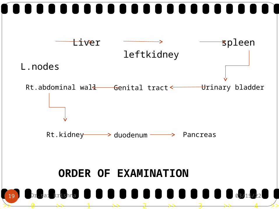
Liver spleen leftkidney L.nodes
Urinary bladderGenital tractRt.abdominal wall
Rt.kidney duodenum Pancreas
ORDER OF EXAMINATION
04/19/2319 Dr.sai krishna

>> 0 >> 1 >> 2 >> 3 >> 4 >>
Free abdominal fluid: Free abdominal fluid is seen as anechoic
angular or triangular areas between abdominal structures.
If a large amount of free fluid is present, the abdominal structures will be separated by large anechoic areas, and the small intestine attached to a highly echogenic mesentery is seen floating freely in the fluid.
Protein-losing diseases, such hepatopathy, nephropathy orenteritis, portal hypertension, or increased pressure in the caudal vena cava secondary to right-sided heart failure
04/19/2320 Dr.sai krishna
>> 0 >> 1 >> 2 >> 3 >> 4 >>
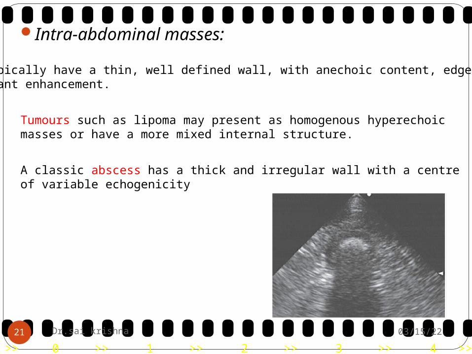
Intra-abdominal masses: Cysts typically have a thin, well defined wall, with anechoic content, edge shadowingand distant enhancement.
Tumours such as lipoma may present as homogenous hyperechoic masses or have a more mixed internal structure.
A classic abscess has a thick and irregular wall with a centre of variable echogenicity
04/19/2321 Dr.sai krishna

>> 0 >> 1 >> 2 >> 3 >> 4 >>
Diaphragmatic and abdominal hernias:
Loss of the curvilinear appearance of the diaphragm, the presence of liver or other abdominal structures close to the heart, and pleural or abdominal effusion are typical signs.
An intercostal approach using a parasternal window.
04/19/2322 Dr.sai krishna

>> 0 >> 1 >> 2 >> 3 >> 4 >>
Abdominal ultrasound Liver
ParenchymaGallbladderPortal veinsHepatic veinsDiaphragm
Indications:
Abnormality in x-ray Metastasis Ascites Elevated liver enzymes Biopsy
04/19/2323 Dr.sai krishna

>> 0 >> 1 >> 2 >> 3 >> 4 >>
Due to the location of the liver within the ribcage, a curvilinear transducer is very helpful to allow full penetration of the ultrasound beam.
Transducer frequency of 7-10 MHz should be sufficient.
Both subxiphoid and right intracostal windows should be used for complete evaluation of the liver and gallbladder.
By using the ribcage to mark the extent of the normal caudal hepatic margin an assessment of marked microhepatia and/or hepatomegaly can be made.
04/19/2324 Dr.sai krishna

>> 0 >> 1 >> 2 >> 3 >> 4 >>
Start with cross sectional view.
Place transducer near the xyphoid process
Scan to the patients right and back to the left Angle the probe as needed to visualise all of the
liver.
Apply fair amount of pressure to expel the gas. then move to longitudinal section
Compare the echogenicity of the liver with the surrounding
fat (hepatic lipidosis).04/19/2325 Dr.sai krishna

>> 0 >> 1 >> 2 >> 3 >> 4 >>
What to examine? Texture Echogenicity Liver , central vessels , gall bladder , cystic
and common bile duct.
04/19/2326 Dr.sai krishna
>> 0 >> 1 >> 2 >> 3 >> 4 >>
Normal Liver is:
Homogenous with hepatic & Portal veins &
caudal vena cava Portal veins with echogenic walls(due
to adjacent fat) . Hepatic veins with out walls.
04/19/2327 Dr.sai krishna
>> 0 >> 1 >> 2 >> 3 >> 4 >>
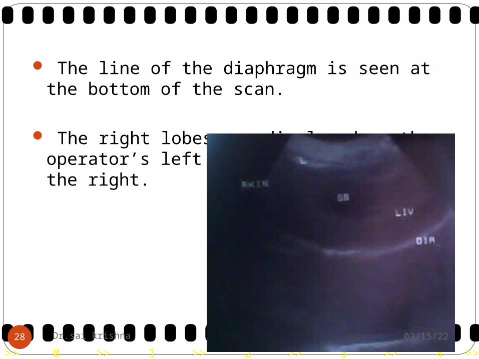
The line of the diaphragm is seen at the bottom of the scan.
The right lobes are displayed on the operator’s left and the left lobes on the right.
04/19/2328 Dr.sai krishna

>> 0 >> 1 >> 2 >> 3 >> 4 >>
04/19/2329 Dr.sai krishna

>> 0 >> 1 >> 2 >> 3 >> 4 >>
04/19/2330 Dr.sai krishna
>> 0 >> 1 >> 2 >> 3 >> 4 >>
Echogenecity:Hepatic echogenicity must be assessed only in
comparison with neighbouring organs at the same depth and preferably within the same image.
Compare with left kid (less) & spleen(more) .
Hyperechoic , Hypoechoic , Mixed
echoic
Diffused , Focal
04/19/2331 Dr.sai krishna
>> 0 >> 1 >> 2 >> 3 >> 4 >>
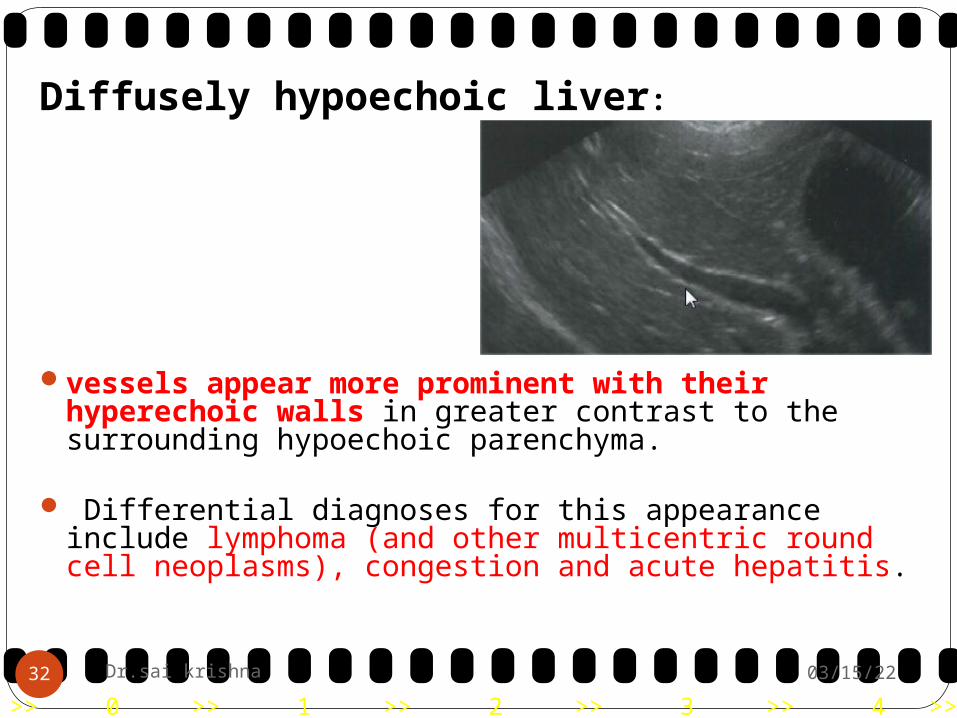
Diffusely hypoechoic liver:
vessels appear more prominent with their hyperechoic walls in greater contrast to the surrounding hypoechoic parenchyma.
Differential diagnoses for this appearance include lymphoma (and other multicentric round cell neoplasms), congestion and acute hepatitis.
04/19/2332 Dr.sai krishna

>> 0 >> 1 >> 2 >> 3 >> 4 >>
Diffusely hyperechoic liver:
Indistinctness of the vessel walls (border effacement).
Differential diagnoses for generalized hyperechogenicity
include vacuolar diseases, fibrosis(cirrhosis) & lymphoma.
Vacuolar diseases include hyperadrenocorticism, hypothyroidism, non-specific hepatopathies and fatty infiltration.
04/19/2333 Dr.sai krishna
>> 0 >> 1 >> 2 >> 3 >> 4 >>
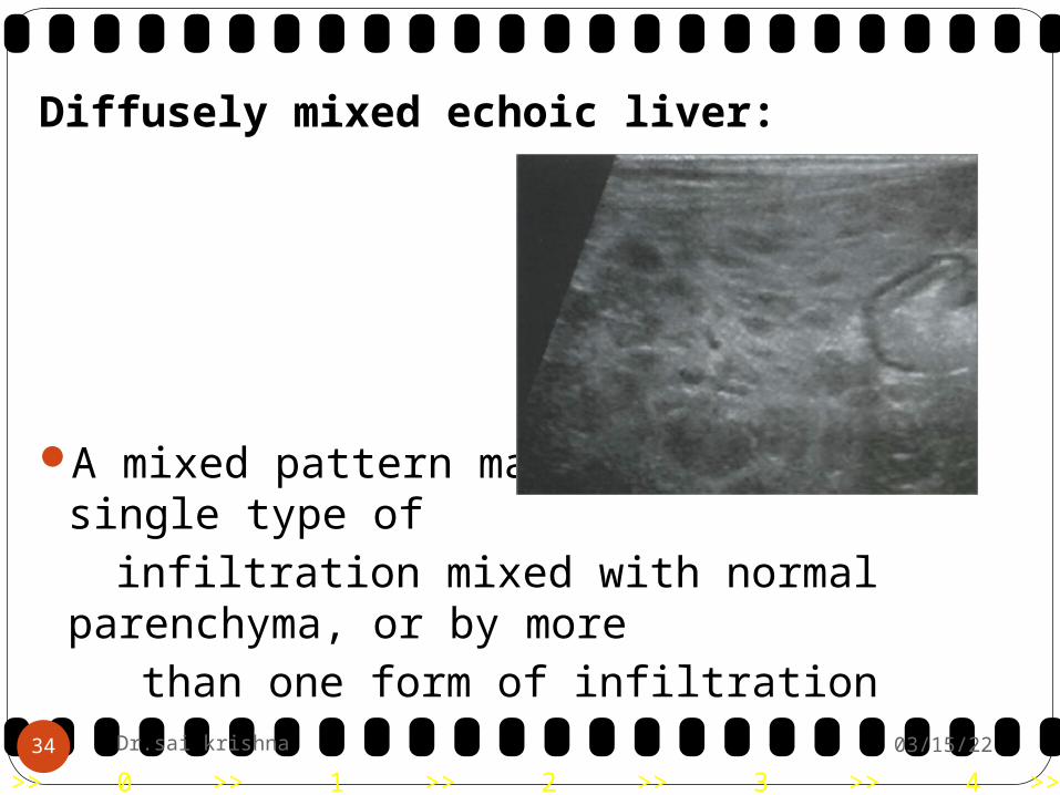
Diffusely mixed echoic liver:
A mixed pattern may be caused by a single type of
infiltration mixed with normal parenchyma, or by more
than one form of infiltration 04/19/2334 Dr.sai krishna

>> 0 >> 1 >> 2 >> 3 >> 4 >>
Differential diagnoses include infiltrative neoplasia , such as lymphoma or mast cell tumour, and histiocytic diseases.
Patchy mixed patterns are seen with advanced fibrosis (cirrhosis), hepatocutaneous syndrome and feline amyloidosis .
The classic appearance of cirrhosis is a hyperechoic parenchyma with hypoechoic regenerative nodules, free peritoneal fluid, small overall size and irregular liver margins.
04/19/2335 Dr.sai krishna
>> 0 >> 1 >> 2 >> 3 >> 4 >>
Feline amyloidosis is characterized by a coarse, patchy mixed echogenicity with hyperechoic specks and hypoechoic foci.
Hepatocutaneous syndrome is associated with mucocutaneous ulcerative lesions and liver failure. The ultrasonographic appearance is a Swiss cheese pattern with hypoechoic regenerative nodules and surrounding regions of hepatocyte collapse
04/19/2336 Dr.sai krishna

>> 0 >> 1 >> 2 >> 3 >> 4 >>
Focal changes in echogenicity:
Nodules are very common in older dogsBoth benign and malignant nodules can be
hypoechoic, mixed or hyperechoic.Benign nodular hyperplasia, metastatic nodules,
haematomas and primary liver neoplasia. 04/19/2337 Dr.sai krishna

>> 0 >> 1 >> 2 >> 3 >> 4 >>
An outer hypoechoic area surrounding a hyperechoic centre is more commonly seen with, but is not unique to, metastatic neoplasia.
Feline biliary cystadenomas are hyperechoic with small to large cavitated (anechoic) portions.
Haemangiosarcoma, fibrosarcoma, leiomyosarcoma and extra-skeletal osteosarcoma.
04/19/2338 Dr.sai krishna
>> 0 >> 1 >> 2 >> 3 >> 4 >>
Changes in liver contour and architecture: Interruption of the regular hepatic
architecture, deviation of adjacent vascular structures and bulging of the hepatic margins are indicative of a mass lesion.
Differential diagnoses include neoplasia, benign nodular hyperplasia, haematoma, abscess, granuloma, cyst and torsion.
04/19/2339 Dr.sai krishna

>> 0 >> 1 >> 2 >> 3 >> 4 >>
Microhepatica:Where there appears to be reduced liver volume .Where there is little space between the liver and
intestine, that the liver size is small.Cirrhosis & portosystemic shunt.The liver is small and is usually poorly vascularised.
The abnormal shunting vessel may be seen relatively easily in portosystemic shunt with smooth, sharp edges.
cirrhotic nodules present, which are isoechoic and are not easily
identified but liver margins are often rounded and irregular
04/19/2340 Dr.sai krishna

>> 0 >> 1 >> 2 >> 3 >> 4 >>
04/19/2341 Dr.sai krishna

>> 0 >> 1 >> 2 >> 3 >> 4 >>
Diffuse necrosis and cirrhosisin a dog.
04/19/2342 Dr.sai krishna

>> 0 >> 1 >> 2 >> 3 >> 4 >>
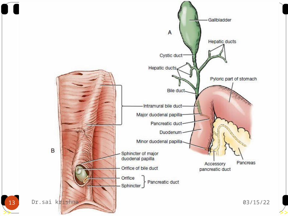
Biliary disease: Gall bladder: It is usually anechoic and ovoid in shape with a
tapered neck.
The intraparenchymal ducts are not usually seen unless dilated and they can then be differentiated from the hepatic vessels by their branching pattern and tortuous appearance.
Chronic cholangitis: Cases of long standing cholangitis or
cholangiohepatitis the wall of the gall bladder may be quite hyperechoic or even thickened.
04/19/2343 Dr.sai krishna

>> 0 >> 1 >> 2 >> 3 >> 4 >>
Gall bladder wall is thickened andhyperechoic.
Chronic cholangitis
04/19/2344 Dr.sai krishna
>> 0 >> 1 >> 2 >> 3 >> 4 >>
Obstructive disease: Cholelithiasis As hyperechoic structures casting a strong
acoustic shadow. Bile duct this may be dilated and tortuous
Mural causes : inflammation or occasionally neoplasia
The bile duct carcinomas may be seen as an echogenic mass within the dilated and obstructed ducts.
04/19/2345 Dr.sai krishna

>> 0 >> 1 >> 2 >> 3 >> 4 >>
Cholelithiasis
Bile duct within which hyperechoiccalculi can be seen
04/19/2346 Dr.sai krishna

>> 0 >> 1 >> 2 >> 3 >> 4 >>
Spleen:
The head of the spleen is often located within the ribcage in the cranial left dorsal abdomen in dogs so left intercostal approach may be necessary to fully evaluate the spleen.
The entire spleen in the cat is caudal to the ribcage and can be fully evaluated from a ventral approach.
The spleen should be uniform in echogenicity. The splenic veins are visualized at the splenic
hilus and for a short distance within the parenchyma. The splenic veins can be evaluated with colour Doppler ultrasonography for assessment of blood flow
04/19/2347 Dr.sai krishna

>> 0 >> 1 >> 2 >> 3 >> 4 >>
Dog
Cat
04/19/2348 Dr.sai krishna

>> 0 >> 1 >> 2 >> 3 >> 4 >>
04/19/2349 Dr.sai krishna
>> 0 >> 1 >> 2 >> 3 >> 4 >>
Doppler ultrasonography interrogation of splenic vein blood flow is important for evaluation of splenic torsion, thrombosis and certain mass lesions.
As the spleen is superficial avoid excessive pressure on the transducer as this may result in it not being readily seen. In most cases (including large dogs) a 7.5 MHz transducer is more than adequate to image this well.
04/19/2350 Dr.sai krishna

>> 0 >> 1 >> 2 >> 3 >> 4 >>
Normal appearance: The spleen is a strap like organ which
originates on the left side of the body close to the left kidney, gastric fundus and colon.
The spleen has a fine-grained texture and should be wholly homogeneous.
04/19/2351 Dr.sai krishna

>> 0 >> 1 >> 2 >> 3 >> 4 >>
Spleenic vessels
04/19/2352 Dr.sai krishna

>> 0 >> 1 >> 2 >> 3 >> 4 >>
The splenic capsule is seen as a very fine hyperechoic rim and the splenic vessels are seen entering through this.
In some cases hyperechoic areas are seen around the point of entry of the vessels and these are thought to be fat deposits and nothing sinister.
04/19/2353 Dr.sai krishna

>> 0 >> 1 >> 2 >> 3 >> 4 >>
Abnormal appearance: Parenchymal Diffuse1. Conditions which result in diffuse
splenic change include lymphoma and mast cell neoplasia (especially in cats) where there is diffuse infiltration of the organ by neoplastic cells.
2. uniform increase or decrease in echogenicity
3. Hard to detect and it is especially important to compare this to the liver and kidneys
04/19/2354 Dr.sai krishna

>> 0 >> 1 >> 2 >> 3 >> 4 >>
Mast cellinfiltrate in a cat
04/19/2355 Dr.sai krishna
>> 0 >> 1 >> 2 >> 3 >> 4 >>
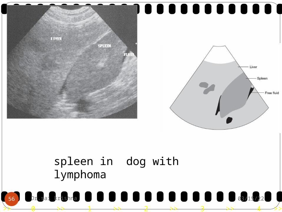
spleen in dog withlymphoma
04/19/2356 Dr.sai krishna

>> 0 >> 1 >> 2 >> 3 >> 4 >>
Focal: focal lesions of the spleen may be hypoechoic,
hyperechoic, anechoic or mixed and may vary in their size.
Lesions of the spleen have been associated with both benign and malignant conditions such as lymphoid hyperplasia, adenocarcinoma, lymphoma and haemangiosarcoma .
04/19/2357 Dr.sai krishna
>> 0 >> 1 >> 2 >> 3 >> 4 >>
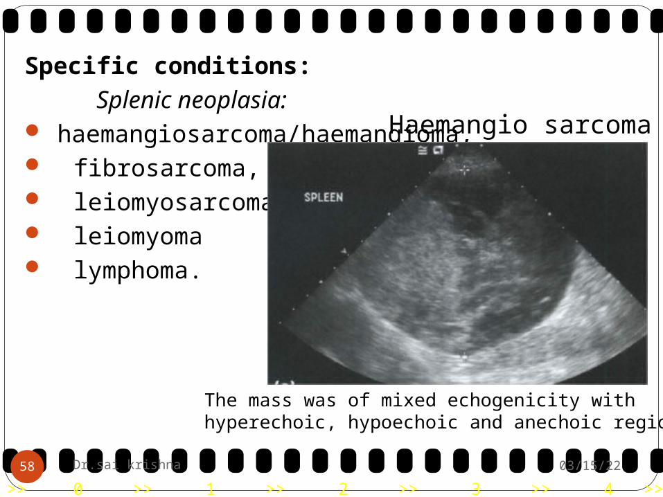
Specific conditions: Splenic neoplasia: haemangiosarcoma/haemangioma, fibrosarcoma, leiomyosarcoma, leiomyoma lymphoma.
The mass was of mixed echogenicity withhyperechoic, hypoechoic and anechoic regions
Haemangio sarcoma
04/19/2358 Dr.sai krishna

>> 0 >> 1 >> 2 >> 3 >> 4 >>
Splenic abscesses: Splenic abscesses are occasionally seen. There may be some anechoic areas
present but the fluid is usually quite echogenic due to its cellular content.
Splenic haematoma: Arise secondary to trauma or bleeding
disorders and may present as a single or multiple masses with mainly hypoechoic and anechoic regions within
04/19/2359 Dr.sai krishna

>> 0 >> 1 >> 2 >> 3 >> 4 >>
Splenic haematoma in a dog.
04/19/2360 Dr.sai krishna
>> 0 >> 1 >> 2 >> 3 >> 4 >>
Splenic torsion: Torsion of the spleen is an uncommon
condition, which may be found in association with gastric dilation-volvulus or may be found as an unrelated condition.
The spleenic parenchyma may be mottled with hypoechoic areas, separated by irregular anechoic areas, suggestive of areas of necrosis, or it may be diffusely hypoechoic.
04/19/2361 Dr.sai krishna

>> 0 >> 1 >> 2 >> 3 >> 4 >>
Typical hypoechoic, lacy parenchymal appearance of the spleen
Splenic torsion
04/19/2362 Dr.sai krishna

>> 0 >> 1 >> 2 >> 3 >> 4 >>
Extramedullary haemopoiesis: Extramedullary haemopoiesis is often seen
ultrasonographically as hyperechoic or hypoechoic nodules.
04/19/2363 Dr.sai krishna
>> 0 >> 1 >> 2 >> 3 >> 4 >>
Gastro-intestinal Tract including Pancreas:
Survey abdominal radiographs should be taken prior to a sonographic examination to assess for possible diagnosis, to rule out obvious obstruction or radio-opaque foreign body and to avoid artifact arising from residual ultrasound gel on the skin.
04/19/2364 Dr.sai krishna

>> 0 >> 1 >> 2 >> 3 >> 4 >>
General stomach: The fundus and body of the stomach lie on the
left side of the abdomen immediately caudal to the liver.
The pylorus lies to the right of midline in the dog and near midline in the cat.
The duodenum is the only part of the small intestine which can be identified, due to its characteristic location and demonstrable connection to the pylorus.
The duodenum runs from the pylorus laterally for a short distance in the right cranial abdomen, then caudally before forming a U-shaped loop at its caudal flexure. 04/19/2365 Dr.sai krishna

>> 0 >> 1 >> 2 >> 3 >> 4 >>
The loops of the jejunum and ileum between the duodenal flexure and the iliocaecocolic junction cannot be differentiated from one another sonographically , due to lack of reference points and the mobility of the small intestine within the abdomen.
The caecum is comma shaped in the cat and spiral in the dog.
04/19/2366 Dr.sai krishna
>> 0 >> 1 >> 2 >> 3 >> 4 >>
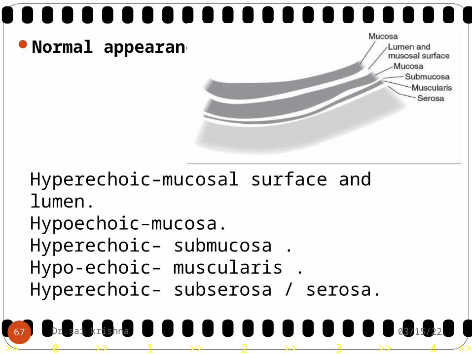
Normal appearance:
Hyperechoic–mucosal surface and lumen.Hypoechoic–mucosa.Hyperechoic– submucosa .Hypo-echoic– muscularis .Hyperechoic– subserosa / serosa.
04/19/2367 Dr.sai krishna

>> 0 >> 1 >> 2 >> 3 >> 4 >>
The hypoechoic muscularis and mucosal layers predominate , the overall appearance of the gastrointestinal wall is of a hypoechoic structure.
The duodenal papilla may be visible as a small hyperechoic indentation in the dorsal wall.
The mucosal layer of the duodenal wall is more prominent than that of jejunum or ileum.
The duodenal wall is often slightly thicker than that in the rest of the small intestine.
04/19/2368 Dr.sai krishna

>> 0 >> 1 >> 2 >> 3 >> 4 >>
Abnormal appearance: The sonographic changes associated
with inflammatory conditions of the gastro-intestinal tract include an increase in wall thickness or altered wall echogenicity, usually with preservation of the layering pattern and symmetry of the gastro-intestinal wall.
1 wall thickness2 wall layering3 presence or absence of lymphadenopathy4 involvement of other organs5 involvement
04/19/2369 Dr.sai krishna

>> 0 >> 1 >> 2 >> 3 >> 4 >>
StomachThe stomach can be visualized by placing the
ultrasound probe caudal to the ribcage in a sagittal plane and sweeping it from right to left in a craniodorsal direction.
Offering water to drink before the examination can be helpful as visibility of the outline of the gastric wall is best in a moderately fluid distended stomach.
Food should be withheld from the patient for at least 12 hours.
A high-frequency transducer of 5-7.5 MHz, or even 10 MHz in small animals such as cats, is needed in order to assess the wall structure and layering appropriately.
04/19/2370 Dr.sai krishna

>> 0 >> 1 >> 2 >> 3 >> 4 >>
04/19/2371 Dr.sai krishna
>> 0 >> 1 >> 2 >> 3 >> 4 >>
The stomach can be recognized by its position just caudal to the liver, its size, contents and rugal folds .
The gastric fundus is seen in the left craniodorsal abdomen and has the most prominent rugal folds when empty.
By following the greater curvature of the stomach ventrally and to the right, the body and antrum are examined.
Alternatively, to visualize the gastric antrum and pyloric canal, the duodenum can be imaged ventro lateral to the right kidney and followed cranially to the pyloric antrum.
A helpful landmark to identify the pylorus is the portal vein at the liver hilus; the pylorus and cranial duodenal flexure are just caudal to the liver hilus and ventral to the portal vein.
04/19/2372 Dr.sai krishna

>> 0 >> 1 >> 2 >> 3 >> 4 >>
04/19/2373 Dr.sai krishna
>> 0 >> 1 >> 2 >> 3 >> 4 >>
Gastritis:Ultrasonography is more sensitive than
radiography for detecting changes associated with inflammatory diseases of the stomach.
Uraemic gastropathy is characterized by ulceration, oedema, mineralization, submucosal arteriopathy, gastric gland atrophy and necrosis .Ultrasonographic changes include:
Poor definition of gastric wall layers Mineralization of the gastric mucosa, seen
as a hyperechoic line adjacent to the gastric lumen, usually not thick enough to cause acoustic shadowing
Thickened gastric wall.04/19/2374 Dr.sai krishna

>> 0 >> 1 >> 2 >> 3 >> 4 >>
Ultrasonographic signs of gastritis are very nonspecific and can include:
• Diffuse thickening of the stomach wall • Increased or decreased echogenicity of the
wall • Fluid accumulation within the gastric lumen • Enlarged rugal folds • Decreased definition of the wall layers In severe cases, loss of wall layering can be
present.
04/19/2375 Dr.sai krishna

>> 0 >> 1 >> 2 >> 3 >> 4 >>
stomach wall is irregularly thickened.There is almost complete loss of layering
04/19/2376 Dr.sai krishna

>> 0 >> 1 >> 2 >> 3 >> 4 >>
Typically a deep ulcer is seen as ahyperechoic region in the wall. This reflects gas trapped in theulcer crater.
04/19/2377 Dr.sai krishna
>> 0 >> 1 >> 2 >> 3 >> 4 >>
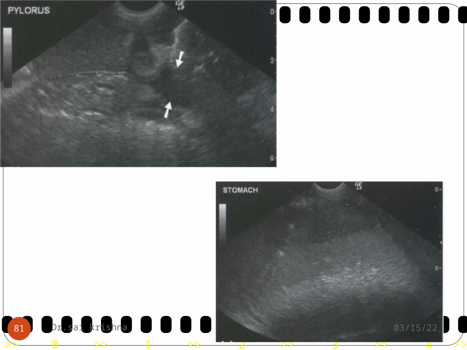
Gastric dilatation and gastric dilatation-volvulus:Ultrasonography is not indicated in patients with
suspected gastric torsion.The large size and amount of gas within the stomach
make it impossible to assess the cranial abdomen.MUCOSAL MINERALISATION:Mucosal mineralisation appears sonographically as a
highly echogenic line at the level of the gastric mucosa which produces minimal acoustic shadowing.
The differential diagnosis of gastric wall mineralisation includes gastric neoplasia and disease processes causing dystrophic calcification such as primary hyperparathyroidism, hypercalcaemia of malignancy, or hyperadrenocorticism.
04/19/2378 Dr.sai krishna

>> 0 >> 1 >> 2 >> 3 >> 4 >>
Chronic hypertrophic pyloric gastropathy:
Gastric distension with fluid, gas or a mixture of these, vigorous antegrade and retrograde peristalsis failing to propel ingesta into the duodenum and an even increase in thickness of the muscular layer in the pyloric canal.
04/19/2379 Dr.sai krishna

>> 0 >> 1 >> 2 >> 3 >> 4 >>
Pyloric outflow obstruction: Pyloric outflow obstruction can be
difficult to diagnose ultrasonographically . Secondary signs of chronic outflow obstruction
include: - Distended stomach with an enlarged
gastric antrum. - A large amount of ingesta in the lumen,
which can have a layered appearance with the solid particles in the dependent part and the liquids and gas in the non-dependent portion .
- Antral contractions with minimal propelling of ingesta into the duodenum.
- Decreased motility. 04/19/2380 Dr.sai krishna
>> 0 >> 1 >> 2 >> 3 >> 4 >>
04/19/2381 Dr.sai krishna

>> 0 >> 1 >> 2 >> 3 >> 4 >>
Foreign bodies:
In the gastric fundus there is a round structure with a hyperechoic surface and a strong distal acoustic shadow consistent with a gastric foreign body.
04/19/2382 Dr.sai krishna

>> 0 >> 1 >> 2 >> 3 >> 4 >>
Gastric neoplasms: Malignant gastric neoplasms in the dog and
cat are malignant lymphoma, adenocarcinoma and leiomyosarcoma.
Benign neoplasms are occasionally seen, such as leiomyomas or adenomas.
The lesser curvature and gastric antrum are the most common sites for gastric neoplasia.
04/19/2383 Dr.sai krishna

>> 0 >> 1 >> 2 >> 3 >> 4 >>
Tumour type Ultrasonography characteristics
Leiomyoma Focal well defined lesionHypo- or hyperechoic
Leiomyosarcoma Large, complex mass Often ulcerated and can lead to wall perforation.Most commonly found in the pyloric antrum
Carcinoma Heteroechoic mass 'Pseudolayering' -layers of hyper- and hypoechoic material not consistent with normal wall layers caused by tumour cell invasion Commonly associated with regional lymphadenopathy
Lymphosarcoma Most common gastric tumour that leads to diffuse wall thickening, especially in cats The most common appearance of gastric lymphosarcoma is diffuse wall thickening.However, focal, mostly hypoechoic, mass lesions can occurDiffusely hypoechoic wall Circumferential transmural thickening Complete loss of wall layering Decreased motilityAlmost always associated
Histiocytic Has been described to cause diffuse hyper- or sarcoma hypoechoic wall thickening with loss of layering
04/19/2384 Dr.sai krishna
>> 0 >> 1 >> 2 >> 3 >> 4 >>
Small intestine: The descending duodenum can be identified ventral to
the right kidney.The intestine should be assessed for the overall wall
thickness, presence of layering, the echogenicity of each layer and the relative width of the each layer.
The layers should be visible in both longitudinal and transverse section.
Thickness of intestinal wall: The duodenum is usually thicker Motility: • Proximal duodenum: 4-5 contractions/minute • Rest of the small intestine: 1-3 contractions/minute.
04/19/2385 Dr.sai krishna

>> 0 >> 1 >> 2 >> 3 >> 4 >>
Luminal interface – hyperechoic
Mucosa (widest layer) –hypoechoic
Submucosa – hyperechoic
Muscularis – hypoechoic
Serosal interface - hyperechoic.
04/19/2386 Dr.sai krishna

>> 0 >> 1 >> 2 >> 3 >> 4 >>
04/19/2387 Dr.sai krishna
>> 0 >> 1 >> 2 >> 3 >> 4 >>
Luminal contents:Mucous pattern: empty bowel, mucus in lumen
that is seen as an echogenic line with no shadowing
Gas: intraluminal, highly reflective interfaces with shadowing/reverberation. Will usually move on with peristalsis or gentle pressure from the ultrasound probe
Fluid: anechoic luminal pattern. Allows optimal assessment of the bowel wall
Alimentary: food particles present within the lumen; echogenic, usually no acoustic shadowing, although some food (e.g. bone
fragments) will shadow04/19/2388 Dr.sai krishna

>> 0 >> 1 >> 2 >> 3 >> 4 >>
Enteritis
04/19/2389 Dr.sai krishna

>> 0 >> 1 >> 2 >> 3 >> 4 >>
Neoplasia: The characteristic sonographic appearance of
intestinal neoplasia is of a focal, asymmetrical thickening of the intestinal wall, with loss of layering and hypomotility.
04/19/2390 Dr.sai krishna
>> 0 >> 1 >> 2 >> 3 >> 4 >>
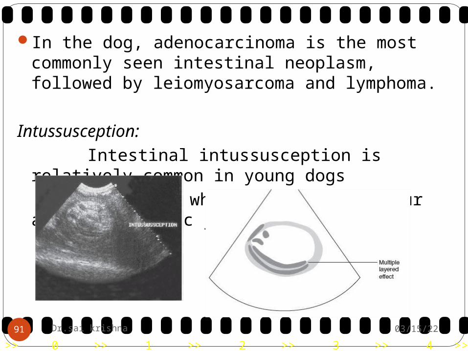
In the dog, adenocarcinoma is the most commonly seen intestinal neoplasm, followed by leiomyosarcoma and lymphoma.
Intussusception: Intestinal intussusception is relatively
common in young dogs and cats, where it tends to occur at the
ileocolic junction.
04/19/2391 Dr.sai krishna
>> 0 >> 1 >> 2 >> 3 >> 4 >>
Ultrasonography can determine the length of the intussusception, the patency of the bowel lumen and the presence of peritonitis or lymph node enlargement.
The intussuscipiens is often swollen and edematous and appears as a hypoechoic rim.
The typical appearance in cross section is that of concentric hypoechoic and hyperechoic rings, which are caused by invaginated layers of the hyperechoic intussusceptum and hypoechoic intussuscipiens.
This is often termed the "ring" or "bull's eye" sign.
04/19/2392 Dr.sai krishna

>> 0 >> 1 >> 2 >> 3 >> 4 >>
Typical "bull's eye" or "target sign” associated with intussusception
04/19/2393 Dr.sai krishna

>> 0 >> 1 >> 2 >> 3 >> 4 >>
Ileus: Accumulation of anechoic fluid within the
intestine is always indicative of an abnormality.
Mechanical ileus, which may be due to obstruction by foreign body or tumour.
Paralytic (functional) ileus secondary to enteritis are possible causes of intestinal dilation.
04/19/2394 Dr.sai krishna

>> 0 >> 1 >> 2 >> 3 >> 4 >>
Foreign body: The presence of a foreign body of soft
tissue opacity may easily be overlooked on a radiograph.
Sonography can be useful to detect a gastrointestinal foreign body and assess for complications such as obstruction.
Metallic foreign bodies are highly echogenic
and cause characteristic reverberation and comet tail artefacts
04/19/2395 Dr.sai krishna

>> 0 >> 1 >> 2 >> 3 >> 4 >>
Rubber ball
04/19/2396 Dr.sai krishna

>> 0 >> 1 >> 2 >> 3 >> 4 >>
INTESTINAL STRICTURE: The bowel may be constricted as a result of
intrinsic causes or as a result of pressure from without.
If outlined by fluid, the site of stricture may be identified ultrasonographically.
04/19/2397 Dr.sai krishna

>> 0 >> 1 >> 2 >> 3 >> 4 >>
THE LARGE INTESTINE: Located adjacent to the bladder. Peristalsis is not a feature. Neoplastic masses may occasionally be
identified. Thickening of the wall is difficult to appreciate
because of the intraluminal gas.
04/19/2398 Dr.sai krishna

>> 0 >> 1 >> 2 >> 3 >> 4 >>
FECAL RETENTION (Constipation):
04/19/2399 Dr.sai krishna
>> 0 >> 1 >> 2 >> 3 >> 4 >>
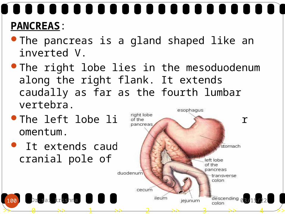
PANCREAS:The pancreas is a gland shaped like an inverted
V. The right lobe lies in the mesoduodenum along
the right flank. It extends caudally as far as the fourth lumbar vertebra.
The left lobe lies within the greater omentum. It extends caudally as far as the cranial pole of
the left kidney.
04/19/23100 Dr.sai krishna

>> 0 >> 1 >> 2 >> 3 >> 4 >>
Ultrasonographically: The pancreas is a difficult organ to find and
evaluate,particularly in the normal dog. Gastrointestinal gas may make it difficult to find the
pancreas. A high resolution 5 MHz or 7.5 MHz transducer
using low gain and output settings is required. Dorsal or right lateral recumbency are the usual
positions. Clipping of the cranial abdomen and right paracostal
region is necessary.In the normal dog, the pancreas is visualised as a
region of high echogenicity relative to the renal cortex and liver parenchyma but is not well defined as it has a similar echogenicity to the surrounding mesentery and lacks capsular margination.
04/19/23101 Dr.sai krishna

>> 0 >> 1 >> 2 >> 3 >> 4 >>
The duodenum and the pancreaticoduodenalvein are useful landmarks for the pancreas as seen here
04/19/23102 Dr.sai krishna

>> 0 >> 1 >> 2 >> 3 >> 4 >>
Pancreatitis:Acute : Decreased echogenicity in acute pancreatitis
reflect oedema, haemorrhage and necrosis.
Chronic: Mineralisation and scarring which cause acoustic shadowing.
Pancreatitis, pancreatic abscess and pancreatic carcinoma can produce very similar sonographic abnormalities.
Pancreatic pseudocysts are visualised as well defined anechoic lesions showing distal enhancement.
04/19/23103 Dr.sai krishna

>> 0 >> 1 >> 2 >> 3 >> 4 >>
Pancreatic nodule in a dog.
04/19/23104 Dr.sai krishna
>> 0 >> 1 >> 2 >> 3 >> 4 >>
Pancreatic abscess: Thick walls and hypoechoic
contents, similar to other intra-abdominal abscesses.
Pancreatic neoplasia: Adenocarcinoma is common. There may be a secondary biliary obstruction.
04/19/23105 Dr.sai krishna

>> 0 >> 1 >> 2 >> 3 >> 4 >>
URINARY SYSTEMKidneys:Can be imaged from a ventral or lateral approach, and usually, even in large dogs, a 7.5 MHz transducer provides sufficient penetration to image the entire kidney.
04/19/23106 Dr.sai krishna
>> 0 >> 1 >> 2 >> 3 >> 4 >>
linear transducers are ideal for renal imaging in cats.
Site: On the left side this is just ventral to the sublumbar
musculature caudal to the last rib .Between the 11th and 12th intercostal space on the
right.
Position:Lateral recumbencythe transducer is held parallel to the long axis of the
animal for longitudinal images .Perpendicular to the spine for transverse images.
04/19/23107 Dr.sai krishna
>> 0 >> 1 >> 2 >> 3 >> 4 >>
Normal appearance:Bean shaped.The left kidney is located immediately caudal to
the fundus of the stomach and caudomedial to the head of the spleen.
The cranial pole of the right kidney sits in the renal fossa of the caudate lobe of the liver.
The normal renal outline is smooth and well defined.
Compared to the medulla, the cortex has a higher echogenicity, but is clearly hypoechoic compared to the spleen.
04/19/23108 Dr.sai krishna

>> 0 >> 1 >> 2 >> 3 >> 4 >>
04/19/23109 Dr.sai krishna
>> 0 >> 1 >> 2 >> 3 >> 4 >>
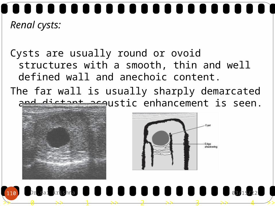
Renal cysts:
Cysts are usually round or ovoid structures with a smooth, thin and well defined wall and anechoic content.
The far wall is usually sharply demarcated and distant acoustic enhancement is seen.
04/19/23110 Dr.sai krishna

>> 0 >> 1 >> 2 >> 3 >> 4 >>
Renal neoplasia: The echogenicity or the echotexture is not
characteristic for a specific tumour type. Differential diagnoses of solid kidney masses include
lymphoma, cystadenocarcinoma, sarcomas and most metastatic tumours.
Hyperechoic masses are relatively rare but seem to
be associated with well vascularised tumours.(haemangiosarcoma, haemangioma, metastatic thyroid carcinoma and chondrosarcoma).
The final diagnosis is usually based on
history,laboratory work, and fine-needle aspiration or core biopsy
04/19/23111 Dr.sai krishna

>> 0 >> 1 >> 2 >> 3 >> 4 >>
04/19/23112 Dr.sai krishna

>> 0 >> 1 >> 2 >> 3 >> 4 >>
Acute infarcts usually present as mass-like lesions with decreased or mixed echogenicity.
Chronic infarcts as triangular or wedge-shaped lesions with the tip at the corticomedullary junction.
The renal contour in the area of an infarct may be flattened or indented.
Parenchymal mineralisation, fibrosis, gas and chronic renal infarcts may lead to hyperechoic areas without mass effect.
04/19/23113 Dr.sai krishna
>> 0 >> 1 >> 2 >> 3 >> 4 >>
Diffuse parenchymal abnormality: - Diffuse renal disease is difficult to assess
sonographically.
- Ultrasound examination of the kidney is helpful in the differentiation of acute and chronic renal disease where there is acute onset of clinical signs.
04/19/23114 Dr.sai krishna

>> 0 >> 1 >> 2 >> 3 >> 4 >>
Acute glomerulonephritis , interstitial nephritis, amyloidosis, diffuse neoplastic infiltrate (e.g. lymphoma), acute tubular necrosis or nephrosis (e.g. ethylene glycol toxicity) and metastatic neoplasia, such as squamous cell carcinoma and mast cell tumour, may all lead to increased echogenicity of the renal cortex.
The kidneys are often unilaterally or bilaterally enlarged.
Increased echogenicity of the renal cortex with loss of
corticomedullary differentiation has been described in polycystic renal disease.
04/19/23115 Dr.sai krishna
>> 0 >> 1 >> 2 >> 3 >> 4 >>
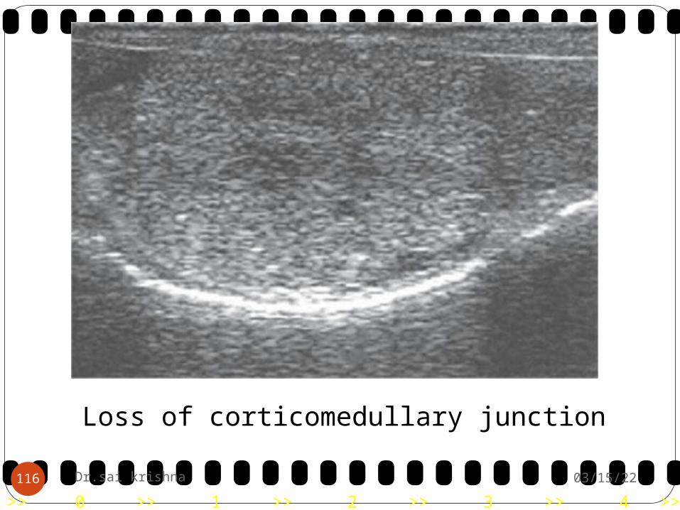
Loss of corticomedullary junction
04/19/23116 Dr.sai krishna
>> 0 >> 1 >> 2 >> 3 >> 4 >>
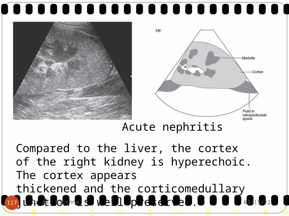
Acute nephritis
Compared to the liver, the cortex of the right kidney is hyperechoic. The cortex appearsthickened and the corticomedullary junction is well preserved.
04/19/23117 Dr.sai krishna

>> 0 >> 1 >> 2 >> 3 >> 4 >>
Decreased cortical echogenicity:Decreased echogenicity of the renal cortex has
been described as a result of necrosis and with lymphoma infiltrate which may also present with multiple, small hypoechoic nodules or masses
04/19/23118 Dr.sai krishna

>> 0 >> 1 >> 2 >> 3 >> 4 >>
Collecting system, pelvis and ureters:
Hydronephrosis:In normal animals, the renal pelvis does not
retain fluid and is not visible sonographically.Ultrasound is a sensitive technique for
investigating the potential causes.Differential diagnoses include congenital
anomaly, ureteric obstruction, pyelonephritis, and diuresis following administration of intravenous fluids or diuretic.
04/19/23119 Dr.sai krishna

>> 0 >> 1 >> 2 >> 3 >> 4 >>
The normally hyperechoic structures of the peripelvic fat gradually
disappear and the pelvic diverticula and proximal ureter become more dilated. This specific appearance is also helpful in differentiating a dilated renal pelvis from a renal cyst.
In hydronephrosis kidney is seen as a round or ovoid anechoic sac.
A thin rim of parenchyma and several hyperechoic bands extending from the hilus to the capsule are typical findings.
04/19/23120 Dr.sai krishna

>> 0 >> 1 >> 2 >> 3 >> 4 >>
Congenital anomalies such as ectopic ureters and ureteroceles are common causes of hydronephrosis.
Acute pyelonephritis may or may not cause dilation of the renal pelvis but in more chronic cases this may be mild to moderate and may be uni- or bilateral.
04/19/23121 Dr.sai krishna

>> 0 >> 1 >> 2 >> 3 >> 4 >>
Renal calculi: Renal calculi are usually easy to detect
sonographically. Both radiopaque and radiolucent renal calculi
are intensely hyperechoic, with clean acoustic shadowing.
Calculi produce maximal shadowing if the highest possible transducer frequency is chosen.
04/19/23122 Dr.sai krishna
>> 0 >> 1 >> 2 >> 3 >> 4 >>
Perinephric fluid accumulation:Subcapsular or extra capsular fluid accumulation
has been described in cats.Sonographically, the kidneys are surrounded by
encapsulated anechoic fluid.Trauma with ruptured kidney, renal pelvis or
ureter, infection, toxicity or tumours.
04/19/23123 Dr.sai krishna
>> 0 >> 1 >> 2 >> 3 >> 4 >>
Bladder: The superficial position of the urinary bladder
and the inherent contrast produced by the anechoic urine makes it ideal for ultrasound examination.
It is recommended that the bladder be
moderately full for the examination.
Animal in dorsal recumbency and the transducer positioned at the ventral or ventro lateral aspect of the caudal abdominal wall.
04/19/23124 Dr.sai krishna
>> 0 >> 1 >> 2 >> 3 >> 4 >>
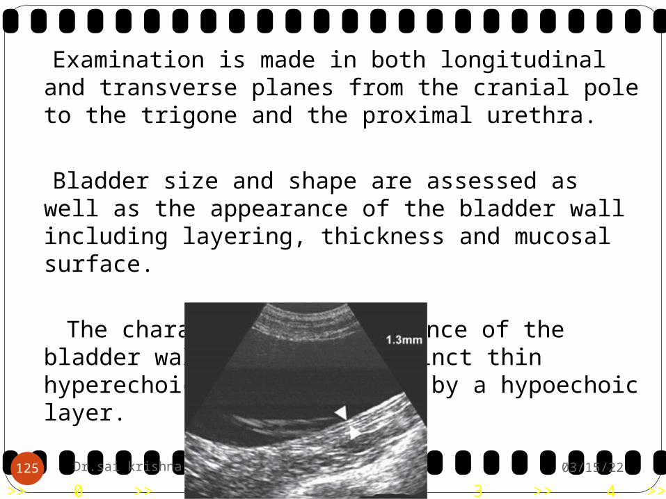
Examination is made in both longitudinal and transverse planes from the cranial pole to the trigone and the proximal urethra.
Bladder size and shape are assessed as well as
the appearance of the bladder wall including layering, thickness and mucosal surface.
The characteristic appearance of the bladder
wall is of two distinct thin hyperechoic lines separated by a hypoechoic layer.
04/19/23125 Dr.sai krishna

>> 0 >> 1 >> 2 >> 3 >> 4 >>
Bladder neoplasia:Thickening of the bladder wall is most commonly
associated with neoplastic or inflammatory infiltration.
Transitional cell carcinoma , but other epithelial and mesenchymal tumours such as leiomyoma, leiomyosarcoma, fibrosarcoma and lymphoma
04/19/23126 Dr.sai krishna

>> 0 >> 1 >> 2 >> 3 >> 4 >>
Transitional cell carcinomas are most often located in the area of the trigone, dorsal wall and proximal urethra.
inflammatory polyps, blood clots, cystitis and calculi may ‘mimic’ a bladder tumour.
04/19/23127 Dr.sai krishna

>> 0 >> 1 >> 2 >> 3 >> 4 >>
Cystitis: Acute cystitis usually causes no sonographic
abnormalities. Long-standing and severe cystitis often leads
to diffuse bladder wall thickening, with a hyperechoic wall and irregular mucosal surface, and is usually more pronounced cranio ventrally but may involve the entire bladder wall in severe cases.
04/19/23128 Dr.sai krishna

>> 0 >> 1 >> 2 >> 3 >> 4 >>
Other abnormalities:Mural haematomas and diffuse haemorrhage
associated with bleeding.Diffuse hyperechoic wall thickening associated
with echogenic luminal content is the predominant feature of haematuria.
Cystic calculi are easy to detect sonographically where they present as hyperechoic lesions with shadowing in the dependent portion of the bladder.
Floating echogenicities without shadowing may be seen where the urine contains cellular debris, blood or fibrin (hyperechoic strands).
04/19/23129 Dr.sai krishna

>> 0 >> 1 >> 2 >> 3 >> 4 >>
04/19/23130 Dr.sai krishna

>> 0 >> 1 >> 2 >> 3 >> 4 >>
Prostate: Performed by rectal or trans-abdominal routes. The examination should be performed with the
bladder moderately distended. Dorsal or lateral recumbency
Small area of hair clipped on one side of the prepuce just in front of the pubic brim. Once the bladder has been identified it is possible to move caudally towards the neck of the bladder and thence to the prostate.
04/19/23131 Dr.sai krishna

>> 0 >> 1 >> 2 >> 3 >> 4 >>
Normal appearance: Spherical to bilobed. The prostatic parenchyma is normally moderately
echogenic with a coarse but even texture throughout.
Linear echogenic streaks may be detected running longitudinally through the middle of the gland. This is the ‘hilar echo’ and is thought to represent peri-urethral fibrous tissue.
The prostatic urethra may be seen running through the parenchyma as a fine, linear anechoic structure.
04/19/23132 Dr.sai krishna
>> 0 >> 1 >> 2 >> 3 >> 4 >>
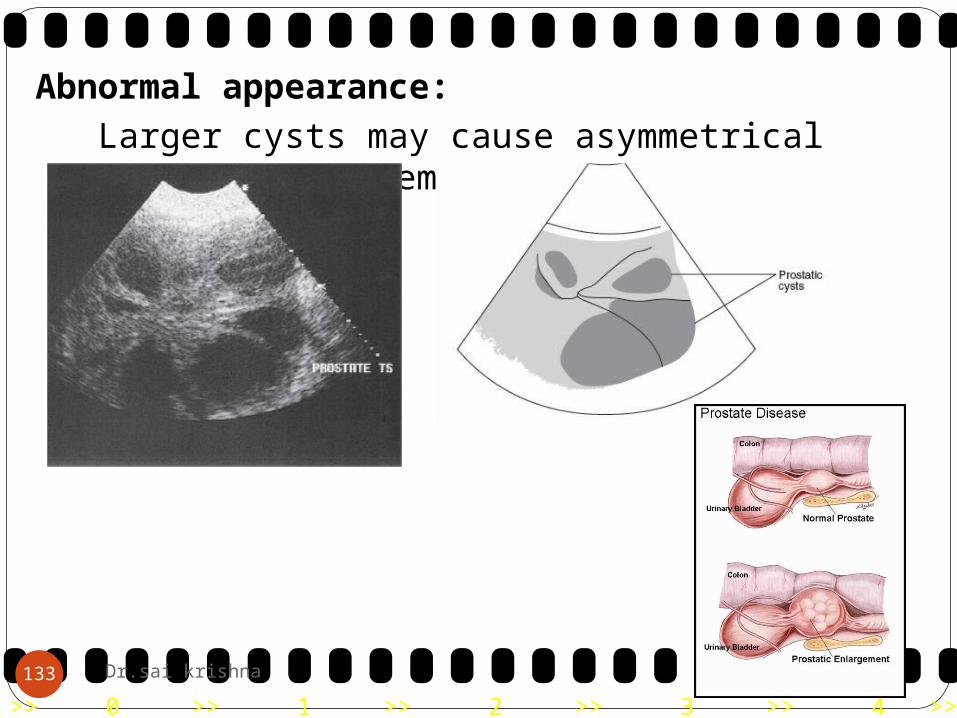
Abnormal appearance: Larger cysts may cause asymmetrical prostatic
enlargement.
04/19/23133 Dr.sai krishna

>> 0 >> 1 >> 2 >> 3 >> 4 >>
Prostratic abcess:
04/19/23134 Dr.sai krishna

>> 0 >> 1 >> 2 >> 3 >> 4 >>
Acute prostatitis, the prostate is usually enlarged.Chronic prostatitis may also result in a patchy
density but in this instance the overall impression is of an increase in echogenicity.
Prostatic hyperplasia in an entire dog. Diffusely coarsened appearance produced by multiple, small parenchymal cysts.
04/19/23135 Dr.sai krishna

>> 0 >> 1 >> 2 >> 3 >> 4 >>
The echotexture is patchy with anoverall increase in echogenicity.
Prostatic tumours in the dog
04/19/23136 Dr.sai krishna
>> 0 >> 1 >> 2 >> 3 >> 4 >>
04/19/23Dr.sai krishna137
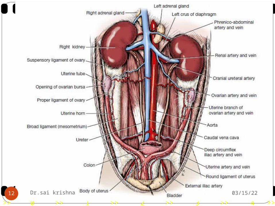
The uterus:The cervix in the bitch and queen is situated between
thedorsal aspect of the bladder neck and the ventral aspect ofthe colon.
Cranial to this is the short uterine body, whichbranches into two long uterine horns. These run craniallythrough the abdomen towards the ovaries, which are located caudal or caudoventral to the ipsilateral kidney.
A 7.5 MHz transducer is the most appropriate for assessment of the uterus.
As the normal, non-gravid uterus is often difficult to locate, the bladder and colon are used as landmarks to allow visualisation
of the uterus in its position between these two structures.

>> 0 >> 1 >> 2 >> 3 >> 4 >>
04/19/23Dr.sai krishna138
Gravid uterus: The embryo first becomes visible around
day 21 of gestationas a small echogenic structure located close to
the endometrium. careful examination will reveal a small
flicker within the embryo representing the heartbeat and indicating viability.
By day 25, the gestational sacs are approximately 1 cm in diameter and are more oval in shape than spherical.

>> 0 >> 1 >> 2 >> 3 >> 4 >>
04/19/23Dr.sai krishna139
By day 35, the fetus has a distinct head, trunk and abdomen, and fetal movements can be observed.
After approximately day 40, it produces increasingly marked distal acoustic shadowing as calcification progresses

>> 0 >> 1 >> 2 >> 3 >> 4 >>
04/19/23Dr.sai krishna140
>> 0 >> 1 >> 2 >> 3 >> 4 >>
04/19/23Dr.sai krishna141
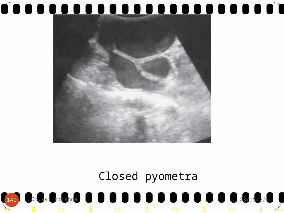
Closed pyometra

>> 0 >> 1 >> 2 >> 3 >> 4 >>
04/19/23Dr.sai krishna142
Multiple, small, anechoic cysts locatedthroughout the endometrium, indicatesthe presence of cystic endometrialhyperplasia.

>> 0 >> 1 >> 2 >> 3 >> 4 >>
04/19/23Dr.sai krishna143
Non-gravid uterus:The cervix appears as a round, hypoechoic
structure positioned between the anechoic bladder and the hyperechoic semi-circle representing the colon.
Uterus is relatively homogenous in appearance without an apparent lumen.
Horns are oval-shaped,hypoechoic structures located one on each side of the midline

>> 0 >> 1 >> 2 >> 3 >> 4 >>
Non-Cardiac Thoracic Ultrasound:Ultrasound examination of the thoracic cavity is
hampered by the surrounding bony rib cage and the air-filled lungs within.
acoustic windowUltrasound examination is preceded by radiography
with orthogonal projections of the thoracic cavity being preferred.
Parasternal , suprasternal (through the thoracic inlet), subcostal (through the liver) or directly over the lesion.
Suprasternal is useful for assessment of the cranial mediastinum in some cases.
The subcostal approach is especially useful for lesions in the caudal lung lobes and the caudal mediastinum.
04/19/23144 Dr.sai krishna
>> 0 >> 1 >> 2 >> 3 >> 4 >>
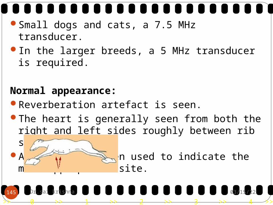
Small dogs and cats, a 7.5 MHz transducer.In the larger breeds, a 5 MHz transducer is
required.
Normal appearance:Reverberation artefact is seen.The heart is generally seen from both the right
and left sides roughly between rib spaces 3 to 7.Apex beat is often used to indicate the most
appropriate site.
04/19/23145 Dr.sai krishna

>> 0 >> 1 >> 2 >> 3 >> 4 >>
Pleural effusion:The sonographic appearance of a pleural
effusion varies with the character and volume of fluid present.
Fluids such as transudate and modified transudate are usually totally
black with the pleural reflections seen as fine echogenic lines ‘floating’ in the surrounding fluid.
As the cellular content of the fluid increases it becomes more echogenic and may even, in some cases, develop an almost granular appearance.
04/19/23146 Dr.sai krishna

>> 0 >> 1 >> 2 >> 3 >> 4 >>
Mediastinal masses: Masses contained within the cranial and middle
mediastinum are most easily seen.Pulsed-wave and colour-flow Doppler
examination of the mediastinum is helpful, identifying the mediastinal vessels and assessing the vascularity of the mass itself. This may be important for surgical planning.
04/19/23147 Dr.sai krishna

>> 0 >> 1 >> 2 >> 3 >> 4 >>
Lymphoma: The typical appearance of lymphoma is
of rounded, discrete, hypoechoic masses. Discrete hypoechoicmass with a hyperechoic rim is almost pathognomonic for lymphoma
These often have a fine echogenic capsule and a central echogenic line. The positioning adjacent to the mediastinalvessels is characteristic and regardless of echogenicity theyare usually quite discrete.
04/19/23148 Dr.sai krishna
>> 0 >> 1 >> 2 >> 3 >> 4 >>
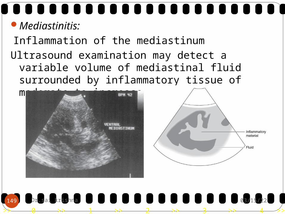
Mediastinitis: Inflammation of the mediastinumUltrasound examination may detect a variable
volume of mediastinal fluid surrounded by inflammatory tissue of moderate to increased echogenicity.
04/19/23149 Dr.sai krishna

>> 0 >> 1 >> 2 >> 3 >> 4 >>
Pulmonary consolidation: Consolidated lung has an appearance very
similar to that of the liver. In consolidated lung there are small irregular
hyperechoic area, which correspond to small pockets of remaining gas.
04/19/23150 Dr.sai krishna
>> 0 >> 1 >> 2 >> 3 >> 4 >>
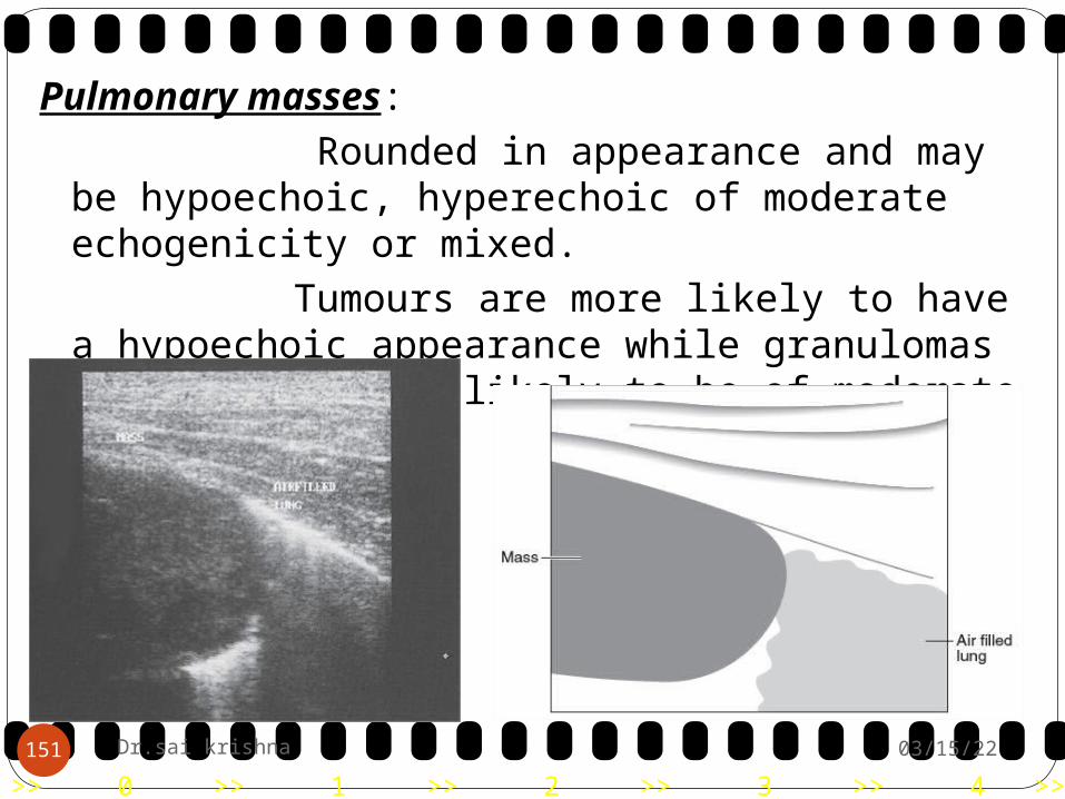
Pulmonary masses: Rounded in appearance and may be
hypoechoic, hyperechoic of moderate echogenicity or mixed.
Tumours are more likely to have a hypoechoic appearance while granulomas are perhaps more likely to be of moderate echogenicity.
04/19/23151 Dr.sai krishna

>> 0 >> 1 >> 2 >> 3 >> 4 >>
Disrupted diaphragm: Mirror-image artefact from herniated liver
lobe. Mirror-image artefact is not seen when
there is a pleural effusion.
04/19/23152 Dr.sai krishna

>> 0 >> 1 >> 2 >> 3 >> 4 >>
Imaging the Heart: Two-dimensional (2-D) echocardiography
produces real-time, cross-sectional images of the heart and great vessels and allows differentiation of the blood-filled lumen from the soft tissue structures of the heart chambers, valves and vessels.
M-mode imaging allows quantitative analysis of the dimensions and motion of chambers and valves.
Doppler analysis provides information on the dynamics of the blood flow through the heart chambers, along vessels and across the valves.
04/19/23153 Dr.sai krishna

>> 0 >> 1 >> 2 >> 3 >> 4 >>
Narrow rib spaces and lung surrounding the heart result in relatively small acoustic windows.
The ideal transducer therefore should have a small footprint, allowing coupling of the transducer between ribs and producing a wedgeshaped beam which fans out from the thoracic wall.
Sector or phased array transducers are therefore the recommended choice for cardiac sonography.
The frequency of the transducer depends on the size of the patient and the type of examination.
For most dogs both 2-D and M-mode examinations require a 5 MHz transducer.
04/19/23154 Dr.sai krishna
>> 0 >> 1 >> 2 >> 3 >> 4 >>
Dogs weighing more than 50 kg may require lower frequencies (3 to 3.5 MHz) whereas small dogs and cats can be examined with a 7.5 MHz transducer.
Haircoat is clipped between the costochondral junction and the sternum on both sides of the thorax in the area of the right and left parasternal windows.
The right parasternal window can be found anywhere between the third and sixth intercostal spaces on the right, it is found most frequently in the fourth and fifth spaces, ventrally between the sternum and the costochondral junction.
04/19/23155 Dr.sai krishna

>> 0 >> 1 >> 2 >> 3 >> 4 >>
The left caudal or apical parasternal window, the transducer is placed in the fifth to the seventh intercostal space and as close to the sternum as possible (ideally over the apex of the heart).
For the left cranial parasternal window, the transducer is placed in the third or fourth intercostal space between the sternum and the costochondral junction.
04/19/23156 Dr.sai krishna

>> 0 >> 1 >> 2 >> 3 >> 4 >>
2-D echocardiography:2-D echocardiography produces real-time, cross-
sectional images of the heart and great vessels. These images are relatively easy to interpret
because they are similar to anatomical sections through the heart.
The images obtained depend on the location of the transducer over the heart and the orientation of the beam plane.
In long-axis views, the index marker of the transducer is directed towards the base of the heart, in short-axis views towards the head of the patient.
04/19/23157 Dr.sai krishna

>> 0 >> 1 >> 2 >> 3 >> 4 >>
M-mode echocardiography: M-mode echocardiography uses a single,
thin ultrasound beam rather than a fan-shaped beam, and is used to record and analyse thickness and motion of the soft tissue structures of the heart (heart chamber walls, valves, vessels).
The distance of individual structures from the transducer is displayed on the vertical axis and time is displayed on the horizontal axis.
M-mode examinations are carried out almost exclusively from the right parasternal approaches.
04/19/23158 Dr.sai krishna
>> 0 >> 1 >> 2 >> 3 >> 4 >>
Examination from the right parasternal window:
Long-axis views:The ultrasound beam is directed parallel to the
long axis of the heart and normally two views are obtained.
The four chamber view shows both ventricles, atria and atrioventricular valves and by slight clockwise rotation or angling the ultrasound beam in a cranial direction, the left ventricle outflow tract is displayed with the left ventricle, aortic valve and naortic root. The ventricles are displayed on the left side of the monitor with the atria or aorta on the right side
04/19/23159 Dr.sai krishna

>> 0 >> 1 >> 2 >> 3 >> 4 >>
04/19/23160 Dr.sai krishna

>> 0 >> 1 >> 2 >> 3 >> 4 >>
Short-axis views: From the long-axis view, the transducer
is rotated 90° in a clockwise direction and the ultrasound beam is orientated perpendicular to the long axis of the ventricles.
The examination normally begins at the level of the apex of the heart, followed by planes through the papillary muscles, chordae tendineae, mitral valve, aortic root, left atrium (M-mode) and main pulmonary artery.
04/19/23161 Dr.sai krishna

>> 0 >> 1 >> 2 >> 3 >> 4 >>
04/19/23162 Dr.sai krishna

>> 0 >> 1 >> 2 >> 3 >> 4 >>
The view through the papillary muscles is described as having a mushroom shape, while the view through the mitral valve is known as the fish mouth view.
Right parasternal short-axis views
04/19/23163 Dr.sai krishna

>> 0 >> 1 >> 2 >> 3 >> 4 >>
Examination from the left caudal parasternalwindow:
After placement of the transducer over the apex, the ultrasound beam is directed almost perpendicular to the sternum and parallel to the long axis of the heart.
By rotating the ultrasound beam in a left caudal to right cranial direction and angling dorsally towards the base of the heart, a four-chamber view is obtained showing the left ventricle and atrium on the right side and the right ventricle and atrium on the left side.
By tilting the ultrasound beam slightly cranially, the left ventricular outflow tract can be displayed between the left ventricle and a portion of the right atrium (five-chamber view).
04/19/23164 Dr.sai krishna

>> 0 >> 1 >> 2 >> 3 >> 4 >>
04/19/23165 Dr.sai krishna
>> 0 >> 1 >> 2 >> 3 >> 4 >>
Congenital heart disease:
Patent ductus arteriosus (PDA):visualise a PDA from the left cranial parasternal
long-axis view.recognition of secondary changes.If the shunt is small, secondary changes include
mild dilation of the pulmonary artery and a mild increase in left ventricular end-diastolic dimension.
In larger shunts, volume overload in the lung, left cardiac chambers and aorta to the level of the ductus leads to dilation of the main pulmonary artery and left atrium.
04/19/23166 Dr.sai krishna
>> 0 >> 1 >> 2 >> 3 >> 4 >>
M-mode examination allows analysis of most of the secondary changes. Increased left ventricular end-diastolic dimensions with normal septal and left ventricular thickness indicate eccentric hypertrophy.
A PDA leads to an abnormal flow pattern within the main pulmonary artery. This can be detected and quantified using Doppler sonography. Detection of the characteristic turbulent flow confirms the diagnosis even where the PDA cannot be visualised.
04/19/23167 Dr.sai krishna

>> 0 >> 1 >> 2 >> 3 >> 4 >>
Pulmonic stenosis (PS):Mild right ventricular hypertrophy may be difficult to
detect, whereas in moderate to severe cases, thickening of the septum, right ventricular free wall and papillary muscles are seen.
Thickened and immobile cusps are seen with valvular stenosis and a narrow subvalvular or infundibular region with subvalvular stenosis. This, together with post-stenotic dilation of the main pulmonary artery, helps confirm the diagnosis of pulmonic stenosis.
M-mode examination is not usually helpful in the diagnosis of pulmonic stenosis.
Pulsed-wave Doppler is used to localise the narrowing and continuous-wave Doppler is then used to quantify the severity by determining the systolic peak velocity and pressure gradient between the right ventricle and pulmonary artery.
04/19/23168 Dr.sai krishna

>> 0 >> 1 >> 2 >> 3 >> 4 >>
Pressure gradients of less than 50 mm Hg are classified as mild whereas gradients of greater than 125 mm Hg are classified as severe cases.
Aortic stenosis (AS): In the right parasternal short-axis view,
narrowing between the interventricular septum and the base of the anterior leaflet of the mitral valve are the main features of subaortic stenosis.
appears as a hyperechoic ridge or ring-like structure in the left ventricular outflow tract.
M-mode examination shows diastolic thickening of the left ventricular free wall and interventricular septum (concentric hypertrophy) with normal fractional shortening.
04/19/23169 Dr.sai krishna
>> 0 >> 1 >> 2 >> 3 >> 4 >>
Doppler echocardiography can detect mild stenosis with no clear abnormalities in 2-D or M-mode examinations, localise (pulsed-wave Doppler) and quantify the stenosis (continuous-wave Doppler).
Pressure gradients of less than 50 mm Hg are classified as mild, between 50 and 75 mm Hg as moderate and greater than 75 mm Hg as severe.
04/19/23170 Dr.sai krishna
>> 0 >> 1 >> 2 >> 3 >> 4 >>
Mitral valve dysplasia (MVD):Mitral valve dysplasia (MVD) is one of the most
commonly seen congenital heart diseases in the cat.The pathophysiological mechanism is volume
overload of the left atrium and left ventricle with elevated left atrial pressure.
The main features of MVD, the often markedly enlarged left atrium and the dilatation of the left ventricle, are easily recognised using 2-D or M-mode echocardiography (right parasternal views).
The main features of MVD, the often markedly enlarged left atrium and the dilatation of the left ventricle, are easily recognised using 2-D or M-mode echocardiography (right parasternal views)
04/19/23171 Dr.sai krishna
>> 0 >> 1 >> 2 >> 3 >> 4 >>
Tricuspid dysplasia: Similar to MVD
Eisenmenger’s syndrome and other malformations:
Eisenmenger’s syndrome, a communication between the right and the left side of the heart (at the level of the atria, ventricular septum (VS) or great vessels), pulmonary hypertension and right-to-left shunt and many other malformations
04/19/23172 Dr.sai krishna
>> 0 >> 1 >> 2 >> 3 >> 4 >>
Acquired heart disease: Cardiomyopathy: Dilated cardiomyopathy describes a condition
which includes hypokinesis with reduced ejection and shortening fractions, diastolic and systolic dilatation of the affected ventricle, dilated atria and finally congestive heart failure.
M-mode and 2-D examinations readily identify these features even in milder forms of DCM and are very helpful in the differentiation of primary myocardial dysfunction from other heart diseases.
Can be quantified using M-mode measurements. Doppler echocardiography may be helpful to
detect and quantify dynamic outflow obstruction and MV regurgitation.
04/19/23173 Dr.sai krishna

>> 0 >> 1 >> 2 >> 3 >> 4 >>
Right parasternal long-axis view with hypertrophic cardiomyopathy in diastole.
04/19/23174 Dr.sai krishna

>> 0 >> 1 >> 2 >> 3 >> 4 >>
Pericardial disease: Pericardial effusion (PE) is the most common
pericardial disease in dogs and cats and may or may not be
associated with cardiac tamponade.
Clinical signs of right heart failure are usually present.
Feline infectious peritonitis and tumours (lymphosarcoma) are the most common causes.
04/19/23175 Dr.sai krishna

>> 0 >> 1 >> 2 >> 3 >> 4 >>
Features of pericardial effusion include a hypo- or anechoic space between the heartwall and pericardial sac.
In more pronounced cases, diminished right ventricle and left ventricle internal dimensions and swinging of the heart in the fluid-filled pericardial sac (bizarre motion in the M-mode examination) during the heart cycle are present.
04/19/23176 Dr.sai krishna
>> 0 >> 1 >> 2 >> 3 >> 4 >>
MASSES: Right atrium masses, most often
haemangiosarcomas, are best imaged from the right parasternal views, but the left cranial views are also important.
They arise from the right auricle, right atrium wall or the junction of right atrium and right ventricle, most of them projecting into the pericardial sac.
Small cavities often give them an irregular appearance
04/19/23177 Dr.sai krishna

>> 0 >> 1 >> 2 >> 3 >> 4 >>
Right parasternal long-axis plane in a dog with a large mass arising from the right atrium.
04/19/23178 Dr.sai krishna

>> 0 >> 1 >> 2 >> 3 >> 4 >>
Heart base tumours (aortic body, ectopic thyroid tumours) originate around the ascending aorta and may grow invasively in any direction and may penetrate the heart wall or chambers.
They require multiple-plane imaging from both sides of the thorax and are more homogenous compared to right atrium tumours.
04/19/23179 Dr.sai krishna

>> 0 >> 1 >> 2 >> 3 >> 4 >>
Infective endocarditis (IE): 2-D echocardiography readily identifies the
shaggy appearance or the hyperechoic irregular vegetations at the normally thin aortic cusps.
The typical feature of aortic valve insufficiency is the turbulent jet from the aortic valve into the right ventricle during diastole.
04/19/23180 Dr.sai krishna

>> 0 >> 1 >> 2 >> 3 >> 4 >>
04/19/23181 Dr.sai krishna
>> 0 >> 1 >> 2 >> 3 >> 4 >>
REFERENCES1. Small Animal Radiology and Ultrasound by
Ronald L. Burk.2. Diagnostic Ultrasound in Small Animal Practice
by Paddy Mannion.3. BSAVA canine and feline abdominal imaging.4. Diagnostic radiology and ultrasonografhy of the
dogs and cats by J. Kevin Kealy.
04/19/23182 Dr.sai krishna