umexpert.um.edu.my€¦ · Web viewThe questionnaire consists of 8 short item questions, covering...
49
Original Manuscript Title: Epidemiology, clinical presentation and respiratory sequelae of adenovirus pneumonia in children in Kuala Lumpur, Malaysia Li Min Lim 1 , Yen Yen Woo 2 , Jessie Anne de Bruyne 1 , Anna Marie Nathan 1* , Sze Ying Kee 3 , Yoke Fun Chan 2 , Chun Wei Chiam 2 , Kah Peng Eg 1 , Surendran Thavagnanam 1 , I-Ching Sam 2 1 Department of Paediatrics, University Malaya, Kuala Lumpur, Malaysia 2 Department of Medical Microbiology, University Malaya, Kuala Lumpur, Malaysia 3 Department of Paediatrics, University Putra Malaysia, Serdang, Selangor Darul Ehsan, Malaysia *Corresponding author: [email protected] SHORT TITLE: Adenovirus infection in children, Malaysia 1 1 2 3 4 5 6 7 8 9 10 11 12 13 14 15 16 17 18
Transcript of umexpert.um.edu.my€¦ · Web viewThe questionnaire consists of 8 short item questions, covering...
Original Manuscript
Title: Epidemiology, clinical presentation and respiratory sequelae of adenovirus
pneumonia in children in Kuala Lumpur, Malaysia
Li Min Lim1, Yen Yen Woo2, Jessie Anne de Bruyne1, Anna Marie Nathan1*, Sze
Ying Kee3, Yoke Fun Chan2, Chun Wei Chiam2, Kah Peng Eg1, Surendran
Thavagnanam1, I-Ching Sam2
1 Department of Paediatrics, University Malaya, Kuala Lumpur, Malaysia
2 Department of Medical Microbiology, University Malaya, Kuala Lumpur, Malaysia
3Department of Paediatrics, University Putra Malaysia, Serdang, Selangor Darul
Ehsan, Malaysia
*Corresponding author:
SHORT TITLE: Adenovirus infection in children, Malaysia
1
1
2
3
4
5
6
7
8
9
10
11
12
13
14
15

Abstract
Objectives: To describe the severity, human adenovirus (HAdV) type and respiratory
morbidity following adenovirus pneumonia in children. Methodology: Retrospective
review of children under 12 years of age, admitted with HAdV pneumonia, between
January 2011 and July 2013, in a single centre in Malaysia. HAdV isolated from
nasopharyngeal secretions were typed by sequencing hypervariable regions 1-6 of the
hexon gene. Patients were reviewed for respiratory complications. Results: HAdV
was detected in 131 children of whom 92 fulfilled inclusion criteria. Median (range)
age was 1.1 (0.1-8.0) years with 80% under 2 years. Twenty percent had severe
disease with a case-fatality rate of 5.4%. Duration of admission (p=0.02) was
independently associated with severe illness. Twenty-two percent developed
respiratory complications, the commonest being bronchiolitis obliterans (15.2%) and
recurrent wheeze (5.4%). The predominant type shifted from HAdV1 and HAdV3 in
2011 to HAdV7 in 2013. The commonest types identified were types 7 (54.4%),
1(17.7%) and 3 (12.6%). Four out of the five patients who died were positive for
HAdV7. Infection with type 7 (OR 8.90, 95% CI 1.32, 59.89), family history of
asthma (OR 14.80, 95% CI 2.12-103.21) and need for invasive or non-invasive
ventilation (OR 151.84, 95% CI 9.93-2.32E) were independent predictors of
respiratory complications. Conclusions: One in five children admitted with HAdV
pneumonia had severe disease and 22% developed respiratory complications. Type 7
was commonly isolated in children with severe disease. Family history of asthma need
for invasive or non-invasive ventilation and HAdV 7 were independent predictors of
respiratory complications.
Word count: 247
2
16
17
18
19
20
21
22
23
24
25
26
27
28
29
30
31
32
33
34
35
36
37
38
39
40

Key words: Adenovirus; children; respiratory; complications; Malaysia
3
41
42

Introduction
Human adenoviruses (HAdV) are a common cause of disease including respiratory
infections, gastroenteritis, and conjunctivitis, particularly in children below 5 years of
age[1] and HAdV accounts for about 5 – 10% of acute lower respiratory tract
infections (ALRTIs). [2] Over 85 genotypes of HAdVs have been recognized by the
Human Addenovirus Working Group based on bioinformatics analysis of complete
genomic sequences.[3,4]. HAdV respiratory infections are predominantly caused by
species B (including types 3, 7, 11, 14, 21 and 55), C (types 1, 2, 5, and 6) and E (type
4).
Recent worldwide epidemics of respiratory infections due to HAdV have resulted in
renewed interest in this virus. The increase in cases of HAdV infection has also been
reported in Asia. These include outbreaks due to established types such as HAdV type
7 (HAdV7), which have been reported in the community and in military and police
camps between 2011 and 2013, in Taiwan, Singapore, China and Malaysia. [5-9]
There have also been more recently emerging types such as HAdV55, described in
China.[10]
Respiratory infections due to HAdV cause significant morbidity and mortality, with
case fatality rates as high as 12%.[11] There is also a risk of up to 30% of developing
long-term respiratory complications such as post-infectious bronchiolitis obliterans
(PIBO) and bronchiectasis. [1,12]
The clinical and molecular epidemiology of HAdV respiratory infections and risk of
complications are relatively understudied in developing countries. In this study, the
aims were to a) describe the clinical presentation, severity, HAdV type and respiratory
morbidity, b) determine risk factors associated with severe illness and the
4
43
44
45
46
47
48
49
50
51
52
53
54
55
56
57
58
59
60
61
62
63
64
65
66
67

development of respiratory complications, c) describe the treatment modalities and
outcomes, and d) assess quality of life of children admitted to our centre with acute
HAdV pneumonia.
Materials and Methods
Hospital setting
University Malaya Medical Centre (UMMC) is a government-funded teaching
hospital in Kuala Lumpur, Malaysia. The paediatric building has 4 Paediatric wards, a
Paediatric Intensive Care Unit (PICU) and a Neonatal Intensive Care unit. It also has
its own Paediatric Emergency Department.
Study design and ethical approval
This is a single centre, retrospective study conducted at UMMC. Ethical approval for
this study was obtained from the institutional ethics committee (MECID.NO: 201410-
659). Verbal consent was taken from patients' parents not on follow-up and
information was obtained over the phone. Questionnaires were administered to
parents whose children were on follow-up. The ethics committee approved this study.
Study population
This study included all children younger than 12 years of age, admitted to UMMC
with laboratory-confirmed HAdV pneumonia between 1st January 2011 and 31st July
2013. Children with pre-existing chronic lung disease, upper respiratory tract
infections, those who could not be contacted or whose medical records could not be
found were excluded. Eligible patients were identified as those whose respiratory
samples tested positive for HAdV by the Department of Medical Microbiology of
UMMC.
5
68
69
70
7172
73
74
75
76
77
78
79
80
81
82
83
84
85
86
87
88
89
90
91

Study definitions
Pneumonia was diagnosed based on history (either fever, cough and/or rapid
breathing) and clinical evidence of pneumonia (either tachypnoea, chest recessions
and/ or adventitious sounds upon lung auscultation) with radiographic signs
(infiltrates or consolidation). HAdV infection was laboratory-confirmed by positive
immunofluorescence assay and/or viral culture
Previous lung infection was defined as any child with a previous history of a lower
respiratory tract infection (LRTI), which was verified by the presence of shortness of
breath during that illness.
Severe HAdV pneumonia was defined as those requiring either invasive or non-
invasive respiratory support, PICU care or illness resulting in death. Disseminated
disease was defined as signs and laboratory evidence of involvement of 2 or more
organ systems.
PIBO was diagnosed in children in the presence of either one of the following signs:
tachypnoea, chest retractions, chest hyperinflation, wheeze, crepitations and
hypoxaemia, for at least 30 days after the initial lung injury.[13] The diagnosis was
made following high-resolution chest tomography showing some or all the following
features: mosaic perfusion, vascular attenuation, atelectasis, expiratory air trapping,
peri-bronchial thickening and bronchiectasis.[14] Respiratory complications include
any form of chronic lung disease (including asthma) and death.
Data collection
Medical records were obtained from the Medical Records Department and reviewed.
Medical records and administration of the questionnaire was between 1st February
2015 till 30th September 2015. Data acquired included: socio-demographic data,
anthropometric measurements, duration of admission, birth history, personal and
6
92
93
94
95
96
97
98
99
100
101
102
103
104
105
106
107
108
109
110
111
112
113
114
115
116

family history of asthma and atopy, vaccination (which includes prior exposure to
pneumococcal and/or influenza vaccines), treatment received (antibiotics and steroids)
and clinical, laboratory and radiological investigations.
Specimen collection, virus identification & typing
Nasopharyngeal aspirates (NPAs) are collected routinely in all children with LRTIs.
Tracheal aspirates were only obtained from intubated children. All respiratory
specimens were tested by direct immunofluorescence (IF) for 8 respiratory viruses:
HAdV, influenza A and B, respiratory syncytial virus (RSV), metapneumovirus and
parainfluenza viruses 1, 2 and 3 (D3 Ultra 8 DFA Respiratory Virus Screening &
Identification Kit (Diagnostic Hybrids, USA). Viral isolation was performed by
inoculating the NPAs into Madin Darby canine kidney (MDCK; ATCC number CCL-
34), Vero (ATCC number CCL-81), A549 (ATCC number CCL-185), and HEp-2
(ATCC number CCL-23) cells. Cultures were incubated at 37°C with 5% CO2.
Infected cells showing cytopathic effect within 10 days were harvested for IF.
HAdV isolates underwent further molecular typing. Viral DNA was extracted from
adenovirus cultures with the QIAamp DNA Blood Mini Kit (Qiagen, Germany). The
hypervariable regions 1–6 of the hexon gene were amplified and sequenced as
previously described[15] with M13 universal priming tails added to the primers to
facilitate sequencing.[16] Sequencing was carried out by First BASE Laboratories
(Selangor, Malaysia). Sequences were edited using Geneious R7 (Biomatters, New
Zealand) and aligned with relevant sequences from GenBank. MEGA7 [17] was used
to construct phylogenetic trees by maximum likelihood with 1000 bootstrap
reiterations, using the general time reversible model with gamma distribution of
evolutionary rates and invariant sites. Separate trees were constructed for sequences
7
117
118
119
120
121
122
123
124
125
126
127
128
129
130
131
132
133
134
135
136
137
138
139
140
141
of HAdV-B and HAdV-C. The sequences reported in this study were deposited into
GenBank with accession numbers KU145006-KU145113.
Treatment modalities
All children received standard treatment for pneumonia, which included supportive
care, and where deemed necessary, invasive or non-invasive ventilation, inotropes and
antibiotics. Intravenous immunoglobulin (IVIG) was given to children with persistent
fever and intravenous pulse methylprednisolone (MTP) was given to children with
significant disease i.e. rapidly increasing respiratory distress, at the discretion of the
treating physician.
Respiratory sequelae
Parents of discharged children with HAdV pneumonia were contacted by a doctor via
telephone and interviewed about their child’s respiratory condition. Children with
respiratory symptoms were recalled for further evaluation. Verbal consent of their
participation was obtained over the phone. All children with severe HAdV pneumonia
were already on regular follow-up. In addition, all parents of children who were
attending clinic answered the translated Malay version of the Parent Cough-specific –
Quality of Life (PC-QOL) questionnaire, as part of clinical management. The PCQOL
is a validated instrument for assessing the burden of chronic cough and quality of life.
[18] The questionnaire provides important information on outcome indicators and aids
in evaluation of efficacy of treatment interventions. The questionnaire consists of 8
short item questions, covering quality of life domains of physical (2 items),
psychological (4 items) and social (2 items) wellbeing. There are 7 options in each
item, with a score scale of 1 – 7 per item. The higher numbers represent fewer
concerns and thus, better quality of life.
8
142
143
144
145
146
147
148
149
150
151
152
153
154
155
156
157
158
159
160
161
162
163
164
165
166

Statistical analysis
Data analysis was performed using Statistical Package for Social Science (SPSS)
software version 16.0 (IBM, USA). Continuous data was expressed as mean (standard
deviation [SD]) or median (interquartile range [IQR]) if not normally distributed. Chi-
square test was used for comparing categorical variables between two groups and
odds ratio (OR) and 95% confidence interval (CI) were reported, where appropriate.
Mann-Whitney U test was used when comparing continuous (numerical) variables
without normal distribution between the two groups. Logistic regression was used to
determine significant factors associated with severe disease and respiratory sequelae.
All tests were calculated in a two-tailed manner and significance was defined by a p
value of less than 0.05.
Results
A total of 131 respiratory samples were positive for HAdV (either by IF and/or viral
culture) between 1st January 2011 and 31st July 2013; however, 92 were included in
the analysis as 39 were excluded for various reasons, as shown in the study flow, Fig
1. Nineteen children (20.6%) had severe infection while 73 (79.4%) had non-severe
cases of HAdV pneumonia.
Fig 1: Study flow of the 92 patients with adenovirus pneumonia
Between 2011 and 2013, there was a sharp increase in both the number and severity
of HAdV cases as shown in Fig. 2.
9
167
168
169
170
171
172
173
174
175
176
177
178
179
180
181
182
183
184
185
186
187
188
189
190

Fig. 2 A sharp increase in the number of adenovirus infections as well as an increase
in the number of severe cases was detected in 2013
Baseline and clinical and laboratory investigations: severe
and non-severe
Severe disease was seen in 20.6% of cases. Demographic characteristics of the
patients with severe and non-severe HAdV infection are shown in Table 1.
Table 1: Baseline characteristics of the 92 patients with adenovirus pneumoniaCharacteristics Total
(n=92)n (%)
Non-severe group(n=73)n (%)
Severe group(n=19)n (%)
P value
Gender Male Female
52 (56.5)40 (43.5)
41 (56.2)32 (43.8)
11 (57.9)8 (42.1)
0.892
Ethnicity Malay Chinese Indian Others
81 (88.0)3 (3.3)6 (6.5)2 (2.2)
65 (89.0)1 (1.4)6 (8.2)1 (1.4)
16 (84.2)2 (10.5)
0 (0)1 (5.3)
0.089
Age, years, median (IQR)1
< 2 years ≥ 2 years
1.08 (0.58– 1.58)
74 (80.4)18 (19.5)
1.08 (0.52-1.64)
57 (78.1)16(21.9)
0.83 (0.41-1.25)
17 (89.5)2 (10.5)
0.218
Gestational age Term Preterm (< 37 weeks)
85 (92.4)7 (7.6)
67 (91.8)6 (8.2)
18 (94.7)1 (5.3)
0.665
Birth weight, kg, mean (S.D.) 1 3.03 (± 0.608) 3.04 (± 0.607) 3.00 (± 0.628)
0.825
Previous LRTI 2
Yes No
23 (25.0)69 (75.0)
15 (20.5)58 (79.5)
8 (42.1)11 (57.9)
0.053
Co-morbid diseases 3
Yes No
8 (8.7)84 (1.3)
5 (6.8)68 (93.2)
3 (15.8)16 (84.2)
0.218
Exposure to passive smoking (n=62) Yes No
28 (30.4)34 (37.0)
22 (30.1)24 (32.9)
6 (31.6)10 (52.6)
0.475
10
191
192
193
194
195
196
197
198

Personal history of asthma/ wheeze Yes No
12 (13.0)80 (87.0)
7 (9.6)66 (90.4)
5 (26.3)14 (73.3)
0.054
Family history of asthma (n=71) Yes No
25 (27.2)46 (50.0)
18 (24.7)36 (49.3)
7 (36.8)10 (52.6)
0.555
Duration of hospitalization, days, median (IQR)
4.5 (1.6-7.4) 4.0 (2.5-5.5) 15.0 (6.5-23.5)< 0.001
1IQR: interquartile range; S.D.: standard deviation
2LRTI: lower respiratory tract infection; 3Congenital heart disease= 3; Achondroplasia=1; Neurological
disease= 2
Most patients (80.4%) were children less than 2 years of age. There were no
significant differences in demographic characteristics between those with severe and
non-severe disease. There were 7 patients with underlying co-morbidities: 3 with
congenital heart disease, 3 with neurological disease and 1 patient with
achondroplasia. Patients in the severe group had a significantly longer duration of
hospitalisation (p<0.001, z= -5.62).
11
199
200
201
202
203
204
205
206
207
208
209
210

Table 2 shows the clinical findings in the children with severe and non-severe HAdV
infection. Children with severe disease presented with more hepatomegaly (p=0.002,
OR 5.64 [95%CI 1.89,16.84]), hepatitis (p=0.01, OR 6.62 [95%CI 4.00,10.92]),
disseminated disease (p<0.001, OR 15.41 [95%CI 4.68, 50.78]) and seizures (p=0.03,
OR 1.38 [95%CI 1.32,142.86]).
Table 3 shows the laboratory investigation findings in children with severe and non-
severe HAdV disease. Review of laboratory investigations showed that in children
with severe disease, albumin (p=0.01, z= -2.33) was significantly lower and
neutrophil/lymphocyte ratio (p = 0.03, z =-2.18) was significantly higher. Viral co-
infection was found in 4 patients, all in the non-severe group (RSV=3,
metapneumovirus=1). One patient had Haemophilus influenzae sepsis, in the severe
group. Bacterial and viral co-infection was not associated with severe adenoviral
infection, as shown in Table 3.
Symptoms & Signs Total
(n=92)
n (%)
Non-severe group
(n=73)
n (%)
Severe group
(n=19)
n (%)
P value
Constitutional
Fever
Duration of fever, median(IQR)
days1
Prolonged fever (>7 days)
87 (94.6)
5.0 (3.0-7.0)
21 (22.8)
69 (94.5)
5.0 (3.0-7.0)
16 (21.9)
18 (94.7)
5.0 (1.5-8.5)
5 (26.3)
0.970
0.662
0.684
Respiratory
Cough
Tachypnoea
Wheezing
Respiratory distress
Rhonchi
Crepitations
86 (93.5)
43 (46.7)
8 (8.7)
62 (67.4)
40 (43.5)
67 (72.8)
68 (93.2)
29 (39.7)
4 (5.5)
47 (64.4)
28 (38.4)
50 (68.5)
18 (94.7)
14 (73.7)
4 (21.1)
15 (78.9)
12 (63.2)
17 (89.5)
0.803
0.008
0.054
0.122
0.052
0.067
Extra-pulmonary
12
211
212
213
214
215
216
217
218
219
220
221
222
223
224
225

Vomiting ± diarrhoea
Conjunctivitis
Hepatomegaly
Seizures
56 (60.9)
10 (10.9)
22 (23.9)
4 (4.4)
46 (63.0)
8 (10.9)
12 (16.4)
1 (1.4)
10 (52.6)
2 (10.5)
10 (52.6)
3 (15.9)
0.409
0.294
0.002
0.027
Disseminated disease
Shock
ARDS2
Pulmonary haemorrhage
Hepatitis
6 (6.5)
7 (7.6)
2 (2.2)
16 (17.4)
0 (0)
0 (0)
0 (0)
9 (12.4)
6 (31.6)
7 (36.8)
2 (10.5)
7 (36.8)
< 0.001
< 0.001
0.041
0.773
Table 2: Clinical Characteristics of 92 children with adenovirus pneumonia
1IQR: interquartile range; 2ARDS: acute respiratory distress syndrome
13
226
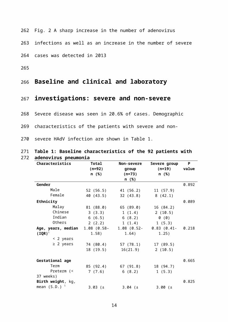
Table 3: Laboratory results of 92 children with adenovirus pneumonia.Laboratory Data Non-severe group
(n=73)
n (%)
Severe group
(n=19)
n (%)
P value
WBC1 (× 10cells/uL) (n=91)
> 15.0
5.0 – 11.0
< 5.0
28 (38.9)
44 (61.1)
0 (0)
4 (21.1)
11 (57.9)
4 (21.1)
0.160
Neutrophil (%), median (IQR)2 52 (47, 61) 67 (59, 74) 0.02
Z=-2.33
Lymphocyte (%), median (IQR) 34 (31, 44) 28 (20, 35) 0.05
Z=-1.98
Monocyte (%), median (IQR) 6.00 (5.5, 9.8) 5.04 (4.33, 7.13) 0.75
Z=-1.36
Haemoglobin, (g/dL) (n=91)
> 11.0
9.0 – 11.0
< 9.0
48 (66.7)
23 (31.9)
1 (1.4)
9 (47.4)
8 (42.1)
2 (10.5)
0.076
Platelet (× 103cells/μL) (n=87)
< 150 1 (1.5) 3 (15.8) 0.031
CRP3 (g/L) (n=79), median (IQR) 1.50 (1.32, 7.89) 2.54 (1.97, 7.32) 0.376
Z=-0.885
Albumin (g/l), median (IQR) 29 (27, 32) 24.58 (22, 27) 0.007
Z=-2.33
ALT4 (n=39)
Abnormal (> 41 U/L) 9 (39.1) 7 (43.8) 0.773
Adenovirus type (n=79)
1
2
3
4
5
7
13 (21.7)
6 (10.0)
9 (15.0)
3 (5.0)
1 (1.7)
28 (46.7)
1 (5.3)
1 (5.3)
1 (5.3)
0 (0)
1 (5.3)
15 (78.9)
0.175
0.665
0.378
0.369
0.300
0.002
Viral co-infection5 4 (5.5) 0 (0) 0.297
Bacterial co-infection6 5 (6.8) 1 (5.3) 0.803
Radiographic findings
Infiltrates ± consolidation 69 (94.5) 17 (89.5)
0.461
1WBC: white blood cell; 2IQR: interquartile range; 3CRP: C-reactive protein; 4ALT: alanine transaminase
14
227
228229

5Viral co-infection: 3 cases of respiratory syncytial virus (RSV), 1 case of metapneumovirus; 6Bacterial co-infection: 1 cases of Streptococcus pneumoniae, 3 cases of Haemophilus influenzae, 1 case of both
Klebsiella pneumoniae and Acinetobacter baumanii in the non-severe group, 1 case of Haemophilus influenzae
bacteraemia in the severe group.
Human adenovirus typesOf the 131 adenovirus-positive samples, 108 were successfully sequenced and typed.
Fig. 3 shows the changing HAdV types over the study period from January 2011 till
December 2013; in 2011, HAdV3 (37.0%) and HAdV1 (23.9%) were the commonest
types, but the proportion of HAdV7 increased from 19.6% in 2011 to 37.5% in 2012
and, finally, 84.8% in 2013.
Fig. 3 Changing human adenovirus types detected over the study period (Jan 2011 to
July 2013). As HAdV3 and HAdV1 declined, HAdV7 increased to become the
predominant circulating type.
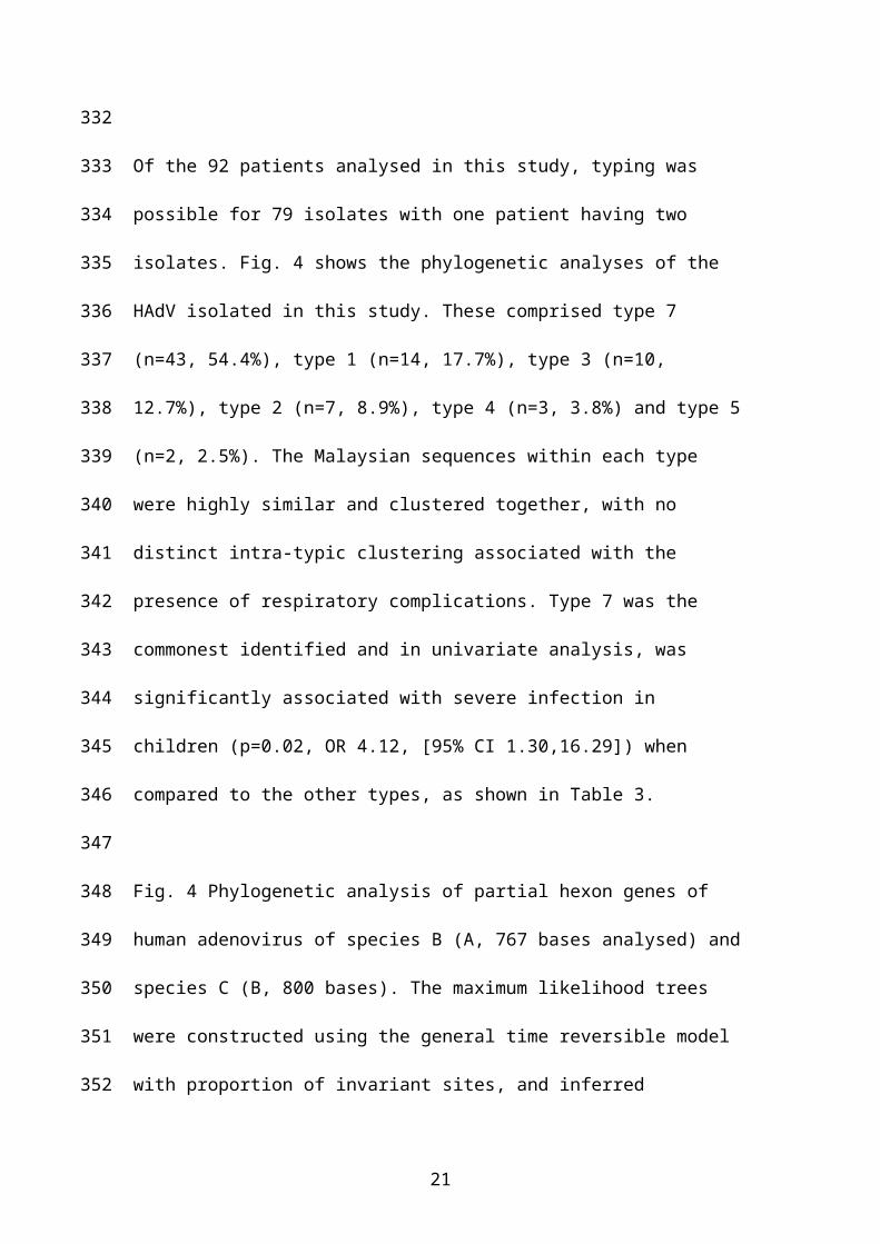
Of the 92 patients analysed in this study, typing was possible for 79 isolates with one
patient having two isolates. Fig. 4 shows the phylogenetic analyses of the HAdV
isolated in this study. These comprised type 7 (n=43, 54.4%), type 1 (n=14, 17.7%),
type 3 (n=10, 12.7%), type 2 (n=7, 8.9%), type 4 (n=3, 3.8%) and type 5 (n=2, 2.5%).
The Malaysian sequences within each type were highly similar and clustered together,
with no distinct intra-typic clustering associated with the presence of respiratory
complications. Type 7 was the commonest identified and in univariate analysis, was
significantly associated with severe infection in children (p=0.02, OR 4.12, [95% CI
1.30,16.29]) when compared to the other types, as shown in Table 3.
Fig. 4 Phylogenetic analysis of partial hexon genes of human adenovirus of species B
(A, 767 bases analysed) and species C (B, 800 bases). The maximum likelihood trees
15
230231
232
233
234235
236
237
238
239
240
241
242
243
244
245
246
247
248
249
250
251
252
253
254
255
256

were constructed using the general time reversible model with proportion of invariant
sites, and inferred following bootstrap analyses using 1000 replicates. Strain names
are in the format: accession number_adenovirus type_strain name_country of
isolation_year of isolation. The Malaysian sequences from this study are coloured red
(with respiratory complications) or blue (without respiratory complications).
Factors associated with severe illness
In univariate analysis, duration of hospitalisation, seizures, disseminated disease
(shock, ARDS, pulmonary haemorrhage), tachypnoea, hepatomegaly, neutrophil (%)
count, albumin, low platelet count and HAdV type 7 (compared to the other types)
were significantly associated with development of severe disease. However, in the
final model, only prolonged hospitalisation was associated with severe illness
(p=0.003, OR 1.54 [95%CI 1.16, 2.06]).
Treatment modalities
Sixty-two children (67.4%) required some form of respiratory support and 17 (18.5%)
required ventilatory support (non-invasive and/or invasive). Thirteen children
received intravenous IVIG (2 in the non-severe and 11 in the severe HAdV group)
and 17 children received steroids (5 in the non-severe and 12 in the severe group).
16
257
258
259
260
261
262
263
264
265
266
267
268
269
270
271
272
273
274
275
276
277

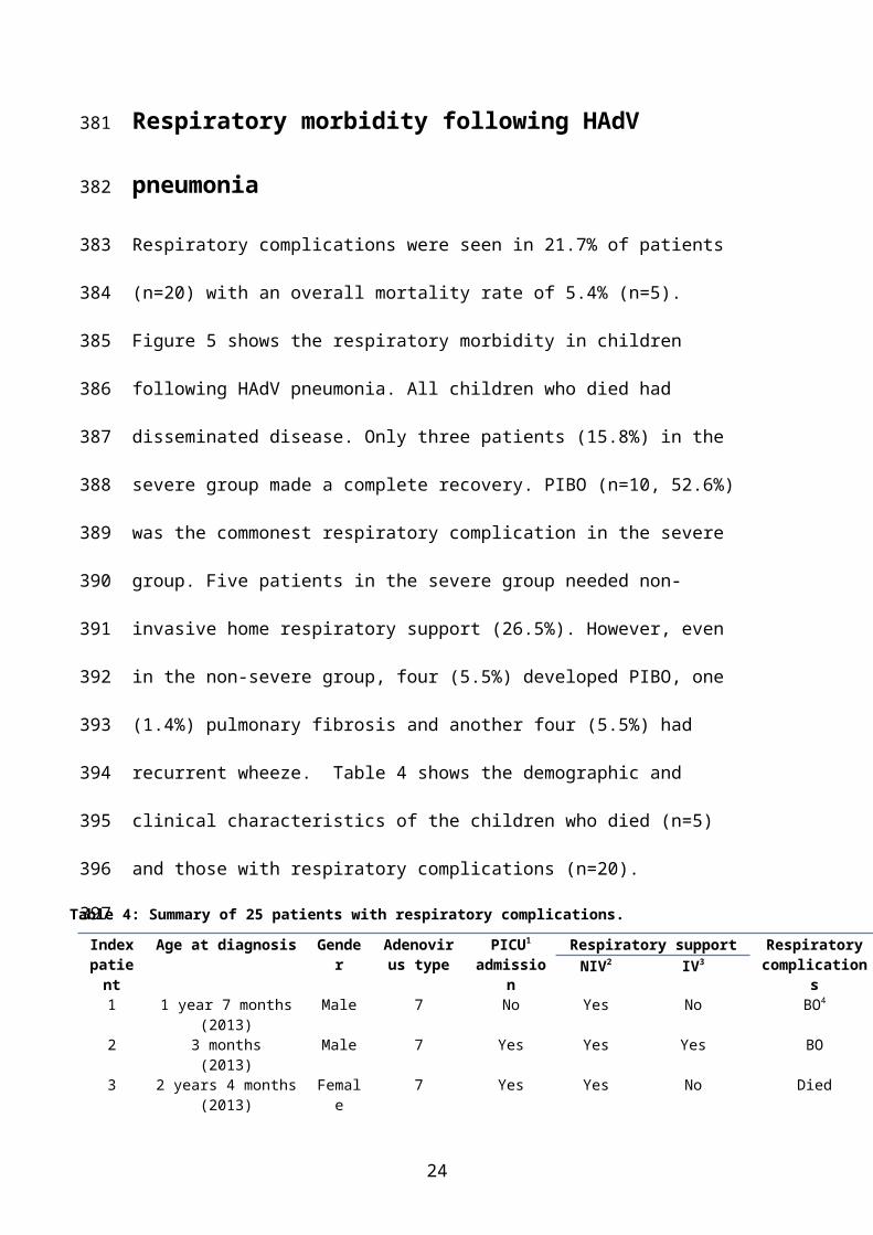
Respiratory morbidity following HAdV pneumonia
Respiratory complications were seen in 21.7% of patients (n=20) with an overall
mortality rate of 5.4% (n=5). Figure 5 shows the respiratory morbidity in children
following HAdV pneumonia. All children who died had disseminated disease. Only
three patients (15.8%) in the severe group made a complete recovery. PIBO (n=10,
52.6%) was the commonest respiratory complication in the severe group. Five patients
in the severe group needed non-invasive home respiratory support (26.5%). However,
even in the non-severe group, four (5.5%) developed PIBO, one (1.4%) pulmonary
fibrosis and another four (5.5%) had recurrent wheeze. Table 4 shows the
demographic and clinical characteristics of the children who died (n=5) and those
with respiratory complications (n=20).
Table 4: Summary of 25 patients with respiratory complications.
Index patient
Age at diagnosis Gender Adenovirus type
PICU1
admissionRespiratory support Respiratory
complicationsNIV2 IV3
1 1 year 7 months(2013)
Male 7 No Yes No BO4
2 3 months(2013)
Male 7 Yes Yes Yes BO
3 2 years 4 months(2013)
Female 7 Yes Yes No Died
4 8 months (2011) Female 1 No No No Recurrent wheeze5 1 year
(2013)Female 7 Yes Yes Yes ARDS,5
pulmonary hemorrhage, BO
6 8 months(2013)
Female 7 Yes Yes No BO
7 6 years 10 months (2011)
Female 4 No No No Recurrent wheeze
8 8 months (2013) Female 7 Yes Yes No BO9 7 months (2013) Female 7 No No No BO10 1 year 1 month (2012) Male - No No No Recurrent wheeze11 1 year 10 months (2011) Female 7 No No No BO12 7 months (2013) Female 7 No No No Recurrent wheeze13 1 year 8 months (2013) Female 7 Yes Yes Yes ARDS, pulmonary
hemorrhage, died14 1 year 6 months (2013) Male 7 No No No Respiratory
failure, died15 4 months (2013) Male 7 Yes Yes Yes BO16 5 months (2013) Male 7 Yes Yes Yes BO17 4 years 4 months (2013) Male 7 No No No BO18 9 months (2013) Male 7 No Yes No BO19 1 year 4 months (2013) Male 7 Yes Yes Yes BO20 7 months (2011) Female 5 Yes Yes Yes Recurrent wheeze
17
278
279
280
281
282
283
284
285
286
287
288
289

21 1 year 4 months (2013) Male 7 Yes Yes No BO22 1 year (2013) Female 7 No No No BO23 10 months (2013) Female 2 Yes No Yes Respiratory
failure, died24 8 months (2013) Female 7 Yes No Yes ARDS, died25 5 years 6 months (2013) Male - No No No Pulmonary fibrosis
1PICU: paediatric intensive care unit; 2NIV: non-invasive ventilation; 3IV: invasive ventilation; 4BO: bronchiolitis obliterans; 5ARDS: acute respiratory distress syndrome;
18
290291
292

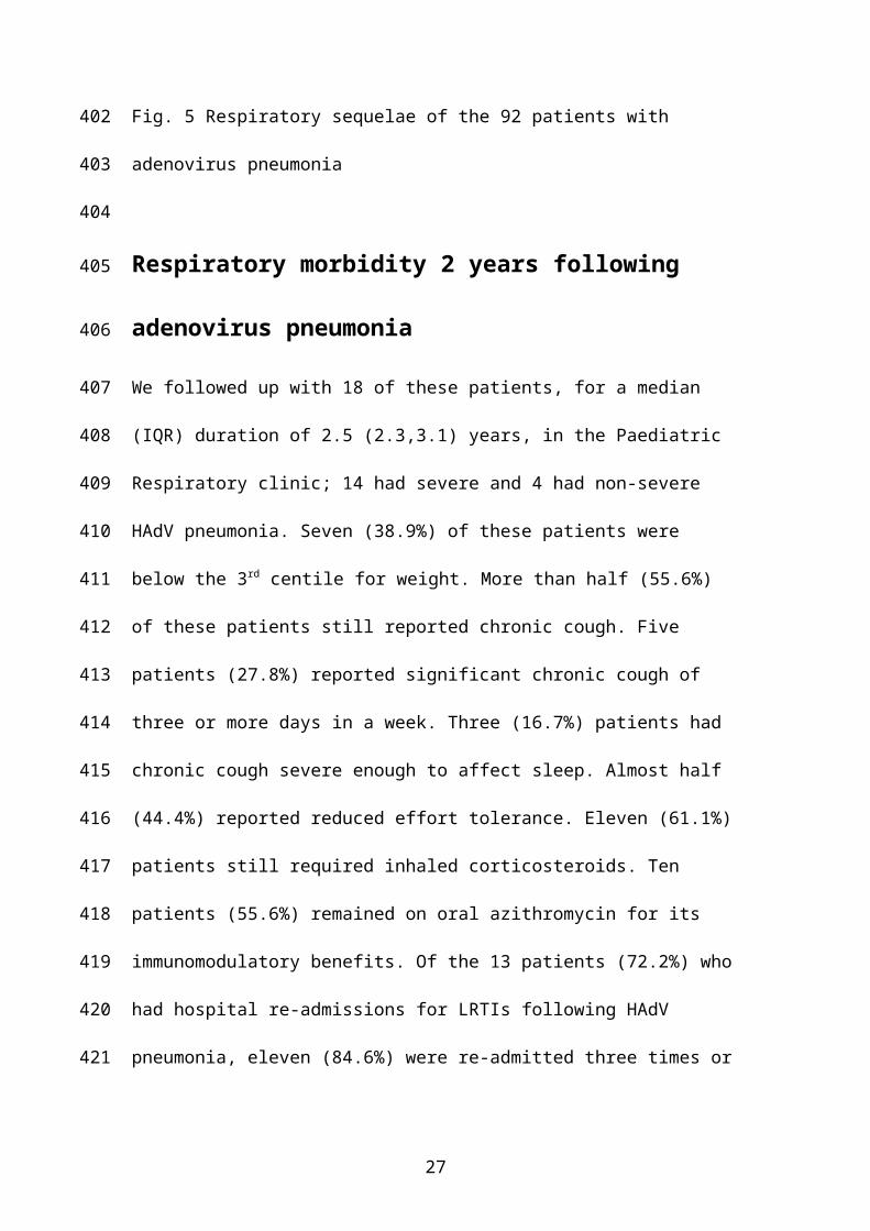
Fig. 5 Respiratory sequelae of the 92 patients with adenovirus pneumonia
Respiratory morbidity 2 years following adenovirus
pneumonia
We followed up with 18 of these patients, for a median (IQR) duration of 2.5 (2.3,3.1)
years, in the Paediatric Respiratory clinic; 14 had severe and 4 had non-severe HAdV
pneumonia. Seven (38.9%) of these patients were below the 3rd centile for weight.
More than half (55.6%) of these patients still reported chronic cough. Five patients
(27.8%) reported significant chronic cough of three or more days in a week. Three
(16.7%) patients had chronic cough severe enough to affect sleep. Almost half
(44.4%) reported reduced effort tolerance. Eleven (61.1%) patients still required
inhaled corticosteroids. Ten patients (55.6%) remained on oral azithromycin for its
immunomodulatory benefits. Of the 13 patients (72.2%) who had hospital re-
admissions for LRTIs following HAdV pneumonia, eleven (84.6%) were re-admitted
three times or less while two patients (15.4%) reported hospital re-admission on more
than three separate occasions.
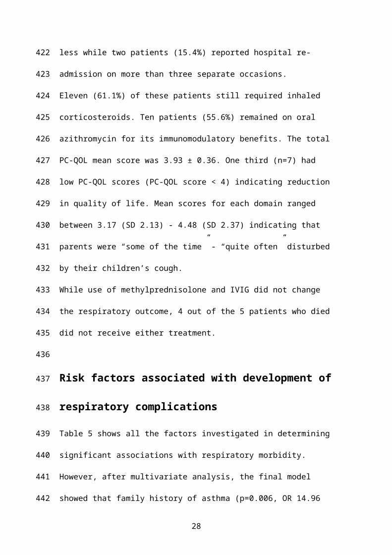
Eleven (61.1%) of these patients still required inhaled corticosteroids. Ten patients
(55.6%) remained on oral azithromycin for its immunomodulatory benefits. The total
PC-QOL mean score was 3.93 ± 0.36. One third (n=7) had low PC-QOL scores (PC-
QOL score < 4) indicating reduction in quality of life. Mean scores for each domain
ranged between 3.17 (SD 2.13) - 4.48 (SD 2.37) indicating that parents were “some of
the time” - “quite often” disturbed by their children’s cough.
While use of methylprednisolone and IVIG did not change the respiratory outcome, 4
out of the 5 patients who died did not receive either treatment.
19
293
294
295
296
297
298
299
300
301
302
303
304
305
306
307
308
309
310
311
312
313
314
315
316
317

Risk factors associated with development of respiratory
complications
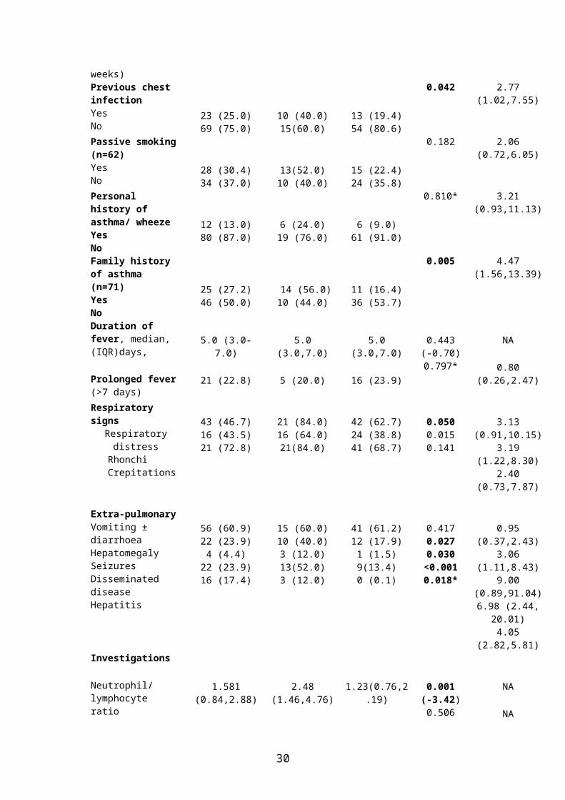
Table 5 shows all the factors investigated in determining significant associations with
respiratory morbidity. However, after multivariate analysis, the final model showed
that family history of asthma (p=0.006, OR 14.96 [95% CI 2.15-104.05]), need for
either invasive or non-invasive ventilatory support (p< 0.001, OR 153.77 [95% CI
10.07-2.32E]) and HAdV type 7 (compared to the other types) (p = 0.025, OR 9.00,
[95% CI 1.34-60.34]) were independent factors associated with the development of
respiratory complications. Table 6 shows the multivariate analysis of risk factors
associated with respiratory complications post-adenovirus pneumonia
Table 5: Baseline characteristics, symptoms and signs of the 92 patients with adenovirus
pneumonia with and without respiratory complications
Characteristics Total(n=92)n (%)
Patient with respiratory
complications(n=25)n (%)
Patient without respiratory
complications(n=67)n (%)
P value(Z score)
Odds ratio95%
(Confidence interval)
GenderMaleFemale
52 (56.5)40 (43.5)
11 (44.0)14 (56.0)
41 (61.2)26 (38.8)
0.139 0.50(0.20,1.26)
EthnicityMalayChineseIndianOthers
81 (88.0)3 (3.3)6 (6.5)2 (2.2)
22 (81.0)1 (4.0)1 (4.0)1 (4.0)
59 (88.1)2 (3.0)5 (7.5)1 (1.5)
0.821 NA
Age, years, median (IQR)1
< 2 Years> 2 Years
1.08 (0.58– 1.58)
74 (80.4)18 (19.5)
1.00 (0.62-1.63)
21 (84.0)4 (16.0)
1.08 (0.67-1.67)
53 (79.1)14(20.9)
0.627(-0.44)
0.770*
NA
1.39(0.49,4.70)
Gestational ageTermPreterm (< 37 weeks)
85 (92.4)7 (7.6)
25 (100.0)0 (0.0)
60 (89.6)7 (10.4))
0.185* 0.71(0.62, 0.81)
Previous chest infectionYesNo
23 (25.0)69 (75.0)
10 (40.0)15(60.0)
13 (19.4)54 (80.6)
0.042 2.77(1.02,7.55)
20
318
319
320
321
322
323
324
325
326
327
328
329
330
Passive smoking (n=62)YesNo
28 (30.4)34 (37.0)
13(52.0)10 (40.0)
15 (22.4)24 (35.8)
0.182 2.06(0.72,6.05)
Personal history of asthma/ wheezeYesNo
12 (13.0)80 (87.0)
6 (24.0)19 (76.0)
6 (9.0)61 (91.0)
0.810* 3.21(0.93,11.13)
Family history of asthma (n=71)YesNo
25 (27.2)46 (50.0)
14 (56.0)10 (44.0)
11 (16.4)36 (53.7)
0.005 4.47(1.56,13.39)
Duration of fever, median, (IQR)days,
Prolonged fever (>7 days)
5.0 (3.0-7.0)
21 (22.8)
5.0 (3.0,7.0)
5 (20.0)
5.0 (3.0,7.0)
16 (23.9)
0.443(-0.70)0.797*
NA
0.80 (0.26,2.47)
Respiratory signsRespiratory distress
Rhonchi Crepitations
43 (46.7)16 (43.5)21 (72.8)
21 (84.0)16 (64.0)21(84.0)
42 (62.7)24 (38.8)41 (68.7)
0.0500.0150.141
3.13 (0.91,10.15)3.19 (1.22,8.30)2.40 (0.73,7.87)
Extra-pulmonaryVomiting ± diarrhoeaHepatomegalySeizuresDisseminated diseaseHepatitis
56 (60.9)22 (23.9)4 (4.4)
22 (23.9)16 (17.4)
15 (60.0)10 (40.0)3 (12.0)13(52.0)3 (12.0)
41 (61.2)12 (17.9)1 (1.5)9(13.4)0 (0.1)
0.4170.0270.030
<0.0010.018*
0.95 (0.37,2.43)3.06 (1.11,8.43)9.00 (0.89,91.04)6.98 (2.44, 20.01)4.05 (2.82,5.81)
Investigations
Neutrophil/ lymphocyte ratio
Platelet
C-reactive protein
Albumin
ALT2
1.581 (0.84,2.88)
278 (213,356)
1.75 (0.98,4.98)
28 (25,32)
39 (24,84)
2.48 (1.46,4.76)
292 (179,356)
2.14 (0.88,4.57)
26 (23,29)
40 (25,82)
1.23(0.76,2.19)
278(227,356)
1.65(1.00,5.12)
31 (29,33)
38 (23,113)
0.001(-3.42)0.506(-0.67)0.920(-0.13)0.015(-2.43)0.990(-0.13)
NA
NA
NA
NA
NA
Adenovirus type(n=79)Type 7
Non-7 types
43 (54.4)
36 (45.6)
19 (82.6)
4 (17.4)
24 (44.4)
30 (55.6)
0.002 5.94 (1.78,19.80)
TreatmentPICU3 admissionVentilation4 Steroids
14 (15.2)47 (51.2)13 (14.1)
13 (52.0)21 (84.0)12 (48.0)
1 (1.5)26 (38.9)1 (1.5)
<0.001<0.001<0.001
71.5 (8.54,598.62)8.28 (2.55, 26.86)
11.49 (3.44, 38.10)
Duration of hospitalization, days, median (IQR)
4.5 (1.6-07.40) 10.0 (4.0-22.50) 4.0 (3.0-6.0) < 0.001(-3.83)
NA
21

*Fishers exact test
1IQR: interquartile range; 2ALT: alanine transferase; 3PICU: Paediatric Intensive Care
Unit; 4invasive or non-invasive ventilation
22
331
332
333
334

Table 6: Multivariate analysis of risk factors associated with respiratory
complications post-adenovirus pneumonia
Risk factors Patients with
respiratory
complications
n=25 (%)
Patients without
respiratory
complications
n=67 (%)
Multivariate analysis
Adjusted OR
(95% CI)
P
value
Family history of asthma
14 (56.0) 11 (16.4) 14.96
(2.15-104.05)
0.006
Need for invasive or non-invasive ventilation
15(60.0) 2(3.0) 153.77
(10.07-2.35E)
<0.001
Adenovirus type 7 versus other types
24(96.0) 19(28.0) 9.00
(1.34-60.34)
0.024
23
335
336
337
338
339340341
Discussion
This comprehensive report on HAdV pneumonia in Malaysian children summarises
their clinical presentation, identified HAdV types, treatment, risk factors for severe
disease and both short- and medium-term respiratory complications. We found that
one in five children admitted with HAdV pneumonia had severe disease and 22%
developed respiratory complications of which PIBO was the commonest problem.
Type 7 was the commonest type detected. Family history of asthma, need for
ventilation(invasive and non-invasive) and type 7 were independent factors associated
with respiratory complications.[15]
Epidemics of HAdV pneumonia have been reported since 2011, in both children and
adults.[6,8,9,19-21] While predominant types circulating at a given time differ among
countries or regions, and change over time [22] , type 7 was the main type reported in
China, Taiwan and Singapore between 2011 and 2013. Among 632 HAdV cases
reported during the Taiwan outbreak in 2011, HAdV3 was predominantly seen in
children with upper respiratory tract infections while HAdV7 was seen in cases that
required PICU care or died.[23] As seen in our study, neighbouring Singapore also
saw the emergence of HAdV type 7 as the predominant type between 2011 and 2013.
[21] An earlier study from our centre found that of the HAdV isolates from 1999 to
2005, 70% of isolates were human adenovirus C (HAdV-1, HAdV-2, HAdV-5 and
HAdV-6) [24], showing a longer term shift to species B (HAdV3 and HAdV7) in
2011-2013. [24]
In this study, the majority of children were under 2 years old. This is interesting as
recent studies from Taiwan and China found that rate of HAdV infection increased
with age.[23,25] However, in the study from Taiwan, only 12% of patients had
24
342
343
344
345
346
347
348
349
350
351
352
353
354
355
356
357
358
359
360
361
362
363
364
365
366

LRTIs. The experiences in Singapore, where most paediatric patients infected with
HAdV were < 2 years old. [21] and in Seremban, Malaysia, where in 2015 the median
age of inpatients with HAdV was14 months, are more similar to ours.[4] It has been
reported that young children are at increased risk of severe HAdV infections. [26]
Treatment and outcome were the main concerns in this study. Twenty percent of
children with HAdV pneumonia had a severe infection, which we defined as requiring
NIV or IV or PICU admission or death. The case fatality was 5.4%. Our severity and
case fatality are much higher than that previously mentioned in the study from
Seremban, Malaysia, which were 11% and 2.6% respectively. [5] This could be
explained as we are a tertiary referral centre that accepts ill patients from peripheral
hospitals. While there were many factors associated with severe infection in
univariate analysis, only hospital duration was an independent association. Rajkumar
et al identified age < 2 years old and presence of significant comorbidities as
independent risk factors for severe disease. We did not find this association with age
and in this study, we excluded those with serious comorbidities. In the outbreak in
Taiwan, authors found an association between severe disease and presence of pleural
effusion. Pleural effusion was seen in only 2 children in this study (one in a severe
HAdV and one in a non-severe HAdV) and none required tube thoracostomy
drainage. Immunocompromised children and children with comorbidities have also
been reported to be at increased risk of severe HAdV infections[6,19,27]. Many
laboratory features have been associated with severe HAdV infection e.g. leucopenia,
[23] thrombocytopenia[23] and a positive blood culture.[20]
In this study, 22% of children developed respiratory complications. Even in the non-
severe group, 12.3% (9 out of 73) children developed respiratory complications. This
25
367
368
369
370
371
372
373
374
375
376
377
378
379
380
381
382
383
384
385
386
387
388
389
390
391

is much higher than what has been previously reported locally, where only one child
developed PIBO.[5] However in other reports, especially from Latin America,
respiratory complications of PIBO range between 36 to 47% and mortality rates can
be as high as 15%[1,12] The need for NIV, PICU admission and family history of
asthma were independent risk factors for respiratory complications. Most of our
patients who had respiratory distress or impending respiratory failure would have
received NIV. Therefore, children with severe respiratory compromise from HAdV
have an increased risk of respiratory complications. As for family history of asthma, a
severe infection could trigger asthma, especially if there is a genetic predisposition.
Castro-Rodriquez from Chile, who followed up 45 children with HAdV pneumonia
for 5 years also found that significant respiratory compromise (intensive care
admission, need for mechanical ventilation and for oxygen therapy, and systemic
corticosteroid and beta agonist use) was associated with risk for PIBO.[1]Hence his
results concur with ours. In China, hypoxaemia was the only factor associated with
risk for PIBO.[28]
No antiviral treatments are currently licensed for treatment of severe HAdV disease.
In this study, we used IVIG and IV MTP, but not in a randomised fashion. While we
did not see any effect on subsequent respiratory complications, 4 out of the 5 who
died from HAdV did not receive these medications. Takahashi et al. reported that use
of pulse methylprednisolone (25mg/kg/day) for 3 days, in a case of severe HAdV
pneumonia (type 3) with hypercytokinemia i.e raised lactate dehydrogenase, ferritin,
interferon-gamma and interleukin-6, resulted in relief of respiratory distress. [29] He
also suggested that serum Krebs von den Lungen-6 (Kl-6) could be used as a marker
of future lung disease. Cidofivir is occasionally used in immunocompromised children
with severe HAdV. It has been shown to clear the virus from blood, however
26
392
393
394
395
396
397
398
399
400
401
402
403
404
405
406
407
408
409
410
411
412
413
414
415
416
mortality despite its use stands at 10-70% and it is nephrotoxic.[30] There have also
been case reports of use of ribavirin in immunocompromised children with HAdV,
[31] as well as successful use of oral ribavirin in an adult with respiratory
compromise.[32]However use of only antivirals may not suffice, and other adjuvant
medication like MTP and IVIG may be necessary.
This is the first study looking at the quality of life in children with post-HAdV lung
disease. We found that more than half of the children under follow-up had chronic
cough, more than a third were underweight, nearly half still had reduced effort
tolerance and as many as 72% required unscheduled healthcare visits. Most parents
were still concerned about their child’s cough. More work is needed to address the
long-term consequences of this viral infection, which may lead to reduced lung
function that is not fully reversible.[1]
Limitations of our study are recognised including the small number of patients, not
using polymerase chain reaction to detect HAdV which would increase the sensitivity
of virus detection, the inability to contact all patients who had HAdV pneumonia and
the inability to type all the 92 detected HAdV virus. However, the strength of this
study is that it has comprehensive data including clinical and laboratory parameters,
HAdV type, respiratory outcomes, and a follow-up of up to 2 years, for a fairly large
number of children.
Conclusion
In conclusion, during the sharp increase of HAdV infection in Malaysia between 2011
and 2013, the majority of children admitted for HAdV pneumonia were less than 2
years old. One in five children had severe disease and the case fatality rate was 5.4%.
HAdV 7 was the most frequently detected type isolated amongst children with severe
27
417
418
419
420
421
422
423
424
425
426
427
428
429
430
431
432
433
434
435
436
437
438
439
440
441
pneumonia and those with persistent respiratory sequelae. Severe disease was
associated with prolonged hospitalisation. Twenty-two percent developed respiratory
complications, commonest being bronchiolitis obliterans (15.2%) and recurrent
wheeze (5.4%). Presence of severe respiratory compromise, isolation of HAdV type 7
and family history of asthma, were independent risk factors associated with
respiratory sequelae. Children with respiratory complications reported significant
reduction in quality of life. There is a lack of good and adequately powered studies to
determine the best treatment for this disease which has a high mortality and
significant morbidity.
28
442
443
444
445
446
447
448
449
450
451
452

References
1. Castro-Rodriguez JA, Daszenies C, Garcia M, Meyer R, Gonzales R (2006)
Adenovirus pneumonia in infants and factors for developing bronchiolitis
obliterans: a 5-year follow-up. Pediatr Pulmonol 41;(10):947-953.
2. Tabain I, Ljubin-Sternak S, Cepin-Bogovic J, Markovinovic L, Knezovic I,
Mlinaric-Galinovic G (2012) Adenovirus respiratory infections in hospitalized
children: clinical findings in relation to species and serotypes. Pediatr Infect
Dis J 31;(7):680-684.
3. Hashimoto S, Gonzalez G, Harada S, Oosako H, Hanaoka N, Hinokuma R, et al.
(2018) Recombinant type Human mastadenovirus D85 associated with
epidemic keratoconjunctivitis since 2015 in Japan. J Med Virol 90;(5):881-
889.
4. Group HAW (2017). http://hadvwg.gmu.edu
5. Foong Ng K, Kee Tan K, Hong Ng B, Nair P, Ying Gan W (2015) Epidemiology of
adenovirus respiratory infections among hospitalized children in Seremban,
Malaysia. Trans R Soc Trop Med Hyg 109;(7):433-439.
6. Ng OT, Thoon KC, Chua HY, Tan NW, Chong CY, Tee NW, et al. (2015) Severe
Pediatric Adenovirus 7 Disease in Singapore Linked to Recent Outbreaks
across Asia. Emerg Infect Dis 21;(7):1192-1196.
7. Tsou TP, Tan BF, Chang HY, Chen WC, Huang YP, Lai CY, et al. (2012)
Community outbreak of adenovirus, Taiwan, 2011. Emerg Infect Dis 18;
(11):1825-1832.
8. Yu P, Ma C, Nawaz M, Han L, Zhang J, Du Q, et al. (2013) Outbreak of acute
respiratory disease caused by human adenovirus type 7 in a military training
camp in Shaanxi, China. Microbiol Immunol 57;(8):553-560.
29
453
454
455
456
457
458
459
460
461
462
463
464
465
466
467
468
469
470
471
472
473
474
475
476
477
9. Yusof MA, Rashid TR, Thayan R, Othman KA, Hasan NA, Adnan N, et al. (2012)
Human adenovirus type 7 outbreak in Police Training Center, Malaysia, 2011.
Emerg Infect Dis 18;(5):852-854.
10. Lu QB, Tong YG, Wo Y, Wang HY, Liu EM, Gray GC, et al. (2014)
Epidemiology of human adenovirus and molecular characterization of human
adenovirus 55 in China, 2009-2012. Influenza Other Respir Viruses 8;(3):302-
308.
11. Hong JY, Lee HJ, Piedra PA, Choi EH, Park KH, Koh YY, et al. (2001) Lower
respiratory tract infections due to adenovirus in hospitalized Korean children:
epidemiology, clinical features, and prognosis. Clin Infect Dis 32;(10):1423-
1429.
12. Murtagh P, Giubergia V, Viale D, Bauer G, Pena HG (2009) Lower respiratory
infections by adenovirus in children. Clinical features and risk factors for
bronchiolitis obliterans and mortality. Pediatr Pulmonol 44;(5):450-456.
13. Colom AJ, Teper AM (2009) Clinical prediction rule to diagnose post-infectious
bronchiolitis obliterans in children. Pediatr Pulmonol 44;(11):1065-1069.
14. Chang AB, Masel JP, Masters B (1998) Post-infectious bronchiolitis obliterans:
clinical, radiological and pulmonary function sequelae. Pediatr Radiol 28;
(1):23-29.
15. Lu X, Erdman DD (2006) Molecular typing of human adenoviruses by PCR and
sequencing of a partial region of the hexon gene. Arch Virol 151;(8):1587-
1602.
16. Gray GC, McCarthy T, Lebeck MG, Schnurr DP, Russell KL, Kajon AE, et al.
(2007) Genotype prevalence and risk factors for severe clinical adenovirus
infection, United States 2004-2006. Clin Infect Dis 45;(9):1120-1131.
30
478
479
480
481
482
483
484
485
486
487
488
489
490
491
492
493
494
495
496
497
498
499
500
501
502

17. Kumar S, Stecher G, Tamura K (2016) MEGA7: Molecular Evolutionary Genetics
Analysis Version 7.0 for Bigger Datasets. Mol Biol Evol 33;(7):1870-1874.
18. Newcombe PA, Sheffield JK, Chang AB (2013) Parent cough-specific quality of
life: development and validation of a short form. J Allergy Clin Immunol 131;
(4):1069-1074.
19. Lai CY, Lee CJ, Lu CY, Lee PI, Shao PL, Wu ET, et al. (2013) Adenovirus
serotype 3 and 7 infection with acute respiratory failure in children in Taiwan,
2010-2011. PLoS One 8;(1):e53614.
20. Zampoli M, Mukuddem-Sablay Z (2017) Adenovirus-associated pneumonia in
South African children: Presentation, clinical course and outcome. S Afr Med
J 107;(2):123-126.
21. Rajkumar V, Chiang CS, Low JM, Cui L, Lin RT, Tee NW, et al. (2015) Risk
Factors for Severe Adenovirus Infection in Children during an Outbreak in
Singapore. Ann Acad Med Singapore 44;(2):50-59.
22. Lynch JP, 3rd, Kajon AE (2016) Adenovirus: Epidemiology, Global Spread of
Novel Serotypes, and Advances in Treatment and Prevention. Semin Respir
Crit Care Med 37;(4):586-602.
23. Lin MR, Yang SL, Gong YN, Kuo CC, Chiu CH, Chen CJ, et al. (2017) Clinical
and molecular features of adenovirus type 2, 3, and 7 infections in children in
an outbreak in Taiwan, 2011. Clin Microbiol Infect 23;(2):110-116.
24. Abd-Jamil J, Teoh BT, Hassan EH, Roslan N, Abubakar S (2010) Molecular
identification of adenovirus causing respiratory tract infection in pediatric
patients at the University of Malaya Medical Center. BMC Pediatr 10:46.
25. Sun HQ, Zhang XX, Kuang XN, Gu WJ, Chen ZR, Yan YD, et al. (2017)
[Epidemiological analysis of 440 cases of respiratory adenovirus infections in
31
503
504
505
506
507
508
509
510
511
512
513
514
515
516
517
518
519
520
521
522
523
524
525
526
527

children from the Suzhou area between 2006 and 2015]. Zhongguo Dang Dai
Er Ke Za Zhi 19;(1):34-38.
26. Erdman DD, Xu W, Gerber SI, Gray GC, Schnurr D, Kajon AE, et al. (2002)
Molecular epidemiology of adenovirus type 7 in the United States, 1966-2000.
Emerg Infect Dis 8;(3):269-277.
27. Shen CF, Wang SM, Ho TS, Liu CC (2017) Clinical features of community
acquired adenovirus pneumonia during the 2011 community outbreak in
Southern Taiwan: role of host immune response. BMC Infect Dis 17;(1):196.
28. Wu PQ, Li X, Jiang WH, Yin GQ, Lei AH, Xiao Q, et al. (2016) Hypoxemia is an
independent predictor of bronchiolitis obliterans following respiratory
adenoviral infection in children. Springerplus 5;(1):1622.
29. Takahashi I, Takahashi T, Tsuchida S, Mikami T, Saito H, Hatazawa C, et al.
(2006) Pulse methylprednisolone therapy in type 3 adenovirus pneumonia with
hypercytokinemia. Tohoku J Exp Med 209;(1):69-73.
30. Ganapathi L, Arnold A, Jones S, Patterson A, Graham D, Harper M, et al. (2016)
Use of cidofovir in pediatric patients with adenovirus infection. F1000Res
5:758.
31. Gavin PJ, Katz BZ (2002) Intravenous ribavirin treatment for severe adenovirus
disease in immunocompromised children. Pediatrics 110;(1 Pt 1):e9.
32. Yoon BW, Song YG, Lee SH (2017) Severe community-acquired adenovirus
pneumonia treated with oral ribavirin: a case report. BMC Res Notes 10;
(1):47.
32
528
529
530
531
532
533
534
535
536
537
538
539
540
541
542
543
544
545
546
547
548
549
550551
552
553

Figure legends
S1 Fig. Study Flow of the 92 patients with adenovirus pneumonia
S2 Fig. Human adenovirus incidence chart. A sharp increase in the number of
adenovirus infections as well as an increase in the number of severe cases was detected in
2013
S3 Fig. Changing human adenovirus types over the study period (2011 to July
2013). As HAdV3 and HAdV1 declined, HAdV7 increased to become the
predominant circulating type.
S4 Fig. Phylogenetic analysis of partial hexon genes of human adenovirus of
species B (A, 767 bases analysed) and species C (B, 800 bases). The maximum
likelihood trees were constructed using the general time reversible model with
proportion of invariant sites, and inferred following bootstrap analyses using 1000
replicates. Strain names are in the format: accession number_adenovirus type_strain
name_country of isolation_year of isolation. The Malaysian sequences from this study
are coloured red (with respiratory complications) or blue (without respiratory
complications).
S5 Fig Respiratory sequelae of the 92 patients with adenovirus pneumonia.
33
554
555
556
557
558
559
560
561
562
563
564
565
566
567
568
569
570
571
572
573
574