documents.cap.org · Web viewTemplate for Reporting Results of Biomarker Testing of Specimens...
15
Template for Reporting Results of Biomarker Testing of Specimens From Patients With Gastrointestinal Stromal Tumors Template web posting date: February 2015 Authors Meera Hameed, MD, FCAP Department of Pathology, Memorial Sloan-Kettering Cancer Center, New York, NY Christopher L. Corless, MD, PhD Department of Pathology, Oregon Health & Science University, Portland, OR Suzanne George, MD Center for Sarcoma and Bone Oncology, Dan-Farber Cancer Institute, Boston, MA Jason L. Hornick MD, PhD, FCAP Department of Pathology, Brigham and Women's Hospital, Boston, MA Sanjay Kakar, MD, FCAP Department of Pathology, University of California, San Francisco and the Veterans Affairs Medical Center, San Francisco, CA Alex Lazar, MD, PhD, FCAP Department of Pathology, Sarcoma Research Center, The University of Texas MD Anderson Cancer Center, Houston, Texas Laura Tang, MD, FCAP Department of Pathology, Memorial Sloan-Kettering Cancer Center, New York, NY For the Members of the Cancer Biomarker Reporting Committee, College of American Pathologists
-
Upload
nguyenkhue -
Category
Documents
-
view
214 -
download
0
Transcript of documents.cap.org · Web viewTemplate for Reporting Results of Biomarker Testing of Specimens...
Template for Reporting Results of Biomarker Testing of Specimens From Patients With Gastrointestinal Stromal Tumors
Template web posting date: February 2015
AuthorsMeera Hameed, MD, FCAP
Department of Pathology, Memorial Sloan-Kettering Cancer Center, New York, NYChristopher L. Corless, MD, PhD
Department of Pathology, Oregon Health & Science University, Portland, ORSuzanne George, MD
Center for Sarcoma and Bone Oncology, Dan-Farber Cancer Institute, Boston, MAJason L. Hornick MD, PhD, FCAP
Department of Pathology, Brigham and Women's Hospital, Boston, MASanjay Kakar, MD, FCAP
Department of Pathology, University of California, San Francisco and the Veterans Affairs Medical Center, San Francisco, CA
Alex Lazar, MD, PhD, FCAPDepartment of Pathology, Sarcoma Research Center, The University of Texas MD Anderson Cancer Center, Houston, Texas
Laura Tang, MD, FCAPDepartment of Pathology, Memorial Sloan-Kettering Cancer Center, New York, NY
For the Members of the Cancer Biomarker Reporting Committee, College of American Pathologists

GIST • BiomarkersGISTBiomarkers 1.0.0.1
© 2015 College of American Pathologists (CAP). All rights reserved.
The College does not permit reproduction of any substantial portion of these templates without its written authorization. The College hereby authorizes use of these templates by physicians and other health care providers in reporting results of biomarker testing on patient specimens, in teaching, and in carrying out medical research for nonprofit purposes. This authorization does not extend to reproduction or other use of any substantial portion of these templates for commercial purposes without the written consent of the College.
The CAP also authorizes physicians and other health care practitioners to make modified versions of the templates solely for their individual use in reporting results of biomarker testing for individual patients, teaching, and carrying out medical research for non-profit purposes.
The CAP further authorizes the following uses by physicians and other health care practitioners, in reporting on surgical specimens for individual patients, in teaching, and in carrying out medical research for non-profit purposes: (1) Dictation from the original or modified templates for the purposes of creating a text-based patient record on paper, or in a word processing document; (2) Copying from the original or modified templates into a text-based patient record on paper, or in a word processing document; (3) The use of a computerized system for items (1) and (2), provided that the template data is stored intact as a single text-based document, and is not stored as multiple discrete data fields.Other than uses (1), (2), and (3) above, the CAP does not authorize any use of the templates in electronic medical records systems, pathology informatics systems, cancer registry computer systems, computerized databases, mappings between coding works, or any computerized system without a written license from the CAP.
Any public dissemination of the original or modified templates is prohibited without a written license from the CAP.
The College of American Pathologists offers these templates to assist pathologists in providing clinically useful and relevant information when reporting results of biomarker testing. The College regards the reporting elements in the templates as important elements of the biomarker test report, but the manner in which these elements are reported is at the discretion of each specific pathologist, taking into account clinician preferences, institutional policies, and individual practice.
The College developed these templates as educational tools to assist pathologists in the useful reporting of relevant information. It did not issue them for use in litigation, reimbursement, or other contexts. Nevertheless, the College recognizes that the templates might be used by hospitals, attorneys, payers, and others. The College cautions that use of the templates other than for their intended educational purpose may involve additional considerations that are beyond the scope of this document.
The inclusion of a product name or service in a CAP publication should not be construed as an endorsement of such product or service, nor is failure to include the name of a product or service to be construed as disapproval.
2

GIST • BiomarkersGISTBiomarkers 1.0.0.1
CAP Gastrointestinal Stromal Tumor Biomarker Template Revision History
Version CodeThe definition of the version code can be found at www.cap.org/cancerprotocols.
Version: GISTBiomarkers 1.0.0.1
Summary of Changes
The following changes were made since the December 2014 version:
Testing MethodsNGS was spelled out: Next-generation sequencing
URL was updated:Human Genome Variation Society: www.hgvs.org/mutnomen/
3

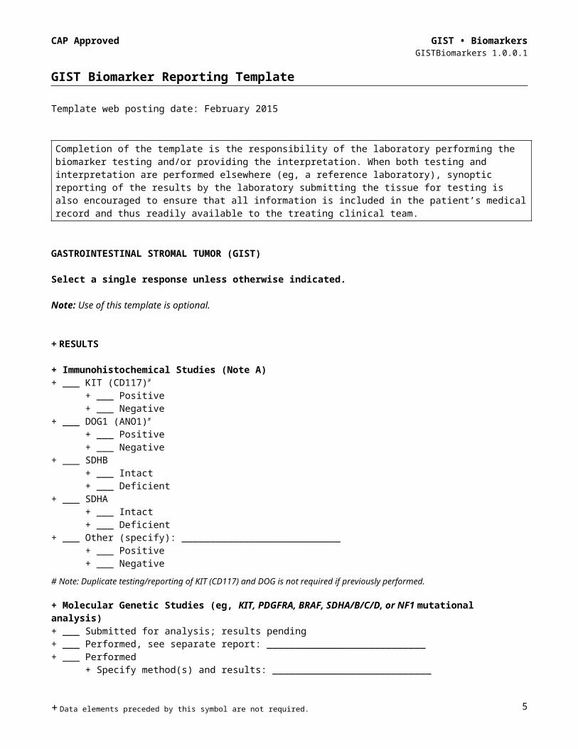
CAP Approved GIST • BiomarkersGISTBiomarkers 1.0.0.1
GIST Biomarker Reporting Template
Template web posting date: February 2015
Completion of the template is the responsibility of the laboratory performing the biomarker testing and/or providing the interpretation. When both testing and interpretation are performed elsewhere (eg, a reference laboratory), synoptic reporting of the results by the laboratory submitting the tissue for testing is also encouraged to ensure that all information is included in the patient’s medical record and thus readily available to the treating clinical team.
GASTROINTESTINAL STROMAL TUMOR (GIST)
Select a single response unless otherwise indicated.
Note: Use of this template is optional.
+ RESULTS
+ Immunohistochemical Studies (Note A)+ ___ KIT (CD117)#
+ ___ Positive+ ___ Negative
+ ___ DOG1 (ANO1)#
+ ___ Positive+ ___ Negative
+ ___ SDHB + ___ Intact+ ___ Deficient
+ ___ SDHA + ___ Intact+ ___ Deficient
+ ___ Other (specify): ____________________________+ ___ Positive+ ___ Negative
# Note: Duplicate testing/reporting of KIT (CD117) and DOG is not required if previously performed.
+ Molecular Genetic Studies (eg, KIT, PDGFRA, BRAF, SDHA/B/C/D, or NF1 mutational analysis)+ ___ Submitted for analysis; results pending+ ___ Performed, see separate report: ____________________________+ ___ Performed
+ Specify method(s) and results: ____________________________+ ___ Not performed
+ KIT Mutational Analysis (Note B)+ ___ No mutation detected + ___ Mutation identified (specify:)____________________+ ___ Cannot be determined (explain): __________________________
+ PDGFRA Mutational Analysis (Note C)+ ___ No mutation detected + ___ Mutation identified (specify): ____________________+ ___ Cannot be determined (explain): __________________________
+ Data elements preceded by this symbol are not required. 4
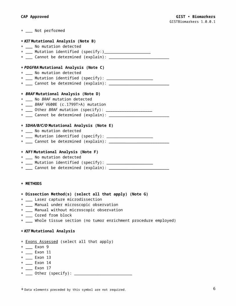
CAP Approved GIST • BiomarkersGISTBiomarkers 1.0.0.1
+ BRAF Mutational Analysis (Note D)+ ___ No BRAF mutation detected + ___ BRAF V600E (c.1799T>A) mutation+ ___ Other BRAF mutation (specify): ____________________+ ___ Cannot be determined (explain): __________________________
+ SDHA/B/C/D Mutational Analysis (Note E)+ ___ No mutation detected + ___ Mutation identified (specify): ____________________+ ___ Cannot be determined (explain): __________________________
+ NF1 Mutational Analysis (Note F)+ ___ No mutation detected + ___ Mutation identified (specify): ____________________+ ___ Cannot be determined (explain): __________________________
+ METHODS
+ Dissection Method(s) (select all that apply) (Note G)+ ___ Laser capture microdissection+ ___ Manual under microscopic observation+ ___ Manual without microscopic observation+ ___ Cored from block+ ___ Whole tissue section (no tumor enrichment procedure employed)
+ KIT Mutational Analysis
+ Exons Assessed (select all that apply)+ ___ Exon 9+ ___ Exon 11+ ___ Exon 13+ ___ Exon 14+ ___ Exon 17+ ___ Other (specify): _________________________
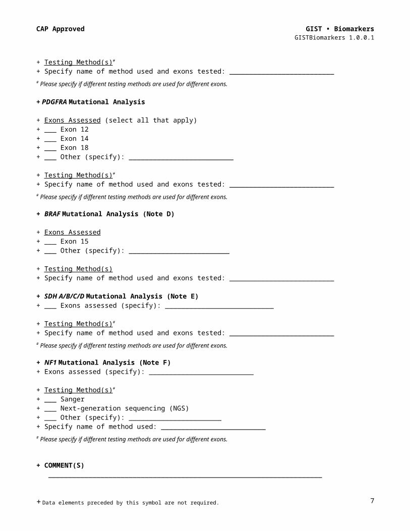
+ Testing Method(s)#
+ Specify name of method used and exons tested: __________________________# Please specify if different testing methods are used for different exons. + PDGFRA Mutational Analysis
+ Exons Assessed (select all that apply)+ ___ Exon 12+ ___ Exon 14+ ___ Exon 18+ ___ Other (specify): __________________________
+ Testing Method(s)#
+ Specify name of method used and exons tested: __________________________# Please specify if different testing methods are used for different exons.
+ Data elements preceded by this symbol are not required. 5
CAP Approved GIST • BiomarkersGISTBiomarkers 1.0.0.1
+ BRAF Mutational Analysis (Note D)
+ Exons Assessed + ___ Exon 15+ ___ Other (specify): _________________________
+ Testing Method(s) + Specify name of method used and exons tested: __________________________
+ SDH A/B/C/D Mutational Analysis (Note E)+ ___ Exons assessed (specify): ___________________________
+ Testing Method(s)#
+ Specify name of method used and exons tested: __________________________# Please specify if different testing methods are used for different exons.
+ NF1 Mutational Analysis (Note F)+ Exons assessed (specify): __________________________
+ Testing Method(s)#
+ ___ Sanger+ ___ Next-generation sequencing (NGS)+ ___ Other (specify): _______________________+ Specify name of method used: __________________________# Please specify if different testing methods are used for different exons.
+ COMMENT(S) ____________________________________________________________________ ____________________________________________________________________
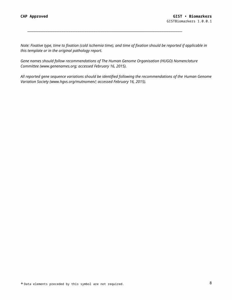
Note: Fixative type, time to fixation (cold ischemia time), and time of fixation should be reported if applicable in this template or in the original pathology report.
Gene names should follow recommendations of The Human Genome Organisation (HUGO) Nomenclature Committee (www.genenames.org; accessed February 16, 2015).
All reported gene sequence variations should be identified following the recommendations of the Human Genome Variation Society (www.hgvs.org/mutnomen/; accessed February 16, 2015).
+ Data elements preceded by this symbol are not required. 6
Background Documentation GIST • BiomarkersGISTBiomarkers 1.0.0.1
Explanatory Notes
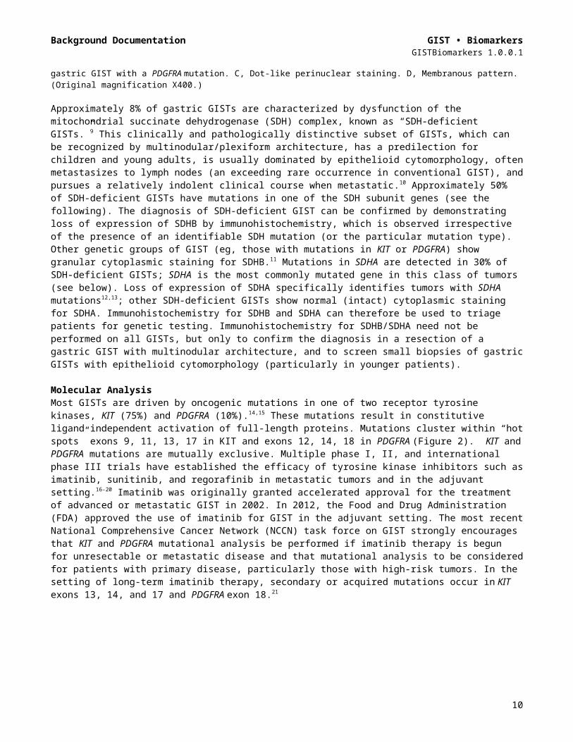
A. Immunohistochemical AnalysisBecause of the advent of small-molecule kinase inhibitor therapy for the treatment of GIST (see the following), it has become imperative to distinguish GIST from its histologic mimics, mainly leiomyoma, leiomyosarcoma, schwannoma, and desmoid fibromatosis.1,2 Immunohistochemistry is instrumental in the workup of GIST. Approximately 95% of GISTs are immunoreactive for KIT (CD117).3 Most KIT-negative GISTs are gastric or omental tumors that harbor mutations in platelet-derived growth factor receptor A (PDGFRA).4 KIT immunoreactivity is usually strong and diffuse but can be more limited in extent in some cases (Figure 1, A and B). It is not unusual for GISTs to exhibit dot-like perinuclear staining (Figure 1, C), while less commonly some cases exhibit membranous staining (Figure 1, D). These patterns do not clearly correlate with mutation type or response to therapy. DOG1 is another highly sensitive and specific marker for GIST, which was discovered by gene expression profiling.5,6 DOG1 (also known as anoctamin 1, ANO1) is particularly useful for KIT-negative tumors and those with limited KIT expression; DOG1 is more sensitive than KIT for gastric epithelioid GISTs.7 Approximately 70% of GISTs are positive for CD34, 30% to 40% are positive for smooth muscle actin, 5% are positive for S100 (usually focal), 5% are positive for desmin (usually focal), and 1% to 2% are positive for keratin (weak/focal).8
Figure 1. Patterns of KIT staining in gastrointestinal stromal tumor (GIST). A, Diffuse and strong immunoreactivity in a typical GIST. B, Focal and weak pattern in an epithelioid gastric GIST with a PDGFRA mutation. C, Dot-like perinuclear staining. D, Membranous pattern. (Original magnification X400.)
Approximately 8% of gastric GISTs are characterized by dysfunction of the mitochondrial succinate dehydrogenase (SDH) complex, known as “SDH-deficient GISTs.”9 This clinically and pathologically distinctive subset of GISTs, which can be recognized by multinodular/plexiform architecture, has a predilection for children and young adults, is usually dominated by epithelioid cytomorphology, often metastasizes to lymph nodes (an exceeding rare occurrence in conventional GIST), and pursues a relatively indolent clinical course when metastatic.10 Approximately 50% of SDH-deficient GISTs have mutations in one of the SDH subunit genes (see the following). The diagnosis of SDH-deficient GIST can be confirmed by demonstrating loss of expression of SDHB by immunohistochemistry, which is observed irrespective of the presence of an identifiable SDH mutation
7
Background Documentation GIST • BiomarkersGISTBiomarkers 1.0.0.1
(or the particular mutation type). Other genetic groups of GIST (eg, those with mutations in KIT or PDGFRA) show granular cytoplasmic staining for SDHB.11 Mutations in SDHA are detected in 30% of SDH-deficient GISTs; SDHA is the most commonly mutated gene in this class of tumors (see below). Loss of expression of SDHA specifically identifies tumors with SDHA mutations12,13; other SDH-deficient GISTs show normal (intact) cytoplasmic staining for SDHA. Immunohistochemistry for SDHB and SDHA can therefore be used to triage patients for genetic testing. Immunohistochemistry for SDHB/SDHA need not be performed on all GISTs, but only to confirm the diagnosis in a resection of a gastric GIST with multinodular architecture, and to screen small biopsies of gastric GISTs with epithelioid cytomorphology (particularly in younger patients).
Molecular AnalysisMost GISTs are driven by oncogenic mutations in one of two receptor tyrosine kinases, KIT (75%) and PDGFRA (10%).14,15 These mutations result in constitutive ligand independent activation of full-length proteins. Mutations cluster within “hot spots” exons 9, 11, 13, 17 in KIT and exons 12, 14, 18 in PDGFRA (Figure 2). KIT and PDGFRA mutations are mutually exclusive. Multiple phase I, II, and international phase III trials have established the efficacy of tyrosine kinase inhibitors such as imatinib, sunitinib, and regorafinib in metastatic tumors and in the adjuvant setting.16-20 Imatinib was originally granted accelerated approval for the treatment of advanced or metastatic GIST in 2002. In 2012, the Food and Drug Administration (FDA) approved the use of imatinib for GIST in the adjuvant setting. The most recent National Comprehensive Cancer Network (NCCN) task force on GIST strongly encourages that KIT and PDGFRA mutational analysis be performed if imatinib therapy is begun for unresectable or metastatic disease and that mutational analysis to be considered for patients with primary disease, particularly those with high-risk tumors. In the setting of long-term imatinib therapy, secondary or acquired mutations occur in KIT exons 13, 14, and 17 and PDGFRA exon 18.21
* Refers to exons involved most frequently by secondary/acquired mutations.
Figure 2. Locations and frequency of activating KIT and PDGFRA mutations in GIST. Adapted with permission from Heinrich et al.14 Copyright 2003 by the American Society of Clinical Oncology. All rights reserved.
B. KIT Mutational Analysis The most common mutations affect the juxta membrane domain encoded by exon 11 (two-thirds of GIST). These mutations include in-frame deletions, substitutions, and insertions. Deletions (in particular codon 557 and/or 558) are associated with shorter progression free and overall survival.22-25 About 7% to 10% of the tumors harbor mutations in the extracellular domain encoded by exon 9 (most commonly insAY502-503).26 Primary mutations in the activation loop (exon 17) and ATP binding region (exon 13) are uncommon (1%). Majority of these mutations are substitutions.27 KIT exon 8 mutations are extremely rare (0.15%).28 Secondary or resistance mutations occur
8

Background Documentation GIST • BiomarkersGISTBiomarkers 1.0.0.1
commonly in tumors harboring primary exon 11 mutations. The newly acquired secondary mutations are always located in exons encoding tyrosine kinase domain (exons 13, 14, 17).29
C. PDGFRA Mutational AnalysisMore than 80% of KIT-negative GISTS have PDGFRA mutations. Activation of PDGFRA is seen in GISTs harboring mutations in juxta membranous domain (exon 12), the ATP binding domain (exon 14), or the activation loop (exon 18).30 Mutations include substitutions and deletions. Primary resistance to imatinib is seen with the most common PDGFRA exon 18 D842V mutation.
D. BRAF Mutational AnalysisActivating mutations of BRAF (V600E) has been identified in a small subset (7%) of KIT/PDGFRA wild-type GISTs. These tumors show a predilection for small bowel location.31
E. SDH A/B/C/D Mutational AnalysisThe succinate dehydrogenase (SDH) complex (mitochondrial complex II) participates in both the Krebs cycle and the electron transport chain of oxidative phosphorylation. About 8% of gastric GISTs (all lacking mutations in KIT and PDGFRA) are caused by dysfunction of the SDH complex ("SDH-deficient GISTs"). Around 50% of patients affected by such tumors harbor germline mutations in one of the SDH subunit genes (SDHA/B/C or D). SDHA-inactivating mutations are most common, detected in about 30% of SDH-deficient GISTs. Mutations involve exons 2, 3, 5, 6, 7, 8, 9, 10, 11, 13, 14 of SDHA; exons 1, 2, 3, 4, 6, 7 of SDHB; exons 1, 4, 5 of SDHC; and exons 4 and 6 of SDHD. While the majority of the mutations are substitutions, deletions, splice-site mutations, frame shift, and duplications have also been reported.9,11,13,32
F. Neurofibromatosis Type 1 (NF1) Mutational AnalysisNF1 is an inherited, autosomal dominant disease characterized by multiple café au lait spots, Lisch nodules, freckling, and development of neurofibromas. GISTs in NF1 patients arise predominantly from the small intestine and can be multicentric and lack KIT and PDGFRA mutations. Until now, no specific genetic alterations have been found in NF1-related GIST.32
G. Dissection Method:While in majority of cases GIST samples show tumor percentage (%) well above the analytical sensitivity of Sanger sequencing (>50% neoplastic cell percentage/20% to 25% mutant allele percentage), in cases of mutation analysis of treated samples, careful macro/microdissection may be necessary to avoid false negative results.
H. Reporting NomenclatureConsistent gene mutation nomenclature is essential for efficient and accurate reporting.33 Following are examples as recommended by Human Genome Variation Society (HGVS) for description of variant changes.34 It is also preferred that protein alterations are mentioned in the report in addition to genomic coordinates.
Examples of DNA, RNA, and Protein NomenclatureDNA: A, G, C, T (example: c.957A>T)RNA: a, g, c, u (example: r.957 a>u)Protein: 3-letter amino acid code, X= Stop codon (example: p. Glu78Gln)
Examples of Nomenclatures for Types of Sequence VariantsTypes of Variation Examples Substitution c.123A>GDeletion c.123delA, c.586_591delTGGTCA or c.586_591del6Duplication c.123dupA, c.586_591dupTGGTCA or c.586_591dup6Insertion c.123_124insC, c.1086_1087insGCGTGAFrame shift p. Arg83 fs or p. Arg83Ser fsX15Deletion/insertions “indel” c.112_117delAGGTCAinsTG
References1. Hornick JL, Fletcher CD. Immunohistochemical staining for KIT (CD117) in soft tissue sarcomas is very
limited in distribution. Am J Clin Pathol. 2002;117(2):188-193.
9
Background Documentation GIST • BiomarkersGISTBiomarkers 1.0.0.1
2. Miettinen M, Sobin LH, Sarlomo-Rikala M. Immunohistochemical spectrum of GISTs at different sites and their differential diagnosis with a reference to CD117 (KIT). Mod Pathol. 2000;13(10):1134-1142.
3. Sarlomo-Rikala M, Kovatich AJ, Barusevicius A, Miettinen M. CD117: a sensitive marker for gastrointestinal stromal tumors that is more specific than CD34. Mod Pathol. 1998;11(8):728-734.
4. Medeiros F, Corless CL, Duensing A, et al. KIT-negative gastrointestinal stromal tumors: proof of concept and therapeutic implications. Am J Surg Pathol. 2004;28(7):889-894.
5. West RB, Corless CL, Chen X, et al. The novel marker, DOG1, is expressed ubiquitously in gastrointestinal stromal tumors irrespective of KIT or PDGFRA mutation status. Am J Pathol. 2004;165(1):107-113.
6. Espinosa I, Lee CH, Kim MK, et al. A novel monoclonal antibody against DOG1 is a sensitive and specific marker for gastrointestinal stromal tumors. Am J Surg Pathol. 2008;32(2):210-218.
7. Miettinen M, Wang ZF, Lasota J. DOG1 antibody in the differential diagnosis of gastrointestinal stromal tumors: a study of 1840 cases. Am J Surg Pathol. 2009;33(9):1401-1408.
8. Miettinen M, Wang ZF, Sarlomo-Rikala M, et al. Succinate dehydrogenase-deficient GISTs: a clinicopathologic, immunohistochemical, and molecular genetic study of 66 gastric GISTs with predilection to young age. Am J Surg Pathol. 2011;35(11):1712-1721.
9. Doyle LA, Hornick JL. Gastrointestinal stromal tumours: from KIT to succinate dehydrogenase. Histopathology. 2014;64(1):53-67.
10. Doyle LA, Nelson D, Heinrich MC, et al. Loss of succinate dehydrogenase subunit B (SDHB) expression is limited to a distinctive subset of gastric wild-type gastrointestinal stromal tumours: a comprehensive genotype-phenotype correlation study. Histopathology. 2012;61(5):801-809.
11. Wagner AJ, Remillard SP, Zhang YX, et al. Loss of expression of SDHA predicts SDHA mutations in gastrointestinal stromal tumors. Mod Pathol. 2013;26(2):289-294.
12. Dwight T, Benn DE, Clarkson A, et al. Loss of SDHA expression identifies SDHA mutations in succinate dehydrogenase-deficient gastrointestinal stromal tumors. Am J Surg Pathol. 2013;37(2):226-233.
13. Fletcher CD, Berman JJ, Corless C, et al. Diagnosis of gastrointestinal stromal tumors: a consensus approach. Hum Pathol. 2002; 33(5):459-465.
14. Heinrich MC, Corless CL, Demetri GD, et al. Kinase mutations and imatinib response in patients with metastatic gastrointestinal stromal tumor. J Clin Oncol. 2003;21(23):4342-4349.
15. Heinrich MC, Corless CL, Duensing A, et al. PDGFRA activating mutations in gastrointestinal stromal tumors. Science. 2003;299(5607):708-710.
16. Hirota S, Isozaki K, Moriyama Y, et al. Gain-of-function mutations of c-kit in human gastrointestinal stromal tumors. Science. 1998;279(5350):577-580.
17. Joensuu H, Roberts PJ, Sarlomo-Rikala M, et al. Effect of the tyrosine kinase inhibitor ST1571 in a patient with metastatic gastrointestinal stromal tumor. N Engl J Med. 2001;344(14):1052-1056.
18. Van Oosterom AT, Judson I, Verweij J, et al. ST1571, an active drug in metastatic gastrointestinal stromal tumors (GIST) an EORTC phase 1 study (abstract). Proc Am Soc Clin Oncol. 2001;20:1a.
19. Demetri GD, von Mehren M, Blanke CD, et al. Efficacy and safety of imatinib mesylate in advanced gastrointestinal stromal tumors. N Engl J Med. 2002;347(7):472-480.
20. Blanke CD, Rankin C, Demetri GD, et al. Phase III randomized intergroup trial assessing imatinib mesylate at two dose levels in patients with unresectable or metastatic gastrointestinal stromal tumors expressing the kit receptor tyrosine kinase: S0033. J Clin Oncol. 2008;26(4):626-632.
21. Verweiji J, Casali PG, Zaleberg J, et al. Progression–free survival in gastrointestinal stromal tumors with high-dose imatinib: randomized trial. Lancet. 2004;364(9440):1127-1134.
22. Gastrointestinal Stromal Tumor Meta-Analysis Group (MetaGIST). Comparison of two doses of imatinib for the treatment of unresectable or metastatic gastrointestinal stromal tumor: a meta-analysis of 1640 patients. J Clin Oncol. 2010;28(7):1247-1253.
23. Heinrich MC, Corless CL, Blanke CD, et al. Molecular correlates of imatinib resistance in gastrointestinal stromal tumors. J Clin Oncol. 2006;24(29):4764-4774.
24. Andersson J, Bumming P, Meis-Kindblom JM, et al. Gastrointestinal stromal tumors with KIT exon 11 deletions are associated with poor prognosis. Gastroenterology. 2006;130(6):1573-1581.
25. Liu XH, Bai CG, Xie Q, et al. Prognostic value of KIT mutation in gastrointestinal stromal tumors. World J Gastroenterol. 2005;11(25):3948-3952.
26. Wardelmann E, Losen I, Hans V, et al. Deletion of Trp-557 and Lys-558 in the juxtamembrane domain of the c-kit protooncogene is associated with metastatic behavior of gastrointestinal stromal tumors. Int J Cancer. 2003;106(6):887-895.
10

Background Documentation GIST • BiomarkersGISTBiomarkers 1.0.0.1
27. Lux ML, Rubin BP, Biase TL, et al. KIT extracellular and kinase domain mutations in gastrointestinal stromal tumors. Am J Pathol. 2000;156(3):791-795.
28. Huss S, Künstlinger H, Wardelmann E, et al. A subset of gastrointestinal stromal tumors previously regarded as wild-type tumors carries somatic activating mutations in KIT exon 8 (p. D419del). Mod Pathol. 2013;26(7):1004-1012.
29. Lasota J, Corless CL, Heinrich MC, et al. Clinicopathologic profile of gastrointestinal stromal tumors (GISTs) with primary KIT exon 13 or 17 mutations: a multicenter study of 54 cases. Mod Pathol. 2008;21(4):476-484
30. LaCosta J, Miettinen M. Clinical significance of oncogenic KIT and PDGFRA mutations in gastrointestinal stromal tumors. Histopathology. 2008;53(3):245-266.
31. Agaram NP, Wong GC, Guo T, et al. Novel V600E BRAF mutations in imatinib-naive and imatinib-resistant gastrointestinal stromal tumors. Genes Chromosomes Cancer. 2008;47(10):853–859.
32. Nannini, M, Biasco B, Astolfi A, et al. An overview on molecular biology of KIT/PDGFRA wild type (WT) gastrointestinal stromal tumours (GIST). J Med Genet. 2013;50(10):653-661.
33. Ogino S, Gulley M, den Dunneb JT, et al. Standard mutation nomenclature in molecular diagnostics: practical and educational challenges. J Mol Diagn. 2007;9(1):1-6.
34 den Dunnen JT, Antonarakis SE. Mutation nomenclature. Curr Protoc Hum Genet. 2003;Chapter 7, Unit 7.13. http://www.currentprotocols.com/WileyCDA/CPUnit/refId-hg0713.html. Accessed October 29, 2014.
11