thandimurape.files.wordpress.com · Web viewGain settings should be applied to avoid artifactual...
6
DEEP VEIN THROMBOSIS SCANNING TECHNIQUE - No preparation is required for this examination - Examination room to be warm to promote vasodilation - Patient history and referral should be read and details confirmed with patient. - A high frequency linear probe, 9MHz is most suitable for most patients because it allows sufficient penetration. A 3-5 MHz curved transducer is used for abdominal vessels and suitable to use on big patients. Gain settings should be applied to avoid artifactual internal echoes in the vein (Hamper, DeJong and Scoutt 2007,527). - B-mode with compression and colour flow imaging are the core of this examination (Zwiebel & Pellerito, 2005, 422). - With patient lying supine semi-erect, examination begins at the groin area. - Using a dual or split screen, all veins are examined in B-mode and in Transverse - Pressure is exerted to compress the vein every centimeter such that the vein collapses completely leaving an adjacent artery slightly distorted. It might be necessary to use the two-hand technique which involves pushing the leg into the transducer with the free hand from behind the thigh/leg to achieve adequate compression eg FV distal (Hamper,DeJong and Scoutt 2007, 529). * Relating to video on scanning technique: - - Check veins per section eg upper leg, behind knee and lower leg – scout scanning - Pay more attention when scanning areas where DVT is prone to occur eg FV dist, pop vein, soleal veins and gastroc veins - Usually where there is pain, swelling or change of skin colour, the vein itself on ultrasound will appear distended. - Some veins are quite small and easily compressed, avoid too much pressure.
-
Upload
phungkhanh -
Category
Documents
-
view
212 -
download
0
Transcript of thandimurape.files.wordpress.com · Web viewGain settings should be applied to avoid artifactual...

DEEP VEIN THROMBOSIS SCANNING TECHNIQUE
- No preparation is required for this examination
- Examination room to be warm to promote vasodilation
- Patient history and referral should be read and details confirmed with patient.
- A high frequency linear probe, 9MHz is most suitable for most patients because it allows sufficient penetration. A 3-5 MHz curved transducer is used for abdominal vessels and suitable to use on big patients. Gain settings should be applied to avoid artifactual internal echoes in the vein (Hamper, DeJong and Scoutt 2007,527).
- B-mode with compression and colour flow imaging are the core of this examination (Zwiebel & Pellerito, 2005, 422).
- With patient lying supine semi-erect, examination begins at the groin area.
- Using a dual or split screen, all veins are examined in B-mode and in Transverse
- Pressure is exerted to compress the vein every centimeter such that the vein collapses completely leaving an adjacent artery slightly distorted. It might be necessary to use the two-hand technique which involves pushing the leg into the transducer with the free hand from behind the thigh/leg to achieve adequate compression eg FV distal (Hamper,DeJong and Scoutt 2007, 529).
* Relating to video on scanning technique: -
- Check veins per section eg upper leg, behind knee and lower leg – scout scanning
- Pay more attention when scanning areas where DVT is prone to occur eg FV dist, pop vein, soleal veins and gastroc veins
- Usually where there is pain, swelling or change of skin colour, the vein itself on ultrasound will appear distended.
- Some veins are quite small and easily compressed, avoid too much pressure.
- DVT study can be done by compression alone but colour Doppler does add finer details of the veins
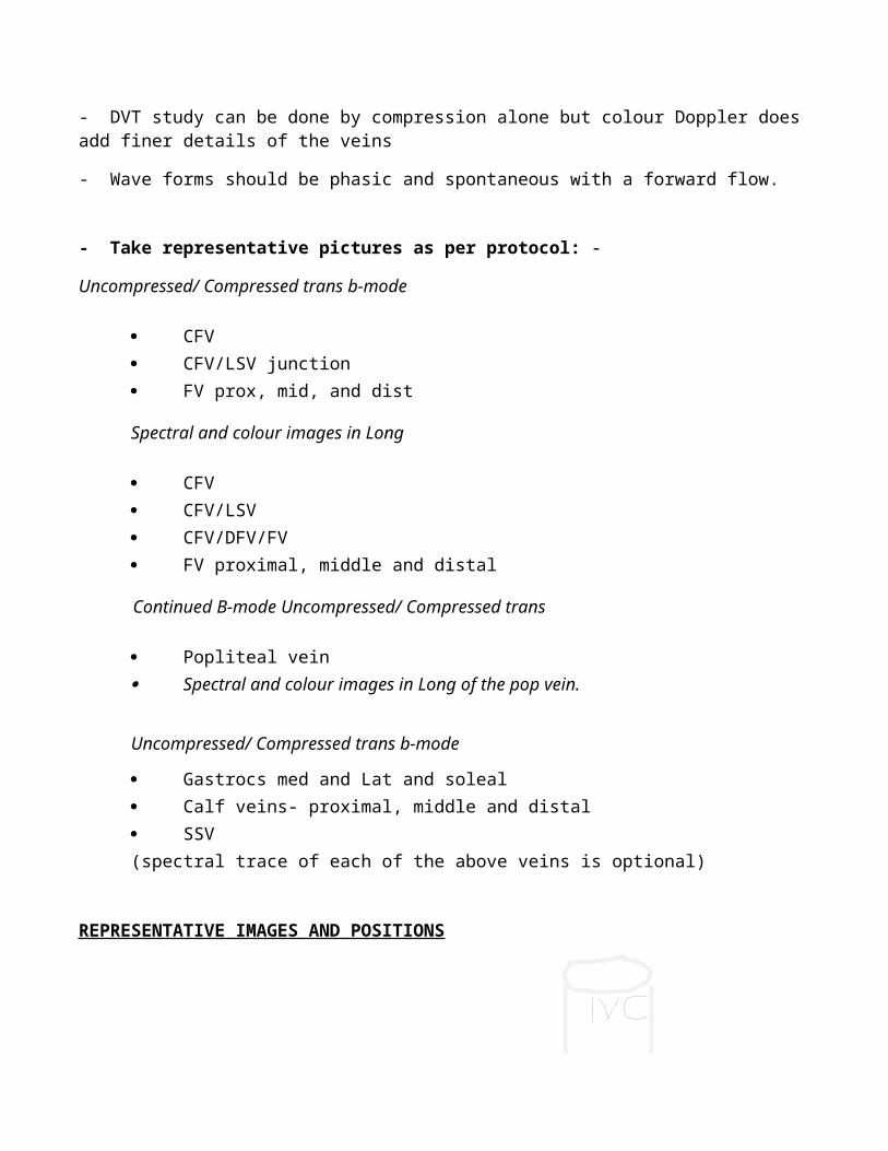
- Wave forms should be phasic and spontaneous with a forward flow.
- Take representative pictures as per protocol: -
Uncompressed/ Compressed trans b-mode
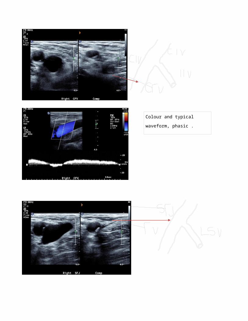
CFV CFV/LSV junction
FV prox, mid, and dist
Spectral and colour images in Long
CFV CFV/LSV CFV/DFV/FV FV proximal, middle and distal
Continued B-mode Uncompressed/ Compressed trans
Popliteal vein Spectral and colour images in Long of the pop vein.
Uncompressed/ Compressed trans b-mode
Gastrocs med and Lat and soleal Calf veins- proximal, middle and distal SSV(spectral trace of each of the above veins is optional)
REPRESENTATIVE IMAGES AND POSITIONS
Colour and typical waveform,
phasic .
SFJ showing forward flow of
blood.

B-mode image of the CFV, FV and
DF

REFERENCES:-
Ulrike M., Hamper., Rober M., DeJong and Leslie M., Scout. 2007. Ultrasound Evaluation of the Lower
Extremity Veins. Radiologic Clinics of North America. Issue 45. USA. Elsevier. Doi:
10.1016/j.rcl.2007.04.013.
William J., Zwiebel and John S., Pellerito. 2005. Introduction to Vascular Ultrasonography. 5th edition.
China. Elsevier Saunders.