عرض تقديمي في PowerPointsite.iugaza.edu.ps/eshaqoura/files/Large-Venis-ar... · Patent...
34
Transcript of عرض تقديمي في PowerPointsite.iugaza.edu.ps/eshaqoura/files/Large-Venis-ar... · Patent...

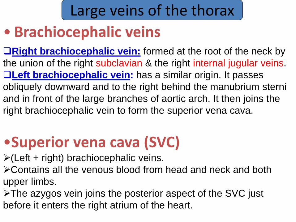
• Brachiocephalic veins
Large veins of the thorax
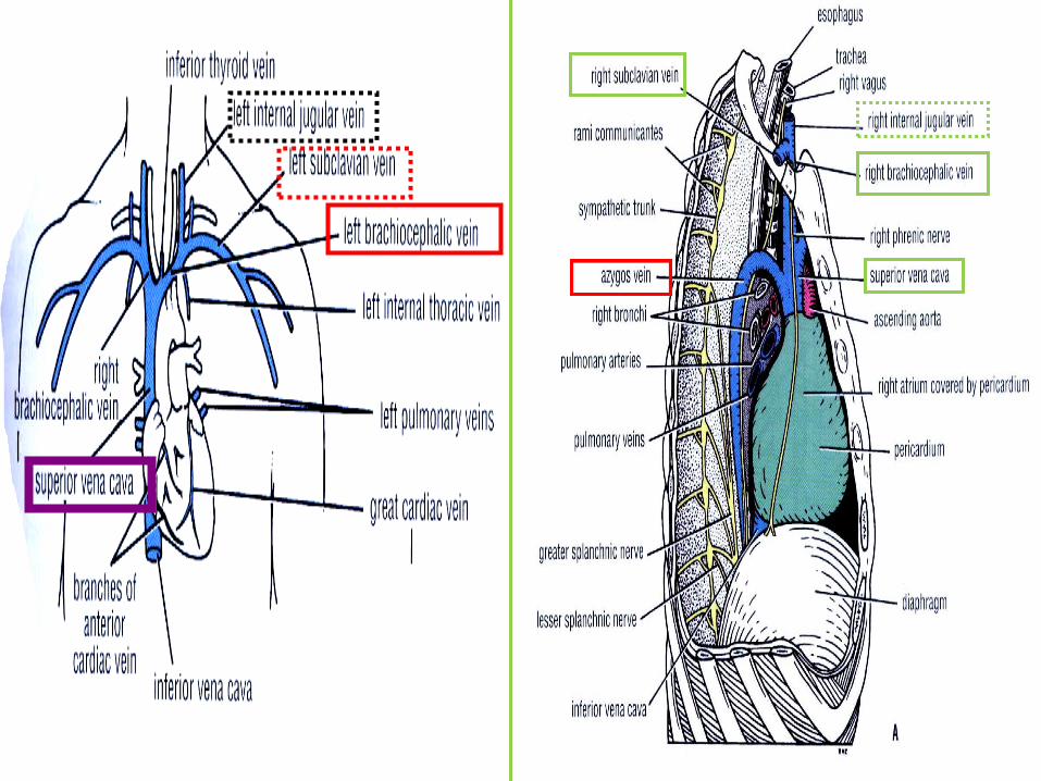
Right brachiocephalic vein: formed at the root of the neck by
the union of the right subclavian & the right internal jugular veins.
Left brachiocephalic vein: has a similar origin. It passes
obliquely downward and to the right behind the manubrium sterni
and in front of the large branches of aortic arch. It then joins the
right brachiocephalic vein to form the superior vena cava.
•Superior vena cava (SVC) (Left + right) brachiocephalic veins.
Contains all the venous blood from head and neck and both
upper limbs.
The azygos vein joins the posterior aspect of the SVC just
before it enters the right atrium of the heart.

Large veins of the thorax
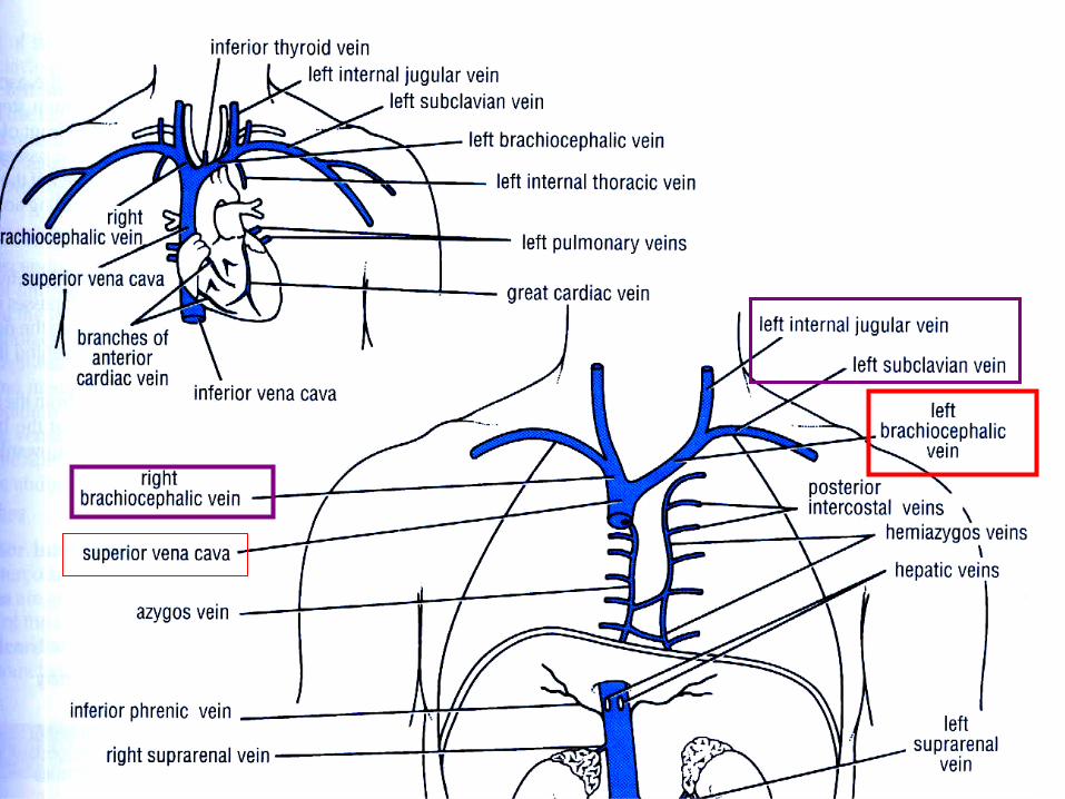
• Azygos veins Main azygos V +inferior hemiazygos V +superior hemiazygos V
They drain blood from:-
The posterior parts of the intercostal spaces
Posterior abdominal wall
The pericardium
The diaphragm
The bronchi
The esophagus
Consist of:

Main Azygous vein : Origin variable, but usually formed by union of the Rt. ascending lumbar vein and the Rt. subcostal vein. At level T5 – it arches forward to empty into post aspect of SVC.
The azygos vein has numerous tributaries, including 8 lower right intercostal veins, the right superior intercostal vein, the superior & inferior hemiazygos veins, and numerous mediastinal veins.
Inferior hemiazygos vein: Formed by LT. ascending lumbar vein + Lt. subcostal vein It ascends through the left crus of diaphragm and, at about the level of the 8th thoracic vertebra, it turns to the right and joins the azygos vein.
Superior hemiazygos vein: formed by union of the 4th to 8th intercostal veins At level T7, it joins the azygos vein

.


Large veins of the thorax
• Inferior vena cava Pierce the central tendon of diaphragm at level T8 Enters the lowest part of the Rt atrium


Azygos vein and caval obstruction
In case of obstruction
of superior or inferior
venae cavae, the azygos
veins provide an
alternative pathway to
return venous blood to
the right atrium of the
heart.
This is possible
because azygos veins
and their tributaries
connect the superior and
inferior venae cavae.

Large veins of the thorax
• Pulmonary veins Leave each lung carrying oxygenated blood to LA


Large arteries of the thorax
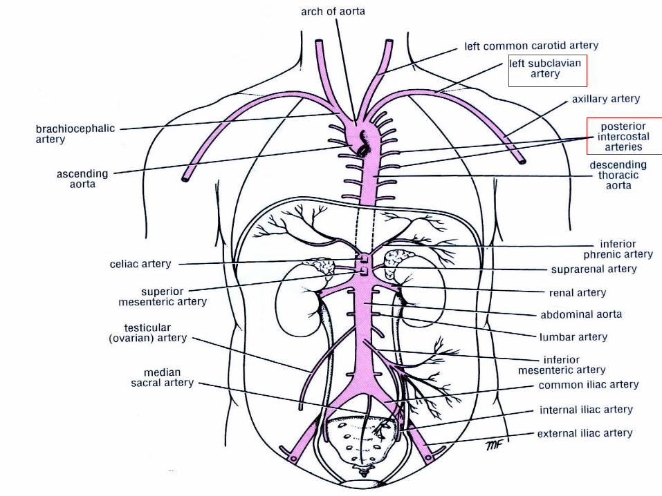
• Aorta is the main arterial trunk that delivers oxygenated blood from
the left ventricle of the heart to the tissues of the body.
Aorta
Ascending aorta
Arch of the aorta
Descending aorta
Abdominal Thoracic

Large arteries of the thorax • Ascending aorta It is contained in the pericardial sac and covered by a visceral
layer of serous pericardium, which also surrounds the pulmonary
trunk in a common sheath
Begins at the base of the left ventricle
Runs upward and forward to Rt half of sternum (sternal angle), At
this point, it enters the superior mediastinum and is then referred
to as the aortic arch. Superior to this point, it posses three bulges
(sinuses) at the root which are posterior, right, and left aortic
sinuses.
•The right and left coronary arteries originate from the anterior and posterior aortic sinuses, respectively.


Large arteries of the thorax • Arch of the aorta Lies behind the manubrium sterni
arches upward, backward, and to the left
At level of sternal angle, it becomes continuous with descending
aorta. Branches of Aorta
Brachiocephalic artery
Left common carotid artery
Left subclavian artery

Large arteries of the thorax • Descending thoracic aorta
Lies in the posterior mediastinum, it begins as a continuation of
the arch of the aorta on the left side of the lower border of the
body of the 4th thoracic vertebra
Reach anterior surface of vertebral column
At level T12, it passes behind the diaphragm and becomes
continuous with the abdominal aorta.
Branches:
Posterior intercostal arteries
Subcostal arteries
Pericardial, esophageal, bronchial arteries


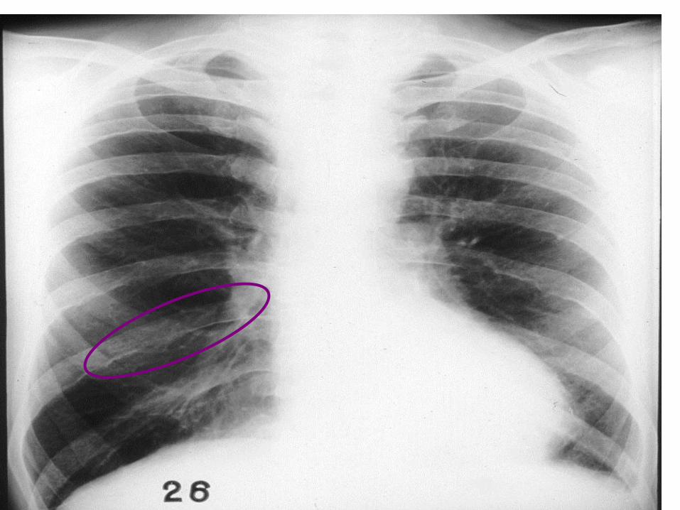
Aneurysm and coarctation of the aorta Aortic aneurysm:
•The arch of the aorta lies behind the manubrium sterni.
•A gross dilatation of the aorta (aneurysm) may show itself as a pulsatile swelling in the suprasternal notch.
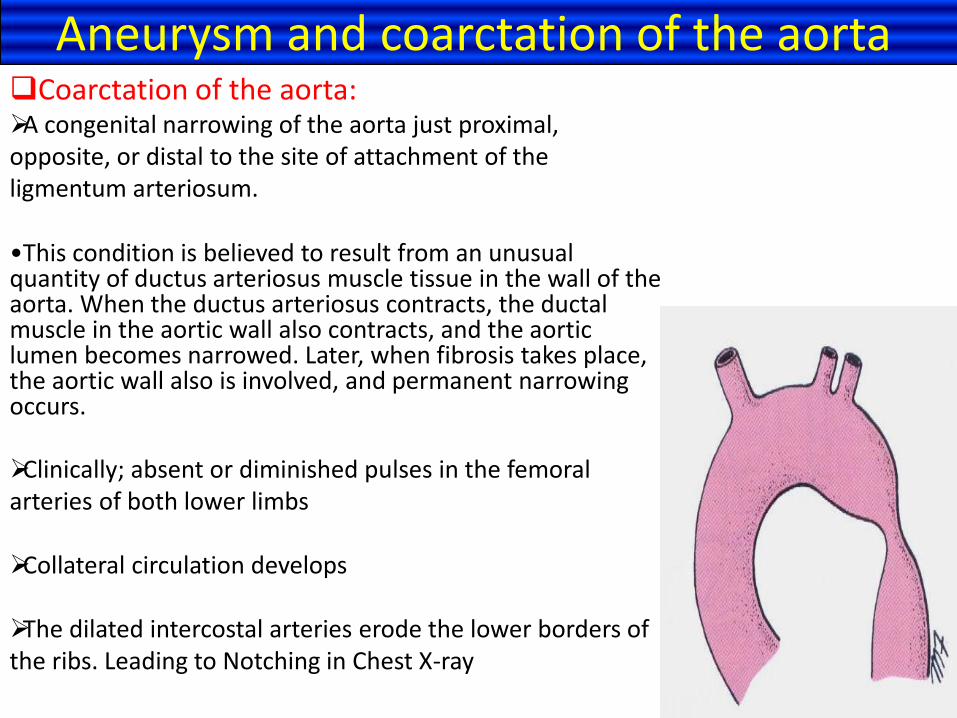
Coarctation of the aorta: A congenital narrowing of the aorta just proximal, opposite, or distal to the site of attachment of the ligmentum arteriosum.
•This condition is believed to result from an unusual quantity of ductus arteriosus muscle tissue in the wall of the aorta. When the ductus arteriosus contracts, the ductal muscle in the aortic wall also contracts, and the aortic lumen becomes narrowed. Later, when fibrosis takes place, the aortic wall also is involved, and permanent narrowing occurs.
Clinically; absent or diminished pulses in the femoral arteries of both lower limbs
Collateral circulation develops
The dilated intercostal arteries erode the lower borders of the ribs. Leading to Notching in Chest X-ray
Aneurysm and coarctation of the aorta

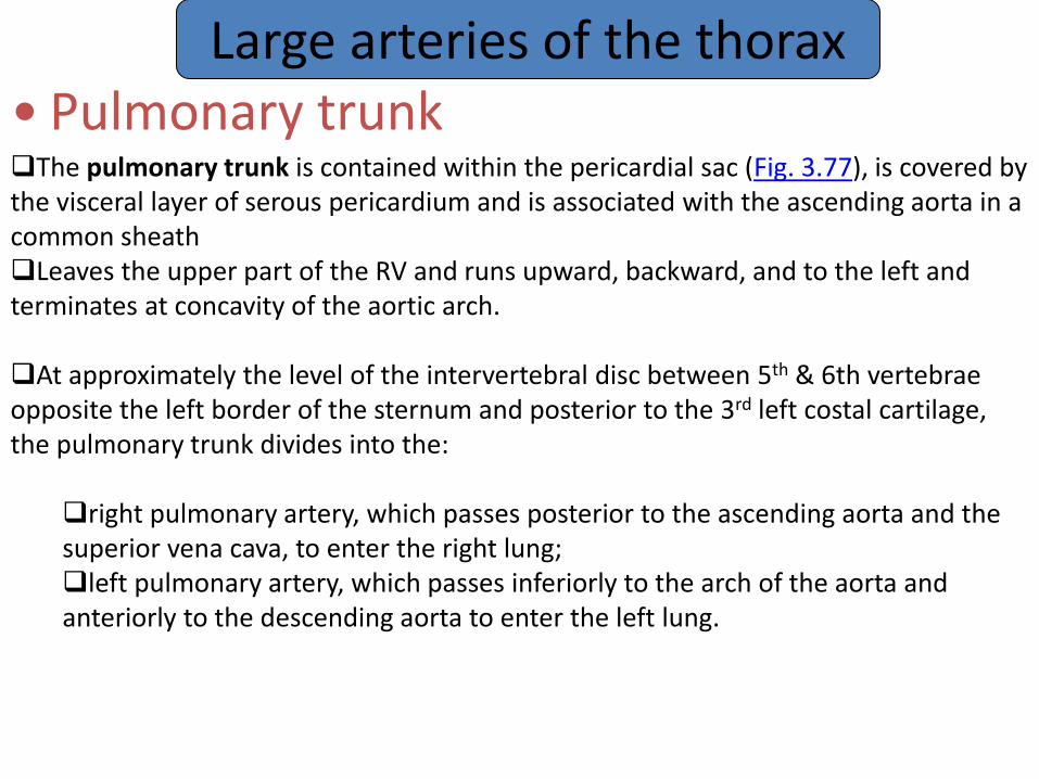
Large arteries of the thorax • Pulmonary trunk The pulmonary trunk is contained within the pericardial sac (Fig. 3.77), is covered by the visceral layer of serous pericardium and is associated with the ascending aorta in a common sheath Leaves the upper part of the RV and runs upward, backward, and to the left and terminates at concavity of the aortic arch.
At approximately the level of the intervertebral disc between 5th & 6th vertebrae opposite the left border of the sternum and posterior to the 3rd left costal cartilage, the pulmonary trunk divides into the:
right pulmonary artery, which passes posterior to the ascending aorta and the superior vena cava, to enter the right lung; left pulmonary artery, which passes inferiorly to the arch of the aorta and anteriorly to the descending aorta to enter the left lung.


Patent ductus arteriosus
Failure of the ductus arteriosus to
close shortly after birth results in a
shunt of blood from the aorta into
the pulmonary trunk, which may
lead to CHF.
PDA occurs in approximately 1 in
2000–2500 births (10% of
congenital heart defects) and can
be treated medically, or surgically
if necessary. The latter treatment
is by direct surgical ligation or via
a less invasive catheter-based
device


Large arteries of the thorax • Ligamentum arteriosum Fibrous band connects the bifurcation of the pulmonary trunk to the lower concave surface of the aortic arch It is the remains of the ductus arteriosus The left recurrent laryngeal nerve hooks around lower border

Lymph nodes and Lymph vessels • Thoracic wall Anterior axillary nodes: anterior thoracic wall Posterior axillary nodes: posterior thoracic wall Internal thoracic nodes: anterior intercostal spaces Posterior intercostal nodes: posterior intercostal spaces
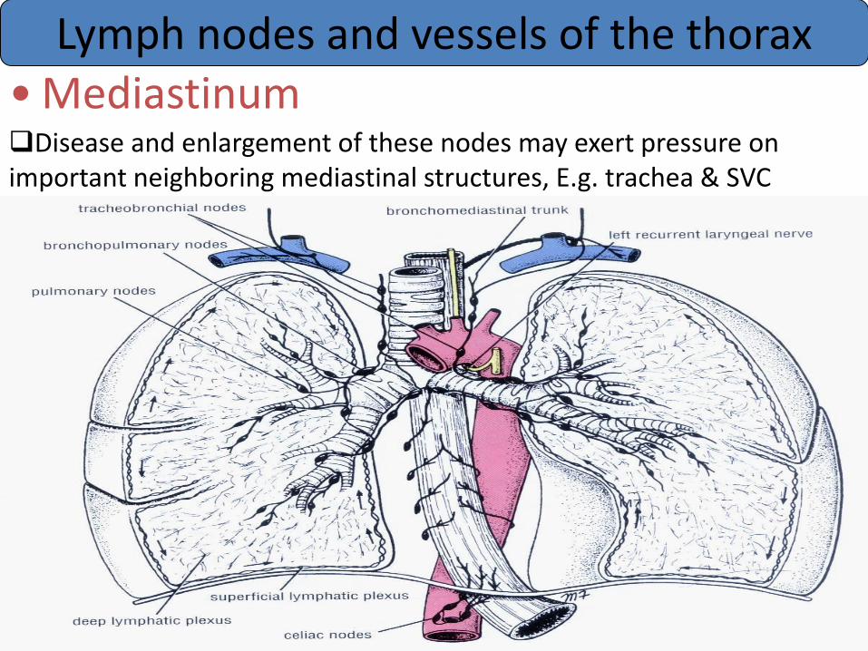
Lymph nodes and vessels of the thorax • Mediastinum Disease and enlargement of these nodes may exert pressure on important neighboring mediastinal structures, E.g. trachea & SVC

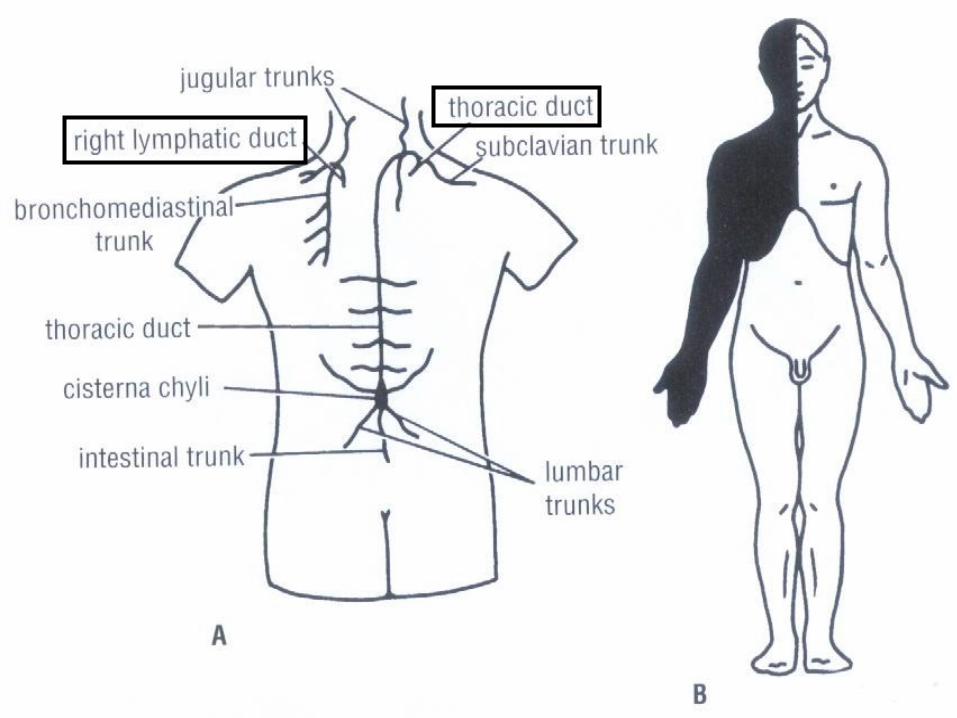
Lymph nodes and vessels of the thorax • Thoracic duct Begins in the abdomen as cisterna chyli Ascends through the aortic opening in the diaphragm Crosses the median plane behind esophagus at sternal angle Runs upward along the left edge of esophagus Enter the beginning of the left brachiocephalic vein
At the root of the neck receives:= Left jugular Subclavian Bronchomediastinal lymph trunks
Conveys to the blood all lymph from Lower limbs, pelvis, abdomen, Left side of (thorax, head, neck,
arm)

Lymph nodes and vessels of the thorax • Right lymphatic duct Opens into the beginning of right brachiocephalic vein
Conveys to the blood all lymph from right side of (thorax, head, neck, arm)