eprints.gouni.edu.ngeprints.gouni.edu.ng/341/1/AKAH, LUCY.docx · Web vieweprints.gouni.edu.ng
71
PHYTOCHEMICAL ANALYSIS AND ANTIMICROBIAL ACTIVITY OF Acalypha wilkesiana EXTRACT AGAINST CLINICAL ISOLATES OF Candida albicans PRESENTED BY AKAH, LUCY AWORI U14/NAS/MCB/042 SUPERVISED BY MRS. ONYINYE OZOKONKWO A PROJECT WORK SUMMITTED TO THE DEPARTMENT OF MICROBIOLOGY, FACULTY OF NATURAL AND APPLIED SCIENCES IN PARTIAL FULFILLMENT OF THE REQUIREMENTS FOR THE FOR AWARD OF BACHELOR OF SCIENCE (B.Sc.) DEGREE IN MICROBIOLOGY 1
Transcript of eprints.gouni.edu.ngeprints.gouni.edu.ng/341/1/AKAH, LUCY.docx · Web vieweprints.gouni.edu.ng

PHYTOCHEMICAL ANALYSIS AND ANTIMICROBIAL ACTIVITY OF Acalypha wilkesiana EXTRACT AGAINST CLINICAL ISOLATES OF
Candida albicans
PRESENTED
BY
AKAH, LUCY AWORIU14/NAS/MCB/042
SUPERVISED BYMRS. ONYINYE OZOKONKWO
A PROJECT WORK SUMMITTED TO THE DEPARTMENT OF MICROBIOLOGY, FACULTY OF NATURAL AND APPLIED
SCIENCES
IN PARTIAL FULFILLMENT OF THE REQUIREMENTS FOR THE FOR AWARD OF BACHELOR OF SCIENCE (B.Sc.) DEGREE IN
MICROBIOLOGY
GODFREY OKOYE UNIVERSITY
JULY, 2018
1
APPROVAL PAGE
This project was submitted and approved by the department of Microbiology, Godfrey Okoye
University, Enugu.
---------------------------------- -----------------------------------AKAH, LUCY AWORI DATESTUDENT
---------------------------------- -----------------------------------MRS. ONYINYE OZOKONKWO DATEPROJECT SUPERVISOR
---------------------------------- -----------------------------------DR. M. N. UNACHUKWU DATEHEAD OF DEPARTMENT
2
DEDICATION
To Cephas , whose existence however brief brought the greatest joy to my life.
To Israel, Abigail, Marvelous, Mildred, Anna, Scholastica, Kate and little Divine.
3
ACKNOWLEDGEMENT
I am eternally grateful to the almighty God for his guidance, immeasurable love, abundant
blessings and unmerited grace. It is said that no individual can accomplish any good venture
without assistance from others. I remain grateful to my supervisor Mrs. Onyinye Ozokonkwo
who despite her tight schedule and engagements patiently took time to nurture, source out help
for me, scrutinize and correct my work. Her immense support, ideas, sacrifices and contributions
during the course of this work is immeasurable. I want to say a big thank you to my HOD Dr.
(Mrs.) M. N. Unachukwu words cannot express my immense gratitude for your support and
counsel.
My profound gratitude goes to my mama, Mrs. Anna Akah a jewel of inestimable value, for her
unfailing support and provision and my Dad, Mr. Kingsley Akah whose advice and teachings
steered me in the right direction and My Uncle, Hon. Ignatius Adida Agabi for his support and
love. I remain grateful to all my lecturers and staff of Microbiology department especially Prof
Nduka Okafor, Mr. Okolo, Prof (Mrs) J. I. Okafor, Miss Irene, Miss Chinenye, Miss Adaeze for
the training, support and assistance given to me at different stages of this study.
To my indescribable siblings (Mrs Agabi Kate, Marc, Papa Ayo and Martha) and my three best
friends Biiwom, Amee and Dalok whose companionship and love made my life infinitely more
rewarding, thank you all for being mine and loving me unconditionally.
A big thank you to the real ones Bassa, Asa, Tejiri, Uncle Mike, Ezechukwu, Cheta, Kosi,
Onyinye, Prosper and my amiable partners Jonathan, Christian and Charity, To Plangnan, Nkay,
Ijay, Dusu, Beccanus, Kylie XY, Jessy, Ochy, to the all of my colleagues and others too
numerous to me. May God bless you all.
4

TABLE OF CONTENTS
Title . . . . . . . . . . . i
Approval . . . . . . . . . . . ii
Dedication. . . . . . . . . . . . iii
Acknowledgements . . . . . . . . . . iv
Table of contents. . . . . . . . . . . v
List of tables . . . . . . . . . . viii
List of figures . . . . . . . . . . ix
Abstract. . . . . . . . . . . . x
CHAPTER ONE: INTRODUCTION
1.1 Introduction . . . . . . . . . 1
1.2 Aim / Objectives. . . . . . . . . 3
CHAPTER TWO: LITERATURE REVIEW
2.1 Historic use of plants as antimicrobials. . . . . . . 4
2.2 Acalypha wilkesiana . 5
2.2.1 Pharmacotherapeutic uses of Acalypha wilkesiana . . 6
2.2.2 Toxicity studies on Acalypha wilkesiana 7
2.3 Candida albicans . . . . . . . . 9
2.3.2 Scientific classification - - - - - - - - - - - - - - - - - - - 10
2.3.3 Morphology of C. albicans - - - - - - - - - - - - - - - - - - - - - - - - - 10
2.3.4 Genome of C. albicans - - - - - - - - - - - - - - - - - - - - - - - - - - - - 11
2.3.5 Role in disease - - - - - - - - - - - - - - - - - - - - - - - - - - - - - - - - - - - -12
5
2.3.5.1 Route of transmission - - - - - - - - - - - - - - - - - - - - - - - - - - - - - - -13
2.3.5.2 Pathogenesis - - - - - - - - - - - - - - - - - - - - - - - - - - - - - - - - - 13
2.3.5.3 Prevention - - - - - - - - - - - - - - - - - - - - - - - - - - - - - - - - - - -14
2.3.5.4 Clinical manifestations - - - - - - - - - - - - - - - - - - - - - - - - - - - - 14
2.3.5.5 Diagnosis - - - - - - - - - - - - - - - - - - - - - - - - - - - - - - - - - - - 15
3.0 CHAPTER THREE
3.1 Materials and methods - - - - - - - - - - - - - - - - - - - - - - - - - - - - - - 16
3.1.1 Collection and maintenance of test microorganism - - - - - - - - - - - - - - - -16
3.1.2 Collection of plant material - - - - - - - - - - - - - - - - - - - - - - - - - - - - 16
3.1.3 Preparation of plant material - - - - - - - - - - - - - - - - - - - - - - - - - - - 16
3.1.4 Hot water extraction of A. wilkesiana - - - - - - - - - - - - - - - - - - - - - - - 16
3.1.5 Ethanol extraction of A. wilkesiana - - - - - - - - - - - - - - - - - - - - - - - -17
3.1.6 Methanol extraction of A. wilkesiana - - - - - - - - - - - - - - - - - - - - - - 17
3.2 Phytochemical analysis (qualitative analysis) - - - - - - - - - - - - - - - - - - - - -17
3.2.1 Test for alkaloids - - - - - - - - - - - - - - - - - - - - - - - - - - - - - - - - - - 17
3.2.2 Test for flavonoids - - - - - - - - - - - - - - - - - - - - - - - - - - - - - - - - - -18
3.2.3 Test for tannin and phenolic compounds - - - - - - - - - - - - - - - - - - - - - - -18
6
3.2.4 Test for saponins - - - - - - - - - - - - - - - - - - - - - - - - - - - - - - - - - -18
3.2.5 Test for steroids - - - - - - - - - - - - - - - - - - - - - - - - - - - - - - - - - - - - - 18
3.2.6 Test for glycoside - - - - - - - - - - - - - - - - - - - - - - - - - - - - - - - - - - 19
3.3 Preparation of media used - - - - - - - - - - - - - - - - - - - - - - - - - - - - - - 19
3.4 Antimicrobial sensitivity assay - - - - - - - - - - - - - - - - - - - - - - - - - - - - - - - 19
3.4.1 Disk diffusion method - - - - - - - - - - - - - - - - - - - - - - - - - - - - - - - - 19
3.4.2 Agar well diffusion method - - - - - - - - - - - - - - - - - - - - - - - - - - - - - - - - - - - - - - - -20
3.5 Determination of Minimum Inhibitory Concentration (MIC) - - - -- -- -- - - - - - - - - - - - 20
3.6 Determination of Minimum Fungicidal Concentration (MFC) - - - - - - - - - - - - - - - - - - 21
4.0 CHAPTER FOUR
4.0 Results - - - - - - - - - -- - - - - - - -- - - - - - - - - - - - - - - - - - - - - - - - - - - - - - - - - - - - - -22
5.0 CHAPTER FIVE
5.1 Discussion - - - - - - - - - - - - - - - - - - - - - - - - - - - -- - - - - - - - - - - - - - - - - - - - - - - - - 31
Conclusion-------------------------------------------------------------------------------------------------34
References ------------------------------------------------------------------------------------------------35
7

LIST OF TABLES
Table 1: Qualitative phytochemical screening of the aqueous, ethanol, and methanol extracts of
Acalypha wilkesiana - - - - - - - - - - - - - - - - - - - - - - - - - - - - - - - - - - - - - - - - - - - - - - - - - - 22
Table 2: Minimum inhibitory concentration (MIC) of ethanol extracts broth dilution method- -26
Table 3: Minimum inhibitory concentration (MIC) of methanol extracts broth dilution method 27
Table 4: Minimum inhibitory concentration (MIC) of ethanol extracts microtiter plate method 28
Table 5: Minimum inhibitory concerntration (MIC) of methanol extracts microtiter plate method - - - - - - - - - - - - - - - - - - - - - - - - - - - - - - - - - - - - - - - - - - - - - - - - - - - - - - - - - - - - -- - - -- 29
Table 6: Minimum fungicidal concentration (MFC) of ethanol extract - - - - - - - - - - - - - - - - - 30
Table 7: Minimum fungicidal concentration (MFC) of methanol extract - - - - - - - - - - - - -- - - 31
8

LIST OF FIGURES
Figure 1: Antimicrobial activity of ethanol extract --------------------------------------- 23
Figure 2: Antimicrobial activity of methanol extract-------------------------------------- 24
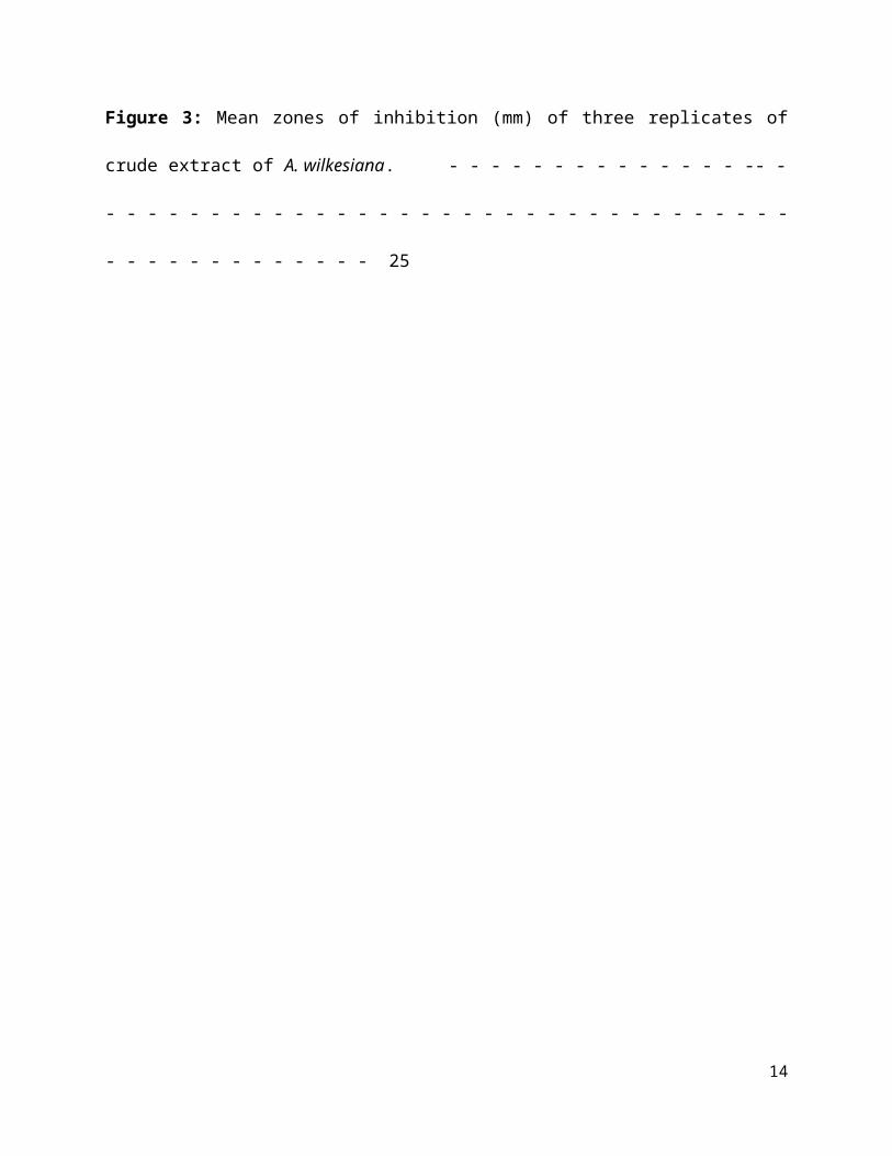
Figure 3: Mean zones of inhibition (mm) of three replicates of crude extract of A. wilkesiana.
- - - - - - - - - - - - - - -- - - - - - - - - - - - - - - - - - - - - - - - - - - - - - - - - - - - - - - - - - - - - - - - 25
9

ABSTRACT
The increased, sustained interest in the production of plant-based drugs for the treatment of many diseases has become a significant reason why people have become more interested in the use of traditional medicine for the treatment of mild and serious illness. Due to increase in the thrust for the production of plant-based antimicrobials, this study was performed on Acalypha wilkesiana against clinical isolates of Candida albicans. The plant extracts were prepared by using ethanol, methanol and hot water. The extracts were evapourated to dryness using a water bath set at 50oC. The plant extracts were tested for the presence of bioactive compounds such as tannins, saponins, glycosides, flavonoids, alkaloid, and steroids. The antimicrobial sensitivity assay of the extracts was studied by agar diffusion method against Candida albicans. The minimum inhibitory concentration was determined by broth dilution method and microtiter plate method. The results of this study showed the presence of the bioactive compounds tested for with the exception of alkaloid. It also showed that in the agar diffusion method the aqueous extracts did not display antimicrobial activity against the test organism, while the ethanol and methanol extracts were effective against the test organism, the methanolic extract demonstrated the highest activity against the test organisms with a mean zone of inhibition of 18mm. The study revealed that the plant contains active bioactive constituents which may hinder the growth of fungi causing skin irritation especially those in the genus of Candida.
10

CHAPTER ONE
1.0 INTRODUCTION
The use and history of herbs dates back to the time of early man, who had the crudest tools as his
implements and used stones to start his fire. Herbs were used in a raw state and cooked forms to
keep fit. Since that time, the use of herbs has been known and accepted by all nations and has
been known also as the first art of treatment available to man (Kafaru, 1994). The search for
natural products to cure diseases has received considerable attentions in which plants have been
the most important source (Okwu, 2001). Herbal preparations form the basis for many
therapeutic drugs and are the first line treatment for many of the world’s population, being
readily available and relatively inexpensive (Olaniyi, 1998; Okpara et al., 2007). Herbal
medicinal products are assuming greater roles in the lives of the people across the world in the
face of global upsurge of drug resistance, toxicity, adverse effects and increasing costs of
synthetic products (Mbi, 1998). In Nigeria, several thousands of plant species have been claimed
to possess medicinal properties and employed in the treatment of many ailments (Oludare, 2011).
Many of these indigenous medicinal plants are used as spices and food plants and for medicinal
purposes (Nwaogu, 20l7). Medicinal plants are believed to be an essential source of new
chemical substances with potential therapeutic effects (Winston, 2008). Medicinal plants are
defined as plants in which one or more of its organs contain substances that can be used for
therapeutic purposes or which its precursors for the manufacturing of drugs are useful for disease
therapy (Sofowora, l982; Balandrin, 1985). Since medicinal plants do not just nearly save people
from the effect of the pathogenic organism but permit them to emerge unscathed, they deserve
investigation. The local use of natural plants as primary health remedies as a result of their
pharmacological properties is quite common in Asia, Latin America, and Africa (Bibitha, 2002).
11

The importance of herbs in the management of human ailments cannot be over emphasized. It is
clear that the plant kingdom harbours an inexhaustible source of active ingredients invaluable in
the management of many intractable diseases. Furthermore, the active components of herbal
remedies have the advantage of being combined with other substances that appear to be inactive.
However, these complementary components give the plant as a whole a safety and efficiency
much superior to that of its isolated and pure active components (Ahmad, 2001). An increasing
reliance on the use of medicinal plants in the industrialized societies has been traced to the
extraction and development of several drugs and chemotherapeutics from these plants as well as
from traditionally used rural herbal remedies (UNESCO, 1998). Moreover, in these societies,
herbal remedies have become more popular in the treatment of minor ailments; this is partly
because of the increasing costs of personal health maintenance. Indeed, the market and public
demand have been so great that there is a great risk that many medicinal plants today face either
extinction or loss of genetic diversity (Idu, 2007). There is no plant that does not have medicinal
value. The active components are normally extracted from all plant structures, but the
concentrations of these components vary from structure to structure. However, parts known to
contain the highest concentration of the bioactive components are preferred to therapeutic
purposes and it can either be the leaves, stems, barks, roots, bulks, corms, rhizomes, woods,
flowerers, fruits or the seeds (Kafaru, 1994). The bitter tastes, pungent and repulsive smell in
some plants; have been found to have repressive ability over the metabolic activities of a wide
range of microorganisms (Mitscher et al., 1992). The genus Acalypha comprises of about 570
species (Riley, 1963). Acalypha wilkesiana belongs to Euphorbiaceae family. The plant is
popularly used for the treatment of malaria, dermatological disorders, gastrointestinal disorders
(Akinde and Odeyemi, 1987) and for its antimicrobial property (Adesina et al., 1980; 2000,
12

Kabir et al., 2005, 0ladunmoye, 2006; Erute and Oyibo, 2008). It is widely used in southern
Nigeria as a remedy for the treatment of skin infections in children (Alade and Irobi, 1992).
Candida albicans belongs to the family Saccharomycetaceae, it is an opportunistic pathogenic
and a common member of the human gut flora. It does not proliferate outside the human body. It
is detected in the gastrointestinal tract and mouth in 40 – 60% of healthy adults. It is usually a
commensal organism, but can become pathogenic in immunocompromised individuals under a
variety of conditions. Children at their tender stage suffer a lot of skin irritation caused by fungi
especially Candida, among these infections is “Nlacha” as proudly called by the Igbo speaking
part of Nigeria. Acalypha wilkesiana have been used by people including educated and local
women in treating this infection, this necessitated this research work to find out the active
components of this plant that confers such therapeutic agent. Also, not many studies have been
conducted on this plant to know its antimicrobial activity against C. albicans.
1.2 AIM AND OBJECTIVES.
Aim: To determine the phytochemical and antimicrobial activity of methanolic, ethanolic and
aqueous extracts of the of Acalypha wilkesiana
Objectives:
1. To determine the phytochemical components of Acalypha wilkesiana
2. To determine the susceptibility of common human pathogen of clinical origin to extracts of
Acalypha wilkesiana
3. To determine the minimum inhibitory concentration and minimum fungicidal concentration of
the plant extracts against the test organism.
13
CHAPTER TWO
2.0 LITERATURE REVIEW.
2.1 HISTORIC USE OF PLANTS AS ANTIMICROBIALS
Historically, plants have provided a source of inspiration for novel drug compounds, as plant
derived medicines have made large contributions to human health and well-being. Their role is
twofold in the development of new drugs: (1) they may become the base for the development of
a medicine, a natural blueprint for the development of new drugs, or; (2) a phytomedicine to be
used for the treatment of disease. There are numerous illustrations of plant derived drugs
(Maurice and lwu, 1999). The isoquinoline alkaloid emetine obtained from the underground part
of Cephaelis ipecacuanha, and related species, has been used for many years as an amoebicidal
drug as well as for the treatment of abscesses due to the spread of Echerichia coli and
Entamoeba histolytica infections. Another important drug of plant origin with a long history of
use is quinine. This alkaloid occurs naturally in the bark of Cinchona tree. Apart from its
continued usefulness in the treatment of malaria, it can be also used to relieve nocturnal leg
cramps. Currently, the widely prescribed drugs are analogs of quinine such as chloroquine. Some
strains of malaria parasite have become resistant to the quinines therefore antimalarial drugs with
novel mode of action were required (Maurice and Iwu, 1999). Similarly, higher plants have made
important contributions in the areas of anti-infectives, such as cancer therapies. Early examples
include the antileukaemic alkaloids, vinblatine and vincristine, which were both obtained from
the Madagascan periwinkle (Catharanthus roseus syn. Vinca roseus) (Nelson, 1982). Other
cancer therapeutic agents include taxol, homoharringgtonine and several derivatives of
camptothein like; a well-known benzylisoquinoline alkaliod, papaverine, has been shown to have
14

a potent inhibitory effect on the replication of several viruses including cytomegalovirus,
measles and HIV (Turano, 1989). Three new atropisomeric naphthylisoquinoline alkaloid
dimers, michellamines A, B, and C were isolated from a newly described species Ancistrocladus
korupensis from the rainforest of Cameroon. The three compounds showed potential anti HIV
with michellamine B being the most potent and abundant member of the series. These
compounds were capable of complete inhibition of the cytopathic effects of HIV-1 and HIV-2 on
human lymphoblastoid target ban in vitro (Boyd, 1994). Due to the lack of adequate medical
facilities in developing countries like Nigeria particularly, especially in rural areas, people make
use of concoctions from medicinal plants in treatment of disease and aliments (UNESCO, 1998).
The pharmacotherapeutic effect of medicinal plants is based on their phytochemical composition.
Different plant parts of Alcalypha wilkesiana possess bioactive constituents; much research work
has not been done to validate its antimicrobial active. Acalypha wilkesiana is frequently used as a
traditional medicine or as a major constituent of many herbal preparations for the management or
treatment of hypertension among its use in the treatment of skin infections.
2.2 Acalypha wilkesiana
Acalypha wilkesiana is a member of the spurge family Euphorbiaceae belonging to the genus
Acalypha and is commonly called copper leaf, Joseph’s coat and fire dragon (Makoshi et al.,
2016). Acalypha wilkesiana is a popular outdoor plant native to Fiji and nearby islands in the
South Pacific, but has spread to most parts of the world, especially the tropics of Africa, America
and Asia. Despite advancement in medical sciences, millions of people in various traditional
systems still resort to the use of medicinal plants to treat their ailments. In southern Nigeria,
expressed juice or boiled decoction of leaves of A. wilkesiana is used in traditional health care
practice, for management of fungal skin infection, hypertension and diabetes and skin diseases, it
15
is also used in the treatment of headache, swelling, cold (Akinyemi et al., 2005). The plant is
also popularly used for the treatment of malaria, dermatological and gastrointestinal disorders
(Akinde and Odeyemi, 1987) and known for its antimicrobial property (Adesina et al., 1980;
2000; Kabir et al., 2005, Oladunmoye, 2006, Erute and Oyibo, 2008). It is also used as a remedy
for the treatment of undefined skin infections in children (Alade and Irobi, 1992). Antimicrobial
screening has been carried out on the leaves of A. wilkesiana. Adesina and coworkers, (2000)
reported a seasonal variation in the distribution of the three natural antimicrobial phenols
(geraniin, corilagin and gallic acid) in the Acalypha.
2.2.1 PHARMACOTHERAPEUTIC USES OF Acalypha wilkesiana
The large armamentarium of diseases reportedly treated using A. wilkesiana has necessitated
scientific inquiry into the biochemical basis of its therapeutic value. Gotep et al. (2010) carried
out in vitro antimicrobial screening using ethanol extracts of A. wilkesiana and reported that the
ethanol extract of the plant had varying antimicrobial activity against Staphylococcus aureus,
Yersinia enterocolitica, Escherichia coli, Salmonella typhi, Pseudomonas aeruginosa and
Klebsiella aerogenes. Since some of these organisms have been implicated in gastrointestinal
diseases and skin diseases, their results provide insight into the acclaimed therapeutic effect of
this plant on skin and gastrointestinal related diseases. The use of A. wilkesiana in the treatment
of diabetes and cardiovascular related diseases, spurred investigation by Ikewuchi and Ikewuchi
who examined the effect of the plant extract administration on blood sugar and cholesterol levels
using rat model (Ikewuchi et al., 2010). It was reported that the aqueous extract of A. wilkesiana
had a lowering effect on blood cholesterol level as well as blood sugar, thereby explaining its use
in the treatment of cardiovascular related diseases. Further studies on fractions of the plant
extract reported its inhibitory effects on methicillin-resistant Staphylococcus aureus (Santiago et
16

al., 2015) as well as bactericidal activities (Din et al., 2013) and antioxidant activities
(Anokwuru et al., 2015). Oxidative stress, a condition where generation of free radicals and
reactive oxygen species overwhelms physiological antioxidant capacity, has been implicated in a
number of diseases, including the aging process. Some plants like curcumin, tumeric among
others are currently known as good antioxidant sources (Sharma et al., 2001). Ogbuehi et al.
(2014) investigated the protective effect of A. wilkesiana on biomarkers of oxidative stress in
liver homogenates. 70% methanol was used for the extraction of A. wilkesiana leaves and the
rats were intraperitoneally administered 50 mg/kg and 100 mg/kg of the extract for 14 days
(Ogbuehi et al., 2014). The results showed significant decreases in malondialdehyde levels in the
liver. There was a significant increase in the liver activity of superoxide dismutase and catalase
in both the 50 mg/kg and 100 mg/kg administered groups compared to control. There was an
insignificant increase in glutathione peroxidase activity in the A. wilkesiana administered groups
compared to control and an increase in glutathione levels in liver homogenates of A. wilkesiana
administered groups compared to control. The results suggested that A. wilkesiana enhanced the
antioxidant capacity of the animals and decreased reactive oxygen species mediated oxidation of
lipids.
2.2.2 TOXICITY STUDIES ON Acalypha wilkesiana
A plant with great therapeutic potential has no potential for use as a drug candidate if it has a
high toxic effect on vital organs at the reported therapeutic dose. Many plants reportedly used in
herbal medicine systems, have not been subjected to extensive toxicity studies (Makoshi et al.,
2016). Studies carried out by Makoshi et al. (2016) examined the toxic effect of A. wilkesiana at
doses of 300, 600, 1200mg/kg using rats models. The results obtained showed a dose dependent
increase in serum aspartate amino transferase (AST), alkaline phosphatase (ALP) and alanine
17
amino transferase (ALT) levels and decrease in serum albumin level at 300, 600 and 1200 mg/kg
compared to the control group administered distilled water, suggesting hepatocellular damage at
the doses administered. Liver histology results of the same animals showed necrosis, hemorrhage
centrilobular degeneration and sinusoidal dilatation at all doses of our study when compared to.
The damage to liver cytoarchitecture observed is consistent with the increase in some serum
markers of tissue damage (AST, ALT), and decreased albumin concentration, further control
suggesting that the leaf decoction was hepatotoxic at all doses of the study. Sule et al. (2012)
tested the effect of A. wilkesiana leaf inclusion on dietary performance and serum biochemical
profiles in Albino rats. At 30% diet inclusion for 28 days, the results showed significant
increases in serum AST, ALT, ALP and lactate dehydrogenase levels compared to the control,
suggesting possible liver and extra hepatic damage at that level and duration of use. Ikewuchi et
al. (2011) evaluated the effect of subcutaneous administration of aqueous extract of A.
wilkesiana on hepatoprotection. The results showed that there was a decreased AST, ALT and
ALP level in Albino rats administered 100mg/kg A. wilkesiana compared to control. However
for the rats treated with 200 and 300mg/kg A. wilkesiana, there were elevated levels of ALT,
AST and ALP compared to control. The results showed that A. wilkesiana provided protection
against carbon tetrachloride induced hepatotoxicity, but only at 100 mg/kg. Ogbuehi et al. (2014)
investigated the protective effect of A. wilkesiana on malaria infected rats. 70% methanol was
used for the extraction of A. wilkesiana leaves and the rats were intraperitoneally administered
50 mg/kg and 100 mg/kg of the extract for 14 days. From their results, there was increase in AST
and ALT levels, while the increase in ALP levels was significant in the A. wilkesiana
administered group compared to control. Their histology results did not indicate liver damage as
the histology results showed no infiltration by inflammatory cells or fatty degeneration. The
18

normal physiological architectural integrity of the rats was maintained despite slight increases in
AST, ALT and ALP, suggesting safety to the liver of the rats at doses of 50 and 100 mg/kg of the
extract A. wilkesiana at doses of 300, 600 and 1200 mg/kg using rats as at doses of 50 and
100mg/kg of the extract.
2.3 Candida albicans
Candida albicans is opportunistic pathogenic yeast and also a normal flora of the human gut
(Gow, 2017). It does not proliferate outside the human body (Odds, 1988). It is detected in the
gastrointestinal tract and mouth in 40-60% of healthy adults (Kerawala et al., 2010). It is usually
a commensal organism, but can become pathogenic in immunocompromised individuals under a
variety of conditions (Erdogan et al., 2015). It is one of the few species of the genus Candida
that causes the human infection candidiasis, which results from an overgrowth of the fungus
(Martins et al., 2014). Candidiasis for example is often observed in HIV-infected patients
(Calderone et al., 2012). C. albicans is the most common fungal species isolated from biofilms
either formed on implanted medical devices or on human tissue (Kumamoto, 2002).
2.3.1 ETYMOLOGY of C. albicans
Candida albicans can be seen as a tautology. Candida comes from the Latin word candidus,
meaning white. Albicans itself is the present participle of the Latin word albicō, meaning
becoming white. This leads to white becoming white, making it a tautology. It is often shortly
referred to as thrush, candidiasis or candida. More than a hundred synonyms have been used to
describe C. albicans (Simi, 1998). Over 200 species have been described within the Candida
genus. The oldest reference to thrush most likely caused by C. albicans, dates back to 400 B.C.
in Hippocrates' work of the Epidemics describing oral candidiasis (McCool, 1998).
19
2.3.2 SCIENTIFIC CLASSIFICATION
Kingdom Fungi
Division Ascomycota
Class Saccharomycetes
Order Saccharomycetales
Family Saccharomycetaceae
Genus Candida
Species Candida albicans
2.3.3 MORPHOLOGY OF C. albicans
C. albicans exhibits a wide range of different morphological phenotypes due to phenotypic
switching and bud to hypha transition. The yeast to hyphae transition (filamentation) is a rapid
process and induced by environmental factors. Phenotypic switching is spontaneous, happens at
lower rates and in certain strains up to seven different phenotypes are known. The best studied
switching mechanism is the white to opaque switching (an epigenetic process). Other systems
have been described as well. Two systems (the high frequency switching system and white to
opaque switching) were discover by David R. Soll and colleagues (Slutsky et al., 1985).
Switching in C. albicans is often, but not always, influenced by environmental conditions such
as the level of CO2, anaerobic conditions, medium used and temperature (Soll, 1992). In its yeast
form C. albicans ranges from to 10-12 microns. Spores can form on the pseudohyphae called
20
chlamydospores in order to survive when put in unfavourable conditions such as dry or hot
seasons (Foss, 2013).
2.3.4 GENOME OF C. albicans
The genome of C. albicans is almost 16Mb for the haploid size (28Mb for the diploid stage) and
consists out of 8 sets of chromosome pairs called chr1A, chr2A, chr3A, chr4A, chr5A, chr6A,
chr7A and chrRA. The second set (C. albicans is diploid) has similar names but with a B at the
end. Chr1B, chr2B, and chrRB. The whole genome contains 6198 Open Reading Frames
(ORFs). 70% of these ORFs have not yet been characterized. The whole genome has been
sequenced making it one of the first fungi to be completely sequenced (next to Saccharomyces
cerevisiae and Schizosaccharomyces pombe) (Calderone et al., 2012). All open reading frames
(ORFs) are also available in gateway adapted vectors. Next to this ORFrames; there is also the
availability of a GRACE (gene replacement and conditional expression) library to study essential
genes in the genome of C. albicans (Roemer et al., 2003). The most commonly used strains to
study C. albicans are the WO-1 and SC5314 strains. The WO-1 strain is known to switch
between white-opaque forms with higher frequency while the SC5314 strain is the strain used for
gene sequence reference. One of the most important features of the C. albicans genome is the
high heterozygosity. At the base of this heterozygosity lies the occurrence of numeric and
structural chromosomal rearrangements and changes as means of generating genetic diversity by
chromosome length polymorphisms (contraction/expansion of repeats), reciprocal translocations,
chromosome deletions, Nonsynonymous single-base polymorphisms and trisomy of individual
chromosomes. These karyotypic alterations lead to changes in the phenotype, which is an
adaptation strategy of this fungus. These mechanisms are further being explored with the
availability of the complete analysis of the C. albicans genome (Jones et al., 2004). An unusual
21

feature of the genus Candida is that in many of its species (including C. albicans and C.
tropicalis, but not, C. glabrata) the CUG codon, which normally specifies leucine, specifies
serine in these species. This is an unusual example of a departure from the standard genetic code,
and most such departures are in start codons or, for eukaryotes, mitochondrial genetic codes
(Ohama et al., 1993). This alteration may, in some environments, help these Candida species by
inducing a permanent stress response, a more generalized form of the heat shock response
(Santos et al., 1999). However this different codon usage makes it more difficult to study C.
albicans protein-protein interactions in the model organism S. cerevisiae. To overcome this
problem a C. albicans specific two-hybrid system was developed (Stynen et al., 2010). The
genome of C. albicans is highly dynamic, contributed by the different CUG translation, and this
variability has been used advantageously for molecular epidemiological studies and population
studies in this species. The genome sequence has allowed for identifying the presence of a
parasexual cycle (no detected meiotic division) in C. albicans (Butler et al., 2009). This study of
the evolution of sexual reproduction in six Candida species found recent losses in components of
the major meiotic crossover-formation pathway, but retention of a minor pathway (Butler et al.,
2009). The authors suggested that if Candida species undergo meiosis with reduced machinery,
or different machinery, and indicated that unrecognized meiotic cycles may exist in many
species. In another evolutionary study, introduction of partial CUG identity redefinition (from
Candida species) into Saccharomyces cerevisiae clones caused a stress response that negatively
affects sexual reproduction. This CUG identity redefinition, occurring in ancestors of Candida
species, was thought to lock these species into a diploid or polyploid state with possible blockage
of sexual reproduction (Silva et al., 2007).
22

2.3.5 ROLE IN DISEASE
Candida is found worldwide but most commonly compromises immunocompromised
individuals diagnosed with serious diseases such as HIV and cancer. Candida is ranked as one of
the most common groups of organisms that cause nosocomial infections especially among high
risk individuals that have undergone surgery, a transplant or are in the Intensive Care Units
(ICU) Brosnahan, (2013), Candida albicans infections is the top source of fungal infections in
critically ill or otherwise immuncompromised patients (Syndor, 2011). These patients
predominantly develop oropharyngeal or thrush candidiasis, which can lead to malnutrition and
interfere with the absorption of medication (Sardi, 2016). Candida continues to be the fourth
most commonly isolated organism in bloodstream infections (Vazquez, 2016). Healthy people
usually do not suffer (severely) from superficial infections caused by a local alteration in cellular
immunity as seen by asthma patients that use oral corticosteroids.
2.3.5.1 Route of transmission: this include mother to infant through childbirth, people-to-
people; this most commonly occur in hospital settings where immunocompromised patients
acquire the yeast from healthcare workers and has a 40% incident rate. Men can become infected
after having sex with a woman that has an existing vaginal yeast infection (Brosnahan, 2013).
Parts of the body that are commonly infected include the skin, genitals, throat, mouth, and blood
(Tortora et al., 2010)
2.3.5.2 Pathogenesis
The pathogenesis of C. albicans is mediated by certain virulence factors. Among these virulence
factors, secreted aspartyl, proteases, adherence, pleomorphism are the most important feature. C.
albicans infections of the skin and superficial mucosal sites are the results of interplay between
23

the fungal virulence and the host defenses. C. albicans can express at least three adhesion
molecules to colonize the host epithelial surfaces an aspartyl proteinase enzymes also facilitates
its penetration of the keratinized cells. Deeper penetration of keratinized epithelia is assisted by
the hypha formation, C. albicans hyphae may use contact sensing (thigomotropism) as a guiding
mechanism. Pathogenesis requires differential expression of virulence factors at each new stage
of the process; a propensity for rapid alteration of the expressed phenotype in C. albicans may
therefore be a significant factor in establishing the comparatively high pathogenic potential of
the organism.
2.3.5.3 Prevention
Candidiasis is mainly caused by overgrowth of the Candida albicans. Keeping a healthy lifestyle
is one of the main keys in protecting an individual from being burdened by the microorganism.
Good hygiene, proper nutrition, and careful antibiotic use prevent C. albicans from
outcompeting other commensal microorganisms. Immunocompromised individuals such as HIV,
cancer, ICU, surgical, and transplant patients can experience recurrent infections or candidemia,
but anti-fungal drugs, such as clotrimazole (Lotrimin, Mycelex); can help in their situation
(Romani, 2000).
2.3.5.4 Clinical manifestations
There are 3 major types of infections caused by Candida albicans: oropharyngeal candidiasis,
vulvovaginal (genital) candidiasis, and invasive candidiasis (candidemia). Oropharyngeal
candidiasis is an infection in the mouth and throat area. Usually, it is characterized by the
formation of white patches on top of the tongue and throughout the mouth, which is also known
as “thrush”. Thrush can be removed with a blade or a cotton-tipped swab, but the underlying
tissue will be irritable and show a distinct redness. This infected area will cause soreness and
24

difficultly during eating (Romani, 2000). Vulvovaginal candidiasis is the infection of the genital
region, typically the vaginal walls, in women. The vaginal yeast infection causes itchiness and a
burning-sensation in the vagina and surrounding tissues. Also, a white discharge – described
with an appearance similar to white cottage cheese – is typically present. Genital candidiasis is
much more prevalent in women, but men can also contract it. Although it is not considered an
STD, men are usually infected after sex with a woman having a vaginal yeast infection.
Symptoms involved rash, irritation on the head and surrounding skin of the penis (Romani,
2000). Invasive candidiasis (or candidemia) is the infection of C. albicans into the bloodstream.
This leads to its invasion of organs throughout the body, such as the kidney, liver, brain, and
many more. Patients began to suffer from fevers, chills, fatigue, muscles aches, and abdominal
pains. Typically, patients with compromised immune systems are only at risk, while healthy
people are susceptible to oral/genital candidiasis. Compromised immune systems can be caused
by chemotherapy, transplantation, broad-spectrum antibiotics, and much more (Romani, 2000).
2.3.5.5 Diagnosis
The diagnosis is done either by microscopic examination or culturing. For microscopic
examination, it is done by the use of light microscope. A scraping or swab of the affected area is
placed on a slide. A drop of 10% potassium hydroxide (KOH) solution is then added to the
specimen. The KOH dissolves the skin cells, but leaves the Candida cells intact, permitting the
visualization of pseudohyphae and budding yeast cells which is a typical feature of C. albicans.
For culturing method, a sterile swab is rubbed on the infected skin surface. The swab is then
streaked on a culture medium for four to five days, to allow the development of yeast colonies.
The characteristics such as formation of pseudohyphae and cream colour allows the diagnosis of
C. albicans
25
CHAPTER THREE
3.1 MATERIALS AND METHODS
3.1.1 Collection and maintenance of test microorganism
The test organism used in this study which is Candida albicans was obtained from Solo Reference
Laboratories, Agbani, Enugu. The isolate was collected in sterile agar plate and subcultured into
sterile agar slant and broth, were incubated at 37oC for 48hrs, preserved as stock culture in the
refrigerator set at 4oC.
3.1.2 Collection of plant material
Fresh, pesticide free leaves of Acalypha wilkesiana were obtained from Trans Ekulu, Enugu State
in the month of April, 2018.
3.1.3 Preparation of plant material
The plant leaves of Acalypha wilkesiana were washed with distilled water and dried at room
temperature. The dried leaves were pulverized using a clean big miller. The powder was stored in
an air tight container. The grinded powder was extracted separately with ethanol, methanol and hot
water. These were prepared using the method described by Oyagade et al. (1999).
3.1.4 Hot water extraction of A. wilkesiana
One hundred grams of the finely ground leaves was weighed with a weighing balance and
suspended in 500milliliter of boiled water, the extraction was done for 72hours. The extract was
decanted and filtered using Whatman No. 1 filter paper. The filtrate was evaporated to dryness
with the aid of a water bath at 60oC
26

3.1.5 Ethanol extraction of A. wilkesiana
One hundred grams of the finely ground leaves were weighed with a weighing balance and
suspended in 500milliliter of ethanol the extraction was done for 72hours. The extract was then
decanted and filtered using Whatman No. 1 filter paper. The filtrate was evaporated to dryness
with the aid of a water bath at 60oC.
3.1.6 Methanol extraction of A. wilkesiana
One hundred grams of the finely ground leaves were weighed with a weighing balance and
suspended in 500milliliter of methanol the extraction was done for 72hours. The extract was then
decanted and filtered using Whatman No. 1 filter paper. The filtrate was evaporated to dryness
with the aid of a water bath at 60oC.
3.2 Phytochemical analysis (qualitative analysis)
Chemical test for the screening and identification of bioactive chemical constituents in the leaves
of the plant were carried out using the methods of Trease and Evans, (1989).
3.2.1 Test for alkaloids
Zero point five grams of the extract was stirred with 3ml of 1% aqueous hydrochloric acid on a
steamed bath and filtered. 1ml of the filtrate was treated with few drops of the following reagents:
1. Mayer’s reagent
2. Picric acid solution
3. Dragendroff’s reagent
27
Precipitation with either of these reagents was taken as preliminary evidence for the presence of
alkaloid.
3.2.2 Test for flavonoids
Two grams of powdered extract was detanned with acetone. The sample was placed on a hot water
bath for all traces of acetone to evaporate. A colour change of the extract is taken as evidence for a
positive result.
3.2.3 Test for tannin and phenolic compounds:
Zero point five grams of the extract was stirred with 1ml of distilled water and filtered. Ferric
chloride solution was added to the filtrate. A blue-black, green or blue-green precipitate was taken
as evidence for the presence of tannins.
3.2.4 Test for saponins
Zero point five grams of the extract was shaken with water in a test tube. Frothing which persists
on warming was taken as evidence for the presence of saponins.
3.2.5 Test for steroids
Zero point five grams of extract was dissolved in 2ml of chloroform, sulphuric acid was carefully
added to form a lower layer. A reddish brown colour at the interphase is indicative of the presence
of steroidal ring.
28
3.2.6 Test for glycoside
Zero point five grams of extract was dissolved in 1ml of glacial acetic acid containing one drop of
ferric chloride solution. One ml of concentrated sulphuric acid was added gently by the side of the
test tube. A brown ring at the interphase was indicative of the presence of glycoside.
3.3 Preparation of media used
Sabouraud dextrose agar: 2.8g of SDA was dissolved in 60ml of water and autoclaved at 121oC
for 15minutes for sterilization.
Mueller hinton agar: 5.32g of MHA was dissolved in 80ml of water and autoclaved at 121oC for
15minutes for sterilization.
Sabouraud dextrose agar slant: 0.93g of SDA was dissolved in 20ml of water and pour in a
sterile bijou bottle and autoclaved at 121oC for 15minutes for sterilization.
3.4 Antimicrobial sensitivity assay
3.4.1 Agar well diffusion method
Zero point five grams of the plant extract was dissolved in 2ml of Dimethyl sulfoxide (DMSO).
Zero point one ml of the broth organism was inoculated into plates of SDA using spread method.
A ditch was aseptically dung on the agar plate using a sterile 6mm cork borer. The ditch was
filled with 0.2ml of the homogenous mixture of the plant extract dissolved in DMSO. The Petri
dishes were allowed to set for 30 minutes. The plates were incubated at 37oC for 24 hours.
29

3.5 Determination of Minimum Inhibitory Concentration (MIC)
Two different methods were used to determine the MIC.
First method was by broth dilution method: 0.512g of plant extract was weighed into a test tube
containing 1ml DMSO and allowed to dissolve. One ml of DMSO was measured into seven
different test tubes, serial dilution of the concentration was done by taking 1ml from 0.512g test
into the first of the seven test tubes and further. The microbial standard was prepared to match
McFarland (0.5%) solution. This was prepared by adding 0.05ml of 1%BaCl2 and 0.95ml of
1%H2SO4. 1ml was taken from the microbial solution into each of the six test tubes containing the
plant extract and was incubated for 18 hours at 37oC.
Second method was by the use of 96-well microtiter plate: 10% (v/v) DMSO was prepared by
measuring 10ml of DMSO in 90ml of water. 2000µg of plant extract was dissolved in 1ml of the
10%DMSO, a twofold dilution of the extract to six places in a 96 well plate. 20µl of overnight
broth suspension of the organisms was added to 180µl of the plant extract dilution in the well plate
and incubate for 18 hours at 37oC.
3.6 Determination of Minimum Fungicidal Concentration (MFC)
The minimum fungicidal concentration of the tested organism was determined by sub-culturing the
test dilution without growth on a fresh solid medium of SDA and MHA and incubated further for
18-24 hours. The lowest dilutions that yielded no fungal growths on solid medium were taken as
MFC.
30
CHAPTER FOUR
4.0 RESULT
The phytochemical analysis of the ethanolic, methanolic and aqueous extracts of Acalypha
wilkesiana shows that the extract contains high level of Tannins, Flavonoids and Steriod. The
result shows that Glycosides and Saponins were present at lower levels and also shows the
absence of Alkaloid as shown in table 1 below. The antimicrobial activity and potency of the
extracts were determined by the presence or absence of zones of inhibition. The extract inhibited
the growth of the test organism in varying degrees indicated by the zone of inhibition with
exception of aqueous extract showing no zones of inhibition. The methanol extract showed the
highest activity against the test organism. The ethanol extract had a varying range of inhibition on
the test organism. Fig. 1 and 2 shows the antimicrobial activities of ethanol and methanol extracts,
while fig. 3 illustrates the mean zones of inhibition of the ethanol, methanol and hot water
extracts on Sabouraud dextrose agar (SDA) after incubation for 24 hours at 37oC. The minimum
inhibitory concentration and Minimum fungicidal concentration of the extracts were determined.
In table 2 and 3, 0.256g/ml and 0.128g/ml had no growth which was indicated by the absence of
turbidity, concentrations 0.016g/ml and 0.008g/ml showed heavy growth. There was no growth in
the MFC for the methanolic and scanty growth was seen with ethanolic extract which are shown
in table 6 and 7 respectively.
31

Table 1: Qualitative phytochemical screening of the aqueous, ethanol, and methanol extracts of
Acalypha wilkesiana
Phytochecmicals Aqueous Ethanol Methanol
Tannins + ++ +++
Flavonoids
Steroids
+
+
++
++
+++
++
Saponins + + +
Glycosides + + +
Alkaloids - - -
(+) = minimum amount (++) = maximum amount (-) = not present
32

Figure 1: Antimicrobial activity of ethanol extract
33

Figure 2: Antimicrobial activity of methanol extract
34
Zones of inhibi
A. wilkesiana against C. albicans
0
2
4
6
8
10
12
14
16
18
20
AqueousEthanolMethanol
Figure 3: Mean zones of inhibition (mm) of three replicates of crude extract of A. wilkesiana.
35
Zones of inhibi
Plant against microbial isolate

Table 2: Minimum inhibitory concentration (MIC) of ethanol extracts by broth dilution method
Test
organism
0.256g/ml 0.128g/ml 0.064g/ml 0.032g/ml 0.016g/ml 0.008g/ml
C. albicans - - + + ++ ++
(-) = no growth, (+) = scanty growth, (++) = medium growth
36

Table 3: Minimum inhibitory concentration (MIC) of methanol extracts by broth dilution
method
Test
organism
0.256g/ml 0.128g/ml 0.064g/ml 0.032g/ml 0.016g/ml 0.008g/ml
C. albicans - - + + ++ ++
(-) = no growth, (+) = scanty growth, (++) = medium growth
37

Table 4: Minimum inhibitory concentration (MIC) of ethanol extracts by microtiter plate method
Test
organism
0.256g/ml 0.128g/ml 0.064g/ml 0.032g/ml 0.016g/ml 0.008g/ml
C. albicans + ++ ++ ++ +++ +++
(-) = no growth, (+) = scanty growth, (++) = medium growth, (+++) = heavy growth
38

Table 5: Minimum inhibitory concerntration (MIC) of methanol extracts by microtiter plate
method
Test
organism
0.256g/ml 0.128g/ml 0.064g/ml 0.032g/ml 0.016g/ml 0.008g/ml
C. albicans + + + + ++ ++
(-) = no growth, (+) = scanty growth, (++) = medium growth, (+++) = heavy growth
39

Table 6: Minimum fungicidal concentration (MFC) of ethanol extract
Test organism Concentration Growth
C. albicans 0.128g/ml +
(+) = scanty growth
40

Table 7: Minimum fungicidal concentration (MFC) of methanol extract
Test organism Concentration Growth
C. albicans 0.128g/ml -
(-) = no growth
41

CHAPTER FIVE
5.1 DISCUSSION
The antimicrobial properties of plants have been investigated by a number of studies worldwide
and many of them have been used as therapeutic alternatives because of their antimicrobial
properties (Adriana, 2007). Plants are the cheaper and safer alternative sources of antimicrobial
(Doughari, 2007). Phytochemical research based on ethnopharmacological information is
generally considered an effective approach to the discovery of antinfective agents in higher plant
(Kloucek et al., 2005). Srinivasan, (2001) stated that the presence of bioactive substances confer
resistance to plants against bacteria, fungi and pests and that is in accordance with this particular
study of which the plant used in this study was able to resist the growth of C. albicans probably
due to presence of tannins, steroids, saponins, flavonoid and glycoside. The results of the
phytochemical analysis of this study showed that the ethanolic, methanolic and aqueous extracts
had high levels of Tannins, Flavonoids and Steroids, while Glycosides, Saponins were present in
lower levels with the absence of Alkaloid as stated in table 1, this differs slightly from Gotep et
al. (2009) who reported the absence of Saponins in the extract, this may be due to the different
locations where the plant was collected. There is a relationship between the chemical
composition of plant and geographical location. The presence of zones of inhibition on the
seeded agar plates as shown in figure 1 showed that the ethanolic and methanolic plant extract
had antimicrobial activity against the test organism while the aqueous extract showed no zones
of inhibition; this may be due to the better solubility of the active components in organic solvents
(de Boer, 2005). Although the zones of inhibition are lower than when compared to zones of
inhibition by standard drugs as reported by (Gotep et al.,2009), this could also be due to the fact
that the plant extract is crude and contains other components that do not possess antifungal
42

properties, also the ability of the extract to diffuse into the gel, may be hindered by the presence
of large molecules (stearic hindrance), at higher concentration of the extract the zones of
inhibition will be comparable with standard drugs. Figure 1 also shows that the methanolic plant
extract has higher antimicrobial activity than the ethanolic extract. In table 2, 0.128g/ml of the
ethanolic extract showed no growth of the test organism in the MIC, but the presence of growth
in the MFC is an indication that the ethanolic A. wilkesiana extract inhibited the growth of the
test organism; therefore the ethanolic extract is fungistatic but not fungicidal. In table 3, the
0.128g/ml of the methanolic extract showed no growth of the test organism in the MIC and the
absence of growth in the MFC is an indication that the methanolic extract of the plant had a cidal
effect on Candida albicans: therefore the methanolic extract is fungicidal. Table 4 and 5 showed
growth of the test organism in both the ethanolic and methanolic at all concentration of the
extract in contrast with the results of the broth dilution method in table 2 and 3, this could be due
to the differences in concentration of the plant extracts, indicating that the plant extracts are more
effective at higher concentration. The difference in the antimicrobial activities of the ethanolic
and methanolic extracts of the plant could also be due to the content level of their bioactive
compound, which could be as a result of the better solubility of the plant extract in a particular
organic solvent.
43
CONCLUSION
This study has provided information that the leaves of Acalypha wilkesiana contain many
bioactive compounds such as Tannins, Saponins, Flavonoids, Glycosides, and Steroids. The
ethanolic and methanolic extracts of the plants had antimicrobial properties against the test
organism (C. albicans) at varying degrees. It could therefore be concluded that the demonstration
of antimicrobial activity against the test organism is an indication that the plant is a potential
source for the production of drugs. The results of the study also supports the traditional
application of the plant and suggests that the plant extracts possess compounds with antifungal
properties that can be used as antifungal agents in novel drugs production for the treatment of
ailments associated with the fungi especially Candida.
44
REFERENCES
Abayomi, S. (1982). The State of Medicinal Plants Research in Nigeria. University of Ife Press, Nigeria. pp. 200.
Adesina S. K., Idowu O., Ogundaini, A. O., Oladimeji, H., Olubgade T. A., Onawunmi, G. O., Pais, M. (2000). Antimicrobial constituents of the leaves of Acalypha wilkesiana and Acalypha Hispida. Phytotherapy Research, 14: 371-374.
Adesina, S. K., Oguntimehin, B. J., Akinwusi, D. D. (1980). Phytochemical and biological examination of the leaves of Acalypha wilkesiana. Journal of Crude Drug Research, 18: 45-48.
Ahmad, I. A. (2001). Antimicrobial studies on 45 Indian medicinal plant against multi-drugs resistant human pathogens. Journal of Ethnopharmacology, 113-123.
Akinde, B. E. and Odeyemi, O. O. (1987). Extraction and microbiological evaluation of the oils from leaves of Acalypha wilkesiana. Nigerian Medical Journal, 17: 163-165.
Alade, P. J. and Irobi, O. N. (1992). Anti-microbial activities of crude leaf extracts of Acalypha wilkesiana. Journal of Ethnopharmacology, 39: 171 -174.
Anesini, C. and Perez, C. (1993). Screening of plants used in Argentine folk medicine for antimicrobial activity. Journal of Ethnopharmacology, 39: 119-128.
Balandrin, M. F. (1985). Natural plant chemicals: sources of industrial and mechanical materials. . Science, 1154-1160.
Baron, J. E. and Fingold, S. M. (1990). Methods for testing antimicrobial effectiveness. In: Bailey Scotts Diagnostic Microbiology. Mosby, C. V. (ed), Missouri pp. 171-194.
Bibitha, B. J. (2002). Antimicrobial activity of different plant extracts. Indian journal of microbiology, 361-363.
Brosnahan, Mandy (2013). Candida albicans. MicrobeWiki. Kenyon College.
Butler G., Rasmussen M. D., Lin M. F. (2009). Evolution of pathogenicity and sexual reproduction in eight Candida genomes. Nature. 459 (7247): 657–62.
Calderone, A. Clancy, C. J., eds. (2012). Candida and Candidiasis (2nd ed.). ASM Press.
de Boer, H. J. (2005). Antifungal and antibacterial activity of some herbal remedies from Tanzania. J. Etnopharm., 96: 461-469.
Davies, J. (1994). Inactivation of antibiotics and the dissemination of resistant genes. Science, 264: 375 -382.
De Silva, T. (2005) Industrial utilization of medicinal plants in developing countries. Industrial sector and Environmental Division UNIDO. pp. 1-11.
45

Desta, B. (1993). Ethiopia traditional herbal drugs part II antimicrobial activity of 63 medicinal plants. Journal of Ethnopharmacology, 42: 129-139.
Erdogan, A. Rao S. S. (2015). Small intestinal fungal overgrowth. Curr Gastroenterol Rep, 17 (4): 16
Erute, M. O. and Oyibo, A. E. (2008). Effects of three plants extract (Occimum gratissimum, Acalypha wilkesiana and Acalypha macrostachya) on post-harvest pathogens of Persia Americana. Journal of Medicinal Plants Research 2: 311-314.
Evans, W. C. (2005). Trease and evans pharmacognosy. 15th edn. Elsevier: pp. 20.
Gotep, J. G., Agada, G. O. A., Gbise, D. S., Choliom, S. (2009). Antibacterial activity of ethanolic extract of Acalypha wilkesiana leaves growing in Jos, Plateau State, Nigeria. Malaysian Journal of Microbiology, 6 (2): 69 – 74.
Gow, N.A.R. (2017). Microbe Profile: Candida albicans: a shape-changing, opportunistic pathogenic fungus of humans. Microbiology. 163 (8): 1145–1147.
Idu, M. A. (2007). Medicinal plants of Edo State, Nigeria. Journal of medicinal plants, 1: (2).
Jones, T.; Federspiel, N. A.; Chibana, H.; Dungan, J.; Kalman, S.; Magee, B. B.; Newport, G.; Thorstenson, Y. R.; Agabian, N.; Magee, P. T.; Davis, R. W.; Scherer, S. (2004). The diploid genome sequence of Candida albicans. Proceedings of the National Academy of Sciences. 101 (19): 7329.
Kabir, O. A., Olukayode. O., Chidi, E. O., Christopher, C. I., Kehinde, A. F. (2005). Screening of crude extracts of six medicinal plants used in South-west Journal of Medicinal Plants Research 4: 312-316
Kerawala C, and Newlands C. (2010). Oral and maxillofacial surgery. Oxford: Oxford University Press. pp. 446, 447.
Kloucek, P., Polesny, Z., Svobadova, B., Vloka, E. and Kokoska, L. (2005) Antibacterial sscreening of some Peruvian medicinal plants used in Calleria District. Journal of Ethnopharmacology 99: 309-312.
Kumamoto, C. A. (2002). Candida biofilms Current Opinion in Microbiology. 5 (6): 608–11.
Liu, C. X. (1987). Development of Chinese medicine based on pharmacology and therapeutics. Journal of Ethnopharmacology 19: 119-123.
Martins, N.; Ferreira, I. C.; Barros, L.; Silva, S. and Henriques, M. (2014). Candidiasis: predisposing factors, prevention, diagnosis and alternative treatment. Mycopathologia. 177 (5–6): 223–240.
Mbi, C. N. (1998). Conventional drug production from medicinal plants with contribution from the Pharmacopoeia. Ibadan: Herbal 98 Abstracts.
46

Nick, A.; Rali, T. and Stitcher, O. (1995). Biological screening of traditional medicinal plants from Papua New Guinea. Journal of Ethnopharmacology, 49: 147-156.
Nwaogu, L. A. (2007). Phytochemical and antimicrobial activty of ethanolic extract of Landolphia owariensis leaf. African journal of Bioechnology, 76: 890-893.
Odds, F. C. and Bernaerts, R (1994). CHROMagar Candida, a new differential isolation medium for presumptive identification of clinically important Candida species. Journal of Clinical Microbiology, 32 (8): 1923–9.
Odds, F. C. (1988). Candida and Candidosis: A Review and Bibliography (2nd ed.). London; Philadelphia: Bailliere Tindall.
Ohama, T.; Suzuki, T.; Mori, M.; Osawa, S.; Ueda, T.; Watanabe, K. and Nakase, T. (1993). Non-universal decoding of the leucine codon CUG in several Candida species. Nucleic Acids Research. 21 (17): 1039–4045.
Okwori, A. E., Dina, C. O., Junaid, S., Adetunji, J. A. and Olabode, A. O. (2007). Antibacterial activities of Ageratum conyzoides extracts on selected bacterial pathogens. The Internet Journal of Microbiology 4: (1).
Okwu, D. (2001). Evaluation of the chemical composition of indigenous plants. Global J. Pure Appl. Sci., 7(3): 455-459.
Oladunmoye, M. K. (2006). Comparative evaluation of antimicrobial activities and phytochemical screening of two varieties of Acalypha wilkesiana. Trends in Applied Science Research, 1: 538-541.
Olaniyi, A. A. (1998). Basic requirements and Strategies for chemical standardization and evaluation of herbal medicine. Ibadan: University of Ibadan press.
Oyagade, J. A. (1999). Antimicrobial activity of some Nigerian medicinal plants. Bio. Res. Comm., 11 (3): 193 - 197.
Pfaller, M. A. and Diekema, D. J. (2007). Epidemiology of Invasive Candidiasis: A Persistent Public Health Problem Clinical Microbiology Reviews. 20 (1): 133–63.
Riley, H. P. (1963). Families of plants of Southern Africa. University of Kenturkey press USA. pp. 73.
Silva, R.M.; Paredes, J. A. Moura, G. R. (2007). Critical roles for a genetic code alteration in the evolution of the genus Candida EMBO J., 26 (21): 4555–65.
Singh, R. and Chakrabarti, A. (2017). "Invasive Candidiasis in the Southeast-Asian Region". In Prasad, Rajendra. Candida albicans: Cellular and Molecular Biology (2 ed.). Switzerland: Springer International Publishing AG. p. 27
47

Slutsky, B.; Buffo, J.; Soll, D. R. (1985). High-frequency switching of colony morphology in Candida albicans. Science. 230 (4726): 666–9.
Slutsky, B.; Staebell, M.; Anderson, J.; Risen, L.; Pfaller, M.; Soll, D. R. (1987). White-opaque transition a second high-frequency switching system in Candida albicans. J. Bacteriol. 1 (169): 189–197.
Soforwora, A. (1982). Medicinal plants and traditional medicine in Africa. Chichester: John Wiley.
Sofowora, A. (1989). Traditional medicine and medicinal plants of Africa. Spectrum books Ltd. Ibadan, Nigeria. pp. 150-153.
Soll, D. R. (2014). The role of phenotypic switching in the basic biology and pathogenesis of Candida albicans. J Oral Microbiol. 6 (2): 895–9.
Soll, D. R. (1992). High-frequency switching in Candida albicans. Clinical Microbiology Reviews. 5 (2): 183–203.
Stynen, B.; Van Dijck, P. and Tournu, H. (2010). A CUG codon adapted two-hybrid system for the pathogenic fungus Candida albicans. Nucleic Acids Res. 38 (19): e184.
Sydnor, E. (2011). Hospital Epidemiology and Infection Control in Acute-Care Settings. Clinical Microbiology Reviews. 24 (1): 141–173.
Tortora, Gerald, J. (2010). Microbiology: an Introduction. San Francisco, CA: Pearson Benjamin Cummings. p.759.
Trease, G. E. and Evans, M. D. (1989). A Textbook of Pharmacognosy. Builler Tindall and Caussel London, 13th edn. pp. 176-180.
Turano, A. G. (1989). Inhibitory effects of papaverine on HIV replication in vitro. AIDS Res. Hum. Retrovir, 5: 183-191.
UNESCO. (1998). promotion of ethnobotany and the sustainable use of plant resources in Africa.
Winston, M. A. (2008). Winston & Kuhn's Herbal Therapy & Supplements: A Scientific & Traditional Approach. Lippincott Williams & Wilkins.
48