Human cells can exist only in a balanced environment. ◦ Cells are enclosed by a cell membrane. ...
51
Intravenous Cannulation and Infusion
-
Upload
carmella-roberts -
Category
Documents
-
view
213 -
download
0
Transcript of Human cells can exist only in a balanced environment. ◦ Cells are enclosed by a cell membrane. ...

Intravenous Cannulation and
Infusion

Human cells can exist only in a balanced environment.◦ Cells are enclosed by a cell membrane.
Small compounds can pass through easily. Larger charged compounds need assistance.
◦ Cell membrane is phospholipid bilayer. Allows selective permeability
Basic Cell Physiology

Total body water (TBW) is 60% of adult weight◦ Intracellular fluid (ICF):
45% ◦ Extracellular fluid: 15%
Interstitial fluid Intravascular fluid
Fluids are composed of solutions (solvent and solute).
Body Fluid Composition

Electrolytes◦ Carry charges◦ Reactive and dangerous if left to circulate
Water stabilizes electrolytes charges. ◦ Cation: positively charged◦ Anion: negatively charged
Body Fluid Composition

Electrolytes (cont’d)◦ Measured by milliequivalent (mEq)
1 mEq of a cation can react completely with 1 mEq of an anion.
◦ Singly charged: monovalent◦ Doubly charged: bivalent
Body Fluid Composition

Electrolytes (cont’d)◦ Sodium: regulates distribution of water◦ Potassium: major role in neuromuscular function
and conversion of glucose into glycogen Sodium-potassium pump helped by insulin and
epinephrine Hypokalemia: low serum levels Hyperkalemia: high serum levels
Body Fluid Composition

Electrolytes (cont’d)◦ Calcium: needed for bone growth
Hypocalcemia: low serum levels Hypercalcemia: high serum levels
◦ Magnesium metabolizes proteins and carbohydrates.
Body Fluid Composition

Electrolytes (cont’d)◦ Bicarbonate: determines metabolic acidosis and
alkalosis◦ Chloride: regulates the pH of the stomach◦ Phosphorus: important component in adenosine
triphosphate (ATP) ATP: the body’s energy source
Body Fluid Composition

A healthy body maintains a balance between intake and output of fluids and electrolytes. ◦ Homeostasis: internal environment’s resistance to
change◦ A healthy person loses approximately 2 to 2.5 L of
fluid daily.
Abnormal States of Fluid and Electrolyte Balance

Dehydration is an inadequate total systemic fluid volume.◦ Causes:
Diarrhea Vomiting Gastrointestinal drainage Infections Metabolic disorders
Abnormal States of Fluid and Electrolyte Balance

Overhydration occurs when the body’s systemic fluid volume increases.◦ Causes:
Unmonitored IVs Kidney failure Water intoxication in
endurance sports Prolonged
hypoventilation
Abnormal States of Fluid and Electrolyte Balance
© Medical-on-Line/Alamy Images

Each bag of IV solution is individually sterilized.◦ Altering IV concentration can
move water into or out of fluid compartment
IV Fluid Composition

IV solutions are categorized by their tonicity.◦ Isotonic: same concentration of sodium as cell◦ Hypertonic: greater concentration of sodium◦ Hypotonic: lower concentration of sodium
Types of IV Solutions

Oxygen-carrying solutions◦ Whole blood is the best replacement for lost
blood.◦ Synthetic blood substitutes are being researched.
Types of IV Solutions

Intravenous (IV) therapy involves cannulation of a vein with a catheter.◦ Keep the IV equipment sterile!
Techniques and Administration

Assembling your equipment◦ Gather and prepare in advance
Elastic tourniquet Cleaning wipe or solution Gauze Tape or adhesive bandage Appropriate size IV catheter IV administration set
Techniques and Administration

Choosing an IV solution◦ Usually limited to
normal saline and LR solution
◦ IV solution bags must be used within 24 hours once opened.
◦ IV bags come in different fluid volumes.
Techniques and Administration

Choosing an administration set◦ Must be used once
piercing spike is exposed◦ Number indicates
number of drops it takes for a milliliter of fluid to pass into the drip chamber Microdrip set: 60 gtt/mL Macrodrip set: 10 or 15
gtt/mL
Techniques and Administration
© Jones & Bartlett Learning.

“Six rights” of medication administration◦ Right patient◦ Right drug◦ Right dose◦ Right route◦ Right time◦ Right documentation

Obtain order from Physician. Understand the physician's orders. Repeat any orders for verification. Ask the patient about medication allergies.
Procedure for Administering Medication

Verify the proper medication and prescription.◦ Read the drug label three times.
When it is in the original box When preparing the drug Before administering
Verify form, dose, and route of medication.
Procedure for Administering Medication

Check the expiration date and condition of the medication.
Confirm medication compatibility. Dispose of syringes and needles safely. Notify and advise the physician of any
changes in the patient condition.
Procedure for Administering Medication

Monitor the patient for adverse side effects. Document actions and patient’s response.
◦ Name of the drug◦ Dose of the drug◦ Time administered ◦ Route of administration◦ Name of person who administered the drug◦ Patient’s response to the medication
Procedure for Administering Medication

Standard precautions◦ Treat any bodily fluid as being potentially
infectious.◦ Handwashing is an effective way to prevent the
spread of disease. Handwashing alone will not prevent infection.
Standard Precautions and Contaminated Equipment Disposal

Disposal of contaminated equipment◦ After an IV catheter
or needle has penetrated a patient’s skin, it is contaminated.
Standard Precautions and Contaminated Equipment Disposal

Disposal of contaminated equipment◦ Immediately dispose of all
sharps in a sharps container.
Standard Precautions and Contaminated Equipment Disposal

Choosing an IV site◦ Avoid areas that contain
valves and bifurcations.◦ Locate vein that looks
straightest, firm, round, and springs when palpitated
◦ Limit IV access to distal areas of extremities.
Techniques and Administration
Cou
rte
sy o
f R
hon
da
Be
ck

Choosing an IV site (cont’d)◦ Bulging veins can
roll from side to side. Pull skin over vein
taut with thumb of free hand.
Flex patient’s hand. Stabilize wrist.
Techniques and Administration

Choosing an IV site (cont’d)◦ Consider the patient’s opinion.◦ Avoid extremity if it shows signs of:
Trauma Injury Infection
◦ Some protocols allow IV cannulation of leg veins.
Techniques and Administration

Choosing an IV catheter◦ Over-the-needle: inserted over a hollow needle◦ Butterfly: hollow, stainless steel needle with two
plastic wings◦ Through-the-needle: inserted through a hollow
needle
Techniques and Administration

Inserting the IV catheter◦ Keep the beveled side
up.◦ Maintain adequate
traction. ◦ Use a constricting
band above the site. Remove the band
while assembling IV equipment.
Techniques and Administration
Courtesy of Rhonda Beck
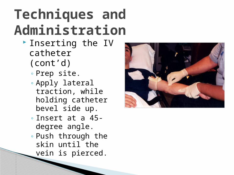
Inserting the IV catheter (cont’d)◦ Prep site.◦ Apply lateral
traction, while holding catheter bevel side up.
◦ Insert at a 45-degree angle.
◦ Push through the skin until the vein is pierced.
Techniques and Administration

Inserting the IV catheter (cont’d)◦ Drop angle to 15 degrees and advance catheter a
few centimeters.◦ Slide sheath off needle into vein.◦ Apply pressure to the vein. ◦ Remove needle.◦ Dispose needle in sharps container
Techniques and Administration

Securing the line◦ Tape the area to
secure the catheter and tubing. Double back the
tubing to create a loop.
◦ Cover the site with sterile gauze and secure with tape.
See Skill Drill 11-2.
Techniques and Administration

Changing an IV bag◦ Stop the flow by closing the roller clamp.◦ Prepare the new IV bag.◦ Remove the piercing spike, and insert it into the
port on the new bag.◦ Ensure the drip chamber is filled, and open the
roller clamp.
Techniques and Administration

Discontinuing the IV line◦ Shut off the flow.◦ Peel tape back.◦ Stabilize the
catheter.◦ Do not remove IV
tubing from hub. ◦ Pull catheter and IV
line from patient’s vein.
◦ Apply pressure.
Techniques and Administration
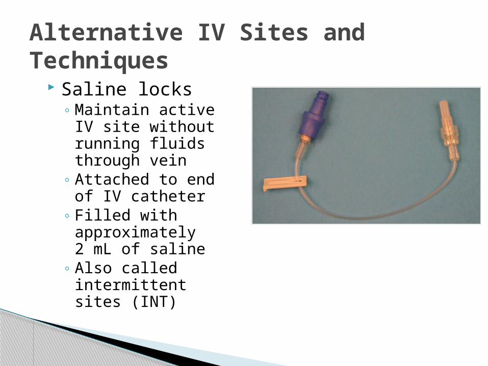
Saline locks◦ Maintain active IV
site without running fluids through vein
◦ Attached to end of IV catheter
◦ Filled with approximately 2 mL of saline
◦ Also called intermittent sites (INT)
Alternative IV Sites and Techniques
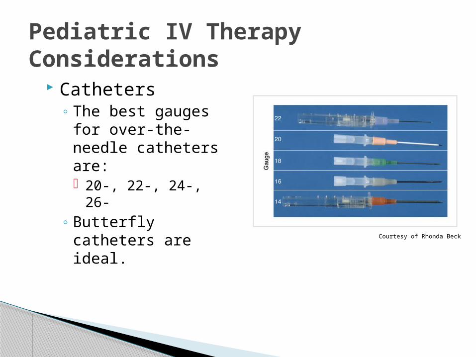
Catheters◦ The best gauges for
over-the-needle catheters are: 20-, 22-, 24-, 26-
◦ Butterfly catheters are ideal.
Pediatric IV Therapy Considerations
Courtesy of Rhonda Beck
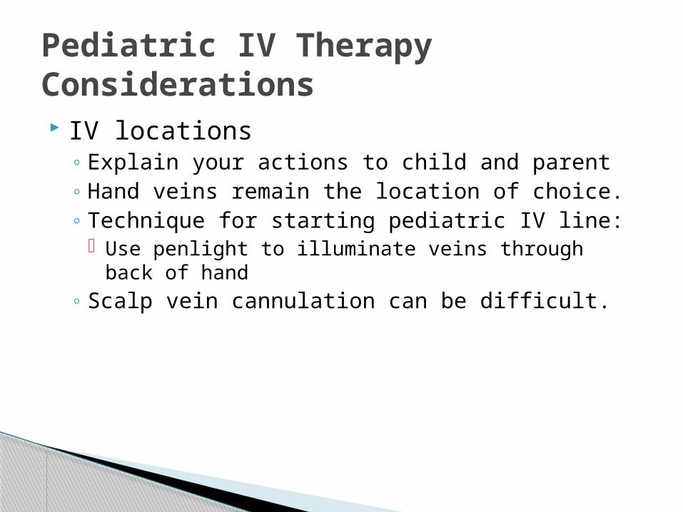
IV locations◦ Explain your actions to child and parent◦ Hand veins remain the location of choice. ◦ Technique for starting pediatric IV line:
Use penlight to illuminate veins through back of hand◦ Scalp vein cannulation can be difficult.
Pediatric IV Therapy Considerations

Checks to perform after IV administration:◦ Fluid◦ Administration set◦ Height of bag◦ Catheter type◦ Constricting band
Factors Affecting IV Flow Rates

Infiltration: escape of fluid into surrounding tissue◦ Causes area of edema◦ Causes include:
Catheter passes through vein and out other side Patient moves excessively Tape becomes lose or dislodged Catheter is inserted at too shallow an angle
Local IV Site Reactions and Complications

Infiltration (cont’d)◦ If infiltration occurs:
Discontinue the IV line. Reestablish IV line in the opposite extremity Apply direct pressure over the area. Do not wrap tape around extremity
Local IV Site Reactions and Complications

Occlusion: blockage of vein or catheter◦ First sign: decreasing drip rate or blood in the IV
tubing◦ May develop due to:
Position of catheter within the vein Patient’s blood pressure overcoming the flow
◦ If occlusion does not dislodge: Discontinue. Reestablish IV in opposite extremity
Local IV Site Reactions and Complications
Vein irritation◦ Often caused by too-rapid infusion rate◦ If redness at the IV site occurs:
Discontinue the IV line. Save the equipment for analysis. Reestablish the IV line in the other extremity with
new equipment.
Local IV Site Reactions and Complications
Thrombophlebitis: inflammation of the vein◦ May be caused by lapses in aseptic technique◦ Pain and tenderness along the vein and redness
and edema at the venipuncture site◦ Appear several hours after IV therapy ◦ Stop the infusion and discontinue the IV at that
site.
Local IV Site Reactions and Complications

Hematoma: accumulation of blood in the tissues surrounding an IV site◦ Often caused by:
Vein perforation Improper catheter removal
◦ Develops while inserting catheter: stop and apply direct pressure
◦ Develops after inserting catheter: evaluate the IV flow
◦ Develops as a result of discontinuing the IV: apply pressure
Local IV Site Reactions and Complications
Courtesy of Rhonda Beck
Nerve, tendon, or ligament damage◦ Results in sudden and severe shooting pain ◦ Remove catheter and select another IV site.
Arterial puncture◦ Bright red blood will spurt through the catheter.◦ Withdraw the catheter and apply direct pressure
for at least 5 minutes.
Local IV Site Reactions and Complications

Allergic reactions◦ Anaphylaxis must be treated aggressively.◦ If an allergic reaction occurs:
Discontinue the line and remove the solution. Leave the catheter in place. Attach a saline lock. Notify medical control. Maintain an open airway; monitor vital signs. Retain the solution or medication for evaluation.
Systemic Complications

Circulatory overload◦ Problems may occur in patients with cardiac,
pulmonary, or renal dysfunction. ◦ To treat:
Slow the IV rate. Raise the patient’s head. Administer high-flow oxygen. Monitor vital signs and breathing adequacy.
Systemic Complications

Air embolus◦ Avoid by properly flushing an IV line and replace
empty IV bags with full ones◦ To treat:
Place patient on left side with head down. Administer 100% oxygen. Transport to closest facility Assist ventilations if needed.
Systemic Complications

Questions?