· For HISTOLOGY. 005-11000-40 7 mL • REAGENT DESCRIPTION . Clone MCM2 26H6.19, MCM2 27C5.6,...
30
For HISTOLOGY 005-11000-40 7 mL REAGENT DESCRIPTION Clone MCM2 26H6.19, MCM2 27C5.6, TOP2A SWT3D1 Ig Class IgG 1 Immunogen Recombinant Human MCM2 and TOP2A 1. INTENDED USE For In Vitro Diagnostic use. For use with automated staining on the Ventana Benchmark ® XT using the iView™ detection chemistry. The ProEx™ C Immunohistochemical Test is intended for the qualitative evaluation of aberrant S-Phase induction in formalin-fixed paraffin-embedded tissue biopsies. Results interpretation must be made by a certified professional within the context of the patient’s history and other diagnostic tests. 2. SUMMARY AND EXPLANATION Minichromosome maintenance (MCM) and topoisomerase II alpha (TOP2A) proteins play an important regulatory role in eukaryotic DNA replication. For example, the HPV oncoproteins E6 and E7 bypass of critical cell-cycle checkpoints resulting in a prolonged and aberrant S-Phase induction cycle. During the transcriptional activation of the aberrant cell cycle, levels of MCM2 and TOP2A proteins increase in the proliferating cells. Both the MCM2 and TOP2A proteins have been shown to be over-expressed in a number of different dysplastic and malignant tissues including cervical neoplasia 1-5 . The over- expression of these proteins in morphologically abnormal cells, as demonstrated by a moderate-to-intense nuclear staining pattern using immunohistochemical (IHC) techniques, is indicative of the presence of aberrant S-Phase induction. 3. REAGENT PROVIDED ProEx™ C Antibody Reagent contains mouse monoclonal anti-MCM2 and anti-TOP2A purified from tissue culture supernatant and diluted in buffered saline solution containing protein stabilizers and 0.09% sodium azide. 4. PRINCIPLES OF PROCEDURE Formalin-fixed paraffin-embedded tissue specimens are sectioned, deposited onto glass slides and deparaffinized. The sectioned specimens are pretreated with a buffer to expose antigenic sites. Blocking agents are added to minimize background staining caused by endogenous peroxidase or non-specific protein binding. The sample is then incubated with the ProEx™ C Antibody Reagent. The addition of an enzyme-linked antibody chromogen system results in the formation of a visible chromogenic product localized at the antigen- antibody binding sites. The specimen is then counterstained with hematoxylin, a bluing agent is applied and the slide is coverslipped. Results are interpreted by a trained professional using a light microscope. 5. MATERIALS AND REAGENTS REQUIRED BUT NOT SUPPLIED (for Ventana Benchmark ® XT Procedure) • 10X Reaction Buffer – Cat # 950-300 2L (Ventana Medical Systems) • LCS (Liquid Coverslip) – Cat # 650-010 2L (Ventana Medical Systems) • 10X SSC – Cat # 950-110 2L (Ventana Medical Systems) • EZ Prep – Cat # 950-102 2L (Ventana Medical Systems) • CC1 (Cell Conditioning) – Cat # 950-124 2L (Ventana Medical Systems) • iView™ DAB Detection Kit – Cat # 760-091 (Ventana Medical Systems) • Amplification Kit (A&B) – Cat # 760-080 (Ventana Medical Systems) • Prep Kit Dispenser with Prep Kit Button – Cat # 786-3034 (Ventana Medical Systems) • Hematoxylin – Cat # 760-2021 (Ventana Medical Systems) • Bluing Reagent – Cat # 760-2037 (Ventana Medical Systems) • Glass Slides (SuperFrost ® Plus or equivalent) • Mounting Media (Acrytol ® or equivalent) • Timer (capable of 1-60 minute intervals) • Distilled H 2 O • Ethanol 95%, 100% • Glass Coverslips • Lab Marker • 20L Carboy (Nalgene ® or equivalent) • Slide Dryer • Slide Rack with Staining Dishes • Xylene or Xylene Substitutes • Light Microscope (10x, 20x [optional], 40x objectives) 6. PRECAUTIONS 6.1. For in vitro diagnostic use. 6.2. Slide clearing steps requiring xylene must be performed in a certified, chemical fume hood. 6.3. The ProEx™ C Antibody Reagent contains sodium azide (NaN 3 ), a chemical highly toxic in pure form. At product concentrations, though not classified as hazardous, sodium azide may react with lead and copper plumbing to form highly explosive build-ups of metal azides. Upon disposal, flush with large volumes of water to prevent metal azide build-up in plumbing. 6.4. DAB (3,3’-Diaminobenzidine) is classified as a suspected carcinogen. Avoid physical contact and prolonged or repeated exposure. Use in a certified, chemical fume hood. 6.5. Specimens and all materials exposed to specimens should be handled as if capable of transmitting infection and disposed of with proper precautions. Never pipette reagents by mouth and avoid contacting the skin and mucous membranes with reagents and specimens. If reagents come in contact with sensitive areas, wash with copious amounts of water. 6.6. Minimize microbial contamination of reagents to avoid nonspecific staining. 6.7. Incubation times, temperatures or methods other than those specified may give erroneous results. 6.8. Do not use the ProEx™ C Antibody Reagent after the expiration date stamped on the package. The user must validate conditions if reagents are stored under any conditions other than those specified in the package insert. 6.9. Wear appropriate Personal Protective Equipment to avoid reagent contact with eyes and skin. Refer to the Material Safety Data Sheet (MSDS) for additional information. 7. INSTRUCTIONS FOR USE 7.1. Specimen Preparation 7.1.1. Cut 4 μm sections from the tissue block and place the sections on SuperFrost ® Plus glass slides. 7.1.2. Label the slides. 7.1.3. Bake the slides in a forced air oven for 20 minutes. If the slides are already dry, touch the slide to a Histo-Orienter until the paraffin melts. 7.2. Reagent Preparation for the Ventana Benchmark ® XT Note: Refer to manufacturer’s instructions. 7.2.1. 10X Reaction Buffer 7.2.1.1. Register a new bottle of the 10X Reaction Buffer concentrate. 7.2.1.2. Add 1 (one) bottle of 10X Reaction Buffer concentrate to 18 liters of distilled water. Mix well. 7.2.1.3. Fill up the EZ Prep bottle #4 on the Benchmark ® XT. Check to ensure that the pH is between 6.5 − 7.1. 7.2.2. EZ Prep (10X) – Deparaffinization Solution 7.2.2.1. Register a new bottle of EZ Prep concentrate. 7.2.2.2. Add 1 (one) bottle of EZ Prep concentrate to 18 liters of distilled water. Mix well. 7.2.2.3. Fill up the EZ Prep bottle #1 on the Benchmark ® XT. Check to ensure that the pH is between 6.90 − 7.2. 7.2.3. SSC (10X) 7.2.3.1. Register 2 (two) bottles of SSC concentrate. 7.2.3.2. Add 2 (two) bottles of SSC concentrate to 16 liters of distilled water. Mix well. 7.2.3.3. Fill up the SSC bottle #3 on the Benchmark ® XT. Check to ensure that the pH is between 7.1 − 7.5. 7.2.4. Fillable Prep Kit 7.2.4.1. Register Prep Kit button 7.2.4.2. Fill Prep Kit Dispenser according to manufacturer’s instructions. 7.3. Staining Procedure Notes 7.3.1. This protocol is for use with automated staining on the Ventana Benchmark ® XT using the iView™ detection chemistry. 7.3.2. Do not allow the slides to dry out at any time during the procedure. Slides that have been allowed to dry out during the procedure may result in increased background staining. IVD 2 o C 8 o C ©October 2008 TriPath Imaging®, Inc. All rights reserved. Doc. No. 779-06555-00 Rev D, 1/30
Transcript of · For HISTOLOGY. 005-11000-40 7 mL • REAGENT DESCRIPTION . Clone MCM2 26H6.19, MCM2 27C5.6,...

For HISTOLOGY 005-11000-40 7 mL
REAGENT DESCRIPTION Clone MCM2 26H6.19, MCM2 27C5.6, TOP2A SWT3D1 Ig Class IgG1 Immunogen Recombinant Human MCM2 and TOP2A
1. INTENDED USE
For In Vitro Diagnostic use.
For use with automated staining on the Ventana Benchmark® XT using the iView™ detection chemistry.
The ProEx™ C Immunohistochemical Test is intended for the qualitative evaluation of aberrant S-Phase induction in formalin-fixed paraffin-embedded tissue biopsies. Results interpretation must be made by a certified professional within the context of the patient’s history and other diagnostic tests.
2. SUMMARY AND EXPLANATION Minichromosome maintenance (MCM) and topoisomerase II alpha (TOP2A) proteins play an important regulatory role in eukaryotic DNA replication. For example, the HPV oncoproteins E6 and E7 bypass of critical cell-cycle checkpoints resulting in a prolonged and aberrant S-Phase induction cycle. During the transcriptional activation of the aberrant cell cycle, levels of MCM2 and TOP2A proteins increase in the proliferating cells.
Both the MCM2 and TOP2A proteins have been shown to be over-expressed in a number of different dysplastic and malignant tissues including cervical neoplasia1-5. The over-expression of these proteins in morphologically abnormal cells, as demonstrated by a moderate-to-intense nuclear staining pattern using immunohistochemical (IHC) techniques, is indicative of the presence of aberrant S-Phase induction.
3. REAGENT PROVIDED ProEx™ C Antibody Reagent contains mouse monoclonal anti-MCM2 and anti-TOP2A purified from tissue culture supernatant and diluted in buffered saline solution containing protein stabilizers and 0.09% sodium azide.
4. PRINCIPLES OF PROCEDURE Formalin-fixed paraffin-embedded tissue specimens are sectioned, deposited onto glass slides and deparaffinized. The sectioned specimens are pretreated with a buffer to expose antigenic sites. Blocking agents are added to minimize background staining caused by endogenous peroxidase or non-specific protein binding. The sample is then incubated with the ProEx™ C Antibody Reagent. The addition of an enzyme-linked antibody chromogen system results in the formation of a visible chromogenic product localized at the antigen-antibody binding sites. The specimen is then counterstained with hematoxylin, a bluing agent is applied and the slide is coverslipped. Results are interpreted by a trained professional using a light microscope.
5. MATERIALS AND REAGENTS REQUIRED BUT NOT SUPPLIED (for Ventana Benchmark® XT Procedure)
• 10X Reaction Buffer – Cat # 950-300 2L (Ventana Medical Systems)
• LCS (Liquid Coverslip) – Cat # 650-010 2L (Ventana Medical Systems)
• 10X SSC – Cat # 950-110 2L (Ventana Medical Systems)
• EZ Prep – Cat # 950-102 2L (Ventana Medical Systems)
• CC1 (Cell Conditioning) – Cat # 950-124 2L (Ventana Medical Systems)
• iView™ DAB Detection Kit – Cat # 760-091 (Ventana Medical Systems)
• Amplification Kit (A&B) – Cat # 760-080 (Ventana Medical Systems)
• Prep Kit Dispenser with Prep Kit Button – Cat # 786-3034 (Ventana Medical Systems)
• Hematoxylin – Cat # 760-2021 (Ventana Medical Systems)
• Bluing Reagent – Cat # 760-2037 (Ventana Medical Systems)
• Glass Slides (SuperFrost® Plus or equivalent)
• Mounting Media (Acrytol® or equivalent)
• Timer (capable of 1-60 minute intervals)
• Distilled H2O
• Ethanol 95%, 100%
• Glass Coverslips
• Lab Marker
• 20L Carboy (Nalgene® or equivalent)
• Slide Dryer
• Slide Rack with Staining Dishes
• Xylene or Xylene Substitutes
• Light Microscope (10x, 20x [optional], 40x objectives)
6. PRECAUTIONS 6.1. For in vitro diagnostic use.
6.2. Slide clearing steps requiring xylene must be performed in a certified, chemical fume hood.
6.3. The ProEx™ C Antibody Reagent contains sodium azide (NaN3), a chemical highly toxic in pure form. At product concentrations, though not classified as hazardous, sodium azide may react with lead and copper plumbing to form highly explosive build-ups of metal azides. Upon disposal, flush with large volumes of water to prevent metal azide build-up in plumbing.
6.4. DAB (3,3’-Diaminobenzidine) is classified as a suspected carcinogen. Avoid physical contact and prolonged or repeated exposure. Use in a certified, chemical fume hood.
6.5. Specimens and all materials exposed to specimens should be handled as if capable of transmitting infection and disposed of with proper precautions. Never pipette reagents by mouth and avoid contacting the skin and mucous membranes with reagents and specimens. If reagents come in contact with sensitive areas, wash with copious amounts of water.
6.6. Minimize microbial contamination of reagents to avoid nonspecific staining.
6.7. Incubation times, temperatures or methods other than those specified may give erroneous results.
6.8. Do not use the ProEx™ C Antibody Reagent after the expiration date stamped on the package. The user must validate conditions if reagents are stored under any conditions other than those specified in the package insert.
6.9. Wear appropriate Personal Protective Equipment to avoid reagent contact with eyes and skin. Refer to the Material Safety Data Sheet (MSDS) for additional information.
7. INSTRUCTIONS FOR USE 7.1. Specimen Preparation
7.1.1. Cut 4 μm sections from the tissue block and place the sections on SuperFrost® Plus glass slides.
7.1.2. Label the slides.
7.1.3. Bake the slides in a forced air oven for 20 minutes. If the slides are already dry, touch the slide to a Histo-Orienter until the paraffin melts.
7.2. Reagent Preparation for the Ventana Benchmark® XT Note: Refer to manufacturer’s instructions.
7.2.1. 10X Reaction Buffer
7.2.1.1. Register a new bottle of the 10X Reaction Buffer concentrate.
7.2.1.2. Add 1 (one) bottle of 10X Reaction Buffer concentrate to 18 liters of distilled water. Mix well.
7.2.1.3. Fill up the EZ Prep bottle #4 on the Benchmark® XT. Check to ensure that the pH is between 6.5 − 7.1.
7.2.2. EZ Prep (10X) – Deparaffinization Solution
7.2.2.1. Register a new bottle of EZ Prep concentrate.
7.2.2.2. Add 1 (one) bottle of EZ Prep concentrate to 18 liters of distilled water. Mix well.
7.2.2.3. Fill up the EZ Prep bottle #1 on the Benchmark® XT. Check to ensure that the pH is between 6.90 − 7.2.
7.2.3. SSC (10X)
7.2.3.1. Register 2 (two) bottles of SSC concentrate.
7.2.3.2. Add 2 (two) bottles of SSC concentrate to 16 liters of distilled water. Mix well.
7.2.3.3. Fill up the SSC bottle #3 on the Benchmark® XT. Check to ensure that the pH is between 7.1 − 7.5.
7.2.4. Fillable Prep Kit
7.2.4.1. Register Prep Kit button
7.2.4.2. Fill Prep Kit Dispenser according to manufacturer’s instructions.
7.3. Staining Procedure Notes 7.3.1. This protocol is for use with automated staining on the Ventana
Benchmark® XT using the iView™ detection chemistry.
7.3.2. Do not allow the slides to dry out at any time during the procedure. Slides that have been allowed to dry out during the procedure may result in increased background staining.
IVD
2oC
8oC
©October 2008 TriPath Imaging®, Inc. All rights reserved. Doc. No. 779-06555-00 Rev D, 1/30

8. AUTOMATED STAINING PROTOCOL (for the Ventana Benchmark® XT) 8.1. Turn on the staining module on the Ventana Benchmark® XT and start the software.
Refer to the manufacturer’s operating instructions for the Ventana Benchmark® XT.
8.2. Select the following protocol parameters on the Benchmark® XT software.
8.2.1. Paraffin (selected)
8.2.2. Deparaffinization (selected)
8.2.3. Cell Conditioning (selected)
8.2.4. Conditioner #1 (selected)
8.2.5. Mild CC1 (selected)
8.2.6. Standard CC1 (selected)
8.2.7. Extended CC1 (selected)
8.2.8. Ab Incubation Temperatures (selected)
8.2.9. 37 C Ab. Inc. (selected)
8.2.10. Antibody (selected)
8.2.11. Apply 1 drop of [PREP KIT 100] (Antibody) and incubate for (1 hour).
8.2.12. Amplify (selected)
8.2.13. Counterstain (selected)
8.2.14. Apply 1 drop of [Hematoxylin] (Counterstain), apply coverslip and incubate for (8 minutes).
8.2.15. Post Counterstain (selected)
8.2.16. Apply 1 drop of [Bluing Reagent] (Post Counterstain), apply coverslip and incubate for (4 minutes).
8.3. Register all reagents being used in the assay.
8.4. Determine the number of slides to be stained (including controls).
8.5. Select or create labels for each slide ensuring that each label corresponds to a single staining protocol.
8.6. Apply the appropriate label to each slide.
8.7. Load the slides onto the slide carousel.
8.8. Load the reagent dispensers and mount the reagent tray onto the reagent carousel.
8.9. Fill the bulk reagent carboys.
8.10. Check the waste module carboy and empty, if necessary.
8.11. Select “RUN” on the main computer screen of the Benchmark® XT to begin staining.
8.12. After the run is completed, print out the protocol summary and staining run reports.
8.13. Remove the slides from the instrument and rinse the slides in soapy water for 3-5 minutes or until Liquid Coverslip residue is no longer visible.
8.14. Dehydrate the slides.
8.14.1. Immerse slides in 95% ethanol, 1 minute or 25 dips.
8.14.2. Immerse slides in absolute alcohol, 4 changes, 1 minute each or 25 dips.
8.14.3. Clear with xylene, 3 changes, 1 minute each or 25 dips.
8.15. Coverslip slides with non-aqueous, permanent mounting media using glass coverslips.
9. STABILITY 9.1. When stored at recommended temperatures, unopened reagent vials are stable until
the expiration date indicated on the vial.
9.2. Once opened, reagents are stabile for ninety (90) days when stored at recommended temperatures.
10. QUALITY CONTROL 10.1. Variability in results is often derived from differences in specimen handling which
deviates from recommended test procedures. Consult the quality control guidelines of the College of American Pathologists (CAP) Certification Program for Immunohistochemistry for additional information.
10.2. A positive tissue control should be included with each stain run to verify the assay performance. If the positive tissue control does not exhibit positive staining, the results with the other test specimens should be considered suspect or invalid.
10.3. A negative tissue control should be included with each stain run to verify the specificity of the primary antibody and to provide an indication of background staining. If the negative tissue control exhibits positive specific staining, the results with the other test specimens should be considered suspect or invalid.
10.4. A non-specific negative control reagent may also be used in place of the primary antibody to evaluate non-specific or background staining.
11. INTERPRETATION Moderate-to-intense brown staining in the nucleus of cells indicates the presence of aberrant S-Phase induction. A pathologist should evaluate the stained slides using a light microscope. Results interpretation must be made by a certified professional within the context of the patient’s history and other diagnostic tests.
12. LIMITATIONS 12.1. Immunohistochemical staining requires specialized training in the selection and
application of reagents.
12.2. This reagent will perform 50 tests assuming 100μL of reagent is applied per slide.
12.3. Some normal cells may stain positive for aberrant S-Phase induction.
12.4. Optimal tissue staining is dependent upon fixation and processing of the specimen.
12.5. Non-specific or increased background staining may occur due to, but not limited to, variations in procedure, inadequate rinsing between assay steps, and/or inadequately processed specimens.
13. TROUBLESHOOTING
Problem Possible Cause Action Reagents applied in improper order.
Review staining protocol. No staining on positive control slides
Omission of any reagent. Repeat staining protocol. Insufficient antigen retrieval.
Check incubation times and temperature of antigen/epitope retrieval buffer.
Incorrect antigen retrieval buffer used.
Review staining protocol.
Inadequate incubation of primary antibody.
Review staining protocol.
Weak staining on positive slides
Primary antibody has been diluted.
Use primary antibody according to manufacturer’s directions.
Inadequate rinsing between assay steps.
Repeat staining protocol.
Excessive incubation times with key reagents.
Review staining protocol.
Excessive background staining
Slides drying out during post assay processing.
Repeat staining protocol.
©October 2008 TriPath Imaging®, Inc. All rights reserved. Doc. No. 779-06555-00 Rev D, 2/30

14. REFERENCES 1. Kastan M and Bartec J. Cell cycle checkpoints and cancer. Nature, 2004. Vol32:316-323.
2. Massague J. G1 cell-cycle control and cancer. Nature, 2004. Vol 432:298-432.
3. Freeman A, Morris LS, Millis AD, Stoeber K, Laskey RA, Williams GH and Coleman N. Minichromosome Maintenance Proteins as Biological Markers of Dysplasia and Malignancy. CI Cancer Research, 1999. Vol 5:2121-2132.
4. Ishimi Y, Okayasu I, Kato C, Kwon H, Kimura H, Yamada K and Song S. Enhanced expression of MCM proteins in cancer cells derived from uterine cervix. Eur. J. Biochem 2003. 270:1089-1101.
5. Lei M and Tye BK. Initiating DNA synthesis: from recruiting to activating the MCM Complex. J Cell Science 2001. Vol. 114 (8): 1447-1454.
15. GLOSSARY OF SYMBOLS
Catalog number
For in vitro diagnostic use
Consult instructions for use
Contains 7 mL
Caution, consult accompanying document
Storage Temperature Limitations
Batch Code
Use by YYYY-MM-DD or YYYY-MM
Manufacturer
IVD
TECHNICAL INFORMATION In the United States, telephone TriPath Technical Services, toll-free 1-866-874-7284.
TriPath Imaging, Inc. 780 Plantation Drive Burlington, NC 27215 USA (800) 426-2176
Developed with technology from Millennium Pharmaceuticals, Inc.
and are trademarks of Millennium Pharmaceuticals, Inc. Millennium Pharmaceuticals, Inc. 40 Landsdowne Street Cambridge, MA 02139 USA www.millennium.com
TriPath Imaging® is a registered trademark of TriPath Imaging, Inc. ProEx is a product and trademark of TriPath Imaging, Inc.
©October 2008 TriPath Imaging®, Inc. All rights reserved. Doc. No. 779-06555-00 Rev D, 3/30

Für die HISTOLOGIE 005-11000-40 7 mL
BESCHREIBUNG DES REAGENZ: Klon MCM2 26H6.19, MCM2 27C5.6, TOP2A SWT3D1 Ig-Klasse IgG1 Immunogen Rekombinantes humanes MCM2 und TOP2A
1. VERWENDUNGSZWECK
Zur In-vitro-Diagnostik.
Zur Anwendung bei automatischer Färbung auf dem Ventana Benchmark® XT mit iView™ Nachweischemie.
Der immunhistochemische Test ProEx™ C dient bestimmungsgemäß zur qualitativen Beurteilung einer abweichenden S-Phasen-Induktion in formalinfixierten, paraffineingebetteten Gewebebiopsien. Die Interpretation der Ergebnisse muss durch eine entsprechend qualifizierte Person im Rahmen der Patienten-Anamnese und anderer diagnostischer Tests erfolgen.
2. ZUSAMMENFASSUNG UND ERLÄUTERUNG Die Proteine Minichromosome Maintenance (MCM) und Topoisomerase II alpha (TOP2A) spielen eine wichtige Rolle bei der Regelung der DNS-Replikation in den Eukaryoten. Beispielsweise umgehen die HPV-Onkoproteine E6 und E7 wichtige Prüfstellen im Zellzyklus, woraus sich ein verlängerter und abweichender S-Phasen-Induktionszyklus ergibt. Bei der transkriptionalen Aktivierung des abnormen Zellzyklus erhöhen sich die Spiegel der Proteine MCM2 und TOP2A in den proliferierenden Zellen.
Die Überexpression der Proteine MCM2 und TOP2A ist für eine Reihe von dysplastischen und malignen Geweben nachgewiesen, darunter auch zervikale Neoplasie1-5. Diese Überexpression in morphologisch abnormen Zellen, die sich bei immunhistochemischer (IHC) Untersuchung in mäßigen bis intensiven nukleären Färbungsmustern äußert, ist ein Indiz für abweichende S-Phasen-Induktion.
3. GELIEFERTES REAGENZ Das ProEx™ C Antikörperreagenz enthält gereinigtes, monoklonales Anti-MCM2 und Anti-TOP2A (Maus) aus Gewebekulturüberstand in einer Verdünnung aus gepufferter Kochsalzlösung mit Proteinstabilisatoren und 0,09 % Natriumazid.
4. PRINZIPIEN DES VERFAHRENS Formalinfixierte, paraffineingebettete Gewebeproben werden geschnitten, auf Glas-Objektträger aufgebracht und entparaffiniert. Die geschnittenen Proben werden mit Puffer vorbehandelt, um die Antigenstellen freizulegen. Blocker werden zugegeben, um die Hintergrundfärbung durch endogene Peroxidase oder unspezifische Proteinbindung zu minimieren. Dann wird die Probe mit dem ProEx™ C Antikörperreagenz inkubiert. Durch Zugabe eines enzymgebundenen Antikörper-Chromogen-Systems bildet sich ein sichtbares chromogenes Produkt an den Antigen-Antikörper-Bindungsstellen. Anschließend wird die Probe mit Hämatoxylin gegengefärbt, ein Bläuungsmittel zugegeben und der Objektträger mit einem Deckglas versehen. Die Ergebnisse werden von einer entsprechend qualifizierten Person unter einem Lichtmikroskop ausgewertet.
5. ERFORDERLICHE, ABER NICHT MITGELIEFERTE REAGENZIEN (für den Einsatz mit Ventana Benchmark® XT)
• 10x Reaktionspuffer – Kat.-Nr. 950-300 2L (Ventana Medical Systems)
• LCS (Liquid Coverslip) – Kat.-Nr. 650-010 2L (Ventana Medical Systems)
• 10x SSC – Kat.-Nr. 950-110 2L (Ventana Medical Systems)
• EZ Prep – Kat.-Nr. 950-102 2L (Ventana Medical Systems)
• CC1 (Cell Conditioning) – Kat.-Nr. 950-124 2L (Ventana Medical Systems)
• iView™ DAB Detection Kit – Kat.-Nr. 760-091 (Ventana Medical Systems)
• Amplification Kit (A&B) – Kat.-Nr. 760-080 (Ventana Medical Systems)
• Prep Kit Dispenser with Prep Kit Button – Kat.-Nr. 786-3034 (Ventana Medical Systems)
• Hämatoxylin – Kat.-Nr. 760-2021 (Ventana Medical Systems)
• Bluing Reagent – Kat.-Nr. 760-2037 (Ventana Medical Systems)
• Glas-Objektträger (SuperFrost® Plus oder Äquivalent)
• Eindeckmittel (Acrytol® oder Äquivalent)
• Stoppuhr (1–60 Minuten)
• destilliertes Wasser
• Äthanol, 95 % und 100 %
• Deckgläser
• Labormarkierstift
• 20-Liter-Ballonflasche (Nalgene® oder Äquivalent)
• Objektträger-Trockner
• Objektträgergestell mit Färbeschalen
• Xylol oder Xylol-Ersatz
• Lichtmikroskop (Vergrößerung 10x, 20x [optional], 40x)
6. SICHERHEITSHINWEISE 6.1. Zur In-vitro-Diagnostik.
6.2. Die Reinigung von Objektträgern mit Xylol darf nur unter einer zugelassenen chemischen Abzugshaube erfolgen.
6.3. Das ProEx™ C Antikörperreagenz enthält Natriumazid (NaN3), eine in reiner Form äußerst giftige Chemikalie. Auch wenn das Natriumazid in diesem Produkt so gering konzentriert ist, dass es als gefahrlos eingestuft ist, kann Natriumazid mit Blei- oder Kupferrohren reagieren und hochexplosive Metallazide bilden. Deshalb beim Ausgießen mit reichlich Wasser spülen, um eine Ansammlung von Metallaziden in den Rohrleitungen zu verhindern.
6.4. DAB (3,3’-Diaminobenzidin) ist als vermutliches Karzinogen eingestuft. Körperkontakt und längere oder wiederholte Exposition vermeiden. Unter einer zugelassenen chemischen Abzugshaube anwenden.
6.5. Proben und alle mit Proben in Berührung kommenden Gegenstände müssen als potenziell infektiös behandelt und entsprechend entsorgt werden. Reagenzien niemals mit dem Mund pipettieren und jeden Kontakt der Haut und Schleimhäute mit Reagenzien und Proben vermeiden. Bei Kontakt der Reagenzien mit empfindlichen Bereichen mit reichlich Wasser waschen.
6.6. Mikrobielle Kontamination von Reagenzien minimieren, um unspezifische Färbung zu vermeiden.
6.7. Von den Angaben abweichende Inkubationszeiten, -temperaturen oder -methoden können zu fehlerhaften Ergebnissen führen.
6.8. Das ProEx™ C Antikörperreagenz nicht nach dem auf der Verpackung aufgedruckten Verfallsdatum verwenden. Werden Reagenzien unter anderen als in der Verpackungsbeilage angegebenen Bedingungen aufbewahrt, müssen diese vom Anwender validiert werden.
6.9. Geeignete Schutzausrüstung tragen, um Kontakt des Reagenzes mit Haut und Augen zu vermeiden. Weitere Hinweise sind dem Materialsicherheitsdatenblatt (MSDS) zu entnehmen.
7. GEBRAUCHSANWEISUNG 7.1. Präparation der Proben
7.1.1. Vom Gewebe 4 μm dicke Scheiben abschneiden und diese auf SuperFrost® Plus Glas-Objektträger aufbringen.
7.1.2. Die Objektträger etikettieren.
7.1.3. Die Objektträger in einem Umluftofen 20 Minuten erhitzen. Sind die Objektträger bereits trocken, an einen Histo-Orienter anlegen, bis das Paraffin schmilzt.
7.2. Ansetzen der Reagenzien für den Ventana Benchmark® XT Hinweis: Anweisungen des Herstellers beachten.
7.2.1. 10x Reaktionspuffer
7.2.1.1. Eine neue Flasche 10x Reaktionspuffer-Konzentrat im System anmelden.
7.2.1.2. Eine (1) Flasche 10x Reaktionspuffer-Konzentrat in 18 Liter destilliertes Wasser geben. Gut mischen.
7.2.1.3. Die EZ Prep Flasche (Nr. 4) am Benchmark® XT füllen. Der pH-Wert muss bei 6,5−7,1 liegen.
7.2.2. EZ Prep (10x) – Entparaffinierungslösung
7.2.2.1. Eine neue Flasche EZ Prep-Konzentrat im System anmelden.
7.2.2.2. Eine (1) Flasche EZ Prep-Konzentrat in 18 Liter destilliertes Wasser geben. Gut mischen.
7.2.2.3. Die EZ Prep-Flasche (Nr. 1) am Benchmark® XT füllen. Der pH-Wert muss bei 6,90−7,2 liegen.
7.2.3. SSC (10x)
7.2.3.1. Zwei (2) Flaschen SSC-Konzentrat im System anmelden.
7.2.3.2. Zwei (2) Flaschen SSC-Konzentrat in 16 Liter destiliertes Wasser geben. Gut mischen.
7.2.3.3. Die SSC-Flasche (Nr. 3) am Benchmark® XT füllen. Der pH-Wert muss bei 7,1−7,5 liegen.
7.2.4. Nachfüllbares Prep Kit
7.2.4.1. Prep Kit Button im System anmelden.
7.2.4.2. Prep Kit Dispenser gemäß Herstelleranweisungen befüllen.
7.3. Hinweise zum Färbeverfahren
IVD
2oC
8oC
©October 2008. TriPath Imaging®, Inc. All rights reserved. Doc. No. 779-06555-00 Rev D, 4/30

7.3.1. Das nachstehende Protokoll ist für die automatische Färbung auf dem Ventana Benchmark® XT mit iView™ Nachweischemie bestimmt.
7.3.2. Die Objektträger dürfen während des gesamten Verfahrens niemals austrocknen. Ausgetrocknete Objektträger können zu erhöhter Hintergrundfärbung führen.
8. AUTOMATISCHES FÄRBEPROTOKOLL (für den Ventana Benchmark® XT) 8.1. Das Färbemodul des Ventana Benchmark® XT einschalten und die Software starten.
Siehe Hersteller-Gebrauchsanweisung für den Ventana Benchmark® XT.
8.2. In der Benchmark® XT Software folgende Parameter einstellen:
8.2.1. Paraffin (gewählt)
8.2.2. Entparaffinierung (gewählt)
8.2.3. Zellenkonditionierung (gewählt)
8.2.4. Konditionierer 1 (gewählt)
8.2.5. CC1 mild (gewählt)
8.2.6. CC1 Standard (gewählt)
8.2.7. CC1 erweitert (gewählt)
8.2.8. Ab. Inkubationstemperaturen (gewählt)
8.2.9. 37 C Ab. Ink. (gewählt)
8.2.10. Antikörper (gewählt)
8.2.11. 1 Tropfen [PREP KIT 100] (Antikörper) aufbringen und eine (1) Stunde inkubieren.
8.2.12. Verstärken (gewählt)
8.2.13. Gegenfärbung (gewählt)
8.2.14. 1 Tropfen [Hämatoxylin] (Gegenfärbung) aufbringen, eindecken und acht (8) Minuten inkubieren.
8.2.15. Nach-Gegenfärbung (gewählt)
8.2.16. 1 Tropfen [Bläuungsmittel] (Nach-Gegenfärbung), eindecken und vier (4) Minuten inkubieren.
8.3. Alle für den Test benutzten Reagenzien im System anmelden.
8.4. Die Anzahl von Objektträgern festlegen, die gefärbt werden sollen (einschließlich Kontrollen).
8.5. Etiketten für alle Objektträger wählen bzw. erstellen, dabei muss jedem Färbeprotokoll je ein Etikett zugeordnet sein.
8.6. Die Etiketten an den jeweiligen Objektträgern anbringen.
8.7. Objektträger in das Objektträgerkarussell laden.
8.8. Die Reagenzdispenser laden und den Reagenzhalter auf das Reagenzkarussell stellen.
8.9. Die Ballonflaschen für Verbrauchsreagenzien füllen.
8.10. Die Ballonflasche des Abfallmoduls prüfen und ggf. leeren.
8.11. Auf dem Hauptbildschirm des Benchmark® XT „RUN“ wählen, um mit der Färbung zu beginnen.
8.12. Nach Ende des Durchlaufs die Zusammenfassung und das Ablaufprotokoll ausdrucken.
8.13. Die Objektträger aus dem Gerät nehmen und in Seifenwasser 3–5 Minuten spülen, oder bis keine Rückstände des flüssigen Eindeckmittels mehr zu sehen sind.
8.14. Objektträger dehydrieren.
8.14.1. Objektträger in 95%iges Äthanol eintauchen (1 Minute bzw. 25 Mal).
8.14.2. Objektträger in reinen Alkohol eintauchen (4 Wechsel, je 1 Minute bzw. 25 Mal).
8.14.3. Mit Xylol klarspülen (3 Wechsel, je 1 Minute bzw. 25 Mal).
8.15. Objektträger mit nicht-wässrigem, permanentem Eindeckmittel und Deckgläsern abdecken.
9. STABILITÄT 9.1. Bei Einhaltung der empfohlenen Lagertemperaturen sind ungeöffnete
Reagenzflakons bis zu dem auf dem Flakon angegebenen Verfallsdatum stabil.
9.2. Nach dem Öffnen sind Reagenzien neunzig (90) Tage stabil, wenn die empfohlenen Lagertemperaturen eingehalten werden.
10. QUALITÄTSKONTROLLE 10.1. Schwankungen der Ergebnisse ergeben sich häufig aus einer Probenhandhabung,
die von den empfohlenen Testverfahren abweicht. Weitere Hinweise hierzu sind den Richtlinien zur Qualitätskontrolle des „College of American Pathologists (CAP) Certification Program for Immunohistochemistry“ zu entnehmen.
10.2. Bei jedem Färbedurchlauf sollte eine positive Gewebekontrolle zur Überprüfung der Testleistung mitlaufen. Weist die positive Gewebekontrolle keine positive Färbung auf, müssen die Ergebnisse der anderen Proben als fragwürdig oder ungültig betrachtet werden.
10.3. Bei jedem Färbedurchlauf sollte eine negative Gewebekontrolle zur Überprüfung der Spezifität des primären Antikörpers und als Anhaltspunkt für die Hintergrundfärbung mitlaufen. Weist die negative Gewebekontrolle eine positive Färbung auf, müssen die Ergebnisse der anderen Proben als fragwürdig oder ungültig betrachtet werden.
10.4. Zur Beurteilung der unspezifischen bzw. Hintergrundfärbung kann anstelle des primären Antikörpers ein unspezifisches negatives Kontrollreagenz verwendet werden.
11. AUSWERTUNG Eine mäßige bis intensive Braunfärbung der Zellkerne zeigt das Vorhandensein einer abweichenden S-Phasen-Induktion an. Die gefärbten Objektträger sollten von einem Pathologen unter einem Lichtmikroskop beurteilt werden. Die Auswertung der Ergebnisse muss durch eine entsprechend qualifizierte Person im Rahmen der Patienten-Anamnese und anderer diagnostischer Tests erfolgen.
12. BESCHRÄNKUNGEN 12.1. Immunhistochemische Färbeverfahren erfordern eine spezielle Ausbildung bzgl. der
Auswahl und Anwendung von Reagenzien.
12.2. Das Reagenz reicht für 50 Tests, wenn 100 μl Reagenz pro Objektträger angewendet werden.
12.3. Einige normale Zellen können sich positiv für abweichende S-Phasen-Induktion verfärben.
12.4. Optimale Gewebefärbung hängt von der Fixierung und Einbettung der Probe ab.
12.5. Unspezifische bzw. erhöhte Hintergrundfärbung kann u. A. auftreten bei abweichender Vorgehensweise, ungenügender Spülung zwischen den Schritten bzw. unsachgemäß verarbeiteten Proben.
13. FEHLERBEHEBUNG
Problem Mögliche Ursache Abhilfe Reagenzien in falscher Reihenfolge angewendet.
Färbeprotokoll überprüfen.
Keine Färbung von positiven Kontrollobjektträgern Auslassen eines
Reagenzes. Färbeprotokoll wiederholen.
Unzureichende Antigendemaskierung.
Inkubationszeit und -temperatur des Antigen-/Epitopdemaskierungs-puffers prüfen.
Falscher Antigendemaskierungs-puffer verwendet.
Färbeprotokoll überprüfen.
Unzureichende Inkubation des Primärantikörpers.
Färbeprotokoll überprüfen.
Schwache Färbung von positiven Objektträgern
Primärer Antikörper wurde verdünnt.
Primärantikörper gemäß Herstelleranweisungen anwenden.
Unzureichendes Spülen zwischen Testschritten.
Färbeprotokoll wiederholen.
Zu lange Inkubationszeiten bei Reagenzien.
Färbeprotokoll überprüfen.
Zu starke Hintergrundfärbung
Objektträger trocknen bei der Verarbeitung nach dem Testen aus.
Färbeprotokoll wiederholen.
©October 2008. TriPath Imaging®, Inc. All rights reserved. Doc. No. 779-06555-00 Rev D, 5/30

14. LITERATURANGABEN 1. Kastan M and Bartec J. Cell cycle checkpoints and cancer. Nature, 2004. Vol32:316-323.
2. Massague J. G1 cell-cycle control and cancer. Nature, 2004. Vol 432:298-432.
3. Freeman A, Morris LS, Millis AD, Stoeber K, Laskey RA, Williams GH and Coleman N. Minichromosome Maintenance Proteins as Biological Markers of Dysplasia and Malignancy. CI Cancer Research, 1999. Vol 5:2121-2132.
4. Ishimi Y, Okayasu I, Kato C, Kwon H, Kimura H, Yamada K and Song S. Enhanced expression of MCM proteins in cancer cells derived from uterine cervix. Eur. J. Biochem 2003. 270:1089-1101.
5. Lei M and Tye BK. Initiating DNA synthesis: from recruiting to activating the MCM Complex. J Cell Science 2001. Vol. 114 (8): 1447-1454.
15. SYMBOLVERZEICHNIS
Katalognummer
Zur In-vitro-Diagnostik
Gebrauchsanleitung beachten
Inhalt 7 ml
Vorsicht, Begleitpapiere beachten
Zulässiger Temperaturbereich für die Aufbewahrung
Chargencode
Bis JJJJ-MM-TT oder JJJJ-MM verbrauchen
Hersteller
IVD
TECHNISCHER KUNDENDIENST Telefon in den USA: TriPath Technical Services, gebührenfreie Hotline 1-866-874-7284.
TriPath Imaging, Inc. 780 Plantation Drive Burlington, NC 27215 USA (800) 426-2176
Mit Technologie von Millennium Pharmaceuticals, Inc. entwickelt
und sind Warenzeichen von Millennium Pharmaceuticals, Inc. Millennium Pharmaceuticals, Inc. 40 Landsdowne Street Cambridge, MA 02139 USA www.millennium.com
TriPath Imaging® ist ein eingetragenes Warenzeichen von TriPath Imaging, Inc. ProEx ist ein Produkt und Warenzeichen von TriPath Imaging, Inc.
©October 2008. TriPath Imaging®, Inc. All rights reserved. Doc. No. 779-06555-00 Rev D, 6/30
Pour HISTOLOGIE 005-11000-40 7 ml
DESCRIPTION DU RÉACTIF Clone MCM2 26H6.19, MCM2 27C5.6, TOP2A SWT3D1
Classe d’Ig IgG1
Immunogène MCM2 et TOP2A humaines recombinantes
1. UTILISATION PRÉVUE
Utilisation diagnostique in vitro.
Utiliser dans le cadre d‘une coloration automatique sur le Ventana Benchmark® XT à l’aide du système de détection iView™.
Le test immunohistochimique ProEx™ C est destiné à l’évaluation qualitative de l’induction aberrante de la phase S sur des biopsies tissulaires fixées au formol et incluses en paraffine. L’interprétation des résultats doit être faite par un professionnel agréé en tenant compte des antécédents du patient et des autres tests diagnostiques.
2. RÉSUMÉ ET EXPLICATION Les protéines MCM (protéines de maintenance du minichromosome) et topoisomérase II alpha (TOP2A) jouent un rôle régulateur important dans la réplication de l’ADN eucaryote : par exemple, le contournement par les oncoprotéines E6 et E7du PVH des points de contrôle du cycle cellulaire ce qui se traduit par un cycle d’induction prolongé et aberrant de la phase S. Pendant l’activation transcriptionnelle du cycle cellulaire aberrant, les niveaux des protéines MCM2 et TOP2A augmentent dans les cellules en mitose.
Il a été montré que les protéines MCM2 et TOP2A sont surexprimées dans de nombreux et divers tissus dysplasiques et malins dont les néoplasies cervicales1-5. La surexpression de ces protéines dans des cellules morphologiquement anormales, démontrée par un profil de coloration nucléaire modérée à intense après emploi de techniques immunohistochimiques (IHC), est indicative de la présence d’une induction d’une phase S aberrante.
3. RÉACTIFS FOURNIS Le réactif anticorps ProEx™ C contient des anticorps monoclonaux de souris anti-MCM2 et anti-TOP2A purifiés à partir d’un surnageant de culture tissulaire et dilués dans une solution saline tamponnée contenant des agents de stabilisation protéiques et de l’azide de sodium à 0,09 %.
4. PRINCIPES DE LA PROCÉDURE Des coupes d’échantillons tissulaires fixés au formol, inclus en paraffine, sont réalisées, déposées sur des lames de verre et déparaffinées. Les coupes d’échantillons sont prétraitées à l’aide d’un tampon afin d’exposer les sites antigéniques. Des agents de blocages sont ajoutés afin de minimiser le bruit de fond due aux peroxydases endogènes et à la liaison aux protéines non spécifiques. L’échantillon est alors incubé avec le réactif anticorps ProEx™ C. L’addition d’un système chromogène constitué d’une enzyme liée à un anticorps se traduit par la formation d’un produit chromogène visible localisé au niveau des sites de liaison antigène-anticorps. L’échantillon fait alors l’objet d’une contre-coloration par l’hématoxyline, un agent bleuissant est appliqué sur la lame puis une lamelle est mise en place. Les résultats sont interprétés par un professionnel expérimenté à l’aide d’un microscope optique.
5. MATÉRIELS ET RÉACTIFS NÉCESSAIRES MAIS NON FOURNIS (pour la procédure Ventana Benchmark® XT)
• Tampon de réaction 10X – n° de cat. 950-300 2 litres (Ventana Medical Systems)
• LCS (Lamelle liquide) – n° de cat. 650-010 2 litres (Ventana Medical Systems)
• SSC 10X – n° de cat. 950-110 2 litres (Ventana Medical Systems)
• EZ Prep – n° de cat. 950-102 2 litres (Ventana Medical Systems)
• CC1 (Conditionnement des cellules) – n° de cat. 950-124 2 litres (Ventana Medical Systems)
• Kit de détection iView™ DAB – n° de cat. 760-091 (Ventana Medical Systems)
• Kit d’amplification (A&B) – n° de cat. 760-080 (Ventana Medical Systems)
• Distributeur Prep Kit avec bouton Prep Kit – n° de cat. 786-3034 (Ventana Medical Systems)
• Hématoxyline – n° de cat. 760-2021 (Ventana Medical Systems)
• Réactif bleuissant – n° de cat. 760-2037 (Ventana Medical Systems)
• Lames de verre (SuperFrost® Plus ou équivalent)
• Milieu de montage (Acrytol® ou équivalent)
• Horloge (capable de mesurer des durées de 1 à 60 minutes)
• H2O distillée
• Éthanol 95 %, 100 %
• Lamelles de verre
• Marqueur de laboratoire
• Bonbonne de 20 litres (Nalgene® ou équivalent)
• Sécheur de lames
• Panier de lames avec bacs de coloration
• Xylène ou substituts du xylène
• Microscope optique (objectifs 10x, 20x (en option), 40x)
6. PRÉCAUTIONS 6.1. Utilisation diagnostique in vitro.
6.2. Les étapes de nettoyage des lames nécessitant du xylène doivent être mises en œuvre sous une hotte aspirante agréée.
6.3. Le réactif anticorps ProEx™ C contient de l’azide de sodium (NaN3), un produit chimique extrêmement toxique à l’état pur. Bien que non classé parmi les composés présentant un danger aux concentrations du produit, l’azide de sodium est susceptible de réagir avec le cuivre et le plomb des canalisations pour donner des azides métalliques hautement explosifs. Lors de l’élimination, rincer avec de grandes quantités d’eau pour éviter l’accumulation d’azides métalliques dans les canalisations.
6.4. Le DAB (3,3’-diaminobenzidine) est classé parmi les produits suspectés d’être cancérigènes. Éviter tout contact physique ou exposition prolongée ou répétée. Utiliser sous une hotte aspirante agréée.
6.5. Les échantillons et tous les matériels exposés aux échantillons doivent être manipulés comme s’ils pouvaient transmettre des infections et éliminés conformément aux précautions en vigueur. Ne jamais pipeter les réactifs avec la bouche et éviter de mettre en contact les réactifs et les échantillons avec la peau et les muqueuses. Laver à grande eau en cas de contact des réactifs avec des régions sensibles.
6.6. Minimiser la contamination microbienne de réactifs pour éviter les colorations non spécifiques.
6.7. Des durées d’incubation, des températures ou des méthodes autres que celles qui sont spécifiées peuvent conduire à des résultats erronés.
6.8. Ne pas utiliser le réactif anticorps ProEx™ C au-delà de la date de péremption figurant sur le conditionnement. L’utilisateur doit valider les conditions si les réactifs sont conservés dans des conditions autres que celles qui sont spécifiées dans la notice.
6.9. Porter des vêtements de protection appropriés pour éviter le contact avec les yeux et la peau. Pour plus d’informations, se référer aux Fiches de données de sécurité (MSDS).
7. MODE D’EMPLOI 7.1. Préparation des échantillons
7.1.1. Réaliser des coupes de 4 μm dans le bloc de tissu et placer les coupes sur des lames de verre SuperFrost® Plus.
7.1.2. Étiqueter les lames
7.1.3. Placer les lames dans une étuve à circulation d’air pendant 20 minutes. Si les lames sont déjà sèches, déposez-les sur un Histo-Orienter jusqu’à ce que la paraffine fonde.
7.2. Préparation du réactif pour le Ventana Benchmark® XT Remarque : Consulter les instructions du fabricant.
7.2.1. Tampon de réaction 10X
7.2.1.1. Enregistrer un nouveau flacon de tampon de réaction 10X concentré
7.2.1.2. Ajouter un (1) flacon de tampon de réaction 10X concentré à 18 litres d’eau distillée. Bien mélanger.
7.2.1.3. Remplir le flacon n°4 EZ Prep sur le Benchmark® XT. Vérifier que le pH est compris entre 6,5 et 7,1.
7.2.2. EZ Prep (10X) – Solution de déparaffinage
7.2.2.1. Enregistrer un nouveau flacon de EZ Prep concentré.
7.2.2.2. Ajouter un (1) flacon de EZ Prep concentré à 18 litres d’eau distillée. Bien mélanger.
7.2.2.3. Remplir le flacon n°1 EZ Prep sur le Benchmark® XT. Vérifier que le pH est compris entre 6,90 et 7,2.
7.2.3. SSC (10X)
7.2.3.1. Enregistrer deux (2) flacons de SSC concentré.
7.2.3.2. Ajouter deux (2) flacons de SSC concentré à 16 litres d’eau distillée. Bien mélanger.
7.2.3.3. Remplir le flacon n°3 SSC sur le Benchmark® XT. Vérifier que le pH est compris entre 7,1 et 7,5.
IVD
2oC
8oC
©October 2008. TriPath Imaging®, Inc. All rights reserved. Doc No. 779-06555-00 Rev D, 7/30

7.2.4. Prep Kit remplissable
7.2.4.1. Enregistrer le bouton Prep Kit
7.2.4.2. Remplir le distributeur Prep Kit conformément aux instructions du fabricant.
7.3. Remarques concernant la procédure de coloration 7.3.1. Ce protocole doit être utilisé dans le cadre d’une coloration automatique sur
le Ventana Benchmark® XT à l’aide du système de détection iView™.
7.3.2. Ne laisser les lames sécher à aucun moment au cours de la procédure. Les lames qui ont pu sécher pendant la procédure peuvent conduire à un accroissement du bruit de fond.
8. PROTOCOLE DE COLORATION AUTOMATIQUE (pour le Ventana Benchmark® XT) 8.1. Allumer le module de coloration du Ventana Benchmark® XT et lancer le logiciel.
Consulter les instructions de fonctionnement fournies par le fabricant du Ventana Benchmark® XT.
8.2. Sélectionner les paramètres du protocole suivants dans le logiciel Benchmark® XT .
8.2.1. Paraffine (sélectionné)
8.2.2. Déparaffinage (sélectionné)
8.2.3. Conditionnement des cellules (sélectionné)
8.2.4. Conditionneur n° 1 (sélectionné)
8.2.5. CC1 léger (sélectionné)
8.2.6. CC1 standard (sélectionné)
8.2.7. CC1 étendu (sélectionné)
8.2.8. Températures d’incubation de l’Ac (sélectionné)
8.2.9. Inc. Ac. 37 °C. (sélectionné)
8.2.10. Anticorps (sélectionné)
8.2.11. Appliquer 1 goutte de [PREP KIT 100] (Anticorps) et incuber pendant (1 heure).
8.2.12. Amplifier (sélectionné)
8.2.13. Colorant de contraste (sélectionné)
8.2.14. Appliquer 1 goutte de [Hématoxyline] (Colorant de contraste), appliquer la lamelle et incuber pendant 8 minutes.
8.2.15. Post colorant de contraste (sélectionné)
8.2.16. Appliquer 1 goutte de [Réactif bleuissant] (Post colorant de contraste), appliquer la lamelle et incuber pendant 4 minutes.
8.3. Enregistrer tous les réactifs utilisés au cours du dosage.
8.4. Déterminer le nombre de lames devant être colorées (y compris les contrôles).
8.5. Sélectionner ou créer les étiquettes de chaque lame en vous assurant que chaque étiquette correspond à un seul protocole de coloration.
8.6. Appliquer l’étiquette appropriée sur chaque lame.
8.7. Charger les lames sur le carrousel.
8.8. Charger les distributeurs de réactifs et monter le plateau des réactifs sur le carrousel.
8.9. Remplir les bonbonnes de réactifs en vrac.
8.10. Vérifier la bonbonne du module d’élimination des déchets et la vider si nécessaire.
8.11. Sélectionner “EXÉCUTER” sur l’écran de l’ordinateur principal du Benchmark® XT avant de commencer la coloration.
8.12. Une fois le cycle terminé, imprimer le résumé du protocole et les rapports d’exécution des colorations.
8.13. Retirer les lames de l’instrument et les rincer dans de l’eau savonneuse pendant 3 à 5 minutes ou jusqu’à ce que le résidu de lamelle liquide ne soit plus visible.
8.14. Déshydrater les lames.
8.14.1. Immerger les lames dans de l’éthanol 95 % pendant 1 minute, ou à 25 reprises.
8.14.2. Immerger les lames dans l’alcool absolu à 4 reprises de 1 minute, ou 25 fois, chacune.
8.14.3. Nettoyer avec du xylène, à 3 reprises de 1 minute, ou 25 fois, chacune
8.15. Recouvrir les lames d’une lamelle avec un milieu de montage non aqueux.
9. STABILITÉ
9.1. Quand ils sont conservés à la température recommandée, les flacons de réactifs non ouverts sont stables jusqu’à la date de péremption indiquée sur le flacon.
9.2. Une fois ouverts, les réactifs sont stables pendant quatre vingt dix (90) jours s’ils sont conservés à la température recommandée.
10. CONTRÔLE QUALITÉ 10.1. La variabilité des résultats provient souvent de différences de manipulations des
échantillons qui s’écartent des procédures de test recommandées. Pour plus d’informations, consulter les directives en matière de contrôle qualité du College of American Pathologists (CAP) Certification Program for Immunohistochemistry.
10.2. Chaque cycle de coloration doit comporter un contrôle tissulaire positif afin de vérifier les performances du dosage. Si le contrôle tissulaire positif ne présente pas de coloration positive, les résultats obtenus pour les autres échantillons du test doivent être considérés comme suspects ou invalides.
10.3. Chaque cycle de coloration doit comporter un contrôle tissulaire négatif afin de vérifier la spécificité de l’anticorps primaire et de fournir une indication relative à la coloration de fond. Si le contrôle tissulaire négatif présente une coloration positive, les résultats obtenus pour les autres échantillons du test doivent être considérés comme suspects ou invalides.
10.4. Un réactif de contrôle négatif non spécifique peut également être utilisé à la place de l’anticorps primaire afin d’évaluer la coloration non spécifique ou la coloration de fond.
11. INTERPRÉTATION Une coloration brune, modérée à intense, dans le noyau des cellules signale la présence d’une induction de la phase S aberrante. Un pathologiste doit évaluer les lames colorées à l’aide d’un microscope optique. L’interprétation des résultats doit être faite par un professionnel agréé en tenant compte des antécédents du patient et des autres tests diagnostiques.
12. LIMITES 12.1. La coloration immunohistochimique exige une formation spécialisée dans le domaine
de la sélection et de l'application des réactifs.
12.2. Ce réactif permet de réaliser 50 tests en supposant que l’on applique 100 μ1 de réactif par lame.
12.3. Un certain nombre de cellules normales peuvent être colorées positivement vis-à-vis de l’induction d’une phase S aberrante.
12.4. La coloration optimale des tissus dépend de la fixation et du traitement de l’échantillon.
12.5. Une coloration non spécifique ou une coloration de fond accrue peuvent survenir en raison, mais se limiter à, des variations de la procédure, d’un rinçage inadéquat entre les étapes du dosage, et/ou d’un traitement inadéquat des échantillons.
13. RÉSOLUTION DES PROBLÈMES
Problème Cause possible Action Réactifs appliqués dans un ordre incorrect.
Revoir le protocole de coloration.
Absence de coloration des lames de contrôle positives Omission d’un réactif. Recommencer le
protocole de coloration. Restauration de l’antigène insuffisante
Vérifier les durées d’incubation et les températures du tampon de restauration de l’antigène/épitope.
Emploi d’un tampon de restauration de l’antigène incorrect.
Revoir le protocole de coloration.
Incubation inadéquate de l’anticorps primaire.
Revoir le protocole de coloration.
Faible coloration des lames de contrôle positives
L’anticorps primaire a été dilué.
Emploi de l’anticorps primaire conformément aux instructions du fabricant.
Rinçage inadéquat entre les étapes du dosage.
Recommencer le protocole de coloration.
Durées d’incubation excessives avec les réactifs clés.
Revoir le protocole de coloration.
Coloration de fond excessive
Lames soumises à un dessèchement pendant le traitement postérieur au dosage.
Recommencer le protocole de coloration.
©October 2008. TriPath Imaging®, Inc. All rights reserved. Doc No. 779-06555-00 Rev D, 8/30
14. BIBLIOGRAPHIE 1. Kastan M and Bartec J. Cell cycle checkpoints and cancer. Nature, 2004. Vol32:316-323.
2. Massague J. G1 cell-cycle control and cancer. Nature, 2004. Vol 432:298-432.
3. Freeman A, Morris LS, Millis AD, Stoeber K, Laskey RA, Williams GH and Coleman N. Minichromosome Maintenance Proteins as Biological Markers of Dysplasia and Malignancy. CI Cancer Research, 1999. Vol 5:2121-2132.
4. Ishimi Y, Okayasu I, Kato C, Kwon H, Kimura H, Yamada K and Song S. Enhanced expression of MCM proteins in cancer cells derived from uterine cervix. Eur. J. Biochem 2003. 270:1089-1101.
5. Lei M and Tye BK. Initiating DNA synthesis: from recruiting to activating the MCM Complex. J Cell Science 2001. Vol. 114 (8): 1447-1454.
15. GLOSSAIRE DE SYMBOLES
Numéro de catalogue
Utilisation diagnostique in vitro.
Consulter les instructions d’emploi
Contenu 7 ml
Attention, consulter le manuel d’accompagnement
Respecter la température d’entreposage
Code de lot
Utiliser avant AAAA-MM-JJ ou AAAA-MM
Fabricant
INFORMATION TECHNIQUE Aux États-Unis, appeler le service technique TriPath, à frais virés 1 866 874-7284.
TriPath Imaging, Inc. 780 Plantation Drive Burlington, NC 27215 USA (800) 426-2176
Développé avec la technologie de Millennium Pharmaceuticals, Inc.
IVD
et sont des marques commerciales de Millennium Pharmaceuticals, Inc.
Millennium Pharmaceuticals, Inc. 40 Landsdowne Street Cambridge, MA 02139 USA www.millennium.com
TriPath Imaging est une marque commerciale de TriPath Imaging, Inc. ProEx est un produit et une marque commerciale de TriPath Imaging, Inc.
©October 2008. TriPath Imaging®, Inc. All rights reserved. Doc No. 779-06555-00 Rev D, 9/30

Para HISTOLOGÍA 005-11000-40 7 mL
Doc. No. 779-06555-00 Rev D, 10/30
DESCRIPCIÓN DEL REACTIVO Clon MCM2 26H6.19, MCM2 27C5.6, TOP2A SWT3D1 Clase Ig IgG1 Inmunógeno Humano recombinante MCM2 y TOP2A
1. USO PREVISTO
Para uso diagnóstico in vitro.
Para ser utilizado con el proceso de tinción automática de Ventana Benchmark® XT empleando el sistema de detección iView™.
La prueba inmunohistoquímica ProEx™ C tiene como fin realizar una evaluación cualitativa de las inducciones aberrantes de fase S en biopsias de tejidos incluidos en parafina y fijados en formalina. La interpretación de los resultados debe ser realizada por un profesional certificado y con base en el contexto del historial del paciente y otras pruebas diagnósticas.
2. RESUMEN Y EXPLICACIÓN Las proteínas de mantenimiento de minicromosomas (MCM) y topoisomerasa II alfa desempeñan un papel regulador importante en la replicación de ADN eucariota. Por ejemplo, la omisión de puntos críticos de control del ciclo celular, causado por las oncoproteínas de HPV E6 y E7, se deriva en un ciclo de inducción de fase S prolongado y aberrante. Durante la activación transcripcional del ciclo de células aberrante, los niveles de proteínas MCM2 y TOP2A aumenta en las células en proliferación.
Se ha demostrado que las proteínas MCM2 y TOP2A están sobreexpresadas en una serie de diferentes tejidos malignos y displásicos, incluida la neoplasia cervical1-5. La sobreexpresión de estas proteínas en células morfológicamente anormales, tal y como lo demuestra un patrón de tinción nuclear de moderado a intenso utilizando técnicas inmunohistoquímicas (IHC), indica la presencia de una inducción aberrante de fase S.
3. REACTIVO SUMINISTRADO El reactivo de anticuerpos ProEx™ C contiene anticuerpos de MCM2 y TOP2A monoclonales de ratón purificados a partir de cultivos de tejidos sobrenadantes y diluidos en una solución salina tamponada que contiene estabilizadores de proteína y 0,09% de azida de sodio.
4. PRINCIPIOS DEL PROCEDIMIENTO Las muestras de tejido incluidas en parafina y fijadas en formalina se seccionan y colocan en portaobjetos de vidrio, y se desparafinizan. Las muestras seccionadas se tratan previamente con un tampón para dejar expuestos los puntos antigénicos. Se añaden agentes inhibidores para reducir al mínimo la tinción de fondo que produce la peroxidasa endógena o la unión de proteínas no específicas. A continuación, la muestra se incuba con el reactivo de anticuerpos ProEx™ C. La adición de anticuerpos unidos a enzimas y un sistema cromogénico, forman un producto visible localizado en los puntos de unión antígeno-anticuerpo. A continuación, se aplica una contratinción en la muestra con hematoxilina, se aplica un agente de coloración azul y se cubre el portaobjetos. Un profesional calificado interpreta los resultados utilizando un microscopio óptico.
5. MATERIALES Y REACTIVOS NECESARIOS QUE NO VIENEN INCLUIDOS (para el procedimiento de Ventana Benchmark® XT)
• Tampones de reacción 10X – Cat nº 950-300 2L (Ventana Medical Systems)
• LCS (tapa para líquidos) – Cat nº 650-010 2L (Ventana Medical Systems)
• SSC 10X – Cat nº 950-110 2L (Ventana Medical Systems)
• EZ Prep – Cat nº 950-102 2L (Ventana Medical Systems)
• CC1 (acondicionamiento de células) – Cat nº 950-124 2L (Ventana Medical Systems)
• Kit de detección DAB de iView™ – Cat nº 760-091 (Ventana Medical Systems)
• Kit de amplificación (A y B) – Cat nº 760-080 (Ventana Medical Systems)
• Dispensador del kit de preparación con botón del kit de preparación – Cat nº 786-3034 (Ventana Medical Systems)
• Hematoxilina – Cat nº 760-2021 (Ventana Medical Systems)
• Reactivo de coloración azul – Cat nº 760-2037 (Ventana Medical Systems)
• Portaobjetos de vidrio (SuperFrost® Plus o equivalente)
• Medio de montaje (Acrytol® o equivalente)
• Reloj de intervalos (con capacidad para intervalos de 1-60 minutos)
• H2O destilada
• Etanol 95%, 100%
• Cubreobjetos de vidrio
• Lápiz o plumón para laboratorio, resistente al xilol y alcohol
• Garrafa de 20 L (Nalgene® o equivalente)
• Secador de portaobjetos
• Canastilla y tren de tinción
• Xilol o sustituto de xilol
• Microscopio óptico (objetivos 10x, 20x [opcional], 40x)
6. PRECAUCIONES 6.1. Para uso diagnóstico in vitro.
6.2. Los pasos para aclarar los portaobjetos en los que es necesario utilizar xilol se deben realizar en una campana de extracción certificada.
6.3. El reactivo de anticuerpos ProEx™ C contiene azida de sodio (NaN3), un producto químico altamente tóxico en forma pura. En concentraciones del producto, au no estén consideradas como peligrosas, la azida de sodio puede reaccionar con la tubería de plomo y cobre y formar acumulaciones de azidas de metal altamente explosivas. Después de desecharla, lave con grandes volúmenes de agua para evitar la acumulación de azida de metal en la tubería.
6.4. Se sospecha que DAB (3,3’-Diaminobenzidina) es un carcinógeno. Evite el contacto físico y la exposición prolongada o repetida a esta sustancia. Utilícela en una campana de extracción certificada.
6.5. Las muestras y todos los materiales que se expongan a las sustan anteriormente mencionadas, se deben tratar como si tuvieran la capacidad de transmitir infecciones, por lo que se deben desechar con las precauciones adecuadas. Nunca utilice la boca sobre pipetas para medir reactivos y evite el contacto de los reactivos y muestras con la piel y las membranas mucosas. Si los reactivos entran en contacto con zonas sensibles, lávelas con abundante agua.
6.6. Reduzca al mínimo la contaminación microbiana de los reactivos para evitar tinciones no específicas.
6.7. Cualquier tiempo de incubación, temperatura o método que no sea el especificado puede producir resultados erróneos.
6.8. No utilice el reactivo de anticuerpos ProEx™ C después de la fecha de caducidad impresa en la caja. El usuario debe validar las condiciones de almacenamiento de los reactivos si éstas no son las que se especifican en el folleto de la caja.
6.9. Lleve el equipo de protección personal adecuado para evitar el contacto de los reactivos con los ojos y la piel. Consulte las hojas de datos de seguridad de materiales (MSDS) para obtener información adicional.
7. INSTRUCCIONES DE USO 7.1. Preparación de la muestra
7.1.1. Corte secciones de 4 μm del bloque de tejido y colóquelas en los portaobjetos de vidrio SuperFrost® Plus.
7.1.2. Etiquete los portaobjetos.
7.1.3. Caliente los portaobjetos en un horno de laboratorio durante 20 minutos. Si los portaobjetos ya están secos, toque el portaobjetos con un histo-orientador hasta que la parafina se funda.
7.2. Preparación del reactivo para Ventana Benchmark® XT Nota: consulte las instrucciones del fabricante.
7.2.1. Tampón de reacción 10X
7.2.1.1. Marque una botella nueva del tampón de reacción 10X concentrado.
7.2.1.2. Añada 1 (una) botella del tampón de reacción concentrado 10X a 18 litros de agua destilada. Mezcle bien.
7.2.1.3. Llene la botella EZ Prep nº 4 en Benchmark® XT. Asegúrese de que el pH oscila entre 6,5 y 7,1.
7.2.2. EZ Prep (10X) – Solución de desparafinación
7.2.2.1. Marque una botella nueva de EZ Prep. Concentrado.
7.2.2.2. Añada 1 (una) botella de EZ Prep. Concentrado a 18 litros de agua destilada. Mezcle bien.
7.2.2.3. Llene la botella EZ Prep nº 1 en Benchmark® XT. Asegúrese de que el pH oscila entre 6,90 y 7,2.
7.2.3. SSC (10X)
7.2.3.1. Marque 2 (dos) botellas de SSC concentrado.
7.2.3.2. Añada 2 (dos) botellas de SSC concentrado a 16 litros de agua destilada. Mezcle bien.
7.2.3.3. Llene la botella de SSC nº 3 en Benchmark® XT. Asegúrese de que el pH oscila entre 7,1 y 7,5.
7.2.4. Kit de preparación rellenable
7.2.4.1. Marque el fondo del kit de Preparación
7.2.4.2. Rellene el surtidor del kit de Preparación de acuerdo con las instrucciones del fabricante.
IVD
2oC
8oC
©October 2008. TriPath Imaging®, Inc. All rights reserved.

7.3. Notas del procedimiento de tinción
7.3.1. Este protocolo debe ser utilizado con el proceso de tinción automática de Ventana Benchmark® XT empleando el sistema de detección iView™.
7.3.2. No deje que los portaobjetos se sequen durante ningún momento del procedimiento. Los portaobjetos que se hayan secado durante el procedimiento podrían tener más tinción de fondo.
8. PROTOCOLO DE TINCIÓN AUTOMÁTICO (para Ventana Benchmark® XT) 8.1. Conecte el módulo de tinción en Ventana Benchmark® XT e inicie el software. Para
utilizar Ventana Benchmark® XT, consulte las instrucciones del fabricante.
8.2. Seleccione los siguientes parámetros de protocolo en el software Benchmark® XT.
8.2.1. Paraffin (seleccionado)
8.2.2. Deparaffinization (seleccionado)
8.2.3. Cell Conditioning (seleccionado)
8.2.4. Conditioner #1 (seleccionado)
8.2.5. Mild CC1 (seleccionado)
8.2.6. Standard CC1 (seleccionado)
8.2.7. Extended CC1 (seleccionado)
8.2.8. Ab Incubation Temperatures (seleccionado)
8.2.9. 37 C Ab. Inc. (seleccionado)
8.2.10. Antibody (seleccionado)
8.2.11. Apply 1 drop of [PREP KIT 100] (Antibody) and incubate for (1 hour).
8.2.12. Amplify (seleccionado)
8.2.13. Counterstain (seleccionado)
8.2.14. Apply 1 drop of [Hematoxylin] (Counterstain), apply coverslip and incubate for (8 minutes).
8.2.15. Post Counterstain (seleccionado)
8.2.16. Apply 1 drop of [Bluing Reagent] (Post Counterstain), apply coverslip and incubate for (4 minutes).
8.3. Registre todos los reactivos que se están utilizando en el ensayo.
8.4. Determine el número de portaobjetos que se van a teñir (incluidos los controles).
8.5. Seleccione o cree etiquetas para cada portaobjetos y asegúrese de que cada etiqueta se corresponde con un solo protocolo de tinción.
8.6. Aplique la etiqueta correcta a cada portaobjetos.
8.7. Cargue los portaobjetos en el carrusel de portaobjetos.
8.8. Cargue los surtidores de reactivos e instale la bandeja de reactivos en el carrusel de reactivos.
8.9. Llene las garrafas de reactivo.
8.10. Revise la garrafa del módulo de residuos y vacíela si es preciso.
8.11. Seleccione “RUN” en la pantalla del ordenador principal de Benchmark® XT para comenzar la tinción.
8.12. Una vez completado el proceso, imprima el resumen del protocolo y los informes del proceso de tinción.
8.13. Remueva los portaobjetos del instrumento y enjuáguelos con agua jabonosa entre 3 y 5 minutos o hasta que no sea visible ningún residuo del líquido sellador de láminas.
8.14. Deshidrate los portaobjetos.
Doc. No. 779-06555-00 Rev D, 11/30
8.14.1. Sumerja los portaobjetos en etanol de 95% durante 1 minuto o realice 25 inmersiones.
8.14.2. Sumerja los portaobjetos en alcohol absoluto, con 4 cambios, durante 1 minuto cada uno o realice 25 inmersiones.
8.14.3. Aclare con xilol, con 3 cambios, durante 1 minuto cada uno o realice 25 inmersiones.
8.15. Monte los portaobjetos con medio de montaje permanente no acuoso utilizando cubreobjetos de vidrio.
9. ESTABILIDAD 9.1. Si se almacenan a las temperaturas recomendadas, los viales de los reactivos no
abiertos son estables hasta la fecha de caducidad que se indica en el vial.
9.2. Una vez abiertos, los reactivos son estables durante noventa (90) días si se almacenan a las temperaturas recomendadas.
10. CONTROL DE CALIDAD 10.1. Las variaciones en los resultados se suelen deber a diferencias en el tratamiento de
las muestras con respecto a los procedimientos de prueba recomendados. Para obtener información adicional, consulte las directrices de control de calidad del programa de certificación para inmunohistoquímica del Colegio de Patología Norteamericano (CAP).
10.2. Con cada proceso de tinción se deberá incluir un control de tejido positivo para verificar el ensayo. Si el control de tejidos positivo no indica que la tinción es positiva, los resultados de las otras muestras de prueba se deberían considerar sospechosos o no válidos.
10.3. Hay que incluir un control de tejido negativo con cada proceso de tinción para verificar la especificidad del anticuerpo primario y para obtener una indicación de la tinción de fondo. Si el control de tejido negativo indica que la tinción específica es positiva, los resultados de las otras muestras de prueba se deberían considerar sospechosos o no válidos.
10.4. También se puede utilizar un reactivo de control negativo no específico en lugar del anticuerpo primario para evaluar la tinción de fondo o no específica.
11. INTERPRETACIÓN La tinción marrón / café de moderada a intensa en el núcleo de las células indica la presencia de una inducción aberrante de fase S. Los portaobjetos con tinción deberían ser evaluados por un patólogo con un microscopio óptico. La interpretación de los resultados debe ser realizada por un profesional certificado, y con base en el contexto del historial del paciente y otras pruebas diagnósticas.
12. LIMITACIONES 12.1. La tinción inmunohistoquímica requiere de un entrenamiento especializado para
seleccionar y aplicar reactivos.
12.2. El reactivo ProEx™ C alcanza para realizar 50 pruebas suponiendo que se aplican 100μL de reactivo por portaobjetos.
12.3. Algunas células normales pueden indicar una tinción positiva para la inducción aberrante de fase S.
12.4. La tinción de tejidos óptima depende de la fijación y el procesamiento de la muestra.
12.5. La tinción de fondo puede aumentar o ser no específica debido (aunque sin limitarse a estas causas) a variaciones en el procedimiento, a un enjuague incorrecto entre los pasos del ensayo y/o al procesamiento inadecuado de las muestras.
13. RESOLUCIÓN DE PROBLEMAS
Problema Posible causa Acción Los reactivos se han aplicado en el orden incorrecto.
Revise el protocolo de tinción.
No hay tinción en los portaobjetos de control positivo
Se ha olvidado aplicar algún reactivo.
Repita el protocolo de tinción.
La recuperación de antígenos es insuficiente.
Revise los tiempos de incubación y la temperatura del tampón de recuperación de los antígenos / epítopes.
Se ha utilizado un tampón de recuperación de antígenos incorrecto.
Revise el protocolo de tinción.
La incubación del anticuerpo primario es inadecuada.
Revise el protocolo de tinción.
Tinción débil en los portaobjetos positivos
El anticuerpo primario se ha diluido.
Utilice el anticuerpo primario de acuerdo con las instrucciones del fabricante.
El enjuague entre los pasos del ensayo es inadecuado.
Repita el protocolo de tinción.
Se han utilizado tiempos de incubación excesivos con reactivos clave.
Revise el protocolo de tinción.
La tinción de fondo es excesiva
Los portaobjetos se secan durante el procesamiento posterior al ensayo.
Repita el protocolo de tinción.
©October 2008. TriPath Imaging®, Inc. All rights reserved.

14. REFERENCIAS 1. Kastan M and Bartec J. Cell cycle checkpoints and cancer. Nature, 2004. Vol32:316-323.
2. Massague J. G1 cell-cycle control and cancer. Nature, 2004. Vol 432:298-432.
3. Freeman A, Morris LS, Millis AD, Stoeber K, Laskey RA, Williams GH and Coleman N. Minichromosome Maintenance Proteins as Biological Markers of Dysplasia and Malignancy. CI Cancer Research, 1999. Vol 5:2121-2132.
4. Ishimi Y, Okayasu I, Kato C, Kwon H, Kimura H, Yamada K and Song S. Enhanced expression of MCM proteins in cancer cells derived from uterine cervix. Eur. J. Biochem 2003. 270:1089-1101.
5. Lei M and Tye BK. Initiating DNA synthesis: from recruiting to activating the MCM Complex. J Cell Science 2001. Vol. 114 (8): 1447-1454.
15. GLOSARIO DE SÍMBOLOS
Número de catálogo
Para uso diagnóstico in vitro
Consultar instrucciones de uso
Contiene 7 mL
Atención: consultar documentación incluida
Límites de temperatura de almacenamiento
Código de lote
Utilizar antes de AAAA-MM-DD o AAAA-MM
Fabricante
INFORMACIÓN TÉCNICA En los Estados Unidos, llame gratis al Servicio técnico de TriPath al 1-866-874-7284.
TriPath Imaging, Inc. 780 Plantation Drive Burlington, NC 27215 EE.UU. (800) 426-2176
Desarrollado con tecnología de Millennium Pharmaceuticals, Inc.
IVD
y son marcas comerciales de Millennium Pharmaceuticals, Inc. Millennium Pharmaceuticals, Inc. 40 Landsdowne Street Cambridge, MA 02139 USA www.millennium.com
TriPath Imaging® es una marca comercial registrada de TriPath Imaging, Inc. ProEx es un producto y una marca comercial de TriPath Imaging, Inc.
©October 2008. TriPath Imaging®, Inc. All rights reserved. Doc. No. 779-06555-00 Rev D, 12/30
Per ISTOLOGIA 005-11000-40 7 mL
DESCRIZIONE DEI REAGENTIg Clone MCM2 26H6.19, MCM2 27C5.6, TOP2A SWT3D1 Classe Ig IgG1 Immunogeno TOP2A e MCM2 umani ricombinanti
1. USO PREVISTO
Per uso diagnostico in vitro.
Da utilizzare per la colorazione automatica sui sistemi Ventana Benchmark® XT con le soluzioni di rilevazione iView™.
Il test immunoistochimico ProEx™ C è destinato alla valutazione qualitativa dell'induzione della fase S aberrante in biopsie fissate in formalina e incluse in paraffina. L'interpretazione dei risultati deve essere affidata ad un patologo e deve avvenire tenendo presente i risultati di altri test diagnostici e l'anamnesi del paziente.
2. RIEPILOGO E INFORMAZIONI DI BASE Le proteine di mantenimento del minicromosoma (MCM) e la topoisomerasi 2 alfa (TOP2A) rivestono un ruolo importante nella replicazione del DNA eucariotico. Per esempio, le oncoproteine HPV E6 e E7 evitano il punto di controllo di un ciclo cellulare critico causando un ciclo di induzione della fase S prolungato e aberrante. Durante l'attivazione trascrizionale del ciclo cellulare aberrante, i livelli delle proteine MCM2 e TOP2A aumentano nelle cellule proliferanti.
E’ stato dimostrato che entrambe le proteine MCM2 e TOP2A sono sovraespresse in diversi tessuti displasici e maligni, inclusa la neoplasia cervicale.1-5 La sovraespressione di queste proteine in cellule morfologicamente anomale, come dimostrato da una colorazione nucleare da moderata a intensa durante l'utilizzo di tecniche immunoistochimiche (IHC), è indice dell'induzione della fase S aberrante.
3. REAGENTE FORNITO Il reagente anticorpale C ProEx™ contiene anticorpi monoclonali da topo anti-MCM2 e anti-TOP2A purificati da sovranatante di coltura tissutale, diluito in soluzione salina tamponata con stabilizzatori delle proteine e sodio azide allo 0,09%.
4. PRINCIPI DELLA PROCEDURA I campioni di tessuto fissati in formalina e inclusi in paraffina sono sezionati, depositati su vetrino e sparaffinati. I campioni sezionati sono trattati preliminarmente con un tampone al fine di smascherare i siti antigenici. Agenti bloccanti sono aggiunti per minimizzare la colorazione di fondo causata da perossidasi endogena o da legami aspecifici con proteine. Il campione viene quindi incubato con il reagente anticorpale C ProEx™. L'aggiunta di un sistema di rilevazione basato su anticorpi legati ad un enzima provoca la formazione di un prodotto cromogenico visibile, localizzato nei siti di legame antigene-anticorpo. Il campione viene sottoposto a colorazione di contrasto con ematossilina, viene applicato un agente azzurrante e quindi viene montato il coprioggetto. I risultati vengono interpretati al microscopio ottico da un patologo.
5. MATERIALI E REAGENTI NECESSARI MA NON FORNITI (per la procedura su Ventana Benchmark® XT)
• 10X Reaction Buffer (tampone di reazione 10X) – Codice 950-300 2L (Ventana Medical Systems)
• LCS Liquid Coverslip (coprioggetto liquido) – Codice 650-010 2L (Ventana Medical Systems)
• 10X SSC – Codice 950-110 2L (Ventana Medical Systems)
• EZ Prep – Codice 950-102 2L (Ventana Medical Systems)
• CC1 (Cell Conditioning) – Codice 950-124 2L (Ventana Medical Systems)
• Detection kit DAB iView™ (kit di rilevazione DAB) – Codice 760-091 (Ventana Medical Systems)
• Amplification Kit A e B (kit di amplificazione) – Codice 760-080 (Ventana Medical Systems)
• Dispensatore Prep Kit con pulsante Prep Kit – Codice 786-3034 (Ventana Medical Systems)
• Ematossilina – Codice 760-2021 (Ventana Medical Systems)
• Bluing Reagent (reagente azzurrante) – Codice 760-2037 (Ventana Medical Systems)
• Vetrini (SuperFrost® o equivalenti)
• Mezzo di montaggio (Acrytol® o equivalenti)
• Timer (con intervalli di 1-60 minuti)
• H2O distillata
• Etanolo (95% e 100%)
• Vetrini coprioggetti
• Pennarello da laboratorio
• Bottiglione da 20L (Nalgene® o equivalente)
• Essiccatore per vetrini
• Rack per vetrini con vaschette di colorazione
• Xilene o sostituti dello xilene
• Microscopio ottico (obiettivi da 10x, 20x [opzionale], 40x)
6. PRECAUZIONI 6.1. Per uso diagnostico in vitro.
6.2. Le procedure di pulizia dei vetrini che richiedono xilene devono essere eseguite sotto una cappa certificata per l’abbattimento di vapori chimici.
6.3. Il reagente anticorpale C ProEx™ contiene sodio azide (NaN3), una sostanza chimica fortemente tossica in forma pura. Alle condizioni indicate, sebbene non sia classificata come prodotto a rischio, la sodio azide può reagire con il rame o il piombo delle tubature formando accumuli di azidi metalliche altamente esplosive. Durante lo smaltimento, sciacquare le tubature con abbondante acqua per prevenire l'eventuale accumulo di azidi metalliche.
6.4. Il DAB (3,3’-diaminobenzidine) è classificato come prodotto sospetto di essere cancerogeno. Evitare il contatto fisico ed esposizioni prolungate o ripetute. Da utilizzare con una cappa per per l’abbattimento di vapori chimici.
6.5. I campioni e tutti i materiali che entrano in contatto con essi devono essere trattati come materiali potenzialmente infetti e pertanto devono essere smaltiti assumendo tutte le precauzioni necessarie. Non pipettare mai i reagenti con la bocca ed evitare che reagenti e campioni entrino in contatto con la pelle e le membrane mucose. In caso di contatto dei reagenti con zone sensibili, lavare con abbondante acqua.
6.6. Evitare la contaminazione batterica dei reagenti, onde evitare una colorazione aspecifica.
6.7. Tempi, temperature e modalità di incubazione diversi da quelli specificati possono generare risultati erronei.
6.8. Non utilizzare il reagente anticorpale C ProEx™ dopo la data di scadenza indicata sulla confezione. Nel caso in cui i reagenti vengano conservati in condizioni diverse da quelle specificate nel foglietto illustrativo, l'utente dovrà convalidarne lo stato.
6.9. Per evitare il contatto del reagente con occhi e cute, indossare adeguatipresidi di protezione individuale. Per ulteriori informazioni, consultare la scheda di sicurezza del materiale (MSDS).
7. ISTRUZIONI PER L'USO 7.1. Preparazione dei campioni
7.1.1. Tagliare dal blocco tissutale delle sezioni di 4 μm e posizionarle su vetrini SuperFrost® Plus.
7.1.2. Etichettare i vetrini.
7.1.3. Porre i vetrini in un forno a convezione d'aria forzata per 20 minuti. Se i vetrini sono già secchi, appoggiare il vetrino a un Histo-Orienter finché la paraffina non si scioglie.
7.2. Preparazione del reagente per il Ventana Benchmark® XT Nota: fare riferimento alle istruzioni del produttore.
7.2.1. Tampone di reazione 10X
7.2.1.1. Registrare un nuovo flacone del tampone di reazione concentrato 10X.
7.2.1.2. Aggiungere 1 (un) flacone di tampone di reazione concentrato 10X a 18 litri di acqua distillata. Miscelare bene.
7.2.1.3. Riempire completamente il flacone EZ Prep nr. 4 sul Benchmark® XT. Assicurarsi che il pH sia compreso tra 6,5 e 7,1.
7.2.2. EZ Prep (10X) – Soluzione di sparaffinatura
7.2.2.1. Registrare un nuovo flacone di EZ Prep concentrato.
7.2.2.2. Aggiungere 1 (un) flacone di EZ Prep concentrato a 18 litri di acqua distillata. Miscelare bene.
7.2.2.3. Riempire completamente il flacone EZ Prep nr. 1 sul Benchmark® XT. Assicurarsi che il pH sia compreso tra 6,90 e 7,2.
7.2.3. SSC (10X)
7.2.3.1. Registrare 2 (due) flaconi di SSC concentrato.
7.2.3.2. Aggiungere 2 (due) flaconi di SSC concentrato a 16 litri di acqua distillata. Miscelare bene.
7.2.3.3. Riempire completamente il flacone SSC nr. 3 sul Benchmark® XT. Assicurarsi che il pH sia compreso tra 7,1 e 7,5.
7.2.4. Prep Kit riempibile
7.2.4.1. Registrare il pulsante Prep Kit 7.2.4.2. Riempire il dispensatore Prep Kit in base alle istruzioni del
produttore.
IVD
2oC
8oC
©October 2008. TriPath Imaging®, Inc. Tutti i diritti riservati. Doc. No. 779-06555-00 Rev D, 13/30

7.3. Note sulla procedura di colorazione 7.3.1. Questo protocollo deve essere utilizzato per la colorazione automatica su
sistemi Ventana Benchmark® XT con soluzioni di rilevamento iView™ .
7.3.2. Evitare l'essiccazione dei vetrini in qualunque fase della procedura. I vetrini che si sono essiccati durante la procedura possono presentare un aumento della colorazione di fondo.
8. PROTOCOLLO DI COLORAZIONE AUTOMATICA (per Ventana Benchmark® XT) 8.1. Accendere il modulo di colorazione del Ventana Benchmark® XT e avviare il
programma. Fare riferimento alle istruzioni operative del produttore del Ventana Benchmark® XT.
8.2. Selezionare i seguenti parametri di protocollo nel programma Benchmark® XT:
8.2.1. Paraffina (selezionato)
8.2.2. Sparaffinatura (selezionata)
8.2.3. Condizionamento cellulare (selezionato)
8.2.4. Condizionatore nr. 1 (selezionato)
8.2.5. CC1 lieve (selezionato)
8.2.6. CC1 standard (selezionato)
8.2.7. CC1 esteso (selezionato)
8.2.8. Temperatura incubazione Ab (selezionata)
8.2.9. Inc. Ab. 37 C (selezionata)
8.2.10. Anticorpo (selezionato)
8.2.11. Applicare 1 goccia di (anticorpo) [PREP KIT 100] e incubare per (1 ora).
8.2.12. Amplificare (selezionato)
8.2.13. Colorazione di contrasto (selezionato)
8.2.14. Applicare 1 goccia di (colorazione di contrasto) con [ematossilina], applicare il coprioggetto e incubare per (8 minuti).
8.2.15. Post colorazione di contrasto (selezionata)
8.2.16. Applicare 1 goccia di [reagente azzurrante] per (post colorazione di contrasto), applicare il coprioggetto e incubare per (4 minuti).
8.3. Registrare tutti i reagenti utilizzati nel saggio.
8.4. Determinare il numero di vetrini da colorare (inclusi quelli di controllo).
8.5. Selezionare o creare le etichette per ogni vetrino e assicurarsi che a ciascuna etichetta corrisponda un singolo protocollo di colorazione.
8.6. Applicare l'etichetta appropriata a ciascun vetrino.
8.7. Caricare i vetrini sul carosello dei vetrini.
8.8. Caricare i dispensatori di reagenti e mettere il vassoio dei reagenti sul carosello dei reagenti.
8.9. Riempire i bottiglioni dei reagenti sfusi.
8.10. Controllare il bottiglione del modulo rifiuti e, se necessario, svuotarlo.
8.11. Selezionare "ESEGUI" sulla schermata principale del computer del Benchmark® XT per iniziare la colorazione.
8.12. Al termine della colorazione, stampare il sommario del protocollo e i report di colorazione.
8.13. Rimuovere i vetrini dallo strumento e sciacquarli con acqua e sapone per 3-5 minuti o finché i residui del coprioggetti liquido non risultino più visibili.
8.14. Disidratare i vetrini.
8.14.1. Immergere i vetrini in etanolo 95% (1 minuto o 25 immersioni).
8.14.2. Immergere i vetrini in alcol assoluto (4 cambi, 1 minuto ciascuno o 25 immersioni).
8.14.3. Chiarificare con xilene (3 cambi, 1 minuto ciascuno o 25 immersioni).
8.15. Applicare i coprioggetti con mezzo di montaggio permanente non acquoso usando coprioggetti di vetro.
9. STABILITÀ 9.1. Se conservati alle temperature consigliate, i reagenti chiusi rimangono stabili fino alla
data di scadenza indicata sul flacone stessa.
9.2. Una volta aperti, i reagenti sono stabili per novanta (90) giorni se conservati alle temperature consigliate.
10. CONTROLLO DI QUALITÀ 10.1. La variabilità dei risultati dipende spesso da una manipolazione dei campioni che si
discosta dalle procedure consigliate. Per ulteriori informazioni, consultare le linee guida per il controllo di qualità nel Certification Program for Immunohistochemistry del College of American Pathologists (CAP).
10.2. Per verificare la performance del saggio, deve essere incluso un controllo di tessuto positivo in ogni procedura di colorazione. Se il controllo di tessuto positivo non mostra una colorazione positiva, i risultati ottenuti per gli altri campioni sono da considerarsi inattendibili o non validi.
10.3. Un controllo di tessuto negativo deve essere incluso in ogni procedura di colorazione per verificare la specificità dell'anticorpo primario e per fornire indicazioni sulla colorazione di fondo. Se il controllo di tessuto negativo mostra una colorazione specifica positiva, i risultati ottenuti per gli altri campioni sono da considerarsi inattendibili o non validi.
10.4. Per valutare la colorazione di fondo e la colorazione aspecifica, si può utilizzare anche un reagente di controllo negativo aspecifico al posto dell'anticorpo primario.
11. INTERPRETAZIONE Una colorazione marrone da moderata a intensa nei nuclei delle cellule indica la presenza di induzione della fase S aberrante. Il tecnico di patologia deve valutare i vetrini colorati al microscopio ottico. L'interpretazione dei risultati deve essere affidata ad un patologo e deve avvenire tenendo presente i risultati di altri test diagnostici e l'anamnesi del paziente.
12. LIMITAZIONI 12.1. La colorazione immunoistochimica richiede un corso di formazione specifico per la
selezione e l'applicazione dei reagenti.
12.2. Applicando 100 μL di reagente per vetrino, si ha una quantità di reagente necessaria per eseguire 50 test.
12.3. Alcune cellule normali possono assumere una colorazione positiva indicando induzione della fase S aberrante.
12.4. Una colorazione ottimale del tessuto dipende dal fissaggio e dal trattamento del campione.
12.5. Può verificarsi una colorazione aspecifica o aumentata del colore di fondo in alcune condizioni, tra cui, ma non solo, variazioni nella procedura, risciacquo inadeguato durante le varie fasi del saggio e/o trattamento inadeguato del campione.
13. RISOLUZIONE DEI PROBLEMI
Problema Possibile causa Rimedio L'ordine di applicazione dei reagenti è scorretto.
Rivedere il protocollo di colorazione.
Non ha luogo alcuna colorazione dei vetrini di controllo positivo. Omissione di un reagente. Ripetere il protocollo di
colorazione. Insufficiente smascheramento dell'antigene.
Controllare i tempi di incubazione e la temperatura del tampone di smascheramento dell'antigene/epitopo.
È stato utilizzato il tampone sbagliato di smascheramento dell'antigene.
Rivedere il protocollo di colorazione.
L'incubazione dell'anticorpo primario è inadeguata.
Rivedere il protocollo di colorazione.
La colorazione dei vetrini di controllo positivo è molto debole.
L'anticorpo primario è stato diluito.
Utilizzare l'anticorpo primario attenendosi alle istruzioni del produttore.
Il risciacquo tra le fasi del saggio è inadeguato.
Ripetere il protocollo di colorazione.
I tempi di incubazione dei reagenti chiave sono eccessivi.
Rivedere il protocollo di colorazione.
La colorazione di fondo è eccessiva.
I vetrini si sono essiccati nel corso del processo post saggio.
Ripetere il protocollo di colorazione.
©October 2008. TriPath Imaging®, Inc. Tutti i diritti riservati. Doc. No. 779-06555-00 Rev D, 14/30

14. BIBLIOGRAFIA 1. Kastan M and Bartec J. Cell cycle checkpoints and cancer. Nature, 2004. Vol32:316-323.
2. Massague J. G1 cell-cycle control and cancer. Nature, 2004. Vol 432:298-432.
3. Freeman A, Morris LS, Millis AD, Stoeber K, Laskey RA, Williams GH and Coleman N. Minichromosome Maintenance Proteins as Biological Markers of Dysplasia and Malignancy. CI Cancer Research, 1999. Vol 5:2121-2132.
4. Ishimi Y, Okayasu I, Kato C, Kwon H, Kimura H, Yamada K and Song S. Enhanced expression of MCM proteins in cancer cells derived from uterine cervix. Eur. J. Biochem 2003. 270:1089-1101.
5. Lei M and Tye BK. Initiating DNA synthesis: from recruiting to activating the MCM Complex. J Cell Science 2001. Vol. 114 (8): 1447-1454.
15. GLOSSARIO DEI SIMBOLI
Codice
Per uso diagnostico in vitro
Consultare le istruzioni per l'uso
Contiene 7 mL
Attenzione, consultare la documentazione allegata
Limiti di temperatura per la conservazione
Codice lotto
Utilizzare entro AAAA-MM-GG o AAAA-MM
Produttore
IVD
INFORMAZIONI TECNICHE Negli Stati Uniti, telefonare al servizio di assistenza tecnica TriPath al numero gratuito 1-866-874-7284.
TriPath Imaging, Inc. 780 Plantation Drive Burlington, NC 27215 USA (800) 426-2176
Sviluppato con tecnologia di Millennium Pharmaceuticals, Inc.
e sono marchi di fabbrica di Millennium Pharmaceuticals, Inc. Millennium Pharmaceuticals, Inc. 40 Landsdowne Street Cambridge, MA 02139 USA www.millennium.com
TriPath Imaging® è un marchio registrato di TriPath Imaging, Inc. ProEx è un prodotto e marchio commerciale di TriPath Imaging, Inc.
©October 2008. TriPath Imaging®, Inc. Tutti i diritti riservati. Doc. No. 779-06555-00 Rev D, 15/30

Voor HISTOLOGIE 005-11000-40 7 mL
REAGENSBESCHRIJVING Kloon MCM2 26H6.19, MCM2 27C5.6, TOP2A SWT3D1 Ig-klasse IgG1 Immunogeen Recombinant Humaan MCM2 en TOP2A
1. BEOOGD GEBRUIK
Voor in-vitrodiagnostiek.
Voor gebruik met geautomatiseerde kleuring op de Ventana Benchmark® XT met behulp van de iView™-detectiechemie.
De ProEx™ C immunohistochemische test is bedoeld voor de kwalitatieve evaluatie van afwijkende S-fase-inductie van in formaline gefixeerde en in paraffine ingebedde weefselbiopsieën. De resultaten moeten door een bevoegde deskundige worden geïnterpreteerd, rekening houdend met de klinische anamnese van de patiënt en andere diagnostische tests.
2. SAMENVATTING EN UITLEG Minichromosome maintenance (MCM) en topo-isomerase II alfa (TOP2A) eiwitten spelen een belangrijke regulerende rol bij eukaryotische DNA-replicatie. De HPV-oncoproteïnen E6 en E7 bijvoorbeeld, slaan essentiële controlepunten uit de celcyclus over, hetgeen resulteert in een verlengde en afwijkende cyclus van de S-fase-inductie. Tijdens de transcriptionele activering van de afwijkende celcyclus nemen de niveaus van MCM2- en TOP2A-eiwitten in de prolifererende cellen toe.
Bij zowel MCM2- als TOP2A-eiwitten is overexpressie aangetoond in een aantal verschillende dysplastische en maligne weefsels waaronder cervicale neoplasie1-5. De overexpressie van deze eiwitten in morfologisch abnormale cellen, zoals aangetoond door een matig-tot-intens nucleair kleuringspatroon met behulp van immunohistochemische (IHC) technieken, wijst op de aanwezigheid van afwijkende S-fase-inductie.
3. GELEVERD REAGENS ProEx™ C-antilichaamreagens bevat muis-monoklonale anti-MCM2 en anti-TOP2A, gezuiverd uit weefselcultuursupernatans en verdund in gebufferde zoutoplossing met proteïnestabilisatoren en 0,09% natriumazide.
4. PRINCIPES VAN DE PROCEDURE In formaline gefixeerde en in paraffine ingebedde weefselmonsters worden geprepareerd, op glazen objectglaasjes geplaatst en gedeparaffineerd. De geprepareerde monsters worden voorbehandeld met een buffer om antigene gebieden bloot te stellen. Er worden blokkeermiddelen toegevoegd om achtergrondkleuring veroorzaakt door endogene peroxidase of niet-specifieke eiwitbinding tot een minimum te beperken. Het monster wordt vervolgens geïncubeerd met het ProEx™ C-antilichaamreagens. Toevoeging van een aan enzym gekoppeld chromogeensysteem resulteert in de vorming van een zichtbaar chromogeenproduct in de antigeen-antilichaam bindende gebieden. Op het monster wordt vervolgens achtergrondkleuring toegepast met hematoxyline, er wordt een blauwkleurend agens gebruikt en op het objectglaasje wordt een dekglaasje geplaatst. De resultaten worden geïnterpreteerd door een getraind deskundige met behulp van een lichtmicroscoop.
5. VEREISTE MAAR NIET BIJGELEVERDE MATERIALEN EN REAGENTIA (voor Ventana Benchmark® XT-procedure)
• 10X reactiebuffer – Cat # 950-300 2L (Ventana Medical Systems)
• LCS (Liquid Coverslip) – Cat # 650-010 2L (Ventana Medical Systems)
• 10X SSC – Cat # 950-110 2L (Ventana Medical Systems)
• EZ Prep – Cat # 950-102 2L (Ventana Medical Systems)
• CC1 (celconditionering) – Cat # 950-124 2L (Ventana Medical Systems)
• iView™ DAB-detectiekit – Cat # 760-091 (Ventana Medical Systems)
• Amplification Kit (A&B) – Cat # 760-080 (Ventana Medical Systems)
• Prep Kit Dispenser met Prep Kit Button – Cat # 786-3034 (Ventana Medical Systems)
• Hematoxyline – Cat # 760-2021 (Ventana Medical Systems)
• Blauwkleurend reagens – Cat # 760-2037 (Ventana Medical Systems)
• Glazen objectglaasjes (SuperFrost® Plus of gelijkwaardig)
• Preparatiemedium (Acrytol® of gelijkwaardig)
• Timer (met mogelijkheid voor intervallen van 1-60 minuten)
• Gedistilleerd H2O
• Ethanol 95%, 100%
• Glazen dekglaasjes
• Markeerstift voor het laboratorium
• 20 L flessen (Nalgene® of gelijkwaardig)
• Objectglaasjesdroger
• Objectglaasjesrek met kleuringschaaltjes
• Xyleen of xyleenvervangers
• Lichtmicroscoop (10x, 20x (optioneel), 40x objectieven)
6. VOORZORGSMAATREGELEN 6.1. Voor in-vitrodiagnostiek.
6.2. Het reinigen van objectglaasjes met gebruik van xyleen moet gebeuren onder een goedgekeurde afzuigkap voor giftige gassen.
6.3. Het ProEx™ C-antilichaamreagens bevat natriumazide (NaN3), een chemische stof die in zuivere vorm uiterst giftig is. Hoewel natriumazide in productconcentraties niet wordt geclassificeerd als gevaarlijk, kan het toch reageren met loden en koperen leidingen, waarbij uiterst explosieve metaalaziden worden gevormd. Bij het afvoeren spoelen met ruime hoeveelheden water om ophoping van metaalaziden in de leidingen te voorkomen.
6.4. DAB (3,3’-diaminobenzidine) is geclassificeerd als vermoedelijk carcinogeen. Vermijd lichamelijk contact en langdurige of herhaalde blootstelling. Gebruik onder een goedgekeurde afzuigkap voor giftige gassen.
6.5. Monsters en alle aan monsters blootgestelde materialen dienen te worden behandeld als potentiële overdragers van infecties en met inachtneming van de juiste voorzorgsmaatregelen te worden verwijderd. Reagentia nooit met de mond pipetteren en contact van reagentia en monsters met de huid en slijmvliezen vermijden. Indien reagentia in contact komen met gevoelige plekken op het lichaam, dient u deze met een ruime hoeveelheid water te wassen.
6.6. Beperk microbiële verontreiniging van reagentia tot een minimum om niet-specifieke kleuring te vermijden.
6.7. Andere incubatietijden, -temperaturen of –methoden dan vermeld kunnen leiden tot onjuiste resultaten.
6.8. Gebruik het ProEx™ C-antilichaamreagens niet na de uiterste gebruiksdatum die op de verpakking vermeld staat. Als reagentia bewaard worden onder andere omstandigheden dan vermeld in de productbijsluiter, moeten de omstandigheden door de gebruiker worden gecontroleerd.
6.9. Draag de juiste beschermende uitrusting om contact van reagentia met ogen en huid te vermijden. Zie het veiligheidsinformatieblad voor verdere informatie.
7. INSTRUCTIES VOOR GEBRUIK 7.1. Voorbereiding van het monster
7.1.1. Snijd coupes van 4 μm uit het weefselblokje en plaats de coupes op SuperFrost® Plus glazen objectglaasjes.
7.1.2. Voorzie de objectglaasjes van een etiket.
7.1.3. Laat de objectglaasjes gedurende 20 minuten drogen in een heteluchtoven. Als de objectglaasjes al droog zijn, breng ze dan in contact met een Histo-Orienter totdat de paraffine smelt.
7.2. Voorbereiding reagens voor de Ventana Benchmark® XT Opmerking: Zie de instructies van de fabrikant.
7.2.1. 10X reactiebuffer
7.2.1.1. Neem een nieuwe fles van het 10X reactiebufferconcentraat.
7.2.1.2. Voeg 1 (een) fles 10X reactiebufferconcentraat toe aan 18 liter gedistilleerd water. Goed mengen.
7.2.1.3. Vul de EZ Prep-fles #4 op de Benchmark® XT. Controleer of de pH tussen 6,5 – 7,1 ligt.
7.2.2. EZ Prep (10X) – deparaffineringsoplossing
7.2.2.1. Neem een nieuwe fles EZ Prep-concentraat.
7.2.2.2. Voeg 1 (een) fles EZ Prep-concentraat toe aan 18 liter gedistilleerd water. Goed mengen.
7.2.2.3. Vul de EZ Prep-fles #1 op de Benchmark® XT. Controleer of de pH tussen 6,90 – 7,2 ligt.
7.2.3. SSC (10X)
7.2.3.1. Neem 2 (twee) flessen SSC-concentraat.
7.2.3.2. Voeg 2 (twee) flessen SSC-concentraat toe aan 16 liter gedistilleerd water. Goed mengen.
7.2.3.3. Vul de SSC-fles #3 op de Benchmark® XT. Controleer of de pH tussen 7,1 – 7,5 ligt.
7.2.4. Vulbare Prep Kit
7.2.4.1. Neem Prep Kit Button
7.2.4.2. Vul de Prep Kit Dispenser conform de instructies van de fabrikant.
IVD
2oC
8oC
©October 2008. TriPath Imaging®, Inc. Alle rechten voorbehouden. Doc. No. 779-06555-00 Rev D, 16/30
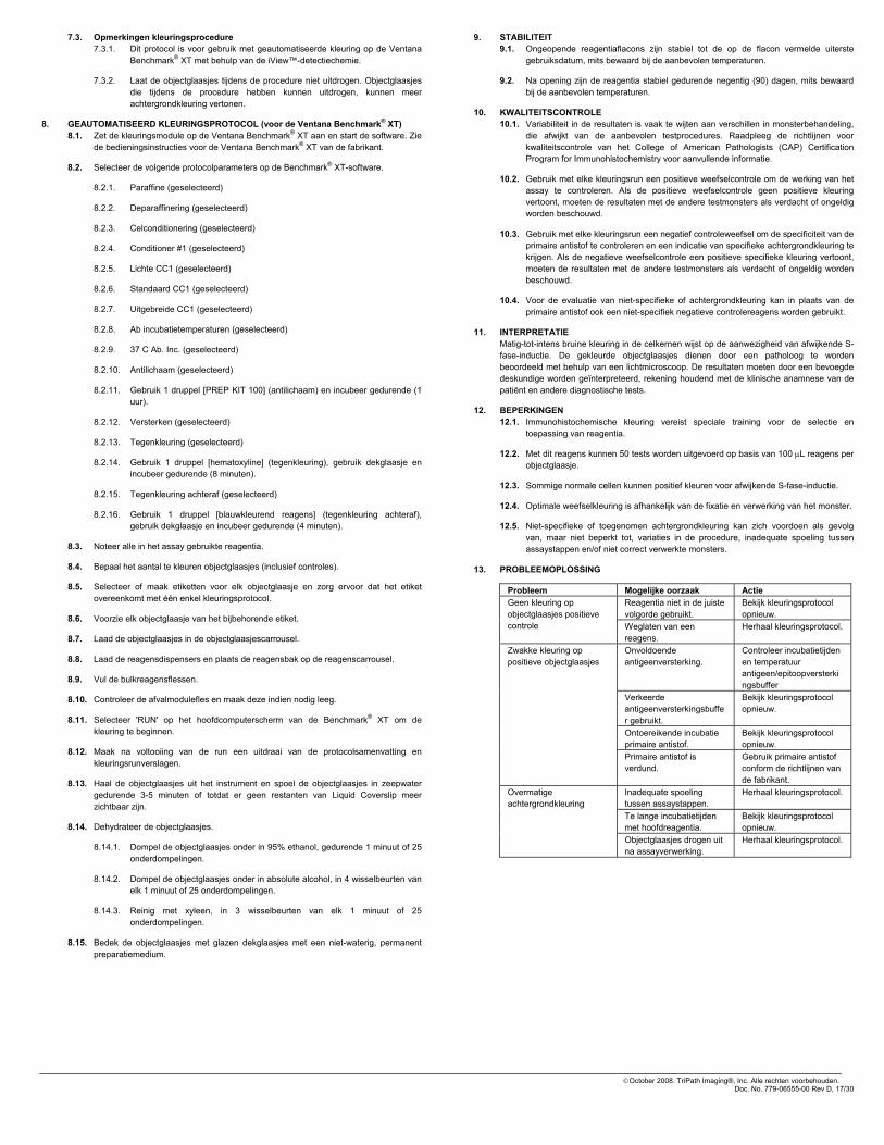
7.3. Opmerkingen kleuringsprocedure 7.3.1. Dit protocol is voor gebruik met geautomatiseerde kleuring op de Ventana
Benchmark® XT met behulp van de iView™-detectiechemie.
7.3.2. Laat de objectglaasjes tijdens de procedure niet uitdrogen. Objectglaasjes die tijdens de procedure hebben kunnen uitdrogen, kunnen meer achtergrondkleuring vertonen.
8. GEAUTOMATISEERD KLEURINGSPROTOCOL (voor de Ventana Benchmark® XT) 8.1. Zet de kleuringsmodule op de Ventana Benchmark® XT aan en start de software. Zie
de bedieningsinstructies voor de Ventana Benchmark® XT van de fabrikant.
8.2. Selecteer de volgende protocolparameters op de Benchmark® XT-software.
8.2.1. Paraffine (geselecteerd)
8.2.2. Deparaffinering (geselecteerd)
8.2.3. Celconditionering (geselecteerd)
8.2.4. Conditioner #1 (geselecteerd)
8.2.5. Lichte CC1 (geselecteerd)
8.2.6. Standaard CC1 (geselecteerd)
8.2.7. Uitgebreide CC1 (geselecteerd)
8.2.8. Ab incubatietemperaturen (geselecteerd)
8.2.9. 37 C Ab. Inc. (geselecteerd)
8.2.10. Antilichaam (geselecteerd)
8.2.11. Gebruik 1 druppel [PREP KIT 100] (antilichaam) en incubeer gedurende (1 uur).
8.2.12. Versterken (geselecteerd)
8.2.13. Tegenkleuring (geselecteerd)
8.2.14. Gebruik 1 druppel [hematoxyline] (tegenkleuring), gebruik dekglaasje en incubeer gedurende (8 minuten).
8.2.15. Tegenkleuring achteraf (geselecteerd)
8.2.16. Gebruik 1 druppel [blauwkleurend reagens] (tegenkleuring achteraf), gebruik dekglaasje en incubeer gedurende (4 minuten).
8.3. Noteer alle in het assay gebruikte reagentia.
8.4. Bepaal het aantal te kleuren objectglaasjes (inclusief controles).
8.5. Selecteer of maak etiketten voor elk objectglaasje en zorg ervoor dat het etiket overeenkomt met één enkel kleuringsprotocol.
8.6. Voorzie elk objectglaasje van het bijbehorende etiket.
8.7. Laad de objectglaasjes in de objectglaasjescarrousel.
8.8. Laad de reagensdispensers en plaats de reagensbak op de reagenscarrousel.
8.9. Vul de bulkreagensflessen.
8.10. Controleer de afvalmodulefles en maak deze indien nodig leeg.
8.11. Selecteer 'RUN' op het hoofdcomputerscherm van de Benchmark® XT om de kleuring te beginnen.
8.12. Maak na voltooiing van de run een uitdraai van de protocolsamenvatting en kleuringsrunverslagen.
8.13. Haal de objectglaasjes uit het instrument en spoel de objectglaasjes in zeepwater gedurende 3-5 minuten of totdat er geen restanten van Liquid Coverslip meer zichtbaar zijn.
8.14. Dehydrateer de objectglaasjes.
8.14.1. Dompel de objectglaasjes onder in 95% ethanol, gedurende 1 minuut of 25 onderdompelingen.
8.14.2. Dompel de objectglaasjes onder in absolute alcohol, in 4 wisselbeurten van elk 1 minuut of 25 onderdompelingen.
8.14.3. Reinig met xyleen, in 3 wisselbeurten van elk 1 minuut of 25 onderdompelingen.
8.15. Bedek de objectglaasjes met glazen dekglaasjes met een niet-waterig, permanent preparatiemedium.
9. STABILITEIT 9.1. Ongeopende reagentiaflacons zijn stabiel tot de op de flacon vermelde uiterste
gebruiksdatum, mits bewaard bij de aanbevolen temperaturen.
9.2. Na opening zijn de reagentia stabiel gedurende negentig (90) dagen, mits bewaard bij de aanbevolen temperaturen.
10. KWALITEITSCONTROLE 10.1. Variabiliteit in de resultaten is vaak te wijten aan verschillen in monsterbehandeling,
die afwijkt van de aanbevolen testprocedures. Raadpleeg de richtlijnen voor kwaliteitscontrole van het College of American Pathologists (CAP) Certification Program for Immunohistochemistry voor aanvullende informatie.
10.2. Gebruik met elke kleuringsrun een positieve weefselcontrole om de werking van het assay te controleren. Als de positieve weefselcontrole geen positieve kleuring vertoont, moeten de resultaten met de andere testmonsters als verdacht of ongeldig worden beschouwd.
10.3. Gebruik met elke kleuringsrun een negatief controleweefsel om de specificiteit van de primaire antistof te controleren en een indicatie van specifieke achtergrondkleuring te krijgen. Als de negatieve weefselcontrole een positieve specifieke kleuring vertoont, moeten de resultaten met de andere testmonsters als verdacht of ongeldig worden beschouwd.
10.4. Voor de evaluatie van niet-specifieke of achtergrondkleuring kan in plaats van de primaire antistof ook een niet-specifiek negatieve controlereagens worden gebruikt.
11. INTERPRETATIE Matig-tot-intens bruine kleuring in de celkernen wijst op de aanwezigheid van afwijkende S-fase-inductie. De gekleurde objectglaasjes dienen door een patholoog te worden beoordeeld met behulp van een lichtmicroscoop. De resultaten moeten door een bevoegde deskundige worden geïnterpreteerd, rekening houdend met de klinische anamnese van de patiënt en andere diagnostische tests.
12. BEPERKINGEN 12.1. Immunohistochemische kleuring vereist speciale training voor de selectie en
toepassing van reagentia.
12.2. Met dit reagens kunnen 50 tests worden uitgevoerd op basis van 100 μL reagens per objectglaasje.
12.3. Sommige normale cellen kunnen positief kleuren voor afwijkende S-fase-inductie.
12.4. Optimale weefselkleuring is afhankelijk van de fixatie en verwerking van het monster.
12.5. Niet-specifieke of toegenomen achtergrondkleuring kan zich voordoen als gevolg van, maar niet beperkt tot, variaties in de procedure, inadequate spoeling tussen assaystappen en/of niet correct verwerkte monsters.
13. PROBLEEMOPLOSSING
Probleem Mogelijke oorzaak Actie Reagentia niet in de juiste volgorde gebruikt.
Bekijk kleuringsprotocol opnieuw.
Geen kleuring op objectglaasjes positieve controle Weglaten van een
reagens. Herhaal kleuringsprotocol.
Onvoldoende antigeenversterking.
Controleer incubatietijden en temperatuur antigeen/epitoopversterkingsbuffer
Verkeerde antigeenversterkingsbuffer gebruikt.
Bekijk kleuringsprotocol opnieuw.
Ontoereikende incubatie primaire antistof.
Bekijk kleuringsprotocol opnieuw.
Zwakke kleuring op positieve objectglaasjes
Primaire antistof is verdund.
Gebruik primaire antistof conform de richtlijnen van de fabrikant.
Inadequate spoeling tussen assaystappen.
Herhaal kleuringsprotocol.
Te lange incubatietijden met hoofdreagentia.
Bekijk kleuringsprotocol opnieuw.
Overmatige achtergrondkleuring
Objectglaasjes drogen uit na assayverwerking.
Herhaal kleuringsprotocol.
©October 2008. TriPath Imaging®, Inc. Alle rechten voorbehouden. Doc. No. 779-06555-00 Rev D, 17/30
14. REFERENTIES 1. Kastan M and Bartec J.
Cell cycle checkpoints and cancer. Nature, 2004. Vol32:316-323.
2. Massague J. G1 cell-cycle control and cancer. Nature, 2004. Vol 432:298-432.
3. Freeman A, Morris LS, Millis AD, Stoeber K, Laskey RA, Williams GH and Coleman N. Minichromosome Maintenance Proteins as Biological Markers of Dysplasia and Malignancy. CI Cancer Research, 1999. Vol 5:2121-2132.
4. Ishimi Y, Okayasu I, Kato C, Kwon H, Kimura H, Yamada K and Song S. Enhanced expression of MCM proteins in cancer cells derived from uterine cervix. Eur. J. Biochem 2003. 270:1089-1101.
5. Lei M and Tye BK. Initiating DNA synthesis: from recruiting to activating the MCM Complex. J Cell Science 2001. Vol. 114 (8): 1447-1454.
15. SYMBOLENLIJST
Bestelnummer Voor in-vitrodiagnostiek
Raadpleeg de gebruiksaanwijzing
Bevat 7 ml
Let op, raadpleeg de bijgeleverde documentatie
Beperkingen bewaartemperatuur
Batchcode
Houdbaar tot jjjj-mm-dd of jjjj-mm
Fabrikant
IVD
TECHNISCHE INFORMATIE In de Verenigde Staten belt u gratis naar de TriPath technische dienst op 1-866-874-7284.
TriPath Imaging, Inc. 780 Plantation Drive Burlington, NC 27215 VS (800) 426-2176
Ontwikkeld met technologie van Millennium Pharmaceuticals, Inc.
en zijn handelsmerken van Millennium Pharmaceuticals, Inc. Millennium Pharmaceuticals, Inc. 40 Landsdowne Street Cambridge, MA 02139 VS www.millennium.com
TriPath Imaging® is een geregistreerd handelsmerk van TriPath Imaging, Inc. ProEx is een product en handelsmerk van TriPath Imaging, Inc.
©October 2008. TriPath Imaging®, Inc. Alle rechten voorbehouden. Doc. No. 779-06555-00 Rev D, 18/30

Til HISTOLOGI 005-11000-40 7 ml
REAGENSBESKRIVELSE Klon MCM2 26H6.19, MCM2 27C5.6, TOP2A SWT3D1 Ig-klasse IgG1 Immunogen Rekombinant humant MCM2 og TOP2A
1. ANVENDELSE
Til in vitro-diagnostisk brug.
Til brug med automatisk farvning i Ventana Benchmark® XT vha. iView™-detektionskemi.
ProEx™ C-immunhistokemisk test er beregnet til kvalitativ evaluering af afvigende S-faseinduktion i formalinfikserede, paraffinindstøbte vævsbiopsier. Fortolkning af resultaterne skal foretages af en faguddannet person under hensyn til patientens kliniske historie samt andre diagnostiske prøver .
2. RESUMÉ OG FORKLARING Minikromosomvedligeholdelse (MCM) og topoisomerase II alpha (TOP2A)-proteiner spiller en vigtig regulerende rolle ved eukaryotisk DNA-replikation. Eksempelvis resulterer HPV oncoprotein E6- og E7-bypass af kritiske cellecyklus-testpunkter i en forlænget og afvigende S-faseinduktionscyklus. Under den transkriptionale aktivering af den afvigende cellecyklus øges niveauerne af MCM2- og TOP2A-proteiner i de prolifererende celler.
Både MCM2- og TOP2A-proteinerne har vist sig at være overudtrykte i et antal forskellige dysplastiske og maligne vævsprøver, herunder cervical neoplasi1-5. Overudtrykket af disse proteiner i morfologisk abnorme celler, der udtrykkes som et moderat-til-intens nukleært farvningsmønster ved brug af immunhistokemiske (IHC) teknikker, indikerer tilstedeværelsen af afvigende S-faseinduktion.
3. MEDFØLGENDE REAGENS ProEx™ C antistofreagens indeholder monoklonalt anti-MCM2 og anti-TOP2A fra mus, oprenset fra vævskultursupernatant og fortyndet i en bufferet saltvandsopløsning, som indeholder proteinstabilisatorer og 0,09% natriumazid.
4. PROCEDUREPRINCIPPER Formalinfikserede, paraffinindstøbte vævsprøver skæres i snit, deponeres på objektglas og afparaffineres. Prøvesnit er forbehandlede med en buffer, der eksponerer antigene steder. Der er tilsat blokeringsstoffer for at minimere baggrundsfarvning, som skyldes endogen peroxidase eller uspecifik proteinbinding. Prøven inkuberes derefter med ProEx™ C-antistofreagens. Tilføjelsen af et enzym-kædet antistofkromogensystem resulterer i dannelsen af et synligt kromogent produkt, lokaliseret ved antigen-antistoffets bindingssteder. Prøven kontrastfarves herefter med hæmatoxylin, der tilføres et blånelsesmiddel, og der sættes dækglas på. Resultaterne skal fortolkes af en faguddannet person ved hjælp af lysmikroskop.
5. NØDVENDIGE, IKKE MEDFØLGENDE MATERIALER OG REAGENSER (til Ventana Benchmark® XT-procedure)
• 10X Reaction Buffer – Kat. nr. 950-300 2L (Ventana Medical Systems)
• LCS (Liquid Coverslip) – Kat. nr. 650-010 2L (Ventana Medical Systems)
• 10X SSC – Kat. nr. 950-110 2L (Ventana Medical Systems)
• EZ Prep – Kat. nr. 950-102 2L (Ventana Medical Systems)
• CC1 (Cell Conditioning) – Kat. nr. 950-124 2L (Ventana Medical Systems)
• iView™ DAB Detection Kit – Kat. nr. 760-091 (Ventana Medical Systems)
• Amplification Kit (A & B) – Kat. nr. 760-080 (Ventana Medical Systems)
• Prep Kit Dispenser med Prep Kit Button – Kat. nr. 786-3034 (Ventana Medical Systems)
• Hematoxylin – Kat. nr. 760-2021 (Ventana Medical Systems)
• Bluing Reagent – Kat. nr. 760-2037 (Ventana Medical Systems)
• Objektglas (SuperFrost® Plus eller tilsvarende)
• Monteringsmedie (Acrytol® eller tilsvarende)
• Minutur (skal kunne indstilles til intervaller på 1-60 minutter)
• Destilleret vand
• Ethanol 95%, 100%
• Dækglas
• Laboratorie-mærkepen
• 20 l glasballon (Nalgene® eller tilsvarende)
• Tørrerack til objektglas
• Objektglasrack med farvningsskiver
• Xylen eller xylenerstatninger
• Lysmikroskop (10x, 20x [valgfrit], 40x objektiver)
6. FORSIGTIGHEDSREGLER 6.1. Til in vitro-diagnostisk brug
6.2. Objektglasrensetrin, der kræver xylen, skal udføres i et certificeret stinkskab.
6.3. ProEx™ C antistofreagenset indeholder natriumazid (NaN3), et kemikalie, der er meget giftigt i ren form. Selvom koncentrationen i produktet ikke er klassificeret som farlig, kan natriumazid reagere med bly og kobber og danne meget eksplosive ophobninger af metalazider. Efter bortskaffelse skylles med rigelige mængder vand for at hindre ophobning af metalazid i afløb.
6.4. DAB (3,3’-diaminobenzidin) er klassificeret som et muligt kræftfremkaldende stof. Undgå fysisk kontakt og længerevarende eller gentagen eksponering af stoffet. Anvend det i et certificeret stinkskab.
6.5. Prøver og alle materialer, der kommer i kontakt med prøver, skal behandles som potentielt smittefarlige og bortskaffes i overensstemmelse med passende forholdsregler. Brug aldrig mundpipette, og undgå, at hud og slimhinder kommer i kontakt med reagenser og præparater. Hvis reagenser kommer i kontakt med følsomme områder, skal de afvaskes med rigelige mængder vand.
6.6. Minimér mikrobiel kontaminering af reagenser for at undgå non-specifik farvning.
6.7. Andre inkubationstider, -temperaturer eller -metoder end de angivne kan medføre fejlagtige resultater.
6.8. Anvend ikke ProEx™ C-antistofreagenset efter den udløbsdato, der er trykt på emballagen. Brugeren skal validere forholdene, hvis reagenser opbevares under andre forhold end dem, der er angivet i pakningsindlægget.
6.9. Brug egnet personligt beskyttelsesudstyr for at undgå kontakt med øjne og hud. Se materialesikkerhedsdatabladet (MSDS) for at få yderligere oplysninger.
7. BRUGSANVISNING 7.1. Prøveforberedelse
7.1.1. Skær snit på 4 μm af vævsblokken, og placér snittene på SuperFrost® Plus-objektglas.
7.1.2. Mærk objektglassene.
7.1.3. Bag glassene i en ovn med varmluft i 20 minutter. Hvis objektglassene allerede er tørre, skal de bringes i kontakt med en Histo-Orienter, indtil paraffinen smelter.
7.2. Reagensforberedelse til Ventana Benchmark® X TBemærk: Følg producentens anvisninger.
7.2.1. 10X Reaction Buffer
7.2.1.1. Registrér en ny flaske 10X Reaction Buffer-koncentrat.
7.2.1.2. Tilsæt 1 (én) flaske 10X Reaction Buffer-koncentrat til 18 liter destilleret vand. Bland grundigt.
7.2.1.3. Fyld EZ Prep-flaske nr. 4 på Benchmark® XT. Kontrollér for at sikre, at pH er mellem 6,5 og 7,1.
7.2.2. EZ Prep (10X) – Afparaffineringsopløsning
7.2.2.1. Registrér en ny flaske EZ Prep-koncentrat.
7.2.2.2. Tilsæt 1 (én) flaske 10X EZ Prep-koncentrat til 18 liter destilleret vand. Bland grundigt.
7.2.2.3. Fyld EZ Prep-flaske nr. 1 på Benchmark® XT. Kontrollér for at sikre, at pH er mellem 6,90 og 7,2.
7.2.3. SSC (10X)
7.2.3.1. Registrér 2 (to) flasker SSC-koncentrat.
7.2.3.2. Tilsæt 2 (to) flasker SSC-koncentrat til 16 liter destilleret vand. Bland grundigt.
7.2.3.3. Fyld SSC-flaske nr. 3 på Benchmark® XT. Kontrollér for at sikre, at pH er mellem 7,1 og 7,5.
7.2.4. Fyldbart Prep-sæt
7.2.4.1. Registrér Prep-sæt-knap
7.2.4.2. Fyld Prep-sæt-dispenseren i henhold til producentens anvisninger.
7.3. Noter om farvningsproceduren 7.3.1. Denne protokol skal anvendes ved automatisk farvning i Ventana
Benchmark® XT vha. iView™-detektionskemi.
7.3.2. Lad ikke objektglassene tørre ud på noget tidspunkt under proceduren. Objektglas, der har været tørret ud under proceduren, kan udvise forøget baggrundsfarvning.
IVD
2oC
8oC
©October 2008. TriPath Imaging®, Inc. All rights reserved. Doc. No. 779-06555-00 Rev D, 19/30

8. PROTOKOL FOR AUTOMATISK FARVNING (for Ventana Benchmark® XT) 8.1. Tænd farvningsmodulet på Ventana Benchmark® XT, og start softwaren. Se
producentens betjeningsvejledning til Ventana Benchmark® XT.
8.2. Indstil nedenstående protokolparametre i Benchmark® XT-softwaren.
8.2.1. Paraffin (valgt)
8.2.2. Deparaffinization (valgt)
8.2.3. Cell Conditioning (valgt)
8.2.4. Conditioner #1 (valgt)
8.2.5. Mild CC1 (valgt)
8.2.6. Standard CC1 (valgt)
8.2.7. Extended CC1 (valgt)
8.2.8. Ab Incubation Temperatures (valgt)
8.2.9. 37 C Ab. Inc. (valgt)
8.2.10. Antibody (valgt)
8.2.11. Apply 1 drop of [PREP KIT 100] (Antibody) and incubate for (1 time).
8.2.12. Amplify (valgt)
8.2.13. Counterstain (valgt)
8.2.14. Apply 1 drop of [Hematoxylin] (Counterstain), apply coverslip and incubate for (8 minutter).
8.2.15. Post Counterstain (valgt)
8.2.16. Apply 1 drop of [Bluing Reagent] (Post Counterstain), apply coverslip and incubate for (4 minutter).
8.3. Registrér alle reagenser, der anvendes i analysen.
8.4. Bestem antallet af objektglas, der skal farves (inklusive kontroller).
8.5. Vælg eller opret etiketter for hvert objektglas, og kontrollér, at hver etiket svarer til en enkelt farvningsprotokol.
8.6. Overfør de relevante etiketter til de enkelte objektglas.
8.7. Sæt objektglassene i objektglaskarrusellen.
8.8. Fyld reagensdispenserne, og montér reagensbakken på reagenskarrusellen.
8.9. Fyld glasballonerne til partivæskereagens.
8.10. Kontrollér glasballonen til affald, og tøm den, hvis det er nødvendigt.
8.11. Vælg “RUN” på computerens hovedskærmbillede. Benchmark® XT indleder farvningsproceduren.
8.12. Udskriv protokoloversigts- og farvekørselsrapporter, når farvningsproceduren er færdig.
8.13. Fjern objektglassene fra instrumentet, og rengør dem i sæbevand i 3-5 minutter, eller indtil der ikke længere ses rester af flydende dækglas.
8.14. Tør objektglassene.
8.14.1. Nedsænk objektglassene i 95% ethanol i 1 minut eller 25 dyp.
8.14.2. Nedsænk objektglassene i ren alkohol, 4 hold, 1 minut hver eller 25 dyp.
8.14.3. Rens med xylen, 3 hold, 1 minut hver eller 25 dyp.
8.15. Dæk objektglassene med ikke-vandholdig, permanent monteringsmedie ved hjælp af dækglas.
9. HOLDBARHED 9.1. Ved opbevaring ved anbefalede temperaturer er uåbnede reagenshætteglas
holdbare, indtil den udløbsdato, der er angivet på hætteglasset.
9.2. Efter åbning er reagenserne holdbare i halvfems (90) dage, hvis de opbevares ved de anbefalede temperaturer.
10. KVALITETSKONTROL 10.1. Varians i resultater skyldes ofte forskelle i prøvehåndteringen, som afviger fra de
anbefalede testprocedurer. Se retningslinjerne for kvalitetskontrol fra College of American Pathologists (CAP) Certification Program for Immunohistochemistry for at få yderligere oplysninger.
10.2. Der bør udføres en positiv vævskontrol ved hver farvningskørsel for at verificere analysens effektivitet. Hvis den positive vævskontrol ikke udviser positiv farvning, skal resultaterne med de øvrige prøver betragtes som mistænkelige eller ugyldige.
10.3. Der bør udføres en negativ vævskontrol ved hver farvningskørsel for at verificere det primære antistofs specificitet samt for at give en indikation af baggrundsfarvningen. Hvis den negative vævskontrol udviser positiv specifik farvning, skal resultaterne med de øvrige prøver betragtes som mistænkelige eller ugyldige.
10.4. Der kan også anvendes en uspecifik negativ kontrolreagens i stedet for det primære antistof til evaluering af uspecifik farvning eller baggrundsfarvning.
11. FORTOLKNING Moderat-til-intens brunfarvning i cellekernerne indikerer tilstedeværelse af afvigende S-faseinduktion. De farvede objektglas bør evalueres af en patolog vha. et lysmikroskop. Fortolkning af resultaterne skal foretages af en faguddannet person under hensyn til patientens kliniske historie samt andre diagnostiske prøver .
12. BEGRÆNSNINGER 12.1. Immunhistokemisk farvning kræver specialuddannelse i udvælgelse og anvendelse
af reagenser.
12.2. Dette reagens kan anvendes til 50 test, forudsat at der anvendes 100 μl reagens pr. objektglas.
12.3. Visse normale celler kan blive farvet positive for afvigende S-faseinduktion.
12.4. Optimal vævsfarvning afhænger af fiksering og behandling af prøven.
12.5. Der kan forekomme uspecifik eller øget baggrundsfarvning på grund af, men ikke begrænset til, procedureafvigelser, utilstrækkelig rensning mellem analysetrin og/eller utilstrækkeligt behandlede prøver.
13. FEJLFINDING
Problem Mulige årsager Afhjælpning Reagens anvendt i forkert rækkefølge.
Gennemgå farvningsprotokollen.
Ingen farvning på positive kontrolglas
Udeladelse af et reagens. Gentag farvningsprotokollen.
Utilstrækkelig antigen-retrieval.
Kontrollér inkubationstider og temperatur for antigen-/epitop-retrievalbuffer.
Forkert antigen-retrievalbuffer er anvendt.
Gennemgå farvningsprotokollen.
Utilstrækkelig inkubation af primært antistof.
Gennemgå farvningsprotokollen.
Svag farvning på positive objektglas
Primært antistof er blevet fortyndet.
Anvend primært antistof i overensstemmelse med producentens anvisninger.
Utilstrækkelig rensning mellem analysetrin.
Gentag farvningsprotokollen.
Lange inkubationstider med nøglereagenser.
Gennemgå farvningsprotokollen.
Kraftig baggrundsfarvning
Objektglas tørret ud under post-analysebehandling.
Gentag farvningsprotokollen.
©October 2008. TriPath Imaging®, Inc. All rights reserved. Doc. No. 779-06555-00 Rev D, 20/30

14. REFERENCER 1. Kastan M and Bartec J. Cell cycle checkpoints and cancer. Nature, 2004. Vol32:316-323.
2. Massague J. G1 cell-cycle control and cancer. Nature, 2004. Vol32:432298.
3. Freeman A, Morris LS, Millis AD, Stoeber K, Laskey RA, Williams GH and Coleman N. Minichromosome Maintenance Proteins as Biological Markers of Dysplasia and Malignancy. CI Cancer Research, 1999. Vol 5:2121-2132.
4. Ishimi Y, Okayasu I, Kato C, Kwon H, Kimura H, Yamada K and Song S. Enhanced expression of MCM proteins in cancer cells derived from uterine cervix. Eur. J. Biochem 2003. 270:1089-1101.
5. Lei M and Tye BK. Initiating DNA synthesis: from recruiting to activating the MCM Complex. J Cell Science 2001. Vol. 114 (8): 1447-1454.
15. SYMBOLFORKLARING
Katalognummer
Til in vitro-diagnostisk brug
Se brugsanvisningen
Indeholder 7 ml
Forsigtig! Se brugsanvisningen
Temperaturbegrænsning under opbevaring
Batchnummer
Anvendes inden ÅÅÅÅ-MM-DD eller ÅÅÅÅ-MM
Producent
IVD
TEKNISKE OPLYSNINGER I USA kan der ringes til TriPath Technical Services, gratisnummer 1-866-874-7284.
TriPath Imaging, Inc. 780 Plantation Drive Burlington, NC 27215 USA (800) 426-2176
Udviklet med teknologi fra Millennium Pharmaceuticals, Inc.
og er varemærker tilhørende Millennium Pharmaceuticals, Inc. Millennium Pharmaceuticals, Inc. 40 Landsdowne Street Cambridge, MA 02139 USA www.millennium.com
TriPath Imaging® er et registreret varemærke tilhørende TriPath Imaging, Inc. ProEx er et produkt og varemærke tilhørende TriPath Imaging, Inc.
©October 2008. TriPath Imaging®, Inc. All rights reserved. Doc. No. 779-06555-00 Rev D, 21/30

Para HISTOLOGIA 005-11000-40 7 mL
DESCRIÇÃO DO REAGENTE Clone MCM2 26H6.19, MCM2 27C5.6, TOP2A SWT3D1 Classe Ig IgG1 Imunogénio Recombinante Humano MCM2 e TOP2A
1. FINALIDADE
Para utilização em diagnóstico in vitro.
Para utilização com a coloração automática no Ventana Benchmark® XT utilizando o kit de detecção química iView™.
A Análise imuno-histoquímica ProEx™ C destina-se a ser utilizada para uma avaliação qualitativa da indução aberrante de uma fase S em biopsias de secções de tecido fixado em formol e impregnado em parafina. A interpretação dos resultados tem de ser efectuada por um profissional qualificado, dentro do contexto do historial do doente e através de outros testes de diagnóstico.
2. RESUMO E EXPLICAÇÃO A proteína de manutenção de minicromossomas (MCM) e a proteína alfa topoisomerase II (TOP2A) desempenham um importante papel regulador na replicação de ADN eucariótico. Por exemplo, o desvio das oncoproteínas E6 e E7 HPV dos pontos de verificação crítica do ciclo celular, que resulta num ciclo de indução de uma fase S prolongado e aberrante. Durante a activação da transcrição do ciclo celular aberrante, os níveis de proteínas MCM2 e TOP2A aumentam nas células proliferativas.
Demonstrou-se que tanto a proteína MCM2 como a TOP2A apresentam uma sobreexpressão em diversos tecidos malignos e displásicos diferentes, incluindo em neoplasias cervicais1-5. A sobreexpressão destas proteínas em células morfologicamente anómalas, conforme demonstrado por um padrão de coloração nuclear moderado a intenso utilizando técnicas de imuno-histoquímica (IHC), é indicadora da presença de indução aberrante de uma fase S.
3. REAGENTES FORNECIDOS O Reagente de Anticorpo ProEx™ C contém anti-MCM2 monoclonal de ratinho e anti-TOP2A purificado de sobrenadante de cultura de tecidos e foi diluído numa solução salina de tampão, que contém estabilizadores de proteínas e 0,09% de azida de sódio.
4. PRINCÍPIOS DO PROCEDIMENTO As amostras de tecido fixadas em formol e impregnadas em parafina são seccionadas, depositadas em lâminas de vidro e desparafinizadas. As amostras seccionadas são pré-tratadas com um tampão para expor os locais antigénicos. São adicionados bloqueadores para minimizar a coloração de fundo causada por peroxidase endógena ou ligação proteica não específica. A amostra é, em seguida, incubada com Reagente de Anticorpo ProEx™ C. A adição de um sistema de cromogénio de anticorpos ligados à enzima resulta na formação de um produto cromogénico visível localizado nos locais de ligação do antigénio-anticorpo. A amostra é, em seguida, sujeita a contrastação com hematoxilina, é aplicado um agente de Bluing e a lâmina é protegida com uma lamela. Os resultados são interpretados por um profissional qualificado utilizando um microscópio óptico.
5. MATERIAIS E REAGENTES NECESSÁRIOS MAS NÃO FORNECIDOS (para Procedimento com Ventana Benchmark® XT)
• 10X Reaction Buffer – Cat # 950-300 2L (Ventana Medical Systems)
• LCS (Liquid Coverslip) – Cat # 650-010 2L (Ventana Medical Systems)
• 10X SSC – Cat # 950-110 2L (Ventana Medical Systems)
• EZ Prep – Cat # 950-102 2L (Ventana Medical Systems)
• CC1 (Cell Conditioning) – Cat # 950-124 2L (Ventana Medical Systems)
• iView™ DAB Detection Kit – Cat # 760-091 (Ventana Medical Systems)
• Amplification Kit (A&B) – Cat # 760-080 (Ventana Medical Systems)
• Prep Kit Dispenser com Prep Kit Button – Cat # 786-3034 (Ventana Medical Systems)
• Hematoxylin – Cat # 760-2021 (Ventana Medical Systems)
• Bluing Reagent – Cat # 760-2037 (Ventana Medical Systems)
• Lâminas de vidro (SuperFrost® Plus ou equivalente)
• Meio de montagem (Acrytol® ou equivalente)
• Cronómetro (com intervalos de 1 a 60 minutos)
• H2O destilada
• Etanol 95%, 100%
• Lamelas de vidro
• Marcador de laboratório
• Garrafão de 20L (Nalgene® ou equivalente)
• Secador de lâminas
• Suporte de lâminas com placas de coloração
• Xilol ou substitutos de xilol
• Microscópio óptico (objectivas de 10x, 20x [opcional], 40x )
6. PRECAUÇÕES 6.1. Para utilização em diagnóstico in vitro.
6.2. Os passos de limpeza das lâminas que requerem xilol têm de ser efectuados numa campânula de fumos químicos certificada.
6.3. O Reagente de Anticorpo ProEx™ C contém azida de sódio (NaN3), um produto químico altamente tóxico na forma pura. Em situações de concentração do produto, embora não sendo classificada como perigosa, a azida de sódio pode reagir com as canalizações de chumbo e de cobre, formando acumulações de azidas metálicas altamente explosivas. Ao eliminar o produto, adicionar água abundante para evitar a acumulação de azidas metálicas na canalização.
6.4. A DAB (3,3’-diaminobenzidina) é classificada como um suposto carcinogénico. Evitar o contacto físico e exposição repetida ou prolongada à mesma. Utilizá-la numa campânula de fumos químicos certificada.
6.5. As amostras e todos os materiais expostos às amostras deverão ser manuseados como materiais passíveis de transmitir infecção e devem ser eliminados com as devidas precauções. Nunca pipetar os reagentes com a boca e evitar o contacto da pele e membranas mucosas com os reagentes e as amostras. Se os reagentes entrarem em contacto com áreas sensíveis, lavar com água abundante.
6.6. Minimizar a contaminação microbiana dos reagentes para evitar uma coloração não específica.
6.7. Os tempos e as temperaturas de incubação ou métodos diferentes dos especificados podem originar resultados erróneos.
6.8. Não utilizar o Reagente de Anticorpo ProEx™ C após o fim do prazo de validade impresso na embalagem. O utilizador deverá validar as condições, caso os reagentes sejam conservados noutras condições que não as especificadas no folheto informativo incluído na embalagem.
6.9. Usar equipamento de protecção pessoal adequado para evitar o contacto do reagente com a pele e os olhos. Consultar a Ficha de Dados de Segurança do Material (MSDS) para obter informações adicionais.
7. INSTRUÇÕES DE UTILIZAÇÃO 7.1. Preparação das amostras
7.1.1. Cortar secções com 4 μm do bloco de tecido e colocá-las em lâminas de vidro SuperFrost® Plus.
7.1.2. Rotular as lâminas com etiquetas.
7.1.3. Aquecer as lâminas numa estufa de ar forçado durante 20 minutos. Se as lâminas já estiverem secas, passá-las para um Histo-Orienter até a parafina derreter.
7.2. Preparação dos reagentes para o Ventana Benchmark® XT Nota: Consultar as instruções do fabricante.
7.2.1. 10X Reaction Buffer
7.2.1.1. Registar um frasco novo do concentrado 10X Reaction Buffer.
7.2.1.2. Adicionar 1 (um) frasco de concentrado 10X Reaction Buffer a 18 litros de água destilada. Misturar bem.
7.2.1.3. Encher o frasco de EZ Prep nº 4 no Benchmark® XT. Verificar para se certificar que o pH está entre 6,5 e 7,1.
7.2.2. EZ Prep (10X) – Solução de desparafinização
7.2.2.1. Registar um frasco novo de concentrado EZ Prep.
7.2.2.2. Adicionar 1 (um) frasco de concentrado EZ Prep a 18 litros de água destilada. Misturar bem.
7.2.2.3. Encher o frasco de EZ Prep nº 1 no Benchmark® XT. Verificar para se certificar que o pH está entre 6,90 e 7,2.
7.2.3. SSC (10X)
7.2.3.1. Registar 2 (dois) frascos de concentrado SSC.
7.2.3.2. Adicionar 2 (dois) frascos de concentrado SSC a 16 litros de água destilada. Misturar bem.
7.2.3.3. Encher o frasco de SSC nº 3 no Benchmark® XT. Verificar para se certificar que o pH está entre 7,1 e 7,5.
7.2.4. Fillable Prep Kit
7.2.4.1. Registar o Prep Kit Button.
7.2.4.2. Encher o dispensador Prep Kit Dispenser de acordo com as instruções do fabricante.
IVD
2oC
8oC
©October 2008. TriPath Imaging®, Inc. Todos os direitos reservados. Doc. No. 779-06555-00 Rev D, 22/30

7.3. Notas ao procedimento de coloração 7.3.1. Este protocolo destina-se a utilização com a coloração automática no
Ventana Benchmark® XT utilizando o kit de detecção química iView™.
7.3.2. Não permitir que as lâminas sequem em nenhum momento durante o procedimento. Caso se permita que as lâminas sequem durante o procedimento, tal resultará num aumento da coloração de fundo.
8. PROTOCOLO DE COLORAÇÃO AUTOMÁTICA (para o Ventana Benchmark® XT) 8.1. Ligar o módulo de coloração no Ventana Benchmark® XT e iniciar o software.
Consultar as instruções de funcionamento do fabricante do Ventana Benchmark® XT.
8.2. Seleccionar os seguintes parâmetros do protocolo apresentados no software do Benchmark® XT.
8.2.1. Parafina (seleccionado)
8.2.2. Desparafinização (seleccionado)
8.2.3. Condicionamento celular (seleccionado)
8.2.4. Condicionador n.º1 (seleccionado)
8.2.5. CC1 suave (seleccionado)
8.2.6. CC1 padrão (seleccionado)
8.2.7. CC1 alargada (seleccionado)
8.2.8. Temperaturas de incubação Ab (seleccionado)
8.2.9. Inc. Ab 37 C (seleccionado)
8.2.10. Anticorpo (seleccionado)
8.2.11. Aplicar uma gota de [PREP KIT 100] (Anticorpo) e incubar durante (1 hora).
8.2.12. Amplificar (seleccionado)
8.2.13. Contrastante (seleccionado)
8.2.14. Aplicar uma gota de [Hematoxilina] (Contrastante), aplicar a lamela e incubar durante (8 minutos).
8.2.15. Após contrastante (seleccionado)
8.2.16. Aplicar uma gota de [Reagente de Bluing] (Após contrastante), aplicar a lamela e incubar durante (4 minutos).
8.3. Registar todos os reagentes que estão a ser utilizados no ensaio.
8.4. Determinar o número de lâminas a serem coradas (incluindo controlos).
8.5. Seleccionar ou criar etiquetas para cada lâmina, certificando-se que cada etiqueta corresponde a um único protocolo de coloração.
8.6. Colar a etiqueta apropriada em cada lâmina.
8.7. Colocar as lâminas no carrossel de lâminas.
8.8. Colocar os dispensadores dos reagentes e montar o tabuleiro de reagentes no carrossel de reagentes.
8.9. Encher os garrafões de reagentes volumosos.
8.10. Verificar o garrafão do módulo de resíduos e esvaziar, se necessário.
8.11. Seleccionar “RUN” (INICIAR PROCESSAMENTO) no ecrã do computador do Benchmark® XT para iniciar a coloração.
8.12. Uma vez concluído o processamento, imprimir o resumo do protocolo e os relatórios dos processos de coloração.
8.13. Remover as lâminas do instrumento e lavar as lâminas em água com detergente durante 3 a 5 minutos ou até o resíduo de Liquid Coverslip deixar de ser visível.
8.14. Desidratar as lâminas.
8.14.1. Imergir as lâminas em etanol a 95%, durante 1 minuto ou 25 vezes.
8.14.2. Imergir as lâminas em álcool absoluto, 4 banhos, 1 minuto cada ou 25 vezes.
8.14.3. Limpar com xilol, 3 banhos, 1 minuto cada ou 25 imersões.
8.15. Cobrir com meios de montagem permanentes não aquosos, utilizando lamelas de vidro.
9. ESTABILIDADE 9.1. Quando conservados às temperaturas recomendadas, os frascos fechados de
reagente permanecem estáveis até à data indicada no frasco.
9.2. Uma vez aberto o frasco, os reagentes permanecem estáveis durante noventa (90) quando conservados às temperaturas recomendadas.
10. CONTROLO DE QUALIDADE 10.1. A variabilidade nos resultados deriva frequentemente de diferenças no
manuseamento das amostras, que se desviam dos procedimentos de teste recomendados. Consultar as directrizes de controlo de qualidade do College of American Pathologists (CAP) Certification Program for Immunohistochemistry para obter mais informações.
10.2. Em cada processo de coloração efectuado deverá ser incluído um controlo tecidular positivo para confirmar o desempenho do ensaio. Se o controlo tecidular positivo não apresentar uma coloração positiva, os resultados das outras amostras de teste deverão ser considerados suspeitos ou inválidos.
10.3. Em cada processo de coloração efectuado deverá ser incluído um controlo tecidular negativo para confirmar a especificidade do anticorpo primário e fornecer uma indicação da coloração de fundo. Se o controlo tecidular negativo apresentar coloração positiva específica, os resultados das outras amostras de teste deverão ser considerados suspeitos ou inválidos.
10.4. Pode também ser utilizado um reagente de controlo negativo não específico em vez do anticorpo primário para avaliar a coloração de fundo ou não específica.
11. INTERPRETAÇÃO Uma coloração castanha moderada a intensa no núcleo das células indica a presença de indução aberrante de uma fase S. Um patologista deverá avaliar as lâminas coradas utilizando um microscópio óptico. A interpretação dos resultados tem de ser efectuada por um profissional qualificado, dentro do contexto do historial do doente e através de outros testes de diagnóstico.
12. LIMITAÇÕES 12.1. A coloração imuno-histoquímica requer uma formação especializada na selecção e
aplicação dos reagentes.
12.2. Este reagente será suficiente para efectuar 50 testes, partindo do princípio que se aplica 100 μL de reagente por lâmina.
12.3. Algumas células normais podem apresentar uma coloração positiva quanto à indução aberrante de uma fase S.
12.4. A coloração tecidular ideal está dependente da fixação e processamento da amostra.
12.5. Pode ocorrer uma coloração de fundo não específica ou um aumento da coloração de fundo devido a, mas não se limitando a, variações no procedimento, lavagem inadequada entre as etapas do ensaio e/ou amostras processadas inadequadamente.
13. RESOLUÇÃO DE PROBLEMAS
Problema Possível causa Medida Reagentes aplicados pela ordem incorrecta.
Rever protocolo de coloração.
As lâminas de controlo positivo não apresentam qualquer coloração. Omissão de qualquer um
dos reagentes. Repetir protocolo de coloração.
Recuperação insuficiente do antigénio.
Verificar tempos de incubação e temperatura do tampão de recuperação do antigénio/epítopo.
O tampão utilizado para a recuperação do antigénio é incorrecto.
Rever protocolo de coloração.
Incubação inadequada do anticorpo primário.
Rever protocolo de coloração.
As lâminas de controlo positivo apresentam uma coloração fraca.
O anticorpo primário foi diluído.
Utilizar o anticorpo primário de acordo com as instruções do fabricante.
Lavagem inadequada entre as etapas do ensaio.
Repetir protocolo de coloração.
Tempos de incubação excessivos com os principais reagentes.
Rever protocolo de coloração.
Coloração de fundo excessiva
As lâminas secaram durante o processamento após o ensaio.
Repetir protocolo de coloração.
©October 2008. TriPath Imaging®, Inc. Todos os direitos reservados. Doc. No. 779-06555-00 Rev D, 23/30

14. BIBLIOGRAFIA 1. Kastan M and Bartec J. Cell cycle checkpoints and cancer. Nature, 2004. Vol32:316-323.
2. Massague J. G1 cell-cycle control and cancer. Nature, 2004. Vol 432:298-432.
3. Freeman A, Morris LS, Millis AD, Stoeber K, Laskey RA, Williams GH and Coleman N. Minichromosome Maintenance Proteins as Biological Markers of Dysplasia and Malignancy. CI Cancer Research, 1999. Vol 5:2121-2132.
4. Ishimi Y, Okayasu I, Kato C, Kwon H, Kimura H, Yamada K and Song S. Enhanced expression of MCM proteins in cancer cells derived from uterine cervix. Eur. J. Biochem 2003. 270:1089-1101.
5. Lei M and Tye BK. Initiating DNA synthesis: from recruiting to activating the MCM Complex. J Cell Science 2001. Vol. 114 (8): 1447-1454.
15. GLOSSÁRIO DE SÍMBOLOS
Número de catálogo
Para utilização em diagnóstico in vitro
Consultar as instruções de utilização
Contém 7 mL
Cuidado, consultar a documentação fornecida
Limitações da temperatura de conservação
Código do lote
Utilizar até AAAA-MM-DD ou AAAA-MM
Fabricante
IVD
INFORMAÇÕES TÉCNICAS Nos Estados Unidos da América, telefone para os Serviços Técnicos da TriPath, número gratuito 1-866-874-7284.
TriPath Imaging, Inc. 780 Plantation Drive Burlington, NC 27215 USA (800) 426-2176
Desenvolvido com tecnologia da Millennium Pharmaceuticals, Inc.
e são marcas comerciais da Millennium Pharmaceuticals, Inc. Millennium Pharmaceuticals, Inc. 40 Landsdowne Street Cambridge, MA 02139 USA www.millennium.com
TriPath Imaging® é uma marca comercial registada da TriPath Imaging, Inc. ProEx é um produto e marca comercial da TriPath Imaging, Inc.
©October 2008. TriPath Imaging®, Inc. Todos os direitos reservados. Doc. No. 779-06555-00 Rev D, 24/30

Για ΙΣΤΟΛΟΓΙΑ 005-11000-40 7 mL
ΠΕΡΙΓΡΑΦΗ ΑΝΤΙΔΡΑΣΤΗΡΙΟΥ Κλώνος MCM2 26H6.19, MCM2 27C5.6, TOP2A SWT3D1 Τάξη Ig IgG1 Ανοσογόνο Ανασυνδυασμένες MCM2 και TOP2A ανθρώπου
1. ΠΡΟΟΡΙΖΟΜΕΝΗ ΧΡΗΣΗ
Για in vitro διαγνωστική χρήση.
Για χρήση με αυτοματοποιημένη χρώση σε Benchmark® XT του οίκου Ventana χρησιμοποιώντας τη διαδικασία εντοπισμού iView™.
Η ανοσοϊστοχημική εξέταση ProEx™ C προορίζεται για τον ποιοτικό προσδιορισμό της έναρξης της παρεκκλίνουσας S-φάσης σε βιοψίες ιστού μονιμοποιημένου με φορμαλίνη και εγκλεισμένου σε παραφίνη. Η ερμηνεία των αποτελεσμάτων πρέπει να πραγματοποιείται από πιστοποιημένο επαγγελματία και εντός των πλαισίων του ιστορικού του ασθενούς και άλλων διαγνωστικών εξετάσεων.
2. ΠΕΡΙΛΗΨΗ ΚΑΙ ΕΠΕΞΗΓΗΣΗ Η συντήρηση των μίνι χρωμοσωμάτων (MCM) και οι α πρωτεΐνες της τοποϊσομεράσης II (TOP2A) διαδραματίζουν σημαντικό ρόλο στη ρύθμιση της αντιγραφής του DNA στα ευκαρυωτικά κύτταρα. Για παράδειγμα, οι ογκοπρωτεΐνες E6 και E7 του HPV παρακάμπτουν τα κρίσιμα σημεία ελέγχου του κυτταρικού κύκλου συντελώντας στην έναρξη ενός κύκλου με παρατεταμένη και παρεκκλίνουσα S φάση. Κατά τη μεταγραφική ενεργοποίηση του ανώμαλου κυτταρικού κύκλου, αυξάνονται τα επίπεδα των πρωτεϊνών MCM2 και TOP2A στα πολλαπλασιαζόμενα κύτταρα.
Έχει διαπιστωθεί ότι τόσο η MCM2 όσο και η TOP2A υπερεκφράζονται σε πλήθος διαφορετικών δυσπλαστικών και κακοηθών ιστών, μεταξύ των οποίων και οι νεοπλασίες του τραχήλου της μήτρας1-5. Όπως διαπιστώθηκε με τεχνικές ανοσοϊστοχημείας (IHC) χρησιμοποιώντας το σχήμα της μέτριας έως έντονης πυρηνικής χρώσης, η υπερέκφραση των παραπάνω πρωτεϊνών σε μορφολογικά ανώμαλα κύτταρα είναι ενδεικτική της ύπαρξης παρεκκλίνουσας έναρξης της S φάσης.
3. ΠΑΡΕΧΟΜΕΝΟ ΑΝΤΙΔΡΑΣΤΗΡΙΟ Το αντιδραστήριο αντισώματος ProEx™ C περιέχει μονοκλωνικό αντίσωμα ποντικού έναντι των MCM2 και TOP2A, τα οποία έχουν απομονωθεί από υπερκείμενο ιστοκαλλιέργειας και είναι αραιωμένα σε ρυθμισμένο διάλυμα φυσιολογικού ορού με σταθεροποιητές πρωτεϊνών και νατραζίδιο 0,09%.
4. ΟΙ ΑΡΧΕΣ ΤΗΣ ΔΙΑΔΙΚΑΣΙΑΣ Τομές από δείγματα ιστών μονιμοποιημένων με φορμαλίνη και εγκλεισμένων σε παραφίνη, τοποθετήθηκαν επάνω σε γυάλινες αντικειμενοφόρους πλάκες και αποπαραφινώθηκαν. Οι τομές των δειγμάτων υποβλήθηκαν σε προεπεξεργασία με ρυθμιστικό διάλυμα για την αποκάλυψη των αντιγονικών θέσεων. Για την ελαχιστοποίηση της χρώσης του υπόβαθρου που προκαλείται από την ενδογενή υπεροξειδάση και τη μη ειδική δέσμευση σε πρωτεΐνες, προστέθηκαν παράγοντες αποκλεισμού. Στη συνέχεια, το δείγμα επωάστηκε με το αντιδραστήριο αντισώματος ProEx™ C. Η προσθήκη ενός συστήματος χρωμογόνου με αντίσωμα συνδεδεμένο σε ένζυμο έχει ως αποτέλεσμα τον σχηματισμό και τον εντοπισμό ενός ορατού χρωμογονικού προϊόντος στις θέσεις δέσμευσης αντιγόνου-αντισώματος. Στη συνέχεια το δείγμα αντιχρωματίζεται με αιματοξυλίνη, εφαρμόζεται ένας παράγοντας bluing και στην αντικειμενοφόρο τοποθετείται καλυπτρίδα. Τα αποτελέσματα ερμηνεύονται από εκπαιδευμένο επαγγελματία χρησιμοποιώντας φωτονικό μικροσκόπιο.
5. ΥΛΙΚΑ ΚΑΙ ΑΝΤΙΔΡΑΣΤΗΡΙΑ ΠΟΥ ΑΠΑΙΤΟΥΝΤΑΙ ΑΛΛΑ ΔΕΝ ΠΑΡΕΧΟΝΤΑΙ (για τη διαδικασία του Benchmark® XT του οίκου Ventana)
• 10X Ρυθμιστικό διάλυμα αντίδρασης – Αρ. κατ. 950-300 2L (Ventana Medical Systems)
• LCS (Υγρή καλυπτρίδα) – Αρ. κατ. 650-010 2L (Ventana Medical Systems)
• 10X SSC – Αρ. κατ. 950-110 2L (Ventana Medical Systems)
• EZ Prep – Αρ. κατ. 950-102 2L (Ventana Medical Systems)
• CC1 (Προετοιμασία κυττάρου) – Αρ. κατ. 950-124 2L (Ventana Medical Systems)
• Κιτ εντοπισμού iView™ DAB – Αρ. κατ. 760-091 (Ventana Medical Systems)
• Κιτ ενίσχυσης (A&B) – Αρ. κατ. 760-080 (Ventana Medical Systems)
• Διανομέας Prep Kit με κουμπί Prep Kit – Αρ. κατ. 786-3034 (Ventana Medical Systems)
• Αιματοξυλίνη – Αρ. κατ. 760-2021 (Ventana Medical Systems)
• Bluing Reagent – Αρ. κατ. 760-2037 (Ventana Medical Systems)
• Γυάλινες αντικειμενοφόροι πλάκες (SuperFrost® Plus ή άλλο ισοδύναμο)
• Υλικό κάλυψης (Acrytol® ή άλλο ισοδύναμο)
• Χρονόμετρο (με δυνατότητα διαστημάτων 1-60 λεπτά)
• Αποσταγμένο H2O
• Αιθανόλη 95%, 100%
• Γυάλινες καλυπτρίδες
• Μαρκαδόρος εργαστηρίου
• Γυάλινη φιάλη 20L (Nalgene® ή άλλο ισοδύναμο)
• Ξηραντήρας αντικειμενοφόρων
• Στατήρας αντικειμενοφόρων με δίσκους χρώσης
• Ξυλένιο ή υποκατάστατα ξυλενίου
• Μικροσκόπιο ορατού (αντικειμενικούς φακούς 10x, 20x [προαιρετικά], 40x)
6. ΠΡΟΦΥΛΑΞΕΙΣ 6.1. Για in vitro διαγνωστική χρήση.
6.2. Τα στάδια διαύγασης των αντικειμενοφόρων με ξυλένιο πρέπει να πραγματοποιούνται σε πιστοποιημένο απαγωγό χημικών αερίων.
6.3. Το αντιδραστήριο αντισώματος ProEx™ C περιέχει νατραζίδιο (NaN3), μια χημική ένωση πολύ τοξική σε καθαρή μορφή. Στις συγκεντρώσεις του προϊόντος, το νατραζίδιο αν και δεν κατατάσσεται ως επικίνδυνο, μπορεί να αντιδράσει με τις μολύβδινες και χάλκινες σωληνώσεις σχηματίζοντας πολύ εκρηκτικά συσσωματώματα μεταλλικών αζιδίων. Όταν το απορρίπτετε, ξεπλένετε με μεγάλες ποσότητες νερού ώστε να προλάβετε τον σχηματισμό μεταλλικών αζιδίων στις σωληνώσεις.
6.4. Το DAB (3,3’-Διαμινοβενζιδίνη) έχει ταξινομηθεί ως ύποπτο καρκινογόνο. Αποφεύγετε τη σωματική επαφή και την παρατεταμένη ή επανειλημμένη έκθεση. Χρησιμοποιήστε σε πιστοποιημένο απαγωγό χημικών αερίων.
6.5. Πρέπει να χειρίζεστε τα δείγματα και όλα τα υλικά που εκτίθενται στα δείγματα ως πιθανά λοιμογόνα και να τα απορρίπτετε με τις κατάλληλες προφυλάξεις. Ποτέ μην αναρροφάτε τα αντιδραστήρια και τα δείγματα με το στόμα και αποφεύγετε την επαφή τους με το δέρμα και τις βλεννογόνους μεμβράνες. Εάν τα αντιδραστήρια έλθουν σε επαφή με ευαίσθητες περιοχές, ξεπλύνετε με άφθονο νερό.
6.6. Ελαχιστοποιήστε τη μικροβιακή επιμόλυνση των αντιδραστηρίων για την αποφυγή της μη ειδικής χρώσης.
6.7. Χρόνοι επώασης, θερμοκρασίες και μέθοδοι διαφορετικοί από εκείνους που έχουν καθοριστεί μπορεί να οδηγήσουν σε εσφαλμένα αποτελέσματα.
6.8. Μην χρησιμοποιείτε το αντιδραστήριο αντισώματος ProEx™ C μετά την ημερομηνία λήξης που αναγράφεται επάνω στη συσκευασία. Ο χρήστης πρέπει να ελέγχει τις συνθήκες εάν τα αντιδραστήρια φυλάσσονται υπό συνθήκες διαφορετικές από εκείνες που καθορίζει το ένθετο της συσκευασίας.
6.9. Να φοράτε τον κατάλληλο προσωπικό εξοπλισμό προστασίας για να αποφύγετε την επαφή του αντιδραστηρίου με τα μάτια και το δέρμα. Για περισσότερες πληροφορίες, ανατρέξτε στο Φύλλο δεδομένων ασφαλείας υλικού (MSDS).
7. ΟΔΗΓΙΕΣ ΧΡΗΣΗΣ 7.1. Προετοιμασία του δείγματος
7.1.1. Από το τεμάχιο του ιστού, κόψτε τομές πάχους 4 μm και τοποθετήστε τις σε γυάλινες αντικειμενοφόρους πλάκες SuperFrost® Plus.
7.1.2. Επισημάνετε τις αντικειμενοφόρους πλάκες.
7.1.3. Θερμάνετε τις αντικειμενοφόρους σε κλίβανο με αέρα επί 20 λεπτά. Εάν οι αντικειμενοφόροι πλάκες είναι ήδη ξηραμένες, ακουμπήστε την αντικειμενοφόρο σε έναν Histo-Orienter μέχρι τήξεως της παραφίνης.
7.2. Προετοιμασία του αντιδραστηρίου για το Benchmark® XT τ υ οίκου Ventana ο
Σημείωση: Ανατρέξτε στις οδηγίες του κατασκευαστή. 7.2.1. 10X Ρυθμιστικό διάλυμα αντίδρασης
7.2.1.1. Καταχωρήστε μια καινούρια φιάλη για το πυκνό διάλυμα του 10X Ρυθμιστικού διαλύματος αντίδρασης.
7.2.1.2. Προσθέστε μία (1) φιάλη πυκνού διαλύματος 10X ρυθμιστικού διαλύματος αντίδρασης σε 18 λίτρα αποσταγμένου νερού. Αναμίξτε καλά.
7.2.1.3. Γεμίστε τη φιάλη #4 EZ Prep στο Benchmark® XT. Ελέγξτε για να βεβαιωθείτε ότι το pH είναι μεταξύ 6,5 − 7,1.
7.2.2. EZ Prep (10X) – Διάλυμα αποπαραφίνωσης
7.2.2.1. Καταχωρήστε μια καινούρια φιάλη για το πυκνό διάλυμα EZ Prep.
7.2.2.2. Προσθέστε μία (1) φιάλη πυκνού διαλύματος EZ Prep σε 18 λίτρα αποσταγμένου νερού. Αναμίξτε καλά.
7.2.2.3. Γεμίστε τη φιάλη #1 EZ Prep στο Benchmark® XT. Ελέγξτε για να βεβαιωθείτε ότι το pH είναι μεταξύ 6,90 − 7,2.
7.2.3. SSC (10X)
7.2.3.1. Καταχωρήστε 2 (δύο) φιάλες του πυκνού διαλύματος SSC.
7.2.3.2. Προσθέστε 2 (δύο) φιάλες πυκνού διαλύματος SSC σε 16 λίτρα αποσταγμένου νερού. Αναμίξτε καλά.
IVD
2oC
8oC
©October 2008. TriPath Imaging®, Inc. Με επιφύλαξη παντός δικαιώματος. Doc. No, 779-06555-00 Rev D, 25/30

7.2.3.3. Γεμίστε τη φιάλη #3 SSC στο Benchmark® XT. Ελέγξτε για να βεβαιωθείτε ότι το pH είναι μεταξύ 7,1 − 7,5.
7.2.4. Συμπληρωμένο Prep Kit
7.2.4.1. Καταχωρήστε το κουμπί Prep Kit
7.2.4.2. Γεμίστε τον διανομέα του Prep Kit σύμφωνα με τις οδηγίες του κατασκευαστή.
7.3. Σημειώσεις για τη διαδικασία χρώσης
7.3.1. Το παρόν πρωτόκολλο προορίζεται για χρήση με αυτοματοποιημένη χρώση σε Benchmark® XT του οίκου Ventana χρησιμοποιώντας τη διαδικασία εντοπισμού iView™.
7.3.2. Μην επιτρέψετε στις αντικειμενοφόρους να στεγνώσουν οποιαδήποτε στιγμή κατά τη διάρκεια της διαδικασίας. Οι αντικειμενοφόροι που αφέθηκαν να στεγνώσουν κατά τη διάρκεια της διαδικασίας μπορεί να έχουν ως αποτέλεσμα την αυξημένη χρώση του υπόβαθρου.
8. ΠΡΩΤΟΚΟΛΛΟ ΑΥΤΟΜΑΤΟΠΟΙΗΜΕΝΗΣ ΧΡΩΣΗΣ (για το Benchmark® XT του οίκου Ventana) 8.1. Ενεργοποιήστε τη μονάδα χρώσης του Benchmark® XT του οίκου Ventana και
εκκινήστε το λογισμικό. Ανατρέξτε στις οδηγίες λειτουργίες του κατασκευαστή για το Benchmark® XT του οίκου Ventana.
8.2. Επιλέξτε τις ακόλουθες παραμέτρους πρωτοκόλλου στο λογισμικό Benchmark® XT.
8.2.1. Παραφίνη (επιλεγμένο)
8.2.2. Αποπαραφίνωση (επιλεγμένο)
8.2.3. Προετοιμασία κυττάρου (επιλεγμένου)
8.2.4. Conditioner #1 (επιλεγμένο)
8.2.5. Ήπιο CC1 (επιλεγμένο)
8.2.6. Τυπικό CC1 (επιλεγμένο)
8.2.7. Εκτεταμένο CC1 (επιλεγμένο)
8.2.8. Θερμοκρασίες επώασης Ab (επιλεγμένο)
8.2.9. 37 C Ab. Inc. (επιλεγμένο)
8.2.10. Αντίσωμα (επιλεγμένο)
8.2.11. Απλώστε 1 σταγόνα του [PREP KIT 100] (Αντίσωμα) και επωάστε επί (1 ώρα).
8.2.12. Ενίσχυση (επιλεγμένο)
8.2.13. Αντίχρωση (επιλεγμένο)
8.2.14. Απλώστε 1 σταγόνα [Αιματοξυλίνης] (Αντίχρωση), εφαρμόστε καλυπτρίδα και επωάστε επί (8 λεπτά).
8.2.15. Μέτα αντίχρωση (επιλεγμένο)
8.2.16. Απλώστε 1 σταγόνα [Bluing Reagent] (Μέτα αντίχρωση), εφαρμόστε καλυπτρίδα και επωάστε επί (4 λεπτά).
8.3. Καταχωρήστε όλα τα αντιδραστήρια που χρησιμοποιούνται στη δοκιμασία.
8.4. Προσδιορίστε τον αριθμό των αντικειμενοφόρων που πρόκειται να χρωματιστούν (συμπεριλαμβανομένων και των μαρτύρων).
8.5. Επιλέξτε ή δημιουργήστε ετικέτες για κάθε αντικειμενοφόρο και εξασφαλίστε ότι κάθε ετικέτα αντιστοιχεί σε ένα μόνο πρωτόκολλο χρώσης.
8.6. Κολλήστε την κατάλληλη ετικέτα σε κάθε αντικειμενοφόρο.
8.7. Τοποθετήστε τις αντικειμενοφόρους επάνω στην περιστρεφόμενη βάση αντικειμενοφόρων.
8.8. Τοποθετήστε τους διανεμητές αντιδραστηρίου και εγκαταστήστε τον δίσκο αντιδραστηρίου επάνω στην περιστρεφόμενη βάση αντιδραστηρίου.
8.9. Γεμίστε τις γυάλινες φιάλες αντιδραστηρίου μεγάλου όγκου.
8.10. Εάν είναι απαραίτητο, ελέγξτε και αδειάστε τη γυάλινη φιάλη της μονάδας αποβλήτων.
8.11. Για να ξεκινήσετε τη χρώση, επιλέξτε «RUN» στην κύρια οθόνη του υπολογιστή του Benchmark® XT.
8.12. Μετά την ολοκλήρωση του κύκλου, εκτυπώστε τη σύνοψη του πρωτοκόλλου και τις αναφορές εκτέλεσης της χρώσης.
8.13. Αφαιρέστε τις αντικειμενοφόρους από το όργανο και ξεπλύνετέ τις σε νερό με σαπουνάδα επί 3-5 λεπτά ή έως ότου δεν υπάρχουν ορατά κατάλοιπα της Υγρής καλυπτρίδας.
8.14. Αφυδατώστε τις αντικειμενοφόρους.
8.14.1. Εμβαπτίστε τις αντικειμενοφόρους σε αιθανόλη 95%, 1 λεπτό ή 25 εμβαπτίσεις.
8.14.2. Εμβαπτίστε τις αντικειμενοφόρους σε απόλυτη αλκοόλη, 4 αλλαγές, 1 λεπτό έκαστη ή 25 εμβαπτίσεις.
8.14.3. Διαυγάστε με ξυλένιο, 3 αλλαγές, 1 λεπτό έκαστη ή 25 εμβαπτίσεις.
8.15. Καλύψτε τις αντικειμενοφόρους με μη υδατικό μονιμοποιητικό υλικό κάλυψης χρησιμοποιώντας γυάλινες καλυπτρίδες.
9. ΣΤΑΘΕΡΟΤΗΤΑ 9.1. Όταν τα φιαλίδια αντιδραστηρίων που δεν έχουν ανοιχτεί, φυλάσσονται στις
προτεινόμενες θερμοκρασίες παραμένουν σταθερά έως την ημερομηνία λήξης που αναγράφεται στο φιαλίδιο.
9.2. Μόλις ανοιχθούν τα αντιδραστήρια παραμένουν σταθερά επί ενενήντα (90) ημέρες όταν φυλάσσονται στις προτεινόμενες θερμοκρασίες.
10. ΕΛΕΓΧΟΣ ΠΟΙΟΤΗΤΑΣ 10.1. Η μεταβλητότητα στα αποτελέσματα συχνά προέρχεται από διαφορές στον χειρισμό
των δειγμάτων, η οποία αποκλίνει από τις προτεινόμενες διαδικασίες της εξέτασης. Για περισσότερες πληροφορίες συμβουλευτείτε τις κατευθυντήριες οδηγίες του College of American Pathologists (CAP) Certification Program (CAP).
10.2. Σε κάθε κύκλο χρώσης πρέπει να περιλαμβάνεται ένας ιστός θετικού μάρτυρα για την επαλήθευση της απόδοσης της δοκιμασίας. Όταν ο ιστός θετικού μάρτυρα δεν παρουσιάσει θετική χρώση, τα αποτελέσματα των υπόλοιπων δειγμάτων της εξέτασης πρέπει να θεωρηθούν ύποπτα ή άκυρα.
10.3. Σε κάθε κύκλο χρώσης πρέπει να περιλαμβάνετε και έναν ιστό αρνητικού μάρτυρα για να επαληθεύετε την ειδικότητα του πρωτοταγούς αντισώματος και ως ένδειξη για τη χρώση του υπόβαθρου. Όταν ο ιστός αρνητικού μάρτυρα παρουσιάσει θετική ειδική χρώση, τα αποτελέσματα των υπόλοιπων δειγμάτων της εξέτασης πρέπει να θεωρηθούν ύποπτα ή άκυρα.
10.4. Για την αξιολόγηση της μη ειδικής χρώσης ή της χρώσης του υπόβαθρου, μπορείτε στη θέση του πρωτοταγούς αντισώματος να χρησιμοποιήσετε το αντιδραστήριο του μη ειδικού αρνητικού μάρτυρα.
11. ΕΡΜΗΝΕΙΑ Η μέτρια έως έντονη καφέ χρώση του πυρήνα των κυττάρων αποτελεί ένδειξη παρουσίας παρεκκλίνουσας έναρξης της S φάσης. Οι χρωματισμένες αντικειμενοφόροι πρέπει να αξιολογηθούν από ειδικό ιστοπαθολόγο χρησιμοποιώντας μικροσκόπιο ορατού. Η ερμηνεία των αποτελεσμάτων πρέπει να πραγματοποιείται από πιστοποιημένο επαγγελματία και εντός των πλαισίων του ιστορικού του ασθενούς και άλλων διαγνωστικών εξετάσεων.
12. ΠΕΡΙΟΡΙΣΜΟΙ 12.1. Η ανοσοϊστοχημική χρώση απαιτεί ειδικευμένη εκπαίδευση για την επιλογή και την
εφαρμογή των αντιδραστηρίων.
12.2. Το παρόν αντιδραστήριο επαρκεί για 50 εξετάσεις με την υπόθεση ότι εφαρμόζετε 100μL αντιδραστηρίου ανά αντικειμενοφόρο πλάκα.
12.3. Ορισμένα φυσιολογικά κύτταρα μπορεί να χρωματίζονται θετικά για παρεκκλίνουσα έναρξη S φάσης.
12.4. Η βέλτιστη χρώση του ιστού εξαρτάται από τη μονιμοποίηση και την επεξεργασία του δείγματος.
12.5. Η μη ειδική ή αυξημένη χρώση του υπόβαθρου μπορεί να οφείλεται, μεταξύ άλλων, σε αποκλίσεις στη διαδικασία, ανεπαρκές ξέπλυμα μεταξύ των βημάτων της δοκιμασίας ή/και ανεπαρκή επεξεργασία των δειγμάτων.
©October 2008. TriPath Imaging®, Inc. Με επιφύλαξη παντός δικαιώματος. Doc. No, 779-06555-00 Rev D, 26/30

14. ΒΙΒΛΙΟΓΡΑΦΙΑ 1. Kastan M and Bartec J. Cell cycle checkpoints and cancer. Nature, 2004. Vol32:316-323.
2. Massague J. G1 cell-cycle control and cancer. Nature, 2004. Vol 432:298-432.
3. Freeman A, Morris LS, Millis AD, Stoeber K, Laskey RA, Williams GH and Coleman N. Minichromosome Maintenance Proteins as Biological Markers of Dysplasia and Malignancy. CI Cancer Research, 1999. Vol 5:2121-2132.
4. Ishimi Y, Okayasu I, Kato C, Kwon H, Kimura H, Yamada K and Song S. Enhanced expression of MCM proteins in cancer cells derived from uterine cervix. Eur. J. Biochem 2003. 270:1089-1101.
5. Lei M and Tye BK. Initiating DNA synthesis: from recruiting to activating the MCM Complex. J Cell Science 2001. Vol. 114 (8): 1447-1454.
13. ΑΝΤΙΜΕΤΩΠΙΣΗ ΠΡΟΒΛΗΜΑΤΩΝ
Πρόβλημα Πιθανή αιτία Ενέργεια Τα αντιδραστήρια εφαρμόστηκαν με λάθος σειρά.
Εξετάστε και πάλι το πρωτόκολλο χρώσης.
Καμία χρώση στις αντικειμενοφόρους θετικού μάρτυρα
Παράλειψη κάποιου αντιδραστηρίου.
Επαναλάβετε το πρωτόκολλο χρώσης.
Ανεπαρκής ανάκτηση αντιγόνου.
Ελέγξτε τους χρόνους επώασης και τη θερμοκρασία του ρυθμιστικού διαλύματος ανάκτησης αντιγόνου/επιτόπου.
Χρήση εσφαλμένου ρυθμιστικού διαλύματος ανάκτησης αντιγόνου.
Εξετάστε και πάλι το πρωτόκολλο χρώσης.
Ανεπαρκής επώαση του πρωτοταγούς αντισώματος.
Εξετάστε και πάλι το πρωτόκολλο χρώσης.
Ασθενής χρώση σε θετικές αντικειμενοφόρους
Αραιώθηκε το πρωτοταγές αντίσωμα.
Χρησιμοποιήστε το πρωτοταγές αντίσωμα σύμφωνα με τις οδηγίες του κατασκευαστή.
Ανεπαρκές ξέπλυμα μεταξύ των βημάτων της δοκιμασίας.
Επαναλάβετε το πρωτόκολλο χρώσης.
Υπερβολικοί χρόνοι επώασης με τα βασικά αντιδραστήρια.
Εξετάστε και πάλι το πρωτόκολλο χρώσης.
Υπερβολική χρώση του υπόβαθρου
Στέγνωσαν οι αντικειμενοφόροι κατά την επεξεργασία μετά τη δοκιμασία.
Επαναλάβετε το πρωτόκολλο χρώσης.
15. ΓΛΩΣΣΑΡΙΟ ΣΥΜΒΟΛΩΝ
Αριθμός καταλόγου
Για in vitro διαγνωστική χρήση
Συμβουλευτείτε τις οδηγίες χρήσης
Περιέχει 7 mL
Προσοχή, συμβουλευτείτε το έγγραφο που το συνοδεύει
Περιορισμοί θερμοκρασίας φύλαξης
Κωδικός παρτίδας
Χρήση έως ΕΕΕΕ-MM-ΗΗ ή ΕΕΕΕ-MM
Κατασκευαστής
IVD
ΤΕΧΝΙΚΕΣ ΠΛΗΡΟΦΟΡΙΕΣ
Στις Ηνωμένες Πολιτείες της Αμερικής, τηλεφωνήστε στις τεχνικές υπηρεσίες της TriPath, χωρίς χρέωση στο 1-866-874-7284.
TriPath Imaging, Inc. 780 Plantation Drive Burlington, NC 27215 USA (800) 426-2176
Αναπτύχθηκε με την τεχνολογία της Millennium Pharmaceuticals, Inc.
και αποτελούν κατατεθέντα σήματα της Millennium Pharmaceuticals, Inc. Millennium Pharmaceuticals, Inc. 40 Landsdowne Street Cambridge, MA 02139 USA www.millennium.com
Το TriPath Imaging® αποτελεί σήμα κατατεθέν της TriPath Imaging, Inc. Το ProEx αποτελεί σήμα κατατεθέν της TriPath Imaging, Inc.
©October 2008. TriPath Imaging®, Inc. Με επιφύλαξη παντός δικαιώματος. Doc. No, 779-06555-00 Rev D, 27/30

För HISTOLOGI 005-11000-40 7 mL
REAGENSBESKRIVNING Klon MCM2 26H6.19, MCM2 27C5.6, TOP2A SWT3D1 Ig-klass IgG1 Immunogen Rekombinant humant MCM2 och TOP2A
1. AVSEDD ANVÄNDNING
För in vitro-diagnostik.
För användning med automatisk färgning på Ventana Benchmark® XT med användning av iView™ detekteringskemi.
ProEx™ C immunohistokemiskt test är avsett för kvalitativ utvärdering av avvikande S-fasinduktion i formalinfixerade paraffininbäddade vävnadsbiopsier. Tolkning av resultaten måste ske av certifierad personal i kombination med patientens anamnes och andra diagnostiska tester.
2. SAMMANFATTNING OCH FÖRKLARING Minikromosomunderhåll (MCM) och topisomeras II alfa (TOP2A)-proteiner spelar en viktig reglerande roll vi eukaryotisk DNA-replikation. HPV-onkoproteinerna E6 och E7 leder t.ex. förbi kritiska kontrollpunkter för cellcykeln vilket resulterar i en längre och avvikande S-fasinduktionscykel. Under transkriptionsaktiveringen av den avvikande cellcykeln ökar nivåerna av MCM2 och TOP2A vid celldelningen.
Både MCM2- OCH TOP2A-proteiner har visat sig vara överrepresenterade i ett antal olika dysplastiska och maligna vävnader, t.ex. cervixneoplasier.1-5 Överrepresentationen av dessa proteiner i morfologiskt abnorma celler vilket påvisas av en måttlig till intensiv kärninfärgningsmönster med immunohistokemiska (IHC)-tekniker indicerar förekomst av avvikande S-fasinduktion.
3. REAGENSER SOM TILLHANDAHÅLLS ProEx™ C antikroppsreagens innehåller musmonoklonalt anti-MCM2 och anti-TOP2A som renats från vävnadskultursupernatant och spätts med buffrad koksaltlösning som innehåller proteinstabilisatorer och 0,09 % natriumazid.
4. PROCEDURPRINCIPER Formalinfixerade paraffininbäddade vävnadsprover sektioneras, placeras på objektglas och deparaffineras. De sektionerade proverna förbehandlas med en buffert för att exponera antigenplatser. Blockerande medel tillsätts för att minimera bakgrundsfärgning som orsakats av endogen peroxidasbindning eller ickespecifik proteinbindning. Provet inkuberas därefter med ProEx™ C antikroppsreagens. Tillsats av ett enzymlänkat antikroppskromogensystem resulterar i bildning av en synlig kromogen produkt som är lokaliserad till bindningsställena för antigener-antikroppar. Provet kontrollfärgas därefter med hematoxylin, Ett blåfärgande medel appliceras och objektglaset förses med ett täckglas. Resultaten tolkas av utbildad personal med hjälp av ett ljusmikroskop.
5. MATERIAL OCH REAGENSER SOM KRÄVS MEN SOM INTE MEDFÖLJER (för Ventana Benchmark® XT-proceduren)
• 10X reaktionsbuffert – katalognr 950-300 2L (Ventana Medical Systems)
• LCS (flytande täckglas) – katalognr 650-010 2L (Ventana Medical Systems)
• 10X SSC – katalognr 950-110 2L (Ventana Medical Systems)
• EZ Prep – katalognr 950-102 2L (Ventana Medical Systems)
• CC1 (cellkonditionering) – katalognr 950-124 2L (Ventana Medical Systems)
• iView™ DAB detekteringskit – katalognr 760-091 (Ventana Medical Systems)
• Amplifieringskit (A&B) – katalognr 760-080 (Ventana Medical Systems)
• Prepkitdispenser med prepkitknapp – katalognr 786-3034 (Ventana Medical Systems)
• Hematoxylin – katalognr 760-2021 (Ventana Medical Systems)
• Blåfärgande reagens – katalognr 760-2037 (Ventana Medical Systems)
• Objektglas av glas (SuperFrost® Plus eller motsvarande)
• Monteringsmedia (Acrytol® eller motsvarande)
• Timer (klarar intervaller på 1 – 60 minuter)
• Destillerat H2O
• Etanol 95 % och 100 %
• Täckglas av glas
• Laboratoriemärkpenna
• 20 L damejeanne (Nalgene® eller motsvarande)
• Objektglastork
• Objektglasställ med färgningsfat
• Xylen eller Xylenersättningar
• Ljusmikroskop (10x-, 20x- [tillval], 40x-objektiv)
6. FÖRSIKTIGHETSÅTGÄRDER 6.1. För in vitro-diagnostik.
6.2. Klargöringsprocedurer av objektglas som kräver xylen måste genomföras i godkända dragskåp.
6.3. ProEx™ C antikroppsreagens innehåller natriumazid (NaN3), en mycket toxisk kemikalie i ren form. Trots att det inte klassificerats som farligt vid produktkoncentration, kan natriumazid reagera med bly- och kopparledningar och bilda högexplosiva ansamlingar av metallazider. Vid bortskaffning skall man spola med stora mängder vatten för att förhindra metallazidansamling i ledningarna.
6.4. DAB (3,3’-Diaminobenzidin) är klassat som en misstänkt karcinogen. Undvik fysisk kontakt och långvarig eller upprepad exponering. Använd i ett godkänt dragskåp.
6.5. Proverna och alla material som utsätts för proverna ska hanteras som om de kan överföra infektion och kasseras med tillämpliga försiktighetsåtgärder. Pipettera aldrig reagenser med munnen och undvik kontakt med hud och slemhinnor. Tvätta rikligt med vatten om reagenserna kommer i kontakt med känsliga områden.
6.6. Minimera den mikrobiella kontaminationen av reagenser för att undvika ej specifik färgning.
6.7. Andra inkubationstider, temperaturer eller metoder än vad som anges kan ge felaktiga resultat.
6.8. Använd inte ProEx™ C antikroppsreagens efter det utgångsdatum som är stämplat på förpackningen. Användaren måste validera tillståndet om reagensen förvaras under andra förvaringsvillkor än de som anges på förpackningsinlagan.
6.9. Använd tillämplig personlig skyddsutrustning för att undvika att reagenset kommer i kontakt med ögonen eller huden. Se databladet för materialsäkerhet för ytterligare information.
7. BRUKSANVISNING 7.1. Provberedning
7.1.1. Skär ut 4 μm sektioner från vävnadsblocket och placera sektionerna på SuperFrost® Plus objektglas av glas.
7.1.2. Märk objektglasen.
7.1.3. Torka objektglasen i varmluftsugn i 20 minuter. Om objektglasen redan torkat flyttar du objektglasen till en Histo-Orienter tills paraffinet smälter.
7.2. Reagensberedning för Ventana Benchmark® XT Obs! Se tillverkarens instruktioner.
7.2.1. 10X reaktionsbuffert
7.2.1.1. Registrera en ny flaska med 10X reaktionsbuffertkoncentrat.
7.2.1.2. Tillsätt 1 (en) flaska 10X reaktionsbuffertkoncentrat till 18 liter destillerat vatten. Blanda väl.
7.2.1.3. Fyll EZ prepflaska nr 4 på Benchmark® XT. Kontrollera att pH-värdet ligger på 6,5 − 7,1.
7.2.2. EZ Prep (10X) – deparaffiniseringslösning
7.2.2.1. Registrera en ny flaska med EZ prepkoncentrat.
7.2.2.2. Tillsätt 1 (en) flaska EZ prepkoncentrat till 18 liter destillerat vatten. Blanda väl.
7.2.2.3. Fyll EZ prepflaska nr 1 på Benchmark® XT. Kontrollera att pH-värdet ligger på 6,90 − 7,2.
7.2.3. SSC (10X)
7.2.3.1. Registrera 2 (två) nya flaskor med SSC-koncentrat.
7.2.3.2. Tillsätt 2 (två) flaskor SSC-koncentrat till 16 liter destillerat vatten. Blanda väl.
7.2.3.3. Fyll SSC-flaska nr 3 på Benchmark® XT. Kontrollera att pH-värdet ligger på 7,1 − 7,5.
7.2.4. Fyllbart prepkit
7.2.4.1. Registrera prepkit-knappen
7.2.4.2. Fyll prepkitdispensern enligt tillverkarens instruktioner.
7.3. Anmärkningar om färgningsprocedurer 7.3.1. Detta protokoll gäller för användning med automatisk färgning på Ventana
Benchmark® XT med användning av iView™ detekteringskemi.
7.3.2. Låt inte objektglasen torka någon gång under proceduren. Objektglas som har torkat ut under proceduren kan öka bakgrundsfärgningen.
IVD
2oC
8oC
© TriPath Imaging®, Inc. Alla rättigheter förbehålles. Dok. nr. 779-06555-00 Rev D, 28/30
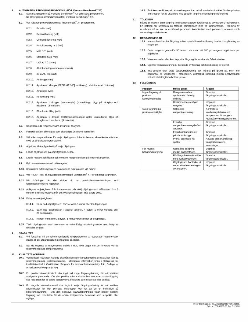
8. AUTOMATISK FÄRGNINGSPROTOKOLL (FÖR Ventana Benchmark® XT) 8.1. Starta färgmodulen på Ventana Benchmark® XT och starta programmet.
Se tillverkarens användarmanual för Ventana Benchmark® XT.
8.2. Välj följande protokollparametrar i Benchmark® XT-programmet.
8.2.1. Paraffin (valt)
8.2.2. Deparaffinering (valt)
8.2.3. Cellkonditionering (valt)
8.2.4. Konditionering nr 1 (valt)
8.2.5. Mild CC1 (valt)
8.2.6. Standard CC1 (valt)
8.2.7. Utökad CC1 (valt)
8.2.8. Ab-inkuberingstemperaturer (valt)
8.2.9. 37 C Ab. Ink. (valt)
8.2.10. Antikropp (valt)
8.2.11. Applicera 1 droppe [PREP KIT 100] (antikropp) och inkubera i (1 timme).
8.2.12. Amplifiera (valt)
8.2.13. Kontrollfärg (valt)
8.2.14. Applicera 1 droppe [hematoxylin] (kontrollfärg), lägg på täckglas och inkubera i (8 minuter).
8.2.15. Efter kontrollfärg (valt)
8.2.16. Applicera 1 droppe [blåfärgningsreagens] (efter kontrollfärg), lägg på täckglas och inkubera i (4 minuter).
8.3. Registrera alla reagenser som används i analysen.
8.4. Fastställ antalet objektglas som ska färgas (inklusive kontroller).
8.5. Välj eller skapa etiketter för varje objektglas och kontrollera att alla etiketter stämmer med ett singelfärgningsprotokoll.
8.6. Applicera tillämplig etikett på varje objektglas.
8.7. Ladda objektglasen på objektglaskarusellen.
8.8. Ladda reagensbehållarna och montera reagensbrickan på reagenskarusellen.
8.9. Fyll damejeannerna med bulkreagens.
8.10. Kontrollera avfallsmodulens damejeanne och töm den vid behov.
8.11. Välj ”RUN” (Kör) på huvuddatorskärmen på Benchmark® XT för att börja färgningen.
8.12. När körningen är klar skriver du ut protokollsammanfattningen och färgningskörningens rapporter.
8.13. Avlägsna objektglasen från instrumentet och skölj objektglasen i tvålvatten i 3 – 5 minuter eller tills resterna från det flytande täckglaset inte längre syns.
8.14. Dehydrera objektglasen.
8.14.1. Sänk ned objektglasen i 95 % etanol, 1 minut eller 25 doppningar.
8.14.2. Sänk ned objektglasen i absolut alkohol, 4 byten, 1 minut vardera eller 25 doppningar.
8.14.3. Klargör med xylen, 3 byten, 1 minut vardera eller 25 doppningar.
8.15. Täck objektglasen med permanent ej vattenlösligt monteringsmedel med hjälp av täckglas av glas.
9. STABILITET 9.1. Vid förvaring vid de rekommenderade temperaturerna är oöppnade reagensvialer
stabila till det utgångsdatum som anges på vialen.
9.2. När de öppnats är reagenserna stabila i nittio (90) dagar när de förvarats vid de rekommenderade temperaturerna.
10. KVALITETSKONTROLL 10.1. Variabilitet i resultaten härleds ofta från skillnader i provhantering som avviker från de
rekommenderade testprocedurerna. Ytterligare information finns i riktlinjerna för kvalitetskontroll i Certification Program for Immunohistochemistry från College of American Pathologists (CAP).
10.2. En positiv vävnadskontroll ska ingå vid varje färgningskörning för att verifiera analysens prestanda. Om den positiva vävnadskontrollen inte visar positiv färgning ska resultaten för de andra testproverna betraktas som suspekta eller ogiltiga.
10.3. En negativ vävnadskontroll ska ingå i varje färgningskörning för att verifiera specificiteten för den primära antikroppen och för att ge en indikation på bakgrundsfärgning. Om den negativa vävnadskontrollen visar positiv specifik färgning ska resultaten för de andra testproverna betraktas som suspekta eller ogiltiga.
10.4. En icke-specifik negativ kontrollreagens kan också användas i stället för den primära antikroppen för att utvärdera icke-specifik färgning eller bakgrundsfärgning.
11. TOLKNING Måttlig till intensiv brun färgning i cellkärnorna anger förekomst av avvikande S-fasinduktion. En patolog bör utvärdera de färgade objektglasen med ett ljusmikroskop. Tolkning av resultaten måste ske av certifierad personal i kombination med patientens anamnes och andra diagnostiska tester.
12. BEGRÄNSNINGAR 12.1. Immunohistokemisk färgning kräver specialiserad utbildning i val och applicering av
reagenser.
12.2. Detta reagens genomför 50 tester och antar att 100 μL reagens appliceras per objektglas.
12.3. Vissa normala celler kan få positiv färgning för avvikande S-fasinduktion.
12.4. Optimal vävnadsfärgning är beroende av fixering och bearbetning av provet.
12.5. Icke-specifik eller ökad bakgrundsfärgning kan inträffa på grund av, men inte begränsat till variationer i proceduren, otillräcklig sköljning mellan analysstegen och/eller felaktigt bearbetade prover.
13. FELSÖKNING
Problem Möjlig orsak Åtgärd Reagenserna har applicerats i felaktig ordning.
Granska färgningsprotokollet.
Ingen färgning på positiva kontrollobjektglas
Utelämnande av något reagens.
Upprepa färgningsprotokollet.
Otillräcklig antigenåtervinning.
Kontrollera inkuberingstiderna och temperturen för antigen-/epitopåtervinningsbufferten.
Felaktig antigenåtervinningsbuffert används.
Granska färgningsprotokollet.
Felaktig inkubation av primär antikropp.
Granska färgningsprotokollet.
Svag färgning på positiva objektglas
Primär antikropp har spätts.
Använd primär antikropp enligt tillverkarens anvisningar.
Otillräcklig sköljning mellan analysstegen.
Upprepa färgningsprotokollet.
För långa inkubationstider med nyckelreagenser.
Granska färgningsprotokollet.
För mycket bakgrundsfärgning
Objektglasen har torkat ut under efterbearbetningen av analysen.
Upprepa färgningsprotokollet.
© TriPath Imaging®, Inc. Alla rättigheter förbehålles. Dok. nr. 779-06555-00 Rev D, 29/30

14. REFERENSER 1. Kastan M and Bartec J. Cell cycle checkpoints and cancer. Nature, 2004. Vol32:316-323.
2. Massague J. G1 cell-cycle control and cancer. Nature, 2004. Vol 432:298-432.
3. Freeman A, Morris LS, Millis AD, Stoeber K, Laskey RA, Williams GH and Coleman N. Minichromosome Maintenance Proteins as Biological Markers of Dysplasia and Malignancy. CI Cancer Research, 1999. Vol 5:2121-2132.
4. Ishimi Y, Okayasu I, Kato C, Kwon H, Kimura H, Yamada K and Song S. Enhanced expression of MCM proteins in cancer cells derived from uterine cervix. Eur. J. Biochem 2003. 270:1089-1101.
5. Lei M and Tye BK. Initiating DNA synthesis: from recruiting to activating the MCM Complex. J Cell Science 2001. Vol. 114 (8): 1447-1454.
15. SYMBOLFÖRTECKNING
Katalognummer
För in vitro-diagnostik
Se bruksanvisningen
Innehåller 7 mL
Försiktigt! Se medföljande dokumentation
Begränsningar för förvaringstemperaturen
Batchkod
Använd senast ÅÅÅÅ-MM-DD eller ÅÅÅÅ-MM
Tillverkare
IVD
TEKNISK INFORMATION I USA: Ring TriPath Technical Services, avgiftsfritt 1 866 874 7284
TriPath Imaging, Inc. 780 Plantation Drive Burlington, NC 27215, USA (800) 426-2176
Utvecklat med teknik från Millennium Pharmaceuticals, Inc.
och är varumärken som tillhör Millennium Pharmaceuticals, Inc. Millennium Pharmaceuticals, Inc. 40 Landsdowne Street Cambridge, MA 02139 USA www.millennium.com
TriPath Imaging® är ett registrerat varumärke som tillhör TriPath Imaging, Inc. ProEx är en produkt och ett varumärke som tillhör TriPath Imaging, Inc.
© TriPath Imaging®, Inc. Alla rättigheter förbehålles. Dok. nr. 779-06555-00 Rev D, 30/30