, €*7 . Q Risk Assessment of a Genetically-Engineered Microorganism: Erwinia carotovora bv David...
136
Environmental Risk Assessment of a Genetically-Engineered Microorganism: Erwinia carotovora bv David Robert Orvos . Dissertation submitted to the Graduate Faculty of the Virginia Polytechnic Institute and State University in partial fulfillment of the requirements for the degree of Doctor of Philosophy in Biology APPROVED: gf" E (V 2£” John Cairns, Jr., Ch an Georg/e H. Lacy , €*7 . Q A Ü/évttg Bruce Wallace Robert A. Paterson ~ „ 'YF ~ „ * { Eric P. Smith William Yongu «¤< 6%o;„.„ J6., fw? Vw — April, 1989 Blacksburg, Virginia
Transcript of , €*7 . Q Risk Assessment of a Genetically-Engineered Microorganism: Erwinia carotovora bv David...

Environmental Risk Assessment of a Genetically-EngineeredMicroorganism:
Erwinia carotovorabv
David Robert Orvos .
Dissertation submitted to the Graduate Faculty of theVirginia Polytechnic Institute and State University
in partial fulfillment of the requirements for the degree of
Doctor of Philosophyin
Biology
APPROVED:
gf" E (V 2£”
John Cairns, Jr., Ch an Georg/e H. Lacy
, €*7 . QA Ü/évttgBruce Wallace Robert A. Paterson
~ „ 'YF ~ „ *{
Eric P. Smith William Yongu
«¤< 6%o;„.„ J6., fw? Vw —
April, 1989
Blacksburg, Virginia

ENVIRONMENTAL RISK ASSESSMENT OF A GENETICALLY-ENGINEERED
MICROORGANISM:
ERWINIA CAROTOVORA
bv
David Robert Orvos
John Cairns, Jr., Chairman
Biology
(ABSTRACT)
Environmental use of genetically-engineered microorganisms (GEMs) has raised
concerns over potential ecological impact. Development of microcosm systems
useful in preliminary testing for risk assessment will provide useful information for
predicting potential structural, functional, and genetic effects of GEM release. This
study was executed to develop techniques that may be useful in risk assessment
and microbial ecology, to ascertain which parameters are useful in determining risk
and to predict risk from releasing an engineered strain of Erwinia carotovora.
A terrestrial microcosm system for use in GEM risk assessment studies was de-
veloped for use in assessing alterations of microbial structure and function that may
be caused by introducing the engineered strain of E. carotovora. This strain is being
developed for use as a biological control agent for plant soft rot. Parameters that
were monitored included survival and intraspecific competition of E. carotovora,
structural effects upon both total bacterial populations and numbers of selected
bacterial genera, effects upon activities of dehydrogenase and alkaline
phosphatase, effects upon soil nutrients, and potential for gene transfer into or out
. of the engineered strain.
No significant difference was found in survival of the engineered strain as com-
pared to its wildtype parent. Both strains survived for over two months in
microcosms. The effects of both strains upon populations of total bacteria and se-
lected bacterial genera were determined; while some effects upon community
structure were observed, they were not significant.
The engineered strain was not found to be a superior competltor compared to its
parent; three different doses of engineered and wildtype strains were used. ln ad-
dition, neither strain affected activities of dehydrogenase or alkaline phosphatase in
soil. Likewise, no effects were observed upon the nutrients monitored.
However, transfer of the kanamycin resistance gene that had been inserted into
the engineered E. carotovora strain may have occurred. Five species of indigenous
bacterla displayed kanamycin resistance 15 days after being exposed to the engi-
neered Erwinia. DNA from these strains was isolated, purified, and hybridization
experiments executed to determine if any homology existed between these DNAs
and the kanamycin resistance gene that had been inserted into E. carotovora. Using
biotin-Iabeled probes and Iow—stringency washing conditions, homology was ob-
served. However, before gene transfer can be proven, additional studies, including
amplification and sequencing, may be required.
Although a simple microcosm design was employed, it yielded sufficient infor-
mation to conclude that release of the engineered Erwinia carotovora will not affect
any of the microbial measures of integrity that were studied in a manner different
from that of the wildtype. Effects upon plant material and other higher taxa will be
the focus of future studies.

ACKNOWLEDGEMENTS
iv

TABLE OF CONTENTSPage
PROLOGUE 1
CHAPTER ONE: MICROCOSMS IN BIOTECHNOLOGY RISK ASSESSMENT 27
Introduction 27
Materials and Methods 30
Results and Discussion 33
References 37
CHAPTER TWO: ACQUISITION OF ANTIBIOTIC RESISTANCE BYINDIGENOUS MICROBES 48
Introduction 48
Materials and Methods 51
Results and Discussion 57
Conclusions 60
References 61
CHAPTER THREE: SURVIVAL OF GENETICALLY-ENGINEEREDERW/N/A CAROTOVORA AND EFFECTSUPON SELECTED BACTERIAL GENERA 74
Introduction 74
Materials and Methods 75
Results 78
Discussion 80
References 85
CHAPTER FOUR: INTRASPECIFIC COMPETITION 94
Introduction 94
Materials and Methods 95
Results and Discussion 98
References 102
CHAPTER FIVE: GEM EFFECTS UPON SOIL CHEMICAL PARAMETERSAND ENZYME ACTIVITIES 108
Introduction ' 108
Materials and Methods 109
Results and Discussion 113
References 117
EPILOGUE 122
VITA n 129
vi

PROLOGUE
During the 1970’s, several techniques were developed that allowed the in vitro
fragmentation and rejolning of molecules of deoxyribonucleic acid (DNA) and the
placement of these molecules into living cells. Use of these recombinant DNA
techniques led to the development of biotechnology, broadly defined as the appli-
cation of biological organisms and their products to commercial processes (Office
of Technology Assessment, 1988). Among types of organisms now being used for
blotechnological applications, bacteria have been used more than others because
of their short generation times, lack of a defined nucleus, and relatively ’simple’
genetics as compared to eukaryotes. Genetic engineering of microorganisms is
used to develop organisms capable of performing specific commercially-desired
tasks. These applications include producing pesticides and pharmaceuticals, fixing
nitrogen, Ieaching ores, recovering oil, degradlng hazardous wastes, and con-
structing biological control agents for pests or diseases. Biotechnology products
will, one hopes, benefit human society.
However, environmental applications of genetically engineered microorganisms
(GEMs) has been limited to small-scale trials because of (1) a lack of knowledge
about their impact on ecosystems and (2) an increased public awareness of concern
over the release of novel organisms. Some ecologlsts, clting releases of starlings,
gypsy moths, the Chestnut blight pathogen, and the Dutch elm disease pathogen,
predict an "inevitabIe environmental disaster" while other scientists, especially
molecular biologists, believe the chance of such an event is remote (Brill, 1988).
Comparison of exotic species release to GEM release may not be valid since exotic
species represent an exotic genome while GEMs may represent lndigenous
genomes altered only to a small extent (Office of Technology Assessment, 1988).
To date, no adverse consequences have resulted from the small-scale release of1

GEMs (Office of Technology Assessment, 1988); however, all of the releases at this
time have entailed the use of microbes that have had parts of their genome re-
moved or have had innocuous insertions that pose no obvious environmental
threats. At some point, however, GEMs with capabilities of altering basic ecosystem
processes, such as nitrogen fixation and nutrient cycling, may be released. The
present research addresses the environmental release of GEMs and the possible
environmental impact that they may have. lt also examines attempts to construct
a risk assessment scheme to predict the fate and effects of a GEM, produced from
the bacterium Erwinia carotovora, released into terrestrial microcosms. The meth-
odologles developed for this organism may be useful in risk prediction with other
GEMs.
Several factors must be considered before GEMs are released in large quantities
into the environment. Survival of an organism under Iaboratory conditions does not
ensure survival in natural ecosystems. Conversely, ability to contain and control
species in the Iaboratory does not preclude their establishment, survival, and ad-
verse effects outside (Cairns and Pratt, 1986). Blotechnological products, especially
the altered or engineered organisms themselves, present unique regulatory prob-
lems. Unlike toxic chemicals, organisms have the potential to increase in quantity
in the environment at the site of release or following transport to some unlntended
and unexpected location.
GEMs may potentially affect the environment in through any one of three ways:
(1) by transferring DNA from or into the GEM, thus altering the genetic composition
of the GEM or indigenous bacterial populations, (2) by affecting the structural integ-
rity of communities, both mlcrobial and macrobial, and (3) by affecting functional
processes, including geochemical cycling of nutrients.
Bacterla possess genetic mechanisms for the dispersal and alteration of their
DNA; such mechanisms enhance bacterial survival in the environment (Grant and2

Long, 1981). Four basic mechanisms for exchanging genetic material are known
(Freifelder, 1987): (1) transformation, the uptake of naked DNA as plasmids in the
case of Gram-negative bacteria or as linear DNA fragments by Gram-positive bac-
teria; (2) transduction, the incorporation of bacterial DNA in bacteriophage particles
and their transfer to recipient bacteria; (3) conjugation, in which the DNA is trans-
ferred by direct cell-to·cell contact; and (4) fusion and recombination, a mechanism
that is limited to actinomycetes.
Most, if not all, bacteria in the environment may exchange genetic information
with nearby microbes (Slater, 1985; Wellington et al., 1988). ln consldering the risk
from potential transfer of the genome from a GEM to indigenous bacteria, several
factors must be considered. First, molecular techniques confirm that gene transfer
has indeed occurred. Such techniques may include DNA: DNA or DNA: RNA
hybridization and sequencing ofthe DNA region in question. Second, if a transfer
has indeed occurred, one must ascertain if the newly created genetic information
will be sustained and expressed in the bacterium which acquired it. Third, if genetic
information is maintained and expressed, the effects that such expression may have
on other biota must be evaluated (Office of Technology Assessment, 1988). Several
criteria, including gene transfer frequency and genetic distance among species, can
be used in executing an assessment along with consideration of the construction
of the gene itself and the vector on which the gene resides (Office of Technology
Assessment, 1988).
While studies on potential genetic exchange mechanisms have been undertaken,
few have examined these mechanisms outside the laboratory. Transfer frequencies
on membranes are useful, but fail to provide information concerning selective
pressures. ln addition, DNA is rarely found in the naked, available state in envi-
ronmental samples; rather, it is either degraded by exogenous nucleases or sorbed
to soil or sediment particles and thus unavailable for transfer (Ogram et al., 1988).3

Such sorption is regulated by soil type, clay and organic material content, ionic
strength, pH, and length of DNA (Ogram et a/., 1988).
Plasmid DNA transfer, via conjugation, in the environment has been studied in
both terrestrial and aquatic systems. Plasmids (autonomously replicating,
extrachromosomal, circular pieces of DNA) are ubiquitous in bacteria. Many envi-
ronmental isolates have been shown to be recipients of plasmids. Genthner et al.
(1988) tested 68 bacteria! isolates from aquatic sources and found 26 of them able
to receive plasmids from Pseudomonas aeruginosa donors. Schilf and Klingmuller
(1983), using pRD1 carried by E. co/i, discovered that 1.3% of soil bacteria and
17.3% of aquatic bacteria could receive the plasmid from a donor. Evidence for
plasmid selection or exchange in the environment is also based upon high incl-
dence of plasmids in polluted areas (Hada and Sizemore, 1981), coexistence of
identical plasmids in different strains, and data from in situ transfer experiments
(Grimes et al., 1988). While plasmids are known to affect the growth of
microorganisms, Cruz-Cruz et al. (1988) found that bacteria containing large (100
megadalton) plasmids may survive in situ equally well as those with smaller
plasmids. They also suggest that genetic alteration of indigenous microorganisms
is made possible by either conjugation or transformation in aquatic habitats. Bac-
terial conjugation has been observed in both non-sterile and sterile soils (Weinberg
and Stotzky, 1972).
Transfer of plasmids also appears to be influenced by environmental influences.
Stotzky and Krasovsky (1981) found that recombination was affected markedly by
. pH with the survival of both donors and recipients increasing up to a pH of 6.9.
Moisture content of soil appears not to be influential ifthis is between 20-100% of
the solls total holding capacity (Trevors and Oddie, 1986). ln addition to transfer in
soil and aquatic systems, plasmids may be transferred in plants (Lacy et al., 1984)Ä

The vast majority of DNA transfers in regard to GEMS appear to be innocuous;
this is expected since antibiotic resistance from the marker plasmid is often the only
parameter monitored. Nevertheless, it is conceivable that transfer of plasmids may
affect important fu nctional characteristics ofthe organism. Rafii and Crawford (1988)
found that several conjugative plasmids were transferred in sterile soll from the
donor, Streptomyces llvidans, to several Streptomyces strains isolated from envi-
ronmental samples. In a concurrent study, Wang and Crawford (1988) found that a
similar Streptomyces strain caused significantly higher soll carbon mineralizatlon
rates in nonsterile soil amended with Iignocellulose. They believe the effect is
caused by a foreign DNA insert. The transfer of such an insert could alter the
function of other organisms. ln addition, Glick et al. (1986) found that introduction
of plasmid DNA into Azotobacter vine/andii, via transformation, reduced its capacity
for nitrogen fixation, mean cell size, and siderophore production.
Because plasmids are frequently passed from one organism to another (Slater,
1985), they may not be suitable for environmental applications. However, because
of ease of manlpulation and potential usefulness, they cannot simply be neglected;
any risk attributed to them, however, must be predicted. Walter et al. (1987b) found
that no single mating technique was superior in predicting the frequency of plasmid
transfer; they developed a new technique, a combination of several older ones, that
predicts the frequency with which plasmids move from one cell to another. Another
group at the EPA’s Corvallis laboratory (Knudsen et al., 1988) developed a predictive
computer simulation model that accurately forecasts transconjugant populations in
both the rhizosphere and on leaf surfaces. This and other models not yet developed
. may eventually lead to accurate assessments of plasmid transfer prior to environ-
mental release.
Transfer of DNA via transduction has also been documented in environmental
samples from both soll and water. Zeph et a/. (1988) found that transduction of E.5

co/i by bacterlophage P1 occurred in soll, both sterile and nonsterlle; however, they
did not observe transduction of indlgenous organisms. lt appears that a minimum
number of bacteria are needed to ensure replication of the phage (Wlggins and
Alexander, 1985). This limit minimlzes viral damage in the environment because
ofthe relatively low populations of individual bacteria (Wlggins and Alexander,
1985). Transduction in aquatic samples has also been demonstrated by Saye et al.
(1987), using P. aeruginosa containing plasmid Rms149, although the presence of
natural microbial communities significantly decreased transduction frequency.
Transformation, especially in Bacl//us subti/is, has been shown in nonsterlle soll
(Graham and lstock, 1978, 1979). However, Greaves and Wilson (1970) found that
DNA or RNA added to nonsterlle soll was rapidly degraded, thus causlng an in-
crease in indlgenous non-transformed populations.
Transfer of genetic information ls a viable concern when deslgnlng a risk as-
sessment scheme to predlct potential environmental consequences. Recent tech-
nologies, such as suiclde vectors (Bej et al., 1988) may eventually prevent
movement of recomblnant genes. Even more important than the actual transfer of
genetic information, however, is the expression ofthat genetic information. lf such
expression ls to be manlfested, the bacterlum must survlve after lt is released.
Alexander (1985) surmises that ifa GEM is to have an effect, good or bad, it must
survlve. The question of survival ls central to both the pro and con arguments re-
garding the efficacy of GEMs.
A great deal of information has been acquired regarding survival of single spe-
cies of bacteria, especially Rhizobium (Stacey, 1985). However, scant data have
been obtalned concerning the release of non-root colonizlng bacteria into an envi-
ronment that may or may not have originally contained the bacterlum being re-
leased. The data collected from introductions of Rhizobium (and other bacteria) ari

useful in comprehending biotic and abiotic factors that affect microbial survival.
These data will be reviewed here.
Ablotlc factors that affect bacterial growth in the environment include substrate
type, nutrient availability, temperature, desiccation, salinlty, and pH (Grant and
Long, 1981; Stacey, 1985). Among the most important for a newly released GEM are
nutrient availability, temperature, and pH. Severe fluctuations in any of these may
severely compromise GEM integrity. Even lf the GEM encounters conditions favor-
able for its establishment, it must contend with predation by phages or protozoans.
Predation has been shown to adversely affect Rhizobium populations (Stacey, 1985).
In addition, deleterious enzymes or antibiotics produced by indigenous microbes
may cause elimination ofthe introduced species. However, effects of many ofthese
factors can be minimized by increasing concentrations of bacteria being applied to
the area of interest. With this rationale, though, comes an increased chance of
producing runoff that may carry the GEM to an area not originally intended.
lt has been demonstrated that bacteria, engineered or not, do not survive for
extended periods of time because of environmental pressures exerted on them.
Indeed, ifthe bacteria being introduced have been derived from indigenous species,
they will have to outcompete these species which are already firmly established in
their niche. To make these introduced microbes predomlnate, strong selective
pressure will have to be applied (Stacey, 1985). Alexander (1985) summarizes the
situation bacteria encounter by saying that bacteria die when they fall to overcome
any of the biotic or abiotic factors that they encounter.
Predlcting survival prior to release is an important parameter in risk assessment.
Walter et a/. (1987a) developed a technique that allows determinatlon of survival of
bacteria in soll extracts. This technique appears to be useful for an industrial assay
ln initial data generation. The group found that engineered strains survive longer
in sterile soll than in nonsterile soll. This ls not surprising since a lack of competi}

tion would encourage survival. Using antibiotic-resistant strains produced by
. mutagenesis, Van Elsas et a/. (1986) released P. f/uorescens and Baci//us subti/is into
field plots. They observed that B. subti/is declined rapidly while P. f/uorescens de-
clined at a slow, steady rate and survived for up to 120 days at detectable levels.
Using rifampin—resistant spontaneous mutants of P. fluorescens and P. putida,
Compeau et al. (1988) found that limited sites exist in soil for colonization by either
bacterial strain. ln addition, they determined that rifampin resistance is not always
harmless and may, in fact, confer a competitive disadvantage to the bacterium.
They suggest that all strains being used in a study be compared for growth rate in
media to ensure that no differences are present. ln all of the GEM soil survival
studies to date, bacteria have always declined in concentration over time; this is not
unexpected because of the enormous competition they encounter.
Little data exists for GEM release into aquatic systems. Like their terrestrial
counterparts, bacteria decline when released into aquatic microcosms (Burton et
al., 1987). Sostarec et al. (1988) found that engineered E. carotovora, the same strain
as used in this research, declined at the same rate as its wildtype parent.
While recent documentation exists for survival of GEMs in soil, few studies have
been executed to measure aerial dispersion of microorganisms from plants and soil;
little or no research has been undertaken to elucidate the mechanisms, such as
wind speed, plant leaf condition and age, inoculum age, and moisture, that may in-
fluence dispersal of GEMs into unanticipated areas. Because of their small mass,
bacteria and viral particles take extensive periods of time to settle out of the at-
mosphere; therefore, they may be transported over long distances (Barnthouse and
Palumbo, 1986). Bacteria have been shown to travel for hundreds of kilometers on
air currents, however, bacteria carried by plant tissue or airborne soil particles
settle far more rapidly (Barnthouse and Palumbo, 1986).8

° Bacterial plant pathogens have been found to disseminate in aerosols produced
by raindrop impacts, irrigation systems, and mechanical manipulation (Harrison,
1980) and are capable of inducing pathogenesis after movement. Lindemann &
Upper (1985) observed that bacteria reentrain on plant surfaces and soil after being
disseminated in aerosols; in addition, they discovered that plant canopies are ex-
cellent sources of these aerosols when leaves are dry on sunny days. More re-
cently, two studies have documented that bacteria altered by natural processes do
indeed aerially disperse from a site of introduction. Marshall et al. (1988) found that
E. co/i, containing a Tn5 derivative of the plasmid CoIE1, dispersed from the sites
of introduction, a laboratory office and a barn, with maximal dispersion being ob-
served in the first 30 minutes after release. While rapid aerosol die-off was ob-
served, it is conceivable that many of the bacteria attained a viable, but
non-culturable status (Colwell et al., 1985) that was not detected. Such bacteria
could increase in concentration if favorable conditions are encountered. Marshall
et al. also learned that, in several instances, E. co/i containing ColE1 and Tn5 dem-
. onstrated enhanced survival, apparently related to a growth advantage from the
transposon. In a more environmentally realistic study, Lindow et al. (1988) deter-
mined that viable Pseudomonas syringae cells experienced an exponential de-
crease in concentration as distance from aerosol application site increased. While
survival rate of bacteria was 10 times higher on the intended plants than on adjacent
plants, such dispersion was demonstrated even with relatively calm winds (0.73 to
1.5 m/s) that were not favorable to dissemination. Organisms on the soil surface
appeared to die rapidly as expected since P. syringae is an epiphyte.
In a study of the first releases of a recombinant bacterium, Seidler and Hern
(1988) reported that low numbers of P. syringae and P. f/uorescens left test plots
even in the presence of minimal wind conditions; they also found that strong vertical
mixing occurred at three spray releases. Even though 99.99% of the introduced9

bacteria remalned on the soll and plants for which they were intended, some 1.2 x
10° colony forming units (CFU) of bacteria migrated onto a 15 meter buffer zone
during the course of one experiment; bacteria could be found around the site two
days after the test. ln addition, a greater drift of bioaerosol particles occurred in an
experiment conducted at Tulelake, CA as compared to experiments carried out at
Brentwood, CA. Seidler and Hern believe that a larger sprayed area and a greater
source strength of bacterial inoculum may have significantly affected aerosol drift
during the one release. Clearly, additional investigation is warranted to determine
which factors contribute to the aerial dispersion of GEMs during and after applica-
tion.
A major problem in ascertaining GEM survival are techniques available to the
investigator. Currently, indirect methods such as the plate count are limited in that
no one medium will support the growth of all bacterial species present in a partic-
ular sample (Mills and Bell, 1986). However, in the studies described later in this
work, the plate count has proven to be an inexpensive, yet reliable, method of de-
termining concentrations of GEMs and indigenous bacteria despite the views of
many microbial ecologists (Mills and Bell, 1986). This conclusion is also supported
by Olsen and Bakken (1987). ln addition, use of selective media allows enumeration
of communities of interest. Direct counting of bacteria using simple or fluorescent
(Daley, 1979) stains contributes little information since it is impossible to detect the
organism of interest from the remainder of the sample. Use of fluorescent anti-
bodies, specific for the organism in question, have not been as useful as initially
hoped because of low sensitivity and presence of contaminating substances in en-
vironmental samples (Levin et al., 1987).
Molecular techniques for detection of GEMs show promise but are more compli-
cated and expensive than conventional technologies. Methods have been devel-
oped to ascertain the amount of dissolved environmental DNA (DeFlaun et al., 198%

‘as well as DNA from bacterial cells (Holben et a/., 1988; Steffan et a/., 1988). Use
of this DNA in hybridization studies allows detection of GEMs by genotype rather
than phenotype as well as allowing detection of nonculturable microbes (Barkay and
Sayler, 1988). Application of these techniques have allowed detection of as few as
10 cells/g soll (Fredrickson et a/., 1988). However, such use of probes in the past
usually resulted in low sensitivity (Barkay and Sayler, 1988; Levin et a/., 1987). The
application of the polymerase chain reaction (PCR) (Saiki et a/., 1988) for use in en-
vironmental samples by Steffan and Atlas (1988) has shown promise for GEM de-
tection. Steffan and Atlas (1988) reported the detection of 1 cell/gram sediment
using P. cepacia containing 15 to 20 copies of a 1.3 kilobase (kb) gene. However,
use of the PCR technique with GEMs containing only one gene per organism has
not been especially productlve because of environmental contamination and low
initial DNA amounts (James Tiedje, personal communication). Hence, further
methods development is lndicated before the technique can be exploited to its
maximum efficiency.
The problem in using GEM survival in developing a risk assessment protocol is
that the GEM may indeed be present but undetectable. Colwell et al. (1985) coined
the term ’viable, but non—culturable’ for such a situation. In their study, Colwell’s
group found that, in both microcosms and field tests, Vibrio cho/erae were present
in higher numbers when determined by immunofluorescent microscopy than by
plate counts. Thus, even when plate counts yielded negative information concern-
ing survival, bacteria were indeed present.
Two other parameters exist where GEMs may cause an unexpected effect. One
is that GEMs may cause alterations in community structure. Ecosystems have two
fundamental properties: 1) they are composed of structures, and 2) these structures
facilitate specific functions (Cairns & Pratt, 1986). Since structure and function are
closely related, an alteration in one may soon affect the other. A variety of studieisl

will be required to estimate probable effects resulting from the controlled or un-
controlled release of GEMs (Chakrabarty et al., 1984; Rissler, 1983; Cairns & Pratt,
1986). Complex ecological effects resulting from displacement of native bacterial
populations may occur, especially when GEMs are derived from common free-living
taxa (Liang et al., 1982). Displacement of bacterial groups could produce an alter-
ation of detrital processing and nutrient cycling that could have broad conse-
quences, especially if introduced organisms have high fitness and are transferred
among ecosystems (Cairns & Pratt, 1986). Many ecologists believe that important
processing bacteria are the same for virtually all comparable ecosystems. The
probability of such adverse effects may be small, but failure to evaluate the potential
risks could be disastrous.
Functional effects would have severe consequences ifa crucial parameter was
affected or if the assimilative capacity of the ecosystem (Cairns, 1977) was ex-
ceeded. Bacteria decomposing detritus produce the majority of energy in most
aquatic ecosystems (Wetzel, 1983); the minerallzation of this material is the most
important role of bacteria (Fenchel & Blackburn, 1979). Most processing occurs in
sediments and is linked tightly to the processing of other vital nutrients such as ni-
trogen, sulfur, and phosphorus (Fenchel & Blackburn, 1979). These processes,
mediated mostly by bacteria, are vital to continued ecosystem function. Surface-
dwelling communities of microorganisms are exceedingly complex (Shapiro, 1985)
and demonstrate the rapid exchange of materials being recycled. ln addition to
having similar functional attributes, most ecosystems boundaries are imprecisely
defined. The exports of energy and nutrient bearing materials from one ecosystem
become the imports of another. At the microbial level, most imported and exported
materials carry a portion of the resident biota (Pimentel, 1985) thus providing the
opportunity for introduction of GEMs into new habitats. However, no consensus
exists on what parameters in the environment should be monitored even when 12

dealing with chemical impacts. The number of parameters that may be monitored
are numerous and include nutrlent and enzyme concentrations,
production/respiration ratlos, oxidation-reductionl potentlals, and pH shifts. How-
ever, no agreement exists on what is a "normaI" value for a particular parameter
under a particular set of circumstances (Cairns and Pratt, 1986). lndeed, far more
research ls indicated in this area for both chemicals and GEMs.
It has been shown that GEMs are able to produce functional effects that are not
observed in the parent organism. Glick et al. (1986) found that the introduction of4
plasmid DNA into Azotobacter vine/andii caused a decrease in nitrogen fixation,
mean cell size, and siderophore synthesis. Glick et al. hypothesized that the
plasmid introduction imparted a metabolic load on the bacterium that impaired its
ability to function. Wang and Crawford (1988) found that a recombinant strain of
Streptomyces lividans significantly affected soil organic carbon turnover rates. They
noted that the organism displayed an increased mineralization rate, particularly in
solls amended with Iignocellulose; these rates remained significantly higher over
the 30 day duration of the test.
Because of the uncertainity of GEM releases, it ls likely that small, easily con-
tained microcosms will form the basis for preliminary assessments of ecologlcalF
effects (A.D. Little, Inc., 1984). Such studies have proved useful for studylng a
number of ecologlcal problems, although results must be interpreted with cautlon
(King, 1980). The small size and contained nature ofthese microcosms make pos-
sible effective dose-response studies for possible dissemination of recombinant
DNA. Small or large innoculum levels of donor and recipient bacteria may be used
with safety since the Iaboratory environments may be autoclaved. Further, the
small size of the chambers makes lntroducing various environmental stresses pos-
sible; such stresses may effect the frequency of DNA exchange. Microcosm con-
struction, optlmization, and limitations will be discussed further in the next chaptelig

Before GEMs can be released into the environment, manufacturers must comply
with existing Federal and local regulations. Most commercial reIeases_ are con-
trolled by the Environmental Protection Agency whose regulation is mandated by
the Toxic Substances Control Act (TSCA) (Cohrssen, 1988). At the present time,
those desiring to release a GEM must first issue a premanufacturing notice (PMN)
describing the organism and its characteristics such as survival and potential ef-
fects. These PMNs must comply with the Coordinated Framework for Regulation
of Blotechnology (Federal Register, 1986). PMNs have been used by the chemical
industry for well over a decade. However, it is important to note that risk assess-
ment protocols have been developed and proven for chemical use whereas they
have not been validated for use with self-multlplying GEMs,
Suitable risk assessment protocols have not yet been developed, in part, be-
cause an agreement of what should be monitored and when has not been reached.
Prior to a release of a chemical or GEM into the environment, data must be collected
so as to provide a baseline for data that is collected after the release (Cairns and
Pratt, 1986). Many suitable risk assessment protocols have been developed for
chemlcals since these have been proven and used by industry for some time; with
modifications to accommodate the unique characteristics of GEMs, these may be
useful (Dean-Ross, 1986). However, molecular biologists and microbial ecologists
charged with assessing the risk from GEM release fall to understand the process
of risk assessment. Many molecular biologists, especially those involved with
commercial applications, feel that the only risk associated with GEM release is that
of gene transfer and they believe that even that risk is remote (Brill, 1988).
Chemical hazard assessment is five-fold: risk identification, risk characterization,
risk source exposure assessment, hazard assessment, and estimation of risk
(Cairns, 1982; Orvos and Cairns, 1987). Risk identification for GEMs may be broadly
thought of in terms of gene transfer, structural effects, and functional effects. Abilitä

to characterize these risks is dependent upon the site under consideration, the GEM
itself, and the parameters being monitored (Molak and Stara, 1988). Assessing ex-
posure will include studies of survival, displacement, and pathogenicity. Hazard
assessment, the subject ofthe majority ofthls dissertation, involves a tier approach
to determine the acute or chronic effects of a particular GEM. With increasing
complexity of the test system comes increased costs, decreased replicability, and
more environmentally realistic data. Estimation of risk is based upon all of the data
collected as well as model development and predictive value.
GEMs, especially non·pathogenic ones, may not have clear-cut effects and the
traditional toxicological dose—response paradigm may not be applicable to evaluat-
ing the ecological effects of introduced organisms. This does not mean that such
a paradigm may not be present nor does it imply that protocols developed for
chemicals are not useful for GEMs. Careful modification of such protocols will make
them useful for GEMs. However, additional technologies are needed to enumerate
GEMs at low numbers and to measure any functional alterations produced by them.
While progress has recently been made on enumeration, such as the polymerase
chain reaction (Steffan and Atlas, 1988; Jain et al., 1988) and improved DNA isolation
procedures (Steffan et al., 1988), a great deal of refinement will be necessary before
they are fully used (J. Tiedje, personal communication).
Efforts by the EPA have centered on methods development since they must
underlie any accepted protocol (Levin et al., 1987). While some of the factors for
establishment of microorganisms are known, far more research is indicated to as-
certain the effects of environmental factors on GEMs and their DNA.
For the research described in subsequent chapters, Erwinia carotovora subsp. U
carotovora has been chosen as a model since this strain (L—833) (Allen et al., 1988)
has been engineered specifically to include two types of genetic alterations in the
bacterial chromosome: deletion of a gene fundamental to plant pathogenesis andls

inclusion of an antibiotic resistance gene. With this model, the possibility that the
antiblotic resistance marker may become integrated into the genomes of other
microbes that come into contact with it will be examined. E. carotovora is a
pathogenic member ofthe Enterobacteriaceae that produces soft rot in plant tissues
and causes more than $100,000,000 in losses of stored food annually despite control
measures that are primarily environmental in nature. Soft-rotting bacteria are
ubiquitous and exist in low populations as epiphytes on underground surfaces of
healthy plants (Stanghellini, 1982). lfthe host is physiologically compromised and
environmental conditions are favorable, these resident bacteria multiply rapidly and
initiate pathogenesis. Therefore, disarmed bacteria applied to seed at planting will
probably protect plants from soft rot since similar studies using fluorescent
pseudomonads and non-pathogenic agrobacteria as biological control agents have
been successful (Suslow, 1982; Lindow, 1983).
Soft rot is a result of a battery of enzymes produced by the pathogen that break
down components of plant cells and release substrates for bacterial growth. These
include pectic enzymes, cellulases, and proteases (Chatterjee & Starr, 1980). Pectic
enzymes are responsible for maceration of plant tissues; other enzymes are inef-
fective without pectic enzymes. Among pectic enzymes, extracellular endo-pectate
Iyases play major roles in plant cell death and tissue maceration (Collmer et al.,
1982; Stack et al., 1980). Endo-pectate Iyases cleave pectic acid which is a compo-
nent of all plant cell walls involved in the structural integrity of plant tissues
(Bateman & Basham, 1976). Cleavage of pectic materials by endo-pectate lyase
causes cell separation or tissue maceration and tissues so affected lose turgidity
and become soft and slimy to the touch. Therefore, the GEM used in these studies
is the precursor to an engineered biological control agent designed for environ-
mental release.16

Routine ecological assessment methods must need to be developed for use by
biotechnology Industries to ensure the soundness of future economic development
of biotechnology products and to provide necessary information to regulatory
agencies. Current case·by—case criteria will soon become useless If the expansion
of biotechnology Industries continues. Site—specific testing will be required and
such testing must be scientifically justifiable and have general transferability to se-
veral potential receiving ecosystems. Testing schemes will need to be easily in·
terpretable by industry and regulatory analysts. lf suitable protocols are not
developed, the environment will undoubtfully be compromised at some point in the
future.
17

LITERATURE CITED
A.D. Little, lnc. 1984. Risk Analysis Methods for Environmental Applications of
Biotechnology. Cambridge, MA. 35 pp.
Alexander, M. 1985. Ecological consequences: reducing the uncertainties. Issues in
Science and Technology. Spring: 57-68
Allen, C., V.K. Stromberg, F.D. Smith, G.H. Lacy, and M.S. Mount. 1986.
Complementation of an Erwinia carotovora subsp. carotovora protease mutant
with a protease-encoding cosmid. Mol. Gen. Genet. 202:276-279.
Barkay, T. and G.S. Sayler. 1988. Gene probes as a tool for the detection of specific
genomes in the environment, In W.J. Adams, G.A. Chapman, and W.G. Landis
(eds.), Aquatic Toxicology and Hazard Assessment: 10th volume, ASTM STP 971.
American Society for Testing and Materials, Philadelphia, pp. 29-36.
Barnthouse, L.W. & A.V. Palumbo. 1986. Assessing the transport and fate of
bioengineered microorganisms in the environment. [ln] J. Fiksel & V.T. Covello
(eds.), Biotechnology Risk Assessment, Pergamon Press, pp. 109-128.
Bateman, D.F., and H.G. Basham. 1976. Degradation of plant cell walls and mem-
branes by microbial enzymes. p.316-355. [In] R. Heitefuss and P.H. Williams,
(eds.), Physiological Plant Pathology, Vol. 4. Encyclopedia of Plant Physiology,
Springer-Verlag, N.Y., 880pp.
Be], A.K., M.H. Perlin, and R.M. Atlas. 1988. Model suicide vector for containment of
genetically engineered microorganisms. Appl. Environ. Micro. 54:2472—2477.
Brill, W.J. 1988. Why engineered organisms are safe. Issues in Science and Technol-
ogy, Spring, 1988.
Burton, G.A., Jr., D. Gunnison, and G.R. Lanza. 1987. Survival of pathogenic bacteria
in various freshwater sediments. Appl. Environ. Micro. 53:633-638.
Cairns, J., Jr. 1977. Aquatic ecosystem assimilative capacity. Fisheries 2(2):5-7,

Cairns, J., Jr. 1982. Environmental Effects. [ln] G.F. Bennett, F.S. Feates, and I. Wilder
(eds.), Hazardous Materials Spills Handbook, McGraw·HilI, New York, pp.
415-421.
Cairns, J., Jr. and J.R. Pratt. 1986. Ecological consequence assessment: effects of
bioenglneered organisms. [ln] J. Flksel & V.T. Covello (eds.), Biotechnology
Risk Assessment, Pergamon Press, pp. 88-108.
Chakrabarty, A.M., J.S. Karns, J.J. Kilbane, and D.K. Chatterjee. 1984. Selective ev-
olution of genes for enhanced degradation of persistent, toxic chemicals. [ln]
W. Arber, K. Illmensee, W.J. Peacock, and P. Starlinger (eds.), Genetic Manipu-
Iation: Impact on Man and Society, Cambridge University Press, NY. pp. 43-54.
Chatterjee, A.K., and M.P. Starr. 1980. Genetics of Erwlnia species. Annu. Rev.
Microbiol. 34:645-676.
Cohrssen, J.J. 1988. United States biotechnology policy. Amer. Biotech. Lab. 1:22-27.
Collmer, A., P. Berman, and M. Mount. 1982. Pectate Iyase regulation and bacterial
soft rot pathogenesis. Pp. 395-422 [In] M.S. Mount and G. H. Lacy (eds.),
Phytopathogenic Prokaryotes. Vol. 1, Academic Press, Inc., N.Y. 541 pp.
Colwell, R.R., P.R. Brayton, D.J. Grimes, D.B. Roszak, S.A. Huq, and L.M. Palmer.
1985. Viable but non-culturable Vibrio cholerae and related pathogens ln the
environment: Implications for release of genetically engineered
microorganisms. Biotechnology. 3:817-820.
Compeau, G., B.J. Al-Achi, E. Platsouka, and S.B. Levy. 1988. Survival of rifampin-
resistant mutants of Pseudomonas f/uorescens and Pseudomonas putida in soll
systems. Appl. Environ. Micro. 54:2432-2438.
Cruz-Cruz, N.E., G.A. Toranzos, D.G. Ahearn, and T.C. Hazen. 1988. In situ survival
of plasmid-bearing and plasmidless Pseudomonas aeruginosa in pristine tropical
waters. Appl. Environ. Micro. 54:2574-2577.19
Daley, R.J. 1979. Direct epifluorescence enumeration of native aquatic bacteria: uses,
limitations, and comparative accuracy. [ln] J.W. Costerton and R.R. Colwell
(eds.), Native Aquatic Bacteria: Enumeration, Activity, and Ecology, ATSM STP
695, American Society for Testing and Materials. pp. 29-45.
Dean-Ross, D. 1986. Release of genetically engineered organisms: hazard assess-
ment. ASM News 52:572-575.
DeFIaun, M.F., J.H. Paul, and D. Davis. 1986. Simplified method for dissolved DNA
determination in aquatic environments. Appl. Environ. Micro. 52:654-659.
Federal Register. 1986. Coordinated framework for regulation of biotechnology. Fed.
Reg. 51(123):23302—23393.
Fenchel, T. and T.H. Blackburn. 1979. Bacteria and mineral cycling, Academic Press,
NY., 225pp.
Fredrickson, J.K., D.F. Bezdicek, F.J. Brockman, and S.W. Li. 1988. Enumeration of
Tn5 mutant bacteria in soll by using a most-probable-number DNA hybridization
procedure and antiblotic resistance. Appl. Enviro. Micro. 54:446-453.
Freifelder, D.M. 1987. Microbial Genetics, Jones and Bartlett, New York, 601 pp.
Genthner, F.J., P. Chatterjee, T. Barkay, and A.W. Bourquin. 1988. Capacity of aquatic
bacteria to act as recipients of plasmid DNA. Appl. Environ. Micro. 54:115-117.
Glick, B.R., H.E. Brooks, and J.J. Pasternak. 1986. Physiological effects of plasmid
DNA transformation on Azotobacter vine/andii. Can. J. Micro. 32:145·148.
Graham, J.B. and C.A. lstock. 1978. Genetic exchange ln Baci//us subti//is in soil. Mol.
Gen. Genet. 166:287-290.
Graham, J.B. and C.A. lstock. 1979. Gene exchange and natural selection cause
Baci//us subti//is to evolve in soll culture. Science 204:637-639.
Grant, W.D. and P.E. Long. 1981. Environmental Microbiology. Halsted Press,
Glasgow, Scotland, 215 pp.20

Greaves, M.P. and M.J. Wilson. 1970. The degradation of nucleic acids and
montmorillonite-nucleic acid complexes by soil microorganisms. Soil Biol.
Biochem. 2:257-268.
Grimes, D.J., C.C. Somerville, W. Straube, D.B. Roszak, B.A. Ortiz-Conde, M.T.
MacDonell, and R.R. Colwell. 1988. [ln] W.J. Adams, G.A. Chapman, and W.G.
Landis (eds.), Aquatic Toxicology and Hazard Assessment: 10th volume, ASTM
STP 971. American Society for Testing and Materials, Philadelphia, pp. 37-42.
Hada, H.S. and R.K. Sizemore. 1981. lncidence of plasmids in marine Vibrio spp. iso-
Iated from an oil field in the northwestern Gulf of Mexico. Appl. Environ.
Microbiol. 41:199-202.
Harrison, M.D. 1980. Aerial dissemination of bacterial plant pathogens. Annals of N.Y.
Aca. Sci. 353:94-104.
Holben, W.E., J.K. Jansson, B.K. Chelm, and J.M. Tiedje. 1988. DNA probe method for
the detection of specific microorganisms in the soil bacterial community. Appl.
Environ. Micro. 54:703·711.
Jain, R.K., R.S. Burlage. 1988. Methods for detecting recomblnant DNA in the envi-
ronment. CRC Critical Rev. Biotech. 8:33-84.
King, D.L. 1980. Some cautions in applying results from aquatic microcosms. [ln] J.P.
Giesy, Jr. (ed.), Microcosm in Ecological Research, National Technical Informa-
tion Service, Springfield, VA. pp. 164-191.
Knudsen, G.R., M.V. Walter, l..A. Porteous, V.J. Prince, J.L. Armstrong, and R.J.
Seidler. 1988. Predictive model of conjugative plasmid transfer in the
rhizosphere and phyllosphere. Appl. Environ. Micro. 54:343-347.
Lacy, G.H., V.K. Stromberg, and N.P. Cannon. 1984. Erwinia amy/ovora mutants and
in p/anta derived transconjugants resistant to oxytetracycline. Can. J. Plant
Pathol. 6:33-39.21
Levin, M.A.,° R. Seidler, A.W. Borquin, J.R. Fowle III, and T. Barkay. 1987. EPA de-
veloping methods to assess environmental release. Blotechnology 5:38-45.
Liang, L.N., J.L. Sinclair, L.M. Mallory and M. Alexander. 1982. Fate in model
ecosystems of microbial species of potential use in genetic engineering. Ap-
plied Env. Micro. 44:708.
Lindemann, J. and C.D. Upper. 1985. Aerial dispersal of epiphytic bacteria over bean
plants. Appl. Environ. Micro. 50:1229-1232.
Lindow, S.E. 1983. Methods of preventing frost injury caused by epiphytic ice-
nucleation-active bacteria. Plant Dis. 67-327-333.
Lindow, S.E., G.R. Knudsen, R.J. Seidler, M.V. Walter, V.W. Lambou, P.S. Amy, D.
Schmedding, V. Prince, and S. Hern. 1988. Aerial dispersal and epiphytic sur-
vival of Pseudomonas syringae during a pretest for the release of genetically
engineered strains into the environment. Appl. Environ. Micro. 54:1557-1563.
Marshall, B., P. Flynn, D. Kamely, and S.B. Levy. 1988. Survival of Escherichia coli in
a laboratory and a farm environment. Appl. Environ. Micro. 54:1776-1783.
Mills, A.L., and P.E. Bell. 1986. Determination of individual organisms and their activ-
ities in situ. [ln] R.L. Tate (ed.), Microbial Autecology, Wiley, New York, pp. 27-60.
Molak, V. and J.F. Stara. 1988. Genetically engineered microorganisms in the aquatic
environment: environmental safety assessment. ln W.J. Adams, G.A. Chapman,
and W.G. Landis (eds.), Aquatic Toxicology and Hazard Assessment: 10th vol-
ume, ASTM STP 971. American Society for Testing and Materials, Philadelphia,
pp. 43-50.
Ogram, A., G.S. Sayler, D. Gustin, and R.J. Lewis. 1988. DNA adsorption to soil and
sediments. Environ. Sci._ Tech. 22:982-984.
Office of Technology Assessment. 1988. New Developments in Blotechnology -
Field-Testing Engineered Organisms: Genetic and Ecological Issues.
OTA-BA-350, Washington, D.C., 150 pp.22

Olsen, R.A. and L.R. Bakken. 1987. Viability of soil bacteria: optimization of plate-
counting technique and comparison between total counts and plate counts
within different size groups. Micro. Ecology 13:59-74.
Orvos, D.R. and J. Cairns, Jr. 1987. Hazmat and Habitat. Fire Engineering 140(7):24-35.
Pimentel, D. 1985. Using genetic engineering for biological control: reducing ecologi-
cal Pectic enzyme complex from risks. [ln] H.O. Halvorson, D. Pramer, and M.
Rogul (eds.), Engineered Organisms in the Environment: Scientific Issues, ASM,
Washington. pp. 129-140.
Rafii, F. and D.L. Crawford. 1988. Transfer of conjugative plasmids and mobilization
of a nonconjugative plasmid between Streptomyces strains on agar and in soil.
Appl. Environ. Micro. 54:1334-1340.
Rissler, J.F. 1983. Research needs for biotic environmental effects of genetically-
engineered microorganisms. AAAS/EPA Science and Engineering Fellows Pro-
gram. 40pp.
Saiki, R.K., D.H. Gelfand, S. Stoffel, S.J. Scharf, R. Higuchi, G.T. Horn, K.B. Mullis, H.A.
Erlich. 1988. Primer-directed enzymatic amplification of DNA with a thermostable
DNA polymerase. Science 239:487-491.
Saye, D.J., O. Ogunseitan, G.S. Sayler, and R.V. Miller. 1987. Potential for transfer of
plasmids in a natural freshwater environment: effect of plasmid donor concen-
tration and a natural microbial community on transduction in Pseudomonas
aeruginosa Appl. Environ. Micro. 53:987-995.
Schilf, W. and W. Klingmuller. 1983. Experiments with E. co/i on the dispersal of l
plasmids in environmental samples. Recomb. DNA Tech. Bull. 6:101-102.
Seidler, R.J. and S. Hern. 1988. Special report: The release of ice minus recombinant
bacteria at California test sites. USEPA, CERL, Corvallis, OR. 83pp. +app.
23

Shapiro, J.A.‘1985. Intercellular communication and genetic change in bacterial pop-
ulations. [ln] H.O. Haivorson, D. Pramer, and M. Rogul (eds.), Engineered
Organisms in the Environment: Scientific Issues, ASM, Washington. pp. 63-69.
Slater, J.H. 1985. Gene transfer in microbial communities. [In] H.O. Haivorson, D.
Pramer, and M. Rogul (eds.), Engineered Organisms in the Environment: Scien-
tific Issues, ASM, Washington. pp. 89-98.
Sostarec, V., D.R. Orvos, G.H. Lacy, and J. Cairns, Jr. 1988. Persistence of genetically
engineered Erwinia carotovora in aquatic microcosms. Abstr. Ninth Ann. Mtg.
Soc. Environ. Toxicol. Chem. 152:177.
Stacey, G. 1985. The Rhizobium experience. [In] H.O. Haivorson, D. Pramer, and M.
Rogul (eds.), Engineered Organisms In the Environment: Scientific Issues, ASM,
Washington. pp. 109-121.
Stack, J.P., M.S. Mount, P.M. Berman, and J.P. Hubbard. 1980. Erwinia carotovora:
A model for degradation and assimilation of host pectic fractions.
Phytopathology 70:267-272.
Stanghellini, M.E. 1982. Soft-rotting bacteria and the rhizosphere. Pp. 225-261 [ln]
M.S. Mount and G.H. Lacy (eds.), Phytopathogenic Prokaryotes, Vol. 1, Academic
Press, Inc., N.Y. 541pp.
Steffan, R.J. and R.M. Atlas. 1988a. DNA amplification to enhance detection of genet-
lcally engineered bacteria in environmental samples. Appl. Environ. Micro.
54:2185·2191.
Steffan, R.J., J. Goksoyr, A. Bej and R.M. Atlas. 1988b. Recovery of DNA from soll and
sediments. Appl. Environ. Micro. 54:2908-2915.
Stotzky, G. and V.N. Krasovsky. 1981. Ecological factors that affect the survlval, es- _
tablishment, growth, and genetic recombination of microbes in natural habitats.
[In] S.B. Levy, R.C. Clowes, and E.L. Koenig (eds.), Molecular Biology,
Pathogenicity, and Ecology of Bacterial Plasmids, Plenum, New York, pp. 31-4äÄ

Suslow, T. V. 1982. Role of root-colonlzing bacteria in plant growth. Pp. 187-223 [In]
M. S. Mount and G. H. Lacy (eds.), Phytopathogenic Prokaryotes, Vol. 1, Aca-
demic Press, Inc., N.Y., 541pp.
Trevors, J.T. and K.M. Oddie. 1986. R-plasmid transfer in soll and water. Can. J. Mi-
cro. 32:610-613.
Van Elsas, J.D., A.F. Dijkstra, J.M. Govaert, amd J.A. van Veen. 1986. Survival of
Pseudomonas fluorescens and Baci//us subtilis lntroduced into two solls of dif-
ferent texture in field microplots. FEMS Micro. Ecology 38:151-160. Vidaver, A.K.
1982. Biological control of Plant Pathogens with prokaryotes. Pp. 387-397 [In]
M.S. Mount and G.H. Lacy (eds.), Phytopathogenic Prokaryotes, Vol. 2, Academic
Press, lnc., N.Y., 506pp.
Walter, M.V., K. Barbour, M. McDoweIl, and R.J. Seidler. 1987a. A method to evaluate
survival of genetically engineered bacteria in soll extracts. Curr. Micro.
15:193-197
Walter, M.V., A. Porteous,and R.J. Seidler. 1987b. Measuring genetic stability in bac-
teria of potential use in genetic engineering. Appl. Environ. Micro. 53:105—109.
Wang, Z. and D.L. Crawford. Effects of wildtype and recombinant Streptomyces on
mineralization of organic carbon. Abstracts of First inter. Conf. Release Genet-
ically Engineered Microorganisms No. 12, p. 13.
Weinberg, S.R. and G. Stotzky. 1972. Conjugation and genetic recombination of
Escherichia coli in soll. Soil Biol. Blochem. 4:171-180.
Wellington, E.M., N. Creswell, and V.A. Saunders. 1988. The fate of seeded
streptomycetes and their plasmlds in soll. Abstracts First lnter. Conf. Release
Genetically Eng. Micro. 39:81.
Wetzel, R.G. 1983. Llmnology, 2nd Ed., W.B. Saunders, Phil., PA.
25

Wiggins, B.A. and M.A. Alexander. 1985. Minimum bacterial densityfor bacteriophage
repllcation: lmplications for signlficance of bacteriophages in natural
ecosystems. Appl. Environ. Micro. 49:19-23.
Zeph, L.R., M.A. Onaga, and G. Stotzky. 1988. Transduction of E. co/i by bacterlophage
P1 in soll. Appl. Environ. Micro. 54:1731—1737.
26

CHAPTER ONE
MICROCOSMS IN BIOTECHNOLOGY RISK ASSESSMENT
INTRODUCTION
Microcosms have been developed to serve as environmental models for de-
termining impacts of anthropogenic factors. Probably no term in the biological
sciences is more ambiguous. Microcosms may range in size from small 10 ml
containers to large 200 liter ones. Ausmus et a/. (1980) define a microcosm as
a "laboratory experimental unit including more than one component." Such
components include trophlc levels and functional groups (Cairns, 1986). lt is
believed that most risk assessments can be accomplished in microcosm sys-
tems before conducting them in more realistic and expensive settings (Cairns,
1986).
Microcosms offer the advantages of small size, replicability, lack of con-
founding factors, realistic interactions between physical, chemical, and biolog-
ical processes, simultaneous estimation of transport, fate and effect parameters,
and an estimation of ecological effects (Ausmus et al., 1980; Harte et al., 1980;
Van Voris et al., 1980; Van Voris et al., 1984). Using microcosms in
biotechnology (GEM) risk assessment offers many advantages including making
a preliminary determination of survival, structural and functional effects, and
potential DNA transfer before the actual environmental release of the microbe.
lf adverse characteristics or behaviors are observed, the microcosm may be
_ easily disposed of. Conversely, if no unusual events are found, the data may
serve to justify smaIl—scale field tests. However, microcosm test results may not
be comparable to each other because of the diversity of systems in present use
(King, 1980; Cairns and Pratt, 1986).27

No clear consensus exists as to what parameters should be monitored
(Ausmus et al., 1980; Cairns, 1986; Pritchard, 1988). Possibilities include struc-
tural components such as biomass and microbial and macroinvertebrate diver-
sity, and functional parameters including respiration, productivity,
decomposition, and nutrient cycling (Cairns, 1986). Although many variables can
be followed, one of the criticisms of using microcosms in biotechnology risk
assessment is that the data generated are not indicative of environmental reali-
ties; furthermore, no one knows what data to collect (Teidje, 1988). Rather than
using microcosms as environmental surrogates, Teidje proposes using them to
answer specific questions, such as gene exchange. However, several studies
have demonstrated the usefulness and replicability of both aquatic and terres-
trial microcosms (Harte et al., 1980; Portier et al., 1983; Levy et al., 1985; Portier,
1985; Federle et al., 1986). Parameters monitored and compared included
microbial density, phosphatase and dehydrogenase activities, ATP,
mineralization (Portier, 1985), community structure, and fatty acid analysis
(Federle et al., 1986).
It is assumed that microcosms are indicative of the systems they are derived
from. However, poor validation of commonly used microcosm systems do not
support that hypothesis (Cairns, 1988). Field validation of a microcosm involves
comparing responses of the microcosm with those that occur in the environ-
ment. Three types of validation are proposed: 1) scientific validation, 2) site-
specific validation, and 3) monitoring validation (Cairns, 1988). Although these
validations were designed for use with chemicals, two of them are applicable for
microcosm use in biotechnology risk assessment. Scientific validation of an
experiment mandates that observations made in a microcosm be validated using
environmental samples. Responses seen in the microcosm prior to inoculation
with either chemicals or GEl\/ls can be assayed in the field site. Site-specific 28

validation confirms results ofthis general validation to a particular site (Cairns,
1988). Validating microcosm response is crucial in biotechnology applications
since results will be used to predict behavior in natural systems (Cairns et al.,
1988.).
Microcosms are classified as single component, single unit, sequential, or
linked (Ausmus et al., 1980). Single component designs are the simplest and
contain specific organisms or substrates. They are useful in screening and
mechanism identification. Single unit microcosms use self-contained exper-
imental units that contain all parameters that are involved in the analysis. Se-
quential microcosms are more complex and involve more than one substrate
type, such as water and soil; however, they are more difficult to validate and
monitor. Linked microcosms involve more than one ecosystem through Iinkage
of experimental. They are rarely used, expensive, and hard to control.
Design of microcosms varies with the parameters being studied. Microcosms
must be carefully designed both for yielding meaningful results and for budget
constraints. They greatly vary in size and composition depending upon research
needs (Pritchard and Bourquin, 1984). Size depends upon the experiment and
the trophic levels being incorporated, sediment/water ratio, and sampling fre-
quency. It appears that most microbial processes can be studied in small
microcosms (Pritchard and Bourquin, 1984) as long as care is taken to avoid
bottle effects. Microcosm scaling and the sediment surface area to water vol-
ume ratio is also important. -Amount of surface area is also important to terres-
trial microcosms since substrate depth and quantity are influential in microbial
processes.
Microcosm construction must be accomplished using biologically inert mate-
rials since many commonly used substances adversely affect microbes (Price
et al., 1986). Carefully cleaned glass containers are preferred along with29

surgical-grade synthetic tubing; however, some tubing, especially neoprene,
. could be toxic (Price et al., 1986). Mixing of the substrate, air, and water column,
if any, must also be considered. Use of inert materials, such as glass and
Teflonm, is crucial. Mixing of water columns is useful to increase replicability
(Pritchard and Bourquin, 1984) but may be questionable since it is not indicative
of environmental conditions; indeed, it may be more realistic to mix the air
above the water column. Light must be provided if it is crucial to parameters
being monitored and its photoperiod and intensity must be optimized.
Few studies have used microcosms in biotechnology risk assessment.
Armstrong et al. (1987) did develop a system to use with plants and insects. The
microcosm demonstrated that GEM effects could be observed using such an
environmental surrogate. Therefore, the purpose of this study was to design an
efficient, low cost microcosm for conducting a preliminary risk assessment of a
genetically engineered microorganism (GEM). Since only microbial effects were
being studied, no plants or macroinvertebrates were to be included. The design
promises to be easy to use and maintain as well as minimizing confounding
factors.
METHODS AND MATERIALS
Microcosms
Terrestrial microcosms consisted of 1-liter glass mason jars containing 200
g of soll. The soil was air dried to 50% of holding capacity to allow for infusion
of the inoculum; this drying did not affect the concentrations of indigenous
microbes (Table 1-1). lnitially, the system was sealed to prevent air movement;
later, humidified, filter-sterilized air was circulated at 5 ml/min through the
microcosm, as shown, for the duration of the study (Fig. 1-1). This modification
minimized evaporation of soil moisture while allowing for elimination of30

“metabolic by-products. Both intake and exhaust air was sterilized by membrane
filtration using a Gelman Maxi Capsule TM (Gelman Sciences, Inc., Ann Arbor,
MI). Soil in the microcosms was gently mixed every third day to ensure a
homogenous mixture. These studies utilized soil from the Virginia Polytechnic
Institute and State University (Virginia Tech) dairy farm on campus and the farm
at Whitethorn on the New River. Soil was a Hayter Ioam in both fields. Dairy
farm soil was no-tillage cropped, planted in alfalfa for at least 3 years, and con-
sisted of 29% sand, 47% silt, and 23% clay. Whitethorn soil was no-till previ-
ously planted with corn for more than 5 years, dark grayish brown in color, and
consisted of 39% sand, 41% silt, and 24% clay. Other soil characteristics, de-
termined by standard methods (Page et al., 1982), are listed in Table 1-2.
Microcosm Inoculation
Erwinia carotovora were grown in 4 Iiters of nutrient broth to a turbidity of
approximately 1.0 at 550 nm. This strain had an ampicillin-resistant gene chro-
mosomally inserted but had no other alterations. Bacterial cells were harvested
by centrifugation at 1000 g for 20 min to provide approximately 6 x10‘°
cells.
The supernatant was discarded and the pellet washed in sterile distilled water
(SDW), resuspended in SDW, and centrifuged at 1000 g This pellet was sus-
pended in SDW each microcosm (200 g soil) to provide approximately 10** cells/g
soil. This amount was chosen after experimentation so that soil would be nei-
ther too moist or dry. Inoculum was added to soil, which had been previousiy
air dried for 48 h and passed through a 4.75 mm sieve after collection, and
throughly mixed. This inoculated soil was passed through a 2 mm sieve, re-
mixed, and then randomly placed into microcosms. Microcosms were incubated
at 20° C.
31

Sampling of Microcosmsl
Soil samples were withdrawn from microcosms on O, 2, 4, 8, 10, 16, 32, and
64 days after inoculation. To allow for diffusion of inoculum, day 0 samples were
withdrawn approximately 2 h after inoculation. After gently mixing soil in the
microcosms, soil samples (1 g) were randomly removed and placed in milk di-
Iution bottles containing 99 ml SDW. These were mixed, first (45 ml) so that 15
ml of inoculum would be delivered to manually for a brief period and then for 20
min on an orbital shaker at 150 rpm, and subsequent dilutions prepared. Di-
Iutlons were plated onto plate count agar (PCA) diluted 1:10 for total bacteria and
PCA containing 40 pg/ml kanamycin monosulfate (Sigma Chemical Co., St.
Louis, MO) for E. carotovora using the spread plate technique. Diluted plate
count agar was chosen because it yielded a significantly higher number of col-
onies than other media in a comparison test (Fig. 1-2). Actinomycete isolation
agar (Al; Difco) was used to isolate Actinomycetes; eosin·methylene blue agar
(EMB; Difco) was used to enumerate gram-negative bacteria; and pseudomonas
isolation agar (PIA; Difco) was used to isolate Pseudomonas spp. lnoculated
Petri dishes were incubated at 30° C for E. carotovora and 25° C for other bac-
teria. Plates with appropriate dilutions were then counted and results expressed
as colony forming units (CFU)/g dry weight. Soil for determination of water
content was removed at time of sampling.
Experimental Design and Statistical Analysis
Four microcosms were inoculated with the genetically-engineered strain of
E. carotovora and four with sterile water as a control. On each sampling day,
one sample from each of the four microcosms containing bacteria was with-
drawn and diluted. Three plates were prepared from each dilution and plates
with approxlmately 30 to 300 colonies were counted. Statistical significance32

- over time was evaluated using a repeated measures analysis (Sokai and Rohlf,
1981) and the Statistical Analysis System (SAS Institute, inc., 1985).
Air Sampling
To ascertain the composition of column air above the soil after a period of
incubation, air from five sealed and five flow-through microcosms was sampled
for oxygen and carbon dioxide concentration using a Drager air sampler
(Dragerwerk Ag Lubeck, Germany) and Drager oxygen (Cat. #67 28081) and
carbon dioxide (Cat. #CH 31401 or #CH 30801) tubes. Oxygen tubes were capa-
ble of detecting 5-24% oxygen with an error of zl: 5% and carbon dioxide tubes
could detect 0.5-10% or 0.03-0.1% carbon dioxide with no error specified. Air
samples were withdrawn through the microcosm rubber stopper 21 days after
inoculation. Concentrations were expressed as % by volume oxygen or carbon
dioxide.
RESULTS AND DISCUSSION
E. carotovora Survival
Genetically-engineered E. carotovora survived for 62 days before falling be-
low the limit of detection (Fig. 1-3). The rate of decline followed a curve similar
to those reported in other studies regardless of whether the bacteria was engi-
neered (Lindow, 1985; Van Elsas et al., 1986) or not (Liang et al., 1982). Since
E. carotovora is a plant pathogen and no such material is provided as a nutrient
source, it is not surprising that the organism declined; however, it was not ex-
pected that E. carotovora would survive for over two months in a simple _
microcosm. lt is believed, therefore, that the design chosen will be satisfactory
for evaluating GEM survival and effects without the presence of confounding
factors.33

Total Bacteria Survival
Survival of total bacteria, on 1:10 PCA, was followed for 34 days (Fig. 1-4) with
no information obtained on day 62 because of dilutlon errors. Populations of
total bacteria demonstrated a significant increase (p < .05) in the first ten days
ofthe study and then subsided to initial concentrations. Such an increase is
probably due to the input of moisture that allowed germination of spores as well
as contributing additional dissolved nutrients. lt is apparent that the design is
suitable for structural effects analyses since these bacteria do not significantly
change over time.
Moisture Content
Moisture content in flow·through microcosms ranged from 13.7 to 16.5%, a
variation of about 3%, over the course of the study (Fig. 1~5). Use of the
humidlfication apparatus allowed the soil to remain moist yet granular through-
out the experiment. This is important since excessively dry conditions could
impair bacterial survival and dispersal (Grant and Long, 1981; Cox, 1987). In
addition, use of humidlfication allows the microcosms to be placed into condi-
tions, such as high temperatures, that favor low humidity. It is evident that
moisture maintenance must be considered in microcosm construction.
Gaseous By-Products
Gas concentrations within the lncubator averaged 20% oxygen and 0.02%
carbon dioxide by volume. Gas concentrations within the aerated microcosms
were 19.5% oxygen and 0.02% carbon dioxide; these did not differ significantly
from the concentrations within the lncubator. However, oxygen concentrations
within the sealed microcosms averaged <5% i while carbon dioxide concen54

trations were >10%. Exact concentrations exceeded the limitations of the
Draeger tube used.
Although the Draeger tubes are not extremely accurate, they did demonstrate
that sealed microcosms should not be used since they promote carbon dioxide
accumulation and oxygen deprivation, even in simple designs that lack higher
trophic levels. These conditions could be unfavorable to aerobic bacteria and
may increase concentrations of anaerobic bacteria (Grant and Long, 1981). No
attempt was made to calculate the gas concentrations within the soil itself; this
often varies significantly from that of the atmosphere (Grant and Long, 1981).
Finally, sealed microcosms are probably not indicative of environmental condi-
tions that bacteria may encounter in the environment in regards to atmospheric
gas accumulation.
CONCLUSIONS
The design chosen for the duration of this study allows survival of engineered
Erwinia as well as populations of bacteria on diluted agar, does not result in the
accumulation or deprivation of atmospheric gases, and does not allow the
dessication of the substrate. The design complexity may be increased to intro-U
duce additional environmental factors, as may be deemed necessary.
lntroduced bacteria survived for over two months in these microcosms with-
out the presence of plant material, a known substrate for E. carotovora. Any
adverse effects upon microbial structural integrity or functional parameters
should occur within this time period when the numbers of E. carotovora are at
detectable levels. The system is also ideal for DNA exchange studies since ex-
ternal selective pressures may be added.
While more environmentally realistic designs may be favored by some in-
vestigators, it has been shown that this microcosm design will allow collectionß

of data regarding the fate and effects of a genetically engineered microorganism
without the presence of confounding factors.
36

LITERATURE CITED '
Armstrong, J.L., G.R. Knudsen, and R.J. Seidler. 1987. Microcosm method to assess
survival of recombinant bacteria associated with plants and herbivorous insects.
Curr. Micro. 15:229—232.
Ausmus, B.S., P. Van Voris, and D.R. Jackson. 1980. Terrestrial microcosms: What
questions do they address? [ln] J.P. Giesy, Jr. (ed.), Microcosms in Ecological
Research. U.S. Dept. of Energy, Springfield, Va., pp. 937-953.
Cairns, J., Jr. 1988. What constitutes field validation of predictions based on labora-
tory evidence? [In] W.J. Adams, G.A. Chapman, and W.G. Landis (eds.), Aquatic
Toxicology and Hazard Assessment: 10th volume, ASTM STP 971. American
Society for Testing and Materials, Philadelphia, pp. 361-368.
Cairns, J., Jr. 1986. Multispecies toxicity testing: a new information base for hazard
evaluation. Curr. Prac. Environ. Sci. Eng. 2:37-49.
Cairns, J., Jr. and J.R. Pratt. 1986. Ecological consequence assessment: effects of
bioengineered organisms. [In] J. Fiksel & V.T. Covello (eds.), Biotechnology
Risk Assessment, Pergamon Press, pp. 88-108.
Cairns, J., Jr., E.P. Smith, and D.R. Orvos. 1988. The problem of validating simulation
of hazardous exposure in natural systems. [ln] C.C. Barnett and W.M. Holmes
(eds.), Proceed. 1988 Summer Computer Simulation Conference, Soc. for Com-
puter Simulation Conf., San Diego, pp. 448-454.
Cox, C.S. 1987. The aerobiological pathway of microorganisms. Chichester, U.K.:
John Wiley & Sons, 293 pp. _
Federle, T.W., R.J. Livingston, L.E. Wolfe, and D.C. White. 1986. A quantitative com-
parison of microbial community structure of estuarine sediments from
microcosms and the field. Can. J. Micro. 32:319-325. 37

Grant, W.D. and P.E. Long. 1981. Environmental Microbiology. Halsted Press,
Glasgow, Scotland, 215 pp.
Harte, J., D. Levy, J. Rees, and E. Saegebarth. 1980. Making microcosms an effective
assessment tool. [ln] J.P. Giesy (ed.), Microcosms in Ecological Research, Na-
tional Technical lnformation Service, Springfield, VA. pp. 105-137.
King, D.L. 1980. Some cautions in applying results from aquatic microcosms. [ln] J.P.
Giesy, Jr. (ed.), Microcosm in Ecological Research, National Technical Informa-
tion Service, Springfield, VA. pp. 164-191.
Levy, D., G. Lockett, J. Oldfather, J. Rees, E. Saegebarth, R. Schneider, and J. Harte.
1985. Realism and replicability of lentic freshwater microcosms. [ln] T.P. Boyle
(ed.), Validation and predictability of laboratory methods for assessing the fate
and effects of contaminants in aquatic ecosystems, ASTM STP 865, Amer. Soc.
Testing Materials, Philadelphia, pp. 43-56.
Liang, L.N., J.L. Sinclair, L.M. Mallory and M. Alexander. 1982. Fate in model
ecosystems of microbial species of potential use in genetic engineering. Ap-
plied Env. Micro. 44:708·714.
Lindow, S.E. 1985. Ecology of Pseudomonas syringae relevant to the field use of ice-
deletion mutants constructed in vitro for plant frost control. [ln] H.O. Halvorson,
D. Pramer, and M. Rogul (eds.), Engineered Organisms in the Environment:
Scientific Issues, ASM, Washington. pp. 23-35.
Page, A.L., R.H. Miller, and D.R. Keeney. 1982. Methods of soil analysis, chemical and
microbiological properties, 2nd edition. Amer. Soc. Agronomy, Madison, WI,
1158 pp.
Portier, R.J. 1985. Comparison of environmental effect and biotransformation of
toxicants on laboratory microcosm and field microbial communities. [ln] T.P.
Boyle (ed.), Validation and predictability of laboratory methods for assessing tigt;

fate and effects of contaminants in aquatic ecosystems, ASTM STP 865, Amer.
Soc. Testing Materials, Philadelphia, pp. 14-30.
Portier, R.J., H.M. Chen, and S.P. Meyers. 1983. Environmental effect and fate of se-
lected phenols in aquatic ecosystems using microcosm approaches. Dev. In-
dustrial Micro., Vol. 24. pp. 409-424.
Price, N.M., P.J. Harrison, M.R. Landry, F. Azam, and K.J.F. Hall. 1986. Toxic effects
of latex and Tygon tubing on marine phytoplankton, zooplankton, and bacteria.
Mar. Ecol. Prog. Ser. 34:41-49.
Pritchard, P.H. 1988. Round table 6: Use of Microcosms. [ln] M. Sussman, C.H. Collins,
F.A. Skinner, and D.E. Steward-Tull (eds.), The Release of Genetically-
engineered Mlcroorganisms, Academic Press, London, pp. 265-269.
Pritchard, P.H. and A.W. Bourquin. 1984. The use of microcosms for evaluation of
interactions between pollutants and microorganlsms. [In] K.C. Marshall (ed.),
Advances in Microbial Ecology, Vol. 7. Plenum Press, New York, pp. 133-215.
SAS Institute, Inc. 1985. SAS User’s Guide: Statistics, version 5. SAS Institute, Inc.,
Cary. N.C.
Sokal, R.R. and F.J. Rohlf. 1981. Biometry. W.H. Freeman and Co., San Francisco, CA.,
776 pp.
Tiedje, J. 1988. Round table 6: Use of Microcosms. [ln] M. Sussman, C.H. Collins, F.A.
Skinner, and D.E. Steward-Tull (eds.), The Release of Genetically-engineered
Mlcroorganisms, Academic Press, London, pp. 268-269.
Van Elsas, J.D., A.F. Dijkstra, J.M. Govaert, amd J.A. van Veen. 1986. Survival of
Pseudomonas f/uorescens and Baci//us subtilis introduced into two soils of dif-
ferent texture in field microplots. FEMS Micro. Ecology 38:151-160.
Van Voris, P., D.A. Tolle, M.F. Arthur, J. Chesson, T.C. Zwick. 1984. Development and
validation of a terrestrial microcosm testing system for assessing ecological ef-
fects of utility wastes. Electric Power Research Institute, Palo Alto, CA. 39

Van Voris, P., R.V. O’NeiII, W.R. Emanuel, H.H. Shugart, Jr. 1980. Functional com-
plexity and ecosystem stability. Ecology 61:1352-1360.
40

TABLE 1-1. Concentrations of bacteria in dried and non·dried soll
Organisms Dried Soil Non-Dried Soil
Log CFU/g Log CFU/g(1 S.D.) (1 S.D.)
Total Bacteriaa 6.56 (0.32) 6.53 (0.19)
Gram-negativeb 5.95 (0.36) 6.20 (0.28)
Actinomycetesc 6.26 (0.12) 6.20 (0.14)
Pseudomonasd 4.00 (0.08) 4.04 (0.11)
aDetermined with 1:10 plate count agar at 20° C.
bDetermined with eosin-methylene blue agar at 20° C.
cDetermined with actinomycete isolation agar at 20° C.
dDetermined with pseudomonas isolation agar at 20° C.
41

TABLE 1-2. Soil characteristics of Hayter Ioam solls from dairy farm (DAIRY) and
Whitethorn Research Farm (WHITE) used in initial soll survival study
Sample pH P K · Ca l\/lg Mn Zn NO:)-N Organic
(ppm) (ppm) (ppm) (ppm) (ppm) (ppm) (ppm) Matter (%)
WHITE 5.6 19 138 876 128 39 2.3 13 3.7
DAIRY 5.4 17 119 852 128 22 3.4 30 3.0
42

FIG. 1-1. Diagram of microcosm apparatus used in study. Soil in bottom of vesselwas subjected to constant flow of humidified air that was sterlized by membranefilters (F), or was sealed for those experiments that did not utilize circulated air.
43

300 A
BB
„_ ._B . B r
B .
3 200 « BQ ·
" .
EA
¤‘°°0
NA NA, 1:5 NA, 1:10 PCA PCA, 1:5 PCA, 1:10
MEDIA
FIG. 1-2. Growth of total numbers of bacteria on different media types and di-Iutlons. Different letters lndicate groups with statistical difference based uponmultiple comparison of means using the Student—Newman-Keuls test. NA- Nutri-ent agar, PCA- Plate count agar,
44

8
7
Du.
° 6UI0.1
5
40 10 20 30 40 50 60 70
TIME (days)
FIG. 1-3. Survival of amplcillln-reslstant (L-827) Erwinia carotovora in sollmicrocosms. Error bars represent one standard deviation unit.
45

8
7DLI.UC7O.J
6
50 1 0 2 0 3 0 4 0
TIME (days)
FIG. 1-4. Total bacteria survival in microcosms containing dairy farm soll. Bac-teria cultured on 1:10 plate count agar and sampled over 32 d.
46

17
16
ä9:: 15EOE
ä 14
130 1 0 20 30 40 50
Time (days)
FIG. 1-5. Soil moisture in mlcrocosms during study. Each point represents av-erage of six samples with moisture determlned by drying soll to constant weightat 105° C.
47

CHAPTER TWO
ACQUISITION OF ANTIBIOTIC RESISTANCE
BY INDIGENOUS MICROBES
INTRODUCTION
Genetic engineering of organisms may have many benefits on society.
However, use of genetically engineered microorganisms (GEMs) could have in-
advertent consequences that may place the environment at risk. Environmental
use of GEMs has been limited to small-scale trials because of a lack of know-
Iedge about their impact on ecosystems and increased public awareness of and
concern over recombinant organism release. GEMs may potentially affect envi-
ronmental processes through any one of three ways: (1) by transferring DNA out
from or into the GEM, thus altering the GEM itself or indigenous populations of
bacteria, (2) by affecting structural integrity of communities, both microscopic
and macroscopic, and (3) by affecting functional processes, including
geochemical cycling of nutrients.
The use of genetic mechanisms for efficient dispersal and alteration of DNA
is lntrinsic to bacterial survival in the environment. Bacteria are known to have
four basic mechanisms for exchanging genetic material: (1) transformation, the
uptake of naked DNA as plasmids for Gram-negative bacteria or linear DNA
fragments by Gram-positive bacteria; (2) transduction, the incorporation of bac-
terial DNA in bacteriophage particles and their transfer to a recipient bacterium;
(3) conjugation, in which the DNA is transferred by direct cell-to-cell contact that
may be plasmid mediated; and (4) fusion and recombination, knowledge of
which is limited at this time to actinomycetes (Freifelder, 1987).
Most, if not all, bacteria in the environment exchange genetic information with
nearby microbes on a routine basis. In assessing risk from potential genomic48

transfer from a GEM to indigenous bacteria, several factors must be considered.
First, using molecular hybridization techniques, it must be confirmed that gene ,
transfer has indeed occurred. Second, if transfer has happened, it must be as-
certained if novel genetic information is sustained and expressed in the
bacterium which just acquired lt. Third, if such new genetic information is
maintained and expressed, effects that such expression may have on other biota
must be evaluated (Office of Technology Assessment, 1988). Several criteria,
including gene transfer frequency and genetic distance between donor and re-
cipient species, can be used (Office of Technology Assessment, 1988).
While some studies have been accomplished on potential genetic exchange
mechanisms, few have examlned these mechanisms in the environment. While
transfer can be evaluated in the laboratory, such studies do not take into account
the various selective pressures that are present in nature. ln addition, DNA is
rarely found in the naked, available state ln environmental samples; rather, lt
may be degraded by exogenous nucleases or absorbed to soll or sediment
partlcles that may make it unavailable for transfer (Ogram et al., 1988). Such
sorption ls regulated by the soll type and ionlc strength, pH, and length of DNA
(Ogram et al., 1988).
Plasmid DNA transfer, via conjugatlon, has been studied in both terrestrial
and aquatic systems. Plasmids — extrachromosomal, circular pieces of DNA -
are ubiquitous in natural bacteria. Many environmental isolates have been
shown to be recipients for plasmid exchange. Genthner et al. (1988) tested 68
bacterlal isolates from aquatic sources and found 26 of them able to receive
plasmids from Pseudomonas aeruginosa donors. Evidence for plasmid selection
or exchange in the environment is based upon the high incidence of plasmids
in polluted areas (Hada and Sizemore, 1981), presence of naked DNA in the en-
vironment, coexistence of identical plasmids in different strains, and data fron;9

in situ transfer experiments (Grimes et al., 1988). Transfer of plasmids also ap-”
pears to be affected by environmental influences. Stotzky and Krasovsky (1981)
found that recomblnation was markedly affected by pH with survival of both do-
nors and recipients increasing as pH was greater. However, moisture content
of soll appears not to be influential in solls adjusted to 20-100% of soll water
holding capacity (Trevors and Oddle, 1986).
The vast majority of such transfers appear to be innocuous; this is not to be
unexpected since antiblotlc reslstance from the marker plasmid ls often the only
parameter monitored. However, it ls conceivable that transfer of plasmids may
affect functional characteristics of the organism. Rafll and Crawford (1988) found
that several conjugative plasmids were transferred in sterile soll from the donor,
Streptomyces /ivic/ans, and several Streptomyces strains lsolated from environ-
mental samples. In a concurrent study, Wang and Crawford (1988) found that
similar Streptomyces strains produced signiflcantly higher soll carbon
mineralization rates in nonsterile soll amended with lignocellulose. They be-
lieve this effect ls due to expression ofthe foreign DNA insert. lt, therefore, is
plausible that transfer of such an insert could convey such alteration in function
to other organisms. ln addition, Glick et al. (1986) found that introduction of
plasmid DNA into Azotobacter vine/andii, via transformation, was not without
consequence since capacity for nitrogen fixation, mean cell size, and
siderophore production were decreased.
In the present study, evidence for acquisition of a kanamycin-reslstance gene
by indigenous bacteria is presented. ln addition, survival of engineered and
wildtype bacteria over a 15 day period is evaluated.
50

NIETHODS AND MATERIALS
Erwinia Strains
The bacterium used in this study was Erwinia carotovora subsp. carotovora
strain EC14 (Allen et al., 1986), a phytopathogenic enterobacterlum that causes
soft rot in plants and is found in plant, soll, and aquatic habitats. Pectate Iyase
genes from EC14 were cloned into plasmid pBR322 on a 1.3 kilobase pair (kbp)
fragment and expressed in Escherichia coli strain HB101 (Roberts et al., 1986b).
A 5.5 kbp fragment of this inserted EC14 DNA was deleted by digestion with
Bglll and the remaining plasmid DNA was ligated using T4 Iigase. The resulting
plasmid, pDR41, no longer mediated pectate Iyase activity as determlned by
isoelectric focusing and activity stain overlays (Roberts et al., 1986a). Plasmid
pDR41 was further modified by inserting the Pstl kbp fragment of Tn903
(kanamycin resistance) isolated from pUC4K (Vieira and Messing, 1982) into the
Pstl site of pDR41. The new plasmid, pVS20, mediating kanamycin resistance,
was transferred into EC14 by conjugal triparental mating with the helper plasmid
pRK2013 (Ditta et al., 1980). Substitutional mutagenesls was attempted by curing
pVS20 from EC14 in low phosphate medium (Torriani, 1960). Kanamycin-
resistant strain L-833 was recovered that contained no detectable plasmids, was
reduced 30% in the ability to rot tuber tissue, but continued to produce pectate
Iyase. The Tn903-derived fragment therefore is probably inserted in a cis-ori-
entation to the wildtype genes in the EC14 chromosome. Strain L-863 was a
wildtype mutant of EC14 resistant by spontaneous mutation to 150 pg/ml
rifampicin; strain L-864 is a spontaneous rifamplcln-resistant (150 pg/ml) mutant
of L-833. Both L-863 and L-864 strains displayed growth on laboratory media and
biochemical characteristics identical to other (Fig. 2-1; Table 2-1). These char-
acteristics were also the same for wildtype Erwinia.51
Media and chemicals
Plate count agar (PCA; Difco Laboratories, Detroit,l\/ll), diluted 1:10, was used to
enumerate total bacteria. This dilution was chosen based upon preliminary ex-
periments for maximum colonies recovered (Fig. 2-2). PCA with 40 ug/ml
kanamycin monosulfate and 150 ug/ml rifampicin (Sigma Chemical Co., St.
Louis, l\/lO) were used to detect the genetically-engineered and wildtype E.
carotovora, respectively.
Microcosms
Microcosms consisted of sealed 1-liter mason jars (Fig. 2-3) containing 200 g of
soil from the Virginia Polytechnic institute and State University (Virginia Tech)
dairy farm complex at Blacksburg. Soil was a Hayter Ioam type, no-till previ-
ously planted with grass, dark brown in color, and consisted of 29% sand, 47%
silt, and 24% clay with a pH of 6.1.
Microcosm inoculation
Erwinia carotovora were grown in 4 liters of nutrient broth to a turbidity of ap-
proximately 1.0 at 550 nm. Bacterial cells were harvested by centrifugation at
1000 g for 20 min to provide approximately 6 x10‘°
cells. The supernatant was
discarded, the pellet washed in sterile distilled water (SDW), resuspended in
SDW, and centrifuged at 1000 g. This pellet was suspended in SDW (45 ml) so
that 15 ml of inoculum would be delivered to each microcosm (200 g soil) to
provide approximately 10° cells/g soil. inoculum was added to soil, which had
been previously air dried for 48 h and passed through a 4.75 mm sieve after
collection, and throughly mixed. This inoculated soil was passed through a252

mm sleve, remixed, and then randomly placed into microcosms. Microcosms
were incubated at 20° C.
Sampling of microcosms
Samples were withdrawn from microcosms on 0, 1, 3, 6, 15 and 24 days after
inoculation. To allow for diffusion of inoculum, day 0 samples were withdrawn
approximately 2 h after inoculation. After gently mixing the soll in the
microcosms, soil samples (1 g) were randomly removed and placed in milk di-
Iutlon bottles containing 99 ml SDW. These were mixed, first manually for a brief
period and then for 20 min on an orbital shaker at 150 rpm, and subsequent di-
Iutions prepared. Dilutions were plated onto appropriate media using the spread
plate technique. lnoculated Petri dishes were incubated at 25° C for 48-96 h and
then plates with appropriate dilutions were counted. Results were expressed
as CFU/g dry weight. Soil for determination of water content was removed at
time of sampling.
Isolation and Identification of Unknown Bacteria
Bacteria displaying resistance to kanamycin in media were isolated and cultured
on plate count agar containing 40 pg/ml kanamycin. Standard biochemical and
morphological tests were conducted including substrate utilization, enzyme
production, gas production, and staining (Gerhardt et al., 1981). In addition, fatty
acids were analyzed by gas chromatography using the HewIett—Packard 5898A
Microbial Identification System (Microbial ID, Inc., Newark, DE).
Isolation of Genomic DNA
Chromosomal DNA from each of the unknown bacteria as well as Erwinia strains
used was isolated using modifications of standard techniques (l\/laniatis et a/.,53

1982) as follows. Erwinia were grown in 300 ml of nutrient broth overnlght and
the cells harvested by centrifugation in a Sorvall GSA rotor at 5,000 rpm for 10
min. After pouring the supernate off, the pellet was washed in 50 ml of
saline-EDTA (0.15 M NaCl; 0.1 M EDTA; 0.05 M Tris HCI, pH 8.0) and centrifuged
as before. The supernate was discarded, 1 g of pellet placed into a new
centrifuge bottle, and 40 ml of saline-EDTA added. The pellet was dissolved in
this buffer and 100 mg of Iysozyme (Sigma Chemical Co., St. Louis, MO; Cat.
L-6876) and 20,000 units of TI ribonuclease (EC 3.1.27.3; Boehringer Mannheim,
Indianapolis, IN; Cat. 109-207) added. The mixture was swirled and incubated
at 37° C for 20 min. After incubation, 8 ml of 5 M sodium perchlorate, 2 mg
proteinase K, and 4.5 ml 10% sodium dodecyl sulfate (SDS) were added, mixed,
and then the mixture incubated at 45° C for 40 min. The bottles were gently
mixed at about 10 min intervals during this process. Upon completion, 40 ml of
1:1 phenol-chloroform were added, the solution gently mixed by rolling, and then
incubated for 30 min followed by centrifugation for 10 min at 2,500 rpm. The
phenol was removed and the extraction process repeated. After this final phenol
extraction was complete, a final extraction was performed using 40 ml of 1:24
isoamyl-chloroform added for 30 min. After completion, the aqueous layer was
dialyzed against 0.2x TEN Buffer (1x TEN = 0.05M NaCI; 0.005M EDTA; 0.05M Tris
HCI, pH 8.0) for 24 h with changes of buffer at 4, 8, and 12 h. DNA was con-
centrated by ethanol precipitation and stored at 4° C. DNAs were checked for
concentration and relative purity by electrophoresis.
Construction of DNA Probe
A nucleic acid probe for DNA hybridization was prepared by digesting pUC4K
(Pharmacia LKB Biotechnology, Piscataway, NJ; Cat. 27-4958-01) with 30 units
of Pstl (Bethesda Research Laboratories, Gaithersburg, MD) for 3 h at 37° C. Tt;

pUC4K plasmid contains a kanamycin resistance marker from Tn903 which en-
codes an aminoglycoside 3’-phosphotransferase gene. This is the same antibi-·
otic marker used in the engineered strain of E. carotovora and it was felt that this
gene, or parts of it, may have moved. The digested mixture was then
electrophoresed in low melting point agar (Pharmacia LKB Biotechnology) at 17
v overnight. The band containing a 1.3 kb fragment mediating resistance to
kanamycin was visualized by UV light, removed by cutting, melted at 70° C, and
analyzed for purity by electrophoresis. This DNA was then nick~translated with
biotin-11-dUTP using the manufacturers instructions (Bethesda Research Labo-
ratories). The Iabeled DNA was purified by either ethanol precipitation or by
size-exclusion chromatography with Sephadex G-50 (Pharmacia LKB
Biotechnology). DNA was then checked for probe purity by developing a 2 pl
aliquot using the BluGENE DNA detection system according to the manufacturers
directions (Bethesda Research Laboratories).
Hybridization with Biotinylated Probe
Biotin-labeled DNA was used to probe for DNA homology in both E.
carotovora and unknown bacteria. Genomic DNA was digested using 30 units
of one of the following restriction enzymes, EcoRl, Hincll, or Pstl. After
electrophoresis at 17 volts, Southern transfer (Maniatis et al., 1982) of the DNA
onto Nitroplus 2000 (Micron Separations, lnc.) nitrocellulose was conducted
using a Vacublot apparatus at 25 pKa vacuum. The filter was then baked at 80°
C for one hour and stored in a plastic bag.
Hybridization was performed by first wetting the membrane in 2x SSC (1x SSC
= 0.15 M NaCl; 0.15 M sodium citrate). A prehybridization solution consisting
of 6x SSC, 0.5% sodium dodecyl sulfate (SDS), 5x Denhardt’s solution (Maniatis
et al., 1982), 45% formamide, and 100 pg/ml denatured salmon sperm DNA wag

added into a plastic bag containing the membrane at a volume of 200 ul/cmz of
nitrocellulose filter. After removing air and sealing the bag, the mixture was in-
cubated at 42° C for 3-4 hours in a waterbath. The bag was then opened, allowed
to drain, and a hybridization solution added at 100 ul/cmz of nitrocellulose
membrane. The hybridization solution consisted of 6x SSC, 0.01 M EDTA, 5x
Denhardt's solution, 0.5% SDS, 100 pg/ml salmon sperm, 45% formamide, and
the biotin-Iabeled probe at a concentration was 0.4 ug/ml. This mixture was in-
cubated for 12-16 hours at 42° C at which time the bag was opened and the
hybridization fluid collected for later use. The filter was washed as followsc
1) 250 ml of 2x SSC/0.1% SDS at room temperature for 3 min. Repeat once.
2) 250 ml of 0.2x SSC/0.1% SDS, room temperature, 3 min. Repeat once.
3) 250 ml of 0.2x SSC/0.1% SDS, 60° C, 15 min. Repeat once.
4) Rinse in 2x SSC at room temperature for 2-3 min.
The membrane was developed using the BluGENE DNA detection system ac-
cording to the manufacturers instructions (Bethesda Research Laboratories).
Experimental design and statistical analysis
Three mlcrocosms were inoculated with the geneticaIly—engineered strain and
three with the wildtype. On each sampling day, one sample from each of the six
mlcrocosms was withdrawn and diluted. Three plates were prepared from each
dilution and plates with approximately 30 to 300 colonies were counted. Statis-
tical significance between groups at each sampling day was evaluated using
Student's ttest and between sampling intervals using a repeated measures
analysis (Sokal and Rohlf, 1981). Statistical analyses were accomplished using
SAS (SAS Institute, 1985).
56

RESULTS AND DISCUSSION
Erwinia Survival
Erwinia decreased in concentration 2 orders of magnitude during the 15 days
they were accurately assessed (Fig. 2-4). Observations made after 15 days were
questionable because ofthe proliferation of indigenous bacteria displaying
kanamycin resistance. No significant difference (p > 0.05) in rate of decline was
observed between the engineered and wildtype strain. Both strains declined
about 0.5 order of magnitude during the first 6 days but then declined about 1.5
orders of magnitude over the subsequent 9 days. The amount of decline over
the duration of the experiment was significant (p < 0.05).
lt is evident that added Erwinia, regardless of its nature, were unable to
colonize the substrate and utilize the soil nutrients present. The organic matter
of the dalry farm soil is rather low (2.7%) and the microcosms were sealed, al-
Iowing metabolic by-products to accumulate. ln addition, no plant material, ex-
cept possible root debris, was available to Erwinia, a plant pathogen. All of
these factors may be responsible for the increased rate of decline observed
during the second half of the experiment.
These results support those of earlier studies, in which added bacteria do not
survive for extended periods of time regardless of their construction or
morphology (Liang et al., 1982; Lindow, 1985; Van Elsas et al., 1986). A wide
range of factors influence bacteria! survival including pH, temperature, nutrient
availability, moisture, and predators (Grant and Long, 1981). While some factors
were controlled, such as moisture and temperature, others were not. The cor-
relation between these observations and those of other investigators seem to
support the use of microcosms for initial data acquisition since container effect
(Pritchard and Bourquin, 1984) does not appear to a problem. The concen-57

trations of Erwinia used appear high (10° CFU/ml), but reflect those concen-
trations that have been used in earlier field releases (Seidler and Hern, 1988)
as well as concentrations that will be commercially applied (M. Walter, personal
communication). However, use of such high inoculum in a closed microcosm
system may contribute to the ultimate demise of the organisms since nutrient
depletion and formation of metabolic wastes is accelerated.
Identification of Unknown Bacteria
lndigenous bacteria, resistant to kanamycin, were observed at day 15 ofthe
study at approximately 10° CFU/g soll. While the time factor appears excessive
for appearance of genetic transfer, experiments with Streptomyces have also
demonstrated similar intervals (E. Wellington, personal communication;
Wellington et al., 1988). The acquisition of kanamycin resistance in this study
was replicated during a second trial using the same dairy farm soll but was not
observed when other soll types (Whitethorn farm) were used. The frequency
observed is greater than commonly reported (Slater, 1985); the reasons for this
remain unclear. Although transfer of plasmids in the environment has been well
documented, no plasmids are reported to be present in the engineered Erwinia
(G. Lacy, personal communication). All of the unknown bacteria were gram-
negative; this is important since gram-negative bacteria more easily exchange
DNA among donors and recipients.
All unknown bacteria were assigned codes based on colony color; codes
were W (white), Y (yellow), or R (red). lndigenous bacteria were identified by
biochemical (Table 2-2) and fatty acid analysis as follows: Agrobacterium
tumefaciens (W·1), F/avobacterium indo/ogenes (W-2), Cytophaga johnsonae (Y—2)
and F/avobacterium spp. (Y-1). Coding numbers are in parentheses. Fatty acid
analysis also revealed that Y-1 may be Cytophaga, but several retention peaksgg

do not correspond, indicating differences between Y—1 and Y-2 (Fig. 2-5). No
identification was possible on the red (R-1) species although it was not found to
be Serratia. Probabilities for these matches, a measure of confidence, were
obtained from fatty acid analysis as compared to a known data bank of fatty acid
profiles. Probabilities were 0.198 for Agrobacterium tumefaciens (W-1), 0.425 for
F/avobacterium indo/ogenes (W-2), 0.386 for Cytophaga johnsonae (Y-2), and only
about 0.01 for F/avobacterium spp. (Y-1). By comparison, values obtained for
known cultures of Erwinia carotovora and Pseudomonas f/uorescens were 0.826
and 0.820, respectively.
lt is apparent that identification of bacteria from environmental samples is not
satisfactory since most testing protocols, including the ones used, were de-
signed for bacteria of medical importance.
Construction of Probe
A suitable probe for hybridization was constructed and labeled with biotin.
The probe was pure, with no contaminating DNA or RNA bands observed by
agarose electrophoresis. DNA extraction from the unknown indigenous
microbes was also satisfactory and DNA appeared to be of a quality suitable for
restriction (Fig. 2-6).
Hybridization
Southern hybridization of the restricted DNAs (Fig. 2-6) appeared to be suc-
cessful (Fig._2—7). However, stringency conditions were low (0.2x SSC) enough
to indicate that the degree of homology is not high. In addition, bands appear
fuzzy and are not tight; this could be due to excessive vacuum during the
blottlng process.59

Regardless, homology is demonstrated in the engineered Erwinia (Lane A;
Fig. 2-7), but not in the wildtype (Lane G). All four indigenous bacteria used (R-1
DNA would not satisfactorily restrict) had homologous regions at approximately
1.4 kbp (Lanes D, F) and 4 kbp (Lanes B, E). These results suggest transfer of
the pUC4K gene present in the engineered strain to indigenous bacteria. How-
ever, before transfer can be assumed, additional studies, including sequencing
and restriction analysis, will have to be executed. Use of radioactive probes
may also be beneficial (Barkay and Sayler, 1988).
CONCLUSIONS
The acquisition of antibiotic resistance by indigenous bacteria was unex-
pected. Although transfer of the chromosomally-inserted kanamycin resistance
gene is possible, it is unlikely. Evidence from the DNA homology experiments
performed in this chapter do indicate that such transfer has indeed occurred, but
the stringency of washing conditions was low. This indicates that the degree of
homology between the target and probe DNA was low. Use of DNA amplification
may help in further proving the transfer of DNA. ln addition, mating experiments
between indigenous organisms not displaying kanamycin resistance and engi-
neered E. carotovora, recommended by EPA personnel, are pianned.
60

LITERATURE CITED
Allen, C., V.K. Stromberg, F.D. Smith, G.H. Lacy, and M.S. Mount. 1986.
Complementation of an Erwinia carotovora subsp. carotovora protease mutant
with a protease·encoding cosmid. Mol. Gen. Genet. 202:276-279.
Barkay, T. and G.S. Sayler. 1988. Gene probes as a tool for the detection of specific
genomes in the environment, In W.J. Adams, G.A. Chapman, and W.G. Landis
(eds.), Aquatic Toxicology and Hazard Assessment: 10th volume, ASTM STP 971.
American Society for Testing and Materials, Philadelphia, pp. 29-36.
Ditta, G., S. Stanfield, D. Corbin, and D.R. Helinski. 1980. Broad host range DNA
cloning system for Gram-negative bacteria: Construction of a gene bank of
Rhizobium me/i/oti. Proc. Nat. Acad. Sci. USA 77:7347-7351.
Freifelder, D.M. 1987. Microbial Genetics, Jones and Bartlett, New York, 601 pp.
Genthner, F.J., P. Chatterjee, T. Barkay, and A.W. Bourquin. 1988. Capacity of aquatic
bacteria to act as recipients of plasmid DNA. Appl. Environ. Micro. 54:115-117.
Gerhardt, P., R.G.E. Murray, R.N. Costilow, E.W. Nester, W.A. Wood, N.R. Krieg, and
G.B. Phillips (eds,). 1981. Manual of Methods for General Bacteriology, American
Society for Microbiology, Washington, D.C., 524 pp.
Glick, B.R., H.E. Brooks, and J.J. Pasternak. 1986. Physiological effects of plasmid
DNA transformation on Azotobacter vine/andii. Can. J. Micro. 32:145-148.
Grant, W.D. and P.E. Long. 1981. Environmental Microbiology. Halsted Press,
Glasgow, Scotland, 215 pp.
Grimes, D.J., C.C. Somerville, W. Straube, D.B. Roszak, B.A. Ortiz-Conde, M.T.
MacDoneII, and R.R. Colwell. 1988. [In] W.J. Adams, G.A. Chapman, and W.G.
Landis (eds.), Aquatic Toxicology and Hazard Assessment: 10th volume, ASTM
STP 971. American Society for Testing and Materials, Philadelphia, pp. 37-42.61

Hada, H.S. and R.K. Sizemore. 1981. lncidence of plasmids in marine Vibrio spp. iso-
lated from an oil field in the northwestern Gulf of Mexico. Appl. Environ. Micro.
41:199-202.
Liang, L.N., J.L. Sinclair, L.M. Mallory and M. Alexander. 1982. Fate in model
ecosystems of microbial species of potential use in genetic engineering. Appl.
Environ. Micro. 44:708-714.
Lindow, S.E. 1985. Ecology of Pseudomonas syringae relevant to the field use of ice-
deletion mutants constructed in vitro for plant frost control. [ln] H.O. Halvorson,
D. Pramer, and M. Rogul (eds)., Engineered Organlsms in the Environment:
Scientific Issues, ASM, Washington. pp. 23-25.
Maniatis, T., E.F. Fritsch, J. Sambrook. 1982. Molecular cloning: A laboratory manual.
Cold Spring Harbor Laboratory, Cold Spring Harbor, New York, 545 pp.
Ogram, A., G.S. Sayler, D. Gustin, and R.J. Lewis. 1988. DNA adsorption to soil and
sediments. Environ. Sci. Tech. 22:982-984.
Office of Technology Assessment. 1988. New Developments in Biotechnology -
Field-Testing Engineered Organisms: Genetic and Ecological Issues.
OTA—BA-350, Washington, D.C., 150 pp.
Pritchard, P.H. and A.W. Bourquin. 1984. The use of microcosms for evaluation of
lnteractions between pollutants and microorganisms, [ln] K.C. Marshall (ed.),
Advances in Microbial Ecology, Vol. 7. Plenum Press, New York, pp. 133-215.
Rafii, F. and D.L. Crawford. 1988. Transfer of conjugative plasmids and mobilization
of a nonconjugative plasmid between Streptomyces strains on agar and in soil.
Appl. Environ. Micro. 54:1334-1340.
Roberts, D.P., P.M. Berman, C. Allen, V.K. Stromberg, G.H. Lacy, and M.S. Mount.
1986a. Erwinia carotovora: Molecular cloning of a 3.4 kilobase DNA fragment
mediating production of pectate lyase. Can. J. Plant Pathol. 8:17-27.62

Roberts, D.P.,‘P.M. Berman, C. Allen, V.K. Stromberg, G.H. Lacy, and M.S. Mount.
1986b. Requlrement for two or more Erwinia carotovora subsp. carotovora
pectolytic gene products for maceration of potato tuber tissue by Escherichia
coli. J. Bacteriol. 167:279-284.
SAS Institute, Inc. 1985. SAS User’s Guide: Statistics, version 5. SAS Institute, Inc.,
Cary. N.C.
Seidler, R.J. and S. Hern. 1988. Special report: The release of ice minus recombinant
bacteria at California test sites. USEPA, CERL, Corvallls, OR. 83 pp.+ app.
Slater, J.H. 1985. Gene transfer in microblal communities. [ln] H.O. Halvorson, D.
Pramer, and M. Rogul (eds.), Engineered Organisms in the Environment: Scien-
tific lssues, ASM, Washington. pp. 89-98.
Sokal, R.R. and F.J. Rohlf. 1981. Blometry. W.H. Freeman and Co., San Francisco, CA.
776 pp.
Stotzky, G. and V.N. Krasovsky. 1981. Ecological factors that affect the survival, es-
tablishment, growth, and genetic recombination of microbes in natural habitats.
[In] S.B. Levy, R.C. Clowes, and E.L. Koenig (eds.), Molecular Biology,
Pathogenlclty, and Ecology of Bacterial Plasmlds, Plenum, New York, pp. 31-42.
Torriani, A. 1960. lnfluence of inorganic phosphate on the fermentation of
phosphatases by E. co/i. Biochem. Biophys. Acta 38:460-469.
Trevors, J.T. and K.M. Oddie. 1986. R-plasmid transfer in soll and water. Can. J. Mi-
cro. 32:610-613
Van Elsas, J.D., A.F. Dijkstra, J.M. Govaert, amd J.A. van Veen. 1986. Survival of
Pseudomonas f/uorescens and Baci//us subti/is introduced into two solls of dif-
ferent texture in field microplots. FEMS Micro. Ecology 38:151-160.
Vieira, J., and J. Messing. 1982. The pUC plasmids, an M13mp7—derived system for
insertion mutagenesis and sequencing with synthetic universal primers. Gene
19:259-268.63

Wang, Z. and D.L. Crawford. 1988. Effects of wlldtype and recombinant Streptomyces
on mlnerallzatlon of organic carbon. Abstracts of First Inter. Conf. Release Ge-
netlcally Englneered Microorganisms No. 12, p. 13.
Wellington, E.l\/I., N. Cresswell, and V.A. Saunders. 1988. The fate of seeded
streptomycetes and their plasmids in soll. Abstracts First Inter. Conf. Release
Genetlcally Eng. Micro. 39:81.
V
64

Table 2-1. Biochemical characteristics of engineered (L-863) and wildtype (L-864)Erwinia carotovora.
Parameter L-863 L-864
Gram stain G- G-Cell lx/lorphology Bacillus BacillusColony l\/lorphology White, circular White, circular
Catalase weak weakGlucose + +Gas - ·Lysine decarboxylase - -Lactose + +Arabinose + +Sorbitol - -V-P - -Urease - -Citrate + +Anaerobic Dextrose + +OF Xylose + +lndole - -
Resistance to:Tetracycline — -Rifampicin + +Kanamycin — +
65

Table 2-2. Biochemical characteristics of unknown bacteria. Unknowns are de-scribed by colony color- Y=Yel|ow, W= White, R = Red.
Parameter Y-1 Y-2 W-1 W-2 R-1
Gram stain G- G- G- G- G-Cell Morphology Bacillus Bacillus Bacillus Bacillus BacillusCell Length long, diplobacilli very long short moderate moderate
Colony Morpho|ogyYeIIow, irregular Yellow, smooth White, mucoid Yellowish-white Reddish-white
mucoidOxidase - - + + -Catalase + + weak strong -
Glucose--·--Gas---·-Lysinedecarboxylase·---·Ornithinedecarboxylase-·---Lactose - - - - +Arablnose—--—·Sorbitol --·-·V-P~-~··Dulcitol - - - · ·Urease - + + · ·Citrate - - — · ·Anaerobic Dextrose - - - - ·Aerobic OF Dextrose + - - - ·Arginine dihydrolase - - - -
—
Hydrogen sulfide - - - - -OF Xylose —---·lndole - - - - ·Resistance to:Tetracycline · - - - ·Rifampicin ----·Kanamycin + + + -+- +
66

Growth Analysis
1.15
0.95
E1:
0.75oinP,
3 0.66z:cuEo3 0.35<
0.15
-0.050 2 4 6 8 1 0 1 2 1 4 1 6 1 8 20
Time (hours)
FIG. 2-1. Analysis of growth characteristics of genetically-engineered Erwiniacarotovora
(•)and wildtype E. carotovora (¤). A 200 pl aliquot of identlcal turbidity
of either strain was inoculated into nutrient broth and absorbance subsequentlymeasured.
67

3(X)A
B B 6 B
6BQ
O1*
.
E
0NA NA, 1:5 NA, 1:10 PCA PCA, 1:5 PCA, 1:10
MEDIA
FIG. 2-2. Effect of different media types and dilutions on growth of total numbers
of bacteria. Different letters indicate groups with statistical difference based
upon multiple comparison of means using the Student-Newman—Keuls test. NA-
Nutrlent agar, analysis by Student-Newman-Kuels test. NA- Nutrlent agar, PCA-
Plate count agar,
68

FIG. 2-3. Diagram of microcosm apparatus used in study. Soil was placed inbottom of vessel and jar sealed.
69

7
\
\6
Du.OU)O..1
5
4O 1 O
TIME (days)
FIG. 2-4. Survival of genetically-englneered (L-833) (•) and wlldtype (¤) Erwiniacarotovora ln terrestrial microcosms. Bacteria were inoculated into 200 g of solland mlxture placed into microcosms.
70

näJ
Z; si
$0$t
GN
E2 2 aä 5*** 2 “°
·= „nä "°n.Ä ui cg : .
(__
..; „.,; E:
äM P2Z32UiF5
P}F2
A E.. F2 ·] IJ5
:'•-n„,,
'“F. · *9 *' ·:·.: lä Q. u ·- I
•'·• Ln.
cl.c:•- 1-.. <¤
FIG. 2-5. Gas chromatograms of fatty acids from Y-1 (F/avobacterium spp.) (top)and Y-2 (Cytophaga johnsonae) (bottom). Although some peaks have identicalretention times, several do not indicating that the organisms are not identical,even though colony morphology is alike.
71


CHAPTER THREE
n SURVIVAL OF GENETICALLY-ENGINEERED ERWINIA CAROTOVORA
AND EFFECTS UPON SELECTED MICROBIAL GENERA
INTRODUCTION
Genetic engineering is being used to develop microorganisms capable of
performing commercial tasks such as producing pesticides and pharmaceu-
ticals, flxing nitrogen, leaching ores, recovering oil, degrading hazardous
wastes, and constructing biological control agents for pests or diseases. Use
of genetically-engineered microorganisms (GEMs) has been limited to small-
scale trials due to lack of knowledge about their survival, impact on ecosystems,
and public concern over release. A need exists for development of risk as-
sessment methodologies to predict environmental risk, if any, prior to release.
A number of factors need to be considered before GEMs are released into the
environment in large quantities. Success of an organism under Iaboratory con-
ditions does not ensure success in natural ecosystems, Conversely, the ability
to contain and control species in the Iaboratory does not preclude their estab-
Iishment, survival, and adverse effects outside the laboratory. ln addition, GEMs
present unique regulatory problems since, unlike toxic chemicals, organisms
have the potential to increase in numbers ln the environment at the site of re-
lease or following transport to some unintended and unexpected location (2).
GEMs may compete with and displace native populations, thereby affecting the
normal processing of materials in solls and sediments (2, 16). Also, the use of
genetic mechanisms for efficient dispersal of DNA is intrinsic to survival of the
organism in the environment; thus DNA from the GEM may be transferred to
native flora (16). Finally, stralns possessing enzymes for the assimilation and
utilization of growth substrates may survive for extended periods when sub-74

jected to nutrient poor environments and thus pose additional problems when
developing risk assessment protocols (2).
Some confusion exists concerning the nature of GEMs for ecological studies
or the environmental fate of the recombinant DNA they contain. Many re-
searchers have proposed models using plasmids containing recombinant DNA;
however, molecular biologists constructing GEMs for environmental release
prefer insertion of the recombinant DNA directly into the bacterial chromosome
since such constructions are more stable (9, 10). In this study, a GEM was used
that was constructed specifically to include two types of genetic alterations in
the bacterial chromosome: deletion of a gene fundamental to plant pathogenesis
and inclusion of an antibiotic resistance gene. In this study, the survival of the
geneticaIly—engineered strain, as well as its parent strain, was examined and the
effect of adding both strains upon populations of indigenous bacteria in
microcosms was determined.
MATERIALS AND METHODS
Bacterial strains and plasmids
The bacterium used in this study was Erwinia carotovora subsp. carotovora
(ECC) strain EC14 (1), a phytopathogenic enterobacterium that causes soft rot in
tissues of many plants and is ubiquitous in nature, occurring in plant, soil, and
aquatic habitats (4, 6, 14, 17). This bacterium may be transmitted by insects (7,
8). Pectate lyase genes from EC14 were cloned into plasmid pBR322 on an 8.7
kilobase pair (kbp) fragment and expressed in Escherichia co/i strain HB101 (19).
A 5.5 kbp fragment of the EC14 DNA was deleted by digestion with Bg/ll and the
remaining plasmid DNA was Iigated using T4 ligase. The resulting plasmid,
pDR41, no longer mediated pectate lyase activity as determined by isoelectric
focusing and activity stain overlays (18). Plasmid pDR41 was further modified75

by inserting the Pstl 1.4 kbp fragment of Tn903 (kanamycin resistance) isolated
from pUC4K (25) into the Pstl site of pDR41. The new plasmid, pVS20, mediating
kanamycin resistance, was transferred into EC14 by conjugal triparental mating
with the helper plasmid pRK2013 (5). Substitutional mutagenesis was attempted
by curing pVS20 from EC14 in low phosphate medium (24). Kanamycin-resistant
strain L-833 was recovered that contained no detectable plasmids and was re-
duced up to 30% in the ability to rot tuber tissue but was not disarmed for the
production of the target pectate lyase. The Tn903 derived fragment, therefore,
is probably inserted in a cis-orientation to the wildtype pectate lyase genes in
the EC14 chromosome. Strain L-863 was a spontaneous mutant of wildtype EC14
resistant to 150 ug/ml rifampicin; strain L-864 is a spontaneous rifampin-resistant
(150 ug/ml) mutant of L-833. Both L-863 and L·864 strains displayed growth and
biochemical characteristics identical to the wildtype strain.
Media and Chemicals
Plate count agar (PCA; Difco Laboratories, Detroit, MI), diluted 1:10, was used
to enumerate total bacteria. This dilution was chosen based upon preliminary
experiments for maximum colonies recovered (data not shown). PCA amended
with 40 ug/ml kanamycin monosulfate and 150 pg/ml rifampicin (Sigma Chemical
Co., St. Louis, MO) were used to detect the genetically-engineered and wildtype
E. carotovora, respectively. Actinomycete isolation agar (AI; Difco) was used to
isolate Actinomycetes; mannitol salt agar (MSA; Difco) was used to enumerate
Staphy/ococcus spp.; and pseudomonas isolation agar (PIA; Difco) was used to
isolate Pseudomonas spp.
76

Microcosmsh
, Terrestrial microcosms consisted of 1-liter glass mason jars containing 200
g of soil (Fig. 3-1). Humidified, sterile air was circulated at 5 ml/min through the
microcosm, as shown, for the duration of the study. This minimized evaporation
of soil moisture while allowing for elimination of metabolic by—products. Both
intake and exhaust air was sterilized by membrane filtration using a Gelman
Maxi Capsule TM (Gelman Sciences, lnc., Ann Arbor, lVll). Soil in microcosms
was gently mixed every other day to ensure a homogenous distribution of
microorganisms. This study utilized soil from the Virginia Polytechnic Institute
and State University (Virginia Tech) farm at Whitethorn on the New River. Soil
was a Hayter Ioam type, no-till previously planted with corn, dark grayish brown
in color, and consisted of 39% sand, 41% silt, and 20% clay. Other soil char-
acteristics, determined by the Virginia Tech soil testing laboratory, are listed in
Table 3-1.
Microcosm lnoculation
Erwinia carotovora were grown in 4 Iiters of nutrient broth to an absorbance
of approximately 1.0 at 550 nm. Bacterial cells were harvested by centrifugation
at 10,000 g for 20 min to provide approximately 6 x10‘°
cells. The supernatant
was discarded and the pellet washed in sterile distilled water (SDW), resus-
pended in SDW, and centrifuged at 10,000 g. This pellet was suspended in SDW
(45 ml) so that 15 ml of inoculum would be delivered to each microcosm (200 g
soil) to provide approximately 10° cells/g soil. Inoculum was added to soil,
which had been previously air dried for 48 h and passed through a 4.75-mm
sieve after collection, and throughly mixed. This inoculated soil was passed
through a 2-mm sieve, remixed, and then randomly placed into microcosms.
Microcosms were incubated at 20°C. ’77
Sampling of Mlcrocosms
Samples were withdrawn from microcosms on 0, 1, 3, 6, 12, 24, 42, and 46
days after inoculation. To allow for diffuslon of inoculum, day 0 samples were
withdrawn approximately 2 h after inoculation. After gently mixing the soll in the
microcosms, soll samples (1 g) were randomly removed and placed in milk di-
Iutlon bottles containing 99 ml SDW. These were mixed, first manually for a brief
period and then for 20 min on an orbital shaker at 150 rpm, and subsequent di-
lutions prepared. Dilutions were plated onto appropriate media using the spread
plate technique. lnoculated Petri dishes were lncubated at 25°C (PCA, Al) or 30°
C (PCA with antibiotics; PIA; MSA) for 48-96 h, and then plates with appropriate
dilutions were counted. Results were expressed as CFU/g dry weight. Soil for
determination of water content was removed at time of sampling.
Experimental Design and Statistical Analysis
Three microcosms were lnoculated with the genetically-engineered strain
and three with the wildtype. On each sampling day, one sample from each ofthe
six microcosms was withdrawn and diluted. Three plates were prepared from
each dilution and plates with approximately 30 to 300 colonies were counted.
Statistical significance between groups at each sampling day was evaluated us-
ing Student’s ttest and between sampling intervals using a repeated measures
analysis (21). Statistical analysis was accomplished using SAS (20).
RESULTS
Soil characteristics after the 46·day study are shown in Table 1. While pH and
concentrations of phosphorus, potassium, calcium, and magnesium remained
essentially unchanged, nitrogen levels increased by a factor of 2.5 and organitäg
carbon decreased by approximately 20% in all microcosms. Using humidified
air, the moisture content of microcosm soii fluctuated only about 3% during the
course of the study.
Populations of genetically-engineered and wiidtype E. carotovora declined at
significantly (p g 0.05) different rates over the duration of the experiment; at
days 1, 3, 6, 12, and 46, the genetically—engineered E. carotovora (l.-664) was
present at a significantly lower concentration (p $ 0.05) than the L-863 (wiidtype)
(Fig. 3-2). lnitial concentrations of engineered and wiidtype E. carotovora al-
though not exactly the same, were not statistically different. Over the course of
the experiment, the engineered strain declined significantly faster (p $ 0.05)
than the wiidtype as determined by the repeated measures analysis. The
threshold of detection for both engineered and wiidtype was approximately 10°
CFU/g dry weight.
Populations of total bacteria remained relatively stable in both treatment
groups and declined only one order of magnitude during the study (Fig. 3-3A).
There were no significant differences in initial populations, but significant de-
creases in populations were observed between 1 and 3 days as well as 6 and
12 days. No difference was observed due to group effect (p > 0.05).
No significant differences were also noted between group effects upon popu-
lations of Pseudomonas spp. (Fig. 3-3B), Staphy/ococcus spp. (Fig. 3-4B), and
Actinomycetes (Fig. 3-4A). While Pseudomonas spp. and Actinomycetes experi-
enced significant declines of 0.5 and 1 order of magnitude, respectively,
Staphy/ococcus spp. of both test groups actually increased in number with sig-
nificant increase being observed between days 24-46. Initial and final popu-
lations of Staphy/ococcus spp. (Fig. 3-4B) and Pseudomonas spp. (Fig. 3-3B) were
not significantly different (p > 0.05); however, beginning concentrations of79

Actinomycetes were significantly (p g 0.05) different while ending populations
were not (p > 0.05) (Fig. 3-4).
DISCUSSION
Both the genetically-engineered and wildtype strains of E. carotovora signif-
icantly declined at different rates over the duration of the experiment. Although
the decline in numbers has been previously observed (D.R. Orvos, G.H. Lacy,
and J. Cairns, Jr., Abstr. Annu. Meet. Soc. Environ. Toxicol. Chem. 1987, p. 212),
the more rapid decline of the engineered strain was unexpected. Although sta-
tistical significance between the genetically-engineered and the wildtype bacte-
ria was observed at day 1, 3, 6, 12 and 46, the reason for this significance is not
clear and may, in fact, simply be a reflection of inherent variability from the
sampling technique rather than any actual difference in rate of decline since no
difference was seen at day 24. lt is possible, however, that insertion of the
kanamycin sequences in the GEM conferred a competitive disadvantage. Re-
gardless, it is important to note that the genetically-engineered strain did not
survive any better compared to the wildtype strain; this was expected based
upon the phenotypes and genotypes of both introduced strains. Overall, it ap-
pears that, even at high initial concentrations, the added bacteria, regardless of
origin, were not able to compete with indigenous oligotrophic bacteria, This has
been observed with wildtype E. carotovora (3) and other species of genetically-
engineered bacteria after release into soil systems (11, 12, 13) as well as for
wildtype E. carotovora (3). The high initial concentration of both strains reflected
concentrations of E. carotovora found near rotting tissue as well as the minimum
number of other rhizobacteria required for colonization (23). Lower detection
L limits for both genetically-engineered and wildtype strains was approximately80

10° CFU/g; this correlates well with other studies (D.R Orvos, G.H. Lacy, and J.
Cairns, Jr., Abstr. Annu. Meet. Soc. Environ. Toxicol. Chem. 1987, p. 212; 22).
Proliferation of indigenous microbes on antibiotic-fortified media was noted
when high inoculum levels were used; detection limits may indeed be even
lower if these problems are overcome. indigenous microbes did not grow on
plates containing rifampin at dilutions less than 10°; however, kanamycin, by it-
self, was not found to be a satisfactory marker antibiotic since some indigenous
soil bacteria, particularly Streptomyces, are already resistant to it. No acquisi-
tion of the marker antibiotic (Kan) was observed in indigenous bacteria as was
suggested but not confirmed in our previous studies (D.R. Orvos, G.H. Lacy, and
J. Cairns, Jr., Abstr. First Int. Conf. Release Genetically Engineered Micro. 1988,
No. 56). lt is important to note that a different soil type was used for this study
as was a new microcosm design allowing air flow through the containers. lt is
not known if these factors may have prevented the potential exchange of the
marker antibiotic resistance genome. Regardless, the need for sensitive de-
tection methodologles remains clear.
Total populations of bacteria experienced a significant increase between day
0 and 1 in both microcosm groups (Fig. 3-3). This has been reported previously
(D.R. Orvos, G.H. Lacy, and J. Cairns, Jr., Abstr. Annu. Meet. Soc. Environ.
Toxicol. Chem. 1987, p. 212) and is probably due to added water and organic
matter, in the form of bacteria, from the inoculum. The small decrease could be
reflective of cropping by nematodes and protozoa though no conclusive evi-
dence was obtained (4). Although no one medium will allow growth of all
indigenous bacteria (15), it is apparent that use of a 1:10 dilution of plate count
agar allowed accurate sampling of a subset of total bacteria. The number sam-
pled remained consistent throughout the experiment and therefore allowed one
to determine the effect of GEM release on this subset. 81

Actinomycetes in both microcosm groups declined significantly (p 3 0.05)
over time, although the decline was less in microcosms containing the engl-
neered E. carotovora. The initial populations in both groups differed significantly
by an order of magnitude, and this may partially explain this findlng (Fig. 3-4).
Actinomycetes may have increased to high levels while soll was air drled, then
declined to equilibrium in moist soil.
Pseudomonas spp. declined by only 0.5 orders of magnitude during the study
and, although no significant treatment effects were observed, some group-time
interactions were noted (Fig. 3-3). increased variability was also noted among
pseudomonads as compared to the other bacteria sampled, posslbly due to
fluctuations in moisture or nutrients in the microcosms. Pseudomonads in
microcosms containing the engineered strain demonstrated a significant de-
crease between day 3 and 6, while those in microcosms containing the wildtype
did not change. While an interaction between pseudomonads and engineered
E. carotovora is possible, it is probably unlikely. The ramifications ofthis ob-
servation are unclear.
Staphy/ococcus spp. were found to have stable populations over time but ex-
perienced a significant increase in both treatment groups between day 24 and
46 (Fig. 4-4). No similar increase was seen in any of the other selected genera
or in total populations. It is hypotheslzed that this may be due to increased
moisture content of the soll, an increase in nutrients due to dead E. carotovcra
or some unknown factor.
The slow decline of total bacteria, Pseudomonas spp., and Actinomycetes, as
well as the small increase of Staphy/ococcus spp., were reflective of a
microcosm system that permitted initial populations of bacteria to remain viable
during the course of the experiment. Lack of other confounding factors, such
as plant growth and other environmental influences, permitted us to determincäz

directly ifthe genetically-engineered bacterium affected these important groups
of soil microorganisms in a manner different than that of the wildtype. Each of
these groups, as well as the total populations, are important within their own
ecological niches, are easy to monitor, and may be indicative of organisms that
may be affected by addition of an exogenous bacterium.
Low standard deviations between and within (data not shown) replicates were
observed. This documents that the spread plate technique method is an inex-
pensive, yet useful, method for gathering preliminary and confirmatory data
when determining the environmental fate of GEMs. Although the viable plate
count method is acknowledged to have many potential sources of error (3, 15),
these sources can be minimized by personnel training and development of
standard operating procedures for a particular study; we have found that these
measures can nearly eliminate operator error regardless of source and increase
the usefulness of the technique.
The increase in nitrogen by-products, especially ammonia, may have contrib-
uted to the large reduction of exogenous bacteria as well as the slow decline
of indigenous species. We speculate that although the relatlvely small reduction
in total organic carbon did not affect indigenous populations, it may have con-
tributed to the decrease of E. carotovora
Even with the small reduction in organic matter and possible accumulation of
nitrogen metabolltes, this microcosm design appeared to be satisfactory for
preliminary studies regarding the risk assessment of geneticalIy·engineered
microorganisms released into the environment. By removing extraneous con-
founding factors, direct determination of GEM effects upon microbial communi-
ties is permissible. ln addition, GEMs and their wildtype survived for over 40
days after inoculation at high concentrations. Perturbation of structural or fung;

tional parameters, as well as potential genetic exchange, would be expected to
occur within this time period.
Although a simple microcosm design was employed, this design yielded pre-
liminary data concerning the survival of a GEM and structural effects upon se-
lected bacterial populations without the presence of confou nding factors. Future
research will examine the effect of such confounding factors as vegetatlon and
temperature fluctuations, upon survival and structural effects as well as potential
functional effects of different GEMs released into the environment.
84

LITERATURE CITED
1. Allen, C., V.K. Stromberg, F.D. Smith, G.H. Lacy, and M.S. Mount. 1986.
Complementation of an Erwinia carotovora subsp. carotovora protease mutant
with a protease-encoding cosmid. Mol. Gen. Genet. 202: 276-279.
2. Cairns, J., Jr., and J.R. Pratt. 1986. Ecological consequence assessment: ef-
fects of bioengineered organisms, p. 88-108. In J. Fiksel and V.T. Covello (ed.),
Biotechnology risk assessment: issues and methods for environmental intro-
ductions. Pergamon Press, New York.
3. DeBoer, S.H. 1982. Survival of phytopathogenic bacteria in soil, p. 285-305.
In M.S. Mount and G.H. Lacy (ed.), Phytopathogenic prokaryotes, vol. 1. Aca-
demic Press Inc., New York.
4. DeBoer, S.H. 1983. Frequency and distribution of Erwinia carotovora
serogroups associated with potato in the Pemberton Valley of British Columbia.
Can. J. Plant Pathol. 5: 279-284.
5. Ditta, G., S. Stanfield, D. Corbin, and D.R. Helinski. 1980. Broad host range
DNA cloning system for Gram-negative bacteria: Construction of a gene bank
of Rhizobium me/iloti. Proc. Natl. Acad. Sci. USA 77: 7347-7351.
6. Harrison, M.D., G.D. Franc, D.A. _Maddox, J.E. Michard, and N.J.
McCarter·Zorner. 1987. Presence of Erwinia carotovora in surface water in North
America. J. Appl. Bacter. 62: 565-570.
85

7. Kloepper, J.W., M.D. Harrison, and J.W. Brewer. 1979. The association of
Erwinia carotovora var. atroseptica and Erwinia carotovora var. carotovora with
insects in Colorado. Am. Potato J. 56: 351-361.
8. Kloepper, J.W., M.D. Harrison, and J.W. Brewer. 1981. Effect of temperature
on differential persistence and insect transmission of Erwinia carotovora var.
atroseptica and Erwinia carotovora var. carotovora. Am. Potato J. 58: 585-592.
9. Lacy, G.H., and J.V. Leary. 1975. Transfer of antibiotic resistance plasmid
RP-1 into Pseudomonas g/ycinea and Pseudomonas phaseo/ico/a in vitro and in
p/anta. J. Gen. Microbiol. 88: 49-57.
10. Lacy, G.H., and J.V. Leary. 1976. Plasmid-mediated transmission of chro-
mosomal genes in Pseudomonas g/ycinea. Genet. Res. (Camb.) 27: 363-368.
11. Liang, L.N., J.L. Sinclair, L. Mallory, and M. Alexander. 1982. Fate in model
ecosystems of microbial species of potential use in genetic engineering. Appl.
Environ. Microbiol. 44: 708-714.
12. Lindow, S.E. 1985. Ecology of Pseudomonas syringae relevant to the field use
of ice- deletion mutants constructed in vitro for plant frost control, p. 23-35. In
H.O. Halvorson, D. Pramer, and M. Rogul (ed.), Engineered organisms in the
environment: scientific issues. American Society of Microbiology, Washington,
D.C.
86

13. Marshall, B., P. Flynn, D. Kamely, and S.B. Levy. 1988. Survival of Escherichia
co/i with and without CoIE1: : Tn5 after aerosol dispersal in a laboratory and a
farm environment. Appl. Environ. Microbiol. 54: 1776-1783.
14. McCarter-Zorner, N.J., G.D. Franc, M.D. Harrison, J.E. Michaud, C.E. Quinn,
l.A. Sells, and D.C. Graham. 1984. Soft rot Erwinia bacteria in surface and
underground waters in southern Scotland and in Colorado, United States. J.
Appl. Bacter. 57: 95-105.
15. Mills, A.L., and P.E. Bell. 1986. Determination of individual organisms and
their activities in situ, p. 27-60. In R.L. Tate (ed.), Microbial autecology. Wiley,
New York.
16. Office of Technology Assessment. 1988. Field-testing engineered organisms:
genetic and ecological issues. OTA-BA-350, U.S. Government Printing Office,
Washington, D.C., 150 pp.
17. Perombelon, M.C.M., and A. Kelman. 1980. Ecology of the soft rot erwinias.
Annu. Rev. Phytopathol. 18: 361-387.
18. Reid, J.L., and A. Collmer. 1985. Activity stain for rapid characterization of
pectic enzymes in isoelectrically focusing and sodium dodecyl sulfate—
polyacrylamide gels. Appl. Environ. Microbiol. 50: 615-622.
19. Roberts, D.P., P.M. Berman, C. Allen, V.K. Stromberg, G.H. Lacy, and M.S.
Mount. 1986. Requirement for two or more Erwinia carotovora subsp.87
carotovora pectolytic gene products for maceration of potato tuber tissue by
Escherichia coli. J. Bacteriol. 167: 279-284.
20. SAS Institute, Inc. 1985. SAS user’s guide: statistics, version 5. SAS Institute,
Inc., Cary, N.C.
21. Sokal, R.R. and F.J. Rohlf. 1981. Biometry. W.H. Freeman, New York.
22. Stanghellini, M.E. 1982. Soft-rotting bacteria in the rhizosphere, p. 249-261.
ln M.S. Mount and G.H. Lacy (ed.), Phytopathogenic prokaryotes, vol. 1. Aca-
demic Press Inc., New York.
23. Suslow, T.V. 1982. Role of root-colonizing bacteria in plant growth, p.
187-223. ln M.S. Mount and G.H. Lacy (ed.), Phytopathogenic prokaryotes, vol.
1. Academic Press Inc., New York.
24. Torrianl, A. 1960. lnfluence of lnorganic phosphate on the fermentation of
phosphatases by E. coli. Biochem. Biophys. Acta 38: 460-469.
25. Vieira, J., and J. Messing. 1982. The pUC plasmids, and M13mp7-derived
system for insertion mutagenesis and sequencing with synthetic universal _
primers. Gene 19: 259-268.
88

TABLE 3-1. Soil characteristics of Hayter Ioam soil before inoculation and 46 days
alter inoculation at experiment termination
Sample pH P K·Ca Mg Mn Zn NO,—N Organic
(ppm) (ppm) (ppm) (ppm) (ppm) (ppm) (ppm) Matter (°/>)
PREa 5.6 19 138 876 128 39 2.3 13 3.7
GEMb 5.4 17 119 852 128 22 3.4 30 3.0
MUTC 5.4 18 124 864 129 22 2.4 33 3.1
aPretest soil sample. .
bPost·experiment soil from microcosm inoculated with genetically-engineered Erwinia
carotovora.
cPost-experiment soil from microcosm inoculated with wildtype bacteria.
89

FIG. 3-1. Diagram of microcosm apparatus used in study. Soil in bottom of vesselwas subjected to constant flow of humidified air that was sterlized by membranefilters (F).
90
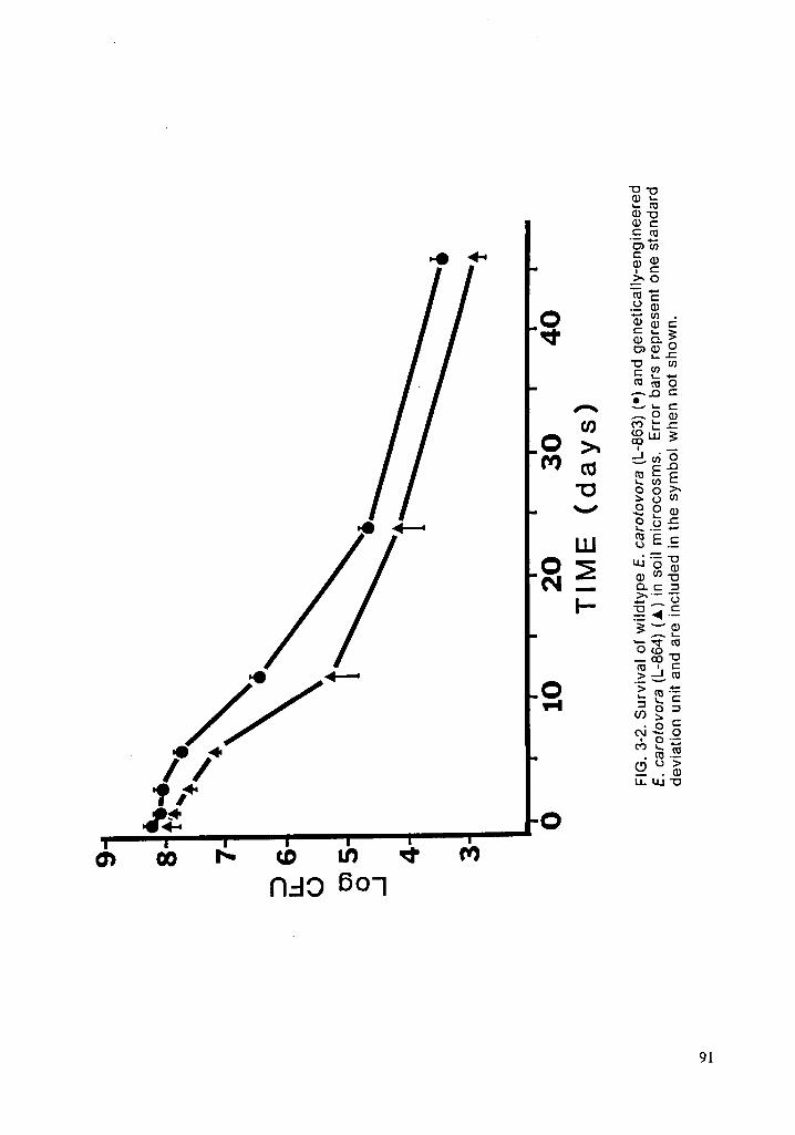
’°‘“
${4/4·
’·
_•
eo
H[_
4_
22E
iii}
mE
,§°
ä·~
gg
>_
Ea
O
zu
ig?
g
gg
ggO
LU
QLEE
‘I
E
Ew?
<r
I:Eäg
M
ärägg
q
$22äéä
Nää
$2*
dä?
Eää
SE2
9 1

•.
ü
¤
m
G!.
g
é
W
.
L
,4
n
”
S
2
H
{
g
6
H
«-nä
ääögä
·¤°
Eeréägä
gägä
ggg?§ä¤g
ggg?
°
92

SEm
ÄCQ
“
·—
·•
«~:|
yuI
ul
3jö=
In},
Em
E
0
¤
I:m
-•22;%
:2%
3

CHAPTER FOUR
INTRASPECIFIC COMPETITION
INTRODUCTION
Environmental release of genetically-engineered microorganisms (GEMs) has
raised concerns over potential ecological effects. Assessment of survival will
be useful in preliminary testing for risk assessment when predicting potential
adverse structural, functional, and genetic effects. Survival is dependent upon
many environmental parameters including substrate, nutrients, temperature, pH,
moisture, and predators (Stacey, 1985; Grant and Long, 1981). Survival and
competition are interrelated and often used interchangably in microbial ecology.
However, while survival is an important parameter, competition of the intro-
duced bacteria with its parental straln is another way to ascertain ifthe GEM will
possess an ability for enhanced survival. ln addition, removing competitors by
substrate sterlization will also provide data on survival in perturbed systems.
Such perturbations may enhance GEM survival (Office of Technology Assess-
ment, 1988).
Competition is defined by Ricklefs (1979) as the use of a resource by an
organism in such a way that it reduces resource availability to other organisms.
Competition may be intraspecific or interspecific; both types are poorly under-
stood ln microbial ecosystems. lnterspeclfic competition in nonsterile soll has
been addressed in earlier chapters.
This study used a genetically-engineered straln of Erwinia carotovora that
was constructed to include two types of genetic alterations in the bacteria!
chromosome: insertion of a disarmed gene fundamental to plant pathogenesis
in a cis-orientation to the wildtype and inclusion of an antibiotic resistance gene.
Either alteration may affect intraspecific competitiveness of the engineered94

strain. The competitiveness ofthe GEM strain with varied concentrations of its
parental strain as well as its survival in sterilized substrate are the topics of this
report.v
MATERIALS AND METHODS
Bacterial strains and plasmids
The bacterium used in this study was Erwinia carotovora subsp. carotovora
(Allen et al., 1986), a phytopathogenic enterobacterium that produces soft rot in
plant tissue. Pectate Iyase genes from E. carotovora were cloned into plasmid
pBR322 on a 1.3 kilobase pair (kbp) fragment and expressed in Escherichia co/i
strain HB101. A 5.5 kbp fragment of the inserted E. carotovora DNA was deleted
by digestion with Bg/Il and the remaining plasmid DNA ligated using T4 Iigase.
The resulting plasmid, pDR41, no longer mediates pectate Iyase activity as de-
termined by isoelectric focusing and activity stain overlays. Plasmid pDR41 was
further modified by inserting the Pst fragment of Tn903 (kanamycin resistance)
isolated from pUC4K into the Pstl site of pDR41. This new plasmid, pVS20, me-
diating kanamycin resistance, was transferred into E. carotovora by conjugal
triparental mating with the helper plasmid pRK2013. Substitutional mutagenesis
was attempted by curing pVS20 from EC14 in low phosphate medium.
Kanamycin·resistant strain L-833 was recovered that contained no detectable
plasmids, was reduced up to 30% in ability to rot tuber tissue, but was not af-
fected in the production ofthe target pectate Iyase. The Tn903 derlved fragment,
therefore, is probably inserted in a cis-orientation to the wildtype genes in the
EC14 chromosome. Strain L-863 was a wildtype mutant of E. carotovora resistant
by spontaneous mutation to 150 ug/ml rifampicin; strain l.-864 is a spontaneous
rifampin-resistant mutant of L-833. Both L-863 and L-864 strains displayed
growth and biochemical characteristics identical to the wildtype strain.95

Media and chemicals '
Plate count agar (PCA; Difco Laboratories, Detroit,Ml), amended with 40
pg/ml kanamycin monosulfate and 150 pg/ml rifampicin (Sigma Chemical Co.,
St. Louis, MO) was used to detect genetlcally-engineered E. carotovora and PCA
with 150 ug/ml rifampicin was used to enumerate wildtype E. carotovora.
Cycloheximide (100 pg/ml; Sigma Chemical Co.) was added to both media to
inhibit fungal growth.
Microcosms
Terrestrial microcosms consisted of 1-liter glass mason jars containing 200
g of soll. Humidified, sterile air was circulated at 5 ml/min through the
microcosm, as shown, for the duration of the study to mlnimize evaporation of
soll moisture and allow for elimination of metabolic by-products. Both intake
and exhaust air was sterilized by membrane filtration usinga Gelman Maxi Cap-
sule TM (Gelman Sciences, Inc., Ann Arbor, Ml). Soil in microcosms was gently
mixed by shaklng every third day to ensure a homogenous mixture. This study
utilized soll from the Virginia Polytechnic Institute and State University farm at
Whitethorn on the New River. Soil was a Hayter loam type, no·till previously
planted with corn, dark grayish brown In color, and consisted of 39% sand, 41%
silt, and 24% clay. Soil moisture was 11%.
Microcosm lnoculation
Erwinia carotovora were grown in 4 liters of nutrient broth to a turbidity of
approximately 1.0 at 550 nm. Bacterial cells were harvested by centrifugation
at 1000 g for 20 min to provide approximately 6 x10‘°
cells. The supernatant
was discarded, the pellet washed in sterile distilled water (SDW), resuspendecäts

in SDW, and centrlfuged at 1000 g. This pellet was suspended in SDW (45 ml) ·
so that 15 ml of lnoculum would be delivered to each microcosm (200 g soll) to
provide approximately 10° cells/g soll. Amount of lnoculum added was caIcu·
Iated dependlng upon the concentration desired in soll, as described below.
Inoculum was added to soll, which had been prevlously air dried for 48 h and
passed through a 4.75 mm sieve after collection, and throughly mixed. Inocu-
Iated soll was passed through a 2 mm sieve, remixed, and then randomly placed
into microcosms. lvlicrocosms were lncubated at 20° C.
Experimental design
Four experiments were conducted: 1) soll containing equal concentratlons
(1:1) of engineered and wildtype bacterla, 2) soil with 10 times as many engi-
neered as wildtype bacterla, 3) soll with 10 times as many wildtype as engi-
neered bacterla, and 4) soll that had been radiation-sterlllzed and inoculated
with either engineered or wildtype strains of E. carotovora.
For each competition study, six microcosms were inoculated with bacteria.
Experiments with sterile soll used three microcosms with genetically-
engineered strain and three with wildtype. On each sampling day, one sample
was randomly withdrawn from each microcosm and diluted. Three plates were
prepared from each dilution and plates with approximately 30 to 300 colonles
were counted.
Sampling of microcosms
Samples were withdrawn from microcosms at varled intervalsz 0, 2, 4, 6, 10,
15, 20, and 25 days for 1:1 competition experiments; 0, 2, 5, 10, and 15 days for
other competition studies; and 0, 2, 5, 10, 16, 20, 40, and 55 days for sterile solls
experiments. To allow for diffusion of inoculum, day 0 samples were wlthdravién]

approximately 2 h after inoculation. After gently mixing the soil in the
microcosms, soll samples (1 g) were randomly removed and placed in milk di-
lution bottles containing 99 ml SDW. These were mixed, first manually for a brief
period and then for 20 min on an orbital shaker at 150 rpm, and subsequent di-
Iutions prepared. Dllutions were spread onto appropriate media. lnoculated
petri dishes were incubated at 30°C for 48 h and then plates with appropriate
dilutions were counted. Results were expressed as CFU/g dry weight. Soil for
determination of water content was removed at time of sampling.
Soil Sterilization
Soil was sterilized by exposing it to 1.5 Mrad of gamma radiation from a °°Co
source over a 15 hour period at the University of Virginia Reactor Facility,
Charlottesville, VA. Soil was checked for sterility by plating onto nonselective
media; no growth was observed.
Statistical analysis
Statistical significance between groups at each sampling day was evaluated
using Student’s ttest and between sampling intervals using a repeated meas-
ures analysis (Sokal and Rohlf, 1981).
V RESULTS AND DISCUSSION
Erwinia Survival in Sterile Soil
Both strains of E. carotovora survived longer in sterile soil (Fig. 4-1) than they
did in nonsterile soil, as described in earlier chapters. Survival in sterile soil
demonstrated that both Erwinia strains persisted for a longer period of time in
the absence of competitors declining only two orders of magnitude over 55 daygé

iLonger survival in sterile seil is net unexpected since less competition fer re- .
sources probably occurs.
. Intraspecific Competition
Both engineered and wildtype E. carotovora declined at similar rates when
added in equal proportions te the same microcosms (Fig. 4-2). Ne significance
difference was observed en sampling days (p > .05) but overall decline over
time was significant (p < .05). Rate ef decline differed from earlier studies
(Chapter 3) in that the characteristic large initial decrease followed by a smaller
rate ef decrease was not seen. instead, decline was linear with density dimin-
ishlng about 2 orders ef magnitude every 10 days.
lnoculating microcosms with concurrent varied doses of engineered and
wildtype E. carotovora did not affect survival. Beth strains declined in concen-
tration at similar rates regardless ef whether the engineered strain concen-
tration was ten times higher (Fig. 4-3) er lower (Fig. 4-4) than the wildtype.
However, rate ef decline was higher in microcosms receiving mixed concen-
trations ef organisms than in microcosms receiving the same concentration ef
both strains. This could be due to the higher number ef bacteria present in
these microcosms as opposed te the 1:1 competition study microcosms. This
higher number would utilize nutrients at a faster rate.
Microbial competition is difficult to understand in nature due te its complexity.
However, intraspecific competition is easily determined in the laboratory and
field-test situation., lt is an important parameter since it may indicate if the en-
gineered strain will survive longer than the parental wildtype. However, field
validation of results (Cairns, 1988) must be executed since laboratory ebserva—
' tions may net correlate with field data. Meade et al. (1985) found that indigenen;

strains of Rhizobium leguminosarum out-competed inoculant strains in the field
but demonstrated no difference in competitiveness in the Iaboratory.
The ability of an engineered strain, often bred in nutrient-rich media, to com-
pete successfully with oligotrophic microbes in an oligotrophic environment is
difficult. However, introduced microbes must displace indigenous organisms
already occupying a niche (Adelberg, 1985) since the engineered strain must
survive long enough to perform its desired task; otherwise, it is of no commer-
cial value. This displacement may be transient. Sparse data exists regarding
introduction of microbes. Introduction of Rhizobium has provided some data for
interspecific competition as described in earlier chapters (Stacey, 1985). In ad-
dition, Lindow (1985) studied intraspecific competition between engineered
(ice-minus) and wlldtype (ice-plus) strains of Pseudomonas syringae. Lindow
found that Ieaf surfaces inoculated with ice-minus strains will effectively inhibit
ice-plus strains by competitive exclusion (Lindow, 1987).
However, it appears that the vast majority of mutations will impart a compet-
itive disadvantage rather than an advantage (Office of Technology Assessment,
1988), especially with transposon insertion (F.J. Brockman, M.S. thesis,
Washington State University, Pullman, WA., 1988). It appears that gene insertion
did not affect the ability of Erwinia to compete.
Factors that may make a system more susceptible to GEM invasion include
environments lacking potential predators, release of ecological generalists,
organisms that can exploit a variety of resources, and perturbed systems. Per-
turbed ecosystems may be more susceptible to invasion than first thought
(Adelberg, 1985; Cairns and Pratt, 1986; Office of Technology Assessment, 1988)
Cairns and Pratt (1986) determined, using protozoan models, that ability to resist
invasion appears to be related to the species richness and the particular100

successional state that the ecosystem is in. Additional studies are warranted to
determine if this also applies to GEl\/ls.
101

LITERATURE CITED
Adelberg, E.A. 1985. Summary of Proceedings. [ln] H.O. Halvorson, D. Pramer, and
M. Rogul (eds.), Engineered Organisms in the Environment: Scientific Issues,
ASM, Washington. pp. 233-235.
Allen, C., V.K. Stromberg, F.D. Smith, G.H. Lacy, and M.S. Mount. 1986.
Complementation of an Erwinia carotovora subsp. carotovora protease mutant
with a protease-encoding cosmid. Mol. Gen. Genet. 202:276-279.
Cairns, J., Jr. 1988. What constitutes field validation of predictions based on labora-
tory evidence? [In] W.J. Adams, G.A. Chapman, and W.G. Landis, (eds.), Aquatic
Toxicology and Hazard Assessment, ASTM STP 971, Phil., pp. 361-368.
Cairns, J., Jr. and J.R. Pratt. 1986. Ecological consequence assessment: effects of
bloengineered organisms. [In] J. Fiksel & V.T. Coveilo (eds.), Biotechnology
Risk Assessment, Pergamon Press, pp. 88-108.
Grant, W.D. and P.E. Long. 1981. Environmental Microbiology. Halsted Press,
Glasgow, Scotland, 215 pp.
Lindow, S.E. 1985. Ecology of Pseudomonas syringae relevant to the field use of ice-
deletion mutants constructed in vitro for plant frost control. [ln] H.O. Halvorson,
D. Pramer, and M. Rogul (eds.), Engineered Organisms in the Environment:
Scientific Issues, ASM, Washington. pp. 23-35. —
Lindow, S.E. 1987. Competitive exclusion of epiphytic bacteria by ice- Pseudomonas
syringae. Appl. Environ. Micro. 53:2520-2527.
Meade, J., P. Higgins, and F. O’Gara. 1985. Studies on the inoculation and
competitiveness of Rhizobium Ieguminosarum in soils containing indigenous
rhizobia. Appl. Environ. Micro. 49:899-903.
102

Office of Technology Assessment. 1988. New Developments in Biotechnology -
Field-Testing Engineered Organisms: Genetic and Ecological Issues.
OTA-BA-350, Washington, D.C., 150 pp.
Ricklefs, R.E. 1979. Ecology. Chiron Press, New York, 966 pp.
Sokal, R.R. and F.J. Rohlf. 1981. Biometry. W.H. Freeman, New York.
Stacey, G. 1985. The Rhizobium experience. [ln] H.O. Haivorson, D. Pramer, and M.
Rogul (eds.), Engineered Organisms in the Environment: Scientific Issues, ASM,
Washington. pp. 109-121.
103

8
\
7
D 6l U
I'!
E?_. 5
4
30 1 0 2 0 3 0 4 0 5 0 6 O
TIME (days)
FIG. 4—1. Survival of genetically-engineered (L·864) (•) and wildtype (L-863) (•)Erwinia carotovora in sterile soil. Error bars represent one standard deviationunit and are contained In the symbol when not shown.
104

10
9 .
6E“\
° 7uio.4
6 -
64
O 10 20Time (days)
FIG. 4-2. lntraspecific competition of genetically-englneered (solid symbol) andwildtype (open symbol) Erwinia carotovora in non-sterile soll. Bacterla wereadded in equal (1:1) amounts to soll prior to placing soll into microcosms.
105

9
8
7DLI.U .U)G 6.1
5
4
30 1 0 2 0
TIME (days)
FIG. 4-3. Intraspecific competition of genetically-engineered (open symbol) andwildtype (solid symbol) Erwinia carotovora in non-sterile soil. Engineered bac-teria were added in 10 times the amount of wildtype bacteria. Error bars repre-sent one standard deviation unit.
106

9
8
7 U
B
häU) 6O.J
5 D
4
30 1 O 2 O
TIME (days)
FIG. 4-4. Intraspeclfic competition of genetically-engineered (open symbol) andwildtype (solid symbol) Erwinia carotovora in non-sterlle soll. Wildtype bacterlawere added in 10 times the amount of engineered bacteria. Error bars representone standard deviation unit.
1107

CHAPTER FIVE
GEM EFFECTS UPON SOIL CHEMICAL PARAMETERS AND.
ENZYME ACTIVITIES
INTRODUCTION
Survival of genetically-engineered microorganisms (GEMs) in the environ-
ment has raised concerns over potential ecological effects. While assessment
of survival, competition, and gene exchange will be useful in preliminary testing
for risk assessment, the ultimate manifestation of any adverse effect will be al-
teration of ecosystem function. While such changes may have profound effects
(Alexander, 1985), others point out that the probability of such adverse effects is
small, especially since few exogenous organisms can even compete with
lndigenous microbes (Hardy and Glass, 1985).
Many functional processes could be affected by the environmental release
of GEMs (Cairns and Pratt, 1986). While some processes may be intentionally
altered for commercial purposes, it is plausible that processes may be inad-
vertently changed. Wang and Crawford (1988) found that an engineered
Streptomyces strain caused signlficantly higher soll carbon mineralization rates
in nonsterile soll amended with lignocellulose. They believe the effect is due to
the expression ofthe foreign DNA insert. lt is plausible that transfer of such an
insert could convey such alteration in function to other organisms.V
While many parameters can be monitored, soll enzyme activities, nutrlent
concentrations, and other chemical variables such as pH and organlc matter, are
among the most important since their stabllity is critical to blogeochemical
processes (Fenchel and Blackburn, 1979). Assaying soll enzyme activities pro-
vides some indication of soil capacity to carry out biochemical processes (Ross,‘
1971). Dehydrogenase enzymes are important in the initial stages of soll organgcä

matter oxidation by transferring hydrogen or electrons from substrates to
acceptors. In actuality, several enzymes and enzyme systems are involved in
total dehydrogenase activity. Phosphatases are capable of cleaving
orthophosphate groups from organic phosphate compounds. Free phosphatase
results from the natural loss of the enzyme either from the periplasmic space
or because of an environmental stress, such as osmotic shock (Sayler, 1979).
Presence of phosphatases in nature directly relates to phosphorous cycling.
This study used a genetically engineered strain of Erwinia carotovora that
was constructed to include two types of genetic alterations in the bacterial
chromosome: deletion of a gene fundamental to plant pathogenesis and inclu-
sion of an antlbiotic resistance gene. Potential functional effects of adding these
strains to microcosms was determined by monitoring concentrations of alkaline
phosphatase, dehydrogenase, nutrlents, and other selected soll attributes such
as organic matter and pH.
MATERIALS AND METHODS
Bacterial strains and plasmids
Erwinia carotovora subsp. carotovora (Allen et al., 1986), a phytopathogenic
enterobacterium that produces soft rot ln plant tissue and is ublqultous in na-
ture, was used in this study. Pectate lyase genes from E. carotovora were
cloned into plasmid pBR322 on a 1.3 kilobase pair (kbp) fragment and expressed
in Escherichia co/i strain HB101 (Roberts et al., 1986). A 5.5 kbp fragment of the
E. carotovora DNA was deleted by digestion with Bg/Il and the remalning
plasmid DNA Iigated using T4 ligase. The resulting plasmid, pDR41, no longer
mediated pectate lyase activity as determined by isoelectric focusing and activ-
ity stain overlays. Plasmid pDR41 was further modified by inserting the Pst
fragment of Tn903 (kanamycin reslstance) isolated from pUC4K into the Pstl sllgä

of pDR41. This new plasmid, pVS20, mediating kanamycin resistance, was
_ transferred into E. carotovora by conjugal triparental mating with the helper
plasmid pRK2013. Kanamycin-resistant strain L-833 was recovered that con-
tained no detectable plasmids, was reduced up to 30% in the ability to rot tuber
tissue, but not reduced in the ability to produce the target pectate lyase. The
Tn903 derived fragment is probably inserted in a cis-orientation to the wildtype
genes in the EC14 chromosome. Strain L-863 was a wildtype mutant of E.
carotovora resistant by spontaneous mutation to 150 p/ml rifampicin; strain L-864
is a spontaneous rifampin-resistant pg/ml) mutant of L-833. Both L-863 and
L-864 strains displayed growth on laboratory media and biochemical character-
istics identical to the wildtype strain.
Media and chemicals
Plate count agar (PCA; Difco Laboratories, Detroit, MI), with 40 pg/ml
kanamycin monosulfate and 150 pg/ml rifampicin (Sigma Chemical Co., St.
Louis, MO) was used to enumerate the genetically-engineered E. carotovora
strain and PCA with 150 pg/ml rifampicin was used to enumerate wildtype E.
carotovora. Cycloheximide (100 pg/ml; Sigma Chemical Co.) was added to both
media to inhibit fungal growth.
Microcosms
Terrestrial microcosms consisted of 1-liter glass mason jars containing 200
g of soil. Humidified, sterile air was circulated at 5 ml/min through the
microcosm for the duration of the study to minimize soil moisture evaporation
and eliminate metabolic by- products. Both intake and exhaust air was filter
sterilized (Gelman Maxi Capsulem; Gelman Sciences, Inc., Ann Arbor, MI). Soil
in microcosms was gently mixed every third day to ensure a homogenous disio

· tribution of microorganisms. This study utillzed soll from the Virginia
Polytechnic Institute and State University farm at Whitethorn on the New River.
Soil was a Hayter Ioam type, dark grayish brown in color, and consisted of 39%
sand, 41% silt, and 24% ciay.
Microcosm inoculatlon
Erwinia carotovora were grown in 4 Iiters of nutrient broth to a turbidity of
approximately 1.0 at 550 nm. Bacterial cells were harvested by centrifugation
at 1000 g for 20 min to provide approximately 6 x 10’° cells. The supernatant
was discarded and the pellet washed in sterile distilled water (SDW), resus-
pended in SDW, and centrifuged at 1000 g. This pellet was suspended in SDW
(45 ml) so that 15 ml of inoculum would be delivered to each microcosm (200 g
soll) to provide approximately 10° cells/g soll. Amount of inoculum added was
calculated depending upon the concentration desired in soll, as described be-
low. inoculum was added to soll, which had been previously air drled for 48 h
and passed through a 4.75 mm sieve after collection, and throughly mixed. ln-
oculated soll was passed through a 2 mm sieve, remlxed, and then randomly
placed into mlcrocosms. Microcosms were incubated at 20° C.
Experimental design
Two experimental series were conducted to (1) determine if soll nutrients or
other soll chemical parameters were affected by the addition of Erwinia as
compared to controls which were inoculated with water, and (2) ascertain lf
exogenous_ bacteria affected soll enzyme activities of dehydrogenase and
alkaline phosphatase.
For each experiment, three microcosms were inoculated with 10° cells of ei-
ther the engineered strain, the wildtype strain, or sterile water as a control.

each sampling day, one sample was randomly withdrawn from each microoosm
in the amount required for the tests described below as well as for dilution so
that the density of bacteria could be monitored.
Dehydrogenase activity
Dehydrogenase activities were determined as follows (Ross, 1971): Two
grams of wet soil/sediment mixture was added to 4 ml 0.5 M Tris HCI (pH 7.5)
plus 2 ml 1% triphenyl-tetrazolium (TTC). One ml of 1% glucose was added and
the solution mixed and incubated at 25-30 C in dark for 96 h. The mixture was
swlrled daily. After incubation, the reaction was stopped by adding 20 ml
methanol, shaking for 30 sec, and incubating an additional 3 h in the dark. The
same amount of methanol was added to each sample. A 1.0 ml aliquot of the
mixture was then centrifuged briefly at 11,000 rpm in a microcentrifuge to re-
move particulate matter and 200 ul of this solution was carefully aspirated and
placed into a microtiter plate. Absorbance was read at 490 nm using a Dynatech
microplate reader and activity calculated as mg formazan/g dry sediment or soil.
Standards were prepared using triphenyl formazan at concentrations of 0.004
mg per 20 ml methanol to 0.4 mg per 20 ml methanol. Blanks contained no
substrate.
Alkaline phosphatase activity
To determine alkaline phosphatase (Sayler et al., 1979), one gram of soll was
added to 4 ml 1 M Tris (pH 8.5) and sonicated for 30 seconds at 10 watts. One
ml of p-nitrophenylphosphate (p-NPP, 1 mg/ml) was added, the mixture vortexed
and incubated in the dark at 30° C for 2-3 h. After incubation, the reaction was
stopped with 1 ml of 1 N NaOH. A 200 pl aliquot was then placed into a microtite;

. plate and read at 410 nm. Activity was calculated as pg p-NP/g dry wt. Stand-
ards were run using p-nitrophenol (p-NP) and blanks contained no substrate.
Soil analysis
Soil was assayed for concentrations of phosphorus, potassium, calcium,
magnesium, zinc, manganese, nitrate, organic matter, soluble salts, and pH by
the Virginia Tech Soils Testing Laboratory using standard methods (Page et al.,
1982). To ascertain the accuracy of tests, pH and organic matter were assayed
according to standard protocols (Page et al., 1982) and not found to be different.
Statistical analysis
Statistical significance between groups for each variable at each sampling
time was evaluated using an analysis of variance rationale and a Student-
Newman-Keuls means analysis (Sokal and Rohlf, 1981). Significance between
sampling intervals was evaluated using a repeated measures analysis (Sokal
and Rohlf, 1981). All analyses were accomplished using the Statistical Analysis
System (SAS institute, 1985).
RESULTS AND DISCUSSION
Enzyme activities
No treatment effects were observed for either alkaline phosphatase (Fig. 5-1)
or dehydrogenase (Fig. 5-2). Both groups with added bacteria did not differ
statistically (p > 0.05) from the control group and no time effects were observed
among either treatment group or the control group (p > 0.05).
Although both phosphatase and dehydrogenase are important natural
enzymes, there is some indication that these proteins are markedly affected orig

when the stress placed on the system is severe, as in the case of chemicals.
Lack of impairment from the engineered and wildtype strains was not unex-
pected since the genetic alteratlons should not have affected such enzyme pro-
duction. However, it was expected that enzyme activities in soll with added
bacteria would be greater than soll with only water added, since microbial ac-
tivity would be higher. This was not observed. Apparently, the added bacteria
did not contribute significant amounts of either dehydrogenase or phosphatase.
Daily fluctuations of enzyme activity have been reported (Huber and Kidby, 1984)
and must be incorporated into experimental designs. Recent studies using
genetically-engineered pseudomonads have produced differences in
dehydrogenase and phosphatase based upon analyses after peak production
periods (J. Doyle, personal communication). These peak periods were detected
by measuring carbon dioxide evolution. Elucidation of this mechanism could
improve the reliabilty of using enzyme activities as lndicators of environmental
stress.
Soil nutrients and other chemical parameters
None of the parameters monitored demonstrated a treatment effect between
engineered and wildtype E. carotovora strains (p > 0.05) (Table 5-1). However,
several time effects were observed.
Decrease in pH from 5.9 on day 0 to 5.63 on day 30 was significant (p= .003)
and indicated that metabolically-active bacteria probably affected soll pH by
excretion of waste products. However, other factors, such as salt and carbon
dioxide content may also affect soll pH (McLean, 1982). Carbon dioxide soll
concentrations were not measured but, based upon air concentrations from
earlier studies (Chapter 1), should not have been very high.114

Other variables also demonstrated changes over time. Soluble salts in-
creased from about 115to 221 ppm over the 30 day course of the study in both
treatment groups; the reasons for this remain unclear but soluble salts from
decaying bacteria may be responsible since data from day 0 samples indicated
that salt concentrations increased from 90 to 120 ppm. Nitrate-nitrogen also in-
creased significantly (p=0.003) by 120% from day 0 to day 30 in both groups,
probably because of the metabolic products excreted by microbes or released
from dying bacteria. Analysis of day 0 soil samples indicated only a 15% in-
crease in nltrate compared to pretest samples. Analysis of urease in future
studies may be beneficial in elucidating ammonia, urea, and nltrate fluctuations.
CONCLUSIONS
The parameters monitored in this research did not demonstrate any appreci-
able change from the engineered or wildtype strains of E. carotovora. This
could be due to one of two reasons: (1) the variables used were not sensitive
enough for use in assessing risk, or (2) the engineered and wildtype strains did
not significantly differ in phenotype as to produce a difference, therefore the risk
posed by the engineered strain is minimal.
Using of enzyme activities to detect functional effects of GEMs has proven
useful at the EPA Research Laboratory at Corvallis, Oregon (J. Doyle, personal
communication). However, such differences in activity were transient and oc-
curred with flushes of carbon dioxide from sealed microcosms. The enzymes
monitored in this study are important in biogeochemical cycles and have been
used with some success in ascertaining chemical effects upon microbial activity
(Sayler et al., 1979; Killham et al., 1983; Arthur and Frea, 1988). However, the
inherent error involved makes results interpretation difficult. 115

Clearly, functional effects will be the ultimate manlfestation of any harmful
GEM release (Cairns and Pratt, 1986). Determlning what functional parameters
to monitor and when to monitor them will require additional research.
116

LITERATURE CITED -
Alexander, M. 1985. Ecological consequences: reducing the uncertainties. Issues in
Science and Technology. Spring: 57-68
Allen, C., V.K. Stromberg, F.D. Smith, G.H. Lacy, and M.S. Mount. 1986.
Complementation of an Erwinia carolovora subsp. carotovora protease mutant
with a protease-encoding cosmid. Mol. Gen. Genet. 202:276—279.
Arthur, M.F. and J.I. Frea. 1988. Microbial activity in soils contamlnated with
2,3,7,8-TCDD. Envlron. Toxicol. Chem. 7:5-13.
Cairns, J., Jr. and J.R. Pratt. 1986. Ecological consequence assessment: effects of
bioengineered organisms. [In] J. Fiksel & V.T. Covello (eds.), Biotechnology
Risk Assessment, Pergamon Press, pp. 88-108.
Fenchel, T. and T.H. Blackburn. 1979. Bacterial and mineral cycling, Academic Press,
NY. 225 pp.
Hardy, R.W.F. and D.J. Glass. 1985. Our investment: what is at stake'? Issues Sci.
Tech. Spring, 69-82.
Huber, A.L., and D.K. Kidby. 1984. An examination of the factors involved in deter-
mining phosphatase activities ln estuarine waters. 2. Sampling procedures.
Hydrobiologla 111:13-19.
McLean, E.O. 1982. Soil pH and lime requirement. [ln] A.L. Page, R.H. Miller, and D.R.
Keeney (eds.), Methods of Soil Analysis, Part 2. Chemical and Microblological
Properties, Amer. Soc. Agronomy, Madison, WI, pp. 199-224.
Page, A.L., R.H. Miller, and D.R. Keeney. 1982. Methods of soil analysis, chemical and
microbiological properties, 2nd edition. Amer. Soc. Agronomy, Madison, WI, _
1158 pp.
Roberts, D.P., P.M. Berman, C. Allen, V.K. Stromberg, G.H. Lacy, and M.S. Mount.
1986. Requirement for two or more Erwinia carotovora subsp. carotovora H7

pectolytic gene products for maceration of potato tuber tissue by Escherichia
coli. J. Bacteriol. 167:279-284.
Ross, D.J. 1971. Some factors influencing the estimation of dehydrogenase activities
of some solls under pasture. Soil Biol. Biochem. 3:97-110.
SAS Institute, Inc. 1985. SAS User’s Guide: Statistics, version 5. SAS Institute, Inc.,
Cary. N.C.
Sayler, G.S., M. Puziss, and M. Silver. 1979. Alkaline phosphatase assay for
freshwater sediments: application to perturbed sediment systems. Appl. Envi-
ron. Micro. 38:922-927.
Sokal, R.R. and F.J. Rohlf. 1981. Biometry. W.H. Freeman and Co., San Francisco, CA.
776 pp.
Wang, Z. and D.L. Crawford. Effects of wildtype and recombinant Streptomyces on
mineralization of organic carbon. Abstracts of First Inter. Conf. Release Genet-
lcally Engineered Microorganisms No. 12, p. 13.
118

:6 Y ¤Q¤Q·Qv vwmu·ä*-“.¤ Egg Omoci gwdg.52:1,;%
~·EgfgllNlglgf vjwgg
$$630 &°U)
06 =¤ „¤;; ...<¤0
am“'>
·¤'>v oo .d
20,Ncnqggü."’
mE·¤¤o ^¤Ö‘OmU U)E QNlqlqv
ww--.--•3„„ ¤.
O 1DLD
N NNN
‘=Lu·¤ Q ^gg > E
LD@cm__ G5 M com . Lg
E""“ 2 ä°¤* 9¤>%° $$+5O·¤C;_?:’O E 2 cv-
»«‘“E°° °?$°Y
NOON.2: —q,w
-9 E¤¤0:>
0::---: ^--
mlqrqw .
·-Q) "' 1—€\]¢') °
«..Y «—vm
•Eg,q;§>-•g> E'; "'**<·· «-~•—„c2.0Q): Q Inäggxaa, ä «6U)gääesä
: CL 0)Q
-¤ 0 E U.; EE 0000cc Lumää
:’;';*;*a 0000 BBB;
119

S
'
II
Q
B
N °L
I
__«•»—Ä
Of-,
EVE
‘_
‘Q‘¤C
1A
@22
MO
ääg
VQS;
O
SSS
ägm
SSS
—N
SSS„;§Am9:**5
2%*
CO;)
U;S2
O
120

°
4
n
LO
-
ro/
B
I
Ln
Ü
-
N,.)
I
·
ä
'
IAI1
äää
g
^
E12
vg
4
oä2
ääö
n-
828
L
·äÄ·§
Ls,
121

EPILOGUE
Risk assessment of genetically-engineered microorganisms is still in its in-
fancy and the lack of literature, including methodology, became more apparent
as this project proceeded. No consensus exists as to which parameters are
important to monitor and which are not, which organisms are "safe" to release
and which are not, and how the scheme of biotechnology risk assessment
should proceed.
Risk is defined as the ”potential for adverse consequences of an event or
activity" (Fiksel and Covello, 1986) while risk assessment is defined as "use of
scientific data to estimate the effects of exposure to hazardous materials or
conditions" (Office of Technology Assessment, 1988). The risk assessment
process for genetically-engineered microorganism (GEM) introduction, accord-
ing to the Office of Technology Assessment (OTA)(1988) is: (1) risk identification,
(2) risk-source characterization, (3) exposure assessment, (4) dose—response
assessment, and (5) risk estimation. This scheme is identical to those used for
chemicals. While it is very effective for predicting risk from chemicals, it falls
when applied to GEMs because, as documented in the OTA report, protocols do
not currently exist for predicting kinds of risk or the magnitude of potential risk.
While primary differences exist between GEMs and chemicals, as described in
earlier chapters, other fundamental differences are: (1) sensitive detection
methodologies, often parts per trillion, exist for many toxic substances, (2) toxic
chemicals, normally, are not present in high amounts prior to the episode being
studied, and (3) the most fundamental principle of environmental toxicology, the
dose-response curve, is easily applied to toxics to allow formulation of such
values as maximum acceptable toxicant concentration, lethal concentration, and
no-observable-effects-concentration (Rand and Petrocelli, 1985). GEMs are ngz

easily detectable, do not lend themselves well to dose-response curves, and are
often already in the environment in the form of parental strains.
Since no clear agreement exists regarding what parameters to monitor, one
of the purposes of this research was to ascertain which variables may be useful
in predicting environmental risk. Survival is probably the most single important
factor to observe. lf the GEM cannot survive, the probability of risk decreases
dramatically (Alexander, 1985; Office of Technology Assessment, 1988). Erwinia
survived for sufficient periods of time (> 42 days) to allow possible adverse ef-
fects to occur. Although the microcosm did not include plant material, the pri-
mary substrate for Erwinia, survival occurred for a longer period than initially
anticipated. Future studies in this Iaboratory will investigate survival with plant
material present as well as bacterial survival using starved cells cultured in
low-nutrient media. Survival must be assessed in more than just the Iaboratory
environment. Both Iaboratory tests with microcosms and small-scale field tests
must be executed in a variety of receiving environments including terrestrial and
aquatic. The environments should include those similar to the anticipated one
as well as environments not analogous to the expected receiving habitat. Vali-
dation of Iaboratory results must occur if such data are to be useful (Cairns,
1988).
Structural effects from GEM introduction appear to be remote because of the
diversity and fitness of indigenous organisms. While such effects are possible,
they may be difficult to detect on the microbial level since enumeration of
microbes is an inexact science at best. This research demonstrated no adverse
effects upon any of the bacterial genera monitored (Chapter 3). Such results
were expected since all genera detected were in large quantities (> 105 CFU/g)
and were already adapted to an oligotrophic environment. However, alteration
of any one of several microbial groups performing critical roles in123
biogeochemical cycling, such as Nitrobacter or Desu/fovibrio, may be disastrous
(Grant and Long, 1981). No attempt was made to examine effects upon higher
taxa. Macroinvertebrates and fish are often used in chemical toxicity tests but
are not particularly useful at this time for GEMS because of the paucity of data
for even lower taxa. However, it is conceivable that GEMs may affect algae,
protoioans, nematodes, and other organisms that feed upon them.
Probability of adverse functional effects may increase as GEM introductions
become more common, if adequate environmental impact assessments are not
performed. Disruption of fundamental ecosystem processes would be the worst
possible ecological impact from a planned introduction (Office of Technology
Assessment, 1988). However, OTA believes that the probability of such an event
occurring in the next few years will be remote since introductions will involve
GEMs with genome deletions (such as ice-minus), rather than genomic in-
sertions that may intentionally or unintentionally be constructed to alter func-
tional processes. Parameters that may be useful to monitor include nutrient
retention and cycling, detritus processing, respiration, productivity, and enzyme
activity (Cairns and Pratt, 1986). No consensus exists as to which parameters
are important and this research did little to eliminate that confusion. This work
did demonstrate that monitoring of enzyme activities and essential soil nutrients
is feasible. Studies at EPA’s Corvallis Environmental Research Laboratory have
shown that geneticaIly·engineered Pseudomonas strains affect phosphatase and
dehydrogenase activities differently than do wildtype strains (J. Doyle, personal
communication). However, these changes were transient and occurred in
sealed microcosms using a microbe carrying a plasmid—bearing pesticide-
degrading gene.
The use of intraspecific competition data in risk assessment protocols should
allow detection of those engineered strains that will survive longer than theirl24

parental strains. The data from this research lndlcated that no difference exists
in intraspecific competition and this was expected based upon the organisms
used and the substrate into which they were inoculated. Competition data will
be particularly useful when the engineered strain is capable of altering natural
functional processes for commercial gain.
Of all the possible adverse GEM effects, none promotes controversy more
than potential transfer of recombinant DNA from the GEM to indigenous
organisms. As discussed in the prologue of this document, DNA exchange in
the environment is natural and allows for enhanced survival of bacteria when
detrimental conditions are encountered. Therefore, expression of recombinant
DNA may occur after the GEM itself has died out. But, as OTA emphasizes, most
engineered strains will differ from the parent in only one or a few genes; such
an alteration should be insignificant since changes in a few genes only rarely
affects survival or function (Office of Technology Assessment, 1988). This re-
search lndicates that transfer of recombinant DNA may have occurred, but, as
described in Chapter 2, concluslve evidence will require use of advanced
methodologies, including gene amplification and sequencing. Such studies
were beyond the scope ofthis dissertation but are planned.
Present guidelines for environmental release require that a premanufacturing
notice (PMN) be filed with the Environmental Protection Agency (EPA). EPA re-
ceives its regulatory mandate under the Toxic Substances Control Act (TSCA),
the statute that is applied to the release of new chemicals and puts the burden
of product safety on the manufacturer. TSCA requires that "adequate data be
developed with respect to the effect of chemical substances on health and the
environment" (West Publishing Co., 1985). But TSCA also requires that such
regulations be executed in a manner "as not to impede unduly or create unnec-
essary economic barriers to technological innovation" (West Publishing Co,125

1985). In regards to engineered organism release, these goals may be at op-
posite ends of the spectrum since biotechnology development has surpassed
the ability of ecotoxicology to devise suitable risk assessment protocols specific
to biotechnology. Blotechnology has immense commercial value and any re-
striction on its development may allow foreign competitors to have unfair ad-
vantages (Hardy and Glass, 1985). However, unregulated development may lead
to environmental impacts, similar to those occurring in the chemical and nuclear
industries. The net result of any such cataclysm would Iikely impair future
biotechnological advances for some time.
Another facet ofthe problem is the influence of the legal system upon envi-
ronmental impact matters. To predict something beyond reasonable doubt is
difficult, since one can question what reasonable doubt is (Anderson et al.,
1984). With the paucity of relevant data available, the mechanisms by which
GEMs could even inflict harm are vague. Such "ignorance of mechanism"
(Anderson et al., 1984) can be used to support release of an unsafe product be-
cause of the uncertainity involved in product regulation. The legal complexities
are numerous when one applies TSCA to GEM releases because TSCA was
written for chemicals in the mid-1970s and many ambiguities are present when
interpretation ofthe law is conducted.
To overcome these scientific and legal complications, the Office of Science
and Technology Policy issued a ”Coordinated Framework for Regulation of
Biotechnology" in 1986 (Federal Register, 1986). The document was an intera-
gency attempt to eliminate jurisdictional duplication in approving safety of
biotechnology products and research. According to this framework, EPA would _
be the primary lead agency for approving environmental releases of GEMs, ex-
cept those used in agriculture. EPA would then conduct an assessment in con-
junction with the Animal and Plant Health Inspection Service (APHIS). A126

Biotechnology Science Coordinating Committee (BSCC) would investigate the
merit of planned releases. While this framework did address some problem
areas, it did not propose assessment protocols, did not recommend extensive
testing of organisms derived from opportunistic pathogens or those containing
non-coding insertions, and exempted "small manufacturers, importers, and
processors" as well as noncommercial research and development firms from
TSCA regulation (Federal Register, 1986). Some of these inconsistencies have
been corrected after public scrutiny.
lt remains clear that risk assessment and regulation of GEMs is complex, in-
accurate, and political. Use of risk assessment to predict risk will only be sat-
isfactory if suitable parameters are determined and monitored. By using a
simplistic hazard assessment scheme (Fig. 6-1) that allows flexibility, diverse
genera and engineering constructs can be evaluated. Factors such as survival,
intraspecific competition, and potential for DNA dissemination must be evalu-
ated using techniques that are commonly available. Evaluating selected groups
of bacteria, such as pseudomonads and nitrifiers, may lndicate if vulnerable
groups are at risk to be affected, but, more likely will not serve any useful pur-
pose. More vague is which functional attributes to monitor and for how long but
far more research is indicated before an answer can be produced.
The purpose of this research was to 1) develop methods to predict risk from
a GEM environmental release, and 2) use these methods to ascertain risk from
engineered Erwinia carotovora. Based upon the acquired data, the engineered
strain is no more likely to cause harm than releasing the parental strain. The
methods developed and refined should be useful in future studies with this and
other organisms.
127

LITERATURE CITED
Alexander, M. 1985. Ecological consequences: reducing the uncertainties. Issues in
Science and Technology. Spring: 57-68.
Anderson, F.R., D.R. Mandelker, and A.D. Tarlock. 1984. Environmental law. Little,
Brown and Co., Boston, 978 pp.
Cairns, J., Jr. 1988. Validating biological monitoring, [In] D. Gruber and J.M. Diamond
(eds.), Automated Biomonitoring, Horwood, Ltd., Chichester, pp. 40-48.
Cairns, J., Jr. and J.R. Pratt. 1986. Ecological consequence assessment: effects of
bioengineered organlsms. Water Resources Bull. 22:171-182.
Federal Register. 1986. Coordinated framework for regulation of biotechnology.
51 :23301 -23350.
Fiksel, J.R. and V.T. Covello. 1986. The suitability and applicability of risk assessment
methods for environmental apllications of biotechnology. [ln] J.R. Fiksel and V.T.
Covello (eds.), Biotechnology Risk Assessment: Issues and Methods for Envi-
ronmental lntroductions, Pergamon Press, New York, pp. 1-34.
Grant, W.D. and P.E. Long. 1981. Environmental Microbiology. Halsted Press,
Glasgow, Scotland, 215 pp.
Hardy, R.W.F. and D.J. Glass. 1985. Our investment: What is at stake? Issues in Sci-
ence and Tech. Spring, 69-82.
Office of Technology Assessment. 1988. New Developments in Biotechnology-Field
Testing Engineered Organisms: Genetic and Ecological Issues. OTA-BA-350,
Washington, D.C., 150 pp.
Rand, G.M. and S.R. Petrocelli. 1985. Fundamentals of Aquatic Toxicology. Hemi-
sphere Publishing, Cambridge, 666 pp.
West Publishing Co. 1985. Selected environmental law statutes. West Publishing Co.,
St. Paul, Minn., 839 pp.
128

pää*6*,+;;
.
}—>.(DLDC*·-*C
.2 ¤·*¤g E
.. 9 gg
OLD
— 8 3%ä
""°LLI Ln
<
;_
an>
C°’
mE¢v9;
E E
Q2;
.2
·—C*'3
>Ä
UmmQ
g
°’
CQQ.
3E
(DLC
mQ_!I-
3E
‘¤ *1*
3
=- EC
äö
$3 LD
N5,
2gn
'QÜSB
E5
2 (D
<an
··•- 22
§+-·
Ei*“ gä
N•-•
Ou.
$3
.E,
EE
ä
äs
LuééO23T6LDC•0)QD')=L“6
129